breathe easy - rotech healthcare – home medical · pdf file ·...
TRANSCRIPT

BREATHE EASY
COMMON SENSE RESPIRATORY
“COPD”
Learning Objectives Common Sense Respiratory is a series of articles written for the “non-Respiratory Care Practitioner” with the purpose of conveying concepts and terminology of respiratory medicine in every-day language. Increasing understanding of these areas will allow Rotech personnel to provide a higher level of service to patients, families, physicians, nurses, respiratory care practitioners, discharge planners, and other markets we serve. In “COPD,” we will endeavor to answer the following questions:
What is COPD? What is chronic bronchitis? What is emphysema? What is asthmatic bronchitis? What causes COPD? How is COPD identified and treated?
“It is incumbent upon primary care physicians to be alert to the possibility of COPD in patients with a productive cough, particularly in those who suffer exercise-related dyspnea and have a family history of the disease, and definitely in those who are
smokers.”
Dr. Thomas L. Petty- Frontline Treatment of COPD

All words shown bold and italicized will be defined at the end of the chapter in the “Glossary of Terms” section. What is COPD? COPD is an acronym for Chronic Obstructive Pulmonary Disease. The term “chronic” refers to a condition which the patient has all the time. Obstruction refers to the fact that these patients have difficulty getting air out of their lungs. Pulmonary tells us the condition is related to the lungs. In a general sense, it could be used to refer to any lung disease that causes chronic problems in getting air out of the lungs. But by convention, it typically is used to refer to three conditions- chronic bronchitis, emphysema, and asthmatic bronchitis. It is very common for patients with one condition to have a component of one of the others as well. That is, a patient with chronic bronchitis will frequently have some emphysema. Or an emphysema patient may have an asthmatic component. Because these conditions frequently coexist, it is often easier and less confusing to group them under the general diagnosis “COPD.”
COPD has been identified in about 16 million Americans- 14 million with chronic bronchitis and 2 million with emphysema. It is estimated that 30-35 million Americans have COPD, but only half are symptomatic to the point they have been identified. This means that half of the population has yet to be identified! It is the fourth leading cause of death in the US and it is the only one of the top four (the others are heart disease, cancer, and stroke) whose numbers are growing. One reason is that as advances are made in the other diseases resulting in decreased numbers, people are living longer and more likely to develop COPD. What is chronic bronchitis? Chronic bronchitis is defined as a productive cough lasting for at least 3 months in two successive years. It is typically the result of changes that occur in the airways secondary to exposure to an irritant such as tobacco smoke. As tobacco smoke or other irritants are inhaled into the lungs, the lungs try to protect themselves in several ways. One way is by increasing the protective layer of mucus that lines our airways. Sticky mucus lines our airways to trap inhaled particles such as bacteria, pollen, dust, smoke, and viruses before they can travel into the lungs and cause damage. It is produced by two specialized structures in our airways- the mucus gland and goblet cells. As our airways are repeatedly exposed to irritants, the mucus glands hypertrophy (get larger) and goblet cells undergo hyperplasia (increase in number) resulting in more mucus being produced in the airways. Mucus is swept out of our airways by small hair-like structures that line the airways called cilia. Cilia move in a rhythmic fashion to push the old mucus (with trapped particles of dust, bacteria, etc.) up and out of our airways to the back of the throat where it is swallowed. Normal individuals produce and swallow about ½ cup per day. The chemicals within tobacco smoke paralyze the cilia for a period each time smoke is
inhaled. This results in the mucus sitting in the airways rather than being swept out. When the smoker quits smoking overnight, the cilia start working again and push the stagnant mucus out of the airways, resulting in the smoker’s familiar “morning cough.” Other things occurring secondary to repeated exposure to irritants are:
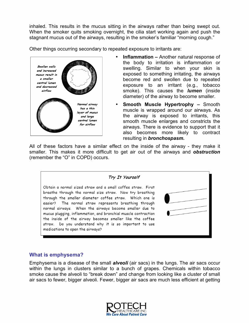
• Inflammation – Another natural response of the body to irritation is inflammation or swelling. Similar to when your skin is exposed to something irritating, the airways become red and swollen due to repeated exposure to an irritant (e.g., tobacco smoke). This causes the lumen (inside diameter) of the airway to become smaller.
• Smooth Muscle Hypertrophy – Smooth muscle is wrapped around our airways. As the airway is exposed to irritants, this smooth muscle enlarges and constricts the airways. There is evidence to support that it also becomes more likely to contract resulting in bronchospasm.
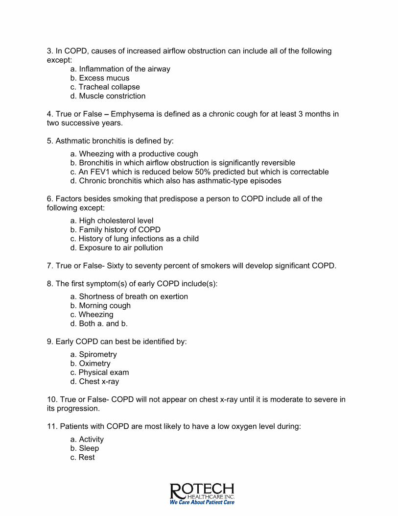
All of these factors have a similar effect on the inside of the airway - they make it smaller. This makes it more difficult to get air out of the airways and obstruction (remember the “O” in COPD) occurs.
What is emphysema? Emphysema is a disease of the small alveoli (air sacs) in the lungs. The air sacs occur within the lungs in clusters similar to a bunch of grapes. Chemicals within tobacco smoke cause the alveoli to “break down” and change from looking like a cluster of small air sacs to fewer, bigger alveoli. Fewer, bigger air sacs are much less efficient at getting

oxygen into the blood stream than several smaller air sacs. It is also more likely to collapse resulting in difficulty getting air out of the lungs. Another change that occurs in the air sacs with emphysema is in their elasticity (stretchiness). The alveoli are normally very elastic, like a balloon. When you take a breath in, you must inflate the alveoli (or balloons), which takes energy to do. However, you exhale by simply relaxing and the stretchy alveoli return to their normal size. Emphysema causes the alveoli to lose their elasticity. Instead of balloons, the air
sacs become more like paper sacks that require energy not only to breathe in, but also to breathe out. What is asthmatic bronchitis? Asthmatic bronchitis is similar to chronic bronchitis, only with a significant asthmatic component. The component of asthma is identified by a significant amount of reversibility in the airflow obstruction when medication is given to open the airways. That is, when a medication like albuterol is given, airflow out of the lungs improves a great deal. What causes COPD? If you haven’t already figured it out, the most common cause of COPD is smoking. Somewhere between 15 and 30% of smokers will develop significant COPD. Other factors also play a role in which smokers will develop COPD including a family history of COPD, childhood infections, and exposure to other irritants (e.g., pollution). How is COPD identified and treated? COPD can be examined in three distinct phases- Early COPD, Moderate COPD, and Severe COPD. Each phase has its own symptoms, testing, and treatments. The American Thoracic Society defines the stages by a measurement called FEV1 (see chart at summary). It is important to identify patients as early in the process as possible so that treatment can be instituted to stabilize their condition as much as possible.
The Three Stages of COPD
Early COPD Symptoms – In early COPD, the patient may have few if any symptoms. The typical early symptoms are morning cough and dyspnea (difficulty breathing) on exertion. Patients who are symptomatic or who have chest x-ray abnormalities have moderately or severely advanced disease. Because the symptoms of COPD develop so slowly, most patients will not seek treatment until their disease is severe. They dismiss the symptoms of shortness of breath and cough as a part of “just getting older.”

Testing – Most testing, including chest x-rays, blood tests, and physical examination, will be totally normal in early COPD. The only test that is sensitive enough to identify COPD in its early stages is spirometry. By having the patient blow into the spirometer, measurements of how much air can be exhaled from the lungs and how fast the air can be exhaled are made. These measurements are very sensitive in determining which patients are developing significant obstruction indicating COPD. Because spirometry is the only test that will identify COPD in its earliest stages, primary care physicians should have a spirometer available in their office to test all smokers, patients with respiratory symptoms, and those at risk for lung problems. Treatment – Treatment in early COPD is directed at stabilizing and reversing the downward trend in the patient’s airflow. Certainly the most important intervention is to remove the irritation from the patient’s airways or to stop smoking. When the patient stops, the rate at which their lung function will decline will slow dramatically. Some may even regain some of their lost lung function.
Secondary to smoking cessation, medications aimed at reversing airflow obstruction are most important. Two types of medications are used as the frontline treatment for opening the airways or bronchodilation - anticholinergics and beta agonists. Anticholinergics are the first line drug for the treatment of COPD. The most commonly used anticholinergic is ipratropium bromide (aka Atrovent®). It works by blocking a chemical (i.e., acetylcholine) that causes the smooth muscle that surrounds the airway to constrict, making the airway smaller. Atrovent also reduces the amount of mucus in the airway, further reducing airway obstruction. Beta agonists include albuterol (aka Proventil®, Ventolin®), metaproterenol (aka, Alupent®), terbutaline (aka Brethine ®), and others. These medications work by relaxing the smooth muscle that surrounds the airways. Because these two medications work by two separate pathways, they can be used together to produce an additive effect. That is, using albuterol and ipratropium together provides more bronchodilation than using either medication alone. Most patients benefit by using both of these medications, referred to as “combination therapy.” Moderate COPD Symptoms – In moderate COPD, dyspnea may begin to worsen. Shortness of breath may be evident during activities of daily living such as bathing, dressing, and household chores. Cough may also worsen and progress from the morning cough to occasional

productive cough throughout the day. This is due to the fact that as the patient continues to smoke, the cilia which move mucus out of the airway are not only paralyzed but are destroyed and disappear from the lining of the airway. As the cilia are destroyed, the only way the mucus can be moved out of the airway is by coughing- thus the increasing productive cough. Because the increased mucus laying stagnant in the airways is an excellent place for bacteria and viruses to grow, patients become more likely to develop respiratory infections. Colds, acute bacterial bronchitis, influenza, and even pneumonia can develop with increasing frequency.
As less and less air is able to get to the alveoli due to worsening obstruction in the airways, oxygen levels begin to fall. This may result in worsening shortness of breath, increased heart rate, and even cyanosis at times of low oxygen level. However, patient’s failure to demonstrate these symptoms does not rule out the possibility of a low oxygen level. Nocturnal (nighttime) symptoms may also develop in moderate COPD. Even more so than activity, sleep may cause severe drops in blood oxygen levels. In all people, breathing tends to be diminished during sleep, especially during the deeper stages of sleep called rapid eye movement or REM sleep. In patients with COPD, breathing during sleep can change significantly.
Because patients with COPD have difficulty getting air out of their lungs, air is trapped in their lungs (“air trapping”) and the lungs are in a constant state of over-inflation (hyperinflation). The diaphragm, which normally is dome shaped, becomes flattened and unable to move sufficiently to adequately ventilate the lungs. The body compensates by enlisting the help of the accessory muscles of ventilation. These muscles located in the chest, shoulders, and abdomen are used by the patient to maintain adequate ventilation. This is often observed in the COPD patient who is sitting with elbows on the table or with hands on knees, stabilizing their shoulders so they can better use their accessory muscles. Often, the upper chest is moving up and down as the patient breathes. During REM sleep, all skeletal muscles, including the accessory muscles of ventilation cease contributing to ventilation and all the work of breathing falls back on the flattened diaphragm. During REM sleep, ventilation may fall to about half of daytime levels as the diaphragm is unable to maintain adequate ventilation. Testing – Typically, the patient’s oxygen level is only measured while the patient is at rest by spot-check oximetry or arterial blood gas (ABG). Their oxygen level is least likely to be low at this time. Possible drops in the patient’s oxygen level during activity or sleep should be measured by oximetry recording.

Oximetry recording is performed by having the patient wear a small probe clipped to the finger or ear. The patient wears the oximeter for an extended period during activity and during sleep. The probe is attached to an oximeter which records the patient’s oxygen level over a period of time (up to 18 hours or longer). A printout of this recording will show if the patient is experiencing drops in their oxygen level during activity or sleep.
Treatment – Treatment is aimed at correcting drops in the oxygen level. Oxygen should be used during periods when the patient’s oxygen level is shown to be low. That is, if the patient’s oxygen level drops only during sleep, they should use oxygen only during sleep. If it drops during activity, the patient should wear oxygen during activity as well. Other medications may also be added as the condition deteriorates. Corticosteroids, theophylline, and other drugs may be beneficial. Spirometry testing should be repeated when adding new medications to evaluate whether a benefit is obtained.
Pulmonary rehabilitation, aimed at minimizing the effects of lung disease, can also play a large role in the treatment of these patients. Patients are educated on their disease process, medications, new breathing techniques, smoking cessation, and ways they can play a more active role in managing their condition. A monitored exercise program is also tailored to each patient to recondition out-of-shape muscles.

Severe COPD Symptoms – Shortness of breath may worsen to the point where patients become dyspneic with any activity or even at rest. Cough can become frequent; however some patients may have little or no cough. Exacerbation (sudden worsening) of their condition can become more frequent due to recurrent infections or fluid retention secondary to heart failure. Signs of heart failure may include swelling in the feet and legs, neck veins (jugular) protruding, and swelling of the liver and abdomen. If patients experience symptoms indicating infection (e.g., cloudy or colored sputum, fever, chills, increased cough, and increased dyspnea) or heart failure, it is important for them to notify their physician immediately. The sooner treatment can be implemented, the greater the likelihood that a hospitalization can be averted. Ventilation of the alveoli continues to worsen to the point where some alveoli are receiving little or no air movement resulting in significant hypoventilation (under-ventilation). The oxygen level continues to drop, resulting in lower oxygen levels, even at rest. Not only does the oxygen level drop, the carbon dioxide level begins to rise. Carbon dioxide is produced as a “waste product” in the body and exhaled from the lungs. However when the lungs are under ventilating, they are not able to get rid of enough carbon dioxide and the carbon dioxide level in the blood starts to rise (called hypercapnia). Testing – Arterial blood gas (ABG) analysis is used to measure the amount of oxygen (referred to as the PaO2) and carbon dioxide (referred to as the PaCO2) in the arterial blood. This test is performed by taking blood out of an artery (usually the radial artery in the wrist). The blood is placed in an ABG analyzer and measurements of blood oxygen, carbon dioxide and pH are made. Other values (SpO2, HCO3-, and base excess) are calculated by the analyzer. A carbon dioxide level greater than 45 mm Hg is considered elevated and is the hallmark sign of hypoventilation. When the PaCO2 level exceeds 50-55 mm Hg in a stable state, the patient is said to have chronic respiratory failure. If patients are suspected to have severe changes in their breathing during sleep, a sleep test called polysomnography may be ordered. During polysomnography (abbreviated PSG) testing, heart EKG, chest movement, air movement out of the nose and mouth, SpO2, EEG, EMG and other measurements are monitored during the patient’s sleep. Treatment – When patients start to under-ventilate, they may need some assistance to maintain acceptable oxygen and carbon dioxide levels. Noninvasive Positive Pressure Ventilation or NPPV is used in patients with hypoventilation. By placing a small mask over the patient’s nose, the NPPV device provides a small boost to the patient’s own breathing resulting in more effective ventilation. The therapy is typically only used during sleep but can improve the patient’s breathing and quality of life when the patient is awake, and not using the device, as well. If the patient’s oxygen level is low while the patient is at rest, oxygen therapy should be used continuously. Studies have demonstrated that the more the patient wears their oxygen, the longer they will live.

Summary While COPD cannot be cured, it is certainly very treatable. Treatment is aimed at slowing any further loss of lung function (i.e., smoking cessation), correcting any reversible changes (i.e., bronchodilators), and treating problems and symptoms of the disease (i.e., oxygen, NPPV). Early identification is a key as the earlier intervention can be implemented, the greater the likelihood of preventing severely debilitating disease.
Questions/Assignments
1. COPD includes all of the following except:
a. Asthma b. Asthmatic Bronchitis c. Chronic Bronchitis d. Emphysema
2. COPD has been identified in _____ million Americans but exists in ______ million.
a. 4, 10 b. 8, 12 c. 12, 20 d. 16, 30
3. In COPD, causes of increased airflow obstruction can include all of the following except:
a. Inflammation of the airway b. Excess mucus c. Tracheal collapse d. Muscle constriction
4. True or False – Emphysema is defined as a chronic cough for at least 3 months in two successive years. 5. Asthmatic bronchitis is defined by:
a. Wheezing with a productive cough b. Bronchitis in which airflow obstruction is significantly reversible c. An FEV1 which is reduced below 50% predicted but which is correctable d. Chronic bronchitis which also has asthmatic-type episodes
6. Factors besides smoking that predispose a person to COPD include all of the following except:
a. High cholesterol level b. Family history of COPD c. History of lung infections as a child d. Exposure to air pollution
7. True or False- Sixty to seventy percent of smokers will develop significant COPD. 8. The first symptom(s) of early COPD include(s):
a. Shortness of breath on exertion b. Morning cough c. Wheezing d. Both a. and b.
9. Early COPD can best be identified by:
a. Spirometry b. Oximetry c. Physical exam d. Chest x-ray
10. True or False- COPD will not appear on chest x-ray until it is moderate to severe in its progression. 11. Patients with COPD are most likely to have a low oxygen level during:
a. Activity b. Sleep c. Rest

d. While laying on their side 12. Which two inhaled drugs are commonly used together as a front line treatment in COPD patients?
a. Albuterol and Azmacort b. Azmacort and Atrovent c. Albuterol and Theophylline d. Albuterol and Atrovent
13. Which testing is best for determining if low oxygen levels are a problem in COPD patients?
a. ABG b. Spot check oximetry c. Oximetry recording d. Spirometry
14. The most likely cause of nighttime oxygen desaturations in COPD is: a. Obstructive apnea b. Central apnea c. Mixed apnea d. Hypoventilation
15. Common causes of exacerbations in COPD include: a. Heart failure b. Infection c. Asthma episodes d. Both a. and b.
16. The test value indicative of hypoventilation is: a. PaCO2 b. PaO2 c. FEV1 d. SpO2
Answers to Test Questions 1. A 5. B 9. A 13. C 2. D 6. A 10. True 14. D 3. C 7. False 11. B 15. D 4. False 8. D 12. D 16. A

Glossary of Terms- COPD Accessory Muscles – Muscles within the chest, shoulders, and abdomen can act as a “backup system” to the diaphragm when it is not able to maintain adequate breathing. Patients using their accessory muscles are often noted to sit with their arms resting on a table or with hands on knees in order to better use these muscles for breathing. Air Trapping/Hyperinflation – Patients with diseases which make it difficult to get air out of the lungs (e.g., COPD, asthma) begin to trap excess air in the chest. Air is trapped in the lungs as airways are narrowed by swelling, excess mucous, airway muscle spasms, and destruction of the lungs themselves. This is analogous to a balloon that is over-inflated. Airflow – Airflow is how fast air is moving in (inspiratory flows) or out (expiratory flows) of the airways. It is usually measured in liters per minute (l/m) or liters per second (l/s). Airways – Airways are the tubes that provide a path for air movement from the atmosphere to the alveoli (air sacs) in the lungs. Alveoli – Microscopic air sacs in the lungs that are responsible for getting oxygen into, and carbon dioxide out of, the bloodstream. Artery or Arterial – The blood vessels that carry blood away from the heart. Arterial Blood Gas (ABG) – ABG’s are obtained by taking a sample of blood from an artery and measuring the blood for the level of oxygen, carbon dioxide, and pH in it. Values may be calculated for other measures such as bicarbonate ion (HCO3-), SaO2, and base excess. Arterial blood gases provide a good measure for how effectively the lungs are functioning in getting oxygen into the bloodstream and removing carbon dioxide (CO2) from the blood. Asthma – Asthma is a condition characterized by periods of decreased airflow through the airways. This is due to blockage of the airways from swelling, spasm of the muscle around the airway, and increased secretions in the airways. Bronchodilation – An increase in the diameter of the bronchiole (airway), usually as the result of relaxation of the smooth muscle that surrounds the airways. Bronchospasm or Bronchoconstriction – A reduction in the diameter of the bronchiole (airway), usually as a result of contraction of the smooth muscle that surrounds the airways. Carbon Dioxide (CO2) – Carbon dioxide is a waste gas that is produced by our bodies as energy is burned. It is normally carried to the lungs by the blood where it is exhaled. Chronic Obstructive Pulmonary Disease (COPD) – COPD is a diagnosis which includes conditions such as emphysema, chronic bronchitis, and asthmatic bronchitis which produce chronic reduction of the airflow out of the lungs. Because these conditions often coexist to some degree, it is often easier to group patients under COPD rather than “emphysema with some chronic bronchitis” or “chronic bronchitis with an asthmatic component.” Chronic Respiratory Failure – A constant state of the patient being unable to maintain adequate ventilation resulting in a low oxygen level and/or a high carbon dioxide level.
Cilia – Microscopic hair-like projections that line the airways. These cilia beat in a rhythmic fashion to push mucus (and trapped, inhaled particles) up the airway, where it is swallowed. Convention – When a group of experts come together to determine the terms and standards surrounding a particular area of care. Corticosteroids – Medication group, includes prednisone, dexamethasone, and triamcinolone, which is used to reduce inflammation. Used in a variety of illnesses including lung disease and arthritis. Cyanosis – A bluish discoloration of the skin as a result of a low oxygen level. This typically is first seen in the nail beds and around the mouth. Desaturate/Desaturation – An abnormal drop in blood oxygen levels. This is typically defined by a drop of at least 4-5% in oxygen saturation. Diaphragm – The diaphragm is the dome shaped muscle under the lungs. When it contracts it causes the size of the chest cavity to expand, drawing air into the lungs (inhalation). When it relaxes it returns to its natural dome shape and exhalation occurs. Dyspnea – A sensation experienced by the patient of having difficulty breathing. Elasticity – The stretchiness of the air sacs in the lungs. Normally the alveoli are like balloons and try to return to their resting volume when inflated. However, with disease such as emphysema, the air sacs can lose their elastic property and become more like paper sacs. Electrocardiogram (ECG or EKG) – Recording of the electrical activity of the heart. Electroencephalogram (EEG) – Recording of the electrical activity of the brain. EEG is used to determine which stage of sleep the patient is in during a sleep study (polysomnography). Electromyogram (EMG) – Recording of the electrical activity of a muscle. In polysomnography studies, EMG of the jaw, legs, and eyes may be recorded. Exacerbation – A sudden worsening of the patient’s condition. In lung disease, the two most common causes of exacerbation are infection and heart failure. Fluid Retention – When the patient begins to accumulate extra fluid in the body, this fluid often gathers in the feet, legs, and body organs including the abdominal organs and the lungs. When the fluid gathers in the lungs, oxygen is less able to move into the bloodstream resulting in worsened breathing. Goblet Cells – Specialized cells that exist in the lining of the airways. These cells produce a part of the mucus that lines and protects the airway. Heart Failure – An inability of the heart to pump enough blood resulting in a “back up” of fluid in the body tissues. Hypercapnia – A high level of carbon dioxide, also referred to as hypercarbia. Hypercapnia is a sign of hypoventilation. Hyperinflation – See “Air Trapping” Hyperplasia – An increase in number.

Hypertrophy – When a body organ or tissue becomes larger in size. Hypoventilation – Less than normal ventilation due to smaller sized breaths (hypopnea), slower breathing (bradypnea), or a combination of the two. Inflammation – Swelling in an area as a natural body response to irritation. One example is when an irritant contacts our skin and the area becomes red and swollen. Lungs – The lungs are the organs of gas exchange in the body. Composed of millions of tiny alveoli (air sacs), they are designed to get oxygen into the bloodstream and carbon dioxide out of the blood. Each alveolus is bordered by its accompanying capillary, which allows red blood cells to flow in very close proximity to the fresh air inside the alveoli. This allows for ready movement of oxygen into the blood and carbon dioxide out of the blood stream. Mucus – A sticky substance that lines our airways and traps inhaled particles such as pollen, dust, bacteria, and viruses to protect the lungs. It is produced by mucus glands and goblet cells. Mucus Gland – Glands that line our larger airways and secrete one part of the mucus that lines the surface of the airways. Nocturnal – Occurring during the night. Noninvasive – Not requiring penetration of the skin (e.g., needle stick) or penetration into a body orifice (e.g., intubation, NG tube). Noninvasive Positive Pressure Ventilation (NPPV) – A therapy using a small bi-level pressure support ventilator and a noninvasive interface (usually a nasal mask) to augment the patient’s sponstaneous breathing. It is used in patients with chronic respiratory failure to treat chronic or periodic hypoventilation. Obstruction or Obstructive Defect – When there is impairment in how quickly the air is able to move out of the lungs, this is an obstructive defect. It is indicative of obstructive diseases such as asthma and COPD. A reduction in the FEV1/FVC ratio is the best indicator of obstruction. Oximetry/Oximetry Recording – This is a noninvasive (i.e., does not require insertion through the skin or a body orifice) measure of the oxygen saturation of hemoglobin. That is, if 97% of the hemoglobin in blood is completely saturated with oxygen the oximetry reading (i.e., SpO2) would be 97%. Oximetry is a useful tool because it allows for continuous recording (18 hours or more) of the blood oxygen level during periods of sleep and activity, when other measures (i.e., ABG’s would be very difficult to perform). Polysomnography (PSG) – Testing performed while the patient is asleep that may include monitoring EKG, chest movement, air movement out of the nose and mouth, SpO2, EEG, EMG, and other measurements. Primary Care Physicians – Physicians including general practitioners, family practitioners, and general internists responsible for vast majority of general healthcare. Along with nurse practitioners and physician assistants, these primary care providers deliver medical care to most patients with lung disease, especially early in the course of their disease.

Rapid Eye Movement (REM) Sleep – The deepest stage of sleep, so called because when it occurs, the eyes begin to move back and forth rapidly which can be measured during sleep testing (i.e., polysomnography). Reversibility – The ability to return to a more normal level. For example, if airflow obstruction is able to be returned toward normal, it is said to be reversible. Saturation (SpO2/SaO2) – A measurement of how much oxygen the hemoglobin is carrying in comparison to how much it could possibly hold. If 97% of the hemoglobin is holding oxygen it is 97% saturated. SpO2 is an abbreviation used when saturation is measured with a pulse oximeter and SaO2 is the abbreviation used when it is measured by an arterial blood gas (ABG). Secondary – When one condition occurs as a result of another condition, it is said to occur secondary to that condition; e.g., “A black eye occurs secondary to a punch in the eye.” Signs – Signs are abnormal physical findings identified by the clinician as indicators of disease. Signs can include items such as cyanosis (a bluish discoloration of the skin), increased work of breathing, or digital clubbing (bulbar enlargement of the finger tips). Smooth Muscle – Muscle that surrounds the airways and other body organs. This muscle is involuntary- that is, it is not controlled by a voluntary decision to “move” it, such as is true of the voluntary skeletal muscles (e.g., biceps, triceps). Instead, it is controlled by the level of certain chemicals (e.g., acetylcholine) that occur in the body. Spirometer – A device used to measure basic lung function by quantifying the volume and flow rate of air out of the lungs during basic spirometry testing. This device should be available to all physicians involved in the treatment of patients with lung disease. Spirometry – Basic spirometry is the term typically used to denote the measurement of basic lung function (e.g., volume and airflow) by the use of a spirometer. Stable State – When the patient is in a stable and well-managed condition. That is, they are not experiencing an acute problem or exacerbation. ABG’s must be done in the “stable state” to use them for qualification for certain home medical equipment (e.g., oxygen). Symptoms – Abnormal physical findings identified by the patient as possible indicators of disease. This may include items such as cough, shortness of breath, or weakness. Theophylline – Medication, part of a group called methylxanthines, which causes dilation of the bronchiole tubes. It also strengthens the contraction of the diaphragm and acts as a respiratory stimulant. Ventilate/Ventilation – The act of moving air in (inhalation) and out (exhalation) of the lungs.
