breath-hold mr imaging of focal hepatic lesions: clinical ... · ventional se t1 wi. flash with or...
TRANSCRIPT

Journal ofthe Korean Radiological Society 1996; 35(6) ’ 929- 937
Breath-Hold MR Imaging of Focal Hepatic lesions: Clinical Usefulness of Breath-Hold TSE T2WI Combined
by Fast Low-Angle Shot (FlASH) MR Imaging 1
Tae Hoon Kim, M.D. , Ki Whang Kim, M.D., Eun Kyung Kim, M.D., Jeong Sik Yu, M.D.
Purpose : To compare the image quality and diagnostic efficacy of turbo spin-echo (TSE) T2W1 with breath-hold turbo SE T2W1 and to evaluate the clinical usefulness combined breath-hold turbo SE T2W1 with FLASH(fast low-angleshot) MR imaging for the eval uation of focal hepatic lesions.
Materials and Methods : A total of 47 patients with known or suspected hepatic mass were prospectively evaluated using a commercially available 1.5 - T MR system. AII patients were examined with conventional spin-echo T1 W1, TSE T2WI,
breath-hold TSE T2W1, and T1 -weighted FLASH with and without Gd-DTPA. The images were compared quantitatively (l iver-Iesion C/N; CN R [contrast-to-noise ratio] and lesion detectability) and qualitatively (sharpness of anatomic structure, artifact, and overall image quality) .
Results : A total of 69 hepatic lesions were detected in 47 patients. Sixty-seven lesions (97.1 %) were detected with Gd-FLASH , 66 (95.7%) with TSE T2W1, 65 (94.2 %) with breath-hold TSE T2W1, 62 (89.9%) with non-enhanced FLASH, and 55 (79.7 %) with conventional 5 E T1 W1. The CN R of cysts and hemangiomas was significantly greater on turbo SE T2W1 and breath-hold TSE T2WI than on other sequences, but there was no significant difference between turbo SE T2WI and breath-hold TSE T2W1. For solid lesions, CNR was greatest on turbo SE T2W1 and was similar on breath-hold TSE T2W1 and Gd-FLASH without statistical significance, but was significantly higher than conventional SE T1 W I. Breath-hold TSE T2W1 and Gd-FLASH were qualitatively superior to other sequences except the vascular pulsation artifact of FLASH. Non-enhanced FLASH was also superior to conventional T1W1 for CNR, lesion detectability, sharpness, respiratory motionartifact, and overall imagequality.
Conclusion : Breath-hold TSE T2W1 may replace turbo SE T2W1, and as well as conventional SE T1 W I. FLASH with or without Gd-DTPA may be used for the evaluation of focal hepatic lesions. The combination of FLASH and breath-hold TSE T2W1 may be an excellent technique that can be used to rapidly evaluate liver lesions, and at the same ti me offer su perio r overa 11 image qual ity.
Index Words : Liver neoplasms, MR Magnetic resonance(MR) , technology
INTRODUCTION
T2-weighted spin-echo magnetic resonance (MR)
' Department 01 Diagnostic Radiology YongDong Severance Hospital , Yonsei Univer
sity, College 01 Medicine
Received June 28. 1996 ; Accepted September 12, 1996
Address reprint requests to: Tae Hoon Kim, M. D. , Department 01 미agn ostic
Radiology, YongDong Severance Hospital, Yonsei University, C이lege 01 Medicine, ’ 146-92 Dokok-Dong, Kangnam-Ku, Seoul, 135-270, Korea
Tel.82-2-3450-3515 Fax.82-2-562-5472
imaging has proved to be a useful and effective means for the detection of hepatic lesions at high field strength (1 -6). However , the limitations of conventional T2-weighted spin-echo (SE) sequences are ; lengthy acquisition times, image degradation due to motion artifacts, and decreased signal-to-noise ratio. Turbo SE sequences can provide high-quality T2-weighted images in a much less time than is needed for conventional SE imaging, but motion-induced artifacts do remain a pro비em (4-7)
Rapid gradient-echo imaging techniques have recently been proposed as another way to scan the abdomen.
- 929 -
Journ al of the Korean Radiologica l Society 1996 : 35(6) : 929~937
These techniques include ; fast low-angle shot (FLASH) (1 , 8, 9) , fast imaging with steady-state precession (FISP) (10) , reversed FISP (3D-PSIF) (2) , and turbo FLASH (1 1). However , rapid gradient-echo imaging techniques provide mainly T1-weighted tissue contrast with dynamic gadolinium-enhanced MR images (6 , 12-14)
The purpose of this study was to compare turbo SE T2-weighted sequences with breath-hold turbo SE T2-weighted sequences for image quality and diagnostic efficacy and to evaluate the clinical usefulness of the combination of breath-hold turbo SE T2-weighted MR imaging and T1-weighted FLASH MR imaging for the evaluation offocal hepatic lesions.
MATERIALS and METHODS
Subjects A total of 47 patients with known or suspected hepatic
mass were prospectively evaluated with a commercially available 1.5 -T MR system (Magnetom VISION ; Siemens , Erlangen , Germany). The patients were between 27 and 85 years old (mean , 52.7years) and included 15 women and 32 men. They had a total of 67 lesions : 17 primary hepatocellular carcinomas , 13 hemangiomas, three metastatic lesions (two stomach
a
c
carcinomas, one colon carcinoma) , two intrahepatic cholangiocarcinomas , and 12 simple hepatic cysts. The hepatomas, metastases, and cholangiocarcinomas were confi rmed pathologically by surgery or fine needle aspiration biopsy. The diagnoses of hemangiomas and simple hepatic cysts were based on characteristic imaging findings on CT scans , sonograms, scintigrams, or MR images.
Imaging Protocol AII patients were examined with conventional SE
T1-weighted images, turbo SE T2-weighted images, breath-hold turbo SE T2-weighted images, and T1-weighted FLASH with and without Gd-DTPA. AII images were acquired in the transaxial plane with a section thickness of8mm and an intersection gap of1.6 mm. The patients were instructed to suspend breathing at half expiration for all breath-hold sequences. Conventional SE T1- weighted imaging (500/12 -16 [TRI TE]) was performed , and tow signals were averaged. A 192 X 256 acquisition matrix and 150Hz sampling bandwidth were used , with an imaging time of 5-6 minutes. Turbo SE T2- weighted images (4500 -50001 130 -138) were obtained with a 210 X 256 matrix , an echo train length of 15, two excitations , and a bandwidth of 130Hz. For the breath-hold turbo SE T2-
b
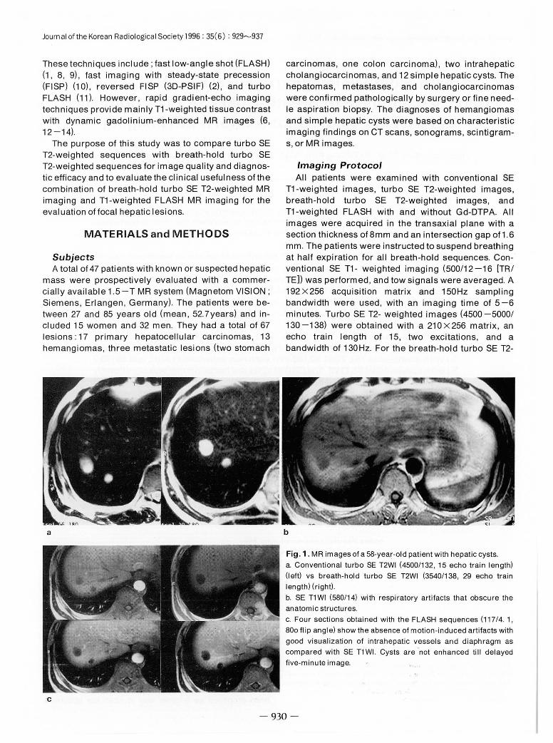
Fig. 1. MR images 01 a 58-yea.r-old patient with hepatic cysts. a. Conventional turbo SE T2WI (4500/132 , 15 echo train length) (l eft) vs breath-hold turbo SE T2WI (3540/138 , 29 echo train length) (right) b. SE T1 WI (580/14) with respiratory artifacts that obscure the anatomic structures c. Four sections obtained with the FLASH sequences (117 /4. 1,
800 fl ip angle) show the absence of motion-induced artifacts with good visualization of intrahepatic vessels and diaphragm as compared with SE T1 WI. Cysts are not enhanced till delayed five-minute image 1
‘
- 930 -
weighted images, parameters were TR , 3500 -4000 msec ; TE , 130 -138 msec ; a 116 X 256 matrix ; one excitation ; a sampling bandwidth of 260Hz ; and an echo train length of 29 , with saturation pulses superior and inferior to the section. Acquisition time was 4-6 minutes on the turbo SE T2-weighted images and was 17-20 sec on the breath-hold turbo SE T2- weighted images, respectively. FLASH imaging was performed with sections encompassing the entire liver in one breath hold. Imaging parameters were 117/4.1 ; one signal average; fl ip angle 80
0
; matrix size , 232 X 256 Acquisition time was 18 -20sec. Following the initial FLASH sequence , 0.1 m mol/kg of gedopentetate dimeglumine was given and as a bolus injection over approximately 20 seconds with the patient positioned in the bore of the magnet. 15mL of normal sal ine solution was rapidly flushed through the 100-cm intravenous extension tubing. Postcontrast FLASH images were obtained at 25, 50 , and 75 seconds and 5 minutes after the saline flush. In all imaging sequences, the field of view was 31 0 -400 mm.
Tae Hoon Kim, etal : Breath -Hold MR Imagingof Focal Hepatic Lesions
performed as follows: CNR=(signal intensity of lesionsignal intensity of liver)/standard deviation of noise signal intensity (15, 26). Region of interest (ROI) analysis of images was performed by a single observer (T. H. K.), for the liver ,an ROI was drawn as large as possible without the inclusion of surrounding tissues, especially blood vessels. The size and contour of the ROls were therefore not exactly the same for images obtained with all sequences. Mean values of two measurements were used. For liver lesions, an ROI was drawn as large as possible to encompass as much of the lesion as possible. Standard deviation of noise signal inten-
Table 1. Lesion Detectability by Image Sequence (n=69)
Sequence
Gd-FLASH Turbo SE T2WI
BH TSET2WI
FLASH
CSE T1WI
N。 oflesion( %)
67(97.1 )
66(96.4)
65(95.6)
62(89.9)
55(79.7)
Gd-FLASH : Gd-enhanced FLASH images, Image Analysis Turbo SE T2WI : turbo spin-echo T2-weighted images,
The signal intensity of I iver I esions and normal I iver BH TSE T2WI : breath-hold turbo spin-echo T2-weighted images,
parenchyma was measured with an electronic cursor. FLASH : fastlow-angleshotimages ,
The calculations of contrast-to-noise ratio (CNR) were CSE T1 WI : conventional spin-echo T1-weighted images
a b
c
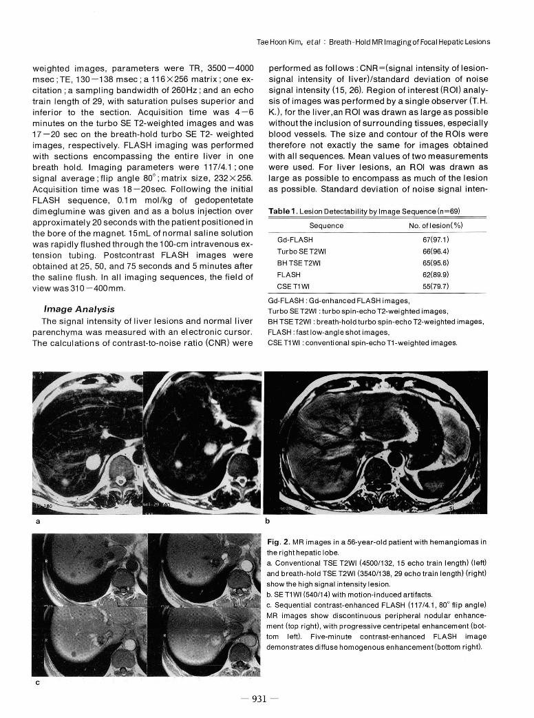
Fig. 2. MR images in a 56-year-old patient with hemangiomas in the right hepatic lobe. a. Conventional TSE T2WI (4500/132 , 15 echo train length) (left) and breath-hold TSE T2WI (3540/138 , 29 echo train length) (right) show the high signal intensity lesion. b. SE T1 WI (540 /14) with motion-induced artifacts c. Sequential contrast-enhanced FLASH (117/4.1 , 80
0 flip angle) MR images show discontinuous peripheral nodular enhancement (top right) , with progressive centripetal enhancement (bottom left). Five-minute contrast-enhanced FLASH image demonstrates diffuse homogenous enhancement (bottom right)
- 931 -

Journal ofthe Korean Radiological Society 1996 ; 35( 6) : 929~937
sity was measured as far as possible from the image in
the phase-encoding direction anterior to the abdomen.
CNRs of hepatic lesions (hemangiomas, cysts , and
solid masses) were compared with various pulse
sequences
AII images were assessed by two radiologists (T. H K. , K.W. K.) in consensus. Lesion detectability was
compared with all other imaging sequences. Qualitat
ive evaluation was based on the following criteria:
sharpness of anatomic structures, presence of respir
atory motion and vascular pulsation artifacts, and over-
all image quality. The sharpness of anatomic structur
es was based on an analysis of the ability to detect
internal structures (intrahepatic vessels) and the de
tection of the edges of normal structures (Iiver , pan
creas, spleen , and kidney) ; the criteria for evaluation
were as follows : extreme blur , moderate blur, mild
blur, and sharp. Artifacts were ranked as none, mild , moderate , or severe. Overall image quality was also
evaluated as poor , fair , good , or excellent. Statistical
analysis was performed with the Wilcoxon signed rank
and sign tests (19) .
Table 2 . Results 01 Ouantitative Evaluation 01 Focal Hepatic Lesions by Imaging Sequence
Total Cyst Hemangioma Solid mass
Sequence (n=69) (n=24) (n=17) (n=28)
Turbo SE T2WI 29.5 :t 17.8* 38.3 :t 1 0.4’ 38.8 :t 18. 7'" 19.1 :t 12.8.
BHTSET2WI 27.2 :t 13.5* 36.8 :t 15.3’ 32.2 :t 1 O. 6. 17.0 :t9.5
Gd-FLASH 15.8 :t 12.4 17.9 :t 15.0 14.5 :t 7.5 16.2:t 12.3
FLASH 13.8 :t7.2 16.4:t7.8 13.4 :t 10.4 11 .7 :t 6.4
CSET1WI 11.1 :t8.8 15.5 :t 11. 7 10.9 :t8.2 8.5 :t6.1
Oata (CNR) : mean + SO. *, . , "', . : statistical signilicance achieved at p ( 0.05Ievel. Total : total mass olthe liver, Turbo SE T2WI : turbo spin-echo T2-weighted images, BH TSE T2WI : breath-hold turbo spin-echo T2-weighted images,
Gd-FLASH ‘ Gd-enhanced last low-angle shot images, FLASH : last low-angle shot images,
CSE T1 WI : conventional spin-echo T1-weighted images
Table 3. Results 01 Oualitative Eval uation in 47 Patients.
Parameter Gd-FLASH
Sequence( %)
SET1WI TSET2W BHTSET2WI FLASH
Motion artilact
Breathing
None
Mild
Moderate
Severe
Vascular pulsation
Non닝
Mild
Moderate
Severe
Edge sharpness
Sharp
Mild blur
Moderate blur
Extreme blur
Overall image quality
Excellent
Good
Fair
Poor
5(11 ) 6(13) 21 (45) 23(49) 21(45)
17(36) 25(53) 16(34) 20(43) 19(40)
19(40) 13(28) 8(17) 2(4) 4(9)
6(13) 3(6) 2(4) 2(4) 3(6)
28(60) 21(45) 19(40) 0(0) 0(0)
13(28) 23(49) 26(55) 16(34) 20(43)
6(13) 3(6) 2(4) 25(53) 19(40)
0(0) 0(0) 0(0) 6(13) 8(17)
5(1 1) 7(15) 26(55) 25(53) 19(40)
20(43) 23(49) 12(26) 16(34) 22(47)
18(38) 15(32) 8(17) 5(11 ) 4(9)
4(9) 2(4) 1 (2) 1 (2) 2(4)
6(13) 7(15) 21(45) 24(51) 15(32)
21(45) 23(49) 15(32) 16(34) 20(43)
15(32) 14(30) 9(19) 6(13) 10(21 )
5(11 ) 3(6) 2(4) 1 (2) 2(4)
SE T1 WI : spin-echo T1-weighted images, TSE T2WI : turbo spin-echo T2-weighted images,
BH TSE T2WI : breath-hold turbo spin-echo T2-weighted images, FLASH : last low-angle shot images ,
Gd-FLASH ‘ Gd-enhanced last low-angle shot images
1 ‘ m n

RESULTS
A total of 69 hepatic lesions were detected in 47 patients. Lesion detectability was 67 (97. 1 %) with GdFLASH , 66 (96.4%) with turbo SE T2-weighted images, 65 (95.6 %) with breath-hold turbo SE images, 62 (89.9 %) with non-enhanced FLASH , and 55 (79.7%) with conventional SE T1-weighted images (Table 1).
For all hepatic lesions, CNR was significantly greater on turbo SE T2-weighted images (29.5) and breath-hold turbo SE T2-weighted images (27.2) than on FLASH with/without Gd-DTPA (15.8/13.8) and conventional SE T1-weighted images (11 .1) (p ( O.05) . There was , however no significant difference between turbo SE T2-weighted images and breath-hold turbo SE T2-weighted images (Table 2). CNR of cysts was significantly greater on turbo SE T2-weighted images (38.3) and breath-hold turbo SE T2-weighted images (36.8) , than on any other sequences (Table 2, Fig. 1). CNR of hemangiomas was the same as for cysts (Table 2, Fig. 2). For solid lesions, CNR was greatest on turbo SE T2-weighted images and was similar on breath-hold turbo SE T2-weighted images and Gd-FLASH without statistical significance, but was significantly higher than on conventional SE T1 -weighted images (Table 2,
TaeHoon Kim, etal : Breath -Hold MR ImagingofFocal Hepatic Lesions
Fig. 3, 4). Breath-hold turbo SE T2-weighted images and Gd-FLASH were qualitativly superior to other sequences except the vascular pulsation artifact of FLASH (Table 3). Breath-hold turbo SE T2-weighted images were inferior to turbo SE T2-weighted images in lesion detectability (Table 1) , but there was no statistical difference in CNR (Table 2). Non-enhanced FLASH was also superior to conventional T1-weighted images for CNR , lesion delectability , sharpness , respiratory motion artifact, and overall image quality (Table 1 -3) .
DISCUSSION
High soft-tissue contrast and the absence of motioninduced image artifacts with rapid acquisition time are the major prerequisites for the detection of liver lesions in MR imaging of the abdomen (9). SE T2-weighted images are superior to T1-weighted SE sequences in lesion detection at higher field strengths because of high soft-tissue contrast (1 -6). However, the limitations of conventional T2-weighted SE sequences are long examination times and high susceptibility to motion induced artifacts.
Turbo SE sequences provide high-quality images in significantly less time than is required for conventional
a
c 1 j
m n
b
Fig. 3. MR images of a 68-year-old patient with hepatocellular carclnoma a. Conventional TSE T2WI (4500/132 , 15 echo train length) (Ieft) vs breath-hold TSE T2WI (3540 /138 , 29 echo train length) (right) and (b) SE T1 WI (580/14).
c. Dynamic contrast-enhanced FLASH (117/4.1 , 800 flip angle) MR
images demonstrate the presence of an early enhancing tumor with rapi d wash-out
Journal ofthe Korean Radiologica l Society 1996 : 35(6) : 929~937
SE imaging, but the problem is respiratory motioninduced artifacts. Turbo SE was developed by Hennig et al. (16). Multiple 1800 refocusing RF p비 ses are applied with a different phase-encoding value , thus decreasing the acquisition time proportional to the echo train length. The reduction in imaging time can be used to improve image quality (4, 7, 8, 11). The increased number of excitations can be used to increase the SNR and to decrease the prominence of respiratory ghost and vascular pulsation artifacts. Spatial resolution can be improved by using a larger matrix. In this study, turbo SE T2-weighted images had an echo train length of 15, a 210 X 256 matrix, two excitations, and acquisition time of 4-6 minutes. Our data still show poor image quality due to respi ratory motioninduced artifacts, however (Table 3). Breath-hold turbo SE T2-weighted images and turbo SE T2-weighted images show similar CNR (Table 1, 2) , the former also decreases motion induced artifacts from respiratory suspension during image acquisition. To reduce the imaging times , parameters are as follows; one acqui-
sition , a 116 X 256 matrix , an echo train length of 29. Eleven sections can be obtained in 16-20 seconds with 。ne breath-holding period. Because twenty-nine 180。refocusing p비 ses per TR interval are applied with varied phase encoding , four TRs are needed for the filling ofthe K-space.
Tissue contrast on turbo SE images is nearly identical to that on conventional SE images, and the former might replace the latter for imaging the brain , spine, and pelvis (17 , 18). Catasca et al. (4) showed , however, that nearly all solid abdominal organs or mass lesions showed a lower signal intensity on turbo SE images than on conventional SE images. The higher signal intensity of abdominal fat on the turbo SE images could account for the decreased range of tissue contrast represented on a relative scale. This effect increases as the number of refocusing pulses increases and as the time between refocusing pulses decreases (4, 5). Tissue contrast will also be influenced by different amounts of T2-decay, was caused by varying refocusing pulses on turbo SE images. In our data, con-
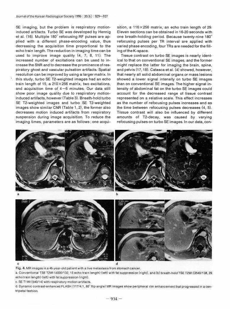
a b
c d Fig . 4. MR images in a 46-year-old patient with a live metastasis from stomach cancer a. Conventional TSE T2WI (4500 /132 , 15 echo train length) (Ieft) with fat suppression (right) , and (b) breath-hold TSE T2WI (3540 /138 , 29
echo train length) (left) with fat suppression (right). c. SE T1 WI (540 /14) with respi ratory motion artifacts‘
d. Dynamic contrast-enhanced FLASH (117 /4.1 , 800 flip angle) MR images show peripheral rim enhancement that progressed in a cen
tripetal fashion
A
냥 ‘
m n

ventional SE sequences were not used , but we thought that turbo SE sequences with echo train length of 15 would influence the tissue contrast. CNR and lesion detectability on turbo SE T2-weighted images were actually superior to those on breath-hold turbo SE T2-weighted images, but differences between them were ot significant (Table 1, 2). On turbo SE images, magnetization transfer contributes to some loss of signal intensity in solid tissues (4, 7, 20 , 21 , 26) Magnetization transfer refers to the cross relaxation between free unbound water protons and protons bound to the surface of macromolecules in protein solutions and tissues ; these effects are thus generated by the m비tiple 1800 refocusing pulses used in the turbo SE sequences (20 , 21 , 26). Increased magnetization transfer effects may lead to relatively lower signal intensity ratios of solid lesions (20 , 21). Thus , as ech。train length increases, tissue contrast and loss of signal intensity of solid lesions will increase. In our study, CNR for solid lesions was greater on turbo SE T2-weighted images than on other sequences, but there was no statistical significance with FLASH sequences (Table 2, Figure 3, 4). Cystic lesions or hemangiomas undergo little or no magnetization transfer effect , however and have more heavily T2-weighted imaging parameters. Consequentiy , the difference in the tissue contrast between cystic lesions or hemangiomas and solid lesions would be relati vely greater on the turbo SE images than on the conventional SE images (4, 7, 21 , 26). Lesion detectability and CNR were actually signi ficantly higher on turbo SE T2-weighted images than on FLASH and conventional SE T1 -weighted images, but those for breath-hold turbo SE T2-weighted images which had longer echo trains were inferior to those for turbo SE T2-weighted images, without statistical significance. Effective TE as well as the effect of magnetization are also major elements that theoretically alter tissue contrast on turbo SE images (4, 7). Multiple 1800 refocusing pulses per TR interval have a different T2 decay. The middle lines of K-space primari ly determine tissue contrast and SNR. Tissue contrast will thus be influenced primarily by the T2 decay consistent with a given operator-selected TE (4)
Overall image quality for the turbo SE T2-weighted sequence was found to be significantly inferior to that obtained for the breath-hold turbo SE T2-weighted se-
Tae Hoon Ki m, et al : Breath -Hold MR Imaging of Foca l Hepatic Lesions
were slightly inferior to turbo SE T2-weighted images for lesion detectability and tissue con rast, but the re was no statistically significat difference between the two sequences. As a breath-hold turbo SE T2-weighted sequence provides high-quality images with fewer arifacts in significantly less time than is possible with a turbo SE T2-weighted sequence, we think that the breath -hold turbo SE T2-weighted sequence is a useful method for the detection of liver lesions. The potential limitation of a breath-hold turbo SE T2-weighted sequence is the restr icted number of slices. In our study , 11 sections can be acquired within 18 seconds. At a section thickness of 8mm and an intersection gap of 1.6 mm , as was used in our study, about 10.5cm in length can be covered. However, if a slice thickness of 1 0 -12 mm and an interslice gap of 2 -3mm were applied , about 16-17cm in length could be covered. On the other hand , the entire l iver can be imaged twice, if necesary , within a short acquisition time ; the imag ing time of breath-hold turbo SE T2-weigedd sequence is only about 18 seconds.
FLASH is an MR imaging sequence that can acquire a T1-weighted image in less than 1 second per section (5 , 6, 9, 12, 23). It has a very short TE , needed to obtain heavy T1-weighted images and to allow for high multi -slice capability. The FLASH technique thus also provides good image quality with less motion-induced artifacts. CNR on the FLASH images was also slightly superior than on conventional SE T1 -wei ghted images, and lesion detectability was also higher on FLASH than 。n conventional SE T1-weighted images (Table 1, 2). Although the CNR ofFLASH was infer ior to that ofturbo SE T2-weighted images , FLASH techniques were useful in dynamic images with Gd-DTPA ; CNR was 14 % hegher on Gd-DTPA enhanced FLASH images than on unenhanced images, and also showed a 38 % increase in CNR for solid mass lesions. In comparison , Edelman et al. (24) reported that CNR was 50 % higher on Gd-DTPA enhanced FLASH images than on unenhanced images. Dynamic FLASH sequence images offer some potential for the characterization of lesions, and lesion detectability on FLASH is also slightly better than on turbo SE T2-weighted images (Table 1). Simple cysts showed relatively well-marginated , oval lesions with low signal intensity, or signal void lesions on enhanced dynamic FLASH imges and very high signal intensity on SE TE-weighted images (Figure 1). Hemangiomas show
- 935 -

Journal ofthe Korean Radiological Society 1996 : 35( 6) : 929~937
regular enhanced patterns on cystic mass lesions such as hemangiomas or simple cysts. Hepatocellular carcinomas showed as relatively high vasucular solid mass lesions and also were of high signal intensity on SE T2-weighted images and of low signal intensity with a less clearly demarcated margin on conventional T1-weighted images or unenhanced FLASH images These lesions demonstrated early inhomogenous enhancement with early wash-out on dynamic enhanced FLASH images (Figure 3). On the other hand , meta
static lesions showed peripheral rim enhancementthat progressed in a centripetal fashion (Figure 4) (6 , 13).
Lesion detectability was slightly inferior on breathhold TSE T2-weighted images (95.6 %) than on conventional TSE T2-weighted images (96.4 %), but with breath-hold TSE T2-weighted images in combination
with pre- and post-enhanced dynamic FLASH images there were no problems in the evaluation of hepatic focal lesions (97.1 %). Since FLASH images were free of
respiratory motion-induced artifacts and were also superior to the conventional SE T1-weighted images with respect to CNR and overall image quality , we thought that breath-hold TSE T2-weighted iamges combined with dynamic enhanced FLASH images might provide good image quality and reduced acquisition time
FLASH sequence limitations include a prominent vascular p비 sation artifact arising from the aorta, which could obscure lesions, especially in the left lobe of the liver (5 , 6, 9) . Presaturation pulses have been used with other fast imaging sequences to decrease this flow artifact (27) . Saturation pulses are not compatible with FLASH sequences , however (23). We are currently
investigating the use of SWAP (changes of phaseencoding direction) , where there is doubt regarding a focal lesion in the left lobe of the liver. Susceptability artifacts play a major role in gradient-echo sequences
and also influence image quality. The TE of 4.1 msec approximates the fat-water in-phase time of the 1.5 T MR system; signal losses at organ interfaces due to signal-canceling artifacts were thereby avoided. Metal implants such as surgical clips produced strong artifacts, which decreased with SE sequences and were also no seen with longer echo train turbo SE sequences (9 , 23)
In conclusion , the breath-hold TSE T2-weighted sequence is slightly inferior to the TSE T2-weighted sequence as regards lesion detectability or tissue c
may be an excellent techn i ques that can be used to rapidly evaluate liver lesions while offering superior overall image quality.
REFERENCES
1. Simm FC, Semelka RC , Recht M, Deimling M, Lenz G, Laub GA. Breath-hold T2-weighted sequences 01 the liver: a com parison 01 lour techniques at 1.0 and 1.5 T. Magn Reson
Imaging 1992 ; 10: 41-47 2. Taupitz M, Speidel A, Hamm B, et al. T2-weighted breath-hold
MR imaging 01 the liver at 1.5 T: results with three-dimensional steady-state Iree precession sequence in 87 patients. Radi
ology 1995 ; 194 : 439-446 3. Rydberg JN , Lomas DJ , Coakley KJ , Hough DM , Ehman RL,
Riederer SJ. Comparison 01 breath-hold last spin-echo and conventional spin-echo pulse sequences lor T2-weighted MR imaging 01 liver lesions. Radiology 1995; 194: 431-437
4. Catasca JV, Mirowitz SA T2-weighted MR imaging 01 the abdomen : last spin-echo vs conventional spin-echo sequen ces. AJR 1994; 162: 61-67
5. Low RN , Fancis IR , Sigeti JS, Foo TK F. Abdominal MR imaging: comparison 01 T2-weighted last and conventional spin-echo, and contrast-enhanced last multiplanar spoiled gradient-recalled imaging. Radiology 1993; 186: 803-811
6. Semelka RC , Shoenut JP, Kroeker MA , et al. Focal liver disease: comparison 01 dynamic contrast-enhanced CT and T2-weighted lat-suppressed, FLASH , and dynamic gadolinium enhanced MR imaging at 1.5 T. Radiology 1992;184:687-694
7. Eric K, Outwater EK , Mitchell DG , Vinitski S. Abdominal MR imaging : evaluation 01 a last spin-echo sequence. Radiology
1994 ; 1 90 : 425-429 8. Edelman RR , Hahn PF , Buxton R, et al. Rapid MR imaging
with suspended respiration : clinical application in the liver Radiology 1986 ; 161 : 125-131
9. Taupitz M, Hamm B, Speidel A, Deimling M, Branding G, Woll KJ. Multisection FLASH: method lor breath-hold MR imaging 01 the entire liver. Radiology 1992;183:73-79
10. Unger EC, Cohen MS, Gatenby RA‘ et al. Single breath-holding scans 01 the abdomen using FISP and FLASH at 1.5 T. J Comput Assit Tomogr 1988; 12: 575-583
11. Edelman RR , Wallner B, Singer A, Atkinson DJ , Saini S Segmented turbo FLASH: method lor breath-hold MR imaging 。1 the liver with Ilexible contrast. Radiology 1990; 177 515-521
12. Mirowitz SA, Lee JK, Gutierrez E, Brown J, Heiken JP,
Eilenberg SS. Dynamic gadolinium-enhanced rapid acquisition spin-echo MR imaging 01 the liver. Radiology 1991; 179 371-376
13. Hamm B, Fischer E, Taupitz M. Differentiation 01 hepatic hemangiomas Irom metastases by dynamic contrast-enhanced MR imaging. J Comput Assist Tomogr 1990; 14(2): 205-216
14. Semelka RC , Brown ED, Ascher SM , et al. Hepatic hemangioma: a multi-institutional study 01 appearance on T2 weighted and serial gadolinium-enhanced gradient-echo MR images. Radiology 1994; 192 .401-406
15. Mirowitz SA, Lee JKT, Brown JJ , Eilenberg SS , Heiken JP,
Perman WH. Rapid acquisition spin-echo (RASE) MR imaging a new technique lor reduction 01 artilacts and acquisition time. Radiology 1990;175:131-135
16. Henig J, Nauerth A, Friedburg H. RARE imaging : a last %

imaging method lor clinical MR. Magn Reson Med 1986 ‘ 3
823-833
17. Johns KM , Mulken RV, Schwartz RB , Oshio K, Barnes PD , Jolez FA. Fast spin-echo MR imaging 01 the brain and spine
current concepts. AJR 1992;158:1315-1320
18. Smith RC , Reinhold C, Lang RC , McCauley TR , Kier R, McCarthy S. Fast spin-echo MR imaging 01 the lemale pelvis
Part 1. Use 01 a whole-volume coi l. Radiology 1992; 184
665-669
19. Sachs L. Applied statistics. 1 st ed. Berlin , Germany
Springer-Verlag , 1982.
20. Constable Rt, Anderson AW, Zhong J, Gore JC. Factors
inlluencing contrast in last spin-echo MR imaging. Magn
Reson Imaging 1992 ;10 :497-511
21. Melki PS , Mulkern RV. Magnetization transler effects in
multi-slice RARE sequences. Magn Reson Imaging 1992; 24
189-195
22. Wood ML , Runge VM , Henkelman RM. Overcoming motion in
abdominal MR imaging. AJR 1988 ;150 : 513-522
Tae Hoon Kim, et al : Breath - Hold MR Imaging of Focal Hepatic Lesions
23. Low RN. Francis IR , Herlkens RJ , et a l. Fast m비tiplanar spoil
ed gradient-recalled imaging 01 the liver : p비se sequence
。ptimization and comparison with spin -echo MR imaging. AJR
1993 ; 160 : 501 -509
24. Edelman RR , Siegel JB , Singer A, et al. Dynamic MR imaging
。1 the liver with Gd-DTPA : initial clinical results. AJR 1989;
1 53 : 1213-1 21 9
25. Mitchell DG , Saini S, Weinreb J, et al. Hepatic metastases
and cavernous hemangiomas : distinction with standard- and
triple-dose gadoteridol-enhanced MR imaging. Radiology
1994 ; 192 ‘ 401-406
26. Outwater E, Schnall M, Braitman LE, Dinsmore BJ‘ Kressel
HY. Magnetization transler 01 hepatic lesions : evaluation 01 a
novel contrast technique in the abdomen. Radiology 1992 ;
182 : 535-540
27. Felmle JP , Ehman Rl. Spatial presaturation : a method lor
suppressing Ilow artilacts and improving depiction 01 vascular
anatomy in MR imaging. Radiology 1987 ;164:559-564
대 한 방사 선 의 학회 지 1996 ; 35( 6) : 929 - 937
국소적 간병변에 대한호흡보상이 가능한자기공명영상: 호흡보상-급속 스핀에코 T2-강조영상과 FLASH 영상 조합의 임상적 유용성 1
1 연세대학교 의과대학 진 단방사선과, 영동세브란스병원
김 태 훈·김 기 황·김 은 경·유 정 식
목 적 : 간병변에 대한 자기공명 영상 검사에서 기존의 급속스핀어|코(turbo spin - echo ; TSE) 방식으1 T2 -강조영상( T2WI )
과 호흘보상( breath - hold ) 0 1 가능한 TSE 방식의 T2WI를 영상의 질과 진단적 효율면에서 비교하고, 호흘보상이 가능한 TSE
T2WI와 FLASH 방식으1 T1 강조영상( T1WI ) 기법만으로도 기존의 고식적, 혹은 TSE 방식을 대체할 수 있는지 알아보고자 하였
다
대상 및 방법 : 간병변이 의심되거나 치료중인 47 명의 환자(남 여 =32: 15, 평균연령 = 52.7)를 대상으로 1.5 T 자기공명
영상장치를 이용하여 검사를 시행하였다. 모든 환자에 대해 고식적 SE T1WI , 기존의 TSE T2WI , breath - hold TSE T2WI 및
FLASH 영상을 얻었다. 대상환자는 원발성 간암이 17 예, 혈관종이 13예, 전이암이 3여1 , 담관암이 2예였으며 단순낭종만 있었
던 경우도 12예가 있었다. 종괴의 대조도와 발견율, 해부학적 경계의 명확도, 영상의 질, 인공유렁물 등을 각 영상방식에 따
라비교하였다.
결 과 47명의 환자에서 69개의 종괴가 발견되었다. 각 영상별로 종괴의 발견율은 Gd-FLASH 97.1 % ( 67/ 69 ) , TSE T2WI
96.4% (66/ 69 ) , breath - hold TSE T2WI 95.6% (65/ 69) , 조영전 -FLASH 89.9% (62/ 69) , SE T1WI 79.7%(55/ 69)였다. 단순낭
종과 혈관종괴에 대한 대초도는 TSE T2WI와 breath - hold TSE T2WI가 다른 세 군에 비해 통계적으로 의의 있게 높았으나 서
로간에는 통계적인 차이가 없었다. 고형종괴의 대조도는 TSE T2WI ( 19.1)에서 가장 높게 나타났으나 breath - hold TSE T2WI
(17.1) , Gd-FLASH( 16.2) 방식에서도 비슷한 결과를 보였으며 이들 세군 사이에서는 통계적 의의가 없었다. 질적인 면에서는
breath - hold TSE T2WI 및 Gd - FLASH 방식이 해부학적 경계의 명확도, 영상의 질, 호흘에 의한 인공유렁물은 우수한 결과를
나타냈으나 혈관 박동에 의한 유렁물은 FLASH 기법에서 높게 관찰되었다. Breath-hold TSE T2WI는 병소발견율에서는 TSE
T2WI보다 다소 낮았으나 대조도 면에서는 통계적인 차이가 없었다. 조영전 -FLASH 방식은 SE T1WI에 비해서 통계적인 차이
는 없었지만 대조도,종괴발견율 및 영상의 질적인 면에서 우수하였다.
결 론 : 국소적 간병변을 발견하는데 있어서 breath-hold TSE T2WI는 기존으1 TSE T2WI를 대체할 수 있으리라 기대되며,
조염전 -FLASH 방식은 고식적 SE T1WI를 대체 가능 하리라 생각된다. 따라서 breath - hold TSE T2WI, 조영전 -FLASH 및
Gd - FLASH 영상들만의 조합으로도짧은시간에 앙질의 간영상을얻을수 있으리라생각된다.
” ”
찍지이21 그 L 톨 L I 를
Internet을 통한 방사선과학 및 관련분야의 정보를 얻을 수
있는 Web site 주소와 찾는 방법에 대하여 월레스기념 침
례병원 진단방사선과의 전동진 선생의 기고가 있었습니다
(대한방사선의학회지 1996; 34 : 299 -300). 전돔진 선생이
추가로 제공한 Web site 주소중 독자의 관심 영역이라고
판단되는 것을 선정하여 알려드립니다.
※ Web site
. ftp: // ftp.xray.hmc.psu.edu/ acr _ codes
ACR [Index for Radiological Diagnoses] (4th Edi
tion)
. http: // www.rad.rpslmc.edu / -ajnr / aj nrhome.html
[AJNR: American Journal of Neuroradiology]
. http: // www.acponline.org / j ournals / annals / annal
toc.htm
[Annals of Internal Medicine]
. http :// www.hwc.ca : 8080 / cma / j ournals / carj / in
dex.html
. http: // www.scar.rad.washington.edu/SCAR/ JD I.
html
[Journal of Digital Imaging]
(Society for Computer Applications in Radiology
[SCAR] )
. http ://www.spie.org / web / journals / j ei_ home.html
[Journal of Electronic Imaging] (SPIE)
. http ://wwwicic.nci.nih.gov / jnci / jnci_issues.html
[Journal of the National Cancer Institute]
. http :// www.thelancet.com
[The Lancet]
. http :// www.ecr.org / journal /index.htm
[Eurean Radiology]
. http: //www.mir.wustl.edu / MIRINFO / focalspot /
FocalSpot _S94.HTML
[Focal Spot] (Mallinckrodt Institute of Radiology)
[ CARJ Online (Canadian Association of Radiologists . http: // www.uky.edu/OtherOrgs / lnvestRadiol/
Journal) ] [ Investigative Radiology]
. http ://www-mitpress.mit.edu / jrnls -catalog / cont - . http: // www.nejm.org/
neuro.html [New England Journal of Medicine Online]
[ Journal of Contemporary Neurology]
(Massachusetts Institute of Technology)
938 -