breast cancer src activity: bad to the bone
TRANSCRIPT
Cancer Cell
Previews
Breast Cancer Src Activity: Bad to the Bone
Dennis C. Sgroi1,2,*1Molecular Pathology Research Unit and Center for Cancer Research, Massachusetts General Hospital2Department of PathologyHarvard Medical School, Boston, MA 02129, USA*Correspondence: [email protected] 10.1016/j.ccr.2009.06.010
Bone metastases are a major cause of breast cancer morbidity and mortality. In this issue of Cancer Cell,Zhang and colleagues identify a Src activation expression signature associated with late-onset breast cancerbone metastases and provide evidence for Src as a key mediator of survival signals in latent bone metastases.
Metastasis, the disseminated spread and
growth of malignant cells to distant
organs, is the final and most devastating
stage of human breast cancer progres-
sion. Bone represents the most common
organ involved by metastatic breast
cancer, and metastases to this site occur
in approximately 30% of all women
diagnosed with invasive breast cancer.
Once breast cancer has metastasized to
bone, the disease often inflicts marked
morbidity and is ultimately incurable.
The development of bone metastases
from a primary invasive breast cancer
involves a complex sequence of interde-
pendent events that include extravasation
of tumor cells into the blood or lymphatic
vasculature, survival within the circulation,
arrest and adhesion at the distant site,
and intravasation and seeding of the
bone marrow microenvironment (Gupta
and Massague, 2006). Once metastatic
cells have successfully seeded the bone
marrow, three potential outcomes may
be distinguished (Brackstone et al., 2007):
(1) the tumor cells can die or be eliminated
by an effective immune response or thera-
peutic intervention; (2) the malignant cells
may survive and remain clinically dormant;
or (3) the tumor cells can survive and enter
into a ‘‘vicious cycle’’ of bone resorption
and tumor outgrowth that gives rise to
clinically significant osteolytic macrome-
tastases (Kang et al., 2003, 2005; Mundy,
2002). Although proliferating metastatic
cells may be of primary clinical concern,
clinically dormant tumor cells and micro-
metastases also have significant clinical
importance as they can and do frequently
serve as the source of ‘‘latent’’ metastatic
breast disease.
In this issue of Cancer Cell, Zhang et al.
(2009) provide both clinical and experi-
mental evidence that Src plays a critical
role in the establishment of latent bone
metastases in breast cancer. Using a bio-
informatic approach that investigated the
association of various signaling pathway-
specific gene expression patterns with
breast cancer outcome, the authors iden-
tified a Src activity gene expression signa-
ture (designated as the Src responsive
signature, or SRS) that was highly associ-
ated with late-onset breast cancer bone
metastases. In a multivariate analysis,
SRS was found to be independent of the
distinct molecular subtypes of human
breast cancer and independent of cell
proliferation, the common biological prin-
ciple driving the prognostic performance
of most previously discovered breast
cancer gene expression signatures. Most
notably, the SRS was found independent
of estrogen receptor (ER) status, the con-
ventional clinicopathological parameter
currently most closely associated with
breast cancer bone metastases.
To address the role of Src in the bone
metastatic process, the authors employed
the use of two SRS-expressing human
breast cancer cell lines that possess
either aggressive or indolent metastatic
bone tropism in a xenograft mouse model.
In the cell line possessing aggressive
metastatic bone tropism, stable knock-
down of Src resulted in a significantly
decreased rate of tumor outgrowth of
bone lesions that was most apparent in
the latter stages of this model, while
knockdown of other Src family kinases
did not impact bone metastatic activity.
In the indolent model of bone metastatic
disease, knockdown of Src resulted in
near complete loss of bone metastatic
activity. Src knockdown did not alter
lung or lymph node metastatic activity
supporting a specific role for Src in bone
metastasis.
Cance
Src has the potential to impact one
or more steps in the highly complex
bone metastatic process. In addition to
its effect on increasing cell proliferation,
Src demonstrates pleiotropic functional
activity that includes cellular differentia-
tion, adhesion, migration, invasion, and
survival. Zhang et al. showed that the
potential pro-proliferative effect of Src
do not account for the bone metastatic
outgrowth in their model system by de-
monstrating that stable knockdown of
Src did not decrease the intrinsic prolifera-
tive activity of the cells in both in vitro- and
in vivo-based assays. These results sug-
gested that Src may play a role in the initial
seeding of or in the sustained survival of
metastatic cells in the bone microenviron-
ment. To better understand the role of Src
as it relates to these two different aspects
of the metastatic cascade, the authors
performed a series of elegant genetic and
pharmacological studies (using dasatinib,
a Src tyrosine kinase inhibitor) that de-
monstrate Src is dispensable for bone
marrow seeding but rate-limiting for the
survival and sustained outgrowth of indo-
lent breast cancer cells in the bone marrow
microenvironment.
Like early stage primary breast cancer,
the successful survival and outgrowth
of bone metastases likely develops from
intrinsic tumor epithelial-specific and
tumor stromal microenvironment-specific
(i.e., bone marrow-specific) factors. In
order to identify cell survival factors in
the bone marrow microenvironment that
contribute to metastatic breast cancer
outgrowth, Zhang et al. performed
comparative microarray gene expression
analysis of metastatic breast cancer
samples derived from human bone, lung,
brain, and liver. Seventeen secreted
factors were differentially upregulated in
r Cell 16, July 7, 2009 ª2009 Elsevier Inc. 1
Cancer Cell
Previews
the human bone metastatic samples
including the chemokine CXCL12/SDF1
(a survival factor for cells expressing the
receptor CXCR4) and the cytokine TRAIL
(a proapoptotic ligand that binds to the
DR4/5 receptor). The authors demon-
strated a similar cytokine expression
profile in metastatic tumor samples ob-
tained from their xenograft model of breast
cancer bone metastases. Zhang at al. also
explored the functional implication of these
observations through Src knockdown and
rescue experiments in their SRS-express-
ing bone metastatic cell lines and demon-
strated that Src is required for CXCL12/
SDF-mediated cell survival and abrogates
TRAIL-mediated apoptosis.
Taken together, the findings of Zhang
et al. provide strong clinical and experi-
mental evidence that Src activation plays
a critical role in bone-specific metastatic
breast cancer cell survival and in the
establishment of latent breast cancer bone
metastases. These findings set the stage
for the development of novel therapeutic
strategies directed at eradicating breast
cancer metastases to bone. Because
metastatic bone colonization consists of
an initial latent phase mediated by a Src
survival response and a ‘‘vicious cycle’’
outgrowth phase mediated by complex
molecular signaling between bone osteo-
clasts and tumor cells (Figure 1), one could
envision therapeutic interventions aimed
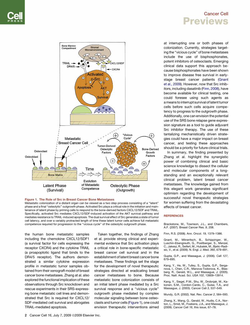
Figure 1. The Role of Src in Breast Cancer Bone MetastasesMetastatic colonization of a distant organ can be viewed as a two step process consisting of a ‘‘latent’’phase and a final ‘‘osteolytic’’ outgrowth phase. Activated Src plays a critical role in the initiation and main-tenance of latent phase by priming cells to respond to the bone-derived factors CXCL12/SDF and TRAIL.Specifically, activated Src mediates CXCL12/SDF-induced activation of the AKT survival pathway andmediates resistance to TRAIL-induced apoptosis. The dual survival effect of Src generates a state of tumorcell latency, and over a variably protracted length of time these latent tumor cells achieve full metastaticcompetence required for progression to the ‘‘vicious cycle’’ of the osteolytic outgrowth phase.
2 Cancer Cell 16, July 7, 2009 ª2009 Elsevier Inc.
at interrupting one or both phases of
colonization. Currently, strategies target-
ing the ‘‘vicious cycle’’ of bone metastases
include the use of bisphosphonates,
potent inhibitors of osteoclasts. Emerging
clinical data support this approach be-
cause bisphosphonates have been shown
to improve disease free survival in early-
stage breast cancer patients (Gnant
et al., 2009). However, now that Src inhib-
itors, including dasatinib (Finn, 2008), have
become available for clinical testing, one
could foresee using such agents as
a means to interruptsurvival of latent tumor
cells before such cells acquire compe-
tency to progress to the outgrowth phase.
Additionally, one can envision the potential
use of the SRS bone relapse gene expres-
sion signature as a tool to guide adjuvant
Src inhibitor therapy. The use of these
tantalizing mechanistically driven strate-
gies could have a major impact on breast
cancer, and testing these approaches
should be a priority for future clinical trials.
In summary, the finding presented by
Zhang et al. highlight the synergistic
power of combining clinical and basic
science knowledge to dissect the cellular
and molecular components of a long-
standing and an exceptionally relevant
clinical problem, latent breast cancer
metastases. The knowledge gained from
this elegant work generates significant
optimism regarding the development of
successful novel therapeutic strategies
for women suffering from the devastating
effects of breast cancer.
REFERENCES
Brackstone, M., Townson, J.L., and Chambers,A.F. (2007). Breast Cancer Res. 9, 208.
Finn, R.S. (2008). Ann. Oncol. 19, 1379–1386.
Gnant, M., Mlineritsch, B., Schippinger, W.,Luschin-Ebengreuth, G., Postlberger, S., Menzel,C., Jakesz, R., Seifert, M., Hubalek, M., Bjelic-Radi-sic, V., et al. (2009). N. Engl. J. Med. 360, 679–691.
Gupta, G.P., and Massague, J. (2006). Cell 127,679–695.
Kang, Y., He, W., Tulley, S., Gupta, G.P., Serga-nova, I., Chen, C.R., Manova-Todorova, K., Blas-berg, R., Gerald, W.L., and Massague, J. (2005).Proc. Natl. Acad. Sci. USA 102, 13909–13914.
Kang, Y., Siegel, P.M., Shu, W., Drobnjak, M., Ka-konen, S.M., Cordon-Cardo, C., Guise, T.A., andMassague, J. (2003). Cancer Cell 3, 537–549.
Mundy, G.R. (2002). Nat. Rev. Cancer 2, 584–593.
Zhang, X., Wang, Q., Gerald, W., Hudis, C.A., Nor-ton, L., Smid, M., Foekens, J.A., and Massague, J.(2009). Cancer Cell 16, this issue, 67–78.