brain tumors in children - gupea: home · med hjälp av kognitiva och motoriska tester samt test...
TRANSCRIPT

Brain tumors in children
Intervening with the aftermath: relapse and long term side effects
Magnus Sabel
Department of Pediatrics
Institute of Clinical Sciences
Sahlgrenska Academy, University of Gothenburg, Sweden
Gothenburg 2017

Cover illustration by Axel Sabel 2017
Brain tumors in children
© Magnus Sabel 2017
ISBN 978-91-629-0131-8 (tryck)
ISBN 978-91-629-0132-5 (pdf)
E-publication: http://hdl.handle.net/2077/51734
Printed in Gothenburg, Sweden, 2017 by
Ineko AB

“Outside of a dog, a book is a man's best friend. Inside of a dog it's too dark
to read”
― Groucho Marx
To Susanne, Axel and Ivan


Brain tumors in children Intervening with the aftermath: relapse and long
term side effects
ABSTRACT
After completing primary treatment, childhood brain tumor patients enter a follow-up
phase. Follow-up is needed for two main reasons; to detect relapse and to diagnose
late side effects. The overall aims of this thesis were; 1) To describe and analyze the
pattern of relapse after treatment for medulloblastoma the most common malignant
brain tumor in childhood with the aim to find potentially successful relapse
treatment and; 2) To investigate ways to lessen the side effects of brain tumor
treatment, especially the cognitive side effects.
Methods: The long-term outcome of 338 medulloblastoma patients enrolled in the
HIT-SIOP-PNET4 trial was investigated, with a focus on relapse diagnosis, pattern
of relapse, and treatment of relapse. In a separate randomized, single-center, single-
blinded, pseudo crossover study, the potential benefit of physically active video
gaming in childhood brain tumor survivors was investigated. Thirteen children, all
previously treated with cranial radiotherapy, were randomized to either active video
gaming (with weekly internet-based coaching sessions) followed by a waiting list
period, or these periods in reverse order. They were assessed before and after each
period, with measures of cognition, motor function and activities of daily living
(ADL). Finally, in a rodent model the potentially protective effect of post-irradiation
hypothermia on the neurogenic areas of the brain the subventricular zone (SVZ)
and the granule cell layer (GCL) of the hippocampus was examined. Young rats
were randomized to either normothermia or hypothermia for eight hours post-
irradiation, or a control group. Their brains were examined one week later, measuring
the SVZ and GCL areas, and counting the number of proliferating cells and
microglia.
Results and conclusions: The ultimately grim prognosis for patients with recurrent
medulloblastoma, irrespective of treatment, is confirmed. Surgery for histological
diagnosis and research should be encouraged, and can in selected cases prolong
survival, but new treatment options are needed. Active video gaming improves body
coordination and the execution of ADL. Positive effects on cognition is a possibility,
although not confirmed in this pilot study. Hypothermia after irradiation of the brain
has a protective effect on the SVZ, but not the GCL, one week post-irradiation. The
long term and functional effect of this finding needs further exploration, together
with studies of the effect of hypothermia on brain tumors.
Keywords: medulloblastoma, relapse, radiotherapy, cognition, exercise therapy,
video games, hypothermia, pediatric, brain tumor ISBN: 978-91-629-0131-8

SAMMANFATTNING PÅ SVENSKA
När barn med hjärntumör är färdigbehandlade påbörjas en uppföljningsfas, av
två viktiga skäl: för att upptäcka eventuella återfall och för att upptäcka sena
biverkningar. Målsättningen med denna avhandling var dels att beskriva och
analysera återfall av medulloblastom, den vanligaste elakartade hjärntumören
hos barn, dels att undersöka sätt att mildra de långsiktiga (kognitiva)
biverkningarna som barn med hjärntumör ofta drabbas av.
I avhandlingen presenteras resultat avseende överlevnad hos 338 patienter
som behandlats för medulloblastom i studien HIT-SIOP-PNET4. Hos de 72
patienter som fick återfall beskrivs diagnostik, återfallsmönster, behandling
och prognos. I en separat studie av 13 barn som fått strålbehandling för en
hjärntumör undersöktes om regelbundet spelande av fysiskt aktiverande
dataspel (Nintendo Wii) kunde påverka motorik, kognition, aktiviteter i
dagliga livet (ADL) och aktivitetsnivåer. I denna randomiserade
singelblindade studie fick barnen antingen börja med fysiskt aktivt
dataspelande (med veckovis internetbaserat coachningstöd) följt av en
”vänteperiod”, eller det omvända. De utvärderades före och efter varje period
med hjälp av kognitiva och motoriska tester samt test avseende ADL-
förmåga. Slutligen undersöktes i en djurmodell om generell nedkylning
(hypotermi) kunde skydda de nervcellsbildande (neurogena) områdena i den
unga hjärnan från att skadas av joniserande strålning. Unga råttor
randomiserades till tre grupper, normal kroppstemperatur alternativt
nedkylning under åtta timmar efter en stråldos mot vänster hjärnhalva, eller
till en kontrollgrupp. En vecka senare undersöktes de neurogena områdena i
hjärnan, den subventrikulära zonen (SVZ) och den subgranulära zonen i
korncellslagret (eng: granule cell layer, GCL) i hippocampus. Områdenas
areor mättes och antalet celler i celldelning samt antalet inflammatoriska
celler (mikroglia) räknades.
Slutsatser: Återfallsrisken efter behandling för medulloblastom är ca 20 %.
Återfall medför en mycket dålig prognos, trots ibland intensiva
behandlingsförsök. I utvalda fall kan kirurgi förlänga överlevnaden. Nya
läkemedel eller angreppssätt behövs. Fysisk aktiverande dataspel förbättrar
kroppskoordinationen samt ADL-förmågan hos barn som behandlats för
hjärntumör. Positiva kognitiva effekter är en möjlighet men kunde inte säkert
påvisas i denna pilotstudie. Hypotermi efter strålning skyddar cellerna i SVZ
men inte i GCL, en vecka efter strålningen. Betydelsen av detta för den
kognitiva förmågan, liksom effekten av hypotermi på tumörer behöver
studeras ytterligare i framtida studier.

i
LIST OF PAPERS
This thesis is based on the following studies, referred to in the text by their
Roman numerals.
I. Sabel M, Fleischhack G, Tippelt S, Gustafsson G, Doz F,
Kortmann R, Massimino M, Navajas A, von Hoff K,
Rutkowski S, Warmuth-Metz M, Clifford, S C, Pietsch T,
Pizer B, Lannering B. Relapse patterns and outcome after
relapse in standard risk medulloblastoma: a report from
the HIT-SIOP-PNET4 study. J. Neurooncol.
2016;129(3):515-524.
II. Sabel M, Sjölund A, Broeren J, Arvidsson D, Saury J. M,
Blomgren K, Lannering B, Emanuelson I. Active video
gaming improves body coordination in survivors of
childhood brain tumours. Disabil Rehabil. 2016;38
(21):2073–2084.
III. Sabel M, Sjölund A, Broeren J, Arvidsson D, Saury J-M,
Gillenstrand J, Emanuelson I, Blomgren K, Lannering B
Effects of physically active video gaming on cognition
and activities of daily living in childhood brain tumor
survivors: a randomized pilot study. Neuro-Oncology
Practice Epub August 29, 2016; doi: 10.1093/nop/npw020
IV. Sabel M, Kalm M, Björk-Eriksson T, Lannering B,
Blomgren K. Hypothermia after cranial irradiation
protects neural progenitor cells in the subventricular
zone but not in the hippocampus. International Journal of
Radiation Biology, accepted for publication April 13, 2017.
doi:10.1080/09553002.2017.1321810

ii
CONTENTS
ABBREVIATIONS ............................................................................................. IV
DEFINITIONS IN SHORT ................................................................................... VI
1 INTRODUCTION ........................................................................................... 7
1.1 Brain tumors in children ....................................................................... 7
1.2 Pediatric brain tumor treatment ............................................................. 9
1.3 Medulloblastoma ................................................................................. 10
1.3.1 Medulloblastoma treatment ......................................................... 13
1.3.2 HIT-SIOP PNET4 ....................................................................... 15
1.3.3 Medulloblastoma relapse ............................................................. 16
1.4 Long term side effects ......................................................................... 18
1.4.1 Cognitive side effects .................................................................. 19
1.4.2 Factors associated with cognitive dysfunction ............................ 21
1.4.3 Neurogenesis in the brain ............................................................ 24
1.4.4 Damaging mechanisms from radiotherapy .................................. 26
1.4.5 Means to mitigate the cognitive side effects ............................... 28
1.4.6 Effects of physical activity on cognition, potential mechanisms 34
2 AIMS ......................................................................................................... 37
3 PAPER I (MEDULLOBLASTOMA RELAPSE STUDY) .................................... 39
3.1 Specific aims for Paper I ..................................................................... 39
3.2 Materials and methods (Paper I) ......................................................... 39
3.2.1 Statistical methods ....................................................................... 39
3.3 Results (Paper I) .................................................................................. 40
3.3.1 Survival after primary treatment ................................................. 40
3.3.2 Diagnosis of relapse .................................................................... 40
3.3.3 Relapse site and timing of relapse ............................................... 41
3.3.4 Relapse in relation to histology and biology ............................... 42
3.3.5 Second malignant neoplasms ...................................................... 42
3.3.6 Relapse treatment ........................................................................ 43

iii
3.3.7 Survival after relapse ................................................................... 43
3.4 Discussion (Paper I) ............................................................................ 45
4 PAPER II AND III (ACTIVE VIDEO GAMING STUDY) ................................... 51
4.1 Specific aims for Paper II and III ........................................................ 51
4.2 Material and methods (Paper II and III) .............................................. 51
4.2.1 Statistical methods ....................................................................... 58
4.3 Results (Paper II and III) ..................................................................... 60
4.3.1 Compliance and gaming time ...................................................... 60
4.3.2 Energy expenditure levels ........................................................... 61
4.3.3 Internet coaching and technical issues ........................................ 62
4.3.4 Effects on cognitive test results ................................................... 63
4.3.5 Effects on physical functioning ................................................... 63
4.3.6 Effects on ADL performance ...................................................... 64
4.4 Discussion (Paper II and III) ............................................................... 66
5 PAPER IV (HYPOTHERMIA STUDY) ........................................................... 69
5.1 Specific aim for Paper IV .................................................................... 69
5.2 Material and methods (Paper IV) ........................................................ 69
5.2.1 Statistical analysis ....................................................................... 72
5.3 Results (Paper IV) ............................................................................... 72
5.3.1 Temperature, body weight, and blood glucose ............................ 72
5.3.2 Effects on the hippocampus (GCL and SGZ) .............................. 72
5.3.3 Effects on the subventricular zone .............................................. 74
5.4 Discussion (Paper IV) ......................................................................... 77
6 FINAL CONCLUSIONS AND FUTURE PERSPECTIVES ................................... 79
6.1 Concluding remarks ............................................................................ 80
7 ERRATA .................................................................................................... 82
ACKNOWLEDGEMENTS .................................................................................. 83
REFERENCES .................................................................................................. 85

iv
ABBREVIATIONS
ACT Auditory Consonant Trigrams
ADD Attention deficit disorder
ADL Activities of daily living
AMPS Assessment of Motor and Process Skills
AT/RT Atypical Teratoid/Rhabdoid Tumor
AVG Active video gaming
BDNF Brain-derived neurotrophic factor
BOT-2 Bruininks–Osteretsky Test of Motor Performance, 2nd Edition
CCNU Lomustine
CCR Continuous complete remission
Chr Chromosome
CI Confidence interval
COWAT Controlled Oral Word Association Test
CPT II Conners’ Continuous Performance Test II
CSF Cerebrospinal fluid
CSI Craniospinal irradiation
CTNNB1 Catenin (cadherin-associated protein), beta 1
D-KEFS Delis-Kaplan Executive Function System
DNMB Desmoplastic/nodular medulloblastoma
EFS Event free survival
EE Energy expenditure
FSIQ Full-Scale Intelligence Quotient
ETMR Embryonal Tumor with Multi-layered Rosettes
GTR Gross total resection
Gy Grey
HDSCR High dose chemotherapy with stem cell rescue
HFRT Hyperfractionated radiotherapy
Iba1 Ionized calcium-binding adapter molecule 1
IHC Immunohistochemistry
i.t. Intrathecal
MBEN Medulloblastoma with Extensive Nodularity
MET Metabolic Equivalent of Task
MID Minimal important difference
MRI Magnetic resonance imaging

v
Mtx Methotrexate
MVPA Moderate and vigorous physical activity
MYC V-MYC avian myelocytomatosis viral oncogene homolog
MYCN V-MYC avian myelocytomatosis viral oncogene
neuroblastoma-derived homolog
NPC Neural progenitor cell
n.s. Non-significant
OS Overall survival
PAAC Physical activity across the curriculum
PBS Phosphate-buffered saline
PF Posterior fossa
PFS Progression-free survival
PNET Primitive neuroectodermal tumor
PHH3 Phosphorylated-Histone H3
QoL Quality of Life
RAVLT Rey Auditory Verbal Learning Test
ROS Reactive oxygen species
SD Standard deviation
SHH Sonic hedgehog
SMN Second malignant neoplasm
SRM Standardized response mean
stPNET Supratentorial PNET
STRT Standard radiotherapy
SVZ Subventricular zone
SWA SenseWear Pro2 armband
WHO World Health Organization
WISC-IV Wechsler Intelligence Scale for Children-version IV
WNT Wingless-related integration site
WT Wild type

vi
DEFINITIONS IN SHORT
Active video gaming Video gaming requiring physical activity
(beyond that of conventional hand-controlled
games), sometimes referred to as exercise
gaming or exergaming
Cognition The mental action or process of acquiring
knowledge and understanding through
thought, experience, and the senses (Oxford
Dictionaries, 2016)
Epigenetics Mitotically heritable changes in gene
expression that are not accompanied by
modifications in primary DNA sequence
(Northcott et al. 2010)
Executive function A psychological construct of the cognitive
processes responsible for planning,
sequencing, and controlling goal-directed
behavior (Banich 2009)
Exergaming A portmanteau of “exercise” and “gaming”
Exercise Exercise is a subset of physical activity that is
planned, structured, and repetitive with the
objective of improving or maintaining
physical fitness (Caspersen et al. 1985)
Physical activity Any bodily movement produced by skeletal
muscles that requires energy expenditure
(WHO)
Standardized Response
Mean (SRM)
Effect size measure, defined as the ratio
between the mean change score and the
standard deviation of that change score within
the same group

Magnus Sabel
7
1 INTRODUCTION
Every year in Sweden, around 300 children are diagnosed with a childhood
cancer, and 28 % of them have a tumor in the central nervous system, CNS
(Gustafsson et al. 2013). The mean annual incidence rate in the Nordic
countries has been estimated to 4.2/100 000, and has remained stable for at
least 20 years (Schmidt et al. 2011). The prognosis for CNS tumors has
improved during the last decades, and 10 year overall survival (OS) in
Sweden is now exceeding 70%, although the prognosis is highly dependent
on the histopathological diagnosis and tumor location, as well as treatment
(Lannering et al. 2009).
Despite the improvement in prognosis, cancer is still the major cause of death
in Swedish children aged 1-14 years, with CNS tumors being the most
common cancer type leading to death in this age group (Socialstyrelsen
2016). Malignant brain tumors have a worse prognosis, and curative
treatment usually requires a combination of neurosurgery, chemotherapy
and/or radiotherapy. For the survivors, cure often comes with a cost of long-
term side effects. This means that annually in Sweden, around 50-60 children
and adolescents join an increasing group of pediatric brain tumor survivors.
Finding effective rehabilitation therapies that promote neural recovery, as
well as preventive programs, will therefore be increasingly important.
If a tumor relapses after primary treatment, chances of cure are reduced. The
additional relapse treatment required, adds to the risk for late side effects
(Conklin et al. 2008). The balancing of increased treatment intensity, with the
aim of increasing the chance for cure, versus the risk for severe long-term
side effects is a major challenge in all pediatric oncology and especially in
pediatric neuro-oncology. Without cure there are no long-term side effects,
but if cure is achieved at the price of a functioning brain there might be life,
but of poor quality. Despite the sometimes severe impact of the
neurocognitive side effects, there have been few studies of methods to
remediate or prevent them, and even fewer empirically supported
interventions available in clinical practice.
1.1 Brain tumors in children
A “brain tumor” is not a single entity, and children are not adults. As for
many childhood cancers, the type and distribution of brain tumors differs

Brain tumors in children
8
from those of adults (Ostrom et al. 2013). The most recent update of the
WHO classification; the 2016 World Health Organization Classification of
Tumors of the Central Nervous System, lists >130 different entities and
variants of brain tumors. In this classification update, brain tumors are for the
first time classified not only according to their histopathological features, but
with the option to also incorporate molecular findings, integrating the
tumors’ phenotypic and genotypic features (Louis et al. 2016). The WHO
classification includes a grading of the malignancy of a tumor type according
to features suggesting malignancy, such as pleomorphic nuclei, high mitotic
rate, and vascular invasion, ranging from I-II (non-malignant) to III and IV
(malignant). The grading can be used as a means of predicting the biological
behavior of a neoplasm (Louis et al. 2007). On top of this
histological/molecular classification, the tumor location in the brain is a
crucial factor, with implications on symptoms, treatment strategy and
prognosis. Thus, a non-malignant tumor such as a pilocytic astrocytoma
(WHO grade I) can have an excellent prognosis when located at a site where
it is amenable to surgical removal (such as the cerebellum), and a poorer
prognosis when located in a more sensitive area, such as the thalamus or
basal ganglia (Gnekow et al. 2012).
Figure 1.Childhood CNS tumors in Sweden 1984-2010. (Gustafsson et al. 2013)
30%
15%
7%7%
5%
5%
5%
5%
3%
3%
3%3%
2%
Childhood CNS tumors in Sweden 1984–2010Astrocytomas - low grade
Medulloblastomas
Ependymomas
Optic/hypothalamic gliomas
Craniopharyngeomas
Astrocytomas - high grade
Mixed/unspecified gliomas
Neuronal and mixed neuroglial
PNETs
Neuroepithelial glial or uncertainoriginUnspecified
Germ cell tumors

Magnus Sabel
9
The childhood brain tumors (Figure 1) can be roughly divided into tumors of
glial origin and those of non-glial origin (Northcott et al. 2015). Examples of
glial tumors include astrocytomas, ependymomas, oligodendrogliomas, and
mixed glial/neuronal tumors (e.g. gangliogliomas). Examples of non-glial
tumor types are embryonal tumors, such as medulloblastomas, atypical
teratoid/rhabdoid tumors (AT/RTs) and Embryonal Tumors with Multi-
layered Rosettes (ETMRs), but also craniopharyngeomas, and germ cell
tumors, among others. The classification is constantly evolving and changes
over time, as we gain more knowledge. For example, in the current WHO
classification, primitive neuroepithelial tumors (PNET) are no longer
recognized as an entity and the PNET terminology is no longer used (Louis et
al. 2016). This change reflects the emerging evidence, using e.g. methylation
profiling, that most “PNETs” are more commonly related to other tumor
types (such as glioblastomas or ependymomas), than to each other (Schwalbe
et al. 2013, Danielsson et al. 2015). There are data however, suggesting the
existence of a true “PNET” group of tumors (Sturm et al. 2016).
The age distribution of diagnosed childhood CNS tumors (between ages 1 to
<15 years) is fairly even, without apparent age peaks (Gustafsson et al.
2013). Just over 50 % of all pediatric CNS tumors are located in the posterior
fossa (PF), the majority of these are in the cerebellum (41 %), and a smaller
fraction in the brainstem (10-13 %). The remainder are found in the cerebral
hemispheres (21-24 %), the midbrain (13-25 %), and spinal cord (3 %).
(Lannering et al. 1990b, Kaatsch et al. 2001).
1.2 Pediatric brain tumor treatment
Finding a brain tumor does not automatically mean it must be treated.
Treatment decisions are based on the (presumed or histologically proven)
tumor type, tumor location, symptoms and the patient´s age. In selected
cases, tumor surveillance by repeated magnetic resonance imaging (MRI) can
be justified (Ali et al. 2014). This option is mainly used in cases of slow-
growing (low-grade) tumors in neurologically sensitive areas, such as the
tectal plate (Stark et al. 2005). In the majority of brain tumor cases, treatment
is necessary without further delay. The first option to consider is to surgically
remove the tumor, completely if possible, partially if not. In sensitive areas of
the CNS only a biopsy might be achievable. A major advantage with an
initial surgical procedure is obtainment of tumor tissue, leading to a

Brain tumors in children
10
histopathological diagnosis. The histopathological diagnosis is the foundation
for further treatment decisions, together with tumor location, tumor stage
(presence or absence of metastases), result of surgery, and the patient´s age.
Staging of CNS tumors is usually done by MRI of the brain and spine,
together with cerebrospinal fluid (CSF) cytology. Some tumor types (germ
cell tumors) secrete substances that can be detected in the CSF and/or blood,
and used as diagnostic/prognostic tumor markers. If the tumor is completely
resected, and is non-malignant (WHO grade I-II), no further treatment is
usually required (Fisher et al. 2001), although exceptions to this rule exist.
Sub-totally resected, non-malignant tumors can benefit from additional
therapy, but a period of watchful waiting is often prudent (Fisher et al. 2008).
The malignant tumors (WHO grade III-IV) cannot be cured by surgery alone,
even if a (macroscopically) complete resection is achieved. Malignant tumors
grow in an infiltrative manner that prevents microscopically complete
resections, without causing inacceptable neurologic damage. They also have
a propensity to metastasize, usually within the CNS. Therefore they are
treated with surgery together with chemotherapy and/or radiotherapy, in
order to get rid of infiltrating tumor cells as well as metastases. An example
of such combined therapy is the treatment for medulloblastoma.
1.3 Medulloblastoma
The term medulloblastoma was coined by Bailey and Cushing around 1925,
when they described “a very cellular tumor of a peculiar kind”, usually
located in the central part of the cerebellum, just over the 4th ventricle.
(Bailey and Cushing 1925). Medulloblastoma is the most common malignant
brain tumor in children, diagnosed in about 15-20 % of children with brain
tumors (Kaatsch et al. 2001, Lannering et al. 2009, Ostrom et al. 2013). Most
cases occur during the first decade of life, with a peak incidence between 5-9
years of age (Lannering et al. 2009, Ostrom et al. 2013), but it can also be
diagnosed in infants, teenagers and young adults. Boys are more commonly
affected, about 1.4-1.5 times as commonly as girls (Lannering et al. 2009,
Kool et al. 2012). Metastases are present at primary diagnosis in ~24 % of all
cases (Kool et al. 2012).
Histological subgroups The four histological medulloblastoma
subgroups defined in the current WHO classification (and their relative
frequency) are: classic medulloblastoma 70 % (CMB), desmoplastic/nodular

Magnus Sabel
11
16 % (DNMB), medulloblastoma with extensive nodularity (MBEN), and
large cell/anaplastic 10 % (LCA) (Kool et al. 2012, Louis et al. 2016).
Medulloblastomas of the large cell or anaplastic histological subtype have
been found to be associated with poorer survival, and these histologies are
now regarded as high risk factors (Brown et al. 2000, Eberhart et al. 2002).
Age The significance of lower age (< 3 years) as an independent negative
prognostic risk factor is unclear (Packer et al. 2003). In many studies,
children < 3 years have a worse prognosis compared to older children (Evans
et al. 1990, Zeltzer et al. 1999, Packer et al. 2001), but comparisons are
confounded by differences in therapy (e.g. no radiotherapy or lower
radiotherapy dose given to the younger children), differences in M-stage
between groups, and the inclusion of (poor prognostic) AT/RTs, in studies of
the earlier era before this diagnosis was described (Rorke et al. 1996).
Younger children have a greater risk for cognitive impairment after
radiotherapy, as discussed below. Looking solely at younger children treated
without radiotherapy, the DNMB/MBEN histological subtypes have a better
prognosis (Rutkowski et al. 2010).
Stage and risk stratification A staging classification (Table 1) was
developed by Chang et al. in the 1960s (Chang et al. 1969), and risk group
stratification according to Chang M-stage and other clinical biomarkers, (i.e.
age at diagnosis, extent of surgical resection), has been used since the 1990s
(Gottardo et al. 2014). The average or standard risk group, has been defined
by: age >3 years, gross total resection (GTR) of the tumor, (or a tumor
residual of ≤ 1.5 cm2), no evidence of metastatic disease (= Chang stage M0),
and no other high risk factors present (Ellison et al. 2003).
Table 1. Chang staging classification for metastasis in medulloblastoma
Stage Definition
Metastasis
M0 No evidence of gross subarachnoid or hematogenous metastasis.
M1 Microscopic tumor cells found in cerebrospinal fluid.
M2 Gross nodular seeding demonstrated in the cerebellar, cerebral
subarachnoid space, or in the third or lateral ventricles
M3 Gross nodular seeding in spinal subarachnoid space
M4 Extraneural metastasis
Table derived from Chang et al. 1969

Brain tumors in children
12
Molecular subgroups Based on the almost identical appearance
(determined by light microscopy and immunohistochemical techniques) of
tumors originating in different parts of the CNS, the concept of PNET was
formed, suggesting that medulloblastomas were a subgroup of PNETs located
in the cerebellum (Becker and Hinton 1983, Rorke 1983). This concept
remained controversial however, especially since different responses to
therapy (and prognosis) were seen for PNETs in different areas of the brain
(Rorke et al. 1997). Using gene expression data, Pomeroy et al. showed that
PNETs from different areas of the brain had different gene expression
profiles, and that medulloblastomas could be separated from supratentorial
PNETs and AT/RTs using the molecular profile (Pomeroy et al. 2002). They
also described a subgroup of medulloblastomas (mainly of the desmoplastic
histological subtype) with a distinct gene expression profile characterized by
activation of the sonic hedgehog (SHH) signaling pathway, and showed that
gene expression data could be used as a prognostic tool (Pomeroy et al.
2002).
In time, several molecular subgroups of medulloblastoma were identified by
different researchers (Thompson et al. 2006, Kool et al. 2008, Fattet et al.
2009, Cho et al. 2011, Northcott et al. 2011), and in a consensus statement
2012, the four principal molecular medulloblastoma subgroups were
described and named: Wnt, Shh, Group 3, and Group 4 (Taylor et al. 2012).
In the revised WHO classification from 2016 medulloblastomas can now, in
addition to the histologically defined variant, also be genetically or
epigenetically defined as either WNT-activated medulloblastomas, SHH-
activated medulloblastomas (TP-53 mutant or TP-53 wildtype) and non-
WNT/non-SHH medulloblastomas (i.e. Group 3 and group 4
medulloblastomas) (Louis et al. 2016).
Molecular subgroups together with other molecular risk factors are now used
in on-going clinical trials, and will be increasingly important in both
diagnosis and treatment stratification in future studies (Gajjar et al. 2004,
Pfister et al. 2009). The clinical and biological risk factors used so far, will
need to be validated in the context of medulloblastoma subgroups. In this
new era, metastatic status and medulloblastoma subgroup seem to be strong
predictive biomarkers. Previously reported biological prognostic biomarkers
are sometimes subgroup driven (e.g. chr6 loss in WNT medulloblastomas),
and sometimes only relevant in a given subgroup (Zhukova et al. 2013, Shih
et al. 2014). A new proposal on risk factor stratification for clinical trials,
combining traditional clinical risk factors with subgroups and
molecular/genetic factors, was recently published (Ramaswamy et al. 2016b),
Table 2.

Magnus Sabel
13
Table 2. Proposed risk stratification for non-infant childhood medulloblastoma, (Ramaswamy et al. 2016a)
1.3.1 Medulloblastoma treatment
Neurosurgical treatment for medulloblastoma was pioneered by Harvey
Cushing and it was soon recognized that more extensive surgery rather than a
biopsy could prolong survival, but only for a limited time (from 6 to 17
months) (Cushing 1930). It was also early noted that radiotherapy prolonged
survival (Bailey and Cushing 1925), and Bailey suggested that craniospinal
irradiation (CSI) was necessary to counter the tumor’s propensity to recur
distally in the CNS, far from the original site (Bailey 1930). In an early
Swedish report, Olivecrona and Lysholm described the treatment of various
gliomas with surgery and irradiation, including an 11 year old boy with a
very cellular tumor “closely resembling a type of gliomatous tumor
WNT SHH Group 3 Group 4
Low risk < 16 years
Standard risk TP53 wt (somatic
or germline)
No MYCN
amplification
Non-metastatic
All of the following:
No MYCN-
amplification
Non-metastatic
All of the
following:
Non-metastatic
Chr.11 loss
High risk One or both:
Metastatic
MYCN
amplification
Metastatic
Very high
risk
TP53 mutation
(metastatic or non-
metastatic)
Metastatic
Unknown Metastatic Non-metastatic with
MYC amplification
Anaplasia
Isochromosome 17q
Anaplasia

Brain tumors in children
14
designated by Bailey and Cushing as medulloblastoma” (Olivecrona and
Lysholm 1926).
For decades to follow, medulloblastoma was a fatal disease for all but a few
patients (Ingraham et al. 1948), but improvements in radiotherapy technique,
surgical and anesthetics procedures continued. The numbers of
medulloblastoma survivors started to increase in the 1960-70s (Bloom et al.
1969). New imaging techniques with computerized tomography (CT) and
MRI also became available, improving diagnosis as well as tumor staging
and risk grouping (Zimmerman et al. 1978, Kramer et al. 1991). Assigning
patients to risk groups and the addition of adjuvant chemotherapy were also
important steps.
Evans and coworkers were among the first to demonstrate a survival benefit
of radiotherapy and adjuvant chemotherapy in the treatment of high (poor)
risk medulloblastoma (Evans et al. 1990). They reported an event free
survival of 49 % in the chemotherapy group versus 0 % in the radiotherapy-
only group (p=0.006), although no benefit from chemotherapy was found in
patients with less advanced disease. In a single-institution trial, the addition
of chemotherapy to radiotherapy was found to significantly improve survival
in poor-risk patients, compared to historical controls (Packer et al. 1991).
Expanding on these encouraging results, a larger, three-institution trial, was
conducted. Patients with high risk medulloblastoma received a combination
of radiotherapy and chemotherapy, resulting in a 5-year progression-free
survival (PFS) of 85% (Packer et al. 1994). The study also included some
younger patients (<5 years) without high-risk disease, who received a lower
dose of CSI (23.4 Gy) with promising results (Packer et al. 1994).
Alarming reports of severe cognitive side effects from radiotherapy
(discussed below) triggered research to find less toxic therapies, without
jeopardizing the chance of cure. The POG 8631/CCG 923 study randomized
standard risk medulloblastoma patients between “reduced-dose” CSI (23.4
Gy) and “standard-dose” CSI (36 Gy) both in combination with a posterior
fossa boost up to 54 Gy but found the reduced CSI dose to be inferior
regarding survival (8-year EFS 52% vs 67%, p=0.080) (Thomas et al. 2000).
However, another study in standard risk medulloblastoma also used reduced-
dose CSI (23.4 Gy, with a PF boost up to 54 Gy), but in combination with
chemotherapy, and presented better survival with a 5-year PFS of 79%
(Packer et al. 1999). The shift towards the current, lower “standard dose” for
standard risk medulloblastoma of 23.4 Gy CSI had thus begun. The positive
results of combining chemotherapy with the lower dose of 23.4 Gy CSI were

Magnus Sabel
15
later confirmed in two large trials. The COG9961 study which used 23.4 Gy
CSI and randomized between two different chemotherapy arms, resulted in a
5-year EFS of 81% (irrespective of randomization arm) (Packer et al. 2006).
Also the HIT-SIOP PNET4 trial, described below, demonstrated similar
results (Lannering et al. 2012). In a retrospective study, the addition of
chemotherapy to radiotherapy improved local control (100% for the
combined-therapy group vs 75% in the group with radiotherapy only)
(Christopherson et al. 2014). Although the significance of adding
chemotherapy to radiotherapy has not been convincingly proven in
randomized controlled trials (Michiels et al. 2015), there are several studies
indicating a benefit of adding chemotherapy.
In order to avoid the detrimental effects of radiotherapy there has been a
consensus within the medical community to avoid radiation therapy in
younger children, (below 3-5 years of age), and to treat these children with
chemotherapy alone (± high dose chemotherapy with stem cell rescue,
HDSCR) (Grill et al. 2005, von Bueren et al. 2011, Cohen et al. 2015). For a
subset of infants (i.e. with DNMB/MBEN histology) this strategy has been
successful (Rutkowski et al. 2010).
1.3.2 HIT-SIOP PNET4
The HIT-SIOP PNET4 trial (2001-2006) was a multinational collaborative
European study with patients from 120 centers in Germany, France, Italy,
UK, Austria, Spain, The Netherlands, Sweden, Norway and Denmark. It
included 338 patients (211 male, 127 female), aged 4 to 21 years, with a non-
metastatic medulloblastoma (i.e. no metastases on craniospinal MRI and
negative CSF cytology). A postoperative residual tumor was allowed, but
second surgery was recommended if it was >1.5 cm2. After an amendment in
2003, patients with large-cell/anaplastic histology tumors were no longer
included, due to reports of inferior outcome in these patients with standard-
risk therapy (Eberhart et al. 2002).
Patients were randomized to either standard radiotherapy (STRT, n=169)
(23.4 Gy to the craniospinal axis and 54 Gy to the whole posterior fossa over
42 days, in 30 fractions of 1.8 Gy, one fraction per day) or hyperfractionated
radiotherapy (HFRT, n=169) (1 Gy/fraction, two fractions per day up to 36
Gy to the craniospinal axis, 60 Gy to the posterior fossa, and an additional
boost to a total of 68 Gy to the tumor bed). Patients in both randomization
arms received concomitant chemotherapy with weekly vincristine during
radiotherapy, followed by adjuvant chemotherapy (eights cycles of cisplatin-
CCNU-vincristine every six weeks), starting 6 weeks after the end of

Brain tumors in children
16
radiotherapy. Tumor histology (99.4 % centrally reviewed) identified classic
medulloblastoma in 81 %, desmoplastic-nodular medulloblastoma in 14 %
and large-cell/anaplastic medulloblastoma in 5 % of cases. Out of 254
assessable tumors, 58 (22.8 %) were WNT-positive medulloblastomas by β-
catenin IHC, 31/195 (15.9 %) harbored CTNNB1 activating mutations
(Clifford et al. 2015). On central review of pre- and post-operative MRIs
(performed in 94 % of cases), 31/338 patients (9 %) had a residual tumor
>1.5 cm2.
No difference in survival was demonstrated after HFRT compared to STRT.
After a median follow up of 4.8 years after diagnosis, the 5-year-EFS for all
patients was 79 %, and the 5-year OS 86 %. The 5-year EFS was 78 % for
the STRT arm and 81 % for the HFRT arm (p=0.9) (Lannering et al. 2012).
Features significantly associated with inferior prognosis were the presence of
a post-operative tumor residue >1.5 cm2 (n=31, p<0.01), and a delay in
radiotherapy start >49 days after surgery (5-year EFS 0.67 vs 0.81, p=0.04)
(Lannering et al. 2012). Patients with WNT-MB had favorable outcomes,
although WNT-MB patients aged ≥16.0 years at diagnosis appeared to have a
lower EFS than younger patients (p=0.058). In the true standard-risk cohort
(i.e. after removal of patients with a tumor residue >1.5 cm2), tumors with
chromosome 17 imbalances/diploid background were associated with a poor
outcome (<60 % 5-year EFS), but tumors with MYC/MYCN amplification or
LCA histology were not (Clifford et al. 2015).
1.3.3 Medulloblastoma relapse
Several studies have shown the prognosis of recurrent medulloblastoma after
standard therapy (i.e. including radiotherapy) to be dismal (Torres et al. 1994,
Bouffet et al. 1998, Pizer et al. 2011). In the French study, by Bouffet et al.,
median survival after progression was only five months after a variety of
treatments including surgery, chemotherapy, radiotherapy and high dose
chemotherapy (Bouffet et al. 1998). Histological subtype was not reported.
Response to salvage therapy and solitary recurrence were clinical factors
associated with longer survival after relapse, but only 2/46 relapsed patients
remained alive, (only one disease-free), at the writing of the report (Bouffet
et al. 1998).
High dose chemotherapy with stem cell rescue (HDSCR) for recurrent brain
tumors (including medulloblastoma) has been tried with some initially
promising results (Finlay et al. 1996, Graham et al. 1997, Guruangan et al.
1998). These studies, together with studies that followed, indicated a benefit
of HDSCR in a subgroup of patients, e.g. patients never treated with

Magnus Sabel
17
radiotherapy as part of their primary therapy, or patients with minimal
residual disease responsive to chemotherapy (Dunkel et al. 1998, Butturini et
al. 2009). There is one caveat however; in many studies the recurrences were
treated with surgery and/or radiotherapy in addition to HDSCR, making the
prognostic impact of HDSCR difficult to evaluate (Dunkel et al. 1998,
Gururangan et al. 2008). In addition, several studies enrolled patients just
prior to the initiation of HDSCR, and not immediately at the diagnosis of
relapse, e.g. (Dunkel et al. 2010). With such a design, patients with chemo-
resistant disease or early disease progression are never enrolled and the
benefit of HDSCR therefore overestimated, based on the total population of
relapsing patients. This selection bias is a problem in several other studies
and makes it difficult to estimate the true benefit of HDSCR, in the absence
of randomized controlled trials comparing HDSCR to other therapies (Gajjar
and Pizer 2010).
Only a few national studies addressing treatment with HDSCR for recurrent
medulloblastoma exist, but they can give an estimate of the benefit of
HDSCR when considering the entire population of patients. The UK CCLG
relapsed PNET study (2000-2007) enrolled 40 patients, (35 with recurrent
medulloblastoma and five with stPNET), all but one previously treated with
radiotherapy (Pizer et al. 2011). The study aimed to first achieve complete or
near-complete remission, and then treat with HDSCR. Of the patients
enrolled, only 22/40 (55 %) proceeded to the HDSCR phase. The remainder
were withdrawn from the study, either due to lack of response to induction
chemotherapy or other reasons, such as toxicity. At a median follow-up of 7.4
years, only three MB patients were still alive. The 5-year EFS and OS was
8.7 % and 8.2 % years respectively (Pizer et al. 2011).
The German HIT-REZ-97 national study tested a non-randomized but
stratified relapse protocol using either intensive chemotherapy with the
addition of HDSCR to good responders (as a potentially curative therapy), or
only oral chemotherapy as a palliative option (Bode et al. 2014). Of 72
patients (87 % medulloblastomas) selected to receive the intensive
chemotherapy option, only 27 (38 %) eventually received HDSCR. The
median PFS was 11.6 months for the whole cohort, and 5-year PFS 0.5 %. In
the HDSCR cohort the median PFS was 8.4 months, and 5-year PFS 0.1 %.
Regarding OS, the median was 21.0 months for the whole cohort, and 5-year
OS 16 %. In patients treated with HDSCR, the median OS was 20.2 months,
and 5-year OS 17 %. There was no difference in survival when comparing
good responders who did, or did not, receive HDSCR. A treatment related
mortality of 8 % was reported. Increased toxicity is an obvious risk from

Brain tumors in children
18
intensive therapies such as HDSCR, and many studies have reported even
higher toxic death rates of 10-16 % (Bouffet et al. 1998, Dunkel et al. 2010).
Oral chemotherapy regimens, e.g. with temozolomide, has been shown to
provide some disease control, albeit not long-lasting. In a study by Cefalo et
al., oral temozolomide gave a response rate of 42.5 %, but a disappointing 1-
year PFS of 7.5 % (Cefalo et al. 2014). Oral etoposide as single therapy has
also been evaluated in small series, with similar but short-lived responses,
and median survival times of 5.5 months from treatment initiation (Ashley et
al. 1996, Chamberlain and Kormanik 1997).
Low intensity multi-agent drug combinations, (often referred to as
‘metronomic chemotherapy’) have been tried in smaller series of relapsed
medulloblastoma, with some promising preliminary results (Sterba et al.
2006, Peyrl et al. 2012). This approach, believed to function through anti-
angiogenesis which indirectly inhibits tumor growth, is currently being
evaluated in the multinational MEMMAT trial.
1.4 Long term side effects
Survivors of pediatric brain tumors often suffer long term side effects. The
risk for these side effects, and their character, depend on several factors.
Treatment is one, but premorbid factors as well as damage from the tumor
itself, also contributes (Iuvone et al. 2011). Before discussing the cognitive
late effects, some other side effects are worth mentioning. Although not a
complete list, late side effects include: impaired motor performance and other
neurological sequelae (Lannering et al. 1990a, Aarsen et al. 2004,
Oyharcabal-Bourden et al. 2005, Ullrich 2009, Piscione et al. 2014), epilepsy
(Sonderkaer et al. 2003), endocrine deficiencies and perturbed growth
(Gurney et al. 2003, Oyharcabal-Bourden et al. 2005, Chemaitilly et al.
2015), impaired vision or visual field defects (Harbert et al. 2012), impaired
hearing (Lannering et al. 1990a, Oyharcabal-Bourden et al. 2005), increased
risk for second malignancies (Tsui et al. 2015), vasculopathy leading to
increased risk for stroke (Gurney et al. 2003, Murphy et al. 2015), reduced
muscle strength and fitness (Ness et al. 2010), and alopecia (Oyharcabal-
Bourden et al. 2005).
Childhood brain tumor survivors often have deficits in activities of daily
living (ADL) (Demers et al. 2016), and are (among childhood cancer
survivors) the group most likely to report restricted abilities to perform

Magnus Sabel
19
personal care, restricted abilities to do routine activities, restricted abilities to
attend work or school, as well as performance limitations (Ness et al. 2005).
In a small study (n=20) by Edelstein et al., 20 % of adult survivors of
childhood medulloblastoma were dependent on caregivers for their daily care
(Edelstein et al. 2011). On the other hand, 55 % were competitively
employed or attended school full time. Studies of quality of life (QoL), have
found radiotherapy and intelligence quotient (IQ) to be associated with lower
health-related QoL (Reimers et al. 2009). In follow-up studies, brain tumor
survivors were at increased risk for adverse outcomes such as unemployment,
having a health condition affecting their ability to work, lower education
level, lower income, and poorer health (Mostow et al. 1991, Boman et al.
2010). Still, in the study by Mostow et al., 85 % of the survivors had some
employment, and 80 % described their health as excellent or good, indicating
the diversities within the brain tumor group (Mostow et al. 1991). Negative
social consequences for brain tumor survivors have also been reported. Adult
childhood brain tumor survivors were less likely to be married or to live in a
relationship, and to have children of their own, compared to controls
(Langeveld et al. 2003, Reimers et al. 2009).
1.4.1 Cognitive side effects
The prevalence of cognitive dysfunction in pediatric brain tumor patients
ranges from 20-70 % (Lannering et al. 1990a, Aarsen et al. 2006, Brinkman
et al. 2016), up to 100 % in selected subgroups (Glauser and Packer 1991).
IQ has been the most common measure of general cognitive function. Most
studies have found a lowered IQ score in survivors, 1-2 standard deviations
below the expected mean (Saury and Emanuelson 2011).
Intelligence quotient (IQ) The finding of lowered IQ is perhaps not surprising, since IQ is a compound
of several different abilities. IQ can be measured by different scales, e.g. the
Wechsler scales, with different scales for different age groups. These scales
are regularly updated. The full-version or the abbreviated version of the
Wechsler scales can be used. The Full-Scale IQ score is a composite of
Verbal and Performance IQ scores, each with a normative mean of 100 and a
standard deviation (SD) of 15. Verbal IQ is composed of several subtests that
measure verbal comprehension and knowledge. Performance IQ includes
subtests that measure visual-perceptual and nonverbal skills. In childhood
brain tumor survivors, the non-verbal (performance) abilities are usually
more affected than the verbal abilities (Grill et al. 1999, Mulhern et al. 1999,
Kieffer-Renaux et al. 2000, Carpentieri et al. 2003, Reimers et al. 2003).

Brain tumors in children
20
After radiotherapy, the IQ score decreases at a rate between 1.7 and 5 IQ
points per year in different series (Copeland et al. 1999, Palmer et al. 2001,
Ris et al. 2001, Spiegler et al. 2004, Saury and Emanuelson 2011, Ris et al.
2013). Longitudinal studies suggest that IQ declines for the first 2-5 years
after diagnosis, but the decline is attenuated 5-10 years after diagnosis
(Palmer et al. 2003, Spiegler et al. 2004, Kieffer-Renaux et al. 2005,
Edelstein et al. 2011). It seems the decline in IQ is not caused by loss of
previously learnt skills, but rather a slower rate of acquiring new knowledge
compared to healthy peers, so that patients lag behind more and more with
time (Palmer et al. 2001). IQ scores can predict certain forms of achievement,
e.g. academic achievement, and subsequently occupational and financial
outcome, although this correlation only accounts for about 25 % of the
variance (Strauss et al. 2006). Other individual factors, such as perseverance,
interest and motivation are probably equally important in academic
achievement (Neisser et al. 1996).
Specific cognitive deficits The most common specific cognitive deficits reported in childhood brain
tumor survivors involve attention, memory especially working memory ,
executive function, processing speed, visual-motor integration and visual-
spatial functioning (Lannering et al. 1990a, Butler and Haser 2006, Edelstein
et al. 2011, Palmer et al. 2013). Attention can be subdivided in sustained and
selective attention. Sustained attention is the capacity to maintain focus and
alertness over time; and selective attention (focused attention) is the ability to
select target information from an array while ignoring irrelevant stimuli
(Mirsky et al. 1991). Working memory can be described as a short-term
memory buffer that allows us to hold information in our mind and mentally
work with it (Cowan 2008). For example, working memory is used when
baking a cake, to avoid adding the same ingredient twice, or when solving an
arithmetic problem in your head. Working memory is also necessary to make
sense of written or spoken language. When reading a sentence, you need to
remember the beginning of the sentence when you reach the end of it, (as you
hopefully just did). Working memory is distinct from short-term memory,
although some consider working memory to be a part of short-term memory
(Cowan 2008). The latter only requires the holding of information in mind,
without manipulation (Diamond 2013). Working memory is generally
divided in two types, verbal and visual-spatial.
Long-term memory can be separated into two broad forms: declarative and
non-declarative (Shohamy and Turk-Browne 2013). Declarative memory
handles long-term, conscious memories of general facts, including new word
meanings (semantic memory), and personal events that have a specific

Magnus Sabel
21
context in space and time (episodic memory). Non-declarative memory
(procedural memory) handles the rest, e.g. nonconscious learning of skills
and habits, perceptual information, and emotional and motor responses
(Squire 2004). Declarative memory relies on the medial temporal lobe
(including the hippocampus), whereas habit learning involves primarily the
striatum, although there is interaction between these two systems (Knowlton
et al. 1996).
Executive function (executive control) is a psychological construct that covers
the cognitive processes responsible for planning, sequencing, and controlling
goal-directed behavior (Banich 2009). These processes allow us to make a
plan, initiate its execution, and persevere on the specific task until its
completion, but also to quickly adapt to diverse situations as well as inhibit
prepotent responses (Jurado and Rosselli 2007). Executive functions consist
of at least three basic functions: shifting (between tasks or mental sets,
sometimes called cognitive flexibility), updating (of working memory), and
inhibition (of automatic, or prepotent responses, when necessary) (Miyake et
al. 2000).
Processing speed can be described as the rate at which a person can take in a
bit of new information, reach some judgment on it and then formulate a
response (Fry and Hale 2000). It has been defined as the general rate at which
a person can complete cognitive operations, and can be viewed as a measure
of the efficiency of the system (Kail 2000). As children develop, they process
information more rapidly, reflecting age-related changes in the CNS, such as
myelination.
Academic achievement is directly related to the skills and knowledge children
acquire at school and is an ecologically valid measure regarding
psychological outcomes, reflective of their daily functioning (Mabbott et al.
2005) After radiotherapy treatment, children fall progressively behind their
peers in academic skills (reading, spelling, and mathematics), due to a
reduced rate of skill acquisition. Although academic achievement is
correlated to IQ, the academic decline remains also when adjusting for the
decline in intelligence, and it is likely that other factors (i.e. fatigue, absence
from school) contribute (Mabbott et al. 2005).
1.4.2 Factors associated with cognitive dysfunction
When survival rates for medulloblastoma began to improve in the 1970s,
initial reports were also optimistic regarding functional outcomes, with e.g.
Bloom et al. reporting 82 % of survivors having no or mild disabilities
(Bloom et al. 1969). However, a number of reports that followed described
serious cognitive side effects in the survivors (Hirsch et al. 1979, Duffner et
al. 1983, Silverman et al. 1984).

Brain tumors in children
22
Radiotherapy
The most important treatment-related factor for cognitive late effects is
probably cranial radiotherapy, and the negative cognitive impact of
radiotherapy has been confirmed in multiple studies (Mulhern et al. 1989,
Mulhern et al. 1992, Palmer et al. 2001, Brinkman et al. 2016). An early
publication by Duffner et al., reported of 10 children with posterior fossa
tumors treated with surgery, craniospinal radiation and chemotherapy. All
children had either mental retardation, or cognitive decline, and/or learning
disorders, with 40 % having IQs <70 (Duffner et al. 1983). Several
subsequent studies confirmed these findings, and also found lower age (at
radiotherapy) to be a risk factor for the most severe cognitive deficits
(Duffner et al. 1988, Packer et al. 1989, Lannering et al. 1990a, Mulhern et
al. 1998). It has also been established that the risk of cognitive deficits
increases with higher radiation dose (Goldwein et al. 1996, Mulhern et al.
1998, Grill et al. 1999, Kieffer-Renaux et al. 2000, Merchant et al. 2014),
larger irradiation fields, or combinations of larger doses and fields (Grill et al.
1999, Kieffer-Renaux et al. 2005, Moxon-Emre et al. 2014).
Surgery The importance of the cerebellum also for non-motor abilities, such as
language, thought modulation, emotions, and planning, has gained increased
attention, but the surgical contribution to deficits found after cerebellar tumor
treatment was for long less known. Several studies have demonstrated that,
although patients with cerebellar tumors (treated with surgery alone) had
FSIQ scores within normal range, a majority had partial cognitive deficits
affecting memory, attention, visual-spatial abilities, and executive function
(Levisohn et al. 2000, Riva and Giorgi 2000, Steinlin et al. 2003).
Furthermore, behavioral problems were described in one third of the patients
(Levisohn et al. 2000, Steinlin et al. 2003). Other studies have found a
cognitive impact from isolated surgical treatment, both after surgery for
cerebellar tumors; affecting sustained attention, visual-spatial function,
executive function, and visual-spatial memory (Aarsen et al. 2004), and for
tumors in supratentorial locations (Carpentieri et al. 2003). The impact on
cognition from the tumor itself (discussed below) is difficult to disentangle
from the surgical impact. Repeated surgery and perioperative complications
have been associated with lower IQ in medulloblastoma patients (Kao et al.
1994), and surgical complications, e.g. cerebellar mutism, with lower Verbal
and Performance IQ in both irradiated and non-irradiated patients (Grill et al.
2004, Ris et al. 2013).

Magnus Sabel
23
Chemotherapy Many studies that evaluated the cognitive effects of multimodal brain tumor
treatment, found no significant impact on cognition from chemotherapy, at
least not compared to the impact from radiotherapy (Grill et al. 1999, Palmer
et al. 2001, Reimers et al. 2003). A medulloblastoma trial for young children
(< 3 years) used intensive postoperative chemotherapy alone (including
intraventricular methotrexate, Mtx, but without radiotherapy), and found the
mean IQ score after treatment to be significantly lower compared to healthy
controls, but significantly higher compared to children treated with
radiotherapy, in an earlier trial, (Rutkowski et al. 2005).
Much of the data on the cognitive effects of chemotherapy alone have come
from studies of children treated for leukemia (without CNS involvement),
where fewer confounding variables are involved. In a study of patients
treated for acute lymphatic leukemia (ALL) with a chemotherapy-only
protocol, no significant differences were found in the survivors´ IQ, academic
skills, learning, or memory, compared to normative expectations (Jacola et al.
2016). However, significantly more children than expected (16 %) performed
below average in measures of sustained attention, and caregivers reported a
greater frequency of learning problems (Jacola et al. 2016). A Nordic study
found progressive deficits in Verbal and Performance IQ in ALL survivors
treated with cranial irradiation, as well as significantly lower test scores in
memory functions, attention and motor functions, compared to ALL patients
treated with chemotherapy only, and to healthy controls (Harila et al. 2009).
Although the chemotherapy-only treated group performed significantly better
than the radiotherapy treated group, they had statistically significant
impairments in VIQ and PIQ, sequential reasoning, working memory and
information processing speed, compared to controls (Harila et al. 2009). The
combination of intrathecal (i.t.) Mtx and cranial irradiation has been found to
be more detrimental for cognition compared to radiotherapy alone (Iuvone et
al. 2002, Riva et al. 2002, Mitby et al. 2003). The sequence of treatment
seems to be important, with a more pronounced IQ decline when Mtx is
given after radiotherapy, rather than before (Balsom et al. 1991).
Other factors
Although treatment often is blamed for the cognitive decline after childhood
brain tumor treatment, several other factors correlate with cognitive outcome.
Negative risk factors include: lower age at diagnosis (Mulhern et al. 2001),
female sex (Mulhern et al. 2004b, Merchant et al. 2014), hydrocephalus
(Merchant et al. 2004, Moxon-Emre et al. 2014, Brinkman et al. 2016),
seizures/epilepsy (Iuvone et al. 2011, Brinkman et al. 2016), and neurologic
complications (Moxon-Emre et al. 2014).

Brain tumors in children
24
One must not forget the child´s pre-morbid abilities, but also the impact from
the brain tumor itself. Several studies have found cognitive difficulties
already at diagnosis (before treatment), in up to 50 % of patients, compared
to norm (Iuvone et al. 2011). Comparing to norms could however be
problematic, since some impairment could be due to the stressful situation,
and the test results negatively affected by anxiety and physical discomfort. In
an attempt to control for this, Margelisch and co-workers compared newly
diagnosed pediatric brain tumor patients to children with an oncological
diagnosis not involving the CNS, and found significantly impaired cognitive
abilities in the brain tumor patients, involving working memory, verbal
memory and attention (but no difference in perceptual reasoning, processing
speed, or verbal comprehension) (Margelisch et al. 2015). Other tumor
related factors include the location of the tumor (Iuvone et al. 2011), tumor
size (Tonning Olsson et al. 2014), and medulloblastoma subgroup (Moxon-
Emre et al. 2016).
To conclude, the cognitive impairment after treatment is most certainly
caused by multiple factors, including host factors, tumor, treatment, and other
factors.
1.4.3 Neurogenesis in the brain
Throughout life, human neurogenesis occurs in the brain, mainly in the
dentate gyrus of the hippocampus and the lateral wall of the lateral ventricles,
in the subventricular zone (SVZ) (Eriksson et al. 1998). The SVZ is the
subependymal cell layer that lies directly subjacent to the ventricular
ependyma. These areas contain neural stem and precursor cells, collectively
known as neural progenitor cells (NPCs).
Hippocampus In the dentate gyrus of the hippocampus, NPCs in the subgranular zone
(SGZ) generate granule cells, which migrate into the granule cell layer (GCL)
where they integrate into neural circuits and become functional neurons (van
Praag et al. 2002). Many neurons fail to integrate, and subsequently die (Zhao
et al. 2008). Hippocampal neurogenesis appears to be involved in memory
formation (Shors et al. 2001), and hippocampus-dependent learning leads to
increased hippocampal neurogenesis, at least in animal models (Gould et al.
1999, van Praag et al. 1999, Curlik and Shors 2011). Furthermore, selective
inhibition of hippocampal neurogenesis leads to impairments in spatial
memory tasks (Deng et al. 2009).

Magnus Sabel
25
The hippocampus is important in declarative memory, something that was
discovered by accident in 1953. After bilateral medial temporal lobe
resections (due to intractable seizures), the patient H.M. experienced
permanent severe anterograde amnesia (Scoville and Milner 1957). The
amnesia manifested itself as a permanent disability in forming new episodic
and semantic memories, and much knowledge has come from the study of
this single patient (Corkin 2002). The hippocampus does not store memories,
but rather processes and prepares incoming information before sending it
back for long-term storage in the neocortex.
The hippocampus also has a role in other cognitive functions, beyond
declarative memory. It has been described as a bridge between perception
and decision making, implicated in imaging the future, keeping track of space
and time, perception and attention as well as reward (Shohamy and Turk-
Browne 2013). The hippocampus is important for the processing of spatial
layouts, topographical memory, and navigation (O'Keefe and Nadel 1978,
Maguire et al. 1997). An indication of this was demonstrated by the posterior
hippocampi of London taxi drivers being significantly larger than those of
controls, and that posterior hippocampal volume correlated positively with
the number of years spent as a taxi driver (Maguire et al. 1997, Maguire et al.
2000). It is also implicated in social navigation, helping to keep track of
memories of social interactions through a “social map” (Tavares et al. 2015).
Subventricular zone Neurogenesis in the SVZ has a less clear role, at least in humans. In rodents,
quiescent radial glia-like cells (B-cells) become activated and give rise to fast
dividing transient amplifying cells (C-cells), which in turn generate
neuroblasts (A-cells), which migrate through the rostral migratory stream
(RMS) to the olfactory bulb, where they end up as interneurons (Doetsch et
al. 1999a, Ming and Song 2011). Although this appears to occur also in
humans (Curtis et al. 2007, Wang et al. 2011), the extent of this migration
seems to be much smaller or even minimal compared to rodents, at least after
the perinatal period (Wang et al. 2011, Bergmann et al. 2012). In humans,
instead of supplying neurons to the olfactory bulb, evidence suggests that
adult neurogenesis in the SVZ produces neurons ending up as interneurons in
the striatum (Ernst et al. 2014). Apart from neurons, type B cells also give
rise to oligodendrocytes and astrocytes (Chaker et al. 2016). Interestingly,
SVZ neurogenesis seems to have a role also in brain repair e.g. in ischemia
(Arvidsson et al. 2002, Jin et al. 2006, Osman et al. 2016), and for providing
remyelinating oligodendrocytes in models of demyelinating disease (Xing et
al. 2014).

Brain tumors in children
26
Several of the studies (e.g. Bergmann et al. 2012, Ernst et al. 2014), proving
(or disproving) post-natal neurogenesis in different areas of the human brain
have relied on a ground-breaking birth-dating method that uses carbon-14
(14C), generated by the cold war atomic bomb tests, to date neurons in human
brain tissue (Spalding et al. 2005). Using this method, it was determined that
cortical neurons are as old as the individual, and that neurons in the cortex are
not replaced by postnatal neurogenesis (Spalding et al. 2005, Bhardwaj et al.
2006). In the cerebellum, the average age of neurons were found to be 2.9
years (after the individual´s birth), indicating neuronal cell proliferation
destined for the cerebellum during the first post-natal years (Spalding et al.
2005).
1.4.4 Damaging mechanisms from radiotherapy
Radiobiology The exact mechanisms of cell death due to ionizing radiation is still an area
of active investigation, but the most important mechanism is through DNA
damage. Radiation ionizes cell molecules either directly, or indirectly via
free-radical intermediates formed from the radiolysis of water. The direct
energy, or energy released by these free radicals, break the phosphodiester
bonds in the backbone of the DNA helix and cause single- or double-strand
breaks. Alterations of the DNA bases and crosslinks between DNA strands
and chromosomal proteins are important reactions as well. Cells respond to
DNA damage by inducing cell-cycle arrest to allow repair. If repair is
unsuccessful or the damage is severe, it results in permanent cell cycle arrest,
p53-mediated apoptosis, or mitotic catastrophe (Gudkov and Komarova
2003). Highly proliferating cells tend to enter apoptosis, whereas low-
proliferating cells (e.g. fibroblasts) tend to enter growth arrest (Gudkov and
Komarova 2003). Damaging reactions with cell membranes in e.g.
endothelial cells and oligodendrocytes can also trigger apoptosis
mechanisms, via sphingomyelinase-mediated release of ceramide (Kolesnick
and Fuks 2003). Furthermore, irradiation causes sustained elevations of
reactive oxygen species (ROS) in cells and tissues, leading to a shift in the
redox homeostasis, which could alter the course of cell proliferation, cell
differentiation, and affect long-term cell survival (Limoli et al. 2004).
Irradiation is damaging to neurogenesis Animal studies have shown that when brains of young rodents are irradiated,
the volume of the irradiated hippocampus is reduced corresponding to an
apoptosis-induced loss of proliferating neural stem and progenitor cells,
rendering the tissue incapable of normal growth (Monje et al. 2002, Fukuda
et al. 2004, Rola et al. 2004). The reduction in neurogenesis leads to a

Magnus Sabel
27
profound reduction in newborn neurons, whereas the production of newborn
astrocytes or oligodendrocytes is relatively spared (Monje et al. 2002). The
damage to the neurogenic regions is greater in younger animals and it is also
sustained, at least until the animals become adult (Hellström et al. 2009). The
reduced neurogenesis is not merely a function of reduced NPC numbers or
proliferative activity, but also due to inhibitory alterations of the neurogenic
microenvironment (Monje et al. 2002). One hypothesis is that chronic
inflammation after radiotherapy influences NPC proliferation as well as cell
fate. Radiation-induced inflammation has been demonstrated to cause
neuronal progenitors to differentiate into glial cells instead of neurons, and an
increase in microglia (Monje et al. 2002). In the juvenile brain neurogenesis
is even more sensitive to irradiation, but there seem to be differences in the
inflammatory response to irradiation compared to the adult brain (Blomstrand
et al. 2014).
In contrast to the profound and long-lasting effect of irradiation,
intraventricular infusion of the chemotherapeutic drug Ara-C (cytarabine)
severely reduced the number of fast-dividing precursors/neuroblasts in the
SVZ, but the effect was transient and the cell population was normalized
within a week (Doetsch et al. 1999b, Ahn and Joyner 2005). Another study
however, found a more long lasting detrimental effect on in vivo
neurogenesis after treatment with BCNU or cisplatin, lasting at least six
weeks (Dietrich et al. 2006).
Disrupting hippocampal neurogenesis causes decreased performance in
hippocampal-dependent memory tasks and behavioral tests in rodents (Rola
et al. 2004, Barlind et al. 2010, Karlsson et al. 2011). A study in humans
treated with chemotherapy and cranial irradiation showed profoundly reduced
hippocampal neurogenesis in combination with reported memory deficits,
supporting the hypothesis that neurocognitive impairment after CNS-directed
therapy to some degree is due to a hampered hippocampal neurogenesis
(Monje et al. 2007). In a study correlating radiation doses to different brain
regions with cognitive outcome, a higher radiation dose to the (right)
hippocampus was associated with reduced reading scores (Merchant et al.
2014).
Irradiation is damaging to white matter and blood vessels Radiation affects oligodendrocytes, and together with vascular changes, this
probably contributes to white matter damage (Mulhern et al. 1999). White
matter damage, (i.e. a reduction of normal appearing white matter or reduced
white matter integrity on MRI), can be seen after radiotherapy, and correlates
to cognitive deficits, i.e. a decrease in IQ, (Mulhern et al. 1999, Palmer et al.

Brain tumors in children
28
2001) working memory (Jacola et al. 2014), and executive function
(Brinkman et al. 2012). In the study by Mulhern et al. (1999), surgery alone
did not affect the amount of normal appearing white matter. Furthermore,
neither the addition of chemotherapy to radiotherapy, nor (surprisingly)
treatment with a higher radiotherapy dose, decreased the amount further
(Mulhern et al. 1999). Younger age at radiotherapy has been associated with
less normal appearing white matter (Mulhern et al. 2001). It is possible that
the white matter is more sensitive to irradiation during the rapid myelination
that occurs in children and adolescents (Moore 2005). A specific form of
MRI abnormality, cerebral microbleeds, has been observed on MRIs after
radiotherapy. In a retrospective MRI study, cerebral microbleeds were
associated with previous cranial radiation (and chemotherapy) (Roddy et al.
2016). A high number of microbleeds correlated with worse cognitive
function (Roddy et al. 2016), but the contribution of these microbleeds to the
cognitive decline is at present unclear. In the hippocampus, the reduction in
proliferating cell numbers has been attributed to a direct effect on NPCs
rather than blood vessels (Boström et al. 2013).
1.4.5 Means to mitigate the cognitive side effects
Prevention Avoiding radiotherapy by substituting it for chemotherapy has for long been
the only strategy available, and is still used especially in the more
radiosensitive younger children (Rutkowski et al. 2005, Lafay-Cousin et al.
2009). With Proton beam therapy, a better delineation of the radiation field is
possible, and irradiation of the hippocampi could thereby be avoided. This
could potentially reduce the cognitive side effects, although it remains to be
tested in a pediatric clinical trial (Blomstrand et al. 2012, Brodin et al. 2014).
A study of hippocampal avoidance in whole brain radiotherapy in adults
found significant (verbal) memory preservation compared to historical
controls (Gondi et al. 2014). Also with conventional modern photon
radiation, a reduction of the radiation field can improve cognitive outcome. In
medulloblastoma treatment, the radiation boost field is nowadays restricted to
the tumor bed (and not the entire posterior fossa), with positive effects on
cognition (Moxon-Emre et al. 2014). As discussed previously, a lower
radiation dose to the brain also reduces the cognitive late effects.
Hypothermia Hypothermia is used in clinical pediatric practice to treat newborn infants
with hypoxic-ischemic encephalopathy, and has in randomized controlled
trials been reported to improve both neurological outcome and survival
(Azzopardi et al. 2009, Jacobs et al. 2013). In hypoxic-ischemic

Magnus Sabel
29
encephalopathy, post-insult hypothermia is neuroprotective through several
different mechanisms that are also relevant after irradiation, including anti-
apoptosis, anti-inflammation, and anti-oxidative mechanisms (Ma et al.
2012). Hypothermia has not been studied in children receiving radiotherapy.
A previous rodent study indicated a possible protective effect on the
neurogenic regions of the brain after irradiation, and inspired the study
presented in Paper IV. Hyperthermia on the other hand, had detrimental
effects on neurogenesis (Fukuda et al. 2005).
Cognitive training In a small study by van´t Hooft and co-workers, children with acquired brain
injury (including brain tumor patients) were randomized to regular cognitive
(and metacognitive) training (daily 30-minute exercises in specific attention
and memory techniques with a coach), vs control (doing a freely chosen daily
interactive activity for 30 minutes). After a 17-week intervention, positive
effects were found in measures of sustained and selective attention, as well as
memory performance (van’t Hooft et al. 2005, van’t Hooft et al. 2007).
A larger randomized multi-center study examined the effects of a cognitive
remediation program (20 two-hour sessions over 4-5 months), including
cognitive and metacognitive training, on brain-irradiated childhood cancer
patients and found a modest positive effect on academic achievement and
parent-reported attention, but not in tests of selective attention, working
memory or vigilance (Butler et al. 2008). A drawback was the substantial
time commitment needed from all involved, with only 60% of participants in
the intervention arm completing the entire program.
Computerized cognitive training In a randomized, single-blind, waiting list controlled study, Conklin and
coworkers evaluated a computerized intervention using Cogmed
(http://www.cogmed.com), in 8-16 years old survivors of ALL (n=47) and
childhood brain tumors (n=21) (Conklin et al. 2015). Cogmed is a
computerized game-like intervention, where repetitive visual–spatial and
verbal working memory exercises of graded difficulty are used. The
intervention period consisted of 25 home-based sessions (30-45 min each),
over 5 to 9 weeks, with weekly telephone-based coaching. Improvements
from pre- to post-intervention tests were seen in measures of working
memory, attention, processing speed and executive function, but not in
academic fluency. (Conklin et al. 2015). Compliance was good at 88 %.

Brain tumors in children
30
Video gaming A large number of studies have reported improvements in various aspects of
cognition from ordinary video (computer) gaming, including improvements
in attention, information processing, task switching flexibility and visual
processing (Powers et al. 2013). A cross-sectional study of video gaming
showed that young adults who played computer games had better spatial and
temporal visual attention, as well as increased visual attention capacity,
compared to non-gamers (Green and Bavelier 2003). Furthermore, the non-
gamers improved their visual attention capacity after training action video
games for just 10 days, compared to a control group playing slower paced
video games (Tetris) (Green and Bavelier 2003).
A longitudinal study in adults examined structural changes in the brain after 2
months of daily video gaming. (Kuhn et al. 2014). In this MRI-study, grey
matter volume changes were observed between pre- and post-test and
between experimental and control group, in the right hippocampus and right
dorsolateral prefrontal cortex. Interestingly, a game with a prominent
navigation component was used, and the cortical areas affected are thought to
be involved in navigation. Changes were also seen in the cerebellum.
Cognitive testing was however, not performed. (Kuhn et al. 2014). Another
study compared different types of games and found that children playing
action video games had shorter reaction times (without making more errors),
compared to those playing non-action video games, and the authors
concluded that action video gamers had enhanced attentional resources (Dye
et al. 2009). There is however, some debate as to whether improvements seen
are a result of bias due to study design flaws (e.g. differential recruitment bias
in cross-sectional studies), or differential placebo effects, and also if the
improvements are transferable to other tasks (Boot et al. 2008, Boot et al.
2011). Concerns also persist about whether video games foster aggression or
addictive play (Calvert et al. 2017).
Active video gaming (exergaming) In a Dutch study, children and young adults with acquired brain damage
exercised using active video gaming (Nintendo Wii), for two hours per week
for 12 weeks. Test results before and after were compared, and showed
improvements in processing speed, attention, and response inhibition tests
(de Kloet et al. 2012). A study of the effects of a single bout of physical
activity studied different combinations of physical activity and cognitive
engagement, and compared active video gaming to sedentary video gaming
or watching a video (Best 2012). The physical activity level was found
important for improving executive function, but with no added effect from
increasing the cognitive engagement level (Best 2012). The authors

Magnus Sabel
31
speculated however, that the activity chosen to deliver high physical activity
but low cognitive engagement, could have been more cognitively demanding
than intended. This speculation might be true, since a similar study found a
significant improvement in executive function (cognitive flexibility) from
cognitively engaging physical activity, compared to simple aerobic activity or
control (Benzing et al. 2016). It should be noted that both studies examined
the acute effects of exercise, and not the effects of long-term physical
activity.
Physical activity A positive correlation of physical exercise with better cognitive function,
including executive function and memory, has been demonstrated in multiple
studies, in both children and the elderly (Barnes et al. 2003, Colcombe et al.
2004, Etnier et al. 2006, Erickson et al. 2011). Some studies provide
correlative evidence, others are cross-sectional studies and some are
randomized intervention trials. A large cohort study on young adult men
found a positive association between cardiovascular fitness and IQ, even after
adjusting for relevant confounders (Åberg et al. 2009). A meta-analysis of
randomized controlled trials in adults found aerobic exercise to be associated
with modest improvements in attention, processing speed, executive function
and memory, but not in working memory (Smith et al. 2010).
A study of pediatric brain tumor survivors investigated the relationship
between cardiorespiratory fitness and executive functioning, and found a
correlation between higher cardiovascular fitness and a more efficient brain
activation pattern (measured with fMRI) during a working memory task
(Wolfe et al. 2012). In a randomized study involving pediatric brain tumor
survivors previously treated with radiotherapy, Riggs and coworkers found
that aerobic exercise, (three 90-minute sessions per week for 12 consecutive
weeks) increased the hippocampal volume, and specific white matter tracts
(i.e. corpus callosum and cingulate gyrus) and decreased reaction times on
tasks of attention, processing speed, and short-term memory (Riggs et al.
2016). Apart from the study presented in this thesis (Paper II and III), this is
so far the only intervention study that has examined the effect of physical
activity on cognition in pediatric brain tumor patients. However, several other
studies have investigated the cognitive effects of physical activity in other
pediatric populations.
In a study by Davis et al., sedentary overweight 7-11 year-olds (n=171) were
randomized to a daily exercise program (20 or 40 min/day), or to a control
group, for a duration of three months. A dose-response benefit from exercise
on executive function (planning) and mathematical academic achievement

Brain tumors in children
32
was found, but not in reading or attention (Davis et al. 2011). Schmidt and
coworkers explored the impact of cognitive engagement during physical
activity on cognitive outcome, by randomizing 10 to 12-year-olds to one of
three different interventions. Group one performed team games with sudden
rules changes (i.e. high level of physical activity + high level of cognitive
engagement), group two did aerobic exercise (high level of physical activity
+ low level of cognitive engagement), and group three did a control activity
(with low level of both physical activity and cognitive engagement) (Schmidt
et al. 2015). The intervention lasted for six weeks with two exercise sessions
per week. Interestingly, although the two high-level physical activities
reached the same level of physical exertion, the team game group had a
stronger improvement in certain aspects of executive function (shifting) but
not in others (updating and inhibition), compared to the other two groups
(Schmidt et al. 2015). A similar study in younger children (5-9 years old),
also found a larger improvement in executive function (attention) when using
a more cognitively challenging form of physical activity (Pesce et al. 2013).
A 9-year long intervention study randomized school children to either daily
physical exercise in school (225 min/week, plus one hour per week of motor
skills training to pupils with specific needs), or two days per week (90
min/week), and found a reduction in motor skills deficits from 47 % to 7 %
and an increased proportion of pupils who qualified for upper secondary
school, in the group with more physical education (Ericsson and Karlsson
2014). In a randomized controlled trial, 24 elementary schools were
randomized to physical activity across the curriculum (PAAC) or control
(Donnelly et al. 2009). The aim was to study the effect of exercise on body
mass index (BMI), but as a secondary outcome academic achievement was
also measured. The intervention consisted of 10-minute sessions of moderate
to vigorous physical activity delivered intermittently throughout the school
day (90 minutes per week) over three years. They found improved academic
results in schools randomized to PAAC, but no effect on BMI (Donnelly et
al. 2009). A randomized, primary school-based, pilot trial with healthy
children, randomized between two hours per week of aerobically intense
physical education vs normal physical education, and found significant
improvements in tests for attention and working memory, but not for
executive function (Fisher et al. 2011).
In a cross-sectional study comparing higher-fit with lower-fit children (8-10
years), the higher-fit children outperformed their lower-fit peers on a
simulated real-world task: street crossing. Interestingly, the fitter children
were not quicker at crossing streets than the less-fit children, but were less
susceptible to distraction and made better decisions about when to cross

Magnus Sabel
33
(Chaddock et al. 2011). In the Fitness Improves Thinking in Kids (FITKids)
trial, 221 children (7-9 years old) were randomized to a 9-month after-school
exercise program (for two hours each school day) or a waiting list control
group (Hillman et al. 2014). Compared to the control group, the exercise
group improved significantly in aerobic fitness, response accuracy, and
cognitive flexibility, but not in reaction time. Improvements measured by
neurophysiological changes (event-related brain potentials derived from
EEGs) were also reported (Hillman et al. 2014).
Regarding the effects of physical activity on executive function, the majority
of studies have been performed in the elderly, and fewer in children,
adolescents and young adults (Verburgh et al. 2014). A meta-analysis of the
effects of acute and chronic physical activity (chronic defined as 6-30 weeks)
on executive function in children, adolescents and young adults, found a
positive effect from acute exercise, but failed to find a significant overall
effect from chronic exercise (Verburgh et al. 2014). However, only five (out
of 24) studies evaluated chronic effects, and the authors concluded that more
studies on the chronic effects of exercise were needed (Verburgh et al. 2014).
Pharmacological treatment Methylphenidate is believed to enhance the function of the fronto-striatal
attentional network, and is probably the most studied medication in pediatric
attention deficit disorder (ADD) (Castellino et al. 2014). In children with
ADD, methylphenidate can improve measures of vigilance, sustained
attention, and reaction time (Chavez et al. 2009). There are a few studies of
methylphenidate in children with brain tumors. Three of them have examined
partly the same patient population: In a short-term (three weeks),
randomized, double-blind, crossover trial, 40 ALL and 43 brain tumor
patients received either placebo or methylphenidate in a low or moderate
dose (0.3 or 0.6 mg/kg). Parents and teachers reported improvements in
behavioral rating scores, and teachers (but not parents) reported
improvements in social rating scores. Academic achievement was not
evaluated (Mulhern et al. 2004a).
Before study entry in the three-week trial, potential patients had to
demonstrate adequate medication tolerance in a separate 2-day, single-dose,
double-blind, randomized, crossover trial between methylphenidate and
placebo (Conklin et al. 2007). The 2-day trial included 122 children (61 brain
tumor patients), and demonstrated improvement after methylphenidate in
Stroop Word-Color Association Test (a measure of attention, cognitive
flexibility, and processing speed) (Conklin et al. 2007). After completing the
three-week trial, patients could enter a 12-month open-label trial (Conklin et

Brain tumors in children
34
al. 2010). In this trial the control group (n=54) was recruited from cancer
survivors that had been screened and found eligible for the previous trials, but
either had declined participation in any medication phase, or had participated
but discontinued the medication, or had been classified as non-responders
(Conklin et al. 2010). Sixty-eight children and adolescents (6-18 years, 35
with malignant brain tumors, 33 with ALL) completed the 12-month
treatment. The methylphenidate treated group demonstrated an improved
performance in sustained attention, as well as on parent, teacher and self-
report measures of attention, and also on parent report of behavior problems
and social competencies. However, controls also showed improvements on
parent report measures of attention, behavior problems, and social
competencies. Neither group improved in IQ or academic skills (Conklin et
al. 2010). A substantial rate of study termination due to medication side
effects among brain tumor survivors has been reported, suggesting an
increased vulnerability with methylphenidate in this patient population
(Conklin et al. 2009).
A small, open-label, pilot study evaluated the feasibility, tolerance, and
cognitive impact of the acetylcholinesterase inhibitor donepezil, in previously
irradiated pediatric brain tumor patients, and reported improvements in tests
of executive function and visual memory (Castellino et al. 2012).
Ecological Last but not least, the child´s environment, especially in school, needs special
attention in order to facilitate rehabilitation and learning. Since academic
problems can appear soon after diagnosis, it is important that pedagogic
interventions are instituted early after diagnosis (Mulhern and Butler 2004).
1.4.6 Effects of physical activity on cognition, potential mechanisms
Physical activity has several different effects on the brain, and the resulting
positive effects on cognition are probably multifactorial. Enhanced
hippocampal neurogenesis is one of the most reproducible effects of exercise
in the rodent brain, resulting in improved learning in younger as well as older
mice (van Praag 1999, van Praag 2005, Fabel et al. 2009). A running study in
brain-irradiated mice, by Naylor et al., was the inspiration for the clinical
study presented in Paper II and III (Naylor et al. 2008). It was found that
voluntary running after brain irradiation improved hippocampal
neurogenesis, with better neuronal integration of new neurons in the dentate
gyrus, compared to a group of non-running mice (Naylor et al. 2008). The
running mice also showed a more normal behavior compared to non-runners.

Magnus Sabel
35
Running has been shown to increase the levels of neurotrophins (e.g. brain-
derived neurotrophic factor, BDNF) in the hippocampus (Neeper et al. 1995),
and also improve hippocampal blood perfusion (van Praag 2005). By using
MRI-methods, an increase in cerebral blood flow in the dentate gyrus was
demonstrated in humans within two weeks after exercise, seemingly
corresponding to an exercise-induced increase in neurogenesis (Pereira et al.
2007). MRI measurements have also shown an increase in hippocampal size
after 12 months of aerobic exercise training, in a randomized trial (walking vs
stretching), involving older adults (Erickson et al. 2011). Cross-sectional
MRI studies of children and adolescents have found some brain regions to be
larger in higher-fit children compared to lower-fit children (Chaddock et al.
2010a, Chaddock et al. 2010b). These brain regions, the hippocampus and the
striatum, are known to be important for declarative learning and habit
learning and have been suggested to play a role in learning while
multitasking (Foerde et al. 2006). In the previously mentioned study by Riggs
et al., hippocampal size was normalized after exercise, in previously brain-
irradiated children (Riggs et al. 2016).
Physical activity seems to improve long-term potentiation (i.e. give a
persistent increase in synaptic strength between two neurons), and improve
synaptic plasticity (Farmer et al. 2004). Evidence points to a positive effect
on white matter structure from physical activity (Chaddock-Heyman et al.
2014, Riggs et al. 2016). Physical activity resulted in an increased efficiency
of cognitive control networks in children, according to a cross-sectional study
(Voss et al. 2011), a finding that was confirmed in a randomized longitudinal
trial (Chaddock-Heyman et al. 2013). Furthermore, the combination of
physical activity and cognitively challenging tasks (enriched environment)
seems to have additive effects on hippocampal neurogenesis (Fabel et al.
2009).

Brain tumors in children
36

Magnus Sabel
37
2 AIMS
The overall aims of this thesis were twofold;
1) To describe and analyze relapses after treatment for standard risk
medulloblastoma, aiming to find potentially successful treatments and to
analyze the impact of clinical and biological factors on prognosis after
relapse.
2) To explore ways to lessen the side effects of pediatric brain tumor
treatment, especially the cognitive side effects.

Brain tumors in children
38
Magnus Sabel
39
3 PAPER I (MEDULLOBLASTOMA RELAPSE STUDY)
3.1 Specific aims for Paper I
The aims for Paper I were to:
1) Estimate the long term survival after treatment for standard
risk medulloblastoma, in the HIT-SIOP PNET4 cohort.
2) Acquire detailed information on medulloblastoma relapse
(diagnosis, site, and timing of relapse), relapse treatment and
outcome after relapse in this cohort.
3.2 Materials and methods (Paper I)
In Paper I, all 338 patients in the randomized HIT-SIOP PNET4 trial
(described above) were included and data collected on survival, as well as
detailed information on all relapses and second malignant neoplasms
(SMNs). Time of relapse was defined as the date of the MRI confirming the
relapse. Central review of the MRI at relapse was performed in Germany, but
in all other countries relapse was diagnosed at the treating centers. Detailed
information on relapse site, reason for performing the MRI, and symptoms
preceding relapse diagnosis was collected, as well as information on the
treatment of relapses, intention of treatment (curative or palliative) and the
date of death. Central review of tumor material was performed at the time of
primary diagnosis. In addition, a series of molecular biomarkers (β-catenin
nuclear accumulation, CTNNB1 exon 3 mutation, MYC and MYCN,
Chromosome 17 alterations) were analyzed as part of the HIT-SIOP PNET4
study. (For further details see Paper I.) The study was approved by each
national/institutional review board and all patients/parents/guardians had
consented to participate.
3.2.1 Statistical methods
The probability of EFS and OS after relapse were estimated using the
Kaplan–Meier method and differences in outcome between patients groups
were tested using the Log Rank method. Relapse, SMN, death in remission,
and death after relapse were defined as events. Comparisons of patient
characteristics between subgroups were performed using Fisher’s exact test,
Chi square test or ANOVA where appropriate. Prognostic factors after

Brain tumors in children
40
relapse were evaluated by using the Cox multivariate, proportional regression
analysis. The significance level was set to p=0.05.
3.3 Results (Paper I)
Of the 338 patients in the HIT-SIOP PNET4 trial, 76 % were still in CCR1
when the database was frozen. The fate of the remaining patients is illustrated
in figure 2. The 72 relapses were studied in detail.
Figure 2. The fate of patients included in the HIT-SIOP PNET4 trial. Adapted from Paper I
3.3.1 Survival after primary treatment
When the database was frozen, the patients in the HIT-SIOP PNET4 trial had
been followed for a median 7.8 years after primary diagnosis. The 10-year
probability of EFS and OS was 76 % and 78 % respectively. There was no
difference in 10-year EFS between the two randomization arms (Figure 3).
3.3.2 Diagnosis of relapse
Diagnosis of relapse was made by craniospinal MRI in all 72 cases, with
central review performed in 29/72 (40 %) of cases. Information on the reason
for performing the MRI was available in 65 cases (90 %). In 45 patients (69
%), the relapse was diagnosed on surveillance MRI in an asymptomatic

Magnus Sabel
41
patient. In the remaining 20 patients the MRI was preformed due to
symptoms. No correlation was found between relapse diagnosis (surveillance
MRI or symptoms) and histological variant, relapse pattern/site, or biological
factors (MYC/MYCN, chr17 alterations, or β-catenin status). There was a
trend for β-catenin nuclear positive relapses more commonly being diagnosed
due to symptoms, compared to non-β-catenin positive tumors (p=0.090,
Fisher´s exact test). About a quarter of recurrent tumors (17/72, 24 %), were
histologically verified by biopsy or surgery.
Figure 3. Probability of event-free survival (EFS) in the two randomization arms of the HIT-
SIOP PNET4 trial. STRT= standard radiotherapy, HFRT=hyperfractionated radiotherapy.
From Sabel et al. 2016, Paper I
3.3.3 Relapse site and timing of relapse
The majority of relapses 59/72 (82 %) were metastatic (all within the CNS),
with or without involvement of the posterior fossa. Most metastatic relapses
(47/59, 80 %) were multifocal, including cases with leptomeningeal
dissemination. The remaining 12 metastatic cases were solitary metastases in

Brain tumors in children
42
the brain (n=5) or spine (n=7). Thirteen patients (18 %) had an isolated local
recurrence in the posterior fossa. In eighteen cases, recurrences were
confined exclusively to the spine, without cranial manifestations. The
majority (12/18, 67 %) of isolated spinal relapses were diagnosed by
surveillance MRI, the remainder due to symptoms. Eight of the 18 MRIs (44
%) displaying an isolated spinal recurrence were centrally reviewed.
Information on CSF cytology was known in 61/72 relapses (85 %), with
malignant cells in 21 cases (34 %). All CSF-positive cases also had tumor
manifestations visible on MRI.
The 72 relapses occurred at a mean/median of 30/26 months (range 2 to 95
months), from diagnosis. Six patients (8 %) had late recurrences, (>5 years
from primary diagnosis), with one relapse occurring almost eight years after
primary diagnosis. The majority of late recurrences, 5/6, were isolated
relapses in PF (n=3) or spine (n=2). Regarding the anatomical distribution of
early/late recurrences there was a trend for more isolated PF recurrences
among the late relapses (p=0.07, Fisher’s exact test). When comparing the
randomization arms, there was no difference regarding the timing of relapses
or relapse sites. The number of chemotherapy courses received in primary
treatment (≤ 4 compared to >4) had no impact on the frequency of relapses
(p=0.398, Fisher’s exact test).
3.3.4 Relapse in relation to histology and biology
At primary diagnosis, the tumors were classified as classic MB in 57 cases,
DMB in 10, and LCA in five cases. There was no relation between
histological subgroup and time in CCR1, localization of relapse, or survival
after relapse. Some biological markers could be analyzed: β-catenin status by
immunohistochemistry (IHC) was known in 80 % of tumors, with a nuclear
positive pattern in 14 % of these. Nuclear β-catenin positive status had no
impact on time in CCR1, localization, or survival after relapse. Of the eight
nuclear β-catenin positive tumors that relapsed, four had other unfavorable
risk factors, (such as age >16 years, delayed start of radiotherapy and/or
residual primary tumor >1.5 cm2). The status of Chr 17(im)/diploid
background could be analyzed in 44 % (positive finding in 31 % of these),
but was not found to be related to time in CCR1, localization, or survival
after relapse. MYC/MYCN status was known in 33/72 (46 %) of cases, with
only 3/33 MYCN amplified cases found (no MYC amplified).
3.3.5 Second malignant neoplasms
Three SMNs were reported as primary events. One patient had an anaplastic
astrocytoma in the pons, one had a glioblastoma in the PF, and one patient

Magnus Sabel
43
(with Li-Fraumeni syndrome) had a rhabdoid tumor in the abdomen. The
SMNs occurred 61, 55 and 35 months after primary diagnosis, and all three
patients died.
3.3.6 Relapse treatment
Data on treatment of the recurrent tumor was known in 71/72 cases. All but
four patients received treatment for relapse. Conventional chemotherapy was
used in the majority of relapses. Surgery was performed in 25 %,
radiotherapy in 22 %, and HDSCR in 21 % of relapsed patients. Surgery was
used more often in isolated PF relapses, but gross total resection was
achieved in less than half of the procedures. HDSCR was also more
commonly used in isolated PF relapses. Focal re-irradiation (20-45 Gy) was
used to treat metastases located in the spine, supratentorial metastases or
relapses in the PF. No patient was re-irradiated with CSI. Data on the use of
intrathecal chemotherapy was available in 66/72 patients. Seventeen (26 %)
received intrathecal chemotherapy. Of those with malignant cells in CSF
(data known in 53/72), 9/21 (43 %) received intrathecal treatment. Five out of
32 without malignant cells in the CSF also received intrathecal therapy.
3.3.7 Survival after relapse
The 3- and 5-year OS after relapse was 20 % and 6 % respectively (Figure 4).
The patients presenting with symptoms had a significantly shorter survival
after relapse than those detected by surveillance MRI (median survival 12
months vs 19 months, p< 0.01), but the OS (after primary diagnosis) did not
differ (p= 0.21, Log Rank test). An isolated local relapse, (i.e. in the posterior
fossa), was associated with longer survival compared to all other relapse
patterns (p= 0.02, figure 5). In Cox multivariate analysis, isolated posterior
fossa relapse was associated with longer survival (p< 0.01), together with
surgery (p< 0.01), but not treatment with RT (p= 0.10) or HDSCR (p= 0.44).
When the data base was frozen, nine patients were still alive. The mean and
median survival after relapse was 23 and 18 months respectively.

Brain tumors in children
44
Figure 4. Probability of survival (OS) after relapse. From Paper I
Figure 5. Probability of survival after relapse in isolated posterior (PF) relapses and
craniospinal ± PF relapses. From Paper I

Magnus Sabel
45
3.4 Discussion (Paper I)
Survival after standard risk medulloblastoma The majority of children diagnosed with standard risk medulloblastoma in the
HIT-SIOP PNET4 trial are survivors, with 10-year EFS and OS around 80 %.
Survival did not differ between treatment arms (STRT and HFRT), after a
median follow up of 7.8 years. These results are comparable to the Children’s
Oncology Group (COG) A9961 study, another large randomized standard
risk medulloblastoma trial, that reported a 10-year OS of 81.3 % which is
very similar to the adjusted OS of 81 % in HIT-SIOP PNET4 (Packer et al.
2012). In both these studies, patients who were not optimally staged at
primary diagnosis (due to incomplete/poor quality MRIs or no central review
of MRIs), or had excess residual tumor (>1.5 cm2) on review, had worse
outcome. The latter observation might however be dependent on
medulloblastoma subgroup. A retrospective analysis of 787 medulloblastoma
patients, with radiographic central review as well as molecular subgrouping,
concluded that the prognostic benefit of increased extent of resection was
attenuated after MB subgroup was taken into account (Thompson et al.
2016). Only in Group 4 tumors a survival benefit from GTR was found, but
not in WNT, SHH, or Group 3 tumors.
Impact of relapse site on prognosis The majority of relapses reported in Paper I were metastatic within the CNS,
with or without involvement of the posterior fossa, consistent with several
previous reports (Bouffet et al. 1998, Pizer et al. 2011, Packer et al. 2012,
Bode et al. 2014). In the paper from Bowers et al. however, a higher
proportion of isolated local relapses (51 %) was reported, the remainder
being metastatic (Bowers et al. 2007).
Isolated PF relapse (i.e. in the posterior fossa only) was found in 18 % of
relapses and was associated with longer survival compared to all other
relapse patterns. We found no difference in survival when comparing all
isolated relapses (i.e. PF or non-PF) vs combined relapses. In contrast,
Bouffet et al. found the opposite association, i.e. an isolated relapse was
associated with better survival compared to a combined relapse, but a local
PF relapse was not associated with longer survival compared to a distant or
combined relapse (Bouffet et al. 1998). In line with our finding, Bowers et al.
reported isolated PF relapse to be associated with longer survival (Bowers et
al. 2007), and also Bode et al. found longer PFS in patients with localized PF

Brain tumors in children
46
relapse compared to disseminated (Bode et al. 2014). Packer et al. found
about the same frequency (20.6 %) of isolated PF relapses in the COG A9961
trial, but the prognostic impact of relapse site was not reported (Packer et al.
2012).
No extra-CNS metastases where reported neither in the HIT-SIOP PNET4
trial nor in the equally large COG-trial A9961 (Packer et al. 2012, Sabel et al.
2016). Such metastases, usually found in bone (but also in lymph nodes, liver
and lungs) were reported quite frequently (in up to 18 % of relapses) when
medulloblastomas were treated with surgery + radiotherapy but without
chemotherapy (Kleinman et al. 1981). This relapse pattern now seem very
rare, probably due to the addition of chemotherapy (Tarbell et al. 1991).
Median time to relapse and late relapses We report in Paper I a median time to relapse of 26 months (mean 30
months). Bouffet et al. reported a shorter median time to relapse, 315 days
from surgery (Bouffet et al. 1998). Other studies have reported a slightly
longer median time to relapse, at 2.3-2.5 years (Pizer et al. 2011, Bode et al.
2014). Bowers et al. reported a mean interval of 1.9 years from primary
diagnosis to relapse (Bowers et al. 2007).
Six of 72 (8.3 %) relapses in Paper I were late relapses (>5 years after
primary diagnosis), which is comparable to previous reports. Others have
reported 8.7 % and 10.3 % late relapses, defined as >4 or >5 years from
primary diagnosis respectively (Bouffet et al. 1998, Packer et al. 2012). In the
latter study a significant proportion of late relapses (57 %), were isolated
relapses in the posterior fossa (Packer et al. 2012). A similar trend for late
relapses more commonly being isolated posterior fossa relapses was found in
the present study (Paper I).
To summarize, the median time from primary diagnosis to relapse varies
from 315 days to 2.5 years in different series, albeit in slightly different
medulloblastoma populations. Late relapses (>4-5 years after primary
diagnosis) are uncommon, but amount to 8-10 % of recurrences. A recent
retrospective epidemiological study found the probability of dying from a
neurological cause was <5 % eight years after medulloblastoma diagnosis,
and stated that patients alive eight years after diagnosis are likely long-term
survivors (Weil et al. 2017).
Survival after relapse We report a median survival time after relapse of 1.5 years (mean 1.9 years),
and a 5-year OS after relapse of 6 % (Paper I). In the study by Bouffet and

Magnus Sabel
47
coworkers, 44/46 patients died, and the median survival after relapse was
only five months. The shorter survival might be explained by the inclusion of
patients with high risk (metastatic) disease, and thus potentially more
aggressive disease, at primary diagnosis (Bouffet et al. 1998). Others have
reported a median OS of 1.6-1.8 years after relapse and a 5-year OS of 8.2 -
29.5% (Bowers et al. 2007, Pizer et al. 2011). The relatively high 5-year OS
of 29.5 % in the study reported by Bowers et al, included only the 41 patients
who received relapse treatment and excluded five patients that did not receive
any treatment, and subsequently died shortly thereafter (Bowers et al. 2007).
Furthermore, a substantial proportion of the patients had not previously
received radiotherapy, which could explain the higher number of survivors
(Bowers et al. 2007). Bode et al reported a median OS of 1.75 years (21.0
months) for the whole cohort (mainly recurrent medulloblastomas, but also
including 12.5 % stPNET/pineoblastomas) (Bode et al. 2014).
Relapse prognosis in relation to histology and biology The histological subgroup (at primary diagnosis) had no impact on time in
first remission, relapse pattern, or survival after relapse. Since biopsy of the
relapsed tumor was not performed in the majority of cases, we could only
analyze histological and biological factors in the primary tumor. This is a
limitation, since it has been demonstrated that significant biological changes
can emerge between primary diagnosis and relapse, e.g. MYC-family gene
amplifications or TP53 pathway defects (Hill et al. 2015). The biological
analyses were also limited by lack of remaining tumor material, and e.g.
retrospective MB subgrouping could not be achieved other than the
prospectively delineated WNT-MB group. It seems however, that MB
subgroup remains stable between the tumor at diagnosis and at relapse
(Ramaswamy et al. 2013, Hill et al. 2015).
Analysis of β-catenin status, Chr 17(im)/diploid background, and
MYC/MYCN status did not reveal an impact on time in CCR1, localization
of the recurrent tumor, or survival after relapse, but there was a limited
number of cases in some biomarker subgroups. For example, there were only
eight relapsed cases with β-catenin nuclear positive status (WNT-MB), but
the prognosis after relapse was equally poor also for these tumors compared
to non-WNT cases.
Diagnosis of relapse Histological verification of the relapse diagnosis was achieved in a minority
of cases. In the majority, diagnosis was based on MRI findings, sometimes
together with CSF cytology. The majority of relapses (69%) were found on
surveillance MRI in asymptomatic patients, and seemingly these patients had

Brain tumors in children
48
a longer survival after relapse compared to patients diagnosed due to
symptoms. This confirms findings in previous studies (Torres et al. 1994,
Bouffet et al. 1998, Minn et al. 2001), but is probably due to the effect of lead
time and length time bias. Eventually almost all patients died due to
progressive disease, and there was no difference in OS (from primary
diagnosis) between the diagnostic groups.
The role for surveillance MRI has been questioned, and especially so for
spinal MRIs, since isolated spinal relapse without concurrent findings on
cranial MRI has been described as rare (or absent) in previous studies
(Saunders et al. 2003, Bartels et al. 2006, Perreault et al. 2014). It should be
noted however, in the study by Saunders and coworkers, a spinal MRI was
included in the surveillance protocol only if the patient had disseminated
disease at primary diagnosis or when a recurrence was detected by cranial
MRI, which might explain the absence of isolated spinal relapses (Saunders
et al. 2003). Perreault and coworkers found 4/27 (14.8 %) isolated spinal
relapses when reviewing the surveillance MRIs of 89 medulloblastoma
patients (Perreault et al. 2014). In Paper I, 17 % of relapses were both
asymptomatic and confined only to the spine. Six more isolated spinal
relapses were diagnosed due to symptoms. A substantial number (44 %) of
MRIs diagnosing these isolated spinal relapse were centrally reviewed,
indicating that this relapse pattern occasionally occurs. The detection rate of
isolated spinal recurrences is however low, and has been estimated to 7/1000
MRIs (Perreault et al. 2014). Considering the poor prognosis after
medulloblastoma relapse, the benefit of early discovery of relapse by
surveillance MRI remains unclear, but could be justified in the perspective of
possible inclusion in a relapse study.
Second malignant neoplasms The number of SMNs (n=3) in Paper I is somewhat lower than expected.
The equally sized COG A9961 study reported fifteen cases, albeit with
almost two years longer (median) follow up (Packer et al. 2012). More SMNs
should therefore be expected with time. Given the fairly low frequency of
tumor biopsies performed at relapse, some SMNs might have been
misdiagnosed as relapses, something that also might explain the low number
of SMNs in Paper I.
Relapse treatment All but four patients received some form of treatment for the recurrent tumor,
most commonly chemotherapy. Since there was no common relapse protocol,
the treatments used in Paper I varied significantly. This makes it difficult to
draw any firm conclusions, e.g. regarding the efficacy of specific

Magnus Sabel
49
chemotherapy regimens, and the results should be interpreted with caution.
No patients received antiangiogenic/metronomic therapy according to the
currently on-going MEMMAT or COMBAT trials (Sterba et al. 2010, Peyrl
et al. 2012).
There was a fairly low usage of intrathecal chemotherapy (17/66, 26 %)
considering that 82 % of patients had a metastatic relapse. Different
intrathecal drugs were used, and the role of intrathecal chemotherapy in
recurrent medulloblastoma was difficult to evaluate. Only 43 % (9/21) of
patients with positive CSF cytology received intrathecal chemotherapy.
Both HDSCR and surgery were more likely to be used in isolated PF
relapses, the relapse pattern associated with the longest OS after relapse. We
found no benefit of HDSCR however (used in 21 % of all cases), or focal re-
irradiation (22 % of cases), whereas surgery was associated with longer OS
after relapse. Even though gross total resection was obtained in < 50 % of
procedures, surgery should be considered in selected cases. This in light of
the positive impact on prognosis, but also due to the value of obtaining tumor
tissue for diagnosis and biological studies.
Strengths and limitations One of the strengths with the study is the long-time follow up of a large
cohort of standard risk medulloblastoma patients, treated according to the
same protocol in multiple centers across Europe. This reduced bias from
treatment at single/few (tertiary) centers and also selection bias. The study
shows the outcome of relapse treatment without the use of a common relapse
protocol and gives a survival curve based on a variety of treatments to which
future studies could be compared.
The study has limitations, e.g. the lack of consistent central review of MRIs
at relapse. Patients were considered to have relapse or secondary tumors
based on institutional determinations, less than half of MRIs were centrally
reviewed. The importance of central review has been shown e.g. in the COG
A9961 study, where central review after the trial found “unequivocal
evidence of residual or metastatic disease” in 30/409 (7.3 %) of included
patients; a misclassification that resulted in under-treatment of these high risk
patients, and subsequently poorer survival (Packer et al. 2006). In Paper I a
central review of all MRIs at relapse would have been ideal, but this was
deemed not feasible given the high number of treatment centers and
difficulties in retrospectively retrieving the MRIs.

Brain tumors in children
50
Another limitation of the study is the low frequency of tumor biopsy at
relapse, 24 %. This low frequency is perhaps understandable, since MRI
often is considered sufficient for relapse diagnosis and a biopsy of a CNS
tumor is no trivial procedure. A biopsy in a patient who is unlikely to be
cured also raises ethical concerns, since (unnecessary) surgery could
potentially lead to further morbidity. Furthermore, the result of the
histopathological/biological analysis is not (at present) used to guide relapse
treatment. On the other hand, not performing biopsies can lead to
misdiagnosis of a SMN as a medulloblastoma relapse, something that could
lead to incorrect therapy. Radiation necrosis, unspecific contrast enhancing
nodules in the radiation field, and even infections can sometimes mimic
recurrent tumor on MRI (Muscal et al. 2009, Fisher et al. 2012, Weintraub et
al. 2014). MRI spectroscopy, MRI perfusion and PET can sometimes be of
help in the differential diagnosis, but histological confirmation should be
used more frequently. Furthermore, if targeted therapy is considered for
treatment of recurrent tumors, it is vital to look for the target in the recurrent
tumor, not only in the primary tumor, since the target might have disappeared
during clonal evolution (Morrissy et al. 2016).

Magnus Sabel
51
4 PAPER II AND III (ACTIVE VIDEO GAMING STUDY)
4.1 Specific aims for Paper II and III
To evaluate if regular, home-based active video gaming (AVG) (with online
coaching), could be used to achieve regular, enjoyable, physical exercise in
children treated for brain tumors and to:
1) Explore the effect of the intervention on physical activity
levels, during gaming sessions, and overall (Paper II).
2) Evaluate compliance to the intervention over time, as well as
the general feasibility of the method (Paper II).
3) Explore the effect on cognitive function (Paper III).
4) Investigate the effect on motor function (Paper II).
5) Investigate the effect on the performance of activities of
daily living (ADL) (Paper III).
4.2 Material and methods (Paper II and III)
Inclusion and exclusion criteria Children, 7-17 years old, who had completed treatment for a brain tumor
between one and five years earlier, were eligible for inclusion. Previous
treatment with cranial radiotherapy was mandatory, but treatment
combinations including surgery and/or chemotherapy were allowed. The cut-
off of one year was chosen to reduce the risk of tumor relapse occurring
during the study period, and the five year cut-off was arbitrarily chosen.
Patients were excluded if not in clinical remission, if they were in a medically
unstable situation (e.g. tumor progression), or if they had another medical or
behavioral condition making them unable to understand or complete the
intervention or assessment measures, e.g. severe motor/sensory deficits.
Patients with conditions that could be worsened by the intervention, such as
photosensitive epilepsy, were also not allowed to participate. Participants had
to speak Swedish. The study was approved by the Regional Ethical Review
Board in Gothenburg.

Brain tumors in children
52
Figure 6. Study design. (SWA: SenseWearPro2 Armband measurements for one week)
Study design Participants were randomized to either start with the intervention period
(intervention group) or a waiting list period (control group) (Figure 6). The
intervention consisted of AVG for 10 weeks, aiming for a minimum of 30
minutes of AVG per day at least five days a week, and weekly coaching
sessions. An extension of the period from 10 up to 12 weeks was allowed, to
compensate for weeks being away or ill. During the waiting list period the
participants were asked to refrain from AVG but otherwise live their life as
normal. Other possible treatments, e.g. physiotherapy or occupational
therapy, could continue as before during both the intervention period and the
waiting list period, although this was not registered. At half-time the groups
crossed over. All participants were tested at three time points, before and
after each period (Figure 6). All testers were blinded to the allocated

Magnus Sabel
53
randomization group. Tests included cognitive tests, assessments of motor
function and execution of ADL. Physical activity levels were recorded
continuously for one week at baseline, and every second week during the
AVG period (see below).
Randomization by minimization The minimization method was used to randomize between treatment groups
(Altman and Bland 2005). With the minimization method, the randomization
group allocated to the next study participant depends partly on the
characteristics of those participants already enrolled, thus minimizing the
imbalance between the groups across multiple factors. The groups were
balanced for the following variables: sex, age at randomization, age at
radiotherapy, age group (7-13 years or >13 years), and type of radiotherapy
(focal vs craniospinal).
Gaming equipment and games An off-the-shelf motion-controlled video console, the Nintendo Wii
(Nintendo Co. Ltd., Kyoto, Japan), was used for active video gaming. The
Wii is controlled by one or two hand-held remote controls, and requires
movement to play the games. Each child received two pairs of controls,
which enabled them to play with friends, and a balance board, the Wii Fit.
The balance board was used for balance games, as well as some aerobic type
games. The games were pre-tested by the investigators and chosen mainly to
stimulate physical activity and gross body movements, but also included less
physically demanding games such as balance games. Since the intensity of
the games varied, participants were instructed to start every session with a
physically more demanding game for at least 10 minutes, before considering
switching to a slower paced game. The participant did not get access to all
games from the start. Instead new games were introduced throughout the
intervention period. This was done by the coach, in dialogue with the
participant, to increase motivation and reduce attrition. The following games
were used: Wii Sports (13/13 participants), Wii Sports Resort (13/13), Wii
Fit (13/13) and Wii Fit plus (11/13). Several dance games were also used:
Just Dance 1–3 (12/13) and Michael Jackson – the Experience (5/13).
Home visits were done at the start and end of the intervention period. During
the first home visit, the study coach and a technician installed and tried out
the equipment together with the family, and the participant performed a
familiarization gaming session. The participants also created their own
unique Wii avatar and were instructed not to let anyone else use it. At the end
of the intervention period all equipment was collected.

Brain tumors in children
54
Monitoring of energy expenditure and gaming time To register energy expenditure (EE) levels, participants wore a multisensory
activity monitor, the SenseWearPro2 Armband (SWA) (BodyMedia Inc.,
Pittsburgh, PA, USA), continuously (daytime) for one week at baseline, and
every second week during the intervention period (Figure 6). The SWA is
attached as an armband, with the sensor touching the skin on the backside of
the upper right arm. It registers input from multiple physiological sensors
(skin temperature, near-body temperature, heat flux, galvanic skin response
and a biaxial accelerometer). It is powered by one AAA battery for up to 14
days and has a data storage capacity of 10 days. Since it is not water proof it
cannot be worn during water-based activities.
With the assistance of the parents, registered SWA data were uploaded to a
secure server at the end of each recording week. Data from these recordings,
together with information on the participant´s age, sex, weight and height,
were used to estimate EE levels, using the SenseWear Professional Software
(version 6.1 with algorithms 2.2). The EE levels were presented as Metabolic
Equivalent of Task (MET). The MET, or just metabolic equivalent, is a
physiological measure of the energy cost of physical activity. It is defined as
the ratio of the metabolic rate during a specific activity to the resting
metabolic rate (Ridley and Olds 2008, Ainsworth et al. 2011). The metabolic
rate during rest is assigned MET=1 and, e.g. an activity with the MET-value
3, would require three times the energy consumption compared to rest. To
assess time spent at different EE levels, the following cut-off points were
used:
Sedentary = MET < 1.5
Light physical activity = MET 1.5–2.9
Moderate physical activity = MET 3.0–5.9
Vigorous physical activity = MET > 6.0
These are cut-off points previously defined for adults, but found to be similar
for physical activities in children (Ridley and Olds 2008). The amount of
moderate physical activity is often combined with vigorous physical activity,
and presented as MVPA (moderate and vigorous physical activity). The
SWA and software have been validated for total EE in children, both for
activities under controlled laboratory conditions (Arvidsson et al. 2007,
Arvidsson et al. 2009b, Lee et al. 2014), and under everyday living
conditions (Arvidsson et al. 2009a, Calabró et al. 2013).
The participants registered the date, start- and stop times of each gaming
session in a gaming diary. In addition, the Wii console recorded how much

Magnus Sabel
55
time was spent playing each game on any given day, and by which avatar
(player). We expected that this information could replace a detailed gaming
diary and used these data to compare the playing time noted by the
participants in the gaming diary, to the gaming time recorded in the console.
Before and after the intervention period, parents and children were
interviewed about the children’s leisure time activities, especially the amount
of regular physical exercise.
Comment: The SWA has a “time stamp” button which we initially thought
could be used to mark the start- and stop times of gaming sessions. However,
there is no indication on the armband itself showing if the button has been
pressed or not, which turned out to be a problem. The participant often forgot
to press it, or forgot he/she had already pressed it and thus pressed it again,
inadvertently indicating a session stop. Fortunately, this was quickly
discovered and the “time stamp” was replaced with the gaming diary. A more
detailed gaming diary, (e.g. including specific games played), would have
been ideal but also more cumbersome for the participants, and we therefore
settled for just start and stop times. As described above, the Wii console
automatically records gaming time and games used, but this registration has
several limitations. Most Wii games consist of several sub-games. These
different games require varying levels of physical activity to be played. The
console only records the main game inserted into the console, and gives no
information on the different sub-games used. Furthermore, it records the time
a specific avatar (gamer) is logged in, but if you forget to log off at the end of
the session (and leave the console on), the registration continues, and logs a
falsely long gaming time. To associate MET levels to specific sub-games, or
extracting the active gaming time solely from console data, was therefore not
possible. It could however be used to confirm that entries in the gaming diary
were correct.
Coaching during the intervention period Coaching was used during the intervention period to encourage participants,
and maintain compliance, and to check that study instructions were being
followed (Hayes and Kalmakis 2007). To provide some background
information, participants completed two surveys before the first coaching
session: one about school, friends and interests and one about how they
usually spend their leisure time. Coaching was performed mainly by the same
research nurse, over the Internet, using laptops with communications
software (Adobe Connect Pro, Adobe Systems Inc., San Jose, CA), webcams,
and headsets. During the weekly coaching sessions, the coach performed
semi-structured interviews with the child regarding the past week’s AVG
sessions. The coach encouraged the participants to perform the AVG,

Brain tumors in children
56
maintaining an optimistic attitude, and also challenged the child to try new
games and/or to compete. No coaching was performed during the waiting list
period.
Cognitive assessment The cognitive tests were selected in close collaboration with a psychologist
experienced in evaluating children with cognitive difficulties after brain
tumor treatment. Tests were chosen to cover the areas most commonly
affected, including IQ, attention (selective, sustained and visual), working
memory (verbal and visual-spatial), memory, verbal learning, executive
function, processing speed, and social ability. For a full description of tests
used, including references, see Supplement table I in Paper III. In short, the
tests were:
Wechsler Intelligence Scale for Children (WISC-IV). A
measure of IQ was computed from an abbreviated version of
WISC-IV, using four subtests: digit span, similarities, block
design, and coding B.
Conners’ Continuous Performance Test II (CPT II)
(Sustained Attention).
Map Mission, a subtest from the Test of Everyday Attention
for children (Selective attention).
Visual Scanning, a subtest in the Trail Making Test version
of the Delis-Kaplan Executive Function System (D-KEFS)
test battery of executive functions (Visual attention).
The Digit Span subtest of the Wechsler Intelligence Scale
for Children-version IV (WISC–IV) (Verbal short-term/
working memory/general working memory).
Auditory Consonant Trigrams (ACT) (Verbal working
memory).
Spatial Span, a subtest from the Wechsler Nonverbal Scale
of Abilities (Spatial working memory).
Rey Auditory Verbal Learning Test (RAVLT)
(Immediate memory, verbal long-term memory and
learning).
The subtest Information from WISC-IV (General
knowledge).
Rey Complex Figure Test (Visual-spatial memory).
Coding, a subtest from the WISC-IV (Processing speed and
implicit learning).
The Controlled Oral Word Association Test (COWAT)
(Executive function Verbal fluency).

Magnus Sabel
57
Stroop Test, Victoria version (Executive function).
The Trail Making Test from D-KEFS (Executive function).
Picture Arrangement, a subtest from the Wechsler
Nonverbal Scale of Abilities (Social ability).
The cognitive tests were administrated in a quiet room, usually at the
hospital, by a trained psychologist. Their order of presentation was designed
to facilitate the child’s engagement, (with easy tests at the beginning), and to
avoid the risk of interference in the period between immediate and delayed
recall of Rey Auditory Verbal Learning Test and Rey Complex Figure Test.
The test battery was delivered in five sets, with four small pauses, to allow
the child to recover. Three tests had alternative forms available: Map
Mission, Rey Auditory Verbal Learning Test and Rey Complex Figure Test.
For these tests, the original form was presented at the first and third
assessments whereas the alternative form was presented at the second
assessment session.
Assessment of motor performance Assessments of physical functioning were done at the hospital’s
physiotherapy department by an experienced physiotherapist, using the
complete form of the Bruininks–Osteretsky Test of Motor Performance,
Second Edition (BOT-2) (Bruininks and Bruininks 2005). BOT-2 is designed
to characterize motor performance and measures fine and gross motor skills
in patients, 4–21 years of age. The BOT-2 has moderate to high inter-rater
and test–retest reliabilities in children (Bruininks and Bruininks 2005, Wuang
and Su 2009). It has been used in studies in pediatric neurological
populations as well as pediatric oncological conditions, including brain
tumors (Johnson et al. 2010, Dahl and Emanuelson 2013, De Luca et al.
2013, Piscione et al. 2014).
BOT-2 has 58 items with eight subscales, which are combined into four
motor area composite scores:
Fine Manual Control (includes the subscales Fine Motor
Precision and Fine Motor Integration),
Manual Coordination (includes Manual Dexterity and
Upper-Limb Coordination),
Body Coordination (includes Bilateral Coordination and
Balance), and
Strength and Agility (includes Running Speed and Agility
and Strength)

Brain tumors in children
58
Assessment of the execution of ADL To evaluate the ability to perform both personal and instrumental ADL we
used the Assessment of Motor and Process Skills (AMPS) (Fisher and Jones
2012). The evaluations took place in each participant’s home. AMPS
measures the quality of a person’s performance during ADL tasks. An
AMPS-trained and calibrated rater (calibrated to compensate for harshness or
softness in the judgment of performance) scores performance in two tasks
according to explicit, specific criteria, using a 4-point ordinal scale;
(1=unacceptable performance, 2=ineffective, 3=questionable, 4=competent).
Tasks are selected from the AMPS collection of analyzed tasks (e.g. setting
the dinner table for four, or making a fruit salad), and should be familiar to
the person tested and identified by him or her as presenting problems in
everyday life.
During the execution of the tasks, the rater evaluates and scores 16 motor
skills and 20 process skills. Motor skills are observable actions used to move
oneself and objects within a task environment, and process skills refer to the
observable actions of a person to organize and adapt task actions to prevent
or overcome problems while performing a functional task (e.g. searching,
locating, and gathering needed objects; initiating, sequencing, and
terminating actions appropriately). Raw scores are adjusted for the difficulty
of the performed task and the severity of the examiner, and converted to a
linear measure of motor and process skills using many-facet Rasch analysis,
and are expressed as interval-scaled log-odds probability units (logits) (Fisher
1993, Linacre 1994). This was done using the AMPS software, version 2005.
A higher ADL motor or ADL process ability measure indicates that the
individual is more able.
AMPS has been validated for use in Sweden (Bernspång and Fisher 1995).
Several studies of the psychometric properties of AMPS, in different
populations, age groups and settings, suggest that it is well suited to both
clinical and research applications (Fisher et al. 1992, Dickerson and Fisher
1993, Gantschnig et al. 2013).
4.2.1 Statistical methods
To detect a mean difference of 1 SD between the two randomized groups,
with a power of 80 % and an alpha of 0.05, a sample size of 34 was
estimated. We expected this number to be difficult to achieve, given the
population base and the rarity of the underlying condition, and therefore
designed an exploratory study with a pseudo crossover design. This design
allowed us to evaluate all children´s performance before and after the

Magnus Sabel
59
intervention period (within-subjects design), as well as to compare the
randomized groups after the first period (parallel group analysis). Since a
carry-over effect from the intervention period could not be excluded, we
choose not to do a conventional crossover analysis.
As a primary analysis we performed Fisher’s paired non-parametric
permutation test of the change in outcome variables during the intervention
period, for all individuals (Good 2000). A parallel group analysis was done as
a sensitivity analysis, comparing the change in outcome variables during the
first period, between the randomized groups. We used analysis of covariance
(ANCOVA) to adjust for the baseline values, with randomization group and
baseline scores as independent variables.
A secondary analysis, measuring the correlation between activity levels and
gaming time and the change in outcome variables, was performed using
Pitman´s non-parametric permutation test (Good 2000), and described with
Pearson correlation coefficient. To calculate the effect sizes (in Paper III)
we used the Standardized Response Mean (SRM), i.e. the ratio between the
mean change score and the standard deviation of that change score within the
same group.
To compare the distribution between the randomization groups we used
unpaired t-test for continuous variables and Fisher´s exact test for categorical.
Wilcoxon signed ranks test was performed to compare EE levels (METs) at
baseline with measurements from the intervention period. To allow
comparison of activity levels regardless of SWA wearing time, the daily
duration of different activity levels were first normalized to 14 hours of
SWA-wearing. This procedure was also used for comparing active video
gaming time between different time periods of the intervention.
To further describe the patient group and allow comparison of BOT-2 and
AMPS results to previous publications and normative data we also, post-hoc,
calculated the Z scores for theses variables. For comparison with normative
data we used one-sample t-test. Post-hoc analyses were done using SPSS
statistics for Windows (version 20, IBM Corp., Armonk, NY, USA). All
other analyses were done using SAS Software (version 9.3, SAS Institute,
Cary, NC, USA). All test were two-sided, and were based on the intention to
treat (ITT) population. For between-group analyses, the significance level
was set to p=0.05, but for comparisons to normative data, the significance
level was reduced to p=0.01 due to multiple comparisons.

Brain tumors in children
60
4.3 Results (Paper II and III)
All patients in Western Sweden diagnosed with a brain tumor after 2003,
who had received radiotherapy and were < 17 years at the start of the study
(n=33), were identified through the Swedish Childhood Cancer Registry and
hospital records. Six had been off treatment for more than five years, four
were too young (< 7 years), four were receiving treatment for a recurrence,
two could not speak Swedish and one was excluded because of autism.
Sixteen patients, fulfilling all criteria, were identified. Three declined to
participate.
The demographics and randomization results are presented in Table 3. Ten of
13 had been treated for high grade (malignant) tumors. The remaining three
had midline supratentorial low grade astrocytomas, not amenable for surgical
removal. The mean time from radiotherapy to randomization was 4.4 years
(median 4.2, range 1-12.3 years), and did not differ between the groups
(p=0.76). The mean IQ score was > 1 SD below the population mean, at 76.8
(median 77, range 63-90). There was no difference between groups regarding
age, sex, tumor location, treatment, and baseline IQ score, or previous Wii
experience (Table 3).
4.3.1 Compliance and gaming time
All included children (n=13) completed the trial. The mean gaming session
lasted 47 minutes (median 38 min). The intervention period (10-12 weeks)
lasted for mean 71 days (median 70 days, range 60-82), and the participants
performed active video gaming on mean (=median) 51 days (72 % of
available days). The preset target of active video gaming on at least five out
of seven days per week was reached (or surpassed) by ten children, who were
within 95 % of the target frequency (or above). Three children reached
between 75-89 % of the target frequency. Compliance was generally good.
The main reasons for non-compliance were illness or travel.
When splitting the intervention period in five equal parts (P1-P5) and
comparing the AVG frequency, the most frequent active video gaming was
seen in the first fifth of the intervention period, with AVG performed on
mean 5.8 days per week. After this intense start the AVG frequency dropped
significantly, to mean 4.7 days/week (p=0.01, Wilcoxon signed ranks test),
but recovered during the following weeks to 5.2 days/week and remained
stable for the remainder of the intervention period.

Magnus Sabel
61
Table 3. Demographics and randomization results. (Adapted from Paper II and Paper III)
Randomization group
Characteristic
All
(n=13)
Intervention first
(n=7)
Waiting list first
(n=6)
p-value
Age at
randomization, y
12.5 (2.9) 11.9 (3.6) 13.2 (1.9) 0.43
Sex
M/F 6/7 (46/54 %) 3/4 (43/57 %) 3/3 (50/50 %) 1.0
Tumor type Anaplastic
astrocytoma
Germinoma (x2)
Medulloblastoma
Central Pilocytic
astrocytoma (x2)
ST-PNET
Choroid plexus
carcinoma
Germinoma
Medulloblastoma (x2)
Central Pilocytic
astrocytoma
ST-PNET
Tumor location
PF/ST 3/10 (23/77 %) 1/6 (14/86 %) 2/4 (33/67 %) 0.56
Type of RT
Focal/CSI 3/10 (23/77 %) 2/5 (29/71 %) 1/5 (17/83 %) 1.0
Age at RT 8.4 (2.6) 8.0 (3.4) 8.9 (1.6) 0.53
Neurosurgery 9 (69 %) 4 (57 %) 5 (83 %) 0.56
Chemotherapy 12 (92 %) 6 (85 %) 6 (100 %) 1.0
VP shunt 4 (31 %) 2 (29 %) 2(33 %) 1.0
Baseline IQ 76.8 (8.0) 77.3 (10.2) 76.2 (5.4) 0.81
Previous Wii
experience
7 (54 %) 4 (57 %) 3 (50 %) 1.0
Regular weekly
physical activity
5 (38 %) 2 (29 %) 3 (50 %) 0.59
For categorical variables n (%) is presented. For continuous variables Mean (SD) is
presented. CSI craniospinal irradiation, PF Posterior fossa, PNET Primitive
neuroectodermal tumor, RT radiotherapy, ST Supratentorial. Continuous variables
analyzed with unpaired t-test, categorical with Fisher´s exact test, both two-sided,
comparing randomization groups.
4.3.2 Energy expenditure levels
The AVG frequency pattern described above (in section 4.3.1), with the most
intensive active video gaming in the first fifth of the intervention, was also

Brain tumors in children
62
evident when analyzing SWA measurements. During the initial weeks (P1),
the sedentary time (MET < 1.5) decreased by 14 % compared to baseline
(p=0.004 Wilcoxon signed ranks test), but slowly rose to baseline levels
thereafter. There was a trend for an increase in MVPA from mean (median)
115 (110) min/day to 139 (116) min/day (p=0.099 Wilcoxon signed ranks
test) during P1, but for the remainder of the intervention there was no change
compared to baseline.
The overall mean daily EE level was not affected by the intervention (1.9
MET at baseline and 2.0 during AVG weeks, n.s.). The mean daily MET
value increased during the intervention period (compared to baseline) in six
children, was unchanged in five and decreased in two, although these changes
were not statistically significant. Five children performed other types of
regular physical exercise, and only one in this group increased the daily mean
MET value (one decreased and three remained at the same level). In the
remaining eight children, without other regular physical exercise, five
increased their daily MET value, in one case it decreased and for the
remaining two it did not change. The mean physical activity level during
AVG sessions was of moderate intensity, at mean 3.0 MET (median 3.0,
range 1.2-5.7).
4.3.3 Internet coaching and technical issues
The mean (and median) number of coaching sessions during the intervention
period was 10 (range 8–11), usually once a week, with a mean duration of 18
minutes. It is our impression that weekly coaching sessions helped the
participants to continue with the study, and it enabled the coach to respond
quickly to signs of attrition, by e.g. introducing a new game, or to help with
technical problems. The participants rated 36 % of the gaming weeks as
‘‘fun’’, 52 % as ‘‘OK’’, and 2 % as ‘‘boring’’. No enjoyment data were
registered in 10 % of the gaming weeks.
The AVG equipment itself did not cause any substantial technical problems.
The children managed to both play and connect to the Internet with little or
no help from their parents. Most technical difficulties were related to the on-
line coaching sessions but the great majority of coaching sessions (89 %)
were performed as planned; however, 11 % had major technical problems. In
these cases coaching was done over the telephone. When functioning as
intended, coaching through video conferencing rather than over the telephone
had several advantages, such as mutual facial contact and making it possible
to instruct also with gestures. If the coaching would have taken place at the

Magnus Sabel
63
participant´s home (or at the hospital), a weekly return trip to each participant
would have amounted to over 1900 km of travelling per week.
4.3.4 Effects on cognitive test results
There was no significant change in cognitive test results between tests before
and after the intervention period (see Paper III for details). There were
trends for improvement in sustained attention (CPT-II omission, T-score) by
mean -8.9 (lower score=better; p=0.090; 95 % CI -21.8 to 4.1) and selective
attention (Map-mission) by mean 0.9 standard points, (p=0.078; 95% CI
-0.02 to 1.9). The improvement in sustained attention was positively
correlated to the total active video gaming time (r=0.61, p=0.047) but showed
no correlation to energy expenditure (MET) parameters. No correlation was
found between selective attention and active video gaming time or MET-
levels.
4.3.5 Effects on physical functioning
At baseline, the participating children had significantly decreased
performance (Z scores) compared to norms in the following BOT-2
composite scores:
Manual Coordination (mean Z score -1.0, 95 % CI -1.6 to -0.4, p=0.0042)
Body Coordination (mean Z score -0.9, 95 % CI -1.4 to -0.4, p=0.0006)
Strength and Agility (mean Z score -0.9, 95 % CI -1.6 to -0.3, p=0.0062)
Performance in Fine Manual Control (and the corresponding sub-tests) was
normal. The reduced performance in Manual Coordination corresponded to a
reduced performance in the sub-test Manual Dexterity (mean Z score -1.0, 95
% CI -1.5 to -0.6, p=0.0007), but not Upper-limb Coordination. The
decreased performance in Body Coordination corresponded to a reduced
Balance score (mean Z score -1.2, 95 % CI -1.9 to -0.6, p=0.002) whereas the
Bilateral Coordination score was within norms (mean Z score -0.4, 95 % CI
-0.8 to 0.10, p=0.11). Finally, the sub-tests making up the composite score
Strength and Agility were both sub-normal, i.e. Running Speed and Agility
(mean Z score -1.0, 95 % CI -1.7 to -0.3, p=0.007) and Strength (mean Z
score -0.9, 95 % CI -1.4 to -0.4, p=0.003). Over the intervention period
(within-subjects analysis), the Body Coordination score improved by 15 %,
(mean improvement 4.55 scale score points, 95 % CI 0.45 to 8.64, p=0.021).
An analysis of the sub-scores forming the Body Coordination score, revealed
a statistically significant 17% improvement in the Bilateral Coordination

Brain tumors in children
64
score over the intervention period, (mean improvement 2.18, 95 % CI 0.31 to
4.05, p=0.047), but no change in Balance score. Regarding Body
Coordination, the statistical significance was lost in the parallel group
analysis (mean improvement 4.13 scale score points, 95 % CI -4.06 to 12.33,
p=0.28).
4.3.6 Effects on ADL performance
At baseline, the participating children had significantly reduced Z scores
compared to norms in both AMPS motor score (mean Z score -1.7; 95 % CI
-2.5 to -0.93, P=0.001), and AMPS process score (mean Z score -1.1; 95 %
CI -1.8 to -0.45, p=0.003).
ADL motor performance The mean motor skills score improved significantly over the intervention
period (within-subjects analysis), by mean 0.51 logits (95 % CI 0.16 to 0.85,
p=0.012). The motor skill Z score also improved after the intervention, but
was still below the norm (mean Z score -1.1, 95 % CI -1.7 to -0.46, p=0.003).
In the parallel group analysis the intervention group improved more than the
control group in motor skills score, but this was not statistically significant
(mean difference 0.47 logits, 95 % CI -0.02 to 0.96, p=0.059).
ADL process performance
The mean process skills score also improved over the intervention period
(within-subjects analysis), by mean 0.50 logits (95 % CI 0.27 to 0.74,
p=0.0020). Translated into Z score, the post-intervention process score
reached the age-expected norm (mean Z score 0.20, 95 % CI -0.24 to 0.64,
p=0.34). In the parallel group analysis, the intervention group improved by
mean 0.47 logits compared to the control group (95 % CI 0.059 to 0.89), an
improvement that was statistically significant (p=0.0296). No correlation was
found between improvement in AMPS motor/process scores and active video
gaming time or MET-levels.
When looking at the AMPS motor and process scores over time, an
improvement was seen after the intervention period but not after the control
period, contradicting a practice effect from multiple testing (Figure 7).
Furthermore, looking at the intervention-first group, the positive effect on
ADL was preserved at the follow up test, 10-12 weeks after the intervention
period, indicating a sustained effect over time.

Magnus Sabel
65
Figure 7. AMPS scores (logits) over time in the two groups. AMPS motor scores in the upper
panel and process scores in the lower.

Brain tumors in children
66
4.4 Discussion (Paper II and III)
This study was, to our knowledge, the first to report on the use of active
video gaming to mitigate late side effects in childhood brain tumor survivors.
AVG, stimulated by weekly online coaching was used to encourage near-
daily, moderately intense, physical activity over a period of three months.
The activity was perceived as fun or OK, and all enrolled participants
completed the study. Coaching was probably an important factor for
compliance. In the absence of a non-coached AVG group we cannot
substantiate this feeling, but it is supported by the results of a randomized
study that compared AVG with or without weekly coaching, and found a
higher gaming participation in the coached group (Errickson et al. 2012).
There were two main reasons for using a crossover design even though a
conventional crossover analysis was not performed:
1) It permitted a within-subjects analysis of the effects of the
intervention, together with a parallel group comparison after
the first period. A pure within-subjects trial would lose the
randomization group comparison, and a pure randomization
study would more likely be underpowered.
2) All children were allowed to play video games at some
point; a motivational factor for participating in the trial.
Active video gaming improved body coordination and ADL performance. It
is likely that the improvement in ADL was due to both motor improvement
and a cognitive effect, although the specific cognitive tests failed to find any
significant improvement. It has been suggested that exercise enhances the
capacity to use previously encoded memories, i.e. you remember effective
task strategies (Hill et al. 2011), something that might explain the positive
impact of exercise on ADL performance. Since no significant changes in
cognitive tests were found, the question if AVG can improve cognition
remains unanswered. A full-sized randomized controlled trial should be done
to answer this question. Building on the findings from this trial, as well as
other exercise trials aiming to improve cognition, the cognitive assessment
battery could be narrower in future trials, and focus on executive function,
working memory, and attention. Tests of academic achievement have in some
studies been more sensitive than cognitive (IQ) tests (Fouladi et al. 2004,
Merchant et al. 2014), and could also be considered. We found statistically
significant improvements in Body Coordination and Bilateral Coordination
after AVG. One way of assessing if these improvements are clinically
significant is by estimating the minimal important difference (MID). MID

Magnus Sabel
67
can be defined as the smallest change in the measurement score considered to
be important by using another anchor score (de Vet et al. 2006). A change
greater than the MID is by definition clinically important. A study of children
with intellectual disabilities explored different aspects of the BOT-2
instrument, and used the physical tasks performance scale as an anchor score
(Wuang and Su 2009). They found the MID for Body coordination to be
1.65, and for Bilateral Coordination 1.11. The mean changes presented in
Paper II were 4.55 and 2.18 respectively, which implies clinically significant
improvements. Regarding ADL, the mean improvement in AMPS score after
the intervention was 0.51 logits in motor score and 0.50 logits in process
score. A change of 0.3 logits is considered clinically relevant (Fisher and
Merritt 2012). The improvement in AMPS score can therefore be transferred
to functional gains that can have an influence on the patients´ daily life.
However, although the effects on ADL performance seem strong and the
results are encouraging, due to the small sample size they must be interpreted
with some caution.
The major limitation of the study is the small sample size, which could lead
to a type II error (i.e. incorrectly retaining a false null hypothesis). There
could also be other potential explanations for the lack of cognitive effects
from the intervention:
1. Physical activity levels were not increased enough. The mean EE
levels during AVG sessions reached moderate levels, which is in
accordance with previous AVG studies (Howcroft et al. 2012,
O'Donovan et al. 2013). Reaching higher EE levels during AVG is
possible, but probably harder to achieve in a home setting compared
to a laboratory setting. The intensity of AVG is more intermittent,
and EE is less uniform across the gaming session in a home setting,
probably contributing to lower mean intensity levels (Biddiss and
Irwin 2010). Regarding the measurement of EE levels with the
SenseWear Armband, there is a possibility of underestimation of the
EE level during some high intensity activities. The signal from an
accelerometer is integrated over a given time interval (epoch), then
summed and stored. The SWA is restricted to a 1-minute epoch due
to storage capacity limitations. This sampling rate may not fully
capture the variability pattern of children, characterized by short,
intermittent bursts of activity, and the SWA (as other activity
monitors), has a tendency of underestimating EE at higher intensities
(Nilsson et al. 2002, Arvidsson et al. 2007). Also, it is possible that
SWA overestimates activities involving primarily upper body
movements, and underestimates activities using lower body

Brain tumors in children
68
movements. A validation study for Wii gaming using SWA would be
desirable.
2. The intervention period was too short. Exercise intervention studies
in older adults have resulted in conflicting results and it has been
proposed that longer intervention periods (≥ 1 year) are necessary to
get cognitive health benefits (Snowden et al. 2011). However,
cognitive benefits from exercise has been found in children after
interventions of similar length as our trial (Davis et al. 2011, de
Kloet et al. 2012, Riggs et al. 2016).
3. Interference from non-active video gaming during the waiting list
period. Non-active video gaming was not restricted and this could
have interfered with the results. We have no way of controlling for
this, as it was not measured. Furthermore, we do not know if some
participants performed active video gaming also during the control
period. The gaming console was retrieved and not available during
the waiting list period for the majority of participants, but some
children had a console of their own. Although we asked the parents
to refrain the children from using it during the control period,
compliance to this was not measured.
4. Interference from other physical exercise or previous AVG
experience. Some participants (5/13, two in the intervention first
group, three in the waiting list group) performed regular physical
exercise (1-3 times/week) along with the trial, which potentially
could have reduced the effect of the intervention. Regarding previous
AVG experience, 7/13 had previous Wii experience (4 in the AVG
group, 3 in the waiting list group). It is possible that higher fit
children or AVG-experienced children had reduced benefit from
AVG interventions, due to a ceiling effect. Unfortunately, the small
trial size prevented reliable sub-analyses regarding this effect.
5. Lastly, it is possible the intervention had no effect on cognition. To
clarify this, a larger trial is needed. A study using Nintendo Wii in a
similar patient population (with acquired brain injury) found
improvements in cognitive parameters after AVG, indicating a
cognitive effect, although the study did not include a control group
(de Kloet et al. 2012).
In conclusion, the children in the study, previously treated for brain tumors
with multimodal therapy (including radiotherapy to the brain), had poor
performance in several functional areas, e.g. motor functions, cognitive
performance and execution of ADL. Their performance was, in some aspects,
improved by the intervention, hopefully helping them to manage their
everyday life better.

Magnus Sabel
69
5 PAPER IV (HYPOTHERMIA STUDY)
5.1 Specific aim for Paper IV
The aim for Paper IV was to study, in a rodent model, if the radiation-
induced damage to the neurogenic regions of the brain could be lessened by
inducing hypothermia directly after brain irradiation.
5.2 Material and methods (Paper IV)
Postnatal day 9 (P9) Wistar rats of either sex were randomized to either a
normothermia group, a hypothermia group or a control group. All subjects
were anesthetized with an intraperitoneal injection of tribromoethanol, and
the control animals were then returned to the dam. The two other groups
received irradiation (8 Gy, single fraction) to the left brain hemisphere using
a linear accelerator. After irradiation, the animals were placed in containers
submerged in temperature-controlled water baths (36˚C and 30˚C
respectively) for eight hours. They were then returned to the dam. Animals
were kept in a 12-h light/dark schedule (lights on at 07:00 h) with food and
water available ad libitum. All animal experimental procedures were
performed in accordance with the European and Swedish animal welfare
regulations and approved by the Regional Ethical Review Board of
Gothenburg.
Tissue preparation and staining Seven days after irradiation the animals were deeply anaesthetised with
sodium pentothal, and transcardially perfusion-fixed with 4%
paraformaldehyde in 0.1 M phosphate-buffered saline (PBS). The brains were
removed and fixed in formaldehyde for 24 hours. After dehydrating the
brains with a graded series of ethanol and xylene, they were then embedded
in paraffin. By this process, the water in the tissue was replaced by paraffin.
Coronal sections (5 μm thick) were mounted on glass slides and stained with
thionin/acid fuchsin for morphological analysis. Adjacent sections were used
for immunostaining. When using paraffin-embedded sections it is beneficial
to perform antigen retrieval before adding the antibody. This was done by
boiling the sections in 10 mM citrate buffer (pH 6.0) for 10 min. Nonspecific
binding was blocked with 4% goat serum in PBS. After blocking, sections
were incubated with primary antibodies (see below) for 24 hours. The
following day all unbound antibody was washed off and the biotinylated

Brain tumors in children
70
secondary antibody was added for one hour at room temperature (goat anti-
rabbit biotinylated secondary antibodies). Endogenous peroxidase activity
was blocked, and visualization was performed using an avidin–biotin–
peroxidase solution.
Figure 8. Illustration of the rat brain (coronal section) at the level of the dentate gyrus (DG)
of the hippocampus (upper panel), and the lateral ventricles (lower panel). The magnification
window in the upper panel shows the different areas of the DG, the granule cell layer (GCL,
in grey) and the subgranular zone (SGZ, in red). The hilus is defined as the area enclosed by
the GCL and the imaginary straight (dotted) line joining the tips of the two GCL blades, not
including the SGZ. The lateral ventricles are seen in the lower panel and the magnification
illustrates the subventricular zone (SVZ, red line) along the lateral wall of the ventricles, and
the rostral migratory stream (red dots).

Magnus Sabel
71
Immunohistochemistry To count the number of proliferating cells an antibody versus phosphorylated
Ser10 in the tails of histone H3 (PHH3), a well-studied marker of cell
mitosis, was used (Hans and Dimitrov 2001). Since neuroinflammation has
been suggested to contribute to the reduced neurogenesis after radiotherapy
(Monje et al. 2002), we also wanted to count the number of microglia.
Microglia are the resident macrophages of the brain that serve both glial (e.g.
monitoring of synapses and synapse pruning) and immune-related functions
(e.g. immune surveillance, phagocytosis, removal of apoptotic and necrotic
cells, regulation of inflammation) (Nimmerjahn et al. 2005, Schlegelmilch et
al. 2011). They develop from embryonal yolk sac myeloid progenitors and
enter into the brain very early in embryonic development (Ginhoux et al.
2010). We used the ionized calcium-binding adapter molecule 1 (Iba1)
monoclonal antibody as a microglia marker. The choice of antibody dilutions
was based on a separate staining experiment. Several different antibody
dilutions were tested, and the optimal dilution for cell counting was chosen.
Counting was performed in two stained sections per animal, one from 5.3
mm (for the SVZ) and one from 1.6 mm (for the hippocampus) rostral to the
vertical zero plane, according to an atlas of rat brain anatomy in P9 rats
(Sherwood and Timiras 1970) (Figure 8). The SVZ and GCL areas (including
the SGZ), as well as the length of the SGZ, in both the ipsi- and contralateral
hemispheres were traced and measured using Stereo Investigator 6.0 (MBF
Bioscience, VT, USA). The examiner was blinded to the randomization
group. In a separate experiment we explored the effect on blood glucose
levels from eight hours of fasting. P9 rats were randomized to two treatment
groups and one control group. The treatment groups were anaesthetized and
put in chambers submerged in temperature-controlled water baths for eight
hours, replicating the conditions from the first irradiation experiment (but
without being irradiated). The control animals were anaesthetized and then
returned to their biological dam. The body temperature was measured after
1.5, 4, and 8 hours. After eight hours the animals were decapitated and blood
was collected in a capillary tube from the neck vessels. Blood glucose was
analyzed immediately in a blood analyzer. For further details regarding
methods see Paper IV.
Comments The brain development in P9 rats corresponds to an infant in humans (Semple
et al. 2013). Since radiotherapy (especially craniospinal radiotherapy) seldom
is used in children below 3-4 years of age, and practically never in newborn
children, the choice of P9 rats is not ideal when translating our findings to
children receiving radiotherapy. The rationale behind this choice was to make

Brain tumors in children
72
the results comparable to the previous experiments, e.g. by Fukuda et al.
(Fukuda et al. 2005). The single dose of ionizing radiation is also different
from the radiotherapy practice in humans, where the total dose is delivered in
multiple daily fractions, (usually every week-day, over a period of weeks).
We could not replicate this however, for different practical reasons: 1)
multiple procedures are more stressful for the animals, 2) the risk of
introducing an infection to the animal facility increases when bringing them
back and forth, 3) multiple sedations using tribromoethanol is not
recommended due to its inflammatory properties.
5.2.1 Statistical analysis
One-way ANOVA followed by Sidak´s multiple comparison test was used to
compare irradiated hemispheres from the normothermia and hypothermia
groups with the control group. The significance level was set to p=0.05. The
same analysis was used to compare the non-irradiated hemispheres between
groups. GraphPad Prism version 7 for Windows (GraphPad Software, CA,
USA), was used for statistical analyses.
5.3 Results (Paper IV)
5.3.1 Temperature, body weight, and blood glucose
The hypothermia group had, as expected, a significantly lower body
temperature (32.2°C) eight hours after irradiation, although the target rectal
temperature of 30°C was not quite achieved. There was no significant
difference in body temperature between the animals in the normothermia
group and the non-irradiated control group. The groups did not differ
significantly in body weight. The blood glucose levels decreased in the
irradiated groups compared to controls, due to the eight-hour separation from
the dam, but there was no significant difference between the normo- and
hypothermia groups.
5.3.2 Effects on the hippocampus (GCL and SGZ)
The GCL area and SGZ length decreased significantly in all irradiated
hemispheres compared to control animals, whereas the non-irradiated
hemispheres were unaffected. There was no significant difference in GCL
size or SGZ length between the irradiated hemispheres in the hypothermia
and normothermia groups. The (absolute) number of proliferating cells
(phospho-Histone H3+, Figure 9A) in the SGZ was significantly higher
(x2.5) in the control group compared to the irradiated groups, but did not
differ between the normo- and hypothermia groups (Figure 9B). There was a

Magnus Sabel
73
non-significant trend towards lower relative numbers of proliferating cells
(phospho-Histone H3+ cells/mm SGZ) after irradiation when comparing the
control group versus the hypothermia group (p=0.053), and versus the
normothermia group (p=0.075), but not between the two treatment groups
(mean difference 0.04, 95 % CI -0.47 to 0.55, p=1.0, not shown).
Furthermore, there was no difference in the number of microglia (Iba1+ cells,
figure 9C) in the GCL, neither in absolute numbers (Figure 9D), nor in cell
density (not shown), between treatment groups or compared to the control
group.
Figure 9. A) Phospho-Histone H3 positive cells (arrows) in the subgranular zone (SGZ) of
the hippocampal granule cell layer (GCL). B) Proliferating (phospho-histone H3+) cells in
the SGZ, in the irradiated hemispheres of treatment groups and controls. C) Iba1 positive
cells (arrows) in the hippocampus. D) Microglia (Iba1+ cells) cell count in the GCL between
randomization groups. Boxplots with the interquartile range (IQR) and median, and Tukey
style whiskers (i.e. extended to the most extreme data point that is no more than 1.5 × IQR
from the edge of the box). IR irradiated, n.s. not significant (p >0.3).
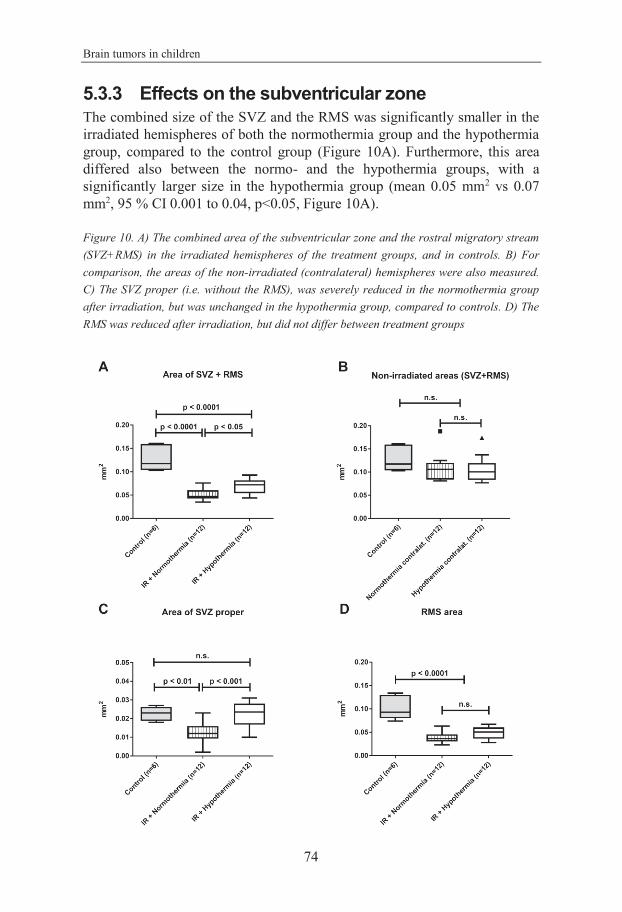
Brain tumors in children
74
5.3.3 Effects on the subventricular zone
The combined size of the SVZ and the RMS was significantly smaller in the
irradiated hemispheres of both the normothermia group and the hypothermia
group, compared to the control group (Figure 10A). Furthermore, this area
differed also between the normo- and the hypothermia groups, with a
significantly larger size in the hypothermia group (mean 0.05 mm2 vs 0.07
mm2, 95 % CI 0.001 to 0.04, p<0.05, Figure 10A).
Figure 10. A) The combined area of the subventricular zone and the rostral migratory stream
(SVZ+RMS) in the irradiated hemispheres of the treatment groups, and in controls. B) For
comparison, the areas of the non-irradiated (contralateral) hemispheres were also measured.
C) The SVZ proper (i.e. without the RMS), was severely reduced in the normothermia group
after irradiation, but was unchanged in the hypothermia group, compared to controls. D) The
RMS was reduced after irradiation, but did not differ between treatment groups

Magnus Sabel
75
The corresponding areas in the non-irradiated hemispheres were not different
in size from control areas (Figure 10B). When the SVZ was measured
separately, excluding the RMS (SVZ proper), the difference between the
treatment groups was even more pronounced, with a 50 % reduction in SVZ
size in the normothermia group compared to controls (0.012 mm2 vs 0.023
mm2 , 95 % CI 0.003 to 0.02, p<0.01). This reduction was completely
prevented in the hypothermia group (0.022 mm2 vs 0.023 mm2 in controls,
95% CI -0.007 to 0.008, p=1.0, Figure 10C). The RMS decreased to mean 43
% of controls after irradiation (p<0.001), but did not differ significantly
between the normo- and hypothermia groups (Figure 10D).
The number of proliferating (phospho-histone H3+) cells was reduced by
more than half in the irradiated normothermic SVZ, but this loss of
proliferating cells was prevented in the hypothermic group (Figure 11A).
Given the larger SVZ size in the irradiated hypothermia group, the cell
density was however not significantly different from the irradiated
normothermia group (Figure 11B).
The absolute numbers of microglia (Iba1+ cells) in the SVZ decreased in
the irradiated groups compared to controls, more significantly in the
hypothermia group than the normothermia group, although the difference
between the treatment groups was not significant (Figure 11C). Due to the
preservation of SVZ area in the hypothermia group, there was a lower
density of microglia in the irradiated SVZ of the hypothermia group
compared to both the normothermia group (Figure 11D), and controls.

Brain tumors in children
76
Figure 11. A) Absolute cell count of proliferating cells (phospho-histone H3+) in the
subventricular zone (SVZ), compared between randomization groups. B) Cell density of
proliferating (phospho-histone H3+) cells in the SVZ proper. The cell density did not differ
between normothermia and hypothermia groups, probably due to the preserved, relatively
larger SVZ in the hypothermia group. C) Microglia (Iba1+) cells in the SVZ+RMS compared
between groups. D) Microglia (Iba1+) cell density in the SVZ+RMS was lower in the
hypothermia group, compared to both the normothermia group and controls, probably due to
the preserved, relatively larger SVZ in the hypothermia group. Boxplots showing the
interquartile range (IQR) and median, with Tukey style whiskers (i.e. extended to the most
extreme data point that is no more than 1.5 × IQR from the edge of the box). IR irradiated,
n.s. not significant (p > 0. 3)

Magnus Sabel
77
5.4 Discussion (Paper IV)
In this animal model, young (P9) rats were randomized to a control group, or
a treatment group consisting of hypo- or normothermia for eight hours
immediately after a single fraction of hemi-brain irradiation. We found a
protective effect from hypothermia on the SVZ area and the proliferative cell
population in the SVZ, but no protection of the hippocampal dentate gyrus,
one week post radiation.
The reason for this selective protection is unclear, although consistent with
previous findings (Fukuda et al. 2005). It could due to different sensitivities
to irradiation, or differences in cell turn-over between the regions. This is
supported by previous finding of differential dynamics between the SVZ and
the GCL in the juvenile rat brain after irradiation, such that the SVZ
recovered with time whereas the GCL did not (Hellström et al. 2009). It is
conceivable that hypothermia in some way can enhance the recovery process
of the SVZ. Another speculative hypothesis is that the close proximity of the
SVZ to the CSF contributes to a less toxic environment after irradiation, as
discussed in Paper IV. It is also possible that NPCs in the hippocampus are
more vulnerable to stress compared to their SVZ counterpart, and that stress
from hypothermia could counter the protective effect. In a study of the effect
of longer-term hypothermia (30˚C for 24 h) on neurogenesis, cell
proliferation was reduced in the hippocampus but not in the SVZ, possibly
mediated by a stress-induced increase in corticosteroid levels (Kanagawa et
al. 2006).
Although the body temperatures in the hypothermia group reached
hypothermic levels (mean body temperature 32.2˚C), the target temperature
of 30˚C was not achieved. During the experiment, it was observed that the
animals tended to clump together in the temperature-controlled container,
something that probably helped them preserve body heat and lessened the
cooling effect. We did however, reach the same hypothermic temperature as a
previous study, but for twice as long, and compared to the previous study we
found a more pronounced protective effect on the SVZ, post-irradiation
(Fukuda et al. 2005). This indicates that hypothermia duration could be an
important factor. The lower cell density of Iba1+ cells in the SVZs of the
hypothermia group could indicate that hypothermia influences the
inflammatory response after irradiation, leading to improved NPC
survival/proliferation in the SVZ, although further studies are needed.
The RMS did not seem to benefit from hypothermia in the same way as the
SVZ. Although the border between these two areas is somewhat arbitrary,
Brain tumors in children
78
this might reflect an underlying mechanism yet to be elucidated. A
hypothesis could be that although the proliferative capacity of the SVZ was
benefited by hypothermia, the previously reported negative effect of
irradiation on the RMS preventing the migration of neuroblasts was
unaffected (Achanta et al. 2012).
Both irradiated groups had a lower mean blood glucose level after eight hours
compared to controls, but there was no difference in blood glucose levels
between the normo- and hypothermia groups that could explain the difference
in SVZ areas. Furthermore, the GCL and SVZ areas in the non-irradiated
(contralateral) hemispheres were not reduced compared to controls. It is
therefore unlikely that blood glucose levels (or other metabolic changes due
to fasting), can explain the difference in SVZ areas.
The strengths of this study is the randomized design with blinded evaluation
of the effect of hypothermia on the proliferative areas of the rodent brain. A
limitation of our study is that we studied the potential protective effect of
hypothermia at one time point only, and that we did not characterize the
proliferating cells further. However, in a stroke model using adult rats, the
size of the SVZ reflected the number of proliferating cells, indicating that
SVZ size could be used as a marker of SVZ precursor proliferation (Parent et
al. 2002).
It may be that hypothermia delayed the detrimental effect on the SVZ area
and that, over time, the area would decrease in a similar way as it did in the
irradiated normothermia group. It is possible however, that hypothermia
protected the NPC population in the SVZ (through anti-apoptosis, anti-
oxidation, or anti-inflammatory mechanisms), leaving a greater number of
viable proliferating cells to repopulate the area. The latter scenario is
supported by the combined findings of a larger preserved SVZ area, together
with higher numbers of proliferating cells in the hypothermia treated group.
This however, needs to be confirmed in future studies.

Magnus Sabel
79
6 FINAL CONCLUSIONS AND FUTURE PERSPECTIVES
The poor survival after medulloblastoma relapse found in Paper I is
consistent with previous findings, and treatment for recurrent
medulloblastoma continues to be a major clinical challenge. In isolated
relapses, surgery can be beneficial. Surgery or biopsy should also be
encouraged to verify the diagnosis of recurrent disease vs SMN or even non-
malignant MRI findings. International collaborative trials for recurrent
medulloblastoma are needed and should be encouraged. These should
preferably be based on biological sampling of recurrent tumors, and use the
results to guide therapy. Tumor biopsy in that context is less ethically
problematic, since the procedure leads to an immediate benefit for the patient
(guided therapy), as well as the research community (more knowledge).
Targeted therapy could be a way forward, e.g. using inhibitors of the sonic
hedgehog pathway in SHH-MB, but the long term side effects of these new
therapies are yet unknown. For example, the sonic hedgehog pathway is an
important stimulating factor for neurogenesis the SVZ and GCL (Ahn and
Joyner 2005), which raises concerns that SHH-inhibition could adversely
affect normal brain function.
The best way to handle relapses is of course to prevent them from happening.
Research in resistance mechanisms is therefore important. Continued
research in tumor (molecular) biology will hopefully provide further insight
into how tumor cells avoid being killed by therapy, and how to best close
their escape routes. As an example, Lithium treatment of radioresistant TP53
mutant medulloblastoma cell lines sensitized the tumor cells to radiation
through activation of the WNT pathway (Zhukova et al. 2014). Interestingly,
Lithium did not activate WNT signaling in normal neuronal stem cells, and
consequently their sensitivity to radiation was not enhanced (Zhukova et al.
2014). In fact, evidence suggests Lithium could have a protective effect on
hippocampal neurogenesis after irradiation, and reduce cognitive side effects
(Zhou et al. 2017).
Finding effective ways to mitigate the long term side effects after childhood
brain tumor treatment is important, since they can have a severe impact on
the survivors’ daily life. In addition, the number or survivors increase every
year. There is growing evidence that exercise is beneficial to cognition, and
exercise interventions are an interesting option for this patient group. But

Brain tumors in children
80
there are challenges in finding interventions that are easily managed, fun to
do, and can be performed on a long term basis. Interventions should probably
be a combination of different activities, varying over time, to enhance
participation and reduce attrition. The value of active video gaming deserves
further exploration, and future research should focus on games that stimulate
both physical activity and cognition, e.g. dance games. Group activities e.g.
real life dance classes, could be considered, as well as methods of proven
benefit, e.g. computerized working memory training with Cogmed.
Inconvenience in travelling to the cancer center, e.g. after school hours, can
hamper clinic-based interventions (Patel et al. 2009), and home-based
interventions are therefore preferable.
Support by Internet coaching is a resource efficient way of doing studies, but
perhaps not easily transferred to large-scale clinical practice. Building
Internet-based communities of brain tumor survivors could however be a
method to stimulate rehabilitation activities, with the added value of
decreasing social isolation. In view of the rapid technical development,
already available virtual reality solutions will probably soon be less
expensive, and could be an interesting option to explore further.
Using hypothermia after radiotherapy to mitigate the harmful effects on
neurogenesis needs further study before it can be used in a clinical setting.
Both the functional and long term effects of the findings in Paper IV need to
be investigated in animal studies, as well as the effect of hypothermia on
brain tumors. Since radiotherapy is given over a period of weeks, the method
has obvious practical challenges as well, although not unsurmountable
provided the gain from the intervention is large enough.
6.1 Concluding remarks
In response to specific aims I. Long term survival after treatment for standard risk
medulloblastoma has improved over the years, now reaching
an estimated 10-year EFS and OS of ~80 %. After a median
follow up of 7.4 years there was no difference in survival
between treatment arms in the HIT-SIOP PNET4 trial. The
prognosis after relapse however, was very poor, with a
median OS of 1.5 years after relapse and a disappointing 5-
year OS of 6 %, irrespective of relapse treatment. Recurrent

Magnus Sabel
81
tumors appeared at a median interval of 26 months from
primary diagnosis, with 8 % late relapses (> 5 years after
primary diagnosis). No benefit from HDSCR or
radiotherapy was detected, but relapse treatments were
diverse, and therefore difficult to evaluate. Patients with
isolated posterior fossa relapses survived longer compared to
patients with other relapse patterns. In selected cases,
surgery seemed to be of benefit. The majority of relapses
however, were metastatic within the CNS. The histological
subgroup or biological factors (at primary diagnosis) had no
impact on time in first remission, relapse pattern, or survival
after relapse.
II. An intervention with active video gaming and regular on-
line coaching achieved an enjoyable, near daily exercise of
moderate intensity in pediatric brain tumor survivors. The
method was feasible and compliance was good, but the
overall physical activity levels were not significantly
increased.
III. No significant improvement was found in cognitive test
scores after AVG, but the evaluation was limited by the
small sample size. Clinically important improvements were
found in motor function (body coordination) and ADL
abilities. Continued research should be encouraged.
IV. Post-irradiation moderate hypothermia for eight hours
protected neurogenesis in the SVZ, but not in the
hippocampus, in a rodent model. The mechanism for the
protective effect and the reason for the selective effect on the
neurogenic areas needs further study, as well as the effect of
post-irradiation hypothermia on brain tumors.

Brain tumors in children
82
7 ERRATA
Paper I: On page 3, the second last sentence on the page should read:
“There was information on CSF cytology in 61/72 patients.”
On page 4: Surgery was performed in 18 cases and radiotherapy was given to
16 patients, as indicated in Table 1.
Paper III: In the Table 2 legend at page 8, the first row should read: “t =T-
score: mean=50, SD=10; s= standard score: mean=10, SD=3, IQ: mean=100,
SD=15”
In Table 3, page 9, the p-value for the change in motor score should be
p=0.0122.

83
ACKNOWLEDGEMENTS
I would like to express my sincere gratitude to all involved, directly or
indirectly, in the work behind this doctoral thesis. I especially want to thank:
All patients that participated in the studies, and their families.
Birgitta Lannering, professor, tutor, and mentor that introduced me to
pediatric neuro-oncology as well as research. I am so grateful for these past
years working beside you, sharing the good times and the bad. Thank you for
your friendship, guidance, encouragement, and well-controlled impatience.
Without you, this thesis would probably not been completed for another year
or so (at least).
Klas Blomgren, professor, co-tutor, and colleague. Thank you for your
friendship, humor and positive attitude, and for sharing your vast knowledge
in the field of neurogenesis. Thank you for your rapid e-mail responses, and
well formulated, encouraging comments.
Ingrid Emanuelson, associate professor, co-tutor and colleague. Thank you
for your friendship and support, your caring and helpful feedback, and for
sharing your knowledge in the rehabilitation field.
Anette Sjölund, for co-authorship and invaluable help in managing the Wii-
study, and especially for your coaching efforts and help to coordinate both
the Wii-study and clinical work. Daniel Arvidsson, Thomas Björk-
Eriksson, Jürgen Broeren, Jonas Gillenstrand, Göran Gustafsson, Marie
Kalm, Jean-Michel Saury, for your co-authorship and collaboration, and
your valued input into the different projects.
The contributions of the SIOP-Europe brain tumor group, national study
group members, and national data managers are gratefully acknowledged.
Members of the SIOP-E brain tumor group, especially the co-authors;
Francois Doz, Steve Clifford, Gudrun Fleischhack, Rolf Kortmann,
Maura Massimino, Aurora Navajas, Torsten Pietsch, Barry Pizer,
Stephan Tippelt, Stefan Rutkowski, Monika Warmuth-Metz, and Katja
von Hoff, thank you for your co-authorship and collaboration.
Rita Grandér, for assistance and help in the hypothermia project and for
teaching me the practicalities of lab work, Christer Ljungberg, for technical
assistance in the Wii-project, Nils-Gunnar Pehrsson, for statistical advice,

Brain tumors in children
84
and Charlotte Simmons, for your indispensable help with BOT-2 testing in
the Wii-study.
My dear pediatric oncology colleagues; Jonas Abrahamsson, Margareta
Bergkvist, Martin Dalin, Anders Fasth, Torben Ek, Magnus Göransson,
Maria Henningsson, Marianne Jarfelt, Cecilia Langenskiöld, Lene
Karlsson, Lars Kawan, Karin Mellgren, Mirka Pinkava, Elizabeth
Schepke, Gustaf Österlundh, for your friendship and excellent clinical
collaboration on an everyday basis. I am most grateful to all of you, and
especially Elizabeth and Birgitta, for taking care of patients during my
absence.
Karin Fritzson, Tony Gavik, Magnus Dahlander, Anna Schröder
Håkansson, Carina Hallberg, Liselotte Neleborn Lingefjärd, for help with
on-going and planned clinical trials, as well as taking care of patients.
All staff at the Children´s Cancer Center at the Queen Silvia Children´s
hospital, for doing what you do, and for keeping the good spirits while doing
it. A special thanks to Carina Fondin, Katarina Fält, Renée Andersson,
Lisbeth Andersson, for coordinating everything, and for helping absent-
minded doctors…
Co-workers in ongoing projects: Frida Abel, Birgitte Berthelsen, Helena
Carén, Rolf Heckemann, Helge Malmgren, Erik Olsson, Hedda Haugen
Cange, Liz Ivarsson, Niklas Klasson, Kerstin Lagerstrand, Isabelle
Rydén, Emily Ruzich, Justin Schneidermann, Simon Skau, Pär-Arne
Svensson, Magnus Thordstein, Magnus Tisell.
Gunnar Steineck, for introducing the research school at the University of
Gothenburg.
My wife and children, family and friends.
This work was supported by the Swedish Research Council, the Swedish
Childhood Cancer Foundation, Frimurare Barnhusfonden, and
Governmental grants to scientists working in health care (ALF) in
Gothenburg.

85
REFERENCES
Aarsen, Van Dongen, Paquier, Van Mourik and Catsman-Berrevoets (2004). "Long-
term sequelae in children after cerebellar astrocytoma surgery." Neurology
62(8): 1311-1316.
Aarsen, Paquier, Reddingius, Streng, Arts, Evera-Preesman and Catsman-Berrevoets
(2006). "Functional outcome after low-grade astrocytoma treatment in
childhood." Cancer 106(2): 396-402.
Achanta, Capilla-Gonzalez, Purger, Reyes, Sailor, Song, Garcia-Verdugo, Gonzalez-
Perez, Ford and Quinones-Hinojosa (2012). "Subventricular zone localized
irradiation affects the generation of proliferating neural precursor cells and
the migration of neuroblasts." Stem Cells 30(11): 2548-2560.
Ahn and Joyner (2005). "In vivo analysis of quiescent adult neural stem cells
responding to Sonic hedgehog." Nature 437(7060): 894-897.
Ainsworth, Haskell, Herrmann, Meckes, Bassett, Tudor-Locke, Greer, Vezina, Whitt-
Glover and Leon (2011). "2011 Compendium of Physical Activities: a
second update of codes and MET values." Med Sci Sports Exerc 43(8):
1575-1581.
Ali, Lang and Sutton (2014). "Conservative management of presumed low-grade
gliomas in the asymptomatic pediatric population." World Neurosurg 81(2):
368-373.
Altman and Bland (2005). "Treatment allocation by minimisation." BMJ 330(7495):
843.
Arvidsson, Collin, Kirik, Kokaia and Lindvall (2002). "Neuronal replacement from
endogenous precursors in the adult brain after stroke." Nat Med 8(9): 963-
970.
Arvidsson, Slinde, Larsson and Hulthen (2007). "Energy cost of physical activities in
children: validation of SenseWear Armband." Med Sci Sports Exerc 39(11):
2076-2084.
Arvidsson, Slinde and Hulthén (2009a). "Free-living energy expenditure in children
using multi-sensor activity monitors." Clin Nutr 28(3): 305-312.
Arvidsson, Slinde, Larsson and Hulthén (2009b). "Energy cost in children assessed
by multisensor activity monitors." Med Sci Sports Exerc 41(3): 603-611.
Ashley, Meier, Kerby, Zalduondo, Friedman, Gajjar, Kun, Duffner, Smith and
Longee (1996). "Response of recurrent medulloblastoma to low-dose oral
etoposide." J Clin Oncol 14(6): 1922-1927.
Azzopardi, Strohm, Edwards, Dyet, Halliday, Juszczak, Kapellou, Levene, Marlow,
Porter, Thoresen, Whitelaw and Brocklehurst (2009). "Moderate
hypothermia to treat perinatal asphyxial encephalopathy." N Engl J Med
361(14): 1349-1358.
Bailey and Cushing (1925). "Medulloblastoma cerebelli: A common type of
midcerebellar glioma of childhood." Arch Neurol Psychiatry 14(2): 192-
224.

Brain tumors in children
86
Bailey (1930). "Further Notes on the Cerebellar Medulloblastomas: The Effect of
Roentgen Radiation." The American Journal of Pathology 6(2): 125-
136.127.
Balsom, Bleyer, Robison, Heyn, Meadows, Sitarz, Blatt, Sather and Hammond
(1991). "Intellectual function in long-term survivors of childhood acute
lymphoblastic leukemia: protective effect of pre-irradiation methotrexate? A
Childrens Cancer Study Group study." Med Pediatr Oncol 19(6): 486-492.
Banich (2009). "Executive Function." Curr Dir Psychol Sci 18(2): 89-94.
Barlind, Karlsson, Bjork-Eriksson, Isgaard and Blomgren (2010). "Decreased
cytogenesis in the granule cell layer of the hippocampus and impaired place
learning after irradiation of the young mouse brain evaluated using the
IntelliCage platform." Exp Brain Res 201(4): 781-787.
Barnes, Yaffe, Satariano and Tager (2003). "A longitudinal study of
cardiorespiratory fitness and cognitive function in healthy older adults." J
Am Geriatr Soc 51(4): 459-465.
Bartels, Shroff, Sung, Dag-Ellams, Laperriere, Rutka and Bouffet (2006). "Role of
spinal MRI in the follow-up of children treated for medulloblastoma."
Cancer 107(6): 1340-1347.
Becker and Hinton (1983). "Primitive neuroectodermal tumors of the central nervous
system." Hum Pathol 14(6): 538-550.
Benzing, Heinks, Eggenberger and Schmidt (2016). "Acute Cognitively Engaging
Exergame-Based Physical Activity Enhances Executive Functions in
Adolescents." PLoS One 11(12): e0167501.
Bergmann, Liebl, Bernard, Alkass, Yeung, Steier, Kutschera, Johnson, Landén,
Druid, Spalding and Frisén (2012). "The Age of Olfactory Bulb Neurons in
Humans." Neuron 74(4): 634-639.
Bernspång and Fisher (1995). "Validation of the Assessment of Motor and Process
Skills for Use in Sweden." Scand J Occup Ther 2(1): 3-9.
Best (2012). "Exergaming immediately enhances children's executive function." Dev
Psychol 48(5): 1501-1510.
Bhardwaj, Curtis, Spalding, Buchholz, Fink, Bjork-Eriksson, Nordborg, Gage,
Druid, Eriksson and Frisen (2006). "Neocortical neurogenesis in humans is
restricted to development." Proc Natl Acad Sci U S A 103(33): 12564-
12568.
Biddiss and Irwin (2010). "Active video games to promote physical activity in
children and youth: A systematic review." Arch Pediatr Adolesc Med
164(7): 664-672.
Blomstrand, Brodin, Munck Af Rosenschold, Vogelius, Sanchez Merino, Kiil-
Berthlesen, Blomgren, Lannering, Bentzen and Bjork-Eriksson (2012).
"Estimated clinical benefit of protecting neurogenesis in the developing
brain during radiation therapy for pediatric medulloblastoma." Neuro
Oncol 14(7): 882-889.
Blomstrand, Kalm, Grander, Bjork-Eriksson and Blomgren (2014). "Different
reactions to irradiation in the juvenile and adult hippocampus." Int J Radiat
Biol 90(9): 807-815.

87
Bloom, Wallace and Henk (1969). "The treatment and prognosis of medulloblastoma
in childen." American Journal of Roentgenology 105(1): 43-62.
Bode, Zimmermann, Moser, Rutkowski, Warmuth-Metz, Pietsch, Kortmann, Faldum
and Fleischhack (2014). "Treatment of recurrent primitive neuroectodermal
tumors (PNET) in children and adolescents with high-dose chemotherapy
(HDC) and stem cell support: results of the HITREZ 97 multicentre trial." J
Neurooncol 120(3): 635-642.
Boman, Lindblad and Hjern (2010). "Long-term outcomes of childhood cancer
survivors in Sweden: a population-based study of education, employment,
and income." Cancer 116(5): 1385-1391.
Boot, Kramer, Simons, Fabiani and Gratton (2008). "The effects of video game
playing on attention, memory, and executive control." Acta Psychol (Amst)
129(3): 387-398.
Boot, Blakely and Simons (2011). "Do action video games improve perception and
cognition?" Front Psychol 2: 226.
Boström, Kalm, Karlsson, Erkenstam and Blomgren (2013). "Irradiation to the
Young Mouse Brain Caused Long-Term, Progressive Depletion of
Neurogenesis but did not Disrupt the Neurovascular Niche." J Cereb Blood
Flow Metab 33(6): 935-943.
Bouffet, Doz, Demaille, Tron, Roche, Plantaz, Thyss, Stephan, Lejars, Sariban,
Buclon, Zucker, Brunat-Mentigny, Bernard and Gentet (1998). "Improving
survival in recurrent medulloblastoma: earlier detection, better treatment or
still an impasse?" Br J Cancer 77(8): 1321-1326.
Bowers, Gargan, Weprin, Mulne, Elterman, Munoz, Giller and Winick (2007).
"Impact of site of tumor recurrence upon survival for children with
recurrent or progressive medulloblastoma." J Neurosurg 107(1 Suppl): 5-
10.
Brinkman, Reddick, Luxton, Glass, Sabin, Srivastava, Robison, Hudson and Krull
(2012). "Cerebral white matter integrity and executive function in adult
survivors of childhood medulloblastoma." Neuro Oncol 14 Suppl 4: iv25-
36.
Brinkman, Krasin, Liu, Armstrong, Ojha, Sadighi, Gupta, Kimberg, Srivastava,
Merchant, Gajjar, Robison, Hudson and Krull (2016). "Long-Term
Neurocognitive Functioning and Social Attainment in Adult Survivors of
Pediatric CNS Tumors: Results From the St Jude Lifetime Cohort Study." J
Clin Oncol 34(12): 1358-1367.
Brodin, Munck af Rosenschold, Blomstrand, Kiil-Berthlesen, Hollensen, Vogelius,
Lannering, Bentzen and Bjork-Eriksson (2014). "Hippocampal sparing
radiotherapy for pediatric medulloblastoma: impact of treatment margins
and treatment technique." Neuro Oncol 16(4): 594-602.
Brown, Kepner, Perlman, Friedman, Strother, Duffner, Kun, Goldthwaite and
Burger (2000). ""Large cell/anaplastic" medulloblastomas: a Pediatric
Oncology Group Study." J Neuropathol Exp Neurol 59(10): 857-865.
Bruininks and Bruininks (2005). BOT-2, Bruininks-Oseretsky Test of Motor
Proficiency. Minneapolis, Minnesota, USA, NCS Pearson.

Brain tumors in children
88
Butler and Haser (2006). "Neurocognitive effects of treatment for childhood cancer."
Ment Retard Dev Disabil Res Rev 12(3): 184-191.
Butler, Copeland, Fairclough, Mulhern, Katz, Kazak, Noll, Patel and Sahler (2008).
"A multicenter, randomized clinical trial of a cognitive remediation
program for childhood survivors of a pediatric malignancy." J Consult Clin
Psychol 76(3): 367-378.
Butturini, Jacob, Aguajo, Vander-Walde, Villablanca, Jubran, Erdreich-Epstein,
Marachelian, Dhall and Finlay (2009). "High-dose chemotherapy and
autologous hematopoietic progenitor cell rescue in children with recurrent
medulloblastoma and supratentorial primitive neuroectodermal tumors: the
impact of prior radiotherapy on outcome." Cancer 115(13): 2956-2963.
Calabró, Stewart and Welk (2013). "Validation of Pattern-Recognition Monitors in
Children Using Doubly Labeled Water." Med Sci Sports Exerc.
Calvert, Appelbaum, Dodge, Graham, Nagayama Hall, Hamby, Fasig-Caldwell,
Citkowicz, Galloway and Hedges (2017). "The American Psychological
Association Task Force assessment of violent video games: Science in the
service of public interest." Am Psychol 72(2): 126-143.
Carpentieri, Waber, Pomeroy, Scott, Goumnerova, Kieran, Billett and Tarbell
(2003). "Neuropsychological functioning after surgery in children treated
for brain tumor." Neurosurgery 52(6): 1348-1356; discussion 1356-1347.
Caspersen, Powell and Christenson (1985). "Physical activity, exercise, and physical
fitness: definitions and distinctions for health-related research." Public
Health Rep 100(2): 126-131.
Castellino, Tooze, Flowers, Hill, McMullen, Shaw and Parsons (2012). "Toxicity and
efficacy of the acetylcholinesterase (AChe) inhibitor donepezil in childhood
brain tumor survivors: A pilot study." Pediatr Blood Cancer.
Castellino, Ullrich, Whelen and Lange (2014). "Developing interventions for cancer-
related cognitive dysfunction in childhood cancer survivors." J Natl Cancer
Inst 106(8).
Cefalo, Massimino, Ruggiero, Barone, Ridola, Spreafico, Potepan, Abate, Mascarin,
Garre, Perilongo, Madon, Colosimo and Riccardi (2014). "Temozolomide is
an active agent in children with recurrent medulloblastoma/primitive
neuroectodermal tumor: an Italian multi-institutional phase II trial." Neuro
Oncol 16(5): 748-753.
Chaddock-Heyman, Erickson, Voss, Knecht, Pontifex, Castelli, Hillman and Kramer
(2013). "The effects of physical activity on functional MRI activation
associated with cognitive control in children: a randomized controlled
intervention." Front Hum Neurosci 7: 72.
Chaddock-Heyman, Erickson, Holtrop, Voss, Pontifex, Raine, Hillman and Kramer
(2014). "Aerobic fitness is associated with greater white matter integrity in
children." Front Hum Neurosci 8: 584.
Chaddock, Erickson, Prakash, Kim, Voss, Vanpatter, Pontifex, Raine, Konkel,
Hillman, Cohen and Kramer (2010a). "A neuroimaging investigation of the
association between aerobic fitness, hippocampal volume, and memory
performance in preadolescent children." Brain Res 1358: 172-183.
89
Chaddock, Erickson, Prakash, Vanpatter, Voss, Pontifex, Raine, Hillman and
Kramer (2010b). "Basal ganglia volume is associated with aerobic fitness in
preadolescent children." Dev Neurosci 32(3): 249-256.
Chaddock, Neider, Lutz, Hillman and Kramer (2011). "Role of Childhood Aerobic
Fitness in Successful Street Crossing." Med Sci Sports Exerc.
Chaker, Codega and Doetsch (2016). "A mosaic world: puzzles revealed by adult
neural stem cell heterogeneity." Wiley Interdiscip Rev Dev Biol 5(6): 640-
658.
Chamberlain and Kormanik (1997). "Chronic oral VP-16 for recurrent
medulloblastoma." Pediatr Neurol 17(3): 230-234.
Chang, Housepian and Herbert (1969). "An operative staging system and a
megavoltage radiotherapeutic technic for cerebellar medulloblastomas."
Radiology 93(6): 1351-1359.
Chavez, Sopko, Ehret, Paulino, Goldberg, Angstadt and Bogart (2009). "An update
on central nervous system stimulant formulations in children and
adolescents with attention-deficit/hyperactivity disorder." Ann
Pharmacother 43(6): 1084-1095.
Chemaitilly, Li, Huang, Ness, Clark, Green, Barnes, Armstrong, Krasin, Srivastava,
Pui, Merchant, Kun, Gajjar, Hudson, Robison and Sklar (2015). "Anterior
hypopituitarism in adult survivors of childhood cancers treated with cranial
radiotherapy: a report from the St Jude Lifetime Cohort study." J Clin
Oncol 33(5): 492-500.
Cho, Tsherniak, Tamayo, Santagata, Ligon, Greulich, Berhoukim, Amani,
Goumnerova, Eberhart, Lau, Olson, Gilbertson, Gajjar, Delattre, Kool,
Ligon, Meyerson, Mesirov and Pomeroy (2011). "Integrative genomic
analysis of medulloblastoma identifies a molecular subgroup that drives
poor clinical outcome." J Clin Oncol 29(11): 1424-1430.
Christopherson, Bradley, Rotondo, Pincus, Fort, Morris, Mendenhall, Marcus and
Indelicato (2014). "Local control in non-metastatic medulloblastoma." Acta
Oncol 53(9): 1151-1157.
Clifford, Lannering, Schwalbe, Hicks, O'Toole, Nicholson, Goschzik, Zur Muhlen,
Figarella-Branger, Doz, Rutkowski, Gustafsson and Pietsch (2015).
"Biomarker-driven stratification of disease-risk in non-metastatic
medulloblastoma: Results from the multi-center HIT-SIOP-PNET4 clinical
trial." Oncotarget 6(36): 38827-38839.
Cohen, Geyer, Miller, Curran, Zhou, Holmes, Ingles, Dunkel, Hilden, Packer,
Pollack, Gajjar and Finlay (2015). "Pilot Study of Intensive Chemotherapy
With Peripheral Hematopoietic Cell Support for Children Less Than 3
Years of Age With Malignant Brain Tumors, the CCG-99703 Phase I/II
Study. A Report From the Children's Oncology Group." Pediatr Neurol
53(1): 31-46.
Colcombe, Kramer, Erickson, Scalf, McAuley, Cohen, Webb, Jerome, Marquez and
Elavsky (2004). "Cardiovascular fitness, cortical plasticity, and aging."
Proc Natl Acad Sci U S A 101(9): 3316-3321.

Brain tumors in children
90
Conklin, Khan, Reddick, Helton, Brown, Howard, Bonner, Christensen, Wu, Xiong
and Mulhern (2007). "Acute neurocognitive response to methylphenidate
among survivors of childhood cancer: a randomized, double-blind, cross-
over trial." J Pediatr Psychol 32(9): 1127-1139.
Conklin, Li, Xiong, Ogg and Merchant (2008). "Predicting change in academic
abilities after conformal radiation therapy for localized ependymoma." J
Clin Oncol 26(24): 3965-3970.
Conklin, Lawford, Jasper, Morris, Howard, Ogg, Wu, Xiong and Khan (2009). "Side
effects of methylphenidate in childhood cancer survivors: a randomized
placebo-controlled trial." Pediatrics 124(1): 226-233.
Conklin, Reddick, Ashford, Ogg, Howard, Morris, Brown, Bonner, Christensen, Wu,
Xiong and Khan (2010). "Long-term efficacy of methylphenidate in
enhancing attention regulation, social skills, and academic abilities of
childhood cancer survivors." J Clin Oncol 28(29): 4465-4472.
Conklin, Ogg, Ashford, Scoggins, Zou, Clark, Martin-Elbahesh, Hardy, Merchant,
Jeha, Huang and Zhang (2015). "Computerized Cognitive Training for
Amelioration of Cognitive Late Effects Among Childhood Cancer Survivors:
A Randomized Controlled Trial." J Clin Oncol 33(33): 3894-3902.
Copeland, deMoor, Moore and Ater (1999). "Neurocognitive development of
children after a cerebellar tumor in infancy: A longitudinal study." J Clin
Oncol 17(11): 3476-3486.
Corkin (2002). "What's new with the amnesic patient H.M.?" Nat Rev Neurosci 3(2):
153-160.
Cowan (2008). "What are the differences between long-term, short-term, and
working memory?" Prog Brain Res 169: 323-338.
Curlik and Shors (2011). "Learning increases the survival of newborn neurons
provided that learning is difficult to achieve and successful." J Cogn
Neurosci 23(9): 2159-2170.
Curtis, Kam, Nannmark, Anderson, Axell, Wikkelso, Holtas, van Roon-Mom, Bjork-
Eriksson, Nordborg, Frisen, Dragunow, Faull and Eriksson (2007).
"Human neuroblasts migrate to the olfactory bulb via a lateral ventricular
extension." Science 315(5816): 1243-1249.
Cushing (1930). "Experiences with the cerebellar medulloblastomas." Acta
Pathologica Microbiologica Scandinavica 7(1-2): 1-86.
Dahl and Emanuelson (2013). "Motor proficiency in children with mild traumatic
brain injury compared with a control group." J Rehabil Med 45(8): 729-
733.
Danielsson, Nemes, Tisell, Lannering, Nordborg, Sabel and Caren (2015).
"MethPed: a DNA methylation classifier tool for the identification of
pediatric brain tumor subtypes." Clin Epigenetics 7(1): 62.
Davis, Tomporowski, McDowell, Austin, Miller, Yanasak, Allison and Naglieri
(2011). "Exercise improves executive function and achievement and alters
brain activation in overweight children: a randomized, controlled trial."
Health psychology : official journal of the Division of Health Psychology,
American Psychological Association 30(1): 91-98.

91
de Kloet, Berger, Verhoeven, van Stein Callenfels and Vlieland (2012). "Gaming
supports youth with acquired brain injury? A pilot study." Brain Inj 26(7-8):
1021-1029.
De Luca, McCarthy, Galvin, Green, Murphy, Knight and Williams (2013). "Gross
and fine motor skills in children treated for acute lymphoblastic leukaemia."
Dev Neurorehabil 16(3): 180-187.
de Vet, Terwee, Ostelo, Beckerman, Knol and Bouter (2006). "Minimal changes in
health status questionnaires: distinction between minimally detectable
change and minimally important change." Health and Quality of Life
Outcomes 4: 54-54.
Demers, Gelinas and Carret (2016). "Activities of Daily Living in Survivors of
Childhood Brain Tumor." Am J Occup Ther 70(1):
7001220040p7001220041-7001220048.
Deng, Saxe, Gallina and Gage (2009). "Adult-born hippocampal dentate granule
cells undergoing maturation modulate learning and memory in the brain." J
Neurosci 29(43): 13532-13542.
Diamond (2013). "Executive Functions." Annu Rev Psychol 64: 135-168.
Dickerson and Fisher (1993). "Age differences in functional performance." Am J
Occup Ther 47(8): 686-692.
Dietrich, Han, Yang, Mayer-Proschel and Noble (2006). "CNS progenitor cells and
oligodendrocytes are targets of chemotherapeutic agents in vitro and in
vivo." J Biol 5(7): 22.
Doetsch, Caillé, Lim, García-Verdugo and Alvarez-Buylla (1999a). "Subventricular
Zone Astrocytes Are Neural Stem Cells in the Adult Mammalian Brain."
Cell 97(6): 703-716.
Doetsch, Garcia-Verdugo and Alvarez-Buylla (1999b). "Regeneration of a germinal
layer in the adult mammalian brain." Proc Natl Acad Sci U S A 96(20):
11619-11624.
Donnelly, Greene, Gibson, Smith, Washburn, Sullivan, DuBose, Mayo, Schmelzle,
Ryan, Jacobsen and Williams (2009). "Physical Activity Across the
Curriculum (PAAC): a randomized controlled trial to promote physical
activity and diminish overweight and obesity in elementary school
children." Prev Med 49(4): 336-341.
Duffner, Cohen and Thomas (1983). "Late effects of treatment on the intelligence of
children with posterior fossa tumors." Cancer 51(2): 233-237.
Duffner, Cohen and Parker (1988). "Prospective intellectual testing in children with
brain tumors." Ann Neurol 23(6): 575-579.
Dunkel, Boyett, Yates, Rosenblum, Garvin, Bostrom, Goldman, Sender, Gardner, Li,
Allen and Finlay (1998). "High-dose carboplatin, thiotepa, and etoposide
with autologous stem-cell rescue for patients with recurrent
medulloblastoma. Children's Cancer Group." J Clin Oncol 16(1): 222-228.
Dunkel, Gardner, Garvin, Goldman, Shi and Finlay (2010). "High-dose carboplatin,
thiotepa, and etoposide with autologous stem cell rescue for patients with
previously irradiated recurrent medulloblastoma." Neuro Oncol 12(3): 297-
303.
Brain tumors in children
92
Dye, Green and Bavelier (2009). "The development of attention skills in action video
game players." Neuropsychologia 47(8-9): 1780-1789.
Eberhart, Kepner, Goldthwaite, Kun, Duffner, Friedman, Strother and Burger
(2002). "Histopathologic grading of medulloblastomas: a Pediatric
Oncology Group study." Cancer 94(2): 552-560.
Edelstein, Spiegler, Fung, Panzarella, Mabbott, Jewitt, D'Agostino, Mason, Bouffet,
Tabori, Laperriere and Hodgson (2011). "Early aging in adult survivors of
childhood medulloblastoma: long-term neurocognitive, functional, and
physical outcomes." Neuro Oncol 13(5): 536-545.
Ellison, Clifford, Gajjar and Gilbertson (2003). "What's new in neuro-oncology?
Recent advances in medulloblastoma." Eur J Paediatr Neurol 7(2): 53-66.
Erickson, Voss, Prakash, Basak, Szabo, Chaddock, Kim, Heo, Alves, White, Wojcicki,
Mailey, Vieira, Martin, Pence, Woods, McAuley and Kramer (2011).
"Exercise training increases size of hippocampus and improves memory."
Proc Natl Acad Sci U S A 108(7): 3017-3022.
Ericsson and Karlsson (2014). "Motor skills and school performance in children
with daily physical education in school--a 9-year intervention study." Scand
J Med Sci Sports 24(2): 273-278.
Eriksson, Perfilieva, Bjork-Eriksson, Alborn, Nordborg, Peterson and Gage (1998).
"Neurogenesis in the adult human hippocampus." Nat Med 4(11): 1313.
Ernst, Alkass, Bernard, Salehpour, Perl, Tisdale, Possnert, Druid and Frisén (2014).
"Neurogenesis in the Striatum of the Adult Human Brain." Cell 156(5):
1072-1083.
Errickson, Maloney, Thorpe, Giuliani and Rosenberg (2012). "'Dance Dance
Revolution' Used by 7- and 8-Year-Olds to Boost Physical Activity: Is
Coaching Necessary for Adherence to an Exercise Prescription?" Games
Health J 1(1): 45-50.
Etnier, Nowell, Landers and Sibley (2006). "A meta-regression to examine the
relationship between aerobic fitness and cognitive performance." Brain Res
Rev 52(1): 119-130.
Evans, Jenkin, Sposto, Ortega, Wilson, Wara, Ertel, Kramer, Chang, Leikin and et
al. (1990). "The treatment of medulloblastoma. Results of a prospective
randomized trial of radiation therapy with and without CCNU, vincristine,
and prednisone." J Neurosurg 72(4): 572-582.
Fabel, Wolf, Ehninger, Babu, Leal-Galicia and Kempermann (2009). "Additive
effects of physical exercise and environmental enrichment on adult
hippocampal neurogenesis in mice." Front Neurosci 3: 50.
Farmer, Zhao, van Praag, Wodtke, Gage and Christie (2004). "Effects of voluntary
exercise on synaptic plasticity and gene expression in the dentate gyrus of
adult male Sprague-Dawley rats in vivo." Neuroscience 124(1): 71-79.
Fattet, Haberler, Legoix, Varlet, Lellouch-Tubiana, Lair, Manie, Raquin, Bours,
Carpentier, Barillot, Grill, Doz, Puget, Janoueix-Lerosey and Delattre
(2009). "Beta-catenin status in paediatric medulloblastomas: correlation of
immunohistochemical expression with mutational status, genetic profiles,
and clinical characteristics." J Pathol 218(1): 86-94.

93
Finlay, Goldman, Wong, Cairo, Garvin, August, Cohen, Stanley, Zimmerman,
Bostrom, Geyer, Harris, Sanders, Yates, Boyett and Packer (1996). "Pilot
study of high-dose thiotepa and etoposide with autologous bone marrow
rescue in children and young adults with recurrent CNS tumors. The
Children's Cancer Group." J Clin Oncol 14(9): 2495-2503.
Fisher, Liu, Velozo and Pan (1992). "Cross-cultural assessment of process skills."
Am J Occup Ther 46(10): 876-885.
Fisher (1993). "The assessment of IADL motor skills: an application of many-faceted
Rasch analysis." Am J Occup Ther 47(4): 319-329.
Fisher, Leighton, Vujovic, Macdonald and Stitt (2001). "Results of a policy of
surveillance alone after surgical management of pediatric low grade
gliomas." Int J Radiat Oncol Biol Phys 51(3): 704-710.
Fisher, Tihan, Goldthwaite, Wharam, Carson, Weingart, Repka, Cohen and Burger
(2008). "Outcome analysis of childhood low-grade astrocytomas." Pediatr
Blood Cancer 51(2): 245-250.
Fisher, Boyle, Paton, Tomporowski, Watson, McColl and Reilly (2011). "Effects of a
physical education intervention on cognitive function in young children:
randomized controlled pilot study." BMC Pediatr 11: 97.
Fisher and Jones (2012). Development, Standardization, and Administration
Manual. In: A. G. Fisher and K. B. Jones (eds) Assessment of Motor and
Process Skills, vol 1, 7th ed. Fort Collins, CO, USA, Three Star Press.
Fisher and Merritt (2012). Current standardization sample, item and task calibration
values, and validity and reliability of the AMPS. In: A. G. Fisher and K. B.
Jones (eds) Assessment of Motor and Process Skills, vol 1, 7th ed. Fort
Collins, CO, USA, Three Star Press.
Fisher, Mitra, Price, Lewis, Johnstone, Gerrard, Hale, Clifford and Bailey (2012).
"The importance of biopsy following radiological diagnosis of relapsed
medulloblastoma." Br J Neurosurg 26(4): 542-544.
Foerde, Knowlton and Poldrack (2006). "Modulation of competing memory systems
by distraction." Proc Natl Acad Sci U S A 103(31): 11778-11783.
Fouladi, Chintagumpala, Laningham, Ashley, Kellie, Langston, McCluggage, Woo,
Kocak, Krull, Kun, Mulhern and Gajjar (2004). "White matter lesions
detected by magnetic resonance imaging after radiotherapy and high-dose
chemotherapy in children with medulloblastoma or primitive
neuroectodermal tumor." J Clin Oncol 22(22): 4551-4560.
Fry and Hale (2000). "Relationships among processing speed, working memory, and
fluid intelligence in children." Biol Psychol 54(1–3): 1-34.
Fukuda, Fukuda, Zhu, Korhonen, Swanpalmer, Hertzman, Leist, Lannering,
Lindholm, Björk-Eriksson, Marky and Blomgren (2004). "Irradiation-
induced progenitor cell death in the developing brain is resistant to
erythropoietin treatment and caspase inhibition." Cell Death Differ 11(11):
1166-1178.
Fukuda, Fukuda, Jonsson, Swanpalmer, Hertzman, Lannering, Bjork-Eriksson,
Marky and Blomgren (2005). "Progenitor cell injury after irradiation to the

Brain tumors in children
94
developing brain can be modulated by mild hypothermia or hyperthermia."
J Neurochem 94(6): 1604-1619.
Gajjar, Hernan, Kocak, Fuller, Lee, McKinnon, Wallace, Lau, Chintagumpala,
Ashley, Kellie, Kun and Gilbertson (2004). "Clinical, histopathologic, and
molecular markers of prognosis: toward a new disease risk stratification
system for medulloblastoma." J Clin Oncol 22(6): 984-993.
Gajjar and Pizer (2010). "Role of high-dose chemotherapy for recurrent
medulloblastoma and other CNS primitive neuroectodermal tumors."
Pediatr Blood Cancer 54(4): 649-651.
Gantschnig, Page, Nilsson and Fisher (2013). "Detecting differences in activities of
daily living between children with and without mild disabilities." Am J
Occup Ther 67(3): 319-327.
Ginhoux, Greter, Leboeuf, Nandi, See, Gokhan, Mehler, Conway, Ng, Stanley,
Samokhvalov and Merad (2010). "Fate Mapping Analysis Reveals That
Adult Microglia Derive from Primitive Macrophages." Science 330(6005):
841-845.
Glauser and Packer (1991). "Cognitive deficits in long-term survivors of childhood
brain tumors." Child's nervous system : ChNS : official journal of the
International Society for Pediatric Neurosurgery 7(1): 2-12.
Gnekow, Falkenstein, von Hornstein, Zwiener, Berkefeld, Bison, Warmuth-Metz,
Driever, Soerensen, Kortmann, Pietsch and Faldum (2012). "Long-term
follow-up of the multicenter, multidisciplinary treatment study HIT-LGG-
1996 for low-grade glioma in children and adolescents of the German
Speaking Society of Pediatric Oncology and Hematology." Neuro Oncol
14(10): 1265-1284.
Goldwein, Radcliffe, Johnson, Moshang, Packer, Sutton, Rorke and D'Angio (1996).
"Updated results of a pilot study of low dose craniospinal irradiation plus
chemotherapy for children under five with cerebellar primitive
neuroectodermal tumors (medulloblastoma)." Int J Radiat Oncol Biol Phys
34(4): 899-904.
Gondi, Pugh, Tome, Caine, Corn, Kanner, Rowley, Kundapur, DeNittis, Greenspoon,
Konski, Bauman, Shah, Shi, Wendland, Kachnic and Mehta (2014).
"Preservation of memory with conformal avoidance of the hippocampal
neural stem-cell compartment during whole-brain radiotherapy for brain
metastases (RTOG 0933): a phase II multi-institutional trial." J Clin Oncol
32(34): 3810-3816.
Good (2000). Permutation Tests. A Practical Guide to Resampling Methods for
Testing Hypotheses. New York, USA, Springer, Inc.
Gottardo, Hansford, McGlade, Alvaro, Ashley, Bailey, Baker, Bourdeaut, Cho, Clay,
Clifford, Cohn, Cole, Dallas, Downie, Doz, Ellison, Endersby, Fisher,
Hassall, Heath, Hii, Jones, Junckerstorff, Kellie, Kool, Kotecha, Lichter,
Laughton, Lee, McCowage, Northcott, Olson, Packer, Pfister, Pietsch,
Pizer, Pomeroy, Remke, Robinson, Rutkowski, Schoep, Shelat, Stewart,
Sullivan, Taylor, Wainwright, Walwyn, Weiss, Williamson and Gajjar
(2014). "Medulloblastoma Down Under 2013: a report from the third

95
annual meeting of the International Medulloblastoma Working Group."
Acta Neuropathol 127(2): 189-201.
Gould, Beylin, Tanapat, Reeves and Shors (1999). "Learning enhances adult
neurogenesis in the hippocampal formation." Nat Neurosci 2(3): 260-265.
Graham, Herndon, Casey, Chaffee, Ciocci, Krischer, Kurtzberg, Laughlin, Longee,
Olson, Paleologus, Pennington and Friedman (1997). "High-dose
chemotherapy with autologous stem-cell rescue in patients with recurrent
and high-risk pediatric brain tumors." J Clin Oncol 15(5): 1814-1823.
Green and Bavelier (2003). "Action video game modifies visual selective attention."
Nature 423(6939): 534-537.
Grill, Renaux, Bulteau, Viguier, Levy-Piebois, Sainte-Rose, Dellatolas, Raquin,
Jambaque and Kalifa (1999). "Long-term intellectual outcome in children
with posterior fossa tumors according to radiation doses and volumes." Int
J Radiat Oncol Biol Phys 45(1): 137-145.
Grill, Viguier, Kieffer, Bulteau, Sainte-Rose, Hartmann, Kalifa and Dellatolas
(2004). "Critical risk factors for intellectual impairment in children with
posterior fossa tumors: the role of cerebellar damage." J Neurosurg 101(2
Suppl): 152-158.
Grill, Sainte-Rose, Jouvet, Gentet, Lejars, Frappaz, Doz, Rialland, Pichon, Bertozzi,
Chastagner, Couanet, Habrand, Raquin, Le Deley and Kalifa (2005).
"Treatment of medulloblastoma with postoperative chemotherapy alone: an
SFOP prospective trial in young children." Lancet Oncol 6(8): 573-580.
Gudkov and Komarova (2003). "The role of p53 in determining sensitivity to
radiotherapy." Nat Rev Cancer 3(2): 117-129.
Gurney, Kadan-Lottick, Packer, Neglia, Sklar, Punyko, Stovall, Yasui, Nicholson,
Wolden, McNeil, Mertens and Robison (2003). "Endocrine and
cardiovascular late effects among adult survivors of childhood brain
tumors: Childhood Cancer Survivor Study." Cancer 97(3): 663-673.
Guruangan, Dunkel, Goldman, Garvin, Rosenblum, Boyett, Gardner, Merchant,
Gollamudi and Finlay (1998). "Myeloablative chemotherapy with
autologous bone marrow rescue in young children with recurrent malignant
brain tumors." J Clin Oncol 16(7): 2486-2493.
Gururangan, Krauser, Watral, Driscoll, Larrier, Reardon, Rich, Quinn,
Vredenburgh, Desjardins, McLendon, Fuchs, Kurtzberg and Friedman
(2008). "Efficacy of high-dose chemotherapy or standard salvage therapy in
patients with recurrent medulloblastoma." Neuro Oncol 10(5): 745-751.
Gustafsson, Kogner and Heyman (2013). Childhood Cancer Incidence and Survival
in Sweden 1984-2010, Report 2013 From the Swedish Childhood Cancer
Registry
http://www.cceg.ki.se/documents/ChildhoodCancerIncidenceandSurvivalinS
weden1984_2010.pdf Accessed 2016-11-13
Hans and Dimitrov (2001). "Histone H3 phosphorylation and cell division."
Oncogene 20(24): 3021-3027.

Brain tumors in children
96
Harbert, Yeh-Nayre, O'Halloran, Levy and Crawford (2012). "Unrecognized visual
field deficits in children with primary central nervous system brain tumors."
J Neurooncol 107(3): 545-549.
Harila, Winqvist, Lanning, Bloigu and Harila-Saari (2009). "Progressive
neurocognitive impairment in young adult survivors of childhood acute
lymphoblastic leukemia." Pediatr Blood Cancer 53(2): 156-161.
Hayes and Kalmakis (2007). "From the sidelines: coaching as a nurse practitioner
strategy for improving health outcomes." J Am Acad Nurse Pract 19(11):
555-562.
Hellström, Bjork-Eriksson, Blomgren and Kuhn (2009). "Differential recovery of
neural stem cells in the subventricular zone and dentate gyrus after ionizing
radiation." Stem Cells 27(3): 634-641.
Hill, Williams, Aucott, Thomson and Mon-Williams (2011). "How does exercise
benefit performance on cognitive tests in primary-school pupils?" Dev Med
Child Neurol 53(7): 630-635.
Hill, Kuijper, Lindsey, Petrie, Schwalbe, Barker, Boult, Williamson, Ahmad,
Hallsworth, Ryan, Poon, Robinson, Ruddle, Raynaud, Howell, Kwok, Joshi,
Nicholson, Crosier, Ellison, Wharton, Robson, Michalski, Hargrave,
Jacques, Pizer, Bailey, Swartling, Weiss, Chesler and Clifford (2015).
"Combined MYC and P53 defects emerge at medulloblastoma relapse and
define rapidly progressive, therapeutically targetable disease." Cancer Cell
27(1): 72-84.
Hillman, Pontifex, Castelli, Khan, Raine, Scudder, Drollette, Moore, Wu and Kamijo
(2014). "Effects of the FITKids randomized controlled trial on executive
control and brain function." Pediatrics 134(4): e1063-1071.
Hirsch, Renier, Czernichow, Benveniste and Pierre-Kahn (1979). "Medulloblastoma
in childhood. Survival and functional results." Acta Neurochir (Wien) 48(1-
2): 1-15.
Howcroft, Klejman, Fehlings, Wright, Zabjek, Andrysek and Biddiss (2012). "Active
Video Game Play in Children With Cerebral Palsy: Potential for Physical
Activity Promotion and Rehabilitation Therapies." Arch Phys Med Rehabil
93(8): 1448-1456.
Ingraham, Bailey and Barker (1948). "Medulloblastoma cerebelli; diagnosis,
treatment and survivals, with a report of 56 cases." N Engl J Med 238(6):
171-174.
Iuvone, Mariotti, Colosimo, Guzzetta, Ruggiero and Riccardi (2002). "Long-term
cognitive outcome, brain computed tomography scan, and magnetic
resonance imaging in children cured for acute lymphoblastic leukemia."
Cancer 95(12): 2562-2570.
Iuvone, Peruzzi, Colosimo, Tamburrini, Caldarelli, Di Rocco, Battaglia, Guzzetta,
Misciagna, Di Giannatale, Ruggiero and Riccardi (2011). "Pretreatment
neuropsychological deficits in children with brain tumors." Neuro Oncol
13(5): 517-524.

97
Jacobs, Berg, Hunt, Tarnow-Mordi, Inder and Davis (2013). "Cooling for newborns
with hypoxic ischaemic encephalopathy." Cochrane Database Syst Rev 1:
Cd003311.
Jacola, Ashford, Reddick, Glass, Ogg, Merchant and Conklin (2014). "The
relationship between working memory and cerebral white matter volume in
survivors of childhood brain tumors treated with conformal radiation
therapy." J Neurooncol 119(1): 197-205.
Jacola, Krull, Pui, Pei, Cheng, Reddick and Conklin (2016). "Longitudinal
Assessment of Neurocognitive Outcomes in Survivors of Childhood Acute
Lymphoblastic Leukemia Treated on a Contemporary Chemotherapy
Protocol." J Clin Oncol 34(11): 1239-1247.
Jin, Wang, Xie, Mao, Zhu, Wang, Shen, Mao, Banwait and Greenberg (2006).
"Evidence for stroke-induced neurogenesis in the human brain."
Proceedings of the National Academy of Sciences 103(35): 13198-13202.
Johnson, MacWilliams, Carey, Viskochil, D'Astous and Stevenson (2010). "Motor
proficiency in children with neurofibromatosis type 1." Pediatr Phys Ther
22(4): 344-348.
Jurado and Rosselli (2007). "The Elusive Nature of Executive Functions: A Review
of our Current Understanding." Neuropsychol Rev 17(3): 213-233.
Kaatsch, Rickert, Kühl, Schüz and Michaelis (2001). "Population-based
epidemiologic data on brain tumors in German children." Cancer 92(12):
3155-3164.
Kail (2000). "Speed of Information Processing: Developmental Change and Links to
Intelligence." J Sch Psychol 38(1): 51-61.
Kanagawa, Fukuda, Tsubouchi, Komoto, Hayashi, Fukui, Shimoya and Murata
(2006). "A decrease of cell proliferation by hypothermia in the hippocampus
of the neonatal rat." Brain Res 1111(1): 36-40.
Kao, Goldwein, Schultz, Radcliffe, Sutton and Lange (1994). "The impact of
perioperative factors on subsequent intelligence quotient deficits in children
treated for medulloblastoma/posterior fossa primitive neuroectodermal
tumors." Cancer 74(3): 965-971.
Karlsson, Kalm, Nilsson, Mallard, Bjork-Eriksson and Blomgren (2011). "Learning
and activity after irradiation of the young mouse brain analyzed in
adulthood using unbiased monitoring in a home cage environment." Radiat
Res 175(3): 336-346.
Kieffer-Renaux, Bulteau, Grill, Kalifa, Viguier and Jambaque (2000). "Patterns of
neuropsychological deficits in children with medulloblastoma according to
craniospatial irradiation doses." Dev Med Child Neurol 42(11): 741-745.
Kieffer-Renaux, Viguier, Raquin, Laurent-Vannier, Habrand, Dellatolas, Kalifa,
Hartmann and Grill (2005). "Therapeutic schedules influence the pattern of
intellectual decline after irradiation of posterior fossa tumors." Pediatr
Blood Cancer 45(6): 814-819.
Kleinman, Hochberg and Richardson (1981). "Systemic metastases from
medulloblastoma: report of two cases and review of the literature." Cancer
48(10): 2296-2309.

Brain tumors in children
98
Knowlton, Mangels and Squire (1996). "A neostriatal habit learning system in
humans." Science 273(5280): 1399-1402.
Kolesnick and Fuks (2003). "Radiation and ceramide-induced apoptosis." Oncogene
22(37): 5897-5906.
Kool, Koster, Bunt, Hasselt, Lakeman, van Sluis, Troost, Meeteren, Caron, Cloos,
Mrsic, Ylstra, Grajkowska, Hartmann, Pietsch, Ellison, Clifford and
Versteeg (2008). "Integrated genomics identifies five medulloblastoma
subtypes with distinct genetic profiles, pathway signatures and
clinicopathological features." PLoS One 3(8): e3088.
Kool, Korshunov, Remke, Jones, Schlanstein and Northcott (2012). "Molecular
subgroups of medulloblastoma: an international meta-analysis of
transcriptome, genetic aberrations, and clinical data of WNT, SHH, Group
3, and Group 4 medulloblastomas." Acta Neuropathol 123(4): 473 - 484.
Kramer, Rafto, Packer and Zimmerman (1991). "Comparison of myelography with
CT follow-up versus gadolinium MRI for subarachnoid metastatic disease in
children." Neurology 41(1): 46-50.
Kuhn, Gleich, Lorenz, Lindenberger and Gallinat (2014). "Playing Super Mario
induces structural brain plasticity: gray matter changes resulting from
training with a commercial video game." Mol Psychiatry 19(2): 265-271.
Lafay-Cousin, Bouffet, Hawkins, Amid, Huang and Mabbott (2009). "Impact of
radiation avoidance on survival and neurocognitive outcome in infant
medulloblastoma." Curr Oncol 16(6): 21-28.
Langeveld, Ubbink, Last, Grootenhuis, Voute and De Haan (2003). "Educational
achievement, employment and living situation in long-term young adult
survivors of childhood cancer in the Netherlands." Psychooncology 12(3):
213-225.
Lannering, Marky, Lundberg and Olsson (1990a). "Long-term sequelae after
pediatric brain tumors: their effect on disability and quality of life." Med
Pediatr Oncol 18(4): 304-310.
Lannering, Marky and Nordborg (1990b). "Brain tumors in childhood and
adolescence in west Sweden 1970-1984. Epidemiology and survival."
Cancer 66(3): 604-609.
Lannering, Sandström, Holm, Lundgren, Pfeifer, Samuelsson, Strömberg and
Gustafsson (2009). "Classification, incidence and survival analyses of
children with CNS tumours diagnosed in Sweden 1984-2005." Acta Paediatr
98(10): 1620-1627.
Lannering, Rutkowski, Doz, Pizer, Gustafsson, Navajas, Massimino, Reddingius,
Benesch, Carrie, Taylor, Gandola, Bjork-Eriksson, Giralt, Oldenburger,
Pietsch, Figarella-Branger, Robson, Forni, Clifford, Warmuth-Metz, von
Hoff, Faldum, Mosseri and Kortmann (2012). "Hyperfractionated versus
conventional radiotherapy followed by chemotherapy in standard-risk
medulloblastoma: results from the randomized multicenter HIT-SIOP PNET
4 trial." J Clin Oncol 30(26): 3187-3193.

99
Lee, Kim, Bai, Gaesser and Welk (2014). "Validation of the SenseWear Mini
Armband in Children during Semi-Structure Activity Settings." J Sci Med
Sport.
Levisohn, Cronin-Golomb and Schmahmann (2000). "Neuropsychological
consequences of cerebellar tumour resection in children: cerebellar
cognitive affective syndrome in a paediatric population." Brain 123 ( Pt 5):
1041-1050.
Limoli, Giedzinski, Rola, Otsuka, Palmer and Fike (2004). "Radiation response of
neural precursor cells: linking cellular sensitivity to cell cycle checkpoints,
apoptosis and oxidative stress." Radiat Res 161(1): 17-27.
Linacre (1994). Many-facet Rasch measurement. Chicago, IL, USA, MESA Press.
Louis, Ohgaki, Wiestler, Cavenee, Burger, Jouvet, Scheithauer and Kleihues (2007).
"The 2007 WHO classification of tumours of the central nervous system."
Acta Neuropathol 114(2): 97-109.
Louis, Perry, Reifenberger, von Deimling, Figarella-Branger, Cavenee, Ohgaki,
Wiestler, Kleihues and Ellison (2016). "The 2016 World Health
Organization Classification of Tumors of the Central Nervous System: a
summary." Acta Neuropathol 131(6): 803-820.
Ma, Sinha, Pandya, Lin, Popp, Li, Yao and Wang (2012). "Therapeutic hypothermia
as a neuroprotective strategy in neonatal hypoxic-ischemic brain injury and
traumatic brain injury." Curr Mol Med 12(10): 1282-1296.
Mabbott, Spiegler, Greenberg, Rutka, Hyder and Bouffet (2005). "Serial evaluation
of academic and behavioral outcome after treatment with cranial radiation
in childhood." J Clin Oncol 23(10): 2256-2263.
Maguire, Frackowiak and Frith (1997). "Recalling Routes around London:
Activation of the Right Hippocampus in Taxi Drivers." J Neurosci 17(18):
7103-7110.
Maguire, Gadian, Johnsrude, Good, Ashburner, Frackowiak and Frith (2000).
"Navigation-related structural change in the hippocampi of taxi drivers."
Proc Natl Acad Sci U S A 97(8): 4398-4403.
Margelisch, Studer, Ritter, Steinlin, Leibundgut and Heinks (2015). "Cognitive
dysfunction in children with brain tumors at diagnosis." Pediatr Blood
Cancer 62(10): 1805-1812.
Merchant, Lee, Zhu, Xiong, Wheeler, Phipps, Boop and Sanford (2004). "The effects
of hydrocephalus on intelligence quotient in children with localized
infratentorial ependymoma before and after focal radiation therapy." J
Neurosurg 101(2 Suppl): 159-168.
Merchant, Schreiber, Wu, Lukose, Xiong and Gajjar (2014). "Critical combinations
of radiation dose and volume predict intelligence quotient and academic
achievement scores after craniospinal irradiation in children with
medulloblastoma." Int J Radiat Oncol Biol Phys 90(3): 554-561.
Michiels, Schouten-Van Meeteren, Doz, Janssens and van Dalen (2015).
"Chemotherapy for children with medulloblastoma." Cochrane Database
Syst Rev 1: Cd006678.
Brain tumors in children
100
Ming and Song (2011). "Adult Neurogenesis in the Mammalian Brain: Significant
Answers and Significant Questions." Neuron 70(4): 687-702.
Minn, Pollock, Garzarella, Dahl, Kun, Ducore, Shibata, Kepner and Fisher (2001).
"Surveillance neuroimaging to detect relapse in childhood brain tumors: a
Pediatric Oncology Group study." J Clin Oncol 19(21): 4135-4140.
Mirsky, Anthony, Duncan, Ahearn and Kellam (1991). "Analysis of the elements of
attention: a neuropsychological approach." Neuropsychol Rev 2(2): 109-
145.
Mitby, Robison, Whitton, Zevon, Gibbs, Tersak, Meadows, Stovall, Zeltzer and
Mertens (2003). "Utilization of special education services and educational
attainment among long-term survivors of childhood cancer: a report from
the Childhood Cancer Survivor Study." Cancer 97(4): 1115-1126.
Miyake, Friedman, Emerson, Witzki, Howerter and Wager (2000). "The unity and
diversity of executive functions and their contributions to complex "Frontal
Lobe" tasks: a latent variable analysis." Cogn Psychol 41(1): 49-100.
Monje, Mizumatsu, Fike and Palmer (2002). "Irradiation induces neural precursor-
cell dysfunction." Nat Med 8(9): 955-962.
Monje, Vogel, Masek, Ligon, Fisher and Palmer (2007). "Impaired human
hippocampal neurogenesis after treatment for central nervous system
malignancies." Ann Neurol 62(5): 515-520.
Moore (2005). "Neurocognitive outcomes in survivors of childhood cancer." J
Pediatr Psychol 30(1): 51-63.
Morrissy, Garzia, Shih, Zuyderduyn, Huang, Skowron, Remke, Cavalli, Ramaswamy,
Lindsay, Jelveh, Donovan, Wang, Luu, Zayne, Li, Mayoh, Thiessen,
Mercier, Mungall, Ma, Tse, Zeng, Shumansky, Roth, Shah, Farooq, Kijima,
Holgado, Lee, Matan-Lithwick, Liu, Mack, Manno, Michealraj, Nor,
Peacock, Qin, Reimand, Rolider, Thompson, Wu, Pugh, Ally, Bilenky,
Butterfield, Carlsen, Cheng, Chuah, Corbett, Dhalla, He, Lee, Li, Long,
Mayo, Plettner, Qian, Schein, Tam, Wong, Birol, Zhao, Faria, Pimentel,
Nunes, Shalaby, Grotzer, Pollack, Hamilton, Li, Bendel, Fults, Walter,
Kumabe, Tominaga, Collins, Cho, Hoffman, Lyden, Wisoff, Garvin, Stearns,
Massimi, Schuller, Sterba, Zitterbart, Puget, Ayrault, Dunn, Tirapelli,
Carlotti, Wheeler, Hallahan, Ingram, MacDonald, Olson, Van Meir, Lee,
Wang, Kim, Cho, Pietsch, Fleischhack, Tippelt, Ra, Bailey, Lindsey,
Clifford, Eberhart, Cooper, Packer, Massimino, Garre, Bartels, Tabori,
Hawkins, Dirks, Bouffet, Rutka, Wechsler-Reya, Weiss, Collier, Dupuy,
Korshunov, Jones, Kool, Northcott, Pfister, Largaespada, Mungall, Moore,
Jabado, Bader, Jones, Malkin, Marra and Taylor (2016). "Divergent clonal
selection dominates medulloblastoma at recurrence." Nature 529(7586):
351-357.
Mostow, Byrne, Connelly and Mulvihill (1991). "Quality of life in long-term
survivors of CNS tumors of childhood and adolescence." J Clin Oncol 9(4):
592-599.
Moxon-Emre, Bouffet, Taylor, Laperriere, Scantlebury, Law, Spiegler, Malkin,
Janzen and Mabbott (2014). "Impact of Craniospinal Dose, Boost Volume,

101
and Neurologic Complications on Intellectual Outcome in Patients With
Medulloblastoma." J Clin Oncol.
Moxon-Emre, Taylor, Bouffet, Hardy, Campen, Malkin, Hawkins, Laperriere,
Ramaswamy, Bartels, Scantlebury, Janzen, Law, Walsh and Mabbott
(2016). "Intellectual Outcome in Molecular Subgroups of
Medulloblastoma." J Clin Oncol.
Mulhern, Horowitz, Kovnar, Langston, Sanford and Kun (1989).
"Neurodevelopmental status of infants and young children treated for brain
tumors with preirradiation chemotherapy." J Clin Oncol 7(11): 1660-1666.
Mulhern, Hancock, Fairclough and Kun (1992). "Neuropsychological status of
children treated for brain tumors: a critical review and integrative
analysis." Med Pediatr Oncol 20(3): 181-191.
Mulhern, Kepner, Thomas, Armstrong, Friedman and Kun (1998).
"Neuropsychologic functioning of survivors of childhood medulloblastoma
randomized to receive conventional or reduced-dose craniospinal
irradiation: a Pediatric Oncology Group study." J Clin Oncol 16(5): 1723-
1728.
Mulhern, Reddick, Palmer, Glass, Elkin, Kun, Taylor, Langston and Gajjar (1999).
"Neurocognitive deficits in medulloblastoma survivors and white matter
loss." Ann Neurol 46(6): 834-841.
Mulhern, Palmer, Reddick, Glass, Kun, Taylor, Langston and Gajjar (2001). "Risks
of young age for selected neurocognitive deficits in medulloblastoma are
associated with white matter loss." J Clin Oncol 19(2): 472-479.
Mulhern and Butler (2004). "Neurocognitive sequelae of childhood cancers and their
treatment." Pediatr Rehabil 7(1): 1-14; discussion 15-16.
Mulhern, Khan, Kaplan, Helton, Christensen, Bonner, Brown, Xiong, Wu,
Gururangan and Reddick (2004a). "Short-term efficacy of methylphenidate:
a randomized, double-blind, placebo-controlled trial among survivors of
childhood cancer." J Clin Oncol 22(23): 4795-4803.
Mulhern, Merchant, Gajjar, Reddick and Kun (2004b). "Late neurocognitive
sequelae in survivors of brain tumours in childhood." Lancet Oncol 5(7):
399-408.
Murphy, Xie, Merchant, Yu, Chao and Suh (2015). "Review of cranial radiotherapy-
induced vasculopathy." J Neurooncol 122(3): 421-429.
Muscal, Jones, Paulino, Bertuch, Su, Woo, Mahoney Jr and Chintagumpala (2009).
"Changes Mimicking New Leptomeningeal Disease After Intensity-
Modulated Radiotherapy for Medulloblastoma." International Journal of
Radiation Oncology*Biology*Physics 73(1): 214-221.
Naylor, Bull, Nilsson, Zhu, Bjork-Eriksson, Eriksson, Blomgren and Kuhn (2008).
"Voluntary running rescues adult hippocampal neurogenesis after
irradiation of the young mouse brain." Proc Natl Acad Sci U S A 105(38):
14632-14637.
Neeper, Goauctemez-Pinilla, Choi and Cotman (1995). "Exercise and brain
neurotrophins." Nature 373(6510): 109-109.

Brain tumors in children
102
Neisser, Boodoo, Bouchard, Boykin, Brody, Ceci, Halpern, Loehlin, Perloff,
Sternberg and Urbina (1996). "Intelligence: Knowns and unknowns." Am
Psychol 51(2): 77-101.
Ness, Mertens, Hudson, Wall, Leisenring, Oeffinger, Sklar, Robison and Gurney
(2005). "Limitations on physical performance and daily activities among
long-term survivors of childhood cancer." Ann Intern Med 143(9): 639-647.
Ness, Morris, Nolan, Howell, Gilchrist, Stovall, Cox, Klosky, Gajjar and Neglia
(2010). "Physical performance limitations among adult survivors of
childhood brain tumors." Cancer 116(12): 3034-3044.
Nilsson, Ekelund, Yngve and Sjöström (2002). "Assessing physical activity among
children with accelerometers using different time sampling intervals and
placements." Pediatr Exerc Sci 14(1): 87-96.
Nimmerjahn, Kirchhoff and Helmchen (2005). "Resting Microglial Cells Are Highly
Dynamic Surveillants of Brain Parenchyma in Vivo." Science 308(5726):
1314-1318.
Northcott, Rutka and Taylor (2010). "Genomics of medulloblastoma: from Giemsa-
banding to next-generation sequencing in 20 years." Neurosurg Focus
28(1): E6.
Northcott, Korshunov, Witt, Hielscher, Eberhart, Mack, Bouffet, Clifford, Hawkins,
French, Rutka, Pfister and Taylor (2011). "Medulloblastoma comprises four
distinct molecular variants." J Clin Oncol 29(11): 1408-1414.
Northcott, Pfister and Jones (2015). "Next-generation (epi)genetic drivers of
childhood brain tumours and the outlook for targeted therapies." Lancet
Oncol 16(6): e293-302.
O'Donovan, Greally, Canny, McNally and Hussey (2013). "Active video games as an
exercise tool for children with cystic fibrosis." J Cyst Fibros.
O'Keefe and Nadel (1978). The Hippocampus as a Cognitive Map, Oxford:
Clarendon Press.
Olivecrona and Lysholm (1926). "Notes on the Roentgen Therapy of Gliomas of the
Brain." Acta Radiol os-7(1-6): 259-268.
Osman, Neumann, Kuhn and Blomgren (2016). "Caspase inhibition impaired the
neural stem/progenitor cell response after cortical ischemia in mice."
Oncotarget 7(3): 2239-2248.
Ostrom, Gittleman, Farah, Ondracek, Chen, Wolinsky, Stroup, Kruchko and
Barnholtz-Sloan (2013). "CBTRUS statistical report: Primary brain and
central nervous system tumors diagnosed in the United States in 2006-
2010." Neuro Oncol 15 Suppl 2: ii1-56.
Oyharcabal-Bourden, Kalifa, Gentet, Frappaz, Edan, Chastagner, Sariban, Pagnier,
Babin, Pichon, Neuenschwander, Vinchon, Bours, Mosseri, Le Gales,
Ruchoux, Carrie and Doz (2005). "Standard-risk medulloblastoma treated
by adjuvant chemotherapy followed by reduced-dose craniospinal radiation
therapy: a French Society of Pediatric Oncology Study." J Clin Oncol
23(21): 4726-4734.
Packer, Sutton, Atkins, Radcliffe, Bunin, D'Angio, Siegel and Schut (1989). "A
prospective study of cognitive function in children receiving whole-brain

103
radiotherapy and chemotherapy: 2-year results." J Neurosurg 70(5): 707-
713.
Packer, Sutton, Goldwein, Perilongo, Bunin, Ryan, Cohen, D'Angio, Kramer,
Zimmerman and et al. (1991). "Improved survival with the use of adjuvant
chemotherapy in the treatment of medulloblastoma." J Neurosurg 74(3):
433-440.
Packer, Sutton, Elterman, Lange, Goldwein, Nicholson, Mulne, Boyett, D'Angio,
Wechsler-Jentzsch and et al. (1994). "Outcome for children with
medulloblastoma treated with radiation and cisplatin, CCNU, and
vincristine chemotherapy." J Neurosurg 81(5): 690-698.
Packer, Goldwein, Nicholson, Vezina, Allen, Ris, Muraszko, Rorke, Wara, Cohen and
Boyett (1999). "Treatment of children with medulloblastomas with reduced-
dose craniospinal radiation therapy and adjuvant chemotherapy: A
Children's Cancer Group Study." J Clin Oncol 17(7): 2127-2136.
Packer, Boyett, Janss, Stavrou, Kun, Wisoff, Russo, Geyer, Phillips, Kieran,
Greenberg, Goldman, Hyder, Heideman, Jones-Wallace, August, Smith and
Moshang (2001). "Growth hormone replacement therapy in children with
medulloblastoma: use and effect on tumor control." J Clin Oncol 19(2):
480-487.
Packer, Rood and MacDonald (2003). "Medulloblastoma: present concepts of
stratification into risk groups." Pediatr Neurosurg 39(2): 60-67.
Packer, Gajjar, Vezina, Rorke-Adams, Burger, Robertson, Bayer, LaFond, Donahue,
Marymont, Muraszko, Langston and Sposto (2006). "Phase III study of
craniospinal radiation therapy followed by adjuvant chemotherapy for
newly diagnosed average-risk medulloblastoma." J Clin Oncol 24(25):
4202-4208.
Packer, Zhou, Holmes, Vezina and Gajjar (2012). "Survival and secondary tumors in
children with medulloblastoma receiving radiotherapy and adjuvant
chemotherapy: results of Children's Oncology Group trial A9961." Neuro
Oncol 15(1): 97-103.
Palmer, Goloubeva, Reddick, Glass, Gajjar, Kun, Merchant and Mulhern (2001).
"Patterns of intellectual development among survivors of pediatric
medulloblastoma: a longitudinal analysis." J Clin Oncol 19(8): 2302-2308.
Palmer, Gajjar, Reddick, Glass, Kun, Wu, Xiong and Mulhern (2003). "Predicting
intellectual outcome among children treated with 35-40 Gy craniospinal
irradiation for medulloblastoma." Neuropsychology 17(4): 548-555.
Palmer, Armstrong, Onar-Thomas, Wu, Wallace, Bonner, Schreiber, Swain,
Chapieski, Mabbott, Knight, Boyle and Gajjar (2013). "Processing speed,
attention, and working memory after treatment for medulloblastoma: an
international, prospective, and longitudinal study." J Clin Oncol 31(28):
3494-3500.
Patel, Katz, Richardson, Rimmer and Kilian (2009). "Cognitive and problem solving
training in children with cancer: a pilot project." J Pediatr Hematol Oncol
31(9): 670-677.

Brain tumors in children
104
Pereira, Huddleston, Brickman, Sosunov, Hen, McKhann, Sloan, Gage, Brown and
Small (2007). "An in vivo correlate of exercise-induced neurogenesis in the
adult dentate gyrus." Proc Natl Acad Sci U S A 104(13): 5638-5643.
Perreault, Lober, Carret, Zhang, Hershon, Decarie, Vogel, Yeom, Fisher and Partap
(2014). "Surveillance imaging in children with malignant CNS tumors: low
yield of spine MRI." J Neurooncol 116(3): 617-623.
Pesce, Crova, Marchetti, Struzzolino, Masci, Vannozzi and Forte (2013). "Searching
for cognitively optimal challenge point in physical activity for children with
typical and atypical motor development." Mental Health and Physical
Activity 6(3): 172-180.
Peyrl, Chocholous, Kieran, Azizi, Prucker, Czech, Dieckmann, Schmook, Haberler,
Leiss and Slavc (2012). "Antiangiogenic metronomic therapy for children
with recurrent embryonal brain tumors." Pediatr Blood Cancer 59(3): 511-
517.
Pfister, Remke, Benner, Mendrzyk, Toedt, Felsberg, Wittmann, Devens, Gerber, Joos,
Kulozik, Reifenberger, Rutkowski, Wiestler, Radlwimmer, Scheurlen,
Lichter and Korshunov (2009). "Outcome prediction in pediatric
medulloblastoma based on DNA copy-number aberrations of chromosomes
6q and 17q and the MYC and MYCN loci." J Clin Oncol 27(10): 1627-1636.
Piscione, Bouffet, Mabbott, Shams and Kulkarni (2014). "Physical functioning in
pediatric survivors of childhood posterior fossa brain tumors." Neuro Oncol
16(1): 147-155.
Pizer, Donachie, Robinson, Taylor, Michalski, Punt, Ellison and Picton (2011).
"Treatment of recurrent central nervous system primitive neuroectodermal
tumours in children and adolescents: results of a Children's Cancer and
Leukaemia Group study." Eur J Cancer 47(9): 1389-1397.
Pomeroy, Tamayo, Gaasenbeek, Sturla, Angelo, McLaughlin, Kim, Goumnerova,
Black, Lau, Allen, Zagzag, Olson, Curran, Wetmore, Biegel, Poggio,
Mukherjee, Rifkin, Califano, Stolovitzky, Louis, Mesirov, Lander and Golub
(2002). "Prediction of central nervous system embryonal tumour outcome
based on gene expression." Nature 415(6870): 436-442.
Powers, Brooks, Aldrich, Palladino and Alfieri (2013). "Effects of video-game play
on information processing: a meta-analytic investigation." Psychon Bull
Rev 20(6): 1055-1079.
Ramaswamy, Remke, Bouffet, Faria, Perreault, Cho, Shih, Luu, Dubuc, Northcott,
Schuller, Gururangan, McLendon, Bigner, Fouladi, Ligon, Pomeroy, Dunn,
Triscott, Jabado, Fontebasso, Jones, Kool, Karajannis, Gardner, Zagzag,
Nunes, Pimentel, Mora, Lipp, Walter, Ryzhova, Zheludkova, Kumirova,
Alshami, Croul, Rutka, Hawkins, Tabori, Codispoti, Packer, Pfister,
Korshunov and Taylor (2013). "Recurrence patterns across
medulloblastoma subgroups: an integrated clinical and molecular
analysis." Lancet Oncol 14(12): 1200-1207.
Ramaswamy, Remke, Adamski, Bartels, Tabori, Wang, Huang, Hawkins, Mabbott,
Laperriere, Taylor and Bouffet (2016a). "Medulloblastoma subgroup-

105
specific outcomes in irradiated children: who are the true high-risk
patients?" Neuro Oncol 18(2): 291-297.
Ramaswamy, Remke, Bouffet, Bailey, Clifford, Doz, Kool, Dufour, Vassal, Milde,
Witt, von Hoff, Pietsch, Northcott, Gajjar, Robinson, Padovani, Andre,
Massimino, Pizer, Packer, Rutkowski, Pfister, Taylor and Pomeroy (2016b).
"Risk stratification of childhood medulloblastoma in the molecular era: the
current consensus." Acta Neuropathol.
Reimers, Ehrenfels, Mortensen, Schmiegelow, Sonderkaer, Carstensen, Schmiegelow
and Muller (2003). "Cognitive deficits in long-term survivors of childhood
brain tumors: Identification of predictive factors." Med Pediatr Oncol
40(1): 26-34.
Reimers, Mortensen, Nysom and Schmiegelow (2009). "Health-related quality of life
in long-term survivors of childhood brain tumors." Pediatr Blood Cancer
53(6): 1086-1091.
Ridley and Olds (2008). "Assigning energy costs to activities in children: a review
and synthesis." Med Sci Sports Exerc 40(8): 1439-1446.
Riggs, Piscione, Laughlin, Cunningham, Timmons, Courneya, Bartels, Skocic, de
Medeiros, Liu, Persadie, Scheinemann, Scantlebury, Szulc, Bouffet and
Mabbott (2016). "Exercise training for neural recovery in a restricted
sample of pediatric brain tumor survivors: a controlled clinical trial with
crossover of training versus no training." Neuro Oncol.
Ris, Packer, Goldwein, Jones-Wallace and Boyett (2001). "Intellectual outcome after
reduced-dose radiation therapy plus adjuvant chemotherapy for
medulloblastoma: a Children's Cancer Group study." J Clin Oncol 19(15):
3470-3476.
Ris, Walsh, Wallace, Armstrong, Holmes, Gajjar, Zhou and Packer (2013).
"Intellectual and academic outcome following two chemotherapy regimens
and radiotherapy for average-risk medulloblastoma: COG A9961." Pediatr
Blood Cancer 60(8): 1350-1357.
Riva and Giorgi (2000). "The cerebellum contributes to higher functions during
development: evidence from a series of children surgically treated for
posterior fossa tumours." Brain 123 ( Pt 5): 1051-1061.
Riva, Giorgi, Nichelli, Bulgheroni, Massimino, Cefalo, Gandola, Giannotta,
Bagnasco, Saletti and Pantaleoni (2002). "Intrathecal methotrexate affects
cognitive function in children with medulloblastoma." Neurology 59(1): 48-
53.
Roddy, Sear, Felton, Tamrazi, Gauvain, Torkildson, Buono, Samuel, Haas-Kogan,
Chen, Goldsby, Banerjee, Lupo, Molinaro, Fullerton and Mueller (2016).
"Presence of cerebral microbleeds is associated with worse executive
function in pediatric brain tumor survivors." Neuro Oncol 18(11): 1548-
1558.
Rola, Raber, Rizk, Otsuka, VandenBerg, Morhardt and Fike (2004). "Radiation-
induced impairment of hippocampal neurogenesis is associated with
cognitive deficits in young mice." Exp Neurol 188(2): 316-330.

Brain tumors in children
106
Rorke (1983). "The cerebellar medulloblastoma and its relationship to primitive
neuroectodermal tumors." J Neuropathol Exp Neurol 42(1): 1-15.
Rorke, Packer and Biegel (1996). "Central nervous system atypical
teratoid/rhabdoid tumors of infancy and childhood: definition of an entity."
J Neurosurg 85(1): 56-65.
Rorke, Trojanowski, Lee, Zimmerman, Sutton, Biegel, Goldwein and Packer (1997).
"Primitive neuroectodermal tumors of the central nervous system." Brain
Pathol 7(2): 765-784.
Rutkowski, Bode, Deinlein, Ottensmeier, Warmuth-Metz, Soerensen, Graf, Emser,
Pietsch, Wolff, Kortmann and Kuehl (2005). "Treatment of early childhood
medulloblastoma by postoperative chemotherapy alone." N Engl J Med
352(10): 978-986.
Rutkowski, von Hoff, Emser, Zwiener, Pietsch, Figarella-Branger, Giangaspero,
Ellison, Garre, Biassoni, Grundy, Finlay, Dhall, Raquin and Grill (2010).
"Survival and prognostic factors of early childhood medulloblastoma: an
international meta-analysis." Journal of clinical oncology : official journal
of the American Society of Clinical Oncology 28(33): 4961-4968.
Sabel, Fleischhack, Tippelt, Gustafsson, Doz, Kortmann, Massimino, Navajas, von
Hoff, Rutkowski, Warmuth-Metz, Clifford, Pietsch, Pizer and Lannering
(2016). "Relapse patterns and outcome after relapse in standard risk
medulloblastoma: a report from the HIT-SIOP-PNET4 study." J Neurooncol
129(3): 515-524.
Saunders, Hayward, Phipps, Chong and Wade (2003). "Surveillance neuroimaging
of intracranial medulloblastoma in children: how effective, how often, and
for how long?" J Neurosurg 99(2): 280-286.
Saury and Emanuelson (2011). "Cognitive consequences of the treatment of
medulloblastoma among children." Pediatr Neurol 44(1): 21-30.
Schlegelmilch, Henke and Peri (2011). "Microglia in the developing brain: from
immunity to behaviour." Curr Opin Neurobiol 21(1): 5-10.
Schmidt, Schmiegelow, Lahteenmaki, Trager, Stokland, Grell, Gustafson, Sehested,
Raashou-Nielsen, Johansen and Schuz (2011). "Incidence of childhood
central nervous system tumors in the Nordic countries." Pediatr Blood
Cancer 56(1): 65-69.
Schmidt, Jager, Egger, Roebers and Conzelmann (2015). "Cognitively Engaging
Chronic Physical Activity, But Not Aerobic Exercise, Affects Executive
Functions in Primary School Children: A Group-Randomized Controlled
Trial." J Sport Exerc Psychol 37(6): 575-591.
Schwalbe, Hayden, Rogers, Miller, Lindsey, Hill, Nicholson, Kilday, Adamowicz-
Brice, Storer, Jacques, Robson, Lowe, Williamson, Grundy, Bailey and
Clifford (2013). "Histologically defined central nervous system primitive
neuro-ectodermal tumours (CNS-PNETs) display heterogeneous DNA
methylation profiles and show relationships to other paediatric brain
tumour types." Acta Neuropathol 126(6): 943-946.
Scoville and Milner (1957). "Loss of recent memory after bilateral hippocampal
lesions." J Neurol Neurosurg Psychiatry 20(1): 11-21.

107
Semple, Blomgren, Gimlin, Ferriero and Noble-Haeusslein (2013). "Brain
development in rodents and humans: Identifying benchmarks of maturation
and vulnerability to injury across species." Prog Neurobiol 106-107: 1-16.
Sherwood and Timiras (1970). A Stereotaxic Atlas of the Developing Rat Brain,
University of California Press.
Shih, Northcott, Remke, Korshunov, Ramaswamy, Kool, Luu, Yao, Wang, Dubuc,
Garzia, Peacock, Mack, Wu, Rolider, Morrissy, Cavalli, Jones, Zitterbart,
Faria, Schuller, Kren, Kumabe, Tominaga, Shin Ra, Garami, Hauser, Chan,
Robinson, Bognar, Klekner, Saad, Liau, Albrecht, Fontebasso, Cinalli, De
Antonellis, Zollo, Cooper, Thompson, Bailey, Lindsey, Di Rocco, Massimi,
Michiels, Scherer, Phillips, Gupta, Fan, Muraszko, Vibhakar, Eberhart,
Fouladi, Lach, Jung, Wechsler-Reya, Fevre-Montange, Jouvet, Jabado,
Pollack, Weiss, Lee, Cho, Kim, Wang, Leonard, Rubin, de Torres, Lavarino,
Mora, Cho, Tabori, Olson, Gajjar, Packer, Rutkowski, Pomeroy, French,
Kloosterhof, Kros, Van Meir, Clifford, Bourdeaut, Delattre, Doz, Hawkins,
Malkin, Grajkowska, Perek-Polnik, Bouffet, Rutka, Pfister and Taylor
(2014). "Cytogenetic prognostication within medulloblastoma subgroups." J
Clin Oncol 32(9): 886-896.
Shohamy and Turk-Browne (2013). "Mechanisms for widespread hippocampal
involvement in cognition." J Exp Psychol Gen 142(4): 1159-1170.
Shors, Miesegaes, Beylin, Zhao, Rydel and Gould (2001). "Neurogenesis in the adult
is involved in the formation of trace memories." Nature 410(6826): 372-
376.
Silverman, Palkes, Talent, Kovnar, Clouse and Thomas (1984). "Late effects of
radiotherapy on patients with cerebellar medulloblastoma." Cancer 54(5):
825-829.
Smith, Blumenthal, Hoffman, Cooper, Strauman, Welsh-Bohmer, Browndyke and
Sherwood (2010). "Aerobic exercise and neurocognitive performance: a
meta-analytic review of randomized controlled trials." Psychosom Med
72(3): 239-252.
Snowden, Steinman, Mochan, Grodstein, Prohaska, Thurman, Brown, Laditka,
Soares, Zweiback, Little and Anderson (2011). "Effect of exercise on
cognitive performance in community-dwelling older adults: review of
intervention trials and recommendations for public health practice and
research." J Am Geriatr Soc 59(4): 704-716.
Socialstyrelsen. (2016). "Statistics on Causes of Death 2015." 2016-8-4. Retrieved
2016-11-29, from
http://www.socialstyrelsen.se/publikationer2016/2016-8-4.
Sonderkaer, Schmiegelow, Carstensen, Nielsen, Muller and Schmiegelow (2003).
"Long-term neurological outcome of childhood brain tumors treated by
surgery only." J Clin Oncol 21(7): 1347-1351.
Spalding, Bhardwaj, Buchholz, Druid and Frisén (2005). "Retrospective Birth
Dating of Cells in Humans." Cell 122(1): 133-143.

Brain tumors in children
108
Spiegler, Bouffet, Greenberg, Rutka and Mabbott (2004). "Change in neurocognitive
functioning after treatment with cranial radiation in childhood." J Clin
Oncol 22(4): 706-713.
Squire (2004). "Memory systems of the brain: A brief history and current
perspective." Neurobiol Learn Mem 82(3): 171-177.
Stark, Fritsch, Claviez, Dorner and Mehdorn (2005). "Management of tectal glioma
in childhood." Pediatr Neurol 33(1): 33-38.
Steinlin, Imfeld, Zulauf, Boltshauser, Lovblad, Ridolfi Luthy, Perrig and Kaufmann
(2003). "Neuropsychological long-term sequelae after posterior fossa
tumour resection during childhood." Brain 126(Pt 9): 1998-2008.
Sterba, Valik, Mudry, Kepak, Pavelka, Bajciova, Zitterbart, Kadlecova and Mazanek
(2006). "Combined biodifferentiating and antiangiogenic oral metronomic
therapy is feasible and effective in relapsed solid tumors in children: single-
center pilot study." Onkologie 29(7): 308-313.
Sterba, Pavelka, Andre, Ventruba, Skotakova, Bajciova, Bronisova, Dubska and
Valik (2010). "Second complete remission of relapsed medulloblastoma
induced by metronomic chemotherapy." Pediatr Blood Cancer 54(4): 616-
617.
Strauss, Sherman and Spreen (2006). A Compendium of Neuropsychological Tests:
Administration, Norms, and Commentary, Oxford University Press.
Sturm, Orr, Toprak, Hovestadt, Jones, Capper, Sill, Buchhalter, Northcott, Leis,
Ryzhova, Koelsche, Pfaff, Allen, Balasubramanian, Worst, Pajtler, Brabetz,
Johann, Sahm, Reimand, Mackay, Carvalho, Remke, Phillips, Perry,
Cowdrey, Drissi, Fouladi, Giangaspero, Lastowska, Grajkowska,
Scheurlen, Pietsch, Hagel, Gojo, Lotsch, Berger, Slavc, Haberler, Jouvet,
Holm, Hofer, Prinz, Keohane, Fried, Mawrin, Scheie, Mobley,
Schniederjan, Santi, Buccoliero, Dahiya, Kramm, von Bueren, von Hoff,
Rutkowski, Herold-Mende, Fruhwald, Milde, Hasselblatt, Wesseling,
Rossler, Schuller, Ebinger, Schittenhelm, Frank, Grobholz, Vajtai, Hans,
Schneppenheim, Zitterbart, Collins, Aronica, Varlet, Puget, Dufour, Grill,
Figarella-Branger, Wolter, Schuhmann, Shalaby, Grotzer, van Meter,
Monoranu, Felsberg, Reifenberger, Snuderl, Forrester, Koster, Versteeg,
Volckmann, van Sluis, Wolf, Mikkelsen, Gajjar, Aldape, Moore, Taylor,
Jones, Jabado, Karajannis, Eils, Schlesner, Lichter, von Deimling, Pfister,
Ellison, Korshunov and Kool (2016). "New Brain Tumor Entities Emerge
from Molecular Classification of CNS-PNETs." Cell 164(5): 1060-1072.
Tarbell, Loeffler, Silver, Lynch, Lavally, Kupsky, Scott and Sallan (1991). "The
change in patterns of relapse in medulloblastoma." Cancer 68(7): 1600-
1604.
Tavares, Mendelsohn, Grossman, Williams, Shapiro, Trope and Schiller (2015). "A
Map for Social Navigation in the Human Brain." Neuron 87(1): 231-243.
Taylor, Northcott, Korshunov, Remke, Cho, Clifford, Eberhart, Parsons, Rutkowski,
Gajjar, Ellison, Lichter, Gilbertson, Pomeroy, Kool and Pfister (2012).
"Molecular subgroups of medulloblastoma: the current consensus." Acta
Neuropathol 123(4): 465-472.

109
Thomas, Deutsch, Kepner, Boyett, Krischer, Aronin, Albright, Allen, Packer,
Linggood, Mulhern, Stehbens, Langston, Stanley, Duffner, Rorke, Cherlow,
Friedman, Finlay, Vietti and Kun (2000). "Low-stage medulloblastoma:
final analysis of trial comparing standard-dose with reduced-dose neuraxis
irradiation." J Clin Oncol 18(16): 3004-3011.
Thompson, Fuller, Hogg, Dalton, Finkelstein, Lau, Chintagumpala, Adesina, Ashley,
Kellie, Taylor, Curran, Gajjar and Gilbertson (2006). "Genomics identifies
medulloblastoma subgroups that are enriched for specific genetic
alterations." J Clin Oncol 24(12): 1924-1931.
Thompson, Hielscher, Bouffet, Remke, Luu, Gururangan, McLendon, Bigner, Lipp,
Perreault, Cho, Grant, Kim, Lee, Rao, Giannini, Li, Ng, Yao, Kumabe,
Tominaga, Grajkowska, Perek-Polnik, Low, Seow, Chang, Mora, Pollack,
Hamilton, Leary, Moore, Ingram, Hallahan, Jouvet, Fevre-Montange,
Vasiljevic, Faure-Conter, Shofuda, Kagawa, Hashimoto, Jabado, Weil,
Gayden, Wataya, Shalaby, Grotzer, Zitterbart, Sterba, Kren, Hortobagyi,
Klekner, Laszlo, Pocza, Hauser, Schuller, Jung, Jang, French, Kros, van
Veelen, Massimi, Leonard, Rubin, Vibhakar, Chambless, Cooper,
Thompson, Faria, Carvalho, Nunes, Pimentel, Fan, Muraszko, Lopez-
Aguilar, Lyden, Garzia, Shih, Kijima, Schneider, Adamski, Northcott, Kool,
Jones, Chan, Nikolic, Garre, Van Meir, Osuka, Olson, Jahangiri, Castro,
Gupta, Weiss, Moxon-Emre, Mabbott, Lassaletta, Hawkins, Tabori, Drake,
Kulkarni, Dirks, Rutka, Korshunov, Pfister, Packer, Ramaswamy and
Taylor (2016). "Prognostic value of medulloblastoma extent of resection
after accounting for molecular subgroup: a retrospective integrated clinical
and molecular analysis." Lancet Oncol.
Tonning Olsson, Perrin, Lundgren, Hjorth and Johanson (2014). "Long-term
cognitive sequelae after pediatric brain tumor related to medical risk
factors, age, and sex." Pediatr Neurol 51(4): 515-521.
Torres, Rebsamen, Silber, Sutton, Bilaniuk, Zimmerman, Goldwein, Phillips and
Lange (1994). "Surveillance scanning of children with medulloblastoma." N
Engl J Med 330(13): 892-895.
Tsui, Gajjar, Li, Srivastava, Broniscer, Wetmore, Kun, Merchant, Ellison, Orr, Boop,
Klimo, Ross, Robison and Armstrong (2015). "Subsequent neoplasms in
survivors of childhood central nervous system tumors: risk after modern
multimodal therapy." Neuro Oncol 17(3): 448-456.
Ullrich (2009). "Neurologic sequelae of brain tumors in children." J Child Neurol
24(11): 1446-1454.
van Praag (1999). "Running increases cell proliferation and neurogenesis in the
adult mouse dentate gyrus." Nat Neurosci 2(3): 266-270.
van Praag, Christie, Sejnowski and Gage (1999). "Running enhances neurogenesis,
learning, and long-term potentiation in mice." Proc Natl Acad Sci U S A
96(23): 13427-13431.
van Praag, Schinder, Christie, Toni, Palmer and Gage (2002). "Functional
neurogenesis in the adult hippocampus." Nature 415(6875): 1030-1034.

Brain tumors in children
110
van Praag (2005). "Exercise Enhances Learning and Hippocampal Neurogenesis in
Aged Mice." J Neurosci 25(38): 8680-8685.
van’t Hooft, Andersson, Bergman, Sejersen, von Wendt and Bartfai (2005).
"Beneficial effect from a cognitive training programme on children with
acquired brain injuries demonstrated in a controlled study." Brain Inj
19(7): 511-518.
van’t Hooft, Andersson, Bergman, Sejersen, von Wendt and Bartfai (2007).
"Sustained favorable effects of cognitive training in children with acquired
brain injuries." NeuroRehabilitation 22(2): 109-116.
Wang, Liu, Liu, Zhao, You, Wang, Zhang, Wei, Ma, Zhang, Zhang, Chen, Song and
Yang (2011). "Identification and characterization of neuroblasts in the
subventricular zone and rostral migratory stream of the adult human
brain." Cell Res 21(11): 1534-1550.
Weil, Wang, Westwick, Ibrahim, Ariani, Crevier, Perreault, Davidson, Tseng and
Fallah (2017). "Survival in pediatric medulloblastoma: a population-based
observational study to improve prognostication." J Neurooncol 132(1): 99-
107.
Weintraub, Miller, Friedman, Abbott and Levy (2014). "Misdiagnosing recurrent
medulloblastoma: the danger of examination and imaging without
histological confirmation." J Neurosurg Pediatr 13(1): 33-37.
Verburgh, Konigs, Scherder and Oosterlaan (2014). "Physical exercise and executive
functions in preadolescent children, adolescents and young adults: a meta-
analysis." Br J Sports Med 48(12): 973-979.
Wolfe, Madan-Swain, Hunter, Reddy, Banos and Kana (2012). "An fMRI
investigation of working memory and its relationship with cardiorespiratory
fitness in pediatric posterior fossa tumor survivors who received cranial
radiation therapy." Pediatr Blood Cancer.
von Bueren, von Hoff, Pietsch, Gerber, Warmuth-Metz, Deinlein, Zwiener, Faldum,
Fleischhack, Benesch, Krauss, Kuehl, Kortmann and Rutkowski (2011).
"Treatment of young children with localized medulloblastoma by
chemotherapy alone: results of the prospective, multicenter trial HIT 2000
confirming the prognostic impact of histology." Neuro Oncol 13(6): 669-
679.
Voss, Chaddock, Kim, Vanpatter, Pontifex, Raine, Cohen, Hillman and Kramer
(2011). "Aerobic fitness is associated with greater efficiency of the network
underlying cognitive control in preadolescent children." Neuroscience 199:
166-176.
Wuang and Su (2009). "Reliability and responsiveness of the Bruininks–Oseretsky
Test of Motor Proficiency-Second Edition in children with intellectual
disability." Res Dev Disabil 30(5): 847-855.
Xing, Roth, Stratton, Chuang, Danne, Ellis, Ng, Kilpatrick and Merson (2014).
"Adult neural precursor cells from the subventricular zone contribute
significantly to oligodendrocyte regeneration and remyelination." J
Neurosci 34(42): 14128-14146.

111
Zeltzer, Boyett, Finlay, Albright, Rorke, Milstein, Allen, Stevens, Stanley, Li, Wisoff,
Geyer, McGuire-Cullen, Stehbens, Shurin and Packer (1999). "Metastasis
stage, adjuvant treatment, and residual tumor are prognostic factors for
medulloblastoma in children: conclusions from the Children's Cancer
Group 921 randomized phase III study." J Clin Oncol 17(3): 832-845.
Zhao, Deng and Gage (2008). "Mechanisms and functional implications of adult
neurogenesis." Cell 132(4): 645-660.
Zhou, Xie, Wickström, Dolga, Zhang, Li, Xu, Culmsee, Kogner, Zhu and Blomgren
(2017). "Lithium protects hippocampal progenitors, cognitive performance
and hypothalamus-pituitary function after irradiation to the juvenile rat
brain." Oncotarget.
Zhukova, Ramaswamy, Remke, Pfaff, Shih, Martin, Castelo-Branco, Baskin, Ray,
Bouffet, von Bueren, Jones, Northcott, Kool, Sturm, Pugh, Pomeroy, Cho,
Pietsch, Gessi, Rutkowski, Bognar, Klekner, Cho, Kim, Wang, Eberhart,
Fevre-Montange, Fouladi, French, Kros, Grajkowska, Gupta, Weiss,
Hauser, Jabado, Jouvet, Jung, Kumabe, Lach, Leonard, Rubin, Liau,
Massimi, Pollack, Shin Ra, Van Meir, Zitterbart, Schuller, Hill, Lindsey,
Schwalbe, Bailey, Ellison, Hawkins, Malkin, Clifford, Korshunov, Pfister,
Taylor and Tabori (2013). "Subgroup-Specific Prognostic Implications of
TP53 Mutation in Medulloblastoma." J Clin Oncol 31(23): 2927-2935.
Zhukova, Ramaswamy, Remke, Martin, Castelo-Branco, Zhang, Fraser, Tse, Poon,
Shih, Baskin, Ray, Bouffet, Dirks, von Bueren, Pfaff, Korshunov, Jones,
Northcott, Kool, Pugh, Pomeroy, Cho, Pietsch, Gessi, Rutkowski, Bognár,
Cho, Eberhart, Conter, Fouladi, French, Grajkowska, Gupta, Hauser,
Jabado, Vasiljevic, Jung, Kim, Klekner, Kumabe, Lach, Leonard, Liau,
Massimi, Pollack, Ra, Rubin, Van Meir, Wang, Weiss, Zitterbart, Bristow,
Alman, Hawkins, Malkin, Clifford, Pfister, Taylor and Tabori (2014). "WNT
activation by lithium abrogates TP53 mutation associated radiation
resistance in medulloblastoma." Acta Neuropathologica Communications
2(1): 1-12.
Zimmerman, Bilaniuk and Pahlajani (1978). "Spectrum of medulloblastomas
demonstrated by computed tomography." Radiology 126(1): 137-141.
Åberg, Pedersen, Toren, Svartengren, Backstrand, Johnsson, Cooper-Kuhn, Aberg,
Nilsson and Kuhn (2009). "Cardiovascular fitness is associated with
cognition in young adulthood." Proc Natl Acad Sci U S A.

Brain tumors in children
112
APPENDIX (PAPER I-IV)