brain research bulletin - dspace.aus.edu:8443
TRANSCRIPT

Contents lists available at ScienceDirect
Brain Research Bulletin
journal homepage: www.elsevier.com/locate/brainresbull
Characterization of transection spinal cord injuries by monitoringsomatosensory evoked potentials and motor behaviorAngelo H. Alla,b,*,1, Hasan Al Nashashc,*,1, Hasan Mirc, Shiyu Luoa, Xiaogang liud
a Department of Biomedical Engineering, Johns Hopkins University, Traylor Building, 720 Rutland Ave., Baltimore, Maryland, 21205, USAb SINAPSE Institute, National University of Singapore, Singaporec Department of Electrical Engineering, College of Engineering, American University of Sharjah, Engineering Building Left, Sharjah, 26666, United Arab EmiratesdDepartment of Chemistry, Faculty of Science, National University of Singapore, 3 Science Drive 3, 117543, Singapore
A R T I C L E I N F O
Keywords:TransectionSpinal cord injurySomatosensory evoked potentialSSEPBBB scoreRat
A B S T R A C T
Standardization of spinal cord injury (SCI) models is crucial for reproducible injury in research settings and theirobjective assessments. Basso, Beattie and Bresnahan (BBB) scoring, the traditional behavioral evaluationmethod, is subjective and susceptible to human error. On the other hand, neuro-electrophysiological monitoring,such as somatosensory evoked potential (SSEP), is an objective assessment method that can be performedcontinuously for longitudinal studies. We implemented both SSEP and BBB assessments on transection SCImodel. Five experimental groups are designed as follows: left hemi-transection at T8, right hemi-transection atT10, double hemi-transection at left T8 and right T10, complete transection at T8 and control group whichreceives only laminectomy with intact dura and no injury on spinal cord parenchyma. On days 4, 7, 14 and 21post-injury, first BBB scores in awake and then SSEP signals in anesthetized rats were obtained. Our results showSSEP signals and BBB scores are both closely associated with transection model and injury progression. However,the two assessment modalities demonstrate different sensitivity in measuring injury progression when it comesto late-stage double hemi-transection, complete transection and hemi-transection injury. Furthermore, SSEPamplitudes are found to be distinct in different injury groups and the progress of their attenuation is increasinglyrapid with more severe transection injuries. It is evident from our findings that SSEP and BBB methods providedistinctive and valuable information and could be complementary of each other. We propose incorporating bothSSEP monitoring and conventional BBB scoring in SCI research to more effectively standardize injury progres-sion.
1. Introduction
Spinal cord injury, characterized by the dysfunction of descendingmotor and ascending sensory pathways, often results in loss of functionat and below the site of lesion (Bellardita et al., 2018). The ensuingdistal and proximal progression of injury would ultimately lead tosecondary injury and result in further degeneration of the neuropath-ways in the spinal cord (Crowe et al., 1997; Talac et al., 2004).
Although axons in the adult central nervous system (CNS) lack anoticeable ability to regenerate and repair, some degree of endogenousrecovery is possible in both complete and incomplete cases of spinalcord injury (SCI) (Bazley et al., 2011; Vipin et al., 2016; Kirshblumet al., 2011; Bazley et al., 2012). Such functional recovery is generallythe result of post-injury adaptive responses in the CNS, namely neuralreorganization and plasticity (Bazley et al., 2011; Vipin et al., 2016;
Bazley et al., 2014). Mostly, these reorganizations are associated withthe sprouting and rewiring of surviving neurons, as have been observedin several axonal tracts systems, including the corticospinal, re-ticulospinal and propriospinal tract (Bareyre et al., 2004; Filli andSchwab, 2015; Ballermann and Fouad, 2006). When sprouting occurs, itwould enable new formation of intra-spinal circuits surrounding thelesion site and could potentially establish some form of endogenous andspontaneous adaptive recovery (Ramer et al., 2014). Sprouting is notalways beneficial since it is also considered as one of the main causes ofneuropathic pain associated with changes in the excitability of neuronsin the CNS, including the spinal cord and supra-spinal sites in the brainafter SCI (Deumens et al., 2008). In addition, the functional recoverycould also be attributed to neuroplasticity, which involves enhancedsynaptic connection and compensational increase of activities in sur-viving axons (Liu et al., 2015). The exact underlying transition
https://doi.org/10.1016/j.brainresbull.2019.12.012Received 27 August 2019; Received in revised form 15 December 2019; Accepted 17 December 2019
⁎ Corresponding authors.E-mail addresses: [email protected] (A.H. All), [email protected] (H. Al Nashash).
1 These authors contributed equally.
Brain Research Bulletin 156 (2020) 150–163
Available online 19 December 20190361-9230/ © 2019 Elsevier Inc. All rights reserved.
T
mechanism from post-SCI adaptive responses (e.g. sprouting) to func-tional recovery is still relatively less known and requires further in-vestigation. Nevertheless, recent years have witnessed the widespreadadoption of therapeutic strategies focusing on neuropathway re-organization and neuroplasticity in conjunction with motor behavioralrehabilitation (Ramer et al., 2014).
The need for evaluation and comparison of different therapeuticmethods calls for appropriate animal SCI models, of which one usefulmodel is the transection SCI, encompassing both complete and partial(hemi) transection. The experimental advantages of the transectionmodel of SCI lie in its simple experimental procedures, reliable injuryoutcomes as well as the chance to perform contralateral side compar-ison (Cheriyan et al., 2014; Lukovic et al., 2015). In this study, partialhemi-transections on either left T8 or right T10, and double hemi-transections on both left T8 and right T10 as well as complete trans-ection on T8 is are performed. One of the major challenges in devel-oping therapeutic strategies for spinal cord injury models is the lack ofan objective and sensitive evaluation method. Conventional char-acterization of rodent model based on the Basso, Beattie and Bresnahan(BBB) locomotor score (from 0 to 21) relies solely on subjective beha-vioral observation of a paralyzed rodent for 4 min in an open field(90 cm diameter), which is susceptible to human error (Basso et al.,1995; Basso et al., 1996; Agrawal et al., 2009a). Furthermore, BBBlocomotion score is limited to providing an overall assessment of motor-neuron functions in the case of moderate to severe SCI. It would not bea viable indicator of function for a specific neural pathway, especially inthe case of mild to moderate SCI. Previous studies in animal modelshave demonstrated that disruption of the dorsal pathways alone maynot result in detectable and significant locomotion deficits (Loy et al.,2002; Rangasamy, 2013). Moreover,BBB score may also be influencedby the occasional low motivation of rodents for moving around an openfield without any incentive or rewards. Another limitation of BBBscoring is in its relatively short observation window (4 min per week)which would inevitably overlook the subtle transient changes in thepost-SCI recovery period (All et al., 2010).
Electroencephalography (EEG) and its many variants such as elec-trocorticography (ECoG), somatosensory evoked potential (SSEP), andmagnetoencephalography (MEG) have been proven valuable for CNSassessment and monitoring. They have shown promise in evaluationand assessments of SCI as well (Rabbani et al., 2019; Al-Nashash et al.,2009; Agrawal et al., 2010a). SSEP is a clinical neuro-electro-physiological tool used to assess the onset and progress of neuronalinjuries. It is also considered to be a sensitive functional marker fordetection of both endogenous recovery and effectiveness of treatments.SSEP monitoring is particularly adopted to verify functionality of neu-ropathways in spinal cord post-injury (Perot, 1973; Dawson, 1947).SSEP could be easily monitored on a real-time basis and be adopted inlong-term observation. Compared with the BBB score, SSEP is also amore direct measurement of cortical projections of neuropathwayfunctionality. Our previous studies have demonstrated that longitudinalSSEP monitoring and signal analysis has the potential to recognize evena small transient insult as well as a very minimal functional recoverypost-SCI (Mir et al., 2018; Sherman et al., 2010; Iyer et al., 2010).Nevertheless, up until now, no comprehensive study has been con-ducted to characterize complete and hemi-transections of the spinalcord using SSEP assessments and in a rodent model.
The aim of this study was to determine whether SSEP is a more validtool for characterizing the severity of various transection SCI modelsthan traditional BBB scoring. Long-term longitudinal SSEP monitoringand subsequent signal processing have been performed for partial hemi-
transection, double hemi-transections and complete transection of thespinal cord in the rodent model
2. Materials and methods
2.1. Animals
All experimental procedures reported in this study conforms to theguidelines published in the Rodent Survival Surgery Manual (Bernalet al., 2009; Flecknell, 2015; Tranquilli et al., 2013; Wayneforth andFlecknell, 1992). All the in vivo experimental design was reviewed andapproved by the Institutional Animal Care and Use Committee (IACUC)at the National University of Singapore. A total of 25 male and femaleadult Sprague-Dawley rats weighing between 200−225 g were pur-chased from Charles River Laboratory for this study. Rats were placedin a prone position during all surgical procedures and electro-physiological assessment. A rodent water heating pad was used tomaintain their body temperature at 37 +/− 0.5 °C. The rats wererandomly divided into four injury groups and one control group (n = 5per group). None of the rats died or needed to be euthanized during theinjury phase and or survival periods. For anesthesia, rats received 0.2-0.3 ml intraperitoneal (ip) injection of a freshly prepared anestheticcocktail containing Ketamine (50 mg/kg), Xylazine (5 mg/kg) andAcepromazine (1 mg/kg). The ip injection was performed to induceanesthesia and was repeated approximately every 30 min to maintaingeneral anesthesia during all surgical procedures. Based on our thoracic(T) transection spinal cord injury experimental design, the four injurygroups were as follow: (i) left T8 hemi-transection (Lxl), (ii) right T10hemi-transection (Rxl), (iii) left T8 and right T10 double hemi-trans-ection (Dxl) and (iv) T8 complete transection (Cxl). The control groupunderwent identical anesthesia and surgical procedures including la-minectomy but did not receive transection injury. Transection injurywas inflicted under microscope using a small-size surgical scalpel. Thehemi-transection consisted of one clean sharp transverse cut preciselythrough half of the spinal cord diameter, starting from the midline. Toensure that rats had no pain, we periodically verified their response tonoxious stimuli. Any positive reflex or sensitive response to noxiousstimuli was considered to be an indication for inadequate (light) an-esthetized animals. We never observed any reflex nor response tonoxious stimuli in our rats under general anesthesia.
2.2. SSEP electrode implantation
We have implanted SSEP electrodes a week prior to the injury dayand monitored SSEP signals in order to (i) verify the quality of signals inhealthy rats, (ii) obtain the baseline SSEP data, (iii) allow rats for en-ough time to recover from implantation procedures and (iv) make surethat the implanted electrodes do not have any side effect on locomotionaffecting BBB scores and overall rats’ health.
Anesthetized rats were subject to midline incision on their scalp.After the skull was exposed and cleaned, 4 transcranial stainless-steelscrew electrodes (E/363/20/SPC; Plastic One, Inc) were inserted intothe somatosensory cortices on both left and right hemispheres corre-sponding to forelimbs and hindlimbs in each rat. The 5th referenceelectrode was implanted above the lambda. The intra-skull bone screwimplantation procedure was performed to secure a very gentle lightcontact of the electrodes with the dura matter while avoiding com-pression of the brain structures. Using micro drills (Ideal Micro Drill;Cellpoint Scientific, Inc), four burr holes were drilled into the skull toaccommodate space needed for the implantation of screw electrodes.
A.H. All, et al. Brain Research Bulletin 156 (2020) 150–163
151

Two drills were made at 0.2 mm posterior and 3.8 mm lateral to thebregma on the left and right hemispheres for the forelimbs recordingwhile another two were made at 2.5 mm posterior and 2.8 lateral to thebregma for the hindlimbs recording. For the reference electrode, a fifthhole was drilled at 3.0 mm lateral to lambda in the right hemisphere. Tostabilize the electrodes permanently and to secure long-term long-itudinal SSEP recordings of the identical spot corresponding to fore-limbs and hindlimbs somatosensory cortices, and to seal the exposedskull, carboxylate dental cement (Jet Denture Repair Package; LangDental Manufacturing Co., Inc) was applied to the 5 drilling electrodeimplanted sites. The dental cement is small with light weight and theprocedure is not harmful to the rats. We did not observe any compli-cation, infection or changes in rats’ behaviors after skull electrode im-plantation during experimental procedures. In addition, post-mortemhistological examination showed no hematoma or compression of cor-tices in areas immediately beneath the electrodes.
2.3. Transection SCI
To verify whether the animals have been adequately anesthetized,responses to noxious stimuli were monitored. Once the animal wasdeemed deeply anesthetized, a dorsal midline skin incision was per-formed, and thoracic vertebrae were exposed and identified. UsingNo.10 sharp-tip scalpel (Swann-Morton), a shallow 2−3 mm deep in-cision was performed on both left and right sides of the para-vertebraemuscles corresponding to injury site at T8 (from T7 to T9) or T10 (fromT9 to T11) and in the case of double hemi-transection corresponding toT8 to T10. The paravertebral muscles were then pulled away from thelamina and held aside by retractors. Subsequently, laminectomy wasperformed with the help of a Nikon operating microscope (SMZ745 T;Nikon Corporation), by removing the lamina (posterior portion ofvertebras) and exposing the dorsal surface of the spinal cord that iseasily identifiable under the vertebras (Fig. 1).
Transection injuries were induced using a No.11 stainless steelscalpel (Swann-Morton). The procedure was assisted by the NikonMicroscope to ensure the consistency of injury across all subjects.Before performing hemi-transections, the midline of the spinal corddorsal pathway was first identified. From the midline, the scalpel wasperpendicularly inserted into the spinal cord. Once complete penetra-tion was achieved, the scalpel was moved laterally on to transect theleft T8 or right T10 or both combined. Complete transection, on the
other hand, consisted of cutting the full diameter of an entire spinalcord segment on the transverse plane completely. It is noteworthy thatthe injury induction requires the scalpel always moves on the bone (theremaining parts of the vertebras in situ), avoiding any possibility ofcausing damages in the surrounding tissues.
2.4. Post-operative animal care
After inducing the transection injury, the para-vertebrae muscleswere sutured back carefully, and the skin was closed using standardwound clips. The incision site was cleaned with povidone-iodine padentirely. Cleaning of the incision sites was repeated daily for 3 days.After surgery, the temperature of the rats was kept at 37 ± 0.5 °C byplacing the animals on a heating pat. The bladder of rats was expressedmanually twice daily until the micturition function of bladder returned.For the first 5 days flowing surgery, analgesic buprenorphine (0.06 mg/kg) and antibiotic gentamicin (8 mg/kg) were provided to all ratssubcutaneously twice a day.
2.5. Definitions
We will be using the following notations. LxI: Left T8 hemi-trans-ection injury; RxI: Right T10 hemi-transection injury; DxI: Left T8 andRight T10 double hemi-transection injury; CxI: T8 complete transectioninjury; RxS: Right hindlimb stimulation; LxS: Left hindlimb stimula-tion; RxR: Right hemisphere SSEP cortical recording; LxR: Left hemi-sphere SSEP cortical recording.
2.6. Multi-limb SSEP recording
As we reported previously, our well-established SSEP monitoring setup consists of the following components: skull screw electrodes (E363/20, Plastics One, Inc., Roanoke, VA), a Tucker-Davis Technologies(TDT; Tucker-Davis Technologies Inc., Alachua, FL) workstation, whichincluded a 64-channel head-stage amplifier (RA64LI), a low-noise di-gital pre-amplifier (RA4PA), and a Bio-amplifier processor (RZ5), anisolated current stimulator (Letchworth DS3; Digitimer Ltd., WelwynGarden City, UK) used with pair of stainless steel subdermal needleelectrodes (RI Safelead F-E3-48; Grass Technologies, West Warwick, RI)(Bazley et al., 2014; Agrawal et al., 2010b; Agrawal et al., 2009b).
The cranium screw electrodes were connected to an amplifier forrecording the SSEP signals. Four pairs of stainless-steel stimulatingneedle electrodes were inserted in proximity to the Median and Tibialnerves (any direct contact between the electrodes and the nerve bundlecautiously avoided) in the left and right forelimbs and hindlimbs, re-spectively. The needle electrodes were connected to the stimulus gen-erator. The OpenEx software controlled the stimulator to generate sti-mulating pulses of 3.5 mA intensity and with a pulse width 200 μsec ata rate of 1 Hz. It was set to sequentially stimulate each of the four limbs,in this order: right forelimb - left forelimb - left hindlimb - and righthindlimb (and back to right forelimb again). Upon each stimulation,SSEP signals were recorded simultaneously from all four cortical screwelectrodes via the amplifier and data acquisition setup.
For anesthesia during SSEP recordings, we used a mixed flow of 1.5% isoflurane (Singapore Aerrane Isoflurane; Baxter Healthcare,Singapore), 90 % oxygen, and room air at a flow rate of 1.5 L/min usinga Patterson Scientific Versa II isoflurane vaporizer (Patterson Scientific,Foster City, CA). Anesthesia was maintained through a rodent-sizedanesthesia mask, connected to a diaphragm with a C-pram circuit de-signed to administer and evacuate the gas. Rats were moved onto aheating pad within a Faraday cage to maintain the temperature of theanimals at 37 ± 0.5 °C during recording. This dosage allowed us to notonly maintain general anesthesia without the need for booster doses,
Fig. 1. Diagram of left and right paravertebral muscles, lamina, dorsal segmentvertebras, midline and exposed dorsal surface of the spinal cord after one la-minectomy (middle).
A.H. All, et al. Brain Research Bulletin 156 (2020) 150–163
152

but also to carry the SSEP recording for the entire duration of recordingwithout any changes in the rats’ depth of anesthesia or body tempera-ture. As we reported previously, ip anesthesia, such as Ketaminecocktails, cannot be considered the anesthesia of choice for SSEP re-cordings (Iyer et al., 2010; Agrawal et al., 2009b). In addition, duringthe SSEP recording, both anesthesia depth and body temperature of ratsmust remain unchanged and the recording should be done within theFaraday cage in order to obtain comparable and noise-free SSEP signals.
The stimulator was individually triggered from the Bioamp pro-cessor at 0.5 Hz, to deliver through the subdermal needle electrodesnear the Median and Tibial nerves. Simultaneously, synchronized SSEPdata from skull electrodes were collected through the TDT workstationin 1-sec epochs at a sampling rate of 4882 Hz. In total, each limb re-ceived 150 consecutive positive monophasic current pulses, with acorresponding 150 samples of SSEPs of 1-sec length collected from thesomatosensory cortices.
One week after skull screw electrode implantation, SSEP was re-corded from healthy rats to ensure their functionality as well as es-tablishing the baseline for future comparisons. The above procedurewas also repeated to collect SSEP on days 4, 7, 14, and 21 post-injury.The SSEP recordings from the forelimbs’ non-injured pathways were
also monitored and used as for internal control. This ensures the sta-bility of electrodes and the quality of data recording. In this experiment,the forelimbs’ SSEP recordings were found to be stable.
All signal processing was performed in MATLAB R2018b fromMathWorks Inc. The SSEP signal was first bandpassed with bandwidth20 Hz to 1KHz. The power line interference was reduced using a notchfilter. Ensemble averaging was performed to improve the signal to noiseratio of the SSEP signal. Peak detection was then applied to locate theN1 and P2 peaks as shown in Fig. 2. This wave shape may have somevariability among different subjects. The SSEP amplitude used for theanalysis is measured between N1-P2 hindlimb peaks as shown in Fig. 2(Agrawal et al., 2009c; Agrawal et al., 2008).
2.7. BBB behavioral assessment
The Basso, Beattie, Bresnahan (BBB) scoring is a motor behavioralassessment test adopted to evaluate the locomotor function of the ro-dents before and after spinal cord injury (Basso et al., 1995; Basso et al.,1996). BBB score classifies the behavior of the rats on a 21-point scalebasis. Depending on the score, each rat is assigned to one of the fol-lowing three categories. If the animal demonstrated isolated jointmovements with little or no hindlimb movement, the rat would beclassified into an early stage (BBB score 0–7). Rats with occasionaluncoordinated stepping are categorized into an intermediate stage (BBBscore 8–13). Late stage consists of rats capable of hindlimb coordina-tion, equilibrium and stepping (14–21) (Bazley et al., 2012; Basso et al.,1995; Basso et al., 1996; Maybhate et al., 2012).
The BBB locomotor scoring (in awake rats) was always done prior toSSEP monitoring (that requires inducing general anesthesia) on thesame day for all the experimental groups. The BBB scoring was con-ducted by two experienced personnel, who were blinded to the injury(location and severity) and SSEPs results.
2.8. Statistical analysis
The statistical software package Minitab® 18.1 was used for statis-tical data analysis. Various statistical tests were performed on SSEPdata over 5 time points: baseline, day 4, day 7, day 14 and day 21. The
Fig. 2. Amplitude (in mV) of a representative SSEP signal.
Fig. 3. SSEP Amplitude from representative animals for the four types of injury before injury (baseline) and after the injury on days 4, 7, 14 and 21 respectively.
A.H. All, et al. Brain Research Bulletin 156 (2020) 150–163
153

Log Transformation of SSEP signal amplitude was used on data re-corded from the four types of injury data: LxI, RxI, DxI and CxI. This isused to reduce the variability and skewness of observed data to improvethe validity of statistical inference results. We compared the trans-formed data using a simple main-effect analysis of variance (ANOVA)on data obtained from all animals of all four injury groups with both leftand right hindlimb stimulation. The Log value is adopted as the re-sponse variable while the injury types as the factors. This was followedby pairwise multivariate tests using Fisher’s Least Significant Differencemethod for multiple comparisons between different injuries with leftand right hindlimb stimulation. The null hypothesis was that the dif-ferent types of injuries have the same effect on the response variable
with equal variance. In addition, we performed two-way ANOVA[Injury × Day of Recovery] to investigate interactions between thesetwo factors. All of the above statistical tests were also performed si-milarly on the BBB scores data (Sherman et al., 2010; Kortelainen et al.,2014; Kortelainen et al., 2016; Mir et al., 2010).
3. Results
Animal subjects of this research were classified into four injurygroups and one control group. Group 1 underwent left hemi-transectionat T8 (LxI); Group 2 underwent right hemi-transection at T10 (RxI);Group 3 underwent left and right hemi-transection at T8 and T10 (DxI);
Fig. 4. Relative SSEP mean recorded from right hemisphere with left hindlimb stimulation on different days for four injuries groups including Left T8 hemi-transection injury (LxI), Right T10 hemi-transection injury (RxI), Left T8 and Right T10 double hemi-transection injury (DxI) and T8 complete transection injury(CxI).
Fig. 5. Relative SSEP mean recorded from left hemisphere with right hindlimb stimulation on different days for four injury groups including LxI, RxI, DxI and CxI.
A.H. All, et al. Brain Research Bulletin 156 (2020) 150–163
154

and Group 4 underwent complete transection at T8 (CxI). The rats inthe control group were subject to identical surgical procedures but withonly laminectomy preformed and no injury.
3.1. SSEP analysis
Since all transection injuries were performed at the lower thoraciclevels, either T8 or T10 or both combined, only the hindlimb ascendingsensory pathways (mainly Gracile Fasciculus) were disrupted and theascending sensory pathways originating from forelimbs, projectingfrom the spinal cord (mainly Cuneate Fasciculus) and reaching higherstructures in the central nervous system (brainstem and somatosensorycortices) remained intact. Because of this, the SSEP signals recordedfrom forelimbs were considered as an internal control and used to verifystability of the experimental setting for the longitudinal studies.
Fig. 2 shows the amplitude (in mV) of a representative SSEP signal,generated from electrical stimulation of peripheral nerve and recordedfrom the contralateral somatosensory cortex. The time (in millisecond)
of appearance for the five components of an SSEP response (Stimulus,P1, N1, P2 and N2) are presented. The three major identifiable com-ponents of forelimbs SSEP signals (P1, N1 and P2) were observed to beunaffected both before and after injury, indicating the stability of therecording system and SSEP signals and eliminating the possibility thatfactors other than SCI influenced the recording.
Since all four transection models of injury were localized in thelower segments of thoracic spinal cord, the main significant changes areobviously expected to be in the hindlimbs SSEP signals. Hence, we onlyreported hindlimb SSEP data here after. The before and after injurySSEP data of forelimbs were also analyzed and used as internal control,though they are not reported.
Fig. 3 shows the visual changes in SSEP signals over time on days 4,7, 14 and 21. It shows the averaged SSEP signal amplitude recordedfrom four representative animals for the four types of injury before theinjury (baseline) and after the injury on four different time points: days4, 7, 14 and 21 respectively. The SSEP signals were recorded from theright and left hemispheres with left and right hindlimb stimulation
Fig. 6. Interval plot of the relative SSEPs from (a) right and (b) left hemisphere recording with (a) left and (b) right hindlimb simulation respectively vs four types oftransection injuries with 95 % confidence interval on day 21.
A.H. All, et al. Brain Research Bulletin 156 (2020) 150–163
155

(contralateral) respectively. As illustrated in Fig. 3, we observe a steadydecrease in SSEP amplitude signals over three-week post-injury mon-itoring in all rats belonging to DxI and CxI injury groups. On the otherhand, the SSEP amplitude of rats from LxI and RxI injury groups fluc-tuated over the three weeks following injury, which is perhaps due tothe occurrence of higher possibility for endogenous acute and sub-acutecompensatory mechanisms such as plasticity and reorganization ofneuropathways at and around the epicenter of injury. However, duringthe later stage of injury progression, their amplitudes were well-stabi-lized with values similar to the baseline SSEP amplitude for the non-injured side, while clearly showing a visual decrease for the injured sideof the spinal cord. No significant SSEP signal attenuation was observedin the control group. It is noteworthy that analysis of SSEP signals,which are reported later, demonstrated both the onset and naturalprogress of injuries as well as some of the recovery dynamics in anobjective manner as compared to the BBB scorings assessment.
The SSEP averaged signals were then normalized relative to therespective baseline signal. Relative here means that the amplitude of
the SSEP is measured relative to the amplitude of the correspondingbaseline SSEP signal. This was done by dividing the N1-P2 peak-to-peakamplitude of SSEPs by the N1-P2 peak-to-peak of the correspondingbaseline. Fig. 4 shows the relative SSEP amplitude recorded from theright hemisphere with left hindlimb stimulation on different days forfour injuries groups including LxI, RxI, DxI and CxI. The value of 1corresponds to the normalized baseline relative SSEPs. The relativeSSEP signals shown here are the average signal of all animals. Similarly,Fig. 5 shows the relative SSEP recorded from left hemisphere with righthindlimb stimulation on different days for four injuries groups in-cluding LxI, RxI, DxI and CxI.
Statistical analysis of relative SSEPs revealed different progressionof injury across the four injury groups. Based on SSEP analysis, the twohemi-transections, double transection, and full transection groups ex-hibited different progress of injury patterns as well as modes of re-covery. To characterize the degree of functional impairment after in-jury, we compared the Log (SSEP relative amplitude) using a simplemain-effect ANOVA on data obtained from all animals of all four injury
Fig. 7. Pairwise multivariate test results according to Fisher’s Least Significant Difference method and compensation for multiple SSEP amplitude comparisons on day21 for both (a) right and (b) left hindlimb stimulation.
A.H. All, et al. Brain Research Bulletin 156 (2020) 150–163
156
groups with both left and right hindlimb stimulation. ANOVA test re-jects the null hypothesis that all means are the same with p < 0.05. Inaddition, we compared the statistical means for the four different injurygroups with 95 % confidence interval. Fig. 6 shows the 95 % confidenceinterval plot of the Log (relative SSEP amplitudes) obtained from (a)right and (b) left somatosensory cortices and left and right hindlimbstimulations respectively for the four groups of injury at day 21.
In addition, Fig. 7 shows the pairwise test results using Fisher’s LeastSignificant Difference method for multiple comparisons with (a) rightand (b) left hindlimb stimulation respectively. The null hypothesis wasthat the means of the corresponding Log (SSEP relative amplitude) areequal for each pair of different injuries. It is observed that the intervalsof the mean difference involving DxI or CxI do not include zero.Therefore, the difference between these means is statistically significant(p< 0.05). Meanwhile, the difference between the means of the LxI andRxI confidence intervals include zero confirming that the mean
difference is not statistically significant with (a) RxS (p= 0.10) and (b)LxS (p= 0.18). Note that although the edge of the difference is close tozero, the mean is away from zero with p< 0.05. Furthermore, it is alsonotable how far the mean difference (central bullet point of each in-terval) is from the zero line (vertical dashed line), which emphasizesignificant differences among the different severities of injury.
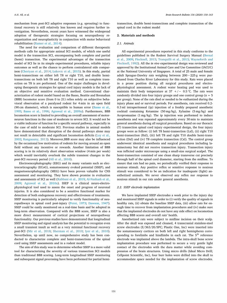
Subsequently, we performed a two-way ANOVA test using the twofactors [Injury × Day of Recovery]. Results are shown in Fig. 8 for both(a) left and (b) right hindlimb stimulation, which revealed significanteffects for interaction between injury(s) and day(s). The mean of Log(relative SSEP amplitude) is generally below the zero baseline for alltypes of injury regardless of the site of stimulation. In fact, the negativemean Log value is associated with the type of injury. In addition, themean Log value is at or above zero when stimulating the non-injuredsite (contralateral to the hemi-transection site).
Fig. 8. Two-way ANOVA [Injury × day of Recovery] test results with (a) Right and (b) Left hindlimb stimulation. It shows the interaction or relationship between thevariables (day and injury type) for all animals under test. Results show that while there is no significant interaction between the different days following injury, thereis a trend between injury type and day following injury.
A.H. All, et al. Brain Research Bulletin 156 (2020) 150–163
157
3.2. Behavioral outcome
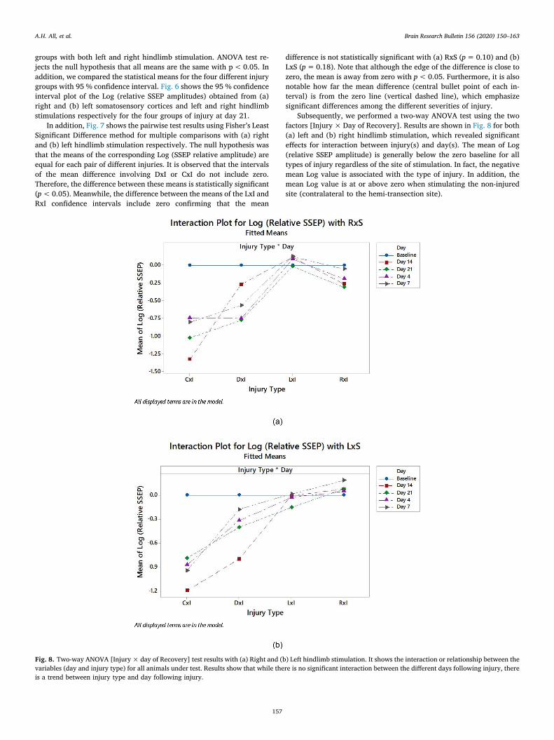
Traditional motor-behavioral assessment, BBB scoring, was used toevaluate the open-field (90 cm) locomotion performance of rats. TheBBB score shown in figures Figs. 9 and 10 demonstrated a short-termrecovery pattern for the two more severe groups which underwent ei-ther complete transection at T8 or double hemi-transection at left T8and right T10. Significant functional loss was observed for the completetransection group during the first week following SCI. The rats de-monstrated slight movement of the joints. On the other hand, thedouble hemi-transection group showed less severe motor functional lossimmediately after injury with extensive joint movement, some plantarstepping and limited weight bearing. However, for rats of these twoinjury groups, an increase of locomotor function was witnessed withinthe three-week observation window, indicating some functional re-covery following injury. In addition, substantial improvements in BBB
score patterns were detected from the two hemi-transection groups,with only mild functional loss flowing injury. Rats that underwent onehemi-transection injury (either at T8 or at T10) showed consistentplanter stepping and coordinated limb movement starting at the end ofweek 1 and the beginning of week 2, which continued till the end ofweek 3. Interestingly BBB scores show similar values for left and righthemi-transection injuries as well. However, only limited improvementof BBB score was recorded for these two groups during the span of ourstudy. The rats in control group (only laminectomy without injury) didnot show any sign of function loss and their BBB scores remained 21throughout the observation period, with the exception of days 1–3 dueto the surgical procedures (but not related to the injury).
Statistical analysis of BBB score (functional loss) revealed a differentprogression of the injury across the four injury groups. To characterizethe degree of functional impairment after injury, we performed mul-tiple comparisons between the different types of injury with 95 %
Fig. 9. Left hindlimb BBB scores on different days post-injury.
Fig. 10. Right hindlimb BBB scores on different days post-injury.
A.H. All, et al. Brain Research Bulletin 156 (2020) 150–163
158
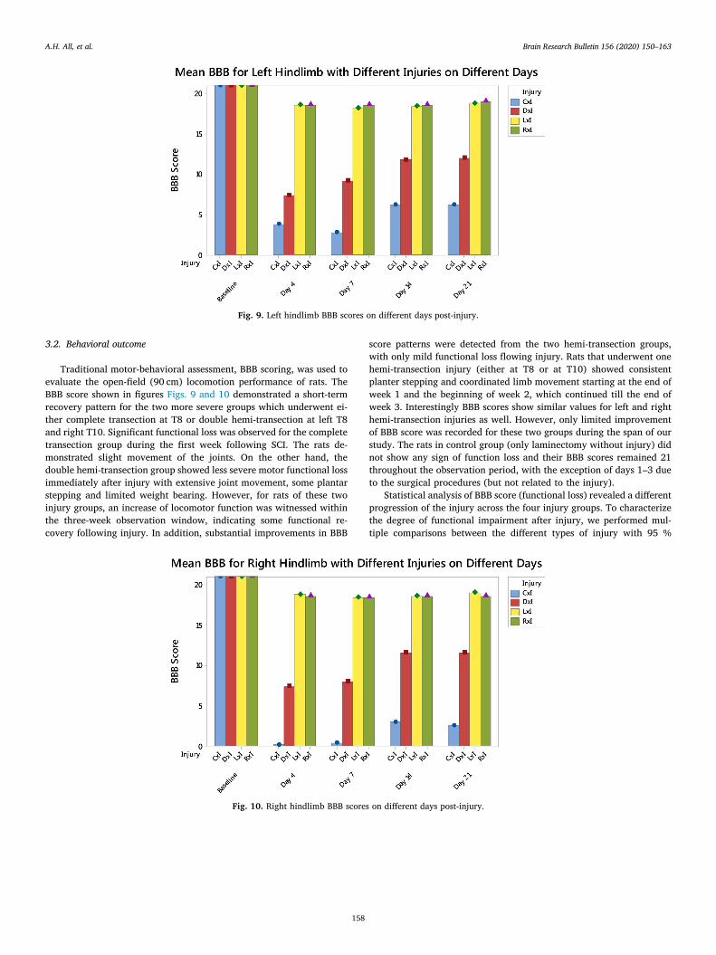
confidence interval for both (a) right and (b) left hindlimbs. ANOVAtest rejects the null hypothesis that all means are the same (p < 0.05) asshown in Fig. 11. Furthermore, the BBB score was unable to differ-entiate between left and right hemi-transection types of injuries.
In addition, Fig. 12 shows the confidence interval for the BBB meanscore difference between the four groups of injury. It is observed thatintervals of the mean difference involving CxI and DxI with LxI and RxIdo not include zero. This implies that the difference between thesemeans is statistically significant (p < 0.05). The difference between the
means of the LxI and RxI confidence intervals in the SSEP measure-ments shown earlier include zero confirming that the mean difference isnot statistically significant with (a) RxS (p = 0.10) and (b) LxS(p = 0.18). On the other hand, the difference between the means of theLxI and RxI confidence intervals of the BBB include zero and not sta-tistically significant (p = 0.93).
Subsequently we performed two-way analysis of variance (ANOVA)[Injury × day of Recovery] on BBB Scores obtained from types of injuryand on all days. ANOVA analysis results are shown in Fig. 13 for both
Fig. 11. Interval plot of the BBB scores for (a) right and (b) left hindlimbs for the four types of transection injuries with 95 % confidence interval (CI) on day 21.
A.H. All, et al. Brain Research Bulletin 156 (2020) 150–163
159
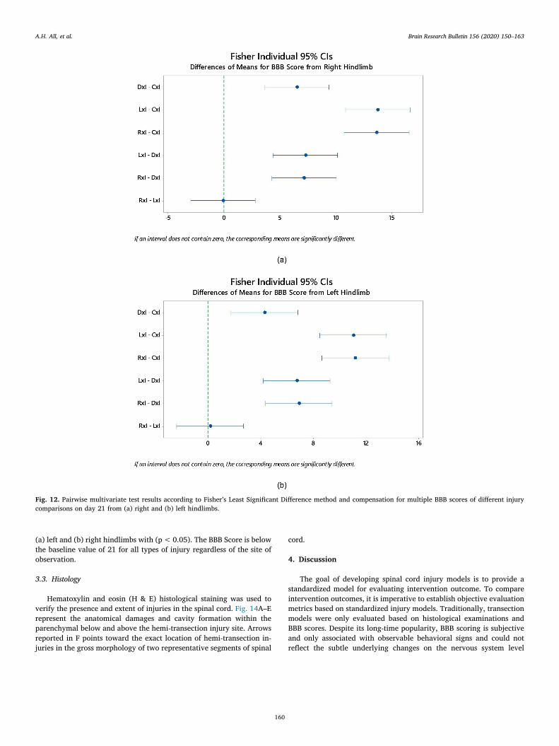
(a) left and (b) right hindlimbs with (p < 0.05). The BBB Score is belowthe baseline value of 21 for all types of injury regardless of the site ofobservation.
3.3. Histology
Hematoxylin and eosin (H & E) histological staining was used toverify the presence and extent of injuries in the spinal cord. Fig. 14A–Erepresent the anatomical damages and cavity formation within theparenchymal below and above the hemi-transection injury site. Arrowsreported in F points toward the exact location of hemi-transection in-juries in the gross morphology of two representative segments of spinal
cord.
4. Discussion
The goal of developing spinal cord injury models is to provide astandardized model for evaluating intervention outcome. To compareintervention outcomes, it is imperative to establish objective evaluationmetrics based on standardized injury models. Traditionally, transectionmodels were only evaluated based on histological examinations andBBB scores. Despite its long-time popularity, BBB scoring is subjectiveand only associated with observable behavioral signs and could notreflect the subtle underlying changes on the nervous system level
Fig. 12. Pairwise multivariate test results according to Fisher’s Least Significant Difference method and compensation for multiple BBB scores of different injurycomparisons on day 21 from (a) right and (b) left hindlimbs.
A.H. All, et al. Brain Research Bulletin 156 (2020) 150–163
160

(Agrawal et al., 2009a; All et al., 2010). Additionally, BBB scoring isprone to human error and lack objectivity. Therefore, this study in-vestigates the practical value of SSEP monitoring, signal analysis andmethods, which is a promising evaluation method of SCI and a reliableassessment for sensory pathways, in transection models. SSEP is a directfunctional representation of specific dorsal sensory neuropathways thatoriginate from the periphery and reach to a well-defined cortical areawith precise activities. Therefore, this study investigates the SSEPneuro-electrophysiological responses for evaluation of SCI on transec-tion injury model.
The transient nature of recovery in post-transection SCI also limitsthe use of BBB locomotion scoring. SSEP, on the other hand, wouldprovide the capability for long-term longitudinal monitoring of SCIwith residual function, benefiting both the assessment of injury, itsprogression and eventual recovery due to treatment strategies. Thecollection of longitudinal SSEP data could also offer a new perspectivein the natural history of injury development.
Severity and length of injuries are the two main causes of SSEPsignal attenuation after trauma. During the three-week observationperiod, SSEP demonstrated to be undoubtedly capable of distinguishing
onset, different severities of injury, progress, and even minuscule en-dogenous recovery in the spinal cord. This can certainly be extended tothe objective assessment of possible benefit for various form of ther-apeutic strategies as well. Additionally, two distinct modes of recoverywere also observed by separating the complete transection group fromdouble hemi-transection and other two hemi-transection groups. Forinstance, following SCI, both the double hemi-transection and twohemi-transection groups demonstrate significant temporal improve-ment of SSEP amplitude during the study. However, no significantimprovement was detected for the complete transection group. Thiswould be attributed mainly to the fact that only in the completetransection groups, bilateral Gracile Fasciculi of a given spinal cordsegment are disrupted, essentially eliminating the possibility of func-tional recovery for the complete transection group. In contrast, for thedouble hemi-transection and two hemi-transection groups, the uni-lateral transections would invariably leave some functional part ofGracile Fasciculi intact in the spinal cord and hence, preserving thepotential for compensatory responses and connectivity to the brain(Athanasiou et al., 2017; Blight and Young, 1989). Additionally, the all-or-none response mechanism of the cortex means maximal response in
Fig. 13. Two-way ANOVA [Injury × day of Recovery] test results of BBB Score for (a) Left and (b) Right hindlimbs.
A.H. All, et al. Brain Research Bulletin 156 (2020) 150–163
161

the cortical regions would occur as long as sufficient number of mye-linated axons survive and reach the target. Since complete transectiongroup could not possibly retain sufficient functional axons post injury,it is highly unlikely that spontaneous recovery would occur in thisgroup. Unlike the major disruption inflicted upon the complete trans-ection group, the disruption to the double and hemi-transection groupsare intermediate and mild, respectively. Both demonstrated a sig-nificant recovery pattern from week 1 to week 2. For the two hemi-transection groups, the SSEP amplitude becomes stable after week 2,indicating maximal recovery, which is below the pre-injury baseline.For the double hemi-transection group, the SSEP amplitude at week 3 isstill substantially below the pre-injury amplitude and the amplitudestabilization phenomenon is less prominent.
The SSEP signal pattern is found to be different from that of the BBBscoring scale. For the complete transection group, the BBB scores showsigns of functional improvement, whereas the SSEP signal could notsubstantiate any degree of recovery. Although early signs of recoverycould be interpreted from both the BBB scores and the SSEP signals,BBB scores indicate a stable functional recovery in later stages (week 3)while the SSEP indicates more volatile and unstable signals during laterphases. For the two hemi-transection groups, however, it is the SSEPsignal that shows mild improvement while BBB score does not show anysignificant improvement after injury. The conflicting results could beattributed to the different modes of recovery of the sensory pathwaysand motor locomotion following spinal cord injury. The isolated
improvement of BBB scores without SSEP signals improvements couldalso be attributed to subsiding spinal shock or to the neuroplasticityamong non-injured descending pathways – to be investigated.
In conclusion, we have demonstrated the SSEP-based method as apromising tool to detect the onset and characterize the severity of injuryfollowing transection spinal cord injury. The results from such methodwere compared to the results from the BBB-based scoring method overthe 3-week observation window following SCI. As we reported in ourresults, the BBB mean score shows one score difference in both the LxIand RxI three week after injury (Fig. 11), while the SSEP shows ap-proximately 20 % and 40 % reduction relative to the baseline in thecorresponding LxI and RxI (Fig. 6). Similarly, the BBB mean score onday 21 shows 9 and 17 average score difference in DxI and CxI re-spectively. The SSEP, on the other hand, shows approximately 50 % and80 % reduction relative to the baseline of the corresponding DxI and CxIrespectively. This noticeable significant differences between the twomethods were discovered. Nevertheless, we found the two methodscomplementary to each other. Hence, we believe it will be beneficial forfuture studies to adopt both the SSEP-based and BBB-based method toevaluate the transection SCI model, which is a model used extensivelyin SCI research experiments.
Author contribution
Angelo All, Shiyu Luo, Hasan Mir and Hasan Al-Nashash contributed
Fig. 14. Histology and morphology of transection injury. (a) The control group (no transection), (b) Sagittal view of a hemi-transection, (c) Internal cavity formationafter a hemi-transection, (d) Longitudinal view of a hemi-transection. (e) Full transection. (f) Gross morphology of a hemi-transection on the left and double hemi-transection on the right. (Scale: H & E staining in A–E are 5 × magnification and gross anatomy in F is 1 × magnification.).
A.H. All, et al. Brain Research Bulletin 156 (2020) 150–163
162
to the conception and design.Angelo All, Shiyu Luo and Hasan Al-Nashash did literature search.Hasan Mir and Hasan Al-Nashash performed data analysis.Angelo All, Shiyu Luo and Hasan Al-Nashash drafted the paper.Xiaogang Liu provided critical revision of the work.Angelo All and Xiaogang Liu supervised and reviewed this work.
Declaration of Competing Interest
The authors declare no potential conflict of interest.
Acknowledgement
We would like to thank research assistants: Ms. Ashwati Vipin, Mr.Thow Xin Yuan, Ms. Chua Soo Min, Ms. Astrid, and research associateDr. Janani Manivannan as well as Dr. Jukka Kortelainen for theircontribution in experimental procedures, SSEP monitoring, behavioralassessment, animal care and administrative role in this project.
References
Agrawal, G., Sherman, D., Thakor, N., All, A., 2008. A novel shape analysis technique forsomatosensory evoked potentials. Conf. Proc. IEEE Eng. Med. Biol. Soc. 4688–4691.
Agrawal, G., Thakor, N.V., All, A.H., 2009a. Evoked potential versus behavior to detectminor insult to the spinal cord in a rat model. J. Clin. Neurosci. 16 (8), 1052–1055.
Agrawal, G., Sherman, D., Maybhate, A., Gorelik, M., Kerr, D.A., Thakor, N.V., All, A.H.,2010a. Slope analysis of somatosensory evoked potentials in spinal cord injury fordetecting contusion injury and focal demyelination. J. Clin. Neurosci. 17 (9),1159–1164.
Agrawal, G., Kerr, C., Thakor, N.V., All, A.H., 2010b. Characterization of graded multi-center animal spinal cord injury study contusion spinal cord injury using somato-sensory-evoked potentials. Spine 35 (11), 1122–1127.
Agrawal, G., Iyer, S., All, A.H., 2009b. A comparative study of recording procedures formotor evoked potential signals. Conf. Proc. IEEE Eng. Med. Biol. Soc. 2086–2089.
Agrawal, G., Sherman, D.L., Walczak, P., Bulte, J.W.M., Thakor, N.V., Kerr, D.A., All,A.H., 2009c. Shape analysis of somatosensory evoked potentials to detect a focalspinal cord lesion. Proc. IEEE Annu. Northeast Bioeng. Conf. 1–2.
All, A.H., Agrawal, G., Walczak, P., Maybhate, A., Bulte, J.W., Kerr, D.A., 2010. Evokedpotential and behavioral outcomes for experimental autoimmune encephalomyelitisin lewis rats. Neurol. Sci. 31 (5), 595–601.
Al-Nashash, H., Fatoo, N.A., Mirza, N.N., Ahmed, R.I., Agrawal, G., Thakor, N.V., All,A.H., 2009. Spinal cord injury detection and monitoring using spectral coherence.IEEE Trans. Biomed. Eng. 56 (8), 1971–1979.
Athanasiou, A., Klados, M.A., Pandria, N., Foroglou, N., Kavazidi, K.R., Polyzoidis, K.,Bamidis, P.D., 2017. A systematic review of investigations into functional brainconnectivity following spinal cord injury. Front. Hum. Neurosci. 11, 517.
Ballermann, M., Fouad, K., 2006. Spontaneous locomotor recovery in spinal cord injuredrats is accompanied by anatomical plasticity of reticulospinal fibers. Eur. J. Neurosci.23 (8), 1988–1996.
Bareyre, F.M., Kerschensteiner, M., Raineteau, O., Mettenleiter, T.C., Weinmann, O.,Schwab, M.E., 2004. The injured spinal cord spontaneously forms a new intraspinalcircuit in adult rats. Nat. Neurosci. 7 (3), 269.
Basso, D.M., Beattie, M.S., Bresnahan, J.C., 1995. A sensitive and reliable locomotorrating scale for open field testing in rats. J. Neurotrauma 12 (1), 1–21.
Basso, D.M., Beattie, M.S., Bresnahan, J.C., 1996. Graded histological and locomotoroutcomes after spinal cord contusion using the NYU weight-drop device versustransection. Exp. Neurol. 139 (2), 244–256.
Bazley, F.A., All, A.H., Thakor, N.V., Maybhate, A., 2011. Plasticity associated changes incortical somatosensory evoked potentials following spinal cord injury in rats. Conf.Proc. IEEE Eng. Med. Biol. Soc.
Bazley, F.A., Hu, C., Maybhate, A., Pourmorteza, A., Pashai, N., Thakor, N.V., Kerr, C.L.,All, A.H., 2012. Electrophysiological evaluation of sensory and motor pathways afterincomplete unilateral spinal cord contusion. J. Neurosurg. Spine 16 (4), 414–423.
Bazley, F.A., Maybhate, A., Tan, C.S., Thakor, N.V., Kerr, C., All, A.H., 2014.Enhancement of bilateral cortical somatosensory evoked potentials to intact forelimbstimulation following thoracic contusion spinal cord injury in rats. IEEE Trans. NeuralSyst. Rehabil. Eng. 22 (5), 953–964.
Bellardita, C., Marcantoni, M., Löw, P., Kiehn, O., 2018. Sacral spinal cord transectionand isolated sacral cord preparation to study chronic spinal cord injury in adult mice.Bio Protoc. 8 (7).
Bernal, J., Baldwin, M., Gleason, T., Kuhlman, S., Moore, G., Talcott, M., 2009. Guidelines
for rodent survival surgery. J. Invest. Surg. 22 (6), 445–451.Blight, A.R., Young, W., 1989. Central axons in injured cat spinal cord recover electro-
physiological function following remyelination by Schwann cells. J. Neurol. Sci. 91(1), 15–34.
Cheriyan, T., Ryan, D.J., Weinreb, J.H., Cheriyan, J., Paul, J.C., Lafage, V., Kirsch, T.,Errico, T.J., 2014. Spinal cord injury models: a review. Spinal Cord 52 (8), 588.
Crowe, M.J., Bresnahan, J.C., Shuman, S.L., Masters, J.N., Crowe, M.S., 1997. Apoptosisand delayed degeneration after spinal cord injury in rats and monkeys. Nat. Med. 3(1), 73–76.
Dawson, G.D., 1947. Cerebral responses to electrical stimulation of peripheral nerve inman. J. Neurol. Neurosurg. Psychiatry 10 (3), 134.
Deumens, R., Joosten, E.A., Waxman, S.G., Hains, B.C., 2008. Locomotor dysfunction andpain: The scylla and charybdis of fiber sprouting after spinal cord injury. Mol.Neurobiol. 37 (1) 2008.
Filli, L., Schwab, M.E., 2015. Structural and functional reorganization of propriospinalconnections promotes functional recovery after spinal cord injury. Neural Regen. Res.10 (4), 509.
Flecknell, P., 2015. Laboratory Animal Anaesthesia. Academic press, London.Iyer, S., Maybhate, A., Presacco, A., All, A.H., 2010. Multi-limb acquisition of motor
evoked potentials and its application in spinal cord injury. J. Neurosci. Methods 193(2), 210–216.
Kirshblum, S.C., Burns, S.P., Biering-Sorensen, F., Donovan, W., Graves, D.E., Jha, A.,Johansen, M., Jones, L., Krassioukov, A., Mulcahey, M.J., Schmidt-Read, M., 2011.International standards for neurological classification of spinal cord injury (revised2011). J. Spinal Cord Med. 34 (6), 535–546.
Kortelainen, J., Vipin, A., Yuan, T.X., Mir, H., Thakor, N., Al-Nashash, H., All, A., 2014.Effect of isoflurane on somatosensory evoked potentials in a rat model. Conf. Proc.IEEE Eng. Med. Biol. Soc. 4286–4289.
Kortelainen, J., Al-Nashash, H., Vipin, A., Thow, X.Y., All, A., 2016. The effect of an-aesthesia on somatosensory evoked potential measurement in a rat model. Lab. Anim.50 (1), 63–66.
Liu, Z.H., Yip, P.K., Adams, L., Davies, M., Lee, J.W., Michael, G.J., Priestley, J.V.,Michael-Titus, A.T., 2015. A single bolus of docosahexaenoic acid promotes neuro-plastic changes in the innervation of spinal cord interneurons and motor neurons andimproves functional recovery after spinal cord injury. J. Neurosci. 35 (37),12733–12752.
Loy, D.N., Talbott, J.F., Onifer, S.M., Mills, M.D., Burke, D.A., Dennison, J.B., Fajardo,L.C., Magnuson, D.S., Whittemore, S.R., 2002. Both dorsal and ventral spinal cordpathways contribute to overground locomotion in the adult rat. Exp. Neurol. 177 (2),575–580.
Lukovic, D., Moreno-Manzano, V., Lopez-Mocholi, E., Rodriguez-Jimenez, F.J.,Jendelova, P., Sykova, E., Oria, M., Stojkovic, M., Erceg, S., 2015. Complete rat spinalcord transection as a faithful model of spinal cord injury for translational celltransplantation. Sci. Rep. 5, 9640.
Maybhate, A., Hu, C., Bazley, F.A., Yu, Q., Thakor, N.V., Kerr, C.L., All, A.H., 2012.Potential long term benefits of acute hypothermia after spinal cord injury: assess-ments with somatosensory evoked potentials. Crit. Care Med. 40 (2), 573.
Mir, H., Al-Nashash, H., Kortelainen, J., All, A., 2018. Novel modeling of somatosensoryevoked potentials for the assessment of spinal cord injury. IEEE Trans. Biomed. Eng.65 (3), 511–520.
Mir, H., Al-Nashash, H., Kerr, D., Thakor, N., All, A., 2010. Histogram based quantifi-cation of spinal cord injury level using somatosensory evoked potentials. Conf. Proc.IEEE Eng. Med. Biol. Soc. 4942–4945.
Perot Jr., P.L., 1973. The clinical use of somatosensory evoked potentials in spinal cordinjury. Neurosurgery 20 (1), 367–381.
Rabbani, Q., Milsap, G., Crone, N.E., 2019. The potential for a speech brain–computerinterface using chronic electrocorticography. Neurotherapeutics 16 (1), 144–165.
Ramer, L.M., Ramer, M.S., Bradbury, E.J., 2014. Restoring function after spinal cordinjury: towards clinical translation of experimental strategies. Lancet Neurol. 13 (12),1241–1256.
Rangasamy, S.B., 2013. Locomotor recovery after spinal cord hemisection/contusion in-jures in bonnet monkeys: footprint testing—a mini review. Synapse 67 (7), 427–453.
Sherman, D.L., Wuyyuru, V., Brooke, M.J., Zhang, H.X., Sepkuty, J.P., Thakor, N.V.,Natarajan, A., All, A.H., 2010. Spinal cord integrity monitoring by adaptive co-herence measurement. J. Neurosci. Methods 193 (1), 90–99.
Talac, R., Friedman, J.A., Moore, M.J., Lu, L., Jabbari, E., Windebank, A.J., Currier, B.L.,Yaszemski, M.J., 2004. Animal models of spinal cord injury for evaluation of tissueengineering treatment strategies. Biomaterials 25 (9), 1505–1510.
Tranquilli, W.J., Thurmon, J.C., Grimm, K.A., 2013. Lumb and Jones’ VeterinaryAnesthesia and Analgesia. John Wiley & Sons, New York.
Vipin, A., Thow, X.Y., Mir, H., Vipin, A., Thow, X.Y., Mir, H., Kortelainen, J.,Manivannan, J., Al-Nashash, H., All, A.H., et al., 2016. Natural progression of spinalcord transection injury and reorganization of neural pathways. J. Neurotrauma 33(24), 2191–2201.
Wayneforth, H.B., Flecknell, P.A., 1992. Experimental and Surgical Techniques in the Rat.Academic press, London.
A.H. All, et al. Brain Research Bulletin 156 (2020) 150–163
163