brain basics - homepages.hass.rpi.edu
TRANSCRIPT

Brain Basics
Introduction to Cognitive Science

Neurons • Human brain:
– ~100 billion (1011) neurons – ~100 trillion (1014) neural
connections
• Dozens of different neuro-transmitters … Why?
– And why neurotransmitter in the first place; why not a hard-wired connection?
• Doesn’t fit a simple ‘neurons
fire or not is like 1’s and 0’s in a computer’ story
• Is information-processing done
by chemicals or inside neuron?

‘Gray’ vs ‘White’ Matter
Axons have a myelin sheeth. The myelin sheeth helps the signal travel along the axons. This myelin sheeth will look ‘white’, while the neuronal bodies look ‘grey’. In a brain, there is more gray on the outside (the cortex, basically) and more white on the inside, suggesting that there are more and longer connections on the inside and/or that the inside serves to connect some of the outside regions of the brain

Basic Anatomy
• 4 Main parts of brain – Cerebrum: ‘Cognition’: reasoning, planning,
decision making, ‘complex’ behavior – Cerebellum: Coordination of behavior:
posture, balance, walking – Limbic System: emotion and memory – Brain Stem: basic body regulation: breathing,
heartbeat, sleep, most basic sensory/motor control, e.g. eye movement
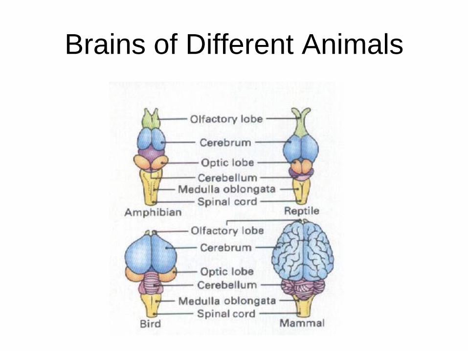
Brains of Different Animals

Cerebrum: 4 lobes Frontal Lobe: executive control, reasoning, decision making, sense of ethics
Parietal lobe: integration sensory information, visuo-spatial processing, movement, numbers
Occipital lobe: visual processing
Temporal lobe: auditory processing, semantics, language

Cortex The cerebral cortex, or neocortex, is the outer layer of the cerebrum. This is the ‘gray matter’: This seems to be where the ‘cognitive’, computational, action is . Underneath the cortex is ‘white matter’ which seems to serve more supporting functions, e.g. connections

Motor and Somatosensory Cortex
Here we find very specialized areas (modules) of the brain: specific areas responsible for specific functions Question: Is the rest of the brain (mind) like that? E.g. are there specific areas of the brain responsible for all knowledge regarding grandmother?
This figure represents how much of the cortex is devoted to the different parts of the body

Limbic System
• Thalamus: ‘relay station’ of sensory input to cortex (exception: smell)
• Hippocampus: formation of memory
• Amygdala: Emotional salience of stimuli
• Hypothalamus: basic drives (4 F’s), breathing, heartrate, and more

Phrenology
• 19th century ‘brain science’
• Assumed localization of cognitive function (personality traits, really
• Bumps or dents on head were thought to reveal more or less neurons devoted to that ‘cognitive function

Modern Brain Nonsense: The 10% of Your Brain Myth
• “Most people only use 10% of their brain!” – The suggestion being that if you could somehow
‘unlock’ the ‘extra power’ of your brain, you could be so much smarter!
– Which is why you should buy this book for $79.99 or take this 7 week course for only $499!!
• Total myth! – First off all: this makes no sense evolutionarily – Indeed, when doing brain imaging, we find activity all
over the brain at all times • Where does the myth come from?
– Probably from exactly those brain images

CAT Scans • CAT or CT (Computed
Axial Tomography) – x-ray – Provides rough
structural/anatomical information
– Mostly to find suspected brain damage

PET Scans • PET (Positron Emission
Tomography) – Radioactive chemicals
inserted in bloodstream – So, PET scans provide
‘metabolic’ information: roughly, reveal functional activity of brain

MRI and fMRI Scans • MRI (Magnetic Resonance Imaging)
– Place subject in powerful magnetic field and measure differences in frequency
– Good spatial resolution; low temporal resolution – f (functional) MRI: higher temporal resolution, lower spatial
It is exactly these kinds of pictures that perpetuate the 10% myth: it looks as if only a small part of the brain is active, but what they’re showing is merely areas whose ‘activity’ (or whatever it really is that the MRI is measuring) is above some defined threshold.
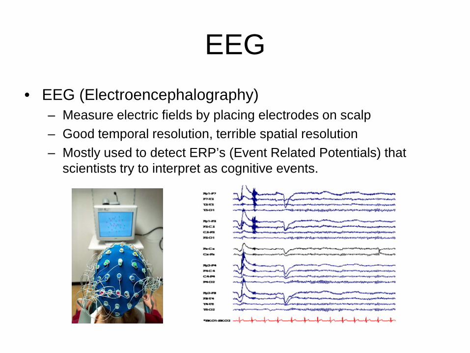
EEG • EEG (Electroencephalography)
– Measure electric fields by placing electrodes on scalp – Good temporal resolution, terrible spatial resolution – Mostly used to detect ERP’s (Event Related Potentials) that
scientists try to interpret as cognitive events.