brain abscess in a patient with rendu-osler-weber · pdf filevalue of proton magnetic...
TRANSCRIPT
35
Case Report
Brain Abscess in a Patient with Rendu-Osler-Weber Syndrome: Value of Proton Magnetic Resonance Spectroscopy
Alex Roberto Ramírez Mejía, Miguel Yus Fuertes, and Manuela Jorquera Moya
Department of Diagnostic Radiology, Hospital Clínico San Carlos, Madrid, Spain
Received: June 4, 2015; Accepted: September 20, 2015
NMC Case Report Journal 2016; 3: 35–37 DOI: 10.2176/nmccrj.cr.2015-0141
Brain MRI was performed by using a 1.5-T MR imaging unit (GE Medical Systems, Milwaukee, Wisconsin, USA) with a quadrature T/R head coil. Routine transverse and sagittal fast spin-echo (FSE) T1-weighted image (WI), transverse and cor-onal FSE T2WI, transverse fluid attenuation inversion recovery (FLAIR) and diffusion weighted imaging (DWI), and post contrast transverse T1WIs three-dimensional fast spoiled gra-dient (3D-FSPGR) images were acquired after intravenous administration of gadobutrol (Gadovist; Bayer Pharma AG, Berlin, Germany) at a dose of 0.1 mmol/kg of body weight. The images were obtained with an acquisition matrix of 256 × 256 (128 × 128 on the DWI) and a field of view of 24 × 24 cm.
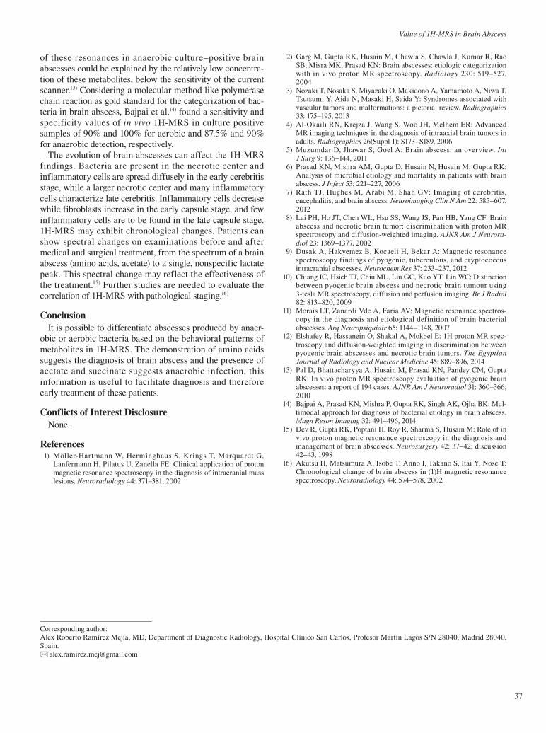
Single-voxel volume-selective water suppressed 1H-MRS was performed by using PROBE-PRESS sequence with repetition time (TR) 1,500 ms and echo time (TE) 35 ms, 144 ms, and 288 ms, and a voxel size of 8–12 ml (mean: 10 ml) in the center of the lesion. We used FLAIR images to locate the voxels for the 1H-MRS (Figs. 1, 2).
The diagnosis of intracranial focal lesions based on imaging studies is a challenge. In brain abscess, proton magnetic resonance spectroscopy (1H-MRS) is useful to define etiology, differentiating anaerobic from aerobic pathogens and inclusive sterile abscess based on the behavior of certain metabolites. We report a patient with Rendu-Osler-Weber syndrome, pulmonary arterio-venous fistulas, and brain abscesses characterized by brain magnetic resonance imaging (MRI), with a spec-troscopy pattern compatible with anaerobic infection.
Keywords: brain abscess , magnetic resonance spectroscopy, Rendu-Osler-Weber syndrome
IntroductionThe differentiation between brain abscesses and other
entities with ring-type contrast enhancement is difficult based only on image, because abscesses simulate other injuries as primary tumors or metastases. In contrast to magnetic resonance imaging (MRI) and computed tomog-raphy (CT) that provide structural data, proton magnetic resonance spectroscopy (1H-MRS) gives completely dif-ferent information related to neuronal integrity, cell prolif-eration or degradation, energy, metabolism, and necrotic transformation of brain or tumor tissues. 1H-MRS allows the study of metabolites normally absent in the normal brain, gives information on the nature of the abscess, and thus increases the specificity of conventional MRI studies.1) 1H-MRS can provide information about the cause of the abscess and their response to treatment, and since anaerobic bacteria typically have a growth pattern in culture media slower than aerobic bacteria, 1H-MRS can be useful in choosing an appropriate antibiotic therapy.2)
Case ReportA 67-year-old-woman with known rheumatoid arthritis,
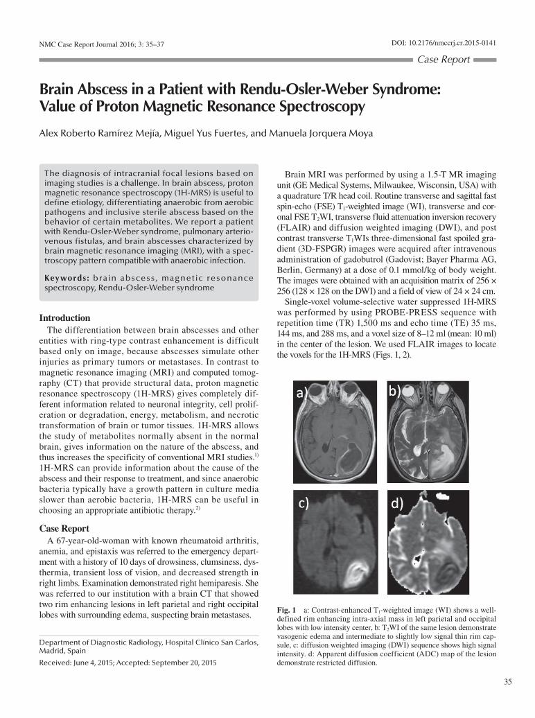
anemia, and epistaxis was referred to the emergency depart-ment with a history of 10 days of drowsiness, clumsiness, dys-thermia, transient loss of vision, and decreased strength in right limbs. Examination demonstrated right hemiparesis. She was referred to our institution with a brain CT that showed two rim enhancing lesions in left parietal and right occipital lobes with surrounding edema, suspecting brain metastases.
Fig. 1 a: Contrast-enhanced T1-weighted image (WI) shows a well-defined rim enhancing intra-axial mass in left parietal and occipital lobes with low intensity center, b: T2WI of the same lesion demonstrate vasogenic edema and intermediate to slightly low signal thin rim cap-sule, c: diffusion weighted imaging (DWI) sequence shows high signal intensity. d: Apparent diffusion coefficient (ADC) map of the lesion demonstrate restricted diffusion.

36
A. R. Ramírez Mejía et al.
The diagnostic criteria are recurrent epistaxis, multiple vas-cular dilatations, arteriovenous malformations or fistulas in the internal organs including lungs, liver, brain, or bone marrow, and a first-degree relative with the condition.3) Pul-monary arteriovenous malformations predispose to the for-mation of brain abscess in immunocompetent patients due to hematogenous spread of pulmonary infectious foci.4)
Brain abscess is a focal intracerebral infection that begins with a localized cerebritis and evolves to a collection of pus surrounded by a housing area; often caused by contiguous spread of infectious foci or hematogenous spread from a distant focus. It can occur at any age, more common in men than in women and the most common presenting symptoms are headache and altered mental status. Although there has been clear progress in diagnostic techniques and treatment in recent decades, brain abscesses still carry significant morbidity and mortality. Because of this, early diagnosis is crucial to optimize the use of antibiotics; the success of treatment is best when the etiologic agent is identified and antimicrobial therapy is targeted.5,6)
1H-MRS instead of showing the signals of the protons as a grayscale image based on their relative signal strength shows the amounts as a spectrum, and metabolites in greater numbers have a greater peak height or greater area under the peak.
Typical 1H-MRS features of brain abscesses include high peaks of amino acids, lactate, alanine, acetate and succinate and, as the abscess cavity lacks normal brain tissue, there are no basic brain metabolites (N-acetylaspartate, creatine, and choline). Rupture of the cell membranes and proteolytic enzymes released by neutrophils produce cytosolic amino acids (valine, leucine, and isoleucine), bacterial fermentation leads to the production of lactate, acetate and succinate; and necrosis produces lactate and lipids. The presence of anaer-obic bacteria tends to cause an increase of acetate and succi-nate while the absence thereof becomes more likely to be an infection by aerobic bacteria.2,7–12)
Resonances of acetate with or without succinate on 1H-MRS favor an abscess of anaerobic bacterial origin and, in some cases, facultative anaerobic microbes. Nonvisibility
Fig. 2 a: Transverse FLAIR image shows the location of the rectangular voxel placed for 1H-MRS in the occipital lobe. b–d: Single-voxel 1H-MRS with echo times of (b) 35 msec (c) 144 msec and (d) 288 msec. The 1H-MRS spectrum shows absence of N-acetylaspartate (2.02 ppm), choline (3.2 ppm), and creatine (3.0 ppm) peaks, and elevation of amino acids (AA) (0.9 ppm), lactate (Lac) (1.33 ppm), acetate (Ac) (1.9 ppm), and succinate (Su) (2.4 ppm) peaks. Inversion of lactate peak with an echo time of 144 msec in (c). Lac/Lip: lactate/lipid. ppm: parts per million, FLAIR: fluid attenuation inversion recovery, 1H-MRS: proton magnetic resonance spectroscopy.
Fig. 3 Contrast-enhanced reformatted thoracic computed tomo-graphic scan with mediastinal soft tissue window settings demonstrates a large pulmonary arteriovenous fistula (arrow) in the lingula.
A diagnosis of brain abscess was made, the 1H-MRS find-ings were consistent with the presence of anaerobic bacteria and antibiotic therapy with cefotaxime, linezolid, and metro-nidazole was initiated. Drainage of the abscess 3 days later revealed purulent content. Cultures were positive for Strepto-coccus milleri, a facultative anaerobic gram-positive bacteria.
A thoracic CT demonstrated pulmonary arteriovenous fistulas up to 2 cm in diameter in middle lobe and lingula and the patient was diagnosed for Rendu-Osler-Weber syndrome (Fig. 3).
Neurological symptoms present at admission resolved. The patient was discharged awaiting embolization of vascular malformations.
DiscussionRendu-Osler-Weber syndrome is a genetic, autosomal
dominant disorder characterized by dilatation of small skin and mucosal blood vessels which leads to epistaxis and hem-orrhage into the digestive tract. It affects 1 in 100,000 people.
Value of 1H-MRS in Brain Abscess
37
2) Garg M, Gupta RK, Husain M, Chawla S, Chawla J, Kumar R, Rao SB, Misra MK, Prasad KN: Brain abscesses: etiologic categorization with in vivo proton MR spectroscopy. Radiology 230: 519–527, 2004
3) Nozaki T, Nosaka S, Miyazaki O, Makidono A, Yamamoto A, Niwa T, Tsutsumi Y, Aida N, Masaki H, Saida Y: Syndromes associated with vascular tumors and malformations: a pictorial review. Radiographics 33: 175–195, 2013
4) Al-Okaili RN, Krejza J, Wang S, Woo JH, Melhem ER: Advanced MR imaging techniques in the diagnosis of intraaxial brain tumors in adults. Radiographics 26(Suppl 1): S173–S189, 2006
5) Muzumdar D, Jhawar S, Goel A: Brain abscess: an overview. Int J Surg 9: 136–144, 2011
6) Prasad KN, Mishra AM, Gupta D, Husain N, Husain M, Gupta RK: Analysis of microbial etiology and mortality in patients with brain abscess. J Infect 53: 221–227, 2006
7) Rath TJ, Hughes M, Arabi M, Shah GV: Imaging of cerebritis, encephalitis, and brain abscess. Neuroimaging Clin N Am 22: 585–607, 2012
8) Lai PH, Ho JT, Chen WL, Hsu SS, Wang JS, Pan HB, Yang CF: Brain abscess and necrotic brain tumor: discrimination with proton MR spectroscopy and diffusion-weighted imaging. AJNR Am J Neurora-diol 23: 1369–1377, 2002
9) Dusak A, Hakyemez B, Kocaeli H, Bekar A: Magnetic resonance spectroscopy findings of pyogenic, tuberculous, and cryptococcus intracranial abscesses. Neurochem Res 37: 233–237, 2012
10) Chiang IC, Hsieh TJ, Chiu ML, Liu GC, Kuo YT, Lin WC: Distinction between pyogenic brain abscess and necrotic brain tumour using 3-tesla MR spectroscopy, diffusion and perfusion imaging. Br J Radiol 82: 813–820, 2009
11) Morais LT, Zanardi Vde A, Faria AV: Magnetic resonance spectros-copy in the diagnosis and etiological definition of brain bacterial abscesses. Arq Neuropsiquiatr 65: 1144–1148, 2007
12) Elshafey R, Hassanein O, Shakal A, Mokbel E: 1H proton MR spec-troscopy and diffusion-weighted imaging in discrimination between pyogenic brain abscesses and necrotic brain tumors. The Egyptian Journal of Radiology and Nuclear Medicine 45: 889–896, 2014
13) Pal D, Bhattacharyya A, Husain M, Prasad KN, Pandey CM, Gupta RK: In vivo proton MR spectroscopy evaluation of pyogenic brain abscesses: a report of 194 cases. AJNR Am J Neuroradiol 31: 360–366, 2010
14) Bajpai A, Prasad KN, Mishra P, Gupta RK, Singh AK, Ojha BK: Mul-timodal approach for diagnosis of bacterial etiology in brain abscess. Magn Reson Imaging 32: 491–496, 2014
15) Dev R, Gupta RK, Poptani H, Roy R, Sharma S, Husain M: Role of in vivo proton magnetic resonance spectroscopy in the diagnosis and management of brain abscesses. Neurosurgery 42: 37–42; discussion 42–43, 1998
16) Akutsu H, Matsumura A, Isobe T, Anno I, Takano S, Itai Y, Nose T: Chronological change of brain abscess in (1)H magnetic resonance spectroscopy. Neuroradiology 44: 574–578, 2002
of these resonances in anaerobic culture–positive brain abscesses could be explained by the relatively low concentra-tion of these metabolites, below the sensitivity of the current scanner.13) Considering a molecular method like polymerase chain reaction as gold standard for the categorization of bac-teria in brain abscess, Bajpai et al.14) found a sensitivity and specificity values of in vivo 1H-MRS in culture positive samples of 90% and 100% for aerobic and 87.5% and 90% for anaerobic detection, respectively.
The evolution of brain abscesses can affect the 1H-MRS findings. Bacteria are present in the necrotic center and inflammatory cells are spread diffusely in the early cerebritis stage, while a larger necrotic center and many inflammatory cells characterize late cerebritis. Inflammatory cells decrease while fibroblasts increase in the early capsule stage, and few inflammatory cells are to be found in the late capsule stage. 1H-MRS may exhibit chronological changes. Patients can show spectral changes on examinations before and after medical and surgical treatment, from the spectrum of a brain abscess (amino acids, acetate) to a single, nonspecific lactate peak. This spectral change may reflect the effectiveness of the treatment.15) Further studies are needed to evaluate the correlation of 1H-MRS with pathological staging.16)
ConclusionIt is possible to differentiate abscesses produced by anaer-
obic or aerobic bacteria based on the behavioral patterns of metabolites in 1H-MRS. The demonstration of amino acids suggests the diagnosis of brain abscess and the presence of acetate and succinate suggests anaerobic infection, this information is useful to facilitate diagnosis and therefore early treatment of these patients.
Conflicts of Interest DisclosureNone.
References 1) Möller-Hartmann W, Herminghaus S, Krings T, Marquardt G,
Lanfermann H, Pilatus U, Zanella FE: Clinical application of proton magnetic resonance spectroscopy in the diagnosis of intracranial mass lesions. Neuroradiology 44: 371–381, 2002
Corresponding author: Alex Roberto Ramírez Mejía, MD, Department of Diagnostic Radiology, Hospital Clínico San Carlos, Profesor Martín Lagos S/N 28040, Madrid 28040, Spain.* [email protected]