botany mitosis and cytokinesis · 12/11/2017 · pembelahan sel adalah transisi g2/m ... ©modul...
TRANSCRIPT

BOTANY Mitosis and Cytokinesis Prof. Dr. S.M. Sitompul Lab. Plant Physiology, Faculty of Agriculture, Universitas Brawijaya Email : [email protected]
Mitosis adalah proses pembelahan sel
tubuh (somatic sel) untuk pertumbuhan
dan perkembangan tananman.
Proses ini, yang terjadi setelah kondisi
khusus (signal) dialami sel, sangat ketat
dikendalikan agar organella kopi
tunggal direplikasi secara tepat untuk
diturunkan pada generasi berikutmya.
Transisi utama yang genting dalam
pembelahan sel adalah transisi G2/M
dan transisi Metaphase/Anaphase.
It is not the strongest of the species that survives, nor the most intelligent that survives. It is the one that is the most adaptable to
change. Charles Darwin
LEARNING OUTCOME Students, after mastering materials of the present lecture, should be able
1. to explain signal transduction 2. to explain cell division
3. to explain cytokinesis in the life of plants 4. to explain the position of mitosis in cell cycle 5. to explain the process of mitosis
LECTURE LAYOUT
1. INTRODUCTION 1. Changes
2. Signals 3. Signal Transduction 4. Yeast Cell Mitosis
2. CELL DIVISION 1. Basic Concept
2. The Signaling Pathways 3. Genetic Material
4. DNA Replication 5. Spindle apparatus
3. MITOSIS AND CYTOKINESIS 1. Mitosis 2. Cytokinesis
12
mtom
MODUL
SELF-PR
OP
AG
ATIN
G EN
TREP
REN
EUR
IAL ED
UC
ATIO
N D
EVELO
PM
ENT
(SPEED
)
©Modul ini tidak boleh digandakan sebagian
atau seluruhnya tanpa izin dari penulis Hak cipta diindungi undangundang
Ha
k ci
pta
dili
nd
un
gi u
nd
an
g-u
nd
an
g.
©M
od
ul i
ni t
ida
k b
ole
h d
iga
nd
aka
n s
elu
ruh
nya
ata
u s
eba
gia
n t
an
pa
izin
da
ri p
enu
lis

Page 2 of 12
Botany/Mitosis/S.M. Sitompul 2017 The University of Brawijaya
QUESTIONS Who orders the cells to start to divide?
or
What triggers mitosis/cell division to start? - There is no one event or set of conditions that will stimulate all cells to begin
mitosis.
- A small number of eukaryotic cells are genetically programmed to pass through the cell cycle and undergo mitosis as soon as they can.
- Yeast cells are good examples of this; so long as there is ample food, they will grow and reproduce as quickly as possible.
I. INTRODUCTION 1. Changes
“You cannot step twice into the same river” Heraclitus of Ephesus (c.535 BCE-475 BCE), a Greek philosopher before
Socrates, is known for his doctrine of change being central to the universe with an epigram;
Everything changes and nothing remains still... and... you cannot step twice into the same stream for other waters are ever flowing on to you.
Good character is not formed in a week or a month. It is created little by
little, day by day. Protracted and patient effort is needed to develop good character.
You literally are not the same person you were in the past - Your bone marrow is producing 2 million red blood cells PER SECOND! - Skin cells divide once every 20-30 minutes.
Changes are due partly to cell division that is required for Growth & Development and Repair.
- Human zygote = 1 cell Mature adult = 1 X 10¹⁴ cells - Cell division is used to replace damaged cells and repair worn out tissues.
2. Signals In multicellular organisms, nearly all cells wait for specific conditions
(signals) before they progress through mitosis. - For example, normal hepatocytes (liver cells) divide about once a year.
Yet if the liver is injured or damaged, within a few hours they begin dividing to replace the damaged cells.
Some cells get a signal to divide so frequently and seem to be dividing
continuously (like yeasts). - The cells lining our stomach are good examples, and replaced every 2
days, and take about 1.5 days to divide, and so appear never to stop.
Other cells rarely, if ever, get the signal to divide. - Until a few years ago, biologists thought neurons never divided once a
mammal reached maturity. - On average, each neuron in a person's nervous system will divide once or
twice in their lifetime.
Page 3 of 12
Botany/Mitosis/S.M. Sitompul 2017 The University of Brawijaya
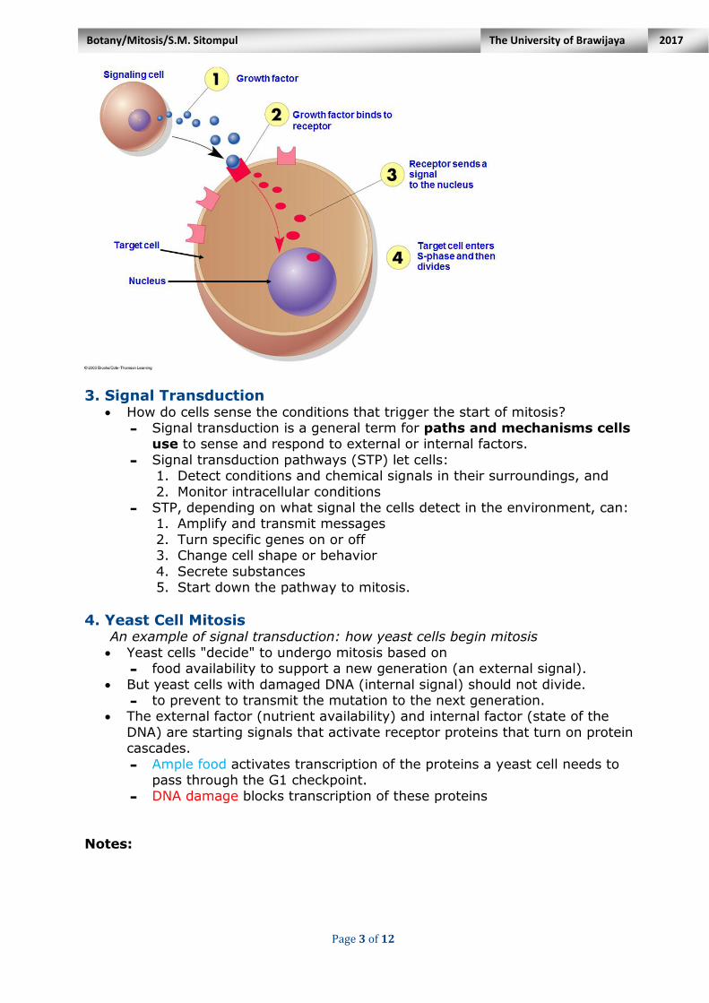
3. Signal Transduction How do cells sense the conditions that trigger the start of mitosis?
- Signal transduction is a general term for paths and mechanisms cells use to sense and respond to external or internal factors.
- Signal transduction pathways (STP) let cells: 1. Detect conditions and chemical signals in their surroundings, and
2. Monitor intracellular conditions - STP, depending on what signal the cells detect in the environment, can:
1. Amplify and transmit messages
2. Turn specific genes on or off 3. Change cell shape or behavior
4. Secrete substances 5. Start down the pathway to mitosis.
4. Yeast Cell Mitosis An example of signal transduction: how yeast cells begin mitosis
Yeast cells "decide" to undergo mitosis based on - food availability to support a new generation (an external signal).
But yeast cells with damaged DNA (internal signal) should not divide. - to prevent to transmit the mutation to the next generation.
The external factor (nutrient availability) and internal factor (state of the
DNA) are starting signals that activate receptor proteins that turn on protein cascades.
- Ample food activates transcription of the proteins a yeast cell needs to pass through the G1 checkpoint.
- DNA damage blocks transcription of these proteins
Notes:

Page 4 of 12
Botany/Mitosis/S.M. Sitompul 2017 The University of Brawijaya
2. CELL DIVISION
1. Basic Concept To ensure successful cell division, all the single-copy organelles must be
precisely replicated, maintained at specific locations, and faithfully
segregated to the daughter cells. Trypanosoma brucei (Fig. 4.1), a unicellular eukaryote and the causative
agent of human sleeping sickness, possesses multiple single-copy organelles.
Fig. 4.1 The cell structure of Trypanosoma brucei. (A) Cross-section of a
trypanosome cell visualized under the transmission EM. The flagellar axoneme,
paraflagellar rod (PFR), flagellum attachment zone (FAZ) filament, and the
subpellicular microtubule corset are indicated. (B) Schematic illustration of a
trypanosome cell showing the relative locations of its single-copied organelles. Zhou
et al. (2014)
- Trypanosomes undergo a closed mitosis in which the mitotic spindle is anchored on the nuclear envelope and connects the kinetochores made of
novel protein components. - Higher eukaryotes undergo an open mitosis, with the nuclear envelope
completely disassembled at the G2/M transition and then reassembled
upon the completion of mitosis. - During trypanosome cell cycle, basal body is the first organelle to be
duplicated, which constitutes the first cytoskeletal event of the cell cycle. - The most commonly used basal body marker, YL 1/2, is a monoclonal
antibody raised against the tyrosinated α–tubulin in the basal body, which only labels the mature basal body (Fig. 4.2).

Page 5 of 12
Botany/Mitosis/S.M. Sitompul 2017 The University of Brawijaya
Fig. 4.2 Duplication and segregation of basal body and flagellum during the cell cycle
in trypanosomes. Procyclic trypanosomes were immunostained with YL 1/2 antibody
and L8C4 antibody to label the basal body and flagellum, respectively, and then
counterstained. Zhou et al. (2014)
2. The signaling pathways Cell division involves two connected processes triggered at the end of G2
phase: - mitosis itself (segregation of the chromosomes, which duplicate in S
phase) and - cytokinesis (division of the cell, per se).
The signaling pathways that operate in G2 phase to control the onset of
mitosis, and those that operate during mitosis to control chromosome segregation and the initiation of cytokinesis, are somewhat different from
other cellular signaling pathways. Two major transitions are required for cell division:
- the G2/M transition and
- the metaphase/anaphase transition. These are regulated by the protein kinase cyclin-dependent kinase 1 (CDK1)
and the anaphase-promoting complex (APC, an E3 ubiquitin ligase), respectively.
Fig. 1. The major events of the cell
cycle. The major events of the cell
cycle are regulated by successive
waves of kinase and ubiquitin ligase
activity. G1-cyclin–CDK activity is
required to initiate the cell cycle
and activate B-type-cyclin–CDK
activity. Low levels of B-type-
cyclin–CDK activity are sufficient to
trigger S phase, but tyrosine
phosphorylation by Wee1 prevents
full activation, preventing
premature mitosis. Full CDK
activation triggers mitosis and
activates APC, which triggers
anaphase and feeds back to inactivate CDK activity. Inactivation of CDK allows exit
from mitosis and the reestablishment of interphase chromosome and nuclear
structure in G1 phase. Rhind & Russell (2012).

Page 6 of 12
Botany/Mitosis/S.M. Sitompul 2017 The University of Brawijaya
Full activation of CDK1, and thus entry into M phase, is restrained by Wee1
and related protein kinases (Fig. 2), which inhibit the activity of CDK1–cyclin complexes that accumulate during S and G2 phases
Wee1 family kinases resemble serine/threonine protein kinases, but actually phosphorylate CDK1 on a tyrosine residue (Y15), as well as the adjacent
threonine residue (T14) once it is bound to a cyclin. Once CDK1 is bound to a cyclin and phosphorylated by CAK and Wee1, it is
primed and ready to be activated through the dephosphorylation of T14 and
Y15. Cdc25, a dual-specificity protein phosphatase, catalyzes this
dephosphorylation reaction to bring about the G2/M transition.
Fig. 2. Signaling at the G2/M transition. The rate-limiting step for the transition from
G2 to mitosis is the dephosphorylation of CDK1 on Y15, and in some organisms T14.
This phosphorylation is catalyzed by the Wee1 family of dual-specificity kinases and
the phosphate is removed by Cdc25 phosphatases. Most of the many signaling
pathways that affect the G2/M transition regulate Wee1 or Cdc25. The DNA damage
and replication checkpoints inactivate Cdc25; the morphogenesis and nutritional
checkpoints activate Wee1. CDK1 regulates its own activation as part of a feedback
loop by directly phosphorylating Wee1 and Cdc25 or doing so indirectly through Plk1.
Rhind & Russell (2012).
To establish metaphase, CDK1 directly, and indirectly through a network of
kinases including Plk1 and Aurora A, phosphorylates substrates that trigger nuclear envelope breakdown, chromosome condensation, centrosome separation, and spindle assembly (Fig. 3).
In addition, in animal cells CDK1 activates the Greatwall kinase, which indirectly inactivates the CDK-antagonizing PP2A-B55 phosphatase via
phosphorylation of the small phosphatase inhibitors Arpp19 and endosulfine. The rate-limiting step for the transition from metaphase to anaphase is the
activation of the APC ubiquitin ligase.
Notes:

Page 7 of 12
Botany/Mitosis/S.M. Sitompul 2017 The University of Brawijaya
Fig. 3. Signaling at the M/A transition. The APC is activated by binding of the Cdc20
regulatory subunit (and later Cdh1) and by CDK1 phosphorylation. Active APC
targets securin for destruction, activating separase to release chromatid cohesion
and trigger anaphase. In the presence of unattached kinetochores, anaphase is
delayed by the mitotic checkpoint complex (MCC), a soluble inhibitor of the APC
produced at unattached kinetochores. The APC also feeds back to inactivate CDK1 by
targeting cyclin B for degradation and activates phosphatases (CDC14 in yeasts and
PP1 and PP2A-B55 in animals) that oppose CDK activity, resetting the cell cycle to a
CDK-free G1 state.
3. Genetic Material 1. During cell division, the genetic material DNA needs to be copied and
divided between the two new cells 2. DNA in cells is divided into long chains called chromosomes (“volumes” of
DNA). 3. Normally chromosomes are spread out, not identifiable, in a form called
chromatin 4. During mitosis, chromosomes condense and then fold up
5. Chromosome DNA is wrapped around proteins called histones to organize it 6. Nucleosome: unit of DNA wrapped around histones. 7. The replicated
chromosomes stay together and are called
sister chromatids. 8. Sister chromatids are
attached at the
centromere by proteins called cohesins.
9. The other side of the centromeres contain other proteins called kinetochore.

Page 8 of 12
Botany/Mitosis/S.M. Sitompul 2017 The University of Brawijaya
4. DNA Replication The DNA molecule is unlocked
and unraveled by an enzyme
called DNA helicase, which breaks the hydrogen bonds
between the nitrogenous bases while topoisomerase keeps the DNA molecule stable.
- Without topoisomerase, the DNA molecule that was not
unwound would become strained and could be damaged.
A replication fork forms as DNA helicase moves up the DNA
molecule, leaving behind two single strands of DNA.
These single strands serve as a template to build a complementary strand of
DNA using complementary base pairing.
- One strand is known as the leading strand, and replicated continuously in the 3' to 5' direction.
- The other strand is known as the lagging strand and formed slowly as a series of small segments (Okazaki fragments) that are later stitched together by DNA ligase.
https://ka-perseus-images.s3.amazonaws.com/319e71fef87236a01bb797379e3e15745790ff19.png

Page 9 of 12
Botany/Mitosis/S.M. Sitompul 2017 The University of Brawijaya
5. Spindle apparatus Spindle apparatus consists of two distinct sets
of microtubules
- Each set extends from one of the cell poles
- Two sets overlap at spindle equator This helps to move chromosomes during
mitosis
In both plant and animal cells, spindle fibers originate from centrosomes; in animal cells,
centrosomes are centrioles.
3. MITOSIS AND CYTOKINESIS
1. Cell Cycle Progress through the cell cycle is carefully
regulated in unicellular and multicellular organisms, and can be devided into;
1. Interphase - G phase
- S phase 2. Mitosis (M Phase)
- Nuclear Division
3. Cytokinesis (C phase) - Cytoplasmic Division
Mitosis can be subdivided into six distinct
phases:
(1) prophase, in which the spindle begins to assemble in the cytoplasm and chromosomes begin to condense in the
nucleus; (2) prometaphase, in which
the nuclear envelope breaks down and chromosomes attach to
the spindle; (3) metaphase, in which
chromosomes align at the spindle midzone;
(4) anaphase A, in which
chromosomes move to the centrosomes, which
form the spindle poles; (5) anaphase B, in which the
spindle elongates; and
(6) telophase, in which the nuclear envelope reforms
around the new daughter nuclei.
Page 10 of 12
Botany/Mitosis/S.M. Sitompul 2017 The University of Brawijaya
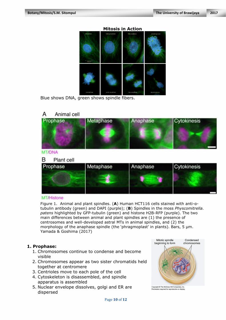
Mitosis in Action
Blue shows DNA, green shows spindle fibers.
Figure 1. Animal and plant spindles. (A) Human HCT116 cells stained with anti-α-
tubulin antibody (green) and DAPI (purple); (B) Spindles in the moss Physcomitrella.
patens highlighted by GFP-tubulin (green) and histone H2B-RFP (purple). The two
main differences between animal and plant spindles are (1) the presence of
centrosomes and well-developed astral MTs in animal spindles, and (2) the
morphology of the anaphase spindle (the ‘phragmoplast’ in plants). Bars, 5 µm.
Yamada & Goshima (2017)
1. Prophase:
1. Chromosomes continue to condense and become visible
2. Chromosomes appear as two sister chromatids held
together at centromere 3. Centrioles move to each pole of the cell
4. Cytoskeleton is disassembled, and spindle apparatus is assembled
5. Nuclear envelope dissolves, golgi and ER are
dispersed

Page 11 of 12
Botany/Mitosis/S.M. Sitompul 2017 The University of Brawijaya
2. Prometaphase:
1. Chromosomes become attached to the microtubules or spindle apparatus at the
kinetochores 2. Each chromosome is oriented such that the
kinetochores of sister chromatids are attached to microtubules from opposite poles
3. Microtubules begin to pull each chromosome
toward the center (the equator) of the cell
3. Metaphase: 1. Microtubules pull the chromosomes to align them at
the center of the cell
2. Metaphase plate: imaginary plane through the center of the cell where the chromosomes align
Kidney cell in mitosis
(metaphase) showing actin by
Dr. Paul Andrews at 100x objective. http://fyeahmedlab.tumblr.com/post/27469407377/fyeahuniverse-kidney-cell-in-mitosis
4. Anaphase:
1. Removal of cohesin proteins causes the
centromeres to separate 2. Microtubules pull sister chromatids toward the
poles 3. In anaphase A the kinetochores are pulled apart 4. In anaphase B the poles move apart
5. Telophase:
1. Spindle apparatus disassembles 2. Nuclear envelope forms around each set of sister
chromatids
3. Chromosomes begin to uncoil 4. Nucleolus reappears in each new nucleus
Onion root tip cells undergoing mitosis (http://tinyurl.com/c42amb3)
Kidney cell in metaphase)
Metaphase plate
Microtubule fibers
Chromosomes
Metaphase plate
Spindle pole

Page 12 of 12
Botany/Mitosis/S.M. Sitompul 2017 The University of Brawijaya
6. Cytokinesis
1. Cytokinesis is division of the cytoplasm to produce two daughter cells, usually begun during
telophase or occurs between late anaphase and end of
telophase. 2. Two mechanisms: (i) Cleavage
(animals), and (ii) Cell plate
formation (plants). Plants: Cell Plate Formation
Cytokinesis-division of the
cytoplasm - In animal cells, the membrane
pinches closed at a point called cleavage furrow.
- In plant cells, vacuoles join
together and form a cell plate. - Result-2 identical daughter cells
with identical copies of genetic material-DNA
Mitosis/Cytokinesis outcome - 1 parent cell 2 identical
daughter cells - Chromosome number remains the same from one
generation to the next Mitosis: plant vs. animal cells
Plant cell Animal Cell
Centrioles Absent Present
Cytokinesis Cell plate formation Cleavage furrow