bones of the upper limb - cnx.org · the distal end of the humerus has two articulation areas,...
TRANSCRIPT

OpenStax-CNX module: m46368 1
Bones of the Upper Limb*
OpenStax
This work is produced by OpenStax-CNX and licensed under the
Creative Commons Attribution License 3.0�
Abstract
By the end of this section, you will be able to:
• Identify the divisions of the upper limb and describe the bones in each region• List the bones and bony landmarks that articulate at each joint of the upper limb
The upper limb is divided into three regions. These consist of the arm, located between the shoulderand elbow joints; the forearm, which is between the elbow and wrist joints; and the hand, which is locateddistal to the wrist. There are 30 bones in each upper limb (see ). The humerus is the single bone of theupper arm, and the ulna (medially) and the radius (laterally) are the paired bones of the forearm. Thebase of the hand contains eight bones, each called a carpal bone, and the palm of the hand is formed by�ve bones, each called a metacarpal bone. The �ngers and thumb contain a total of 14 bones, each ofwhich is a phalanx bone of the hand.
1 Humerus
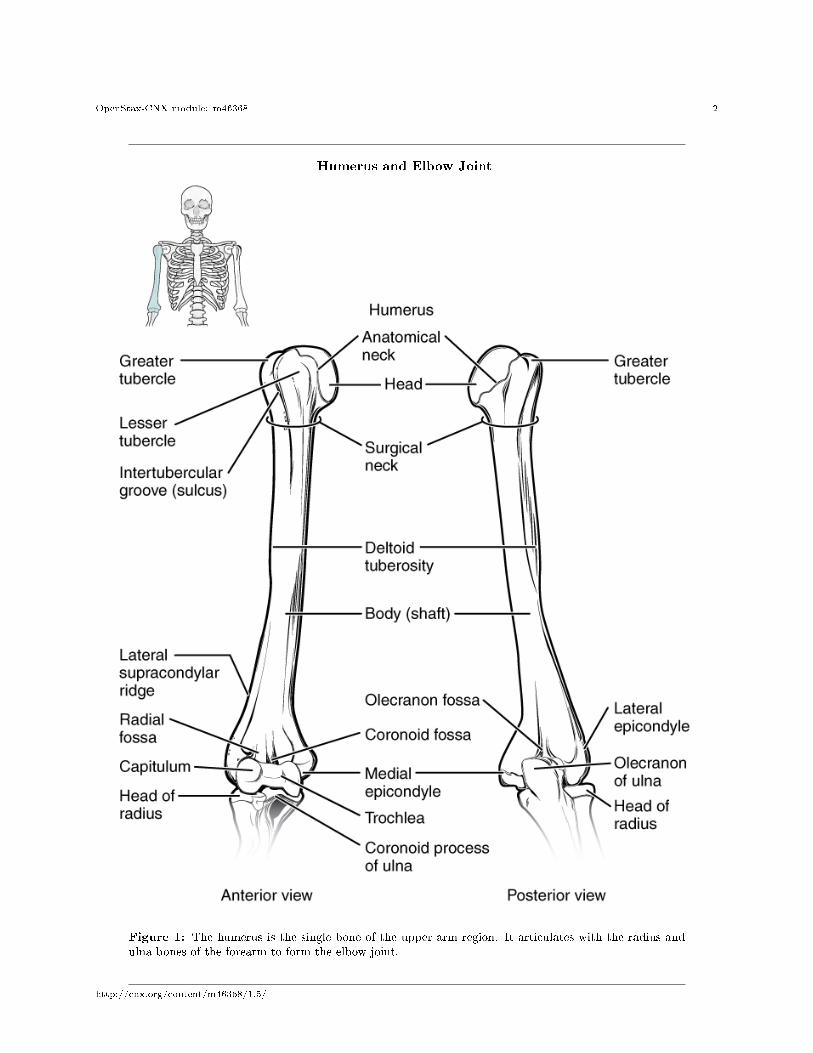
The humerus is the single bone of the upper arm region (Figure 1 (Humerus and Elbow Joint )). At itsproximal end is the head of the humerus. This is the large, round, smooth region that faces medially.The head articulates with the glenoid cavity of the scapula to form the glenohumeral (shoulder) joint. Themargin of the smooth area of the head is the anatomical neck of the humerus. Located on the lateral side ofthe proximal humerus is an expanded bony area called the greater tubercle. The smaller lesser tubercleof the humerus is found on the anterior aspect of the humerus. Both the greater and lesser tubercles serveas attachment sites for muscles that act across the shoulder joint. Passing between the greater and lessertubercles is the narrow intertubercular groove (sulcus), which is also known as the bicipital groovebecause it provides passage for a tendon of the biceps brachii muscle. The surgical neck is located atthe base of the expanded, proximal end of the humerus, where it joins the narrow shaft of the humerus.The surgical neck is a common site of arm fractures. The deltoid tuberosity is a roughened, V-shapedregion located on the lateral side in the middle of the humerus shaft. As its name indicates, it is the site ofattachment for the deltoid muscle.
*Version 1.5: Oct 3, 2013 8:15 pm +0000�http://creativecommons.org/licenses/by/3.0/
http://cnx.org/content/m46368/1.5/
OpenStax-CNX module: m46368 2
Humerus and Elbow Joint
Figure 1: The humerus is the single bone of the upper arm region. It articulates with the radius andulna bones of the forearm to form the elbow joint.
http://cnx.org/content/m46368/1.5/

OpenStax-CNX module: m46368 3
Distally, the humerus becomes �attened. The prominent bony projection on the medial side is themedialepicondyle of the humerus. The much smaller lateral epicondyle of the humerus is found on thelateral side of the distal humerus. The roughened ridge of bone above the lateral epicondyle is the lateralsupracondylar ridge. All of these areas are attachment points for muscles that act on the forearm, wrist,and hand. The powerful grasping muscles of the anterior forearm arise from the medial epicondyle, whichis thus larger and more robust than the lateral epicondyle that gives rise to the weaker posterior forearmmuscles.
The distal end of the humerus has two articulation areas, which join the ulna and radius bones of theforearm to form the elbow joint. The more medial of these areas is the trochlea, a spindle- or pulley-shapedregion (trochlea = �pulley�), which articulates with the ulna bone. Immediately lateral to the trochlea isthe capitulum (�small head�), a knob-like structure located on the anterior surface of the distal humerus.The capitulum articulates with the radius bone of the forearm. Just above these bony areas are two smalldepressions. These spaces accommodate the forearm bones when the elbow is fully bent (�exed). Superior tothe trochlea is the coronoid fossa, which receives the coronoid process of the ulna, and above the capitulumis the radial fossa, which receives the head of the radius when the elbow is �exed. Similarly, the posteriorhumerus has the olecranon fossa, a larger depression that receives the olecranon process of the ulna whenthe forearm is fully extended.
2 Ulna
The ulna is the medial bone of the forearm. It runs parallel to the radius, which is the lateral bone of theforearm (Figure 2 (Ulna and Radius )). The proximal end of the ulna resembles a crescent wrench withits large, C-shaped trochlear notch. This region articulates with the trochlea of the humerus as part ofthe elbow joint. The inferior margin of the trochlear notch is formed by a prominent lip of bone called thecoronoid process of the ulna. Just below this on the anterior ulna is a roughened area called the ulnartuberosity. To the lateral side and slightly inferior to the trochlear notch is a small, smooth area called theradial notch of the ulna. This area is the site of articulation between the proximal radius and the ulna,forming the proximal radioulnar joint. The posterior and superior portions of the proximal ulna makeup the olecranon process, which forms the bony tip of the elbow.
http://cnx.org/content/m46368/1.5/

OpenStax-CNX module: m46368 4
Ulna and Radius
Figure 2: The ulna is located on the medial side of the forearm, and the radius is on the lateral side.These bones are attached to each other by an interosseous membrane.
http://cnx.org/content/m46368/1.5/

OpenStax-CNX module: m46368 5
More distal is the shaft of the ulna. The lateral side of the shaft forms a ridge called the interosseousborder of the ulna. This is the line of attachment for the interosseous membrane of the forearm, asheet of dense connective tissue that unites the ulna and radius bones. The small, rounded area that formsthe distal end is the head of the ulna. Projecting from the posterior side of the ulnar head is the styloidprocess of the ulna, a short bony projection. This serves as an attachment point for a connective tissuestructure that unites the distal ends of the ulna and radius.
In the anatomical position, with the elbow fully extended and the palms facing forward, the arm andforearm do not form a straight line. Instead, the forearm deviates laterally by 5�15 degrees from the line ofthe arm. This deviation is called the carrying angle. It allows the forearm and hand to swing freely or tocarry an object without hitting the hip. The carrying angle is larger in females to accommodate their widerpelvis.
3 Radius
The radius runs parallel to the ulna, on the lateral (thumb) side of the forearm (see Figure 2 (Ulna andRadius )). The head of the radius is a disc-shaped structure that forms the proximal end. The smalldepression on the surface of the head articulates with the capitulum of the humerus as part of the elbowjoint, whereas the smooth, outer margin of the head articulates with the radial notch of the ulna at theproximal radioulnar joint. The neck of the radius is the narrowed region immediately below the expandedhead. Inferior to this point on the medial side is the radial tuberosity, an oval-shaped, bony protuberancethat serves as a muscle attachment point. The shaft of the radius is slightly curved and has a small ridgealong its medial side. This ridge forms the interosseous border of the radius, which, like the similarborder of the ulna, is the line of attachment for the interosseous membrane that unites the two forearmbones. The distal end of the radius has a smooth surface for articulation with two carpal bones to formthe radiocarpal joint or wrist joint (Figure 3 (Bones of the Wrist and Hand ) and Figure 4 (Bones of theHand )). On the medial side of the distal radius is the ulnar notch of the radius. This shallow depressionarticulates with the head of the ulna, which together form the distal radioulnar joint. The lateral end ofthe radius has a pointed projection called the styloid process of the radius. This provides attachmentfor ligaments that support the lateral side of the wrist joint. Compared to the styloid process of the ulna,the styloid process of the radius projects more distally, thereby limiting the range of movement for lateraldeviations of the hand at the wrist joint.
http://cnx.org/content/m46368/1.5/

OpenStax-CNX module: m46368 6
:
Watch this video1 to see how fractures of the distal radius bone can a�ect the wrist joint. Explainthe problems that may occur if a fracture of the distal radius involves the joint surface of theradiocarpal joint of the wrist.
4 Carpal Bones
The wrist and base of the hand are formed by a series of eight small carpal bones (see Figure 3 (Bonesof the Wrist and Hand )). The carpal bones are arranged in two rows, forming a proximal row of fourcarpal bones and a distal row of four carpal bones. The bones in the proximal row, running from the lateral(thumb) side to the medial side, are the scaphoid (�boat-shaped�), lunate (�moon-shaped�), triquetrum(�three-cornered�), and pisiform (�pea-shaped�) bones. The small, rounded pisiform bone articulates withthe anterior surface of the triquetrum bone. The pisiform thus projects anteriorly, where it forms thebony bump that can be felt at the medial base of your hand. The distal bones (lateral to medial) are the
1http://openstaxcollege.org/l/fractures
http://cnx.org/content/m46368/1.5/

OpenStax-CNX module: m46368 7
trapezium (�table�), trapezoid (�resembles a table�), capitate (�head-shaped�), and hamate (�hookedbone�) bones. The hamate bone is characterized by a prominent bony extension on its anterior side calledthe hook of the hamate bone.
A helpful mnemonic for remembering the arrangement of the carpal bones is �So Long To Pinky, HereComes The Thumb.� This mnemonic starts on the lateral side and names the proximal bones from lateral tomedial (scaphoid, lunate, triquetrum, pisiform), then makes a U-turn to name the distal bones from medialto lateral (hamate, capitate, trapezoid, trapezium). Thus, it starts and �nishes on the lateral side.
Bones of the Wrist and Hand
Figure 3: The eight carpal bones form the base of the hand. These are arranged into proximal anddistal rows of four bones each. The metacarpal bones form the palm of the hand. The thumb and �ngersconsist of the phalanx bones.
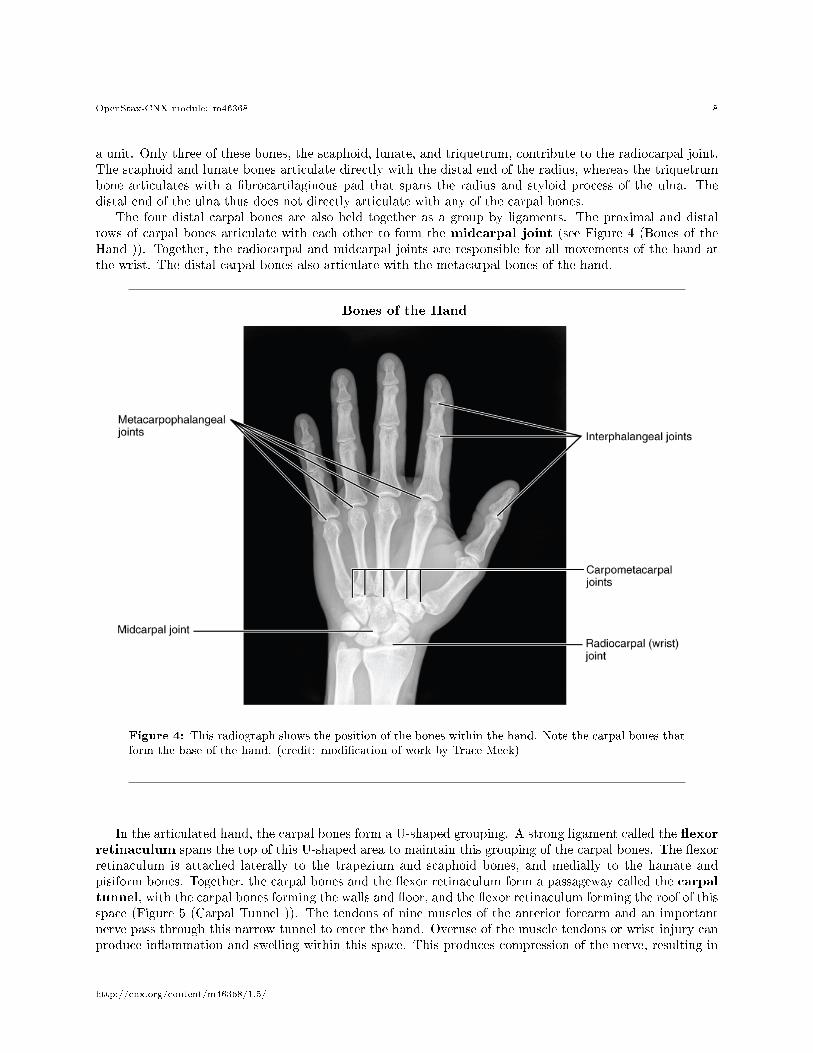
The carpal bones form the base of the hand. This can be seen in the radiograph (X-ray image) of thehand that shows the relationships of the hand bones to the skin creases of the hand (see Figure 4 (Bones ofthe Hand )). Within the carpal bones, the four proximal bones are united to each other by ligaments to form
http://cnx.org/content/m46368/1.5/
OpenStax-CNX module: m46368 8
a unit. Only three of these bones, the scaphoid, lunate, and triquetrum, contribute to the radiocarpal joint.The scaphoid and lunate bones articulate directly with the distal end of the radius, whereas the triquetrumbone articulates with a �brocartilaginous pad that spans the radius and styloid process of the ulna. Thedistal end of the ulna thus does not directly articulate with any of the carpal bones.
The four distal carpal bones are also held together as a group by ligaments. The proximal and distalrows of carpal bones articulate with each other to form the midcarpal joint (see Figure 4 (Bones of theHand )). Together, the radiocarpal and midcarpal joints are responsible for all movements of the hand atthe wrist. The distal carpal bones also articulate with the metacarpal bones of the hand.
Bones of the Hand
Figure 4: This radiograph shows the position of the bones within the hand. Note the carpal bones thatform the base of the hand. (credit: modi�cation of work by Trace Meek)
In the articulated hand, the carpal bones form a U-shaped grouping. A strong ligament called the �exorretinaculum spans the top of this U-shaped area to maintain this grouping of the carpal bones. The �exorretinaculum is attached laterally to the trapezium and scaphoid bones, and medially to the hamate andpisiform bones. Together, the carpal bones and the �exor retinaculum form a passageway called the carpaltunnel, with the carpal bones forming the walls and �oor, and the �exor retinaculum forming the roof of thisspace (Figure 5 (Carpal Tunnel )). The tendons of nine muscles of the anterior forearm and an importantnerve pass through this narrow tunnel to enter the hand. Overuse of the muscle tendons or wrist injury canproduce in�ammation and swelling within this space. This produces compression of the nerve, resulting in
http://cnx.org/content/m46368/1.5/

OpenStax-CNX module: m46368 9
carpal tunnel syndrome, which is characterized by pain or numbness, and muscle weakness in those areas ofthe hand supplied by this nerve.
Carpal Tunnel
Figure 5: The carpal tunnel is the passageway by which nine muscle tendons and a major nerve enterthe hand from the anterior forearm. The walls and �oor of the carpal tunnel are formed by the U-shapedgrouping of the carpal bones, and the roof is formed by the �exor retinaculum, a strong ligament thatanteriorly unites the bones.
5 Metacarpal Bones
The palm of the hand contains �ve elongated metacarpal bones. These bones lie between the carpal bones ofthe wrist and the bones of the �ngers and thumb (see Figure 3 (Bones of the Wrist and Hand )). The proximal
http://cnx.org/content/m46368/1.5/

OpenStax-CNX module: m46368 10
end of each metacarpal bone articulates with one of the distal carpal bones. Each of these articulations is acarpometacarpal joint (see Figure 4 (Bones of the Hand )). The expanded distal end of each metacarpalbone articulates at the metacarpophalangeal joint with the proximal phalanx bone of the thumb or oneof the �ngers. The distal end also forms the knuckles of the hand, at the base of the �ngers. The metacarpalbones are numbered 1�5, beginning at the thumb.
The �rst metacarpal bone, at the base of the thumb, is separated from the other metacarpal bones. Thisallows it a freedom of motion that is independent of the other metacarpal bones, which is very important forthumb mobility. The remaining metacarpal bones are united together to form the palm of the hand. Thesecond and third metacarpal bones are �rmly anchored in place and are immobile. However, the fourth and�fth metacarpal bones have limited anterior-posterior mobility, a motion that is greater for the �fth bone.This mobility is important during power gripping with the hand (Figure 6 (Hand During Gripping )). Theanterior movement of these bones, particularly the �fth metacarpal bone, increases the strength of contactfor the medial hand during gripping actions.
Hand During Gripping
Figure 6: During tight gripping�compare (b) to (a)�the fourth and, particularly, the �fth metatarsalbones are pulled anteriorly. This increases the contact between the object and the medial side of thehand, thus improving the �rmness of the grip.
6 Phalanx Bones
The �ngers and thumb contain 14 bones, each of which is called a phalanx bone (plural = phalanges), namedafter the ancient Greek phalanx (a rectangular block of soldiers). The thumb (pollex) is digit number 1and has two phalanges, a proximal phalanx, and a distal phalanx bone (see Figure 3 (Bones of the Wristand Hand )). Digits 2 (index �nger) through 5 (little �nger) have three phalanges each, called the proximal,middle, and distal phalanx bones. An interphalangeal joint is one of the articulations between adjacentphalanges of the digits (see Figure 4 (Bones of the Hand )).
http://cnx.org/content/m46368/1.5/

OpenStax-CNX module: m46368 11
:
Visit this site2 to explore the bones and joints of the hand. What are the three arches of the hand,and what is the importance of these during the gripping of an object?
: Appendicular System: Fractures of Upper Limb Bones
Due to our constant use of the hands and the rest of our upper limbs, an injury to any of theseareas will cause a signi�cant loss of functional ability. Many fractures result from a hard fall ontoan outstretched hand. The resulting transmission of force up the limb may result in a fracture ofthe humerus, radius, or scaphoid bones. These injuries are especially common in elderly peoplewhose bones are weakened due to osteoporosis.
Falls onto the hand or elbow, or direct blows to the arm, can result in fractures of the humerus(Figure 7 (Fractures of the Humerus and Radius )). Following a fall, fractures at the surgical neck,the region at which the expanded proximal end of the humerus joins with the shaft, can result in an
2http://openstaxcollege.org/l/handbone
http://cnx.org/content/m46368/1.5/

OpenStax-CNX module: m46368 12
impacted fracture, in which the distal portion of the humerus is driven into the proximal portion.Falls or blows to the arm can also produce transverse or spiral fractures of the humeral shaft.
In children, a fall onto the tip of the elbow frequently results in a distal humerus fracture. In these,the olecranon of the ulna is driven upward, resulting in a fracture across the distal humerus, aboveboth epicondyles (supracondylar fracture), or a fracture between the epicondyles, thus separatingone or both of the epicondyles from the body of the humerus (intercondylar fracture). With theseinjuries, the immediate concern is possible compression of the artery to the forearm due to swellingof the surrounding tissues. If compression occurs, the resulting ischemia (lack of oxygen) due toreduced blood �ow can quickly produce irreparable damage to the forearm muscles. In addition,four major nerves for shoulder and upper limb muscles are closely associated with di�erent regionsof the humerus, and thus, humeral fractures may also damage these nerves.
Another frequent injury following a fall onto an outstretched hand is a Colles fracture (�col-lees�) ofthe distal radius (see Figure 7 (Fractures of the Humerus and Radius )). This involves a completetransverse fracture across the distal radius that drives the separated distal fragment of the radiusposteriorly and superiorly. This injury results in a characteristic �dinner fork� bend of the forearmjust above the wrist due to the posterior displacement of the hand. This is the most frequent forearmfracture and is a common injury in persons over the age of 50, particularly in older women withosteoporosis. It also commonly occurs following a high-speed fall onto the hand during activitiessuch as snowboarding or skating.
The most commonly fractured carpal bone is the scaphoid, often resulting from a fall onto the hand.Deep pain at the lateral wrist may yield an initial diagnosis of a wrist sprain, but a radiographtaken several weeks after the injury, after tissue swelling has subsided, will reveal the fracture. Dueto the poor blood supply to the scaphoid bone, healing will be slow and there is the danger of bonenecrosis and subsequent degenerative joint disease of the wrist.
http://cnx.org/content/m46368/1.5/

OpenStax-CNX module: m46368 13
Fractures of the Humerus and Radius
Figure 7: Falls or direct blows can result in fractures of the surgical neck or shaft of the humerus.Falls onto the elbow can fracture the distal humerus. A Colles fracture of the distal radius is the mostcommon forearm fracture.
http://cnx.org/content/m46368/1.5/

OpenStax-CNX module: m46368 14
:
Watch this video3 to learn about a Colles fracture, a break of the distal radius, usually caused byfalling onto an outstretched hand. When would surgery be required and how would the fracture berepaired in this case?
7 Chapter Review
Each upper limb is divided into three regions and contains a total of 30 bones. The upper arm is the regionlocated between the shoulder and elbow joints. This area contains the humerus. The proximal humerusconsists of the head, which articulates with the scapula at the glenohumeral joint, the greater and lessertubercles separated by the intertubercular (bicipital) groove, and the anatomical and surgical necks. Thehumeral shaft has the roughened area of the deltoid tuberosity on its lateral side. The distal humerus is�attened, forming a lateral supracondylar ridge that terminates at the small lateral epicondyle. The medialside of the distal humerus has the large, medial epicondyle. The articulating surfaces of the distal humerus
3http://openstaxcollege.org/l/colles
http://cnx.org/content/m46368/1.5/

OpenStax-CNX module: m46368 15
consist of the trochlea medially and the capitulum laterally. Depressions on the humerus that accommodatethe forearm bones during bending (�exing) and straightening (extending) of the elbow include the coronoidfossa, the radial fossa, and the olecranon fossa.
The forearm is the region of the upper limb located between the elbow and wrist joints. This regioncontains two bones, the ulna medially and the radius on the lateral (thumb) side. The elbow joint isformed by the articulation between the trochlea of the humerus and the trochlear notch of the ulna, plusthe articulation between the capitulum of the humerus and the head of the radius. The proximal radioulnarjoint is the articulation between the head of the radius and the radial notch of the ulna. The proximal ulnaalso has the olecranon process, forming an expanded posterior region, and the coronoid process and ulnartuberosity on its anterior aspect. On the proximal radius, the narrowed region below the head is the neck;distal to this is the radial tuberosity. The shaft portions of both the ulna and radius have an interosseousborder, whereas the distal ends of each bone have a pointed styloid process. The distal radioulnar joint isfound between the head of the ulna and the ulnar notch of the radius. The distal end of the radius articulateswith the proximal carpal bones, but the ulna does not.
The base of the hand is formed by eight carpal bones. The carpal bones are united into two rows ofbones. The proximal row contains (from lateral to medial) the scaphoid, lunate, triquetrum, and pisiformbones. The scaphoid, lunate, and triquetrum bones contribute to the formation of the radiocarpal joint. Thedistal row of carpal bones contains (from medial to lateral) the hamate, capitate, trapezoid, and trapeziumbones (�So Long To Pinky, Here Comes The Thumb�). The anterior hamate has a prominent bony hook.The proximal and distal carpal rows articulate with each other at the midcarpal joint. The carpal bones,together with the �exor retinaculum, also form the carpal tunnel of the wrist.
The �ve metacarpal bones form the palm of the hand. The metacarpal bones are numbered 1�5, startingwith the thumb side. The �rst metacarpal bone is freely mobile, but the other bones are united as a group.The digits are also numbered 1�5, with the thumb being number 1. The �ngers and thumb contain a total of14 phalanges (phalanx bones). The thumb contains a proximal and a distal phalanx, whereas the remainingdigits each contain proximal, middle, and distal phalanges.
8 Interactive Link Questions
Exercise 1 (Solution on p. 17.)
Watch this video4 to see how fractures of the distal radius bone can a�ect the wrist joint. Explainthe problems that may occur if a fracture of the distal radius involves the joint surface of theradiocarpal joint of the wrist.
Exercise 2 (Solution on p. 17.)
Visit this site5 to explore the bones and joints of the hand. What are the three arches of the hand,and what is the importance of these during the gripping of an object?
Exercise 3 (Solution on p. 17.)
Watch this video6 to learn about a Colles fracture, a break of the distal radius, usually caused byfalling onto an outstretched hand. When would surgery be required and how would the fracture berepaired in this case?
9 Review Questions
Exercise 4 (Solution on p. 17.)
How many bones are there in the upper limbs combined?
a. 20
4http://openstaxcollege.org/l/fractures5http://openstaxcollege.org/l/handbone6http://openstaxcollege.org/l/colles
http://cnx.org/content/m46368/1.5/
OpenStax-CNX module: m46368 16
b. 30c. 40d. 60
Exercise 5 (Solution on p. 17.)
Which bony landmark is located on the lateral side of the proximal humerus?
a. greater tubercleb. trochleac. lateral epicondyled. lesser tubercle
Exercise 6 (Solution on p. 17.)
Which region of the humerus articulates with the radius as part of the elbow joint?
a. trochleab. styloid processc. capitulumd. olecranon process
Exercise 7 (Solution on p. 17.)
Which is the lateral-most carpal bone of the proximal row?
a. trapeziumb. hamatec. pisiformd. scaphoid
Exercise 8 (Solution on p. 17.)
The radius bone ________.
a. is found on the medial side of the forearmb. has a head that articulates with the radial notch of the ulnac. does not articulate with any of the carpal bonesd. has the radial tuberosity located near its distal end
10 Critical Thinking Questions
Exercise 9 (Solution on p. 17.)
Your friend runs out of gas and you have to help push his car. Discuss the sequence of bones andjoints that convey the forces passing from your hand, through your upper limb and your pectoralgirdle, and to your axial skeleton.
Exercise 10 (Solution on p. 17.)
Name the bones in the wrist and hand, and describe or sketch out their locations and articulations.
http://cnx.org/content/m46368/1.5/
OpenStax-CNX module: m46368 17
Solutions to Exercises in this Module
to Exercise (p. 15)A fracture through the joint surface of the distal radius may make the articulating surface of the radiusrough or jagged. This can then cause painful movements involving this joint and the early development ofarthritis. Surgery can return the joint surface to its original smoothness, thus allowing for the return ofnormal function.to Exercise (p. 15)The hand has a proximal transverse arch, a distal transverse arch, and a longitudinal arch. These allow thehand to conform to objects being held. These arches maximize the amount of surface contact between thehand and object, which enhances stability and increases sensory input.to Exercise (p. 15)Surgery may be required if the fracture is unstable, meaning that the broken ends of the radius won't stay inplace to allow for proper healing. In this case, metal plates and screws can be used to stabilize the fracturedbone.to Exercise (p. 15)Dto Exercise (p. 16)Ato Exercise (p. 16)Cto Exercise (p. 16)Dto Exercise (p. 16)Bto Exercise (p. 16)As you push against the car, forces will pass from the metacarpal bones of your hand into the carpalbones at the base of your hand. Forces will then pass through the midcarpal and radiocarpal joints into theradius and ulna bones of the forearm. These will pass the force through the elbow joint into the humerusof the arm, and then through the glenohumeral joint into the scapula. The force will travel through theacromioclavicular joint into the clavicle, and then through the sternoclavicular joint into the sternum, whichis part of the axial skeleton.to Exercise (p. 16)The base of the hand is formed by the eight carpal bones arranged in two rows (distal and proximal)of four bones each. The proximal row contains (from lateral to medial) the scaphoid, lunate, triquetrum,and pisiform bones. The distal row contains (from medial to lateral) the hamate, capitate, trapezoid, andtrapezium bones. (Use the mnemonic �So Long To Pinky, Here Comes The Thumb� to remember thissequence). The rows of the proximal and distal carpal bones articulate with each other at the midcarpaljoint. The palm of the hand contains the �ve metacarpal bones, which are numbered 1�5 starting on thethumb side. The proximal ends of the metacarpal bones articulate with the distal row of the carpal bones.The distal ends of the metacarpal bones articulate with the proximal phalanx bones of the thumb and�ngers. The thumb (digit 1) has both a proximal and distal phalanx bone. The �ngers (digits 2�5) allcontain proximal, middle, and distal phalanges.
Glossary
De�nition 7: anatomical neckline on the humerus located around the outside margin of the humeral head
http://cnx.org/content/m46368/1.5/

OpenStax-CNX module: m46368 18
De�nition 7: armregion of the upper limb located between the shoulder and elbow joints; contains the humerus bone
De�nition 7: bicipital grooveintertubercular groove; narrow groove located between the greater and lesser tubercles of thehumerus
De�nition 7: capitatefrom the lateral side, the third of the four distal carpal bones; articulates with the scaphoidand lunate proximally, the trapezoid laterally, the hamate medially, and primarily with the thirdmetacarpal distally
De�nition 7: capitulumknob-like bony structure located anteriorly on the lateral, distal end of the humerus
De�nition 7: carpal boneone of the eight small bones that form the wrist and base of the hand; these are grouped as aproximal row consisting of (from lateral to medial) the scaphoid, lunate, triquetrum, and pisiformbones, and a distal row containing (from lateral to medial) the trapezium, trapezoid, capitate, andhamate bones
De�nition 7: carpal tunnelpassageway between the anterior forearm and hand formed by the carpal bones and �exor retinac-ulum
De�nition 7: carpometacarpal jointarticulation between one of the carpal bones in the distal row and a metacarpal bone of the hand
De�nition 7: coronoid fossadepression on the anterior surface of the humerus above the trochlea; this space receives the coronoidprocess of the ulna when the elbow is maximally �exed
De�nition 7: coronoid process of the ulnaprojecting bony lip located on the anterior, proximal ulna; forms the inferior margin of the trochlearnotch
De�nition 7: deltoid tuberosityroughened, V-shaped region located laterally on the mid-shaft of the humerus
De�nition 7: distal radioulnar jointarticulation between the head of the ulna and the ulnar notch of the radius
De�nition 7: elbow jointjoint located between the upper arm and forearm regions of the upper limb; formed by the articu-lations between the trochlea of the humerus and the trochlear notch of the ulna, and the capitulumof the humerus and the head of the radius
De�nition 7: �exor retinaculumstrong band of connective tissue at the anterior wrist that spans the top of the U-shaped groupingof the carpal bones to form the roof of the carpal tunnel
De�nition 7: forearmregion of the upper limb located between the elbow and wrist joints; contains the radius and ulnabones
De�nition 7: greater tubercleenlarged prominence located on the lateral side of the proximal humerus
De�nition 7: hamatefrom the lateral side, the fourth of the four distal carpal bones; articulates with the lunate andtriquetrum proximally, the fourth and �fth metacarpals distally, and the capitate laterally
http://cnx.org/content/m46368/1.5/

OpenStax-CNX module: m46368 19
De�nition 7: handregion of the upper limb distal to the wrist joint
De�nition 7: head of the humerussmooth, rounded region on the medial side of the proximal humerus; articulates with the glenoidfossa of the scapula to form the glenohumeral (shoulder) joint
De�nition 7: head of the radiusdisc-shaped structure that forms the proximal end of the radius; articulates with the capitulum ofthe humerus as part of the elbow joint, and with the radial notch of the ulna as part of the proximalradioulnar joint
De�nition 7: head of the ulnasmall, rounded distal end of the ulna; articulates with the ulnar notch of the distal radius, formingthe distal radioulnar joint
De�nition 7: hook of the hamate bonebony extension located on the anterior side of the hamate carpal bone
De�nition 7: humerussingle bone of the upper arm
De�nition 7: interosseous border of the radiusnarrow ridge located on the medial side of the radial shaft; for attachment of the interosseousmembrane between the ulna and radius bones
De�nition 7: interosseous border of the ulnanarrow ridge located on the lateral side of the ulnar shaft; for attachment of the interosseousmembrane between the ulna and radius
De�nition 7: interosseous membrane of the forearmsheet of dense connective tissue that unites the radius and ulna bones
De�nition 7: interphalangeal jointarticulation between adjacent phalanx bones of the hand or foot digits
De�nition 7: intertubercular groove (sulcus)bicipital groove; narrow groove located between the greater and lesser tubercles of the humerus
De�nition 7: lateral epicondyle of the humerussmall projection located on the lateral side of the distal humerus
De�nition 7: lateral supracondylar ridgenarrow, bony ridge located along the lateral side of the distal humerus, superior to the lateralepicondyle
De�nition 7: lesser tuberclesmall, bony prominence located on anterior side of the proximal humerus
De�nition 7: lunatefrom the lateral side, the second of the four proximal carpal bones; articulates with the radiusproximally, the capitate and hamate distally, the scaphoid laterally, and the triquetrum medially
De�nition 7: medial epicondyle of the humerusenlarged projection located on the medial side of the distal humerus
De�nition 7: metacarpal boneone of the �ve long bones that form the palm of the hand; numbered 1�5, starting on the lateral(thumb) side of the hand
De�nition 7: metacarpophalangeal jointarticulation between the distal end of a metacarpal bone of the hand and a proximal phalanx boneof the thumb or a �nger
http://cnx.org/content/m46368/1.5/

OpenStax-CNX module: m46368 20
De�nition 7: midcarpal jointarticulation between the proximal and distal rows of the carpal bones; contributes to movementsof the hand at the wrist
De�nition 7: neck of the radiusnarrowed region immediately distal to the head of the radius
De�nition 7: olecranon fossalarge depression located on the posterior side of the distal humerus; this space receives the olecranonprocess of the ulna when the elbow is fully extended
De�nition 7: olecranon processexpanded posterior and superior portions of the proximal ulna; forms the bony tip of the elbow
De�nition 7: phalanx bone of the hand(plural = phalanges) one of the 14 bones that form the thumb and �ngers; these include theproximal and distal phalanges of the thumb, and the proximal, middle, and distal phalanx bonesof the �ngers two through �ve
De�nition 7: pisiformfrom the lateral side, the fourth of the four proximal carpal bones; articulates with the anteriorsurface of the triquetrum
De�nition 7: pollex(also, thumb) digit 1 of the hand
De�nition 7: proximal radioulnar jointarticulation formed by the radial notch of the ulna and the head of the radius
De�nition 7: radial fossasmall depression located on the anterior humerus above the capitulum; this space receives the headof the radius when the elbow is maximally �exed
De�nition 7: radial notch of the ulnasmall, smooth area on the lateral side of the proximal ulna; articulates with the head of the radiusas part of the proximal radioulnar joint
De�nition 7: radial tuberosityoval-shaped, roughened protuberance located on the medial side of the proximal radius
De�nition 7: radiocarpal jointwrist joint, located between the forearm and hand regions of the upper limb; articulation formedproximally by the distal end of the radius and the �brocartilaginous pad that unites the distalradius and ulna bone, and distally by the scaphoid, lunate, and triquetrum carpal bones
De�nition 7: radiusbone located on the lateral side of the forearm
De�nition 7: scaphoidfrom the lateral side, the �rst of the four proximal carpal bones; articulates with the radius proxi-mally, the trapezoid, trapezium, and capitate distally, and the lunate medially
De�nition 7: shaft of the humerusnarrow, elongated, central region of the humerus
De�nition 7: shaft of the radiusnarrow, elongated, central region of the radius
De�nition 7: shaft of the ulnanarrow, elongated, central region of the ulna
De�nition 7: styloid process of the radiuspointed projection located on the lateral end of the distal radius
http://cnx.org/content/m46368/1.5/

OpenStax-CNX module: m46368 21
De�nition 7: styloid process of the ulnashort, bony projection located on the medial end of the distal ulna
De�nition 7: surgical neckregion of the humerus where the expanded, proximal end joins with the narrower shaft
De�nition 7: trapeziumfrom the lateral side, the �rst of the four distal carpal bones; articulates with the scaphoid proxi-mally, the �rst and second metacarpals distally, and the trapezoid medially
De�nition 7: trapezoidfrom the lateral side, the second of the four distal carpal bones; articulates with the scaphoidproximally, the second metacarpal distally, the trapezium laterally, and the capitate medially
De�nition 7: triquetrumfrom the lateral side, the third of the four proximal carpal bones; articulates with the lunatelaterally, the hamate distally, and has a facet for the pisiform
De�nition 7: trochleapulley-shaped region located medially at the distal end of the humerus; articulates at the elbowwith the trochlear notch of the ulna
De�nition 7: trochlear notchlarge, C-shaped depression located on the anterior side of the proximal ulna; articulates at theelbow with the trochlea of the humerus
De�nition 7: ulnabone located on the medial side of the forearm
De�nition 7: ulnar notch of the radiusshallow, smooth area located on the medial side of the distal radius; articulates with the head ofthe ulna at the distal radioulnar joint
De�nition 7: ulnar tuberosityroughened area located on the anterior, proximal ulna inferior to the coronoid process
http://cnx.org/content/m46368/1.5/