bone imaging artifacts
TRANSCRIPT

Bone Imaging Artifacts
Susan Weiss
The Children's Memorial Hospital, Chicago, Illinois
James J. Conway
The Children's Memorial Hospital, and McGaw-Northwestern University Medical Center, Chicago, Illinois
Many artifacts have been observed in studies of the skeleton with 99mTc-phosphate compounds. These artifacts can create difficulties in interpretation and many are preventable with awareness and proper attention by the technologist. Documentation of the etiology of the abnormality can often be achieved by reviewing the patient's record and by examination of the patient and the equipment. The most commonly occurring artifacts are urine contamination, radionuclide extravasation at the injection site, and soft-tissue localization from intramuscular injection of medications. An awareness of the appearance of these typical artifacts will serve to alert the technologist to the possible source of abnormality. In order to prevent errors in interpretation, information as to the etiology of the abnormality should be conveyed to the interpreting physician.
With the development of 99mTc-phosphate radiopharmaceuticals and whole-body imaging devices, radionuclide bone imaging has become a routine diagnostic and staging modality in clinical medicine. As experience with bone imaging increases, it has become evident that a myriad of variables can cause suboptimal results. Thrall et a!. (I) have described several normal variations of radionuclide distribution in the skeleton as well as in extraskeletal tissue. Many artifacts can simulate pathologic lesions and consequently produce difficulties in interpretation. Cognizance of the most commonly occurring artifacts and proper attention by the technologist can assist in the determination of the etiology of these abnormalities. Additional information can be provided to aid the physician in interpretation and to eliminate the possibility of recurrance of the artifact.
The purpose of this paper is to demonstrate the most common artifacts which simulate lesions during bone imaging and their causes. Quality control procedures are proposed to aid in the elimination of such artifacts.
Material and Method
A variety of artifacts were noted on total body images recorded at 2-4 h after iv injection of 99mTc-diphospho-
For reprints contact: Susan Weiss, Chief Technologist, Div. of Nuclear Medicine, The Children's Memorial Hospital, Chicago, IL 60614.
VOLUME 5, NUMBER I
nate. Images were recorded with a Searle Radiographies Pho I Gamma IV scintillation camera and scintiscan table with microdot imaging device, or a PhojGamma III HP scintillation camera with Polaroid images. Adequate patient hydration was insured following injection of the radiopharmaceutical and voiding was encouraged just prior to imaging. Voluntary em tying of the bladder was not often achieved in small children and infants.
Results and Discussion
Urine contamination. Perhaps the most common artifact of bone imaging with 99mTc-phosphate radiopharmaceuticals in children is due to urine contamination. Contamination may appear on the skin, the patient's clothing, or the equipment, and thus simulate abnormal skeletal or extraskeletal localization. Most commonly, artifacts due to urine contamination occur around the buttocks or thighs of the patient (Fig. I) and appear on the skin
FIG. 1. Visualization of bony structures in pelvis is hampered because urine contamination obscures area. Contaminated clothing should lle removed, skin washed, and imaging repeated.
17

surface. Occasionally, urine contamination has been carried to distant sites such as the distal extremities and the chest (Fig. 2). Orthogonal views will frequently demonstrate activity to be on the surface of the skin, but this is not helpful when examining planar or superficial bone structures such as the skull, ribs, and pelvis. Such contamination may be removed by washing the patient (Fig. 3) or by repeat imaging on the following day.
Urine contamination of the patient's clothing and equipment is a lesser problem, since the clothing, sandbags, or sheet can be easily removed. Visualization ofthe artifact on the imaging table or collimator face is easily recognized when the patient is removed from the imaging area and the activity is still evident on a persistence scope.
A full bladder with marked urine radioactivity also presents less than optimal imaging circumstance since much of the recorded information is derived from bladder rather than skeletal activity. If the patient is unable to void prior to imaging, then increased fluid intake is encouraged during imaging of the extremities to increase the patient's need to void. Occasionally, small children and infants will not cooperate by voiding on request. In order to improve quality, a circular piece of lead shielding is placed over the bladder area to attenuate photons from the radioactive urine. At times patients are returned to their room and the nursing staff is instructed
18
FIG. 2. drop of radioactive urine was transferred to patient's skin overlying rib (arrow). In order to exclude metastatic disease, skin was washed but activity remained. Repeat image done next day failed to demonstrate lesion.
FIG. 3. Abnormal abdominal activity (arrow) was removed with washing, ruling out possibility of abdominal mass.
to observe for voiding. After voiding, images of the pelvis are obtained. Very rarely, catheterization is performed in order to remove urine radioactivity from the bladder. This is only done when there is a significant probability of abnormality in the pelvic region.
Soft-tissue localization. Radionuclide localization in soft tissues is also a very commonly occurring artifact. Perhaps the most frequently encountered abnormality is extravasation of the radiopharmaceutical at the injection site (Fig. 4). This is generally due to poor technique and is easily recognized by its localization at the injection site, often with a linear distribution along the longitudinal axis of the draining vessels (Fig. 5). Such an artifact at the injection site can easily simulate abnormal localization within the small bony structures of those areas. As a general rule, indwelling iv infusion tubing which has been used for the administration of medication should not be used for the administration of the radiopharmaceutical, since perivascular inflammation and leakage invariably occur within a matter of hours. Almost any indwelling iv infusion site will produce an abnormality if it has been in location for over 24 h.
Any area of suspected abnormality should be eliminated from consideration as a venipuncture site since perivascular extravasation of the radionuclide can obscure abnormal localization due to real pathology, as
JOURNAL OF NUCLEAR MEDICINE TECHNOLOGY

FIG. 4. Any abnormal localization near injection site could be related to injection technique. Extravasation commonly occurs and can simulate pathology. Lateral view usually demonstrates activity to be in soft tissue.
FIG. 5. Extravasation at injection site can be accompanied by appearance of draining vessels (arrows). Site of radiopharmaceutical injection should be noted on patient's record to aid interpreting physician.
well as create an abnormality in areas where there is no pathology. One should also attempt to avoid venipuncture in the corresponding area on the oposite side of the body, since comparison images are often necessary to determine the relative localization of radionuclide in abnormal sites. Occasionally, the radionuclide will localize in bandages applied to the puncture site
VOLUME 5, NUMBER 1
immediately after infusion, or in other attempted injection sites proximal to the actual administration site due to minimal leakage of blood.
A preventive method for administration of bone imaging radiopharmaceuticals is the use of an iv scalp vein-infusion set attached to a three-way stopcock to which a syringe of normal saline and a syringe of
FIG. 6. Intramuscular injection of medications such as Demerol, Phenergan, Thorazine, and iron dextran prior to bone imaging creates softtissue uptake at site of injection. Usually, this can be documented by examination of patient and review of patient's medical record.
FIG. 7. Radiopharmaceutical localization occurs at site of iatrogenic trauma such as passage of fixation pins. Examination of patient's records will serve to document etiology of abnormality.
19
radioactive material are attached (2). A test dose of normal saline can then be injected to ensure an iv location of the needle before the radioactive material is administered. All injection sites, as well as the name of the individual performing the injection, should be noted on the patient's nuclear medicine record. Any occurrance of extravasation should be noted as well, in order to advise the interpreting physician during his consideration ofthe images obtained.
Other abnormal localizations, not due to the radiopharmaceutical injection technique, are occasionally noted in soft tissues distant from the injection area. One should be aware of the localization of technetium
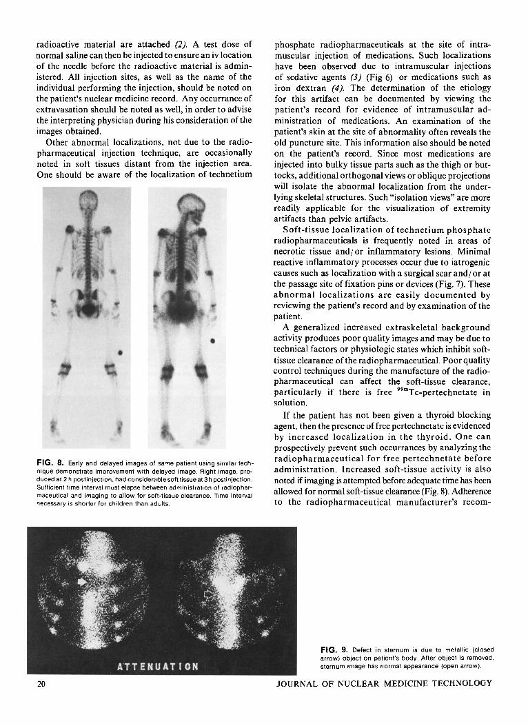
FIG. 8. Early and delayed images of same patient using similar technique demonstrate improvement with delayed image. Right image, produced at 2 h postinjection, had considerable soft tissue at 3h postinjection. Sufficient time interval must elapse between administration of radiopharmaceutical and imaging to allow for soft-tissue clearance. Time interval necessary is shorter for children than adults.
20
phosphate radiopharmaceuticals at the site of intramuscular injection of medications. Such localizations have been observed due to intramuscular injections of sedative agents (3) (Fig 6) or medications such as iron dextran (4). The determination of the etiology for this artifact can be documented by viewing the patient's record for evidence of intramuscular administration of medications. An examination of the patient's skin at the site of abnormality often reveals the old puncture site. This information also should be noted on the patient's record. Since most medications are injected into bulky tissue parts such as the thigh or buttocks, additional orthogonal views or oblique projections will isolate the abnormal localization from the underlying skeletal structures. Such "isolation views" are more readily applicable for the visualization of extremity artifacts than pelvic artifacts.
Soft-tissue localization of technetium phosphate radiopharmaceuticals is frequently noted in areas of necrotic tissue and/ or inflammatory lesions. Minimal reactive inflammatory processes occur due to iatrogenic causes such as localization with a surgical scar and 1 or at the passage site of fixation pins or devices (Fig. 7). These abnormal localizations are easily documented by reviewing the patient's record and by examination of the patient.
A generalized increased extraskeletal background activity produces poor quality images and may be due to technical factors or physiologic states which inhibit softtissue clearance of the radiopharmaceutical. Poor quality control techniques during the manufacture of the radiopharmaceutical can affect the soft-tissue clearance, particularly if there is free 99mTc-pertechnetate in solution.
If the patient has not been given a thyroid blocking agent, then the presence of free pertechnetate is evidenced by increased localization in the thyroid. One can prospectively prevent such occurrances by analyzing the radiopharmaceutical for free pertechnetate before administration. Increased soft-tissue activity is also noted if imaging is attempted before adequate time has been allowed for normal soft-tissue clearance (Fig. 8). Adherence to the radiopharmaceutical manufacturer's recom-
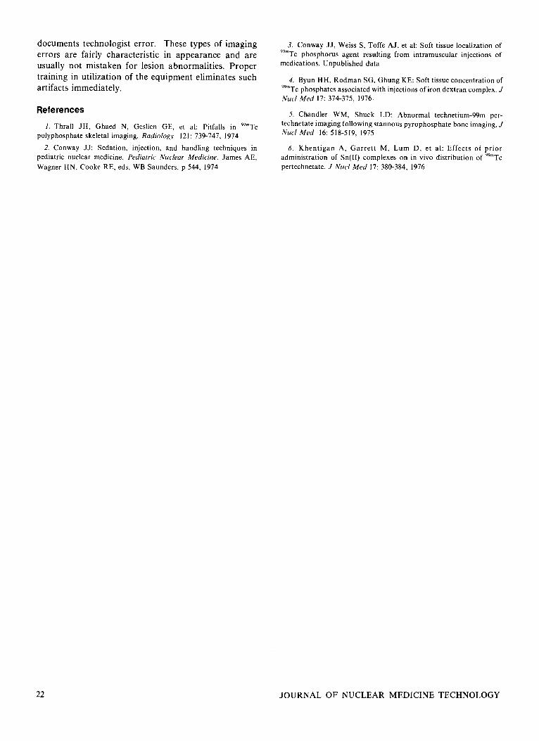
FIG. 9. Defect in sternum is due to metallic (closed arrow) object on patient's body. After object is removed, sternum image has normal appearance (open arrow).
JOURNAL OF NUCLEAR MEDICINE TECHNOLOGY

FIG. 10. Abnormal abdominal activity owing to ureterosigmoidostomy. Etiology of such activity can be documented from patient's history.
mended imaging time will diminish the possibility of this artifact occurring. Patients with diminished renal function, such as nephrotic syndrome, may also present increased extraskeletal background activity.
Miscellaneous Artifacts
The technologist should be aware of many other factors which may adversely affect radionuclide images. These include increased as well as decreased areas of localization, attentuation of photons by metallic objects on the patients or in their clothing (Fig. 9), and diminished localization of radionuclide in areas of previous radiotherapy. Again, examination of and explanation to the patient will eliminate the problem encountered with attentuation by objects in the patient's clothing or on their person. Examination of the records will indicate the history of previous irradiation. Such facts should be duly noted on the patient's nuclear medicine record.
Ectopic localizations of radionuclide can be seen with urinary collection devices or urinary diversions such as ureterosigmoidostomy (Fig. I 0). One should also be aware of residual radioactivity from previous studies such as brain, liver/spleen, or thyroid scans (Fig. 11). Conversly, it has been reported (5,6) that 99mTc-pertechnetate can bind to red blood cells in patients who have received radiopharmaceuticals with high tin content, such as pyrophosphate for bone imaging as well as with several other tin-containing agents. Subsequent brain imaging with 99mTc-pertechnetate may demonstrate increased choroid plexus and blood pool activities (5).
Finally, there is a large number of artifacts caused by equipment malfunctions (Fig. 12) or poor instrumentation technique. Many of these artifacts are related to the imaging device or improper use of the film, which readily
VOLUME 5, NUMBER I
FIG. 11. Residual activity from therapeutic dose of '"I obscures bone scan in neck and chest area.
FIG. 12. Many technical factors can create scan artifacts. Gross example is this bone scan with right and left sides of image reversed owing to improper setting.
21
documents technologist error. These types of imaging errors are fairly characteristic in appearance and are usually not mistaken for lesion abnormalities. Proper training in utilization of the equipment eliminates such artifacts immediately.
References
1. Thrall JH. Ghaed N, Geslien GE, et al: Pitfalls in 9 9 m ~ c polyphosphate skeletal imaging. Radio1og.y 121: 739-747, 1974
2. Conway JJ : Sedation, injection, and handling techniques in pediatric nuclear medicine. Pediatric Nuclear Medicine. James AE, Wagner HN, Cooke RE, eds, WB Saunders, p 544, 1974
3. Conway JJ , Weiss S, Toffe AJ, et al: Soft tissue localization of 9grn Tc phosphorus agent resulting from intramuscular injections of
medications. Unpublished data
4. Byun HH, Rodman SG, Ghung KE: Soft tissue concentration of 99m Tc phosphates associated with injections of iron dextran complex. J Nucl Med 17: 374-375, 1976.
5. Chandler WM, Shuck LD: Abnormal technetium-99m per- technetate imaging following stannous pyrophosphate bone imaging. J Nucl Med 16: 5 18-5 19, 1975
6. Khentigan A, Garre t t M, Lum D, et al: Effects of prior administration of Sn(I1) complexes on in vivo distribution of 9 9 m ~ c pertechnetate. J Nucl Med 17: 380-384, 1976
JOURNAL OF NUCLEAR MEDICINE TECHNOLOGY