bone and soft tissue tumorstissue tumors … · bone and soft tissue tumorstissue tumors fabrizio...
TRANSCRIPT
3/30/2009
1
BONE AND SOFT TISSUE TUMORSTISSUE TUMORS
Fabrizio Remotti MD
DEFINITION• Soft tissue pathology
deals with tumors of the connective tissuesconnective tissues.
• The concept of soft tissue is understood broadly to include non-osseoustumors of extremities, trunk wall, retroperitoneum andretroperitoneum and mediastinum, and head & neck.
• Excluded (with a few exceptions) are organ specific tumors.

3/30/2009
2
DEFINITION• Bone pathology deals
with tumors of the skeletal system.
• Included are subsets of tumors from extra-osseous sites that show osseous and cartilaginous differentiation.
CLASSIFICATION
• Purpose of classification is to link similar tumors in order to understand their behavior, determine the most appropriate treatment, and investigate their pp p gbiology.
• However, purpose of a classification system is simplicity and reproducibility
• Therefore tumors are classified according to the cell type they resemble.
• Refinements are coming from cytogenetics, molecular, and gene expression studies.
• The majority arise from -or show differentiation toward- mesenchymal cells, but some show other differentiation (neuroectodermal, histiocytic).
• A small subset is of unknown histogenesis.

3/30/2009
3
CLASSIFICATION• Many tumors resemble
tissues present in thetissues present in the region of origin.
• These tumors may be derived from stem cells that belong to local, organ-specific pools.
• Other involved stem cells
Vascular leiomyosarcoma
• Other involved stem cells may be bone marrow derived.
Lipoma
CLASSIFICATION• Some tumors have no
resemblance to normal
Alveolar soft part sarcoma
tissue in the region (metaplastic foci within a tumor, or tumors of different histogenesis from the normal cells of the region)
• Some sarcomas have noSome sarcomas have no normal cell counterparts, probably reflecting an unique genetic makeup.
Epithelioid sarcoma, proximal type

3/30/2009
4
CLASSIFICATION
• Tumors are also classified according their bi l i t ti lbiologic potential.
• A three-tiered system is used:– 1. Benign– 2. Borderline (intermediate malignant)– 3 Malignant– 3. Malignant.
EPIDEMIOLOGY• Soft tissue (ST) sarcomas
are rare tumors compared to other malignancies: 8 700other malignancies: 8,700new sarcomas in 2001, with 4,400 deaths.
• The incidence of ST sarcomas in the USA is approximately 3.3 cases per 100,000 people.
• This is roughly 5% of each of some of the most common carcinomas (prostate, breast and lung), half of all brain tumors, and approximately equal to AML.

3/30/2009
5
EPIDEMIOLOGY
• There is a slight male predominance (with some
bt isubtypes more common in women).
• The majority of soft tissue tumors affect older adults (some sub-groups occur predominantly or exclusively in children).
Extra-renal malignant rhabdoid tumor
y )• Incidence of benign soft
tissue tumors not known, but probably outnumber malignant tumors 100:1.
EPIDEMIOLOGY• The knowledge of epidemiologic data may
help in diagnosis.
3/30/2009
6
BONE TUMORS- EPIDEMIOLOGY
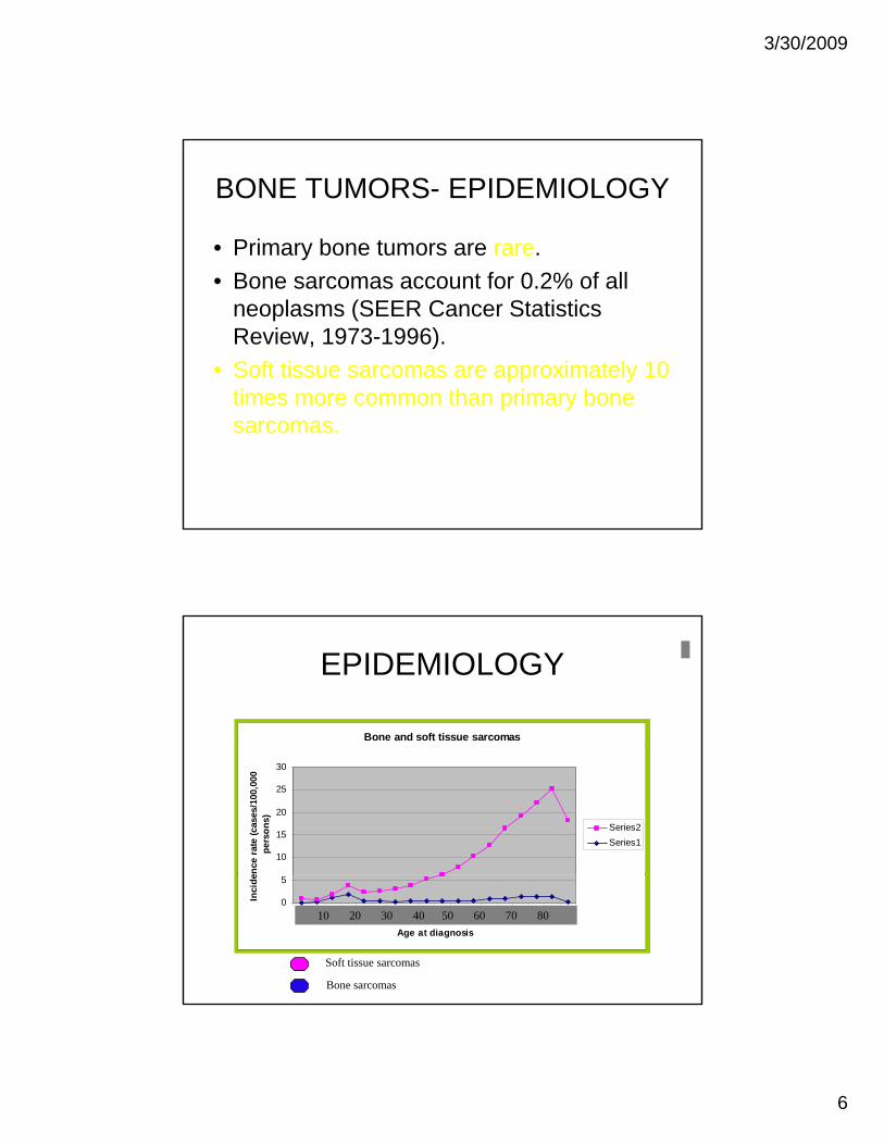
• Primary bone tumors are rare.• Bone sarcomas account for 0.2% of all
neoplasms (SEER Cancer Statistics Review, 1973-1996).
• Soft tissue sarcomas are approximately 10 times more common than primary bonetimes more common than primary bone sarcomas.
EPIDEMIOLOGY
Bone and soft tissue sarcomas
10
15
20
25
30
ence
rat
e (c
ases
/100
,000
pe
rson
s)
Series2Series1
0
5
1 2 3 4 5 6 7 8 9 10 11 12 13 14 15 16 17 18
Age at diagnosis
Inci
de
10 20 30 40 50 60 70 80
Soft tissue sarcomas
Bone sarcomas

3/30/2009
7
BONE TUMORS-EPIDEMIOLOGY• The majority of tumors involving bone
are secondary (or metastatic):- secondary (metastases) (95%)secondary (metastases) (95%)- primary (5%)
MELANOMA TO PROXIMAL HUMERUS
BREAST CANCER TO HIP
Secondary Tumors of Bone
•The carcinomas most frequently involved with bone metastasis originate from:
• Lung• Breast• Prostate• G.I
with bone metastasis originate from:
• Kidney• Thyroid

3/30/2009
8
BONE TUMORS•Bone sarcomas as a group have a bimodal distribution.•The first peak is in the second decade.•The second peak occurs in patients older than sixty.
304050607080
OSCSESCH
01020
0 to 4
10 to
14
20 to
24
30 to
34
40 to
44
50 to
54
60 to
64
70 to
74
80 to
85
MFH
ETIOLOGY
• The etiology of sarcomas is poorly d t d d h t i k l lunderstood, and what is known apply only
to a small fraction of the group.• The known etiologic agents are ionizing
radiation, oncogenic viruses, and chemicals.chemicals.
• These agents are able to cause genetic alterations that can lead to tumorigenesis.
3/30/2009
9
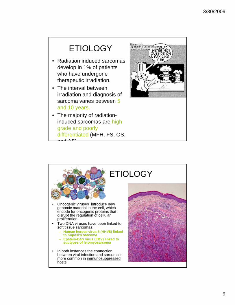
ETIOLOGY• Radiation induced sarcomas
develop in 1% of patients who have undergone therapeutic irradiation.
• The interval between irradiation and diagnosis of sarcoma varies between 5 and 10 earsand 10 years.
• The majority of radiation-induced sarcomas are high grade and poorly differentiated (MFH, FS, OS, and AS)
ETIOLOGY
• Oncogenic viruses introduce new genomic material in the cell, which encode for oncogenic proteins that disrupt the regulation of cellular proliferation.
• Two DNA viruses have been linked to soft tissue sarcomas:– Human herpes virus 8 (HHV8) linked p ( )
to Kaposi’s sarcoma– Epstein-Barr virus (EBV) linked to
subtypes of leiomyosarcoma
• In both instances the connection between viral infection and sarcoma is more common in immunosuppressed hosts.

3/30/2009
10
ETIOLOGY
• Herbicides (“agent orange”) and peripheral soft tissue sarcomas
• Retained metal objects (shrapnel, surgical devices)
d OS AS d MFHand OS, AS and MFH• Vinyl chloride, inorganic
arsenic, Thorotrast, anabolic steroids linked to AS and MFH.
ETIOLOGY• Host factors may
also play a role in the development of soft tissue sarcomas.– Immunosuppression,
besides Kaposi’s sarcoma, may be associated with sarcomassarcomas.
– Lymphedema, congenital or acquired (post-mastectomy) is a rare cause of extremity-based AS.
AS in lymphedema
3/30/2009
11
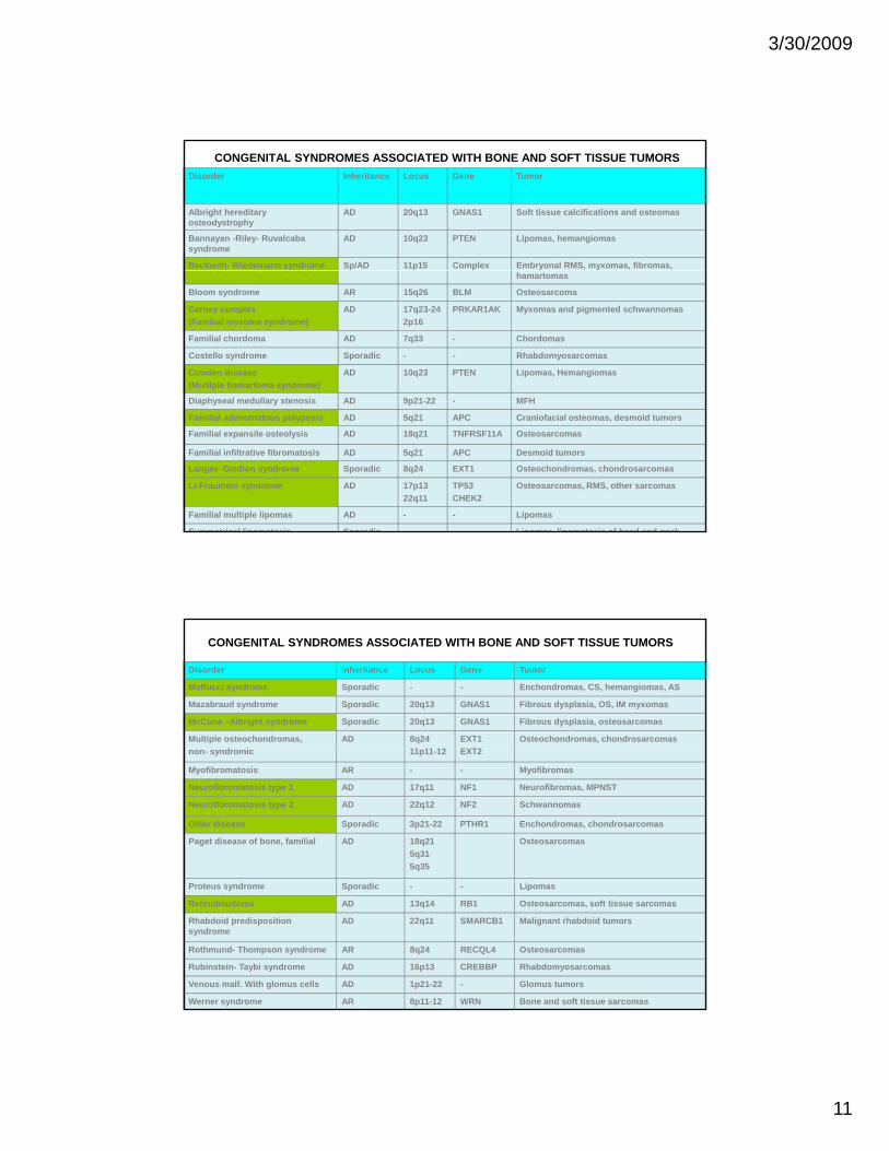
CONGENITAL SYNDROMES ASSOCIATED WITH BONE AND SOFT TISSUE TUMORSDisorder Inheritance Locus Gene Tumor
Albright hereditary osteodystrophy
AD 20q13 GNAS1 Soft tissue calcifications and osteomas
Bannayan -Riley- Ruvalcaba syndrome
AD 10q23 PTEN Lipomas, hemangiomas
Beckwith- Wiedemann syndrome Sp/AD 11p15 Complex Embryonal RMS, myxomas, fibromas, hamartomas
Bloom syndrome AR 15q26 BLM Osteosarcoma
Carney complex(Familial myxoma syndrome)
AD 17q23-242p16
PRKAR1AK Myxomas and pigmented schwannomas
Familial chordoma AD 7q33 - Chordomas
Costello syndrome Sporadic - - Rhabdomyosarcomas
Cowden disease (Multiple hamartoma syndrome)
AD 10q23 PTEN Lipomas, Hemangiomas
Diaphyseal medullary stenosis AD 9p21-22 - MFHDiaphyseal medullary stenosis AD 9p21-22 - MFH
Familial adenomatous polyposis AD 5q21 APC Craniofacial osteomas, desmoid tumors
Familial expansile osteolysis AD 18q21 TNFRSF11A Osteosarcomas
Familial infiltrative fibromatosis AD 5q21 APC Desmoid tumors
Langer- Giedion syndrome Sporadic 8q24 EXT1 Osteochondromas, chondrosarcomas
Li-Fraumeni syndrome AD 17p1322q11
TP53CHEK2
Osteosarcomas, RMS, other sarcomas
Familial multiple lipomas AD - - Lipomas
Symmetrical lipomatosis Sporadic - - Lipomas lipomatosis of head and neck
CONGENITAL SYNDROMES ASSOCIATED WITH BONE AND SOFT TISSUE TUMORS
Disorder Inheritance Locus Gene Tumor
Maffucci syndrome Sporadic - - Enchondromas, CS, hemangiomas, AS
Mazabraud syndrome Sporadic 20q13 GNAS1 Fibrous dysplasia, OS, IM myxomas
McCune –Albright syndrome Sporadic 20q13 GNAS1 Fibrous dysplasia, osteosarcomas
Multiple osteochondromas,d i
AD 8q2411 11 12
EXT1EXT2
Osteochondromas, chondrosarcomasnon- syndromic 11p11-12 EXT2
Myofibromatosis AR - - Myofibromas
Neurofibromatosis type 1 AD 17q11 NF1 Neurofibromas, MPNST
Neurofibromatosis type 2 AD 22q12 NF2 Schwannomas
Ollier disease Sporadic 3p21-22 PTHR1 Enchondromas, chondrosarcomas
Paget disease of bone, familial AD 18q215q315q35
Osteosarcomas
Proteus syndrome Sporadic - - Lipomas
Retinoblastoma AD 13q14 RB1 Osteosarcomas, soft tissue sarcomas
Rhabdoid predisposition syndrome
AD 22q11 SMARCB1 Malignant rhabdoid tumors
Rothmund- Thompson syndrome AR 8q24 RECQL4 Osteosarcomas
Rubinstein- Taybi syndrome AD 16p13 CREBBP Rhabdomyosarcomas
Venous malf. With glomus cells AD 1p21-22 - Glomus tumors
Werner syndrome AR 8p11-12 WRN Bone and soft tissue sarcomas

3/30/2009
12
SOFT TISSUE TUMORSCLASSIFICATION
MAJOR TYPES OF SOFT TISSUE TUMORS C ll t B i t M li t tCell type Benign tumor Malignant tumor(Myo)fibroblast Fibroma, myxoma Fibrosarcoma, MFHAdipocyte Lipoma LiposarcomaSmooth muscle cell Leiomyoma LeiomyosarcomaSkeletal muscle cell Rhabdomyoma RhabdomyosarcomaEndothelial cell Hemangioma AngiosarcomaSchwann cell Schwannoma, neurofibroma MPNSTCartilage cell Chondroma ChondrosarcomaInterstitial cell GIST GISTHistiocyte JXG, GCTTS, RDD True histiocytic sarcomaUnknown No benign counterparts ES, SS, ES, ASPS
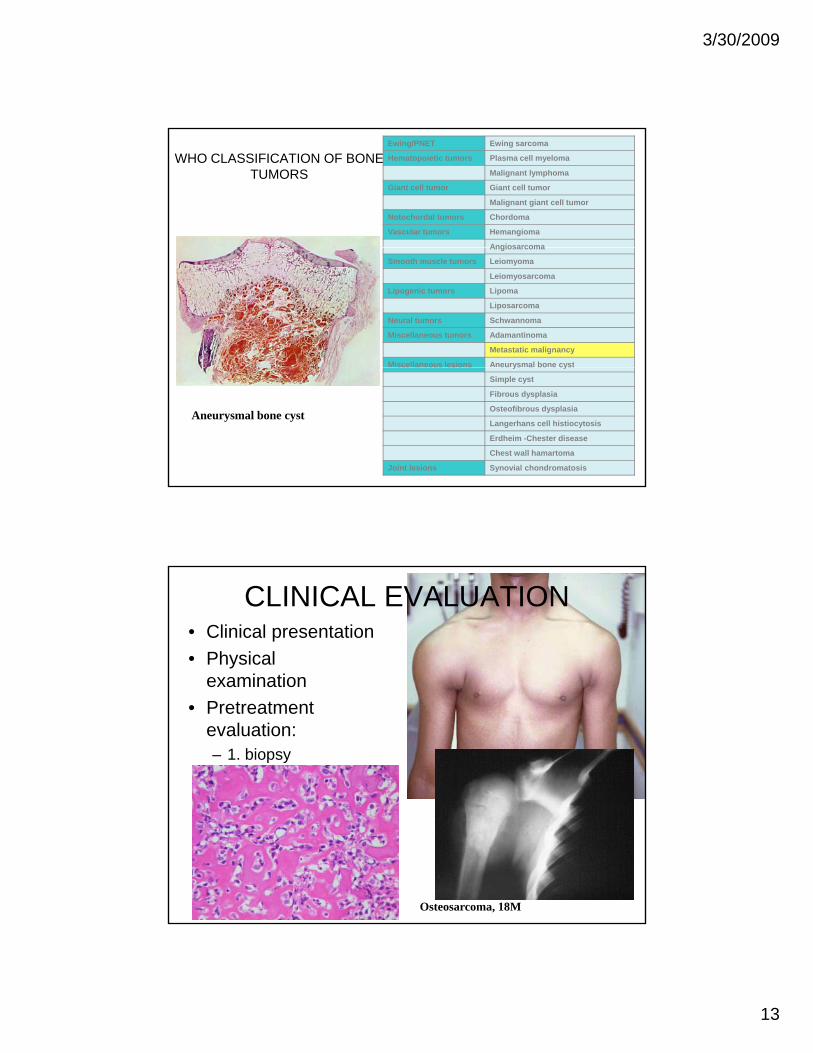
WHO CLASSIFICATION OF BONE TUMORS
Cartilage tumors Osteochondroma
Chondroma Enchondroma
Periosteal chondroma
Mult. chondromatosis
Chondroblastoma
Chondromyxoid fibroma
Chondrosarcoma Central
Peripheral
Dedifferentiated
Mesenchymal
Clear cell
Osteogenic tumors Osteoid osteoma
Osteoblastoma
Osteosarcoma Conventional
Telangiectatic
Small cell
Low grade centralLow grade central
Secondary
Parosteal
Periosteal
High grade surface
Fibrogenic tumors Desmoplastic fibroma
Fibrosarcoma
Fibrohistiocytic tumors Desmoplastic fibroma
Fibrosarcoma
Osteosarcoma
3/30/2009
13
WHO CLASSIFICATION OF BONE TUMORS
Ewing/PNET Ewing sarcoma
Hematopoietic tumors Plasma cell myeloma
Malignant lymphoma
Giant cell tumor Giant cell tumor
Malignant giant cell tumor
Notochordal tumors Chordoma
Vascular tumors Hemangioma
AngiosarcomaAngiosarcoma
Smooth muscle tumors Leiomyoma
Leiomyosarcoma
Lipogenic tumors Lipoma
Liposarcoma
Neural tumors Schwannoma
Miscellaneous tumors Adamantinoma
Metastatic malignancy
Miscellaneous lesions Aneurysmal bone cystsce a eous es o s eu ys a bo e cyst
Simple cyst
Fibrous dysplasia
Osteofibrous dysplasia
Langerhans cell histiocytosis
Erdheim -Chester disease
Chest wall hamartoma
Joint lesions Synovial chondromatosis
Aneurysmal bone cyst
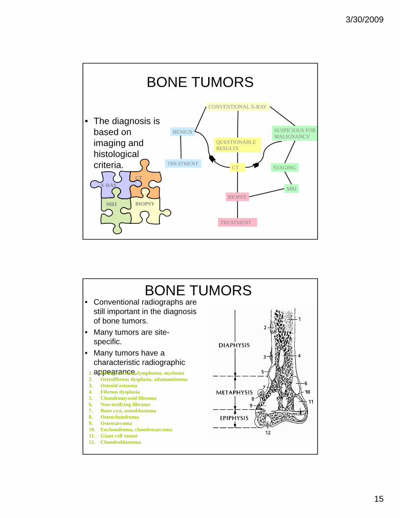
CLINICAL EVALUATION• Clinical presentation• Physical
examinationexamination • Pretreatment
evaluation:– 1. biopsy– 2. radiological
staging g g
Osteosarcoma, 18M

3/30/2009
14
IMAGING STUDIES• The ultimate goal is:
– 1. Detecting lesions– 2. Giving a specific
diagnosis or a reasonable
Liposarcoma
differential diagnosis– 3. Staging the lesion
MFH
IMAGING STUDIES• CT and particularly MRI allow detection
and and staging by delineating anatomical extent in virtually all casesanatomical extent in virtually all cases.
• A relatively specific diagnosis can be given in approximately 25-50% of cases, according to the type.
65 W, FS thigh (MRI)
3/30/2009
15
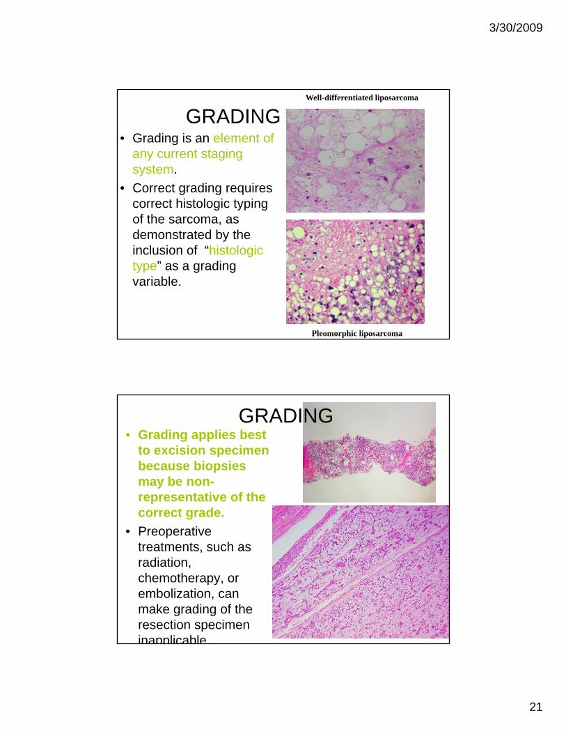
BONE TUMORS
• The diagnosis is
CONVENTIONAL X-RAY
• The diagnosis is based on imaging and histological criteria.
BENIGN
TREATMENT
QUESTIONABLERESULTS
SUSPICIOUS FORMALIGNANCY
STAGINGCT
CT
MRIBIOPSY
TREATMENT
BIOPSYMRI
X-RAY
BONE TUMORS• Conventional radiographs are
still important in the diagnosis of bone tumors.
• Many tumors are site-specific.
• Many tumors have a characteristic radiographic appearance. 1. Ewing sarcoma, lymphoma, myeloma
2. Osteofibrous dysplasia, adamantinoma3. Osteoid osteoma4. Fibrous dysplasia5. Chondromyxoid fibroma6. Non-ossifying fibroma7. Bone cyst, osteoblastoma8. Osteochondroma9. Osteosarcoma10. Enchondroma, chondrosarcoma11. Giant-cell tumor12. Chondroblastoma

3/30/2009
16
BONE TUMORS
Some fancy words from the world of shadows
IMAGING STUDIES
14F R distal femur Osteosarcoma
Soft tissue mass

3/30/2009
17
IMAGING STUDIES• Although imaging studies may give a reasonably
accurate diagnosis on the biological potential of a lesion, there are not many lesions that may be
t l di d b i i t di laccurately diagnosed by imaging studies alone.• The biopsy is the gold standard for diagnosis.
BIOPSY• Select least invasive
technique that allows diagnosis (including grade):
Craig cutting needle with T-handle and sheathfor bone biopsies
diagnosis (including grade):– Percutaneous fine needle
aspiration.– Percutaneous core needle
biopsy (blind or image-guided).
– Incisional biopsy.– Excisional biopsy.
Craig needle set

3/30/2009
18
BIOPSY
• Percutaneous needle
Metastatic myxoid liposarcomato liver
core biopsy usually yield adequate tissue for diagnosis.
• There is enough tissue for morphological studies.
Osteosarcoma
BIOPSY• Core biopsies yield
enough material for extensive i hi t h iimmunohistochemical stains.
MITF
24M, arm, clear cell sarcomaS-100

3/30/2009
19
BIOPSY
• Incisional biopsiesIncisional biopsies are required in many cases.
50M, angiosarcoma of ischium.
SPECIAL DIAGNOSTIC STUDIES
• Many sarcomas require additional studies to confirm the diagnosis and, in some cases, to
fadd prognostic information.

3/30/2009
20
GENETICS OF CONNECTIVE TISSUE NEOPLASMS
• Numerous cancer-specific genetic alterations have been described unfortunately almosthave been described, unfortunately almost exclusively for soft tissue neoplasms.
• Some of them (such as translocations, numerical changes, large deletions and gene amplifications) are seen at the cytogenetic level.
• Subtle changes (such as single base pair• Subtle changes (such as single base pair substitutions, small deletions) require molecular genetic detection.
3/30/2009
21
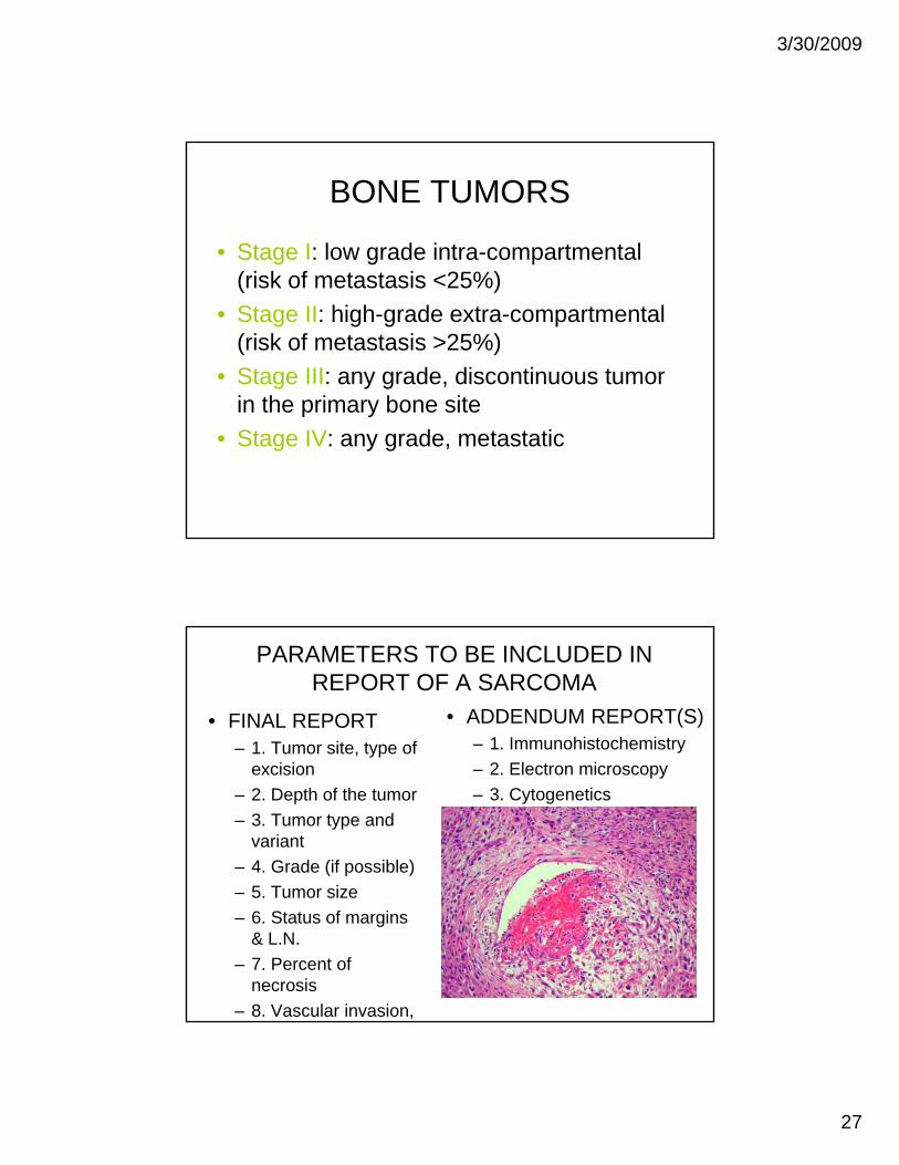
GRADING• Grading is an element of
any current staging system.
Well-differentiated liposarcoma
system.• Correct grading requires
correct histologic typing of the sarcoma, as demonstrated by the inclusion of “histologic gtype” as a grading variable.
Pleomorphic liposarcoma
GRADING• Grading applies best
to excision specimen because biopsies
bmay be non-representative of the correct grade.
• Preoperative treatments, such as radiationradiation, chemotherapy, or embolization, can make grading of the resection specimen inapplicable.

3/30/2009
22
GRADING• Weak points of grading:
– Subjective elementsSubjective elements (number of mitoses, percent of necrosis, tumor differentiation)
– Sampling – Frequent vs. rare tumors
MFH
GRADING• Any diagnostic
entity has a range of malignancy.
• The grade within the overall range depends on the histologic f tfeatures (cellularity, pleomorphism, mitotic activity, necrosis, etc.)

3/30/2009
23
GRADING- ST SARCOMASGRADING SYSTEM SOFT TISSUE SARCOMAS (FFCC)
Score (1-3)TUMOR DIFFERENTIATIONwell diff 1defined histogenetic types 2defined histogenetic types 2poorly diff & undef histogenesis 3
MITOTIC COUNT0-9/10HPF 110-19/HPF 2>20 HPF 3
TUMOR NECROSIS0none 0
<50% 1>50% 2
HISTOLOGIC GRADE Sum of scores1 2 or 32 4 or 53 6, 7 or 8
GRADING-ST SARCOMASDIFFERENTIATION SCORE 1
Well differentiated sarcoma (fibro-, lipo-, leiomyo-, chondro-)Well differentiated MPNST (neurofibroma with malignant transformation)
DIFFERENTIATION SCORE 2Conventional fibrosarcoma, leiomyosarcoma, angiosarcomaConventional MPNSTMyxoid sarcomas (MFH, liposarcoma, chondrosarcoma)Storiform-pleomorphic MFH
DIFFERENTIATION SCORE 3Sarcomas of undefined histog. (ASPS, SS,ES,CCS, undiff. Sarc.,malig. rhabdoid tumor)Ewing family of tumorsPleomorphic sarcomas (lipo leio )Pleomorphic sarcomas (lipo-, leio-)Round cell and pleomorphic liposarcomaRhabdomyosarcoma (except botryoid and spindle cell)Poorly differentiated angiosarcomaTriton tumor, epithelioid MPNSTExtraskeletal mesenchymal CS, and osteosarcomaGiant-cell and inflammatory MFH

3/30/2009
24
STAGING
• The stage is an estimate of the extent or dissemination of a tumor (and in thedissemination of a tumor (and in the current systems includes tumor grade).
• Staging is important for planning of treatment and prognostication.
• Clinical data and imaging studies are part f t iof staging process
• (Visceral sarcomas excluded)
STAGING (G-TNM)- ST SARCOMASSTAGE GRADE PRIMARY TUMOR LYMPH NODES METASTASIS
I - IV LOW OR HIGH T1 (<5 CM) OR T2 (>5 CM) NEG/POSABSENT/PRESENT
IA OW T1 T1b NEGATIVE ABSENTIA LOW T1a or T1b NEGATIVE ABSENT
IB LOW T2a or T2b NEGATIVE ABSENT
IIA HIGH T1a or T1b NEGATIVE ABSENT
IIB HIGH T2a NEGATIVE ABSENT
III HIGH T2b NEGATIVE ABSENT
IV ANY ANY POSITIVE ABSENT
ANY ANYPOSITIVE OR NEGATIVE PRESENT
“a” superficial tumors of trunk and extremities (above fascia)“b” deep tumors of trunk and extremities or intra-abdominal, intra-thoracic or retro-peritoneal

3/30/2009
25
STAGING OF ST SARCOMAS
5-yr survival
Stage %I 86
II 72
III 52III 52
IV 10-20
NEJM 2005; 353: 701-711
BONE SARCOMAS
• Like ST sarcomas, bone sarcomas d t b d d ( di ineed to be graded (grading is an
important element of the staging and determines if the tumor is stage I or II).
• The TNM system for bone sarcomas follows a 2 tier grading system: low-follows a 2 tier grading system: lowand high-grade.

3/30/2009
26
BONE TUMORS
• The staging of bone
Primary tumor (T) TX Primary tumor cannot be assessed
T0 No evidence of primary tumor
T1 T l l t 8 i t t di iof bone sarcomas follows the TNM system.
T1 Tumor less or equal to 8 cm in greatest dimension
T2 Tumor equal or more than 8 cm in greatest dimension
T3 Discontinuous tumors in the primary bone site
Regional lymph nodes (N) NX Regional lymph nodes cannot be assessed
NO No regional lymph node metastasis
N1 Regional lymph node metastasis
Distant metastases (M) MX Distant metastasis cannot be assessed
M0 No distant metastasisM0 No distant metastasis
M1 Distant metastasis:
M1a: lung
M1b: other sites
AJCC Cancer Staging Manual, 6th Edition, Springer, New York
BONE TUMORS
Stage IA T1 N0, NX M0 Low grade
Stage IB T2 N0, NX M0 Low grade
Stage IIA T1 N0, NX M0 High grade
Stage IIB T2 N0, NX M0 High grade
Stage III T3 N0, NX M0 Any grade
Stage IVA Any T N0 NX M1a Any gradeStage IVA Any T N0, NX M1a Any grade
Stage IVB Any T N1 Any M Any grade
Any T Any N M1b Any grade
AJCC Cancer Staging Manual, 6th Edition, Springer, New York
3/30/2009
27
BONE TUMORS
• Stage I: low grade intra-compartmental ( i k f t t i 25%)(risk of metastasis <25%)
• Stage II: high-grade extra-compartmental (risk of metastasis >25%)
• Stage III: any grade, discontinuous tumor in the primary bone sitein the primary bone site
• Stage IV: any grade, metastatic
PARAMETERS TO BE INCLUDED IN REPORT OF A SARCOMA
• FINAL REPORT– 1. Tumor site, type of
• ADDENDUM REPORT(S)– 1. Immunohistochemistry
excision– 2. Depth of the tumor– 3. Tumor type and
variant– 4. Grade (if possible)
5 Tumor size
– 2. Electron microscopy– 3. Cytogenetics
– 5. Tumor size– 6. Status of margins
& L.N.– 7. Percent of
necrosis– 8. Vascular invasion,

3/30/2009
28
TREATMENT• Surgery and pre- or
postoperative external beam radiation treatment in the primary local t t t f t ti ttreatment for most patients with localized disease.
• Adjuvant chemotherapy is usually reserved for patient with high-grade sarcomas.
• Patients with metastatic disease considered fordisease considered for chemotherapy and selected cases may undergo metastasectomy.
TREATMENT• Currently
approximately 90% of patients with localized extremity sarcomas undergo limb-sparing surgery.
31F with OS 9 year s/p surgery