bmc systems biology biomed central - core.ac.uk · biomed central page 1 of 17 (page number not for...
TRANSCRIPT

BioMed CentralBMC Systems Biology
ss
Open AcceResearch articleA computational approach to resolve cell level contributions to early glandular epithelial cancer progressionSean HJ Kim1, Jayanta Debnath2, Keith Mostov3, Sunwoo Park4 and C Anthony Hunt*1,4Address: 1UCSF/UC Berkeley Joint Graduate Group in Bioengineering, University of California, Berkeley, California, 94720, USA, 2Department of Pathology, University of California, San Francisco, California 94143, USA, 3Department of Anatomy, University of California, San Francisco, California 94143, USA and 4Department of Bioengineering and Therapeutic Sciences, University of California, San Francisco, California 94143, USA
Email: Sean HJ Kim - [email protected]; Jayanta Debnath - [email protected]; Keith Mostov - [email protected]; Sunwoo Park - [email protected]; C Anthony Hunt* - [email protected]
* Corresponding author
AbstractBackground: Three-dimensional (3D) embedded cell cultures provide an appropriatephysiological environment to reconstruct features of early glandular epithelial cancer. Althoughthese are orders of magnitude simpler than tissues, they too are complex systems that have provenchallenging to understand. We used agent-based, discrete event simulation modeling methods tobuild working hypotheses of mechanisms of epithelial 3D culture phenotype and early cancerprogression. Starting with an earlier software analogue, we validated an improved in silico epithelialanalogue (ISEA) for cardinal features of a normally developed MDCK cyst. A set of axiomaticoperating principles defined simulated cell actions. We explored selective disruption of individualsimulated cell actions. New framework features enabled recording detailed measures of ISEA cellactivities and morphology.
Results: Enabled by a small set of cell operating principles, ISEA cells multiplied and self-organizedinto cyst-like structures that mimicked those of MDCK cells in a 3D embedded cell culture.Selective disruption of "anoikis" or directional cell division caused the ISEA to develop phenotypicfeatures resembling those of in vitro tumor reconstruction models and cancerous tissues in vivo.Disrupting either process, or both, altered cell activity patterns that resulted in morphologicallysimilar outcomes. Increased disruption led to a prolonged presence of intraluminal cells.
Conclusions: ISEA mechanisms, behaviors, and morphological properties may have biologicalcounterparts. To the extent that in silico-to-in vitro mappings are valid, the results suggestplausible, additional mechanisms of in vitro cancer reconstruction or reversion, and raisepotentially significant implications for early cancer diagnosis based on histology. Further ISEAdevelopment and use are expected to provide a viable platform to complement in vitro methodsfor unraveling the mechanistic basis of epithelial morphogenesis and cancer progression.
Published: 31 December 2009
BMC Systems Biology 2009, 3:122 doi:10.1186/1752-0509-3-122
Received: 27 February 2009Accepted: 31 December 2009
This article is available from: http://www.biomedcentral.com/1752-0509/3/122
© 2009 Kim et al; licensee BioMed Central Ltd. This is an Open Access article distributed under the terms of the Creative Commons Attribution License (http://creativecommons.org/licenses/by/2.0), which permits unrestricted use, distribution, and reproduction in any medium, provided the original work is properly cited.
Page 1 of 17(page number not for citation purposes)

BMC Systems Biology 2009, 3:122 http://www.biomedcentral.com/1752-0509/3/122
BackgroundEpigenetic deregulation of cell activity is thought to be animportant requirement in the preclonal phase of glandu-lar epithelial cancer [1]. What level of deregulation isrequired before the histology becomes abnormal? Can amechanism of deregulation be inferred from the abnor-mal phenotype? To better understand causal linkagesbetween mechanisms and phenotype in an in vitro set-ting, epithelial cells have been cultured and studied inthree-dimensional (3D) gels of extracellular matrix(ECM), such as collagen I or Matrigel®. When grownembedded in 3D culture, Madin-Darby canine kidney(MDCK) cells form identically structured acinar orga-noids enclosing a cell-free, fluid-filled lumen [2]. Prolifer-ation and apoptosis are essential features of the process.When manipulated or exposed to certain factors, the orga-noids and composing cells exhibit phenotypic featuresthat are associated with pre-cancerous or cancerous tissuesin vivo [3]. Such cell culture models are thought to pro-vide an appropriate physiological environment to studyglandular epithelial morphogenesis and cancer progres-sion.
From a systems modeling perspective, epithelial cell cul-tures are abstract, somewhat simplistic models of epithe-lial cells in vivo. They are constructed wet-lab models: acontrollable, careful assemblage of laboratory materialsand equipment, in which one component is alive. There islittle direct overlap between measured in vitro phenotypicattributes and corresponding attributes of epithelial cellsin vivo. Nevertheless, the accumulated literature and themodel's continued study attest that scientifically usefulmappings exist between model and referent at several lev-els, from genetic to systemic phenomena [2,4,5]. How-ever, even though the in vitro cultures are orders ofmagnitude simpler than epithelial cells in a mammaliantissue context, they still are complex systems that haveproven challenging to understand.
We suggest that progress can be made in understandingepithelial cell behavior, morphology, and mechanisms,along with the changes that occur during cancer progres-sion by constructing and studying abstract analogues insoftware, where the system features at all levels can bemodeled, fully explored, and understood. The envisionedproducts are examples of executable biology [6-8], whichare useful for systematically exploring hypotheses aboutreferent mechanisms through virtual experimentation.The approach works synergistically with inductive mathe-matical methods and emerging, hybrid approaches basedon "first principle" physical laws [9-11].
In a previous study [12], we presented a cell-mimetic ana-logue of the envisioned new class, which validated for asmall set of targeted MDCK attributes. Its growth charac-
teristics and the types of stable structures formed mim-icked those of MDCK cells in cultures. Eleven axiomaticoperating principles, and six simulated cell actions, wereadequate for validation. However, it failed to consistentlyproduce cystic structures with a round, convex contour, acardinal feature of normal in vitro phenotype which hasnot been considered in the earlier study.
Starting with the earlier analogue, we explored severalanalogue revision strategies to achieve the expandedattribute set. For the purposes of this study, we focused oncell event (death and division) patterns and multicellularmorphology. Because of the networked nature of axiomuse, some changes intended to have one effect also hadother, unintended, often abiotic consequences. One ofour guidelines was to keep revisions parsimonious. Wesought one new analogue having as few new axioms aspossible to achieve morphological validation against theshape requirement, in addition to the original set of targetattributes. For one, validation was achieved by addition ofonly one new cell action coupled with replacement of oneaxiom. Use of the new action enabled the improved in sil-ico epithelial analogue (ISEA) to form stable cystic struc-tures with smooth, convex margins similar to thoseobserved normally in 3D epithelial cell culture.
We reasoned that if a mapping exists between ISEA'scoarse-grained operating principles and the more com-plex epithelial cell counterparts, then selective disruptionof ISEA's operation should exhibit cancer-like characteris-tics of in vitro epithelial cancer reconstruction models[13], examples of which are provided in the Appendix. Wedesigned and implemented methods to selectively dereg-ulate, at a controlled level of severity, simulated cell oper-ation at the axiom level. We focused on two processesknown to be critical for normal ISEA growth and stabiliza-tion. One process ended with an ISEA version of anoikis,a specific form of cell death due to extracellular matrixdetachment. The other involved directed placement of adaughter cell, the ISEA's version of oriented cell division.A grading measure was developed and used to quantifychanges in morphology.
Dysregulation of either "anoikis" or directed daughter cellplacement, or both, led to manifest changes in ISEA phe-notype that were reminiscent of dysplastic growth associ-ated with in vitro cancer reconstruction and earlyglandular epithelial cancer progression in vivo. Conse-quently, we undertook a detailed analysis of the deregula-tions and their consequences. Varying the level ofdysregulation led to morphologies that could be classifiedinto groups using automated grading. Dysregulation ofISEA's anoikis process had a greater effect on overall mor-phology. Simultaneous dysregulation of the two axiomshad a nonadditive effect. Importantly, dysregulation of
Page 2 of 17(page number not for citation purposes)

BMC Systems Biology 2009, 3:122 http://www.biomedcentral.com/1752-0509/3/122
either process resulted in similar morphological out-comes, which could not be differentiated reliably withoutadditional information on growth dynamics, with poten-tially significant implications for early cancer diagnosisbased on histology. The results also provided an early leadon possible additional mechanisms to reconstruct, orrevert, cancer-like phenotypes in experimental and thera-peutic contexts. Future rounds of development of these insilico methods may lead to a viable platform forunraveling the operational bases of glandular epithelialmorphogenesis and early cancer progression.
MethodsThe ISEA design is based on the methods and principles ofagent-based modeling [14] and discrete event simulation[15]. The ISEA and its predecessors [12] can be categorizedbroadly as cell-based or cell-centered models, whichencompass cellular automata, cellular Potts models, andvarious types of agent-based or individual-based modelsas reviewed in [8,16,17]. Cellular automata and their rela-tionship to ISEA are discussed in the Appendix.
Detailed descriptions of ISEA components and the exper-imentation framework are provided in [12,18]. Anabridged description follows. To clearly distinguish ISEAcomponents and processes from their in vitro counter-parts, hereafter we use small caps when referring theformer. As detailed in [18], ISEA is a standalone systemthat comprises CULTURE analogue and system-level com-ponents for semi-automated experimentation and analy-sis. System-level components include EXPERIMENT
MANAGER, OBSERVER, and CULTURE graphical user interface(GUI). EXPERIMENT MANAGER, the top-level agent, providesexperiment protocol functions and specifications.OBSERVER agent is responsible for recording CULTURE
measurements. CULTURE GUI enables visualization anduser interaction during simulation.
A CULTURE is an agent that maps abstractly to the MDCKcell culture within one well of a multi-well culture plate.It maintains a two-dimensional (2D) hexagonal grid,which represents an arbitrary cross-section through a 3DMDCK cell culture. The grid has toroidal topologies. Dis-crete objects with eponymous names represent the essen-tial cell culture components: CELLS, MATRIX, and FREE SPACE.MATRIX and FREE SPACE are passive objects that map tounits of extracellular matrix (ECM) and matrix-free mate-rial. A MATRIX object maps to a cell-sized volume of ECM.For simplicity, MATRIX represents any media containingsufficient ECM to which MDCK cells can attach. For thetraits targeted, no distinction was needed for differentialphysical ECM characteristics, such as stiffness, density,and viscoelasticity. A FREE SPACE object maps to a similarlysized volume of material that is essentially free of cells andmatrix elements. FREE SPACE also maps to luminal space
and non-matrix material in pockets enclosed by cells. Thelatter are called LUMINAL SPACE when distinction from FREE
SPACE is useful.
CELLS are quasi-autonomous agents. They use the axio-matic operating principles and decision logic (Table 1;Fig. 1) to interact with components in their local environ-ment. Every CELL has the same step function in which anassessment of its environment is made and a call is madefor an appropriate action. The step function is scheduledeach simulation cycle. A set of CELL axioms, discussedbelow, determines CELL action. A CELL selects just oneaxiom and completes its corresponding action duringeach simulation cycle.
CELL axiomatic operating principlesAn agent must have rules and protocols for interactingwith adjacent components. The operating premise is thatthe same is true for cells in culture; what can be describedas rules and protocols are emergent properties of the cell'sexpressed genetics and ongoing biochemistry. Rules cantake any form. We elected to have each rule take the formof an axiom, which specifies a precondition and corre-sponding action. Preconditions correspond to a CELL'S
neighborhood configurations. For action options, wespecified what we determined to be a minimal set toachieve validation: replace an adjacent non-CELL objectwith a CELL copy, DIE (vanish) and leave behind a LUMINAL
SPACE, create MATRIX, destroy an adjacent non-CELL objectand move to that location leaving behind a LUMINAL SPACE,POLARIZE, DEPOLARIZE, and do nothing. For any precondi-tion, only one action was executed.
Detailed descriptions of supporting biological evidenceand assumptions made for ISEA CELL axioms are providedin [12,18]. Briefly, CELL DEATH axioms (Axioms 1, 2, and 5)were based on a general biological principle that cells,such as epithelial cells, undergo a process of cell deathwithin some interval after detaching from ECM [19,20].That behavior is observed in MDCK cell cultures [3,5].Axiom 4, which dictates MATRIX deposition between twoadjacent CELLS, was specified based on observations thatsome matrix is produced de novo between two adheringMDCK cells in suspension culture [21]. A CELL DIVISION
axiom, Axiom 3, follows from experimental observationsthat, when embedded in matrix, single MDCK cells prolif-erate [3,22]. Other CELL DIVISION axioms, Axioms 6, 7, and8, follow from a similar, general principle that epithelialcells proliferate when they adhere to ECM and tend do soin arrangements that maximize intercellular contact[2,23]. CELL POLARIZATION axioms, Axioms 9 and 10,reflect in vitro observations on MDCK cell polarity [2,5].Axiom 11 applied when the CELL achieved mandates thatmap to the three-surfaces principle articulated in [2]. An
Page 3 of 17(page number not for citation purposes)
BMC Systems Biology 2009, 3:122 http://www.biomedcentral.com/1752-0509/3/122
UNPOLARIZED state indicates that the CELL'S three surfacesmandate has not been achieved.
The earlier axioms developed by Grant et al. [12] enabledthe analogue to validate for a set of basic MDCK cell cul-ture attributes. Unlike its referent, the model frequentlyproduced highly irregular, abiotic structures. We revisedthe axioms to enable ISEA to consistently develop CYSTS
having roundish, convex shapes, a cardinal feature of anormal in vitro epithelial cyst. Note that a regular hexagonin a hexagonally discretized space maps to a circle in con-tinuous space. Fig. 1 describes and shows use of all 12ISEA axioms. Axioms 1-10 were carried forward from [12].ISEA variants that were more elaborate also enabled ISEAvariants to achieve the targeted attributes, but they wererejected because we strove to adhere to the guideline ofparsimony.
We considered and explored adding and using limiteddetails about the axis of POLARIZATION. A CELL, followingone of its action options, acquiring CELL neighbor or priorto creating a copy, for example, could assign itself a vecto-
rial axis of POLARIZATION. Thereafter, an axiomatic precon-dition could include reference to the direction ofPOLARIZATION combined with neighborhood information.However, other analogues that were explored, includingISEA, achieved the targeted attributes without that addeddetail. Adhering to the parsimony guideline, we excludedPOLARIZATION details because they were not needed toachieve the attributes targeted, but they can be added eas-ily when the need arises. Axiom 8 requires a CELL to beaware of the positions of its neighboring objects relativeto each other and itself. A higher resolution (more fine-grained) mechanism that would be exchangeable forAxiom 8 could include having and using an axis of POLAR-
IZATION.
Operational disruption of CELL axiomsFollowing implementation (and validation) of the revisedaxiomatic operating principles, our next task was to add amechanism to disrupt the operation of individual CELL
axioms selectively. We added a set of parameters, one peraxiom, which controlled the probability of the decision-making CELL electing to follow the axiom when its precon-
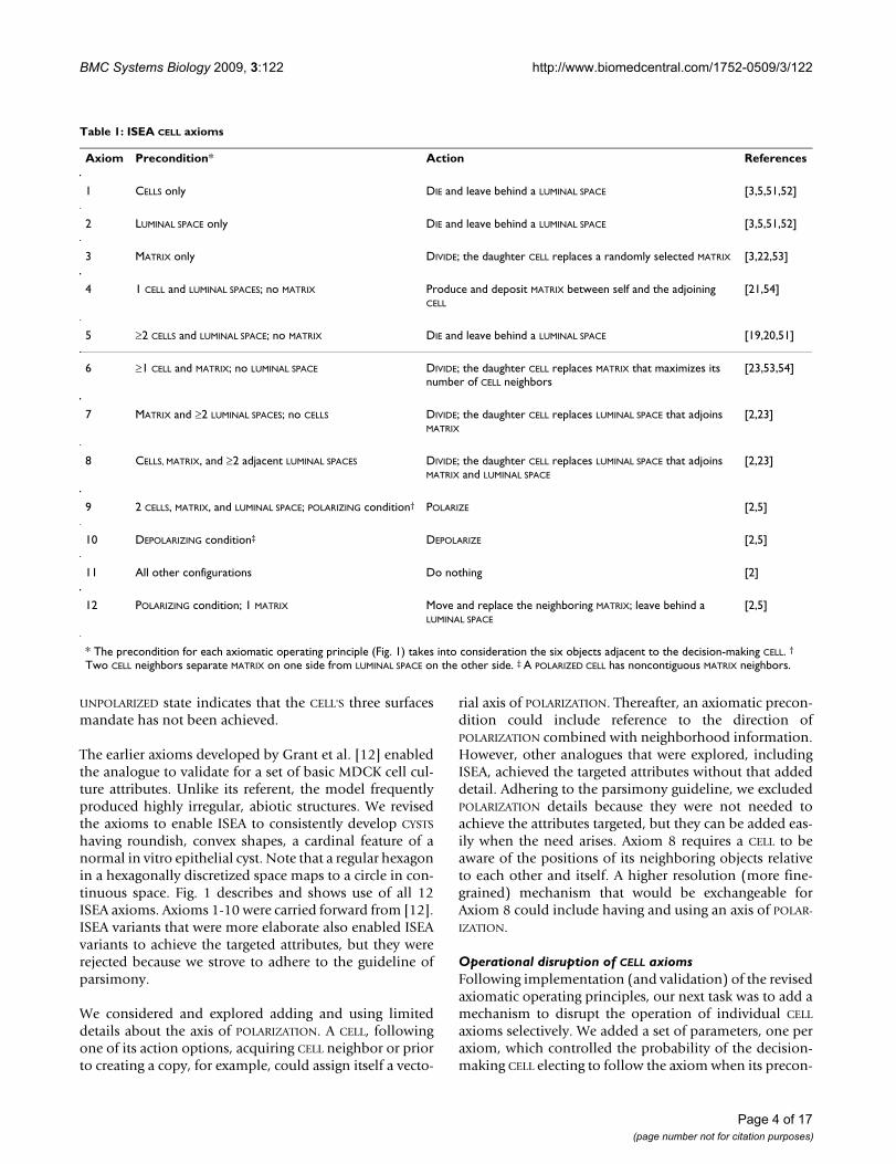
Table 1: ISEA CELL axioms
Axiom Precondition* Action References
1 CELLS only DIE and leave behind a LUMINAL SPACE [3,5,51,52]
2 LUMINAL SPACE only DIE and leave behind a LUMINAL SPACE [3,5,51,52]
3 MATRIX only DIVIDE; the daughter CELL replaces a randomly selected MATRIX [3,22,53]
4 1 CELL and LUMINAL SPACES; no MATRIX Produce and deposit MATRIX between self and the adjoining CELL
[21,54]
5 ≥2 CELLS and LUMINAL SPACE; no MATRIX DIE and leave behind a LUMINAL SPACE [19,20,51]
6 ≥1 CELL and MATRIX; no LUMINAL SPACE DIVIDE; the daughter CELL replaces MATRIX that maximizes its number of CELL neighbors
[23,53,54]
7 MATRIX and ≥2 LUMINAL SPACES; no CELLS DIVIDE; the daughter CELL replaces LUMINAL SPACE that adjoins MATRIX
[2,23]
8 CELLS, MATRIX, and ≥2 adjacent LUMINAL SPACES DIVIDE; the daughter CELL replaces LUMINAL SPACE that adjoins MATRIX and LUMINAL SPACE
[2,23]
9 2 CELLS, MATRIX, and LUMINAL SPACE; POLARIZING condition† POLARIZE [2,5]
10 DEPOLARIZING condition‡ DEPOLARIZE [2,5]
11 All other configurations Do nothing [2]
12 POLARIZING condition; 1 MATRIX Move and replace the neighboring MATRIX; leave behind a LUMINAL SPACE
[2,5]
* The precondition for each axiomatic operating principle (Fig. 1) takes into consideration the six objects adjacent to the decision-making CELL. †
Two CELL neighbors separate MATRIX on one side from LUMINAL SPACE on the other side. ‡ A POLARIZED CELL has noncontiguous MATRIX neighbors.
Page 4 of 17(page number not for citation purposes)

BMC Systems Biology 2009, 3:122 http://www.biomedcentral.com/1752-0509/3/122
dition applies. The parameter values ranged from 0 to 1inclusively. A parameter value = 1 corresponded to 100%adherence to the axiom. Setting the parameter to zerocompletely blocked execution of the prescribed actionand, if specified, dictated an alternate action. At its deci-sion point, each CELL drew a pseudo-random number(PRN) from the standard uniform distribution. The pre-scribed action was followed only when the PRN was ≤ theprobability threshold set for the corresponding parame-ter.
Following exploratory simulations that considered manyoptions, we specified alternative actions that mapped toplausible in vitro cell actions in a deregulated state. Axi-oms 1, 2, and 5 governed CELL DEATH; a reasonable alter-native was to resist DEATH and remain ALIVE (i.e., donothing). Axiom 3 dictated random placement of a CELL
copy; its alternate action was to do nothing and thus pre-vent REPLICATION. We also assigned the alternate action of'do nothing' to Axiom 4 (MATRIX production). Several dys-regulated action options were available to Axiom 6 (ORI-
ENTED CELL DIVISION). One was to do nothing, effectivelysuppressing CELL DIVISION. Another was undirected CELL
DIVISION, placing the CELL copy in a random directionwithout regard for the number of CELL neighbors. We usedthe latter because adequate, supportive biological infor-mation was available. Axiom 7, which dictated CELL DIVI-
SION, had available the same alternative action options.Axiom 8 (CELL DIVISION or POLARIZATION) had many plau-sible options. One was preventing CELL DIVISION; another,as above, was to allow the CELL to place a copy of itself inany available location. Yet, another option was to enablePOLARIZATION. The preconditions prescribing CELL DIVISION
or POLARIZATION also could be swapped. The remainingaxioms, Axioms 9-12, posed a similar problem of havingmany plausible action options. Because no experimentalinformation was available to narrow the options, weelected to defer investigation of those axioms until moreinformation becomes available.
Simulation experimentsCULTURE width and height were set to 100. For EMBEDDED
CELL CULTURE simulation, a single CELL was placed at thecenter of the CULTURE grid filled with MATRIX. One CELL
width mapped to 10 μm. Simulation time advanced dis-cretely. Ordering of CELL events within the same simula-tion cycle was pseudo-random. Each simulationexperiment comprised 100 Monte Carlo (MC) runs. EachMC run was executed for 50 simulation cycles. One simu-lation cycle mapped to 12 h in vitro. A new CULTURE wascreated for each repetition.
Specification and use of morphology indexThe morphology index, M, weighs three basic features ofMULTICELL morphology: local EXTRACELLULAR arrangement,
The twelve ISEA axiomatic operating principlesFigure 1The twelve ISEA axiomatic operating principles. Table 1 is a listing and explanation of the operating principles. The 2D space and all objects within are hexagonally discre-tized. Simulation time advances in steps corresponding to simulation cycles. During a simulation cycle, every CELL, in a pseudo-random order, decides what action to take based on its internal state (POLARIZED or UNPOLARIZED) and the com-position of its adjacent neighborhood. A set of axioms deter-mines what action is taken for each possible neighborhood configurations. Objects represented: POLARIZED CELL (red), UNPOLARIZED CELL (gray), MATRIX (white), and LUMINAL SPACE (black). At the top, selected decision-making CELLS at the start of simulation cycle n are numbered to indicate each of the twelve axiomatic preconditions being satisfied (they are listed in Table 1). For purpose of this illustration, the unnum-bered CELLS are inactive. At the bottom, the system at the start of simulation cycle n + 1 shows the consequences of applying all twelve axioms.
Page 5 of 17(page number not for citation purposes)

BMC Systems Biology 2009, 3:122 http://www.biomedcentral.com/1752-0509/3/122
E, structural discontinuity, D, and a structure's overallshape, S. For each CELL, the algorithm computes a numer-ical score based on its neighborhood arrangement. Anideal arrangement corresponds to the Axiom 12 precondi-tion. Higher scores are assigned to neighborhood config-urations that deviate from that ideal. The collectiveEXTRACELLULAR arrangement score, E, is the mean of indi-vidual CELL scores. A CLUSTER is structurally continuous solong as it remains one connected body of CELLS and FREE
(or LUMINAL) SPACE. When structural continuity is broken,two or more CLUSTERS are formed. The structural disconti-nuity algorithm computes the number of disconnectedbodies; that number translates to D. The shape profilealgorithm takes into consideration a structure's overall 2Dshape in hexagonal space and computes a score, S, whichincreases as shape becomes irregular or deviates from theideal shape, a regular hexagon (a regular hexagon in hex-agonal space maps to a circle in continuous space). Thevalue of the morphology index becomes M = E + D + S.The maximum values of E, D, and S have been set to 3, 2,and 1 respectively, reflecting their assigned relativeweights. The final morphology index value ranges from 1(an ideal CYST) to 6 (disorganized). Lower scores areassigned to configurations that are more organized androundish with a single LUMEN. The measure was imple-mented and calibrated for ISEA simulations, and sufficedfor this study's purposes. However, we will need counter-part metrics for in vitro and in vivo microscopic imageanalysis should we undertake direct, quantitative compar-ison between ISEA and wet-lab morphologies. A detaileddescription of the measure and algorithms are provided inadditional file 1: Supplementary Material.
Tools used for analogue implementation and executionThe model framework was implemented using MASONVersion 11. MASON is a discrete-event, multi-agent simu-lation library coded in Java [24]. Batch simulation experi-ments were performed on a small-scale Beowulf clustersystem. Computer codes and project files are available athttp://biosystems.ucsf.edu/research_epimorph.html
ResultsISEA was validated against normal 3D embedded cell culture traitsWe implemented a common framework and compo-nents, some derived from the earlier analogue [12]. Hav-ing a new general framework was needed in part to reduceunnecessary, cross-model redundancies between differentcandidate CELLS during analogue refinement, and facilitatean iterative model refinement process capable of auto-mated cross-model validation. As done in [12], we vali-dated the revised ISEA for all four growth conditions:monolayer, overlay, suspension, and embedded cultures.Simulation results for monolayer and suspension CUL-
TURES were identical to those in [12]. Results for overlay
cultures were closer in appearance to in vitro observations(not shown). Marked differences were observed for theEMBEDDED CULTURE condition. That CULTURE condition isthe focus hereafter.
At the start of an EMBEDDED CULTURE simulation, a singleCELL was placed in CULTURE space, surrounded by onlyMATRIX. As a simulation progressed, the CELL underwentrepeated rounds of CELL REPLICATION, followed by the for-mation of LUMINAL SPACE and an increase in CELL numberand CYST diameter. The central LUMINAL SPACE grew asCELLS in the inner region DIED or moved out. The growthdynamics and final morphology were similar to thoseobserved for MDCK cells (Fig. 2A). The EMBEDDED CULTURE
always formed stable CYSTS bordered by POLARIZED CELLS
(Fig. 2B), and ISEA consistently produced CYSTS with aroundish, convex shape with smooth margins. During anoccasional simulation, because of their changing, localenvironment, one or more CYST surface CELLS failed toPOLARIZE or DIE before the simulation ended. Such eventsprevented the local rounding out process that a POLARIZED
CELL can undertake, preventing a few structures from sta-bilizing within 50 simulation cycles.
New capabilities were added to simulate epigenetic deregulation of cell processesFollowing validation, we undertook experiments that maysimulate inducement and progression of in vitro cell phe-
MDCK and simulated cystsFigure 2MDCK and simulated cysts. (A) MDCK cells grown in 3D extracellular matrix form lumen-enclosing cystic structures surrounded by a layer of polarized cells. Cells composing cysts maintain three surfaces: apical (red), basal and lateral (green). Note the roundish contour typical of MDCK cysts. For growth and staining details, see [22]. Bar: ~10 μm. (B) Representative, stable ISEA CYSTS. CELLS in EMBEDDED condi-tion produced stable, cystic structures enclosing LUMINAL SPACE; all CELLS were POLARIZED (red). CYSTS had convex shapes. (C) This illustration shows that convex polygonal CYSTS in discretized 2D hexagonal space map to a roundish structures in continuous 2D space. Because such a mapping provides no added scientific or mechanistic information, sub-sequent ISEA structures are shown as they appeared at simu-lation's end in 2D hexagonal space.
Page 6 of 17(page number not for citation purposes)

BMC Systems Biology 2009, 3:122 http://www.biomedcentral.com/1752-0509/3/122
notypes that mimic early, preclonal stages of carcinogene-sis. The progression involves alterations in tissueorganization and morphology but low genetic changes[1]. The experiments were motivated by questions such asthese: what happens when normal cell operation and itsregulation become faulty? To what extent can individualcell activities be disrupted separately and together, andstill maintain an apparently normal morphology? Whatvisible changes accompany relaxation of tight control of acell-level process? Can the operational cause of thechanges be predicted from histological morphology? Toobtain answers for ISEA, we conducted experiments inwhich the probability (p) of proper axiom operation wasvaried from 1 to 0. When proper operation was not fol-lowed, alternate, dysregulated actions were used. From anoperational standpoint, having p = 0 represents a perma-nent change in the CELL'S operating principles throughoutthe simulation. A nonzero value below 1.0 specifies areversible change in the CELL'S operation, or erratic opera-tion. It can be viewed either as the probability of the CELL
behaving properly at any point in time, or the approxi-mate percentage of time that the CELL acts normal (i.e.,strictly adheres to the axiomatic operating principles illus-trated in Fig. 1) during simulation. Individual CELLS
decide to act normal (or not) each simulation cycle, inde-pendent of one another. As a result, CELLS can exhibit dif-ferent behaviors (normal vs dysregulated) during asimulation cycle. With p < 1, a CELL can switch between thetwo behaviors multiple times during a simulation. It is astochastic process having the Markov property: futurechanges in the CELL'S behavior occur probabilistically,independent of its history.
We focused on Axioms 5 and 6. Axioms 2, 3, 4, and 7 werenot essential to normal (i.e., validated) CYST formation inEMBEDDED CULTURE (but they were needed for other simu-lated culture conditions), and were used infrequently.Consequently, they were excluded from this investigation.As described in Methods, selecting an alternate action fordisrupted Axioms 8-11 is not straightforward. We electednot to pursue disruption of those axioms until furtherinsight from wet-lab studies becomes available to narrowoptions. Disrupting Axiom 1 was straightforward but theoutcomes (not shown) offered no significant insight: CELL
CLUSTERS either developed normally into CYSTS (p > 0) orgrew unchecked as a homogenous mass (p = 0). Weexpected that outcome because Axiom 1 is required forLUMINAL SPACE creation but becomes nonessential as soonas the nascent LUMINAL SPACE is formed. Axioms 5 and 6were essential to CYST formation in EMBEDDED CULTURE.Axiom 5 dictates ANOIKIS (in silico counterpart to anoikis,a form of cell death), and is the most frequently used CELL
DEATH axiom during CULTURE growth. Axiom 6 dictatesoriented CELL DIVISION; the action requires making a CELL
copy and placing it selectively. That process accounts for
most of the CELL DIVISION events in the simulations. Theirbiological counterparts are centrally implicated in epithe-lial morphogenesis and carcinogenesis.
To aid investigation, we developed and used a morphol-ogy index to automatically quantify ISEA structure mor-phology. The algorithm analyzed and scored features ofMULTICELL structures. The measure weighed three charac-teristics: local EXTRACELLULAR arrangement, structural con-tinuity, and the overall shape. The index values rangedfrom 1 to 6. Higher values indicated a more disorganizedstate. Lower scores were assigned when the overall shapewas convex, and all CELLS were POLARIZED. The measurewas calibrated for ISEAs, but could be generalized forother model types. The index represents the simplest met-ric that we found to be sufficiently discriminative for thisstudy's purposes. We are exploring alternative metrics thatmay expedite simulation analysis and interpretation.However, it is unlikely that a single, simple metric will suf-fice for analyzing multiple, distinguishable morphologi-cal features, each of which requires differentmeasurements to quantify and characterize. It is morelikely that the complexity of the metric will parallel thecomplexity of the multi-feature attributes being analyzed.A further discussion of the morphology index is providedin additional file 1: Supplementary Material.
Reduced ANOIKIS caused LUMEN fillingFor comparison, selected examples of in vitro structuresformed following dysregulation of normal cell processesare provided in the Appendix.
When cultured within 3D ECM, normal epithelial cellstypically proliferate and organize into hollow spheroids, aprocess that recapitulates certain structural features of aglandular epithelium, such as the presence of a central,hollow lumen [2]. In MDCK and some mammary cell cul-tures, apoptosis contributes centrally to lumen formation:cells in the inner region of the developing structureundergo anoikis upon loss of direct matrix contact [25].Blocking anoikis in vitro has been shown to cause lumenfilling, which resembles a characteristic of most glandularepithelial cancers [3]. In 3D cultures of mammary epithe-lial cell line MCF-10A, focal adhesion kinase-mediatedinhibition of apoptosis by inhibition of metalloprotein-ase 1 (TIMP1) has been shown to induce lumen fillingand disrupt normal acinar development [26]. Also,expression of oncoproteins with anti-apoptotic activities,including receptor tyrosine kinase ErbB2, colony-stimu-lating factor 1 receptor, SRC, and IGF1R, has been shownto elicit abnormal, filled phenotypes that resemblehuman ductal carcinoma in vivo [13]. If ISEA's operatingprinciples have cell culture counterparts, then simulationresults should exhibit (predict) LUMEN filling whenANOIKIS is compromised. We simulated the condition by
Page 7 of 17(page number not for citation purposes)
BMC Systems Biology 2009, 3:122 http://www.biomedcentral.com/1752-0509/3/122
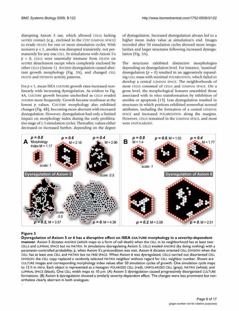
disrupting Axiom 5 use, which allowed CELLS lackingMATRIX contact (e.g., enclosed in the CYST LUMINAL SPACE)to evade DEATH for one or more simulation cycles. Withnonzero p < 1, anoikis was disrupted transiently, not per-manently for any one CELL. In simulations with Axiom 5'sp = 0, CELLS were essentially immune from DEATH onMATRIX detachment except when completely enclosed byother CELLS (Axiom 1). ANOIKIS dysregulation caused aber-rant growth morphology (Fig. 3A), and changed CELL
DEATH and DIVISION activity patterns.
For p < 1, mean ISEA CULTURE growth rates increased non-linearly with increasing dysregulation. As evident in Fig.4A, CULTURE growth became unchecked as CELLS evadedANOIKIS more frequently. Growth became nonlinear at thelowest p values. CULTURE morphology also exhibitedchanges (Fig. 4B), becoming more aberrant with increaseddysregulation. However, dysregulation had only a limitedimpact on morphology index during the early prolifera-tive stage of ≤ 5 simulation cycles. Thereafter, values eitherdecreased or increased further, depending on the degree
of dysregulation. Increased dysregulation always led to ahigher mean index value at simulation's end. Imagesrecorded after 50 simulation cycles showed more irregu-larities and larger structures following increased dysregu-lation (Fig. 3A).
The structures exhibited distinctive morphologiesdepending on dysregulation level. For instance, 'maximal'dysregulation (p = 0) resulted in an aggressively expand-ing CELL mass with minimal POLARIZATION, which failed todevelop a central LUMINAL SPACE. The neighborhoods ofmost CELLS consisted of CELLS and LUMINAL SPACE. On agross level, the morphological features resembled thoseassociated with in vitro transformation by inhibition ofanoikis or apoptosis [13]. Less dysregulation resulted instructures in which portions exhibited somewhat normalattributes, including the formation of a central LUMINAL
SPACE and increased POLARIZATION along the margins.However, CELLS remained in the LUMINAL SPACE, and mostwere UNPOLARIZED.
Dysregulation of Axiom 5 or 6 has a disruptive effect on ISEA CULTURE morphology in a severity-dependent mannerFigure 3Dysregulation of Axiom 5 or 6 has a disruptive effect on ISEA CULTURE morphology in a severity-dependent manner. Axiom 5 dictates ANOIKIS (which maps to a form of cell death) when the CELL in its neighborhood has at least two CELLS and LUMINAL SPACE but no MATRIX. In simulations dysregulating Axiom 5, CELLS evaded ANOIKIS (by doing nothing) with a parameter-controlled probability, p, when Axiom 5's precondition was met. Axiom 6 dictates oriented CELL DIVISION when the CELL has at least one CELL and MATRIX but no FREE SPACE. When Axiom 6 was dysregulated, CELLS carried out disoriented CELL DIVISION: the CELL copy replaced a randomly selected MATRIX neighbor without regard for CELL neighbor number. Shown are CULTURE images and corresponding morphology index values after 50 simulation cycles of growth. One simulation cycle maps to 12 h in vitro. Each object is represented as a hexagon: POLARIZED CELL (red), UNPOLARIZED CELL (gray), MATRIX (white), and LUMINAL SPACE (black). One CELL width maps to 10 μm. (A) Axiom 5 dysregulation caused progressively disorganized CULTURE formations. (B) Axiom 6 dysregulation showed a similarly severity-dependent effect. The changes were less prominent but nev-ertheless clearly aberrant in both analogues.
Page 8 of 17(page number not for citation purposes)

BMC Systems Biology 2009, 3:122 http://www.biomedcentral.com/1752-0509/3/122
Dysregulation also affected CELL activity patterns. In par-ticular, the number of CELL DEATH and CELL DIVISION occur-rences rose steeply over the growth period under severedysregulation (p ≤ 0.4), as illustrated in Fig. 4C-D. Theincrease was not as dramatic, and tended to remain rela-tively steady with less dysregulation. A drop in CELL DIVI-
SIONS was observed when ANOIKIS was blocked (p = 0)relative to strong dysregulation (p = 0.1). The result was
unexpected given the virtually unchecked CULTURE growthmeasured under that condition. However, even thoughgrowth was unchecked, the elimination of ANOIKIS
reduced the relative frequency of CELL DIVISION opportuni-ties compared to extensive but incomplete inhibition ofAxiom 5 use. Analysis of individual axiom use and theirrelative frequencies (additional file 1: SupplementaryMaterial) showed that the relative frequency of CELL DIVI-
Dysregulation of Axiom 5 (ANOIKIS) and its effect on ISEA CULTURE growth and morphologyFigure 4Dysregulation of Axiom 5 (ANOIKIS) and its effect on ISEA CULTURE growth and morphology. Axiom 5 dictates CELL DEATH when a CELL has in its neighborhood at least two CELLS and LUMINAL SPACE but no MATRIX. With a parameter-con-trolled probability, p, CELLS evaded ANOIKIS (by doing nothing) when Axiom 5's precondition was met. Doing so caused distinct changes in growth and structural characteristics of the EMBEDDED CULTURE. (A) CELL CULTURE growth rate increased monoton-ically with the severity of dysregulation. CULTURE growths at six levels of dysregulation are shown. (B) Disrupting operation of Axiom 5 resulted in the formation of progressively aberrant MULTICELL structures, as indicated by the numeric scale. Higher values indicate a more disorganized morphology. Dysregulation had no observable effect on CULTURE morphology in the early stages (~5 simulation cycles) of growth. One simulation cycle maps to 12 h in vitro. The effect became progressively evident as simulation time advanced. (C-D) Axiom 5 dysregulation altered CELL DIVISION and DEATH event patterns. The changes became more evident at later times. In both simulations, the effect on CELL DEATH and DIVISION was monotonic, except when p = 0. The mean occurrence of CELL DIVISION and DEATH fell when p = 0 (vs p = 0.8). The data are mean values of 100 Monte Carlo runs.
Dysregulation of Axiom 5 (ANOIKIS) and its effect on ISEA CULTURE growth and morphologyFigure 4Dysregulation of Axiom 5 (ANOIKIS) and its effect on ISEA CULTURE growth and morphology. Axiom 5 dictates CELL DEATH when a CELL has in its neighborhood at least two CELLS and LUMINAL SPACE but no MATRIX. With a parameter-con-trolled probability, p, CELLS evaded ANOIKIS (by doing nothing) when Axiom 5's precondition was met. Doing so caused distinct changes in growth and structural characteristics of the EMBEDDED CULTURE. (A) CELL CULTURE growth rate increased monoton-ically with the severity of dysregulation. CULTURE growths at six levels of dysregulation are shown. (B) Disrupting operation of Axiom 5 resulted in the formation of progressively aberrant MULTICELL structures, as indicated by the numeric scale. Higher values indicate a more disorganized morphology. Dysregulation had no observable effect on CULTURE morphology in the early stages (~5 simulation cycles) of growth. One simulation cycle maps to 12 h in vitro. The effect became progressively evident as simulation time advanced. (C-D) Axiom 5 dysregulation altered CELL DIVISION and DEATH event patterns. The changes became more evident at later times. In both simulations, the effect on CELL DEATH and DIVISION was monotonic, except when p = 0. The mean occurrence of CELL DIVISION and DEATH fell when p = 0 (vs p = 0.8). The data are mean values of 100 Monte Carlo runs.
Page 9 of 17(page number not for citation purposes)
BMC Systems Biology 2009, 3:122 http://www.biomedcentral.com/1752-0509/3/122
SION (as a consequence of Axiom 6 use) exceeded the useof all CELL DEATH axioms by a factor of > 2 when p = 0.There was a several-fold increase in Axiom 1 activity, butit failed to compensate for the absence of Axiom 5 use;Axiom 2 use showed no observable increase. In partiallydysregulated conditions (p = 0.4-0.8), Axioms 1 and 2showed little activity after the first few simulation cycles.CELL DEATH and DIVISION caused by Axioms 5 and 8occurred more frequently as simulation progressed, indic-ative of active PROLIFERATION and DEATH of CELLS in contactwith LUMINAL SPACE. Both exhibited similar use frequen-cies and changes over time.
Dysregulation of oriented CELL division disrupted CYST formationOriented cell division is central to multicellular morpho-genesis [27-29]. Its disruption is implicated in cancer pro-gression [30]. The cell division axis orientationdetermines the position of the daughter cells, their con-tents and hence their fate. It has been shown that bothmatrix contact and cell adhesions play important roles indetermining the orientation of the division axis in vitro[31,32]. In MDCK cell cultures, disruption of cell polarityby ablating the mammalian ortholog of PALS1, a geneinvolved in epithelial polarity and division orientation inDrosophila, results in incomplete, multiple lumen forma-tions [33]. Also, in Drosophila aurA, mud, and polomutants, improper cell division axis orientation results inabnormal accumulation of dividing cells and tumordevelopment [30]. What impact would deregulation oforiented cell division have on 3D epithelial cell culturephenotype? If any, could it recapitulate features of earlycancer progression? Cell axis and orientation are belowthe current ISEA resolution. Nevertheless, to the degreethat the low granularity mappings between ISEA and invitro systems are acceptable, a dysregulated form of Axiom6, which accounts for most of the CELL DIVISION events thatoccur during CULTURE growth, can be used to explore plau-sible answers. To achieve that aim, we dysregulated Axiom6 by allowing the DIVIDING CELL to place its daughter CELL
in a randomly selected MATRIX location (vs one that maxi-mizes CELL contact). We anticipated that, if Axiom 6'soperation maps abstractly to a form of oriented cell divi-sion in vitro, then the resulting CULTURE phenotype wouldprovide insight into the expected role of oriented cell divi-sion in the development, or disruption, of epithelialarchitecture in vitro.
We ran simulations with Axiom 6's p ranging from 0 to 1and recorded changes in CULTURE growth, morphology,and CELL activity patterns. Some results are shown in Figs.3B and 5. CULTURE growth rate increased monotonicallywith dysregulation (Fig. 5A). The changes were less dra-matic than those observed when Axiom 5 was dysregu-lated. With maximal dysregulation, mean CELL population
after 50 simulation cycles reached 150 CELLS, comparedwith 900 CELLS following Axiom 5 dysregulation. CULTURE
morphology also exhibited changes (Fig. 5B). Whenseverely dysregulated (p ≤ 0.4), the developing structuresexhibited increasing morphological irregularities. Theincrease correlated with the presence of UNPOLARIZED CELLS
inside the LUMINAL SPACE (Fig. 3B). Using CULTURE GUI, wevisualized CULTURE growth and observed CELLS undergo-ing continual, active cycles of PROLIFERATION and DEATH
inside the LUMINAL SPACE.
CELL DEATH and DIVISION activities (Fig. 5 C-D) continuedto register as simulations progressed when highly dysreg-ulated. In fact, the mean number of CELL DIVISIONS andCELL DEATH occurrences increased over time. CELL DEATH
events were offset by an approximately equal number ofCELL DIVISIONS. Their apparent dynamic balance resembledhow a hollow structure is maintained by the increasedapoptosis of cells inside the lumen when proliferation isincreased in mammary epithelial cell culture [3].
Axiom 6 use accounted for most CELL DIVISION events dur-ing early growth (additional file 1: Supplementary Mate-rial), but in a more severely dysregulated state, Axiom 8(another driver of CELL DIVISION) was used more fre-quently as simulations progressed. The increase in CELL
DIVISION was offset by a similar increase in Axiom 5(ANOIKIS) use frequency. Decreased use of Axiom 12 (donothing) provided further evidence for the continual,dynamic CELL turnover occurring inside the LUMINAL SPACE
during growth. That is because current ISEA axiom useassumes that nutrient availability is the same in LUMEN asin the EXTRACELLULAR CULTURE. If that is not the case, thenit is straightforward to make axiom use frequencies nutri-ent dependent.
Combined dysregulation of ANOIKIS and oriented CELL
DIVISION caused phenotypic changes reminiscent of early phase cancer progressionWe also explored conditions in which Axioms 5 and 6were dysregulated simultaneously. The experiments maymap to experimental manipulation of protein complexessuch as activated receptor tyrosine kinases in epithelialcell culture, which can deregulate both cell division andcell death processes [34]. We varied the two axioms' pindependent of each other and conducted 100 MonteCarlo simulation experiments for each condition. Thedysregulated ISEA, illustrated in Fig. 6, exhibited nonlin-ear growth changes. Change was most striking for themaximally dysregulated condition (p = 0), which resultedin a mean cell population of ~2000 CELLS vs ~900 CELLS
when only Axiom 5 was dysregulated, or 150 CELLS fromderegulating only Axiom 6. Changes at other tested levelswere also nonlinear (Fig. 6A). As measured by CELL countand morphology index (Fig. 6A, B), Axiom 5 dysregula-
Page 10 of 17(page number not for citation purposes)

BMC Systems Biology 2009, 3:122 http://www.biomedcentral.com/1752-0509/3/122
tion contributed more to observed phenotypic changes.When Axiom 5 was maximally dysregulated, ISEA pro-duced structures with morphology index values > 4,regardless of Axiom 6's p (Fig. 6B). Simulation images(Fig. 6C) recorded after 50 simulation cycles showed dif-ferentiable morphologies that roughly coincided with dif-ferent gradations observed in Fig. 6B. The altered ISEAmorphologies mapped to characteristics of the in vitrocancer reconstruction model and early cancer progressionin vivo [1,13,35].
In results from the above experiments, we observed simi-lar morphologies regardless of which axiom was dysregu-lated. No new features emerged from simultaneousdysregulation of Axioms 5 and 6. For example, note theCULTURE images in Figs. 3A (p = 0.6), 3B (p = 0.2), and 6C
(Axiom 5's p = 0.8 and Axiom 6's p = 0.4). The similar fea-tures included formation of a central LUMINAL SPACE,which is fully enclosed by a monolayer of POLARIZED CELLS,and the presence of mostly UNPOLARIZED CELLS in the innerregion. The similarities were reflected in the morphologyindex measurements. Consequently, we could not inferfrom CULTURE morphology alone which axiom (Axiom 5,6, or both) had been dysregulated. However, making sucha determination is straightforward given CELL axiom usepatterns. In time, gene or protein expression patterns ofindividual cells may emerge as the wet-lab counterpart toaxiom use patterns.
DiscussionStudies of epithelial cell cultures are providing knowledgeabout how individual cell activities are mediated by
Axiom 6 (oriented CELL DIVISION) dysregulation and its effect on ISEA CULTURE growth and morphologyFigure 5Axiom 6 (oriented CELL DIVISION) dysregulation and its effect on ISEA CULTURE growth and morphology. Axiom 6 dictates CELL DIVISION when a CELL has at least one CELL and MATRIX but no FREE SPACE in its neighborhood. The CELL copy is placed at an adjacent MATRIX position that maximizes its number of CELL neighbors. With a parameter-controlled probability, p, CELLS followed an alternate, dysregulated action (disoriented CELL DIVISION) when the Axiom 6 precondition was met. The CELL copy replaced a randomly selected MATRIX neighbor without regard for CELL neighbor number. Doing so caused changes in growth and structural characteristics of the EMBEDDED CULTURE. (A) CELL CULTURE growth rate increased monotonically with the severity of dysregulation. (B) Shown are changes in growth morphology. Similar to Axiom 5 dysregulation, this analogue showed no observable effects during the early growth stage but obvious differences over time. (C-D) Axiom 6 dysregulation altered CELL DIVISION and DEATH event patterns. Near the maximally dysregulated state (p = 0), the system exhibited a propor-tionately larger increase in CELL DEATH events at later times. The data are mean values of 100 Monte Carlo runs.
Page 11 of 17(page number not for citation purposes)

BMC Systems Biology 2009, 3:122 http://www.biomedcentral.com/1752-0509/3/122
intrinsic and environmental factors to create the diversephenotypes of normal epithelial morphogenesis and epi-thelial cancers. There is a need for additional methods tofacilitate achieving a deeper, integrated understanding ofthe growing body of experimental observations. Pastefforts have demonstrated how combined experimentaland computational approaches contribute to that process[36,37]. Our goal is to broaden and strengthen that effortby developing software analogues that are useful 1) asinstantiated, working hypotheses of epithelial morpho-genesis and tumorigenic phenotype in vitro, and 2) as anextensible, interactive resource of available biologicalknowledge about the mechanisms implicated in thoseprocesses. Progress described herein represents an earlystep towards achieving those goals.
We revised and extended the axiomatic operating princi-ples of an earlier model [12] to those shown in Fig. 1. Therevised ISEA consistently produced roundish, convex
CYSTS with smooth margins, a cardinal feature of normalin vitro MDCK phenotype. We enabled mechanistic trac-ing during simulations of all processes essential for nor-mal ISEA development. Two critical axioms were targetedfor dysregulation: Axiom 5, which controlled ANOIKIS, andAxiom 6 that dictated an abstract form of oriented celldivision. The causal chains of events responsible for ISEAphenotype were explored in detail following dysregula-tion, a process which is infeasible using current state-of-the-art in vitro methods.
Dysregulated ISEA morphology exhibited features remi-niscent of those associated with in vitro cancer reconstruc-tion models and early cancer progression in vivo (seeselected in vitro images in the Appendix). By increasingdysregulation of the two axioms, we altered ISEA mor-phology progressively to mimic features of epigeneticchange that accompany early precursor lesions like atypi-cal ductal hyperplasia [1]. ISEAs using dysregulated
Simultaneous dysregulation of Axioms 5 and 6 and its effect on ISEA CULTURE growth and morphologyFigure 6Simultaneous dysregulation of Axioms 5 and 6 and its effect on ISEA CULTURE growth and morphology. Axioms 5 and 6 dictate ANOIKIS (a form of CELL DEATH) and oriented CELL DIVISION; both are essential to normal CYST growth in EMBED-DED CULTURE. With a parameter-controlled probability, p, for each of the two axioms, CELLS followed an alternate, dysregulated action. For Axiom 5, the alternate action was to evade ANOIKIS (i.e., do nothing). For Axiom 6, it was disoriented CELL DIVISION; the CELL copy replaces a randomly selected matrix neighbor. The lower case letters (a-i) in (A) and (B) correspond to the mor-phologies in (C). (A) ISEA CELL population, (B) morphology index values, and (C) simulation images after 50 simulation cycles of growth. One simulation cycle maps to 12 h in vitro. Each object is represented as a hexagon: POLARIZED CELL (red), UNPOLAR-IZED CELL (gray), MATRIX (white), and LUMINAL SPACE (black). One CELL width maps to 10 μm. The CELL count and morphology measurements are mean values of 100 Monte Carlo runs.
Page 12 of 17(page number not for citation purposes)

BMC Systems Biology 2009, 3:122 http://www.biomedcentral.com/1752-0509/3/122
ANOIKIS (Axiom 5) developed MULTICELLULAR structureshaving ill-formed LUMINAL SPACES containing disorganizednests of CELLS. With increased dysregulation, LUMINAL
CELLS sometimes broke out through the enclosing monol-ayer to PROLIFERATE into the surrounding MATRIX, as illus-trated in Fig. 6C. Although such behavior has not beenobserved in studies of apoptosis inhibition in 3D culture,the activation of certain growth factor receptors able topromote luminal space survival, such as ErbB2, do exhibitsimilar expansive phenotypes in 3D [3,34]. If a mappingdoes exist between those ISEA behaviors and phenomenaof epithelial systems, it suggests that epigenetic changesmay be capable of inducing invasive behaviors in other-wise apparently normal cells in vitro or in vivo [1,38]. Thephenomena merits further in silico exploration.
Similar, but less dramatic changes were observed when wedysregulated oriented CELL DIVISION (Axiom 6). Simulta-neous dysregulation of the two axioms produced nonad-ditive effects but no new morphological features emerged:the structures were virtually indistinguishable from thoseobtained by dysregulating only Axiom 5 or 6. Conse-quently, without a priori dysregulation knowledge, onewould be unable to reliably deduce the operational causeof a change in CULTURE phenotype based solely on mor-phology images. A similar conclusion has been reachedbased on in vitro findings that phenotypic changes such aslumen filling in 3D cultures can be induced by deregula-tion of different molecular mechanisms [13]. To theextent that the in silico-to-in vitro and in vitro-to-in vivomappings are valid, the results support the idea that mor-phologically similar dysplasia can have different causes,and that may have implications for early diagnosis of can-cer based on morphology alone, as very aggressive, earlystage cancers may appear morphologically similar topotentially less aggressive, abnormal, non-cancerousgrowths.
Dysregulation of either axiom enabled some CELLS to sur-vive in the LUMINAL SPACE. That ISEA behavior maps to invitro observations [13,35]. How the latter occurs has notbeen determined. How it occurs within ISEA may provideinsight. A subset of INTRALUMINAL CELLS established MATRIX
contact by producing MATRIX de novo (via Axiom 4 use).So doing enabled them and some other CELLS to survive inaggregates inside the LUMINAL SPACE, where they under-went cycles of PROLIFERATION and DEATH. Blocking theCELLS' ability to produce matrix (Axiom 4) reduced INTRA-
LUMINAL CELL survival dramatically, and facilitated clearingof residual INTRALUMINAL CELLS during LUMINAL develop-ment (data not shown). In vitro, similar phenomena havebeen observed in MCF-10A epithelial cell cultures: cellsaccumulated inside cyst lumens when an anti-apoptoticprotein, Bcl-2 was overexpressed [3]. However, unlike inISEA simulation, the cells eventually died and disap-peared. Mechanisms underlying the latter process are
unknown. Interestingly, some evidence suggests that Bcl-2 activates matrix metalloproteinase (MMP), whichdegrades ECM surrounding cells [39]. Do the above INTRA-
LUMINAL CELL survival observations have an in vitro coun-terpart, or are these ISEA behaviors outside phenotypeoverlap? If there is an in vitro counterpart, then intralumi-nal epithelial cells in 3D embedded culture may evadeapoptosis and further insure their survival by secretingmatrix de novo for anchorage. In such a scenario, MMPactivation could have an opposing effect by degrading thecell-secreted matrix, rendering the cells vulnerable toanchorage-dependent anoikis.
Dysregulation of Axiom 6 demonstrated the importanceof proper DIVISION direction during CULTURE growth. Evi-dence supports a mapping to in vitro counterparts. Simi-lar structures form when cell polarity is disrupted inMDCK cell cultures by ablating the mammalian orthologof PALS1, a gene involved in epithelial polarity in Dro-sophila [33]. Similar to ISEA behaviors, the structures con-tain multiple intraluminal cell clusters and resemblecertain patterns observed in breast ductal carcinomas insitu and prostate hyperplasia [35]. Because cell polarity iscritical to cell division orientation, one could speculatethat a disruption in oriented cell division by PALS1 abla-tion may have contributed to the observed phenomenon.We also note that several groups have discovered that Ric-8 protein plays a key part in the positioning of the divisionaxis in Drosophila morphogenesis [40,41]. It is not yetknown if Ric-8 plays a similar role in oriented mamma-lian cell division in cultures. Nevertheless, ISEA behaviorsindicate that compromising one or more of the mecha-nisms managing oriented cell division can contribute tofeatures that mimic early stage, cancer-like structures in3D cultures.
The ISEA methods used to mimic attributes of cancerreconstruction can be compared to those used to modeltumor growth in vitro and in vivo. Recent models [42-46]have represented cancerous cells as permanently trans-formed cell line. We explored incremental dysregulationof specific ISEA mechanisms. Galle et al. [47] used a sim-ilar, creative, individual cell-based approach to simulateand study epithelial cell monolayer growth. They usedselective "knockouts" of cell level growth regulation andcontrol mechanisms to investigate how those differentmechanisms collectively acted together to influence pop-ulation morphology. More recently, Rejniak and Ander-son [48,49] introduced single cell-based, immersedboundary simulation models of epithelial acini develop-ment in vitro, and applied the models to investigate differ-ent conditions of growth that contribute to normal andabnormal acinar development. Other studies have usedsingle cell-based cellular Potts models and extensions tosimulate various aspects of development includingembryonic cell patterning and tumor invasion [16,17].
Page 13 of 17(page number not for citation purposes)
BMC Systems Biology 2009, 3:122 http://www.biomedcentral.com/1752-0509/3/122
Finally, CELL axioms are high level, low-resolution place-holders for more detailed representations of the actualcomplex mechanisms driving epithelial cell behavior. Useof axioms precludes explicit representations of the abun-dant, detailed subcellular information that is available.However, starting with the current more abstract set of axi-oms provided the simplest method and approach forbuilding a useful, working model, positing principles ofoperation, and testing hypotheses as discussed above. Onthe other hand, a key advantage of the approach built intoISEA and its framework are their adaptability for inclusionof additional attributes and details through an iterativemodel refinement process [8]. The current analogue andits components, including CELL axioms, can be furtherdeveloped to reflect new biological information (e.g., cellpositioning mechanisms). We can elaborate ISEA toinclude higher granularity components and mechanismsthat map to subcellular details such as cell lifecycle path-ways and intercellular signaling networks when validationagainst an expanded set of targeted attributes requiresdoing so. From an engineering perspective, doing so isstraightforward and can be accomplished by swapping thecurrent component (e.g., CELL) for a more detailed com-posite agent as described in additional file 1: Supplemen-tary Material. Replacement could also occur at the intra-component level, for example by replacing CELL axiomswith more detailed logic based on interacting compo-nents. A challenging task will be to insure cross-model val-idation between the different analogue variants, and todevelop appropriate automated validation measures.
ConclusionsThe approach described herein enabled instantiating aworking hypothesis of how individual epithelial cellactions may give rise to cyst organization in vitro, andwhen disrupted selectively, to structures having tumor-like characteristics. Modest dysregulation of one of twokey ISEA operating principles was sufficient to cause man-ifest changes in its original morphology. The results sup-port the position that epigenetic deregulation of a cell'sprinciples of operation is sufficient to cause emergence ofattributes of early stage cancers. We anticipate futurerounds of ISEA refinement and validation will provide anadditional, viable experimental approach to dissect theoperational basis of glandular epithelial morphogenesisand cancer progression.
Authors' contributionsSK and CH conceived the idea. SK designed and per-formed the experiments. SP participated in the design andimplementation. SK, JD, KM, and CH analyzed the exper-iment results. SK and CH wrote the paper with input fromcoauthors. All authors read and approved the final manu-script.
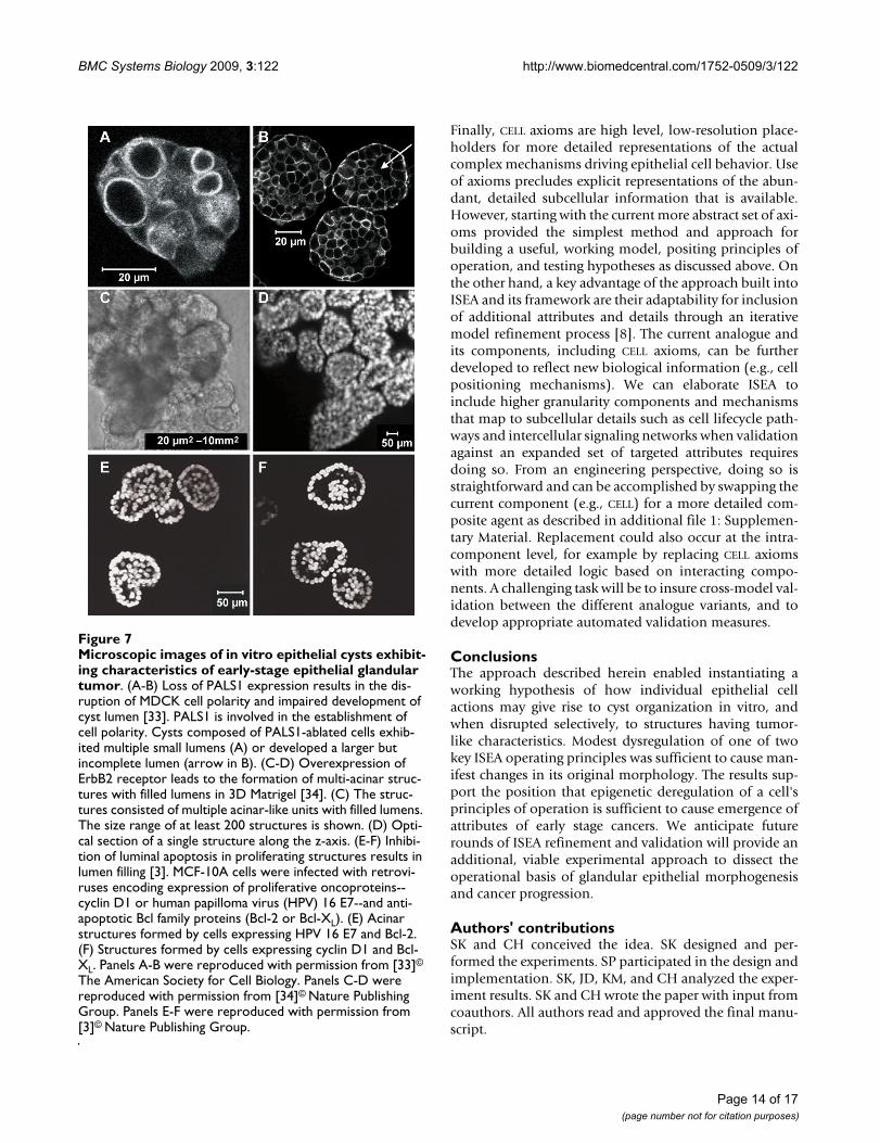
Microscopic images of in vitro epithelial cysts exhibiting char-acteristics of early-stage epithelial glandular tumorFigure 7Microscopic images of in vitro epithelial cysts exhibit-ing characteristics of early-stage epithelial glandular tumor. (A-B) Loss of PALS1 expression results in the dis-ruption of MDCK cell polarity and impaired development of cyst lumen [33]. PALS1 is involved in the establishment of cell polarity. Cysts composed of PALS1-ablated cells exhib-ited multiple small lumens (A) or developed a larger but incomplete lumen (arrow in B). (C-D) Overexpression of ErbB2 receptor leads to the formation of multi-acinar struc-tures with filled lumens in 3D Matrigel [34]. (C) The struc-tures consisted of multiple acinar-like units with filled lumens. The size range of at least 200 structures is shown. (D) Opti-cal section of a single structure along the z-axis. (E-F) Inhibi-tion of luminal apoptosis in proliferating structures results in lumen filling [3]. MCF-10A cells were infected with retrovi-ruses encoding expression of proliferative oncoproteins--cyclin D1 or human papilloma virus (HPV) 16 E7--and anti-apoptotic Bcl family proteins (Bcl-2 or Bcl-XL). (E) Acinar structures formed by cells expressing HPV 16 E7 and Bcl-2. (F) Structures formed by cells expressing cyclin D1 and Bcl-XL. Panels A-B were reproduced with permission from [33]©
The American Society for Cell Biology. Panels C-D were reproduced with permission from [34]© Nature Publishing Group. Panels E-F were reproduced with permission from [3]© Nature Publishing Group.
Page 14 of 17(page number not for citation purposes)

BMC Systems Biology 2009, 3:122 http://www.biomedcentral.com/1752-0509/3/122
AppendixRelationships between ISEA and cellular automataIt can be useful to relate agent-based models to cellularautomata. A CA consists of a regular grid of "cells" whichtransition through a series of states in discrete time steps.The "cells" are immobile. A "cell" transitions its statebased on the states of neighboring "cells". Transitions aresynchronous, meaning that all "cells" are updated eachtime step. A global software executive controls state tran-sitions and time evolution. A fundamental attribute of CAis the realization of non-local, complex behaviors arisingfrom the operation of local rules [50]. A CA can bethought of as a simple type of object-oriented program(OOP), where objects confined to specific locations mapto a CA's "cells" and the transition rules are the objects'methods. The only differences being that 1) the objects inan OOP can determine with which other objects theyinteract, 2) their interactions are not necessarily synchro-nous, and 3) any object may have more than a single statetransition rule. In essence, such an OOP can be viewed asbeing a more heterogeneous and dynamic type of CA.
An agent-based system (ABS) adds considerable heteroge-neity over and above that of an OOP. Whereas an OOP isnot necessarily synchronous, the control of when anobject interacts with another object and which objectsinteract is still handled by a global executive. Objects arereactive slaves to this global executive, even in a paral-lelized OOP. Within an ABS, on the other hand, some ofthe executive's capabilities and responsibilities, includingsome or all of the scheduling of actions, are distributed--delegated--to agents. An agent can be quasi-autonomous.It senses and is part of its environment, which may or maynot be discretized in the form of a grid. It pursues anagenda within a larger script. An agent can choose dynam-ically with which other agents or objects to interact, whento engage other agents or objects, and which of variousactions to take. It can also begin engaging in new actionswithout being told to do so or how to do so by a globalexecutive. Likewise, it can decide to stop engaging in agiven interaction. In fact, an agent can initiate or end theexecution of any of its logic, internal or interactive. Giventhose attributes, "agent" can be defined technically as anobject within an OOP with the ability to schedule its ownactions. In models such as an ISEA, an agent, like an actor,plays a role, participates in a process, or acts on behalf ofsomething else. Importantly, an agent is identifiable by anobserver as a cause of an effect. Some of an agent'sattributes and actions may be designed to represent bio-logical counterparts; others will deal with issues of soft-ware execution.
It can be important to distinguish an ABS from an agent-oriented system. In the former, all the capabilitiesdescribed exist in the software itself. In the latter, the
actual software may not have all the capabilities of anABS, but when the system is used, it is useful to think ofthe simulation as being composed of agents. In that sense,a CA may be agent-oriented but not agent-based. How-ever, an ABS is sufficiently far removed from a CA so thatthe analogy only has pedagogical value.
In vitro morphology observationsA number of molecules contribute to the establishment ofcell polarity and orientation in mammalian epithelialcells. Among these are PALS1 (Proteins Associated withLin Seven 1) and PATJ (PALS1-Associated Tight Junctionprotein). They form macromolecular complexes at tightjunctions. Straight et al. [33] ablated expression of PALS1in MDCK cells in a cyst formation assay, and that led todefects in polarity determination and the failure of cysts toproperly form a lumen. Microscopic images taken at day10 are shown in Fig. 7A-B. Cell masses contained either nolumen or several smaller lumens (Fig. 7A). Occasionally alarger, but incomplete lumen was observed (arrow, Fig.7B). In ISEA simulations, similar structures were observedwhen Axiom 6 was dysregulated. ISEA CELLS developedmultiple, relatively intact LUMENS at different, early timepoints, which disappeared or merged into a larger LUMEN
by simulation cycle 50 (data not shown). Some wereobserved at or around simulation cycle 20, which maps tothe time (day 10) that the in vitro images were captured.These observed in silico features were mostly transient.Time-lapse images at longer time points will be needed toconfirm (or dispute) the in silico observations.
Although MCF-10A and MDCK are different in severalways, when grown under identical 3D culture conditions,their structure formation processes exhibit many similari-ties, including formation of cysts having similar character-istics. We posit that for those conditions, the current ISEAis also an acceptable analogue of MCF-10A cyst forma-tion. Overexpression of ErbB2 oncoprotein receptors andtheir epidermal growth factor (EGF) ligands is implicatedin epithelial glandular cancer progression. To examine theeffects of activating ErbB receptors in a 3D in vitro context,Muthuswamy et al. [34] activated selected ErbB receptorsin preformed acinar structures composed of MCF-10Amammary epithelial cells. To create stable cell linesexpressing chimeric ErbB2 receptors, cells were infectedwith retroviruses encoding the chimeras that could beactivated by synthetic dimerizing ligands without interfer-ing with endogenous receptors and vice versa. When cul-tured in 3D Matrigel, the cells proliferated and organizedinto polarized, lumen-enclosing cysts. Upon activation ofthe chimera, these cysts developed structures consisting ofmultiple acinar-like units with filled lumen (Fig. 7C-D).The units within the multi-acinar structures were con-nected to each other at the base. These altered structuresexhibited characteristics of early-stage epithelial tumors,
Page 15 of 17(page number not for citation purposes)

BMC Systems Biology 2009, 3:122 http://www.biomedcentral.com/1752-0509/3/122
including a high level of proliferation, loss of polarizedorganization, filling of the lumen, and retention of thebasement membrane. Most structures were at least 10times larger than normal acini (~100 μm); some were 100times larger (Fig. 7C). They are similar to the ISEA mor-phology resulting from dysregulation of both Axioms 5and 6, which could map to the in vitro condition whereapoptosis, proliferation, and cell polarity are disrupted byErbB2 expression. As discussed in Results, severely dysreg-ulated CELLS developed large, expanding CELL masses withnumerous, incomplete LUMENS. Like their in vitro counter-parts, the ISEA structures were poorly POLARIZED. Withoutproper POLARIZATION, CELLS at the periphery continued toDIVIDE and expand outward. By simulation cycle 50, somestructures became at least 100-fold larger than stable, 'nor-mal' CYSTS. An example is shown in Fig. 6C.
Debnath et al. [3] used MCF-10A cell cultures to analyzethe role of apoptosis in the formation and maintenance ofluminal space during the in vitro morphogenesis of onco-gene-expressing mammary epithelial acini. They madetwo interventions: one to increase proliferation andanother to inhibit apoptosis. Proliferation was increasedvia the ectopic expression of cyclin D1 or the inactivationof the retinoblastoma protein tumor suppressor pathwayby the E7 oncoprotein from human papilloma virus(HPV) 16. Apoptosis was inhibited by infecting cells withretroviruses encoding for exogenous expression of anti-apoptotic Bcl family proteins. Fig. 7E-F shows typicalstructures formed when cells expressed both the prolifera-tive and anti-apoptotic proteins. Those cells produced aci-nar structures with partially or completely filled lumen.Similar, filled acinar structures were observed in anotherstudy, in which TIMP1, a potent cell survival oncoprotein,was used to inhibit both intrinsic and extrinsic apoptosis[26]. Axiom 5 dysregulation simulated a similar in vitrocondition where anoikis, a specific form of cell death asso-ciated with extrinsic apoptosis pathway, is disrupted. Thedysregulation resulted in structures (Fig. 3A) that exhib-ited morphological characteristics similar to those shownin Fig. 7E-F. Most structures had CELLS inside the CYST LUMI-
NAL SPACE.
Additional material
AcknowledgementsWe thank Mark Grant, Glen Ropella, Wei Yu, Jesse Engelberg, Jon Tang, Teddy Lam, Shahab Sheikh-Bahaei, and members of the BioSystems group for helpful discussions and suggestions. This research was supported in part by the CDH Research Foundation, a graduate fellowship to SK from the International Foundation for Ethical Research, the Culpeper Scholar Award (Partnership For Cures) to JD, and NIH grants R01 DK067153 and R01 DK074398 to KM. The funding bodies had no role in study design; in the collection, analysis, and interpretation of data; in the writing of the manu-script; and in the decision to submit the manuscript for publication.
References1. Tlsty TD, Crawford YG, Holst CR, Fordyce CA, Zhang J, McDermott
K, Kozakiewicz K, Gauthier ML: Genetic and epigenetic changesin mammary epithelial cells may mimic early events in car-cinogenesis. J Mammary Gland Biol Neoplasia 2004, 9:263-274.
2. O'Brien LE, Zegers MM, Mostov KE: Building epithelial architec-ture: insights from three-dimensional culture models. NatRev Mol Cell Biol 2002, 3:531-537.
3. Debnath J, Mills KR, Collins NL, Reginato MJ, Muthuswamy SK,Brugge JS: The role of apoptosis in creating and maintainingluminal space within normal and oncogene-expressing mam-mary acini. Cell 2002, 111:29-40.
4. Montesano R, Schaller G, Orci L: Induction of epithelial tubularmorphogenesis in vitro by fibroblast-derived soluble factors.Cell 1991, 66:697-711.
5. Martín-Belmonte F, Yu W, Rodríguez-Fraticelli AE, Ewald AJ, Werb Z,Alonso MA, Mostov K: Cell-polarity dynamics controls themechanism of lumen formation in epithelial morphogenesis.Curr Biol 2008, 18:507-513.
6. Fisher J, Henzinger TA: Executable cell biology. Nat Biotechnol2007, 25:1239-1249.
7. Hunt CA, Ropella GE, Park S, Engelberg JA: Dichotomies betweencomputational and mathematical models. Nat Biotechnol 2008,26:737-738.
8. Hunt CA, Ropella GE, Lam TN, Tang J, Kim SH, Engelberg JA, Sheikh-Bahaei S: At the biological modeling and simulation frontier.Pharm Res 2009, 26(11):2369-2400.
9. Cristini V, Lowengrub JS, Nie Q: Nonlinear simulation of tumorgrowth. J Math Biol 2003, 46:191-224.
10. Frieboes HB, Zheng X, Sun CH, Tromberg B, Gatenby R, Cristini V:An integrated computational/experimental model of tumorinvasion. Cancer Res. 2006, 66(3):1597-1604.
11. Sanga S, Frieboes HB, Zheng X, Gatenby R, Bearer EL, Cristini V: Pre-dictive oncology: a review of multidisciplinary in silico mod-eling linking phenotype, morphology and growth. NeuroImage2007, 37:S120-S134.
12. Grant MR, Mostov KE, Tlsty TD, Hunt CA: Simulating propertiesof in vitro epithelial cell morphogenesis. PLoS Comput Biol 2006,2:e129.
13. Debnath J, Brugge JS: Modelling glandular epithelial cancers inthree-dimensional cultures. Nat Rev Cancer 2005, 5:675-688.
14. Grimm V, Revilla E, Berger U, Jeltsch F, Mooij WM, Railsback SF,Thulke HH, Weiner J, Wiegand T, DeAngelis DL: Pattern-orientedmodeling of agent-based complex systems: lessons fromecology. Science 2005, 310:987-991.
15. Zeigler BP, Kim TG, Praehofer H: Theory of modeling and simulation:integrating discrete event and continuous complex dynamic systems SanDiego: Academic Press; 2000.
16. Merks RMH, Glazier JA: A cell-centered approach to develop-mental biology. Physica A 2005, 352:113-130.
17. Anderson ARA, Chaplain MAJ, Rejniak KA, Eds: Single-cell based mod-els in biology and medicine Basel, Switzerland: Birkhaüser; 2007.
18. Kim SHJ, Park S, Mostov K, Debnath J, Hunt CA: Computationalinvestigation of epithelial cell dynamic phenotype in vitro.Theor Biol Med Model 2009, 6:8.
19. Frisch SM, Screaton RA: Anoikis mechanisms. Curr Opin Cell Biol2001, 13:555-562.
20. Chiarugi P, Giannoni E: Anoikis: a necessary death program foranchorage-dependent cells. Biochem Pharmacol 2008,76:1352-1364.
21. Nelson WJ: Epithelial cell polarity from the outside looking in.News Physiol Sci 2003, 18:143-146.
Additional file 1Supplementary Material. Provided are detailed descriptions of ISEA morphology index, ISEA CELL axiom use patterns following Axiom 5 or 6 dysregulation, and a diagram illustrating model refinement and cross-model validation.Click here for file[http://www.biomedcentral.com/content/supplementary/1752-0509-3-122-S1.PDF]
Page 16 of 17(page number not for citation purposes)

BMC Systems Biology 2009, 3:122 http://www.biomedcentral.com/1752-0509/3/122
Publish with BioMed Central and every scientist can read your work free of charge
"BioMed Central will be the most significant development for disseminating the results of biomedical research in our lifetime."
Sir Paul Nurse, Cancer Research UK
Your research papers will be:
available free of charge to the entire biomedical community
peer reviewed and published immediately upon acceptance
cited in PubMed and archived on PubMed Central
yours — you keep the copyright
Submit your manuscript here:http://www.biomedcentral.com/info/publishing_adv.asp
BioMedcentral
22. Yu W, Datta A, Leroy P, O'Brien LE, Mak G, Jou TS, Matlin KS, Mos-tov KE, Zegers MM: Beta1-integrin orients epithelial polarityvia Rac1 and laminin. Mol Biol Cell 2005, 16:433-445.
23. Kroschewski R: Molecular mechanisms of epithelial polarity:about shapes, forces, and orientation problems. News PhysiolSci 2004, 19:61-66.
24. Luke S, Cioffi-Revilla C, Panait L, Sullivan K, Balan G: MASON: amultiagent simulation environment. Simulation 2005,81:517-527.
25. Boudreau N, Sympson CJ, Werb Z, Bissell MJ: Suppression of ICEand apoptosis in mammary epithelial cells by extracellularmatrix. Science 1995, 267:891-893.
26. Liu XW, Taube ME, Jung KK, Dong Z, Lee YJ, Roshy S, Sloane BF,Fridman R, Kim HR: Tissue inhibitor of metalloproteinase-1protects human breast epithelial cells from extrinsic celldeath: a potential oncogenic activity of tissue inhibitor ofmetalloproteinase-1. Cancer Res 2005, 65:898-906.
27. Sausedo RA, Smith JL, Schoenwolf GC: Role of nonrandomly ori-ented cell division in shaping and bending of the neural plate.J Comp Neurol 1997, 381:473-488.
28. Gong Y, Mo C, Fraser SE: Planar cell polarity signalling controlscell division orientation during zebrafish gastrulation. Nature2004, 430:689-693.
29. Baena-López LA, Baonza A, García-Bellido A: The orientation ofcell divisions determines the shape of Drosophila organs.Curr Biol 2005, 15:1640-1644.
30. Lee M, Vasioukhin V: Cell polarity and cancer--cell and tissuepolarity as a non-canonical tumor suppressor. J Cell Sci 2008,121:1141-1150.
31. Théry M, Racine V, Pépin A, Piel M, Chen Y, Sibarita JB, Bornens M:The extracellular matrix guides the orientation of the celldivision axis. Nat Cell Biol 2005, 7:947-953.
32. Théry M, Jiménez-Dalmaroni A, Racine V, Bornens M, Jülicher F:Experimental and theoretical study of mitotic spindle orien-tation. Nature 2007, 447:493-496.
33. Straight SW, Shin K, Fogg VC, Fan S, Liu CJ, Roh M, Margolis B: Lossof PALS1 expression leads to tight junction and polaritydefects. Mol Biol Cell 2004, 15:1981-1990.
34. Muthuswamy SK, Li D, Lelievre S, Bissell MJ, Brugge JS: ErbB2, butnot ErbB1, reinitiates proliferation and induces luminalrepopulation in epithelial acini. Nat Cell Biol 2001, 3:785-792.
35. Hebner C, Weaver VM, Debnath J: Modeling morphogenesis andoncogenesis in three-dimensional breast epithelial cultures.Annu Rev Pathol. 2008, 3:313-339.
36. Kitano H: Systems biology: a brief overview. Science 2002,295:1662-1664.
37. Aloy P, Russell RB: Structural systems biology: modelling pro-tein interactions. Nat Rev Mol Cell Biol 2006, 7:188-197.
38. Rizki A, Weaver VM, Lee SY, Rozenberg GI, Chin K, Myers CA, Bas-com JL, Mott JD, Semeiks JR, Grate LR, Mian IS, Borowsky AD, JensenRA, Idowu MO, Chen F, Chen DJ, Petersen OW, Gray JW, Bissell MJ:A human breast cell model of preinvasive to invasive transi-tion. Cancer Res 2008, 68:1378-1387.
39. Choi J, Choi K, Benveniste EN, Rho SB, Hong YS, Lee JH, Kim J, ParkK: Bcl-2 promotes invasion and lung metastasis by inducingmatrix metalloproteinase-2. Cancer Res 2005, 65:5554-5560.
40. David NB, Martin CA, Segalen M, Rosenfeld F, Schweisguth F, Bel-laïche Y: Drosophila Ric-8 regulates Galphai cortical localiza-tion to promote Galphai-dependent planar orientation ofthe mitotic spindle during asymmetric cell division. Nat CellBiol 2005, 7:1083-1090.
41. Wang H, Ng KH, Qian H, Siderovski DP, Chia W, Yu F: Ric-8 con-trols Drosophila neural progenitor asymmetric division byregulating heterotrimeric G proteins. Nat Cell Biol 2005,7:1091-1098.
42. Anderson AR, Weaver AM, Cummings PT, Quaranta V: Tumormorphology and phenotypic evolution driven by selectivepressure from the microenvironment. Cell 2006, 127:905-915.
43. Schaller G, Meyer-Hermann M: Continuum versus discretemodel: a comparison for multicellular tumour spheroids. Phi-los Transact A Math Phys Eng Sci 2006, 364:1443-1464.
44. Harpold HL, Alvord EC Jr, Swanson KR: The evolution of mathe-matical modeling of glioma proliferation and invasion. J Neu-ropathol Exp Neurol 2007, 66:1-9.
45. Wang Z, Zhang L, Sagotsky J, Deisboeck TS: Simulating non-smallcell lung cancer with a multiscale agent-based model. TheorBiol Med Model 2007, 4:50.
46. Engelberg JA, Ropella GE, Hunt CA: Essential operating princi-ples for tumor spheroid growth. BMC Syst Biol 2008, 2:110.
47. Galle J, Loeffler M, Drasdo D: Modeling the effect of deregulatedproliferation and apoptosis on the growth dynamics of epi-thelial cell populations in vitro. Biophys J 2005, 88:62-75.
48. Rejniak KA, Anderson AR: A computational study of the devel-opment of epithelial acini: I. Sufficient conditions for the for-mation of a hollow structure. Bull Math Biol 2008, 70:677-712.
49. Rejniak KA, Anderson AR: A computational study of the devel-opment of epithelial acini: II. Necessary conditions for struc-ture and lumen stability. Bull Math Biol 2008, 70:1450-1479.
50. Wolfram S: A new kind of science Champaign, IL: Wolfram Media; 2002. 51. Meredith JE, Fazeli B, Schwartz MA: The extracellular matrix as a
cell survival factor. Mol Biol Cell 1993, 4:953-961.52. Tang MJ, Hu JJ, Lin HH, Chiu WT, Jiang ST: Collagen gel overlay
induces apoptosis of polarized cells in cultures: disorientedcell death. Am J Physiol 1998, 275:C921-C931.
53. Hall HG, Farson DA, Bissell MJ: Lumen formation by epithelialcell lines in response to collagen overlay: a morphogeneticmodel in culture. Proc Natl Acad Sci USA 1982, 79:4672-4676.
54. Wang AZ, Ojakian GK, Nelson WJ: Steps in the morphogenesisof a polarized epithelium. I. Uncoupling the roles of cell-celland cell-substratum contact in establishing plasma mem-brane polarity in multicellular epithelial (MDCK) cysts. J CellSci. 1990, 95(Pt 1):137-151.
Page 17 of 17(page number not for citation purposes)