blunt cerebrovascular injury: diagnosis at whole-body mdct for multi-trauma · original article...
TRANSCRIPT

ORIGINAL ARTICLE
Blunt cerebrovascular injury: diagnosis at whole-bodyMDCT for multi-trauma
Matteo Bonatti & Norberto Vezzali & Federica Ferro &
Riccardo Manfredi & Nadia Oberhofer &
Giampietro Bonatti
Received: 29 December 2012 /Revised: 10 February 2013 /Accepted: 12 February 2013 /Published online: 21 March 2013# The Author(s) 2013. This article is published with open access at Springerlink.com
AbstractPurpose To analyse the prevalence of blunt cerebrovascularinjuries (BCVIs) in multi-trauma patients by means of apost-contrast acquisition of neck vessels included into thewhole-body multi-detector computed tomography (MDCT)protocol performed at admission and to correlate it with thepresence of risk factors (Memphis approach).Materials and methods A retrospective study was undertak-en for the period January 2005 to November 2011, involv-ing 976 multi-trauma patients. Post-contrast images of neckvessels in MDCT scan were evaluated by two experiencedradiologists; carotid, vertebral and basilar arteries were ratedaccording to the Biffl classification. The presence of clinicaland/or CT risk factors for BCVI was assessed.Results BCVI were present in 32/976 (3.3 %) multi-traumapatients. Risk factors for BCVI were present in 247/976(25.3 %) patients. The group of patients presenting riskfactors showed a significantly higher prevalence of cerebro-vascular injuries (8.1 %) compared with the group of pa-tients without risk factors (1.6 %) (p=0.009); however,12/32 (37.5 %) patients presenting BCVI did not show anyof the risk factors proposed by the Memphis group.
Conclusion An investigation for the presence of BCVIshould be performed on all multi-trauma patients despitethe absence of clinical-radiological risk factors.Key Points• BCVIs are present in 3.3 % of multi-trauma patients.• BCVIs are significantly associated to the Memphis riskfactors.
• Of the multi-trauma patients affected by BCVIs, 37.5 % donot show clinical-radiological risk factors.
• A screening for BCVI should be performed on all multi-trauma patients.
Keywords Carotid artery injury . Vertebral arterydissection . Multiple trauma . Spiral computed tomography .
Spine
Introduction
Blunt cerebrovascular injuries (BCVIs) are considered rela-tively uncommon in patients admitted to an emergencydepartment because of blunt trauma, with reported inci-dences of 0.5–1.5 %, but their incidence increases to about2.7 % if only patients with an Injury Severity Score (that isthe most widely adopted anatomical scoring system forgrading the severity of trauma in patients with multipleinjuries) greater than 16 are considered [1–7].
Clinical presentation of BCVI is similar to spontaneousdissections [8, 9]. Many BCVIs are asymptomatic in theirearly phases and they can be easilymissed at initial evaluation,particularly in severely traumatised patients [1, 10–14].
The outcomes of BCVI can be devastating, includingdebilitating stroke and death, with mortality rates of 8–38 % [3, 7, 9, 15, 16]. Although some conflicting results
M. Bonatti (*) :N. Vezzali : F. Ferro :G. BonattiDepartment of Radiology, San Maurizio Hospital,5 Boehler Street, 39100 Bolzano, Italye-mail: [email protected]
R. ManfrediDepartment of Radiology, Policlinico G.B. Rossi,10 L.A. Scuro Place, Verona, Italy
N. OberhoferDepartment of Physics, San Maurizio Hospital,5 Boehler Street, Bolzano, Italy
Insights Imaging (2013) 4:347–355DOI 10.1007/s13244-013-0235-y

exist [17], it is quite well established that an early treatmentimproves the outcome of patients affected by BCVI, reduc-ing stroke and mortality rates [3, 7, 9, 18–25]. Therefore, anearly diagnosis is fundamental.
Because of its high sensitivity and specificity, four-vesselcervicocerebral digital subtraction angiography (DSA) isconsidered the “gold standard” for the diagnosis of BCVI[26]; however, thanks to the recent technical developments,multi-detector computed tomography (MDCT) angiographyis nowadays regarded as an acceptable screening method forBCVI [27–31]. Moreover, the recent works by Sliker [30]and Rademacher [32] demonstrated that a post-contrast ac-quisition of the neck included into a whole-body MDCTprotocol can be considered an acceptable initial means ofscreening multi-trauma patients for the presence of BCVI.
Many different algorithms have been developed In orderto select which blunt-injured patients need to be aggressive-ly investigated for the presence of BCVI; the ones proposedby the groups of Denver [33] and Memphis [20] being themost widely accepted. All these algorithms are substantiallybased on the presence of clinical/radiological risk factors forBCVI [13], but they seem to lack in sensitivity, because ithas been proven that BCVI can occur even in the absence ofclinical-radiological risk factors [4, 6, 7, 34, 35].
The aim of our study was to analyse the prevalence ofBCVIs in a severely injured patient population, representedby patients classified as multi-trauma, by means of a post-contrast acquisition of neck vessels included in the whole-body MDCT examination that these patients routinely un-dergo at admission to our institution, and to correlate thepresence of BCVI to the presence of the risk factors pro-posed by the Memphis group.
Materials and methods
Patient population
Our retrospective study was approved by the InvestigationalReview Board of our Institution; informed consent was waived.
In the period January 2005 to November 2011, 1153patients admitted to the Emergency Department of our Insti-tution have been classified as multi-trauma, according to theirclinical situation (two or more injuries, of which at least onelife-threatening or Injury Severity Score≥16) and/or to thetrauma dynamic (high-speed trauma or fall from an height>3m), and therefore they have been considered eligible for theperformance of a whole-bodyMDCTaccording to our “multi-trauma protocol”. We considered for inclusion into our studythe 994/1153 (86.2 %) patients who underwent an MDCTexamination within the first 12 h from admission; whereas weexcluded the 52/1153 (4.5 %) patients who underwent theMDCT >12 h from admission and the 107/1153 (9.3 %)
patients who did not undergo MDCT because of death. Eigh-teen out of 994 (1.8 %) patients were excluded because ofinsufficient diagnostic quality of the post-contrast cervicalvessels acquisition due to motion artefacts or technical prob-lems. Thus, our study population encompassed 976 patients,770 men (78.9 %) and 206 women (21.1 %), with a medianage of 43 years (range 18–96 years).
Mechanisms of injury were car (331/976, 33.9 %) ormotorcycle crash (233/976, 23.9 %), skiing (201/976,20.6 %) or bicycle accident (64/976, 6.6 %), pedestrianstruck (50/976, 5.1 %), fall from a great height (42/976,4.3 %) and other less common mechanisms (55/976, 5.6 %).
Clinical data at the time of admission were retrieved fromour institutional informatics system.
Imaging technique: whole-body MDCT multi-traumaprotocol
All the CT examinations were performed on a 16-rowMDCT scanner (Somatom Sensation 16, Siemens MedicalSystems, Erlangen, Germany). The patient was placed su-pine on the table, with the arms lying down along the bodyas close as possible to the abdomen in order to reduce beamhardening artefacts.
The first step of our standard whole body MDCT multi-trauma protocol is a plain helical acquisition of the head,including the mandible, with the following scanning param-eters: 16×0.75 mm collimation, 120 kV tube voltage,320mAs tube current and pitch of 0.55.
After that we administer, through a peripheral venousaccess of at least 18 G, 120–150 ml iodine-containing con-trast medium (Iomeprolo, Iomeron350; Bracco, Milano, It-aly) using a power injector (Spectris; Medrad, Pittsburgh,Pennsylvania, USA) at a flow rate of 4.0 ml/s, followed by asaline flush of 50 ml at a flow rate of 4.0 ml/s.
The post-contrast scan is acquired using a bolus trackerplaced in the aortic arch, with a threshold trigger of 100 HUand a delay trigger of 9 s and extends cranio-caudally from theskull basis to the femoral lesser trochanters, with a 16×1.5-mmcollimation and a pitch of 1.15, using the CARE dose 4Dautomatic exposure control (SiemensMedical Systems). More-over a portal venous phase scan of the abdomen is acquiredwith a fixed delay of 70 s after injection start, extending cranio-caudally with a 16×1.5-mm collimation, pitch 0.75 and usingCARE dose 4D automatic exposure control modality.
According to the clinical question and to the imagingfindings, additional pre- and/or post-contrast acquisitionscan be performed.
The obtained axial source images are reformatted asmulti-planar reconstructions (MPRs) and maximum intensi-ty projections (MIP) with different kernels and slice thick-nesses according to the different anatomical districts; inparticular, the axial source images of craniocervical vessels
348 Insights Imaging (2013) 4:347–355

are reformatted as 2-mm MPRs with a reconstruction incre-ment of 1 mm, using a sharp kernel.
Image analysis
Two radiologists (with 22 and 12 years of experience inemergency radiology, respectively), who were unaware ofthe trauma dynamics and of the clinical conditions of thepatient, independently evaluated the post-contrast CT im-ages of cerebrovascular vessels. Discrepancies were solvedby consensus. Common carotid arteries, internal carotidarteries, vertebral arteries and basilar artery were evaluatedon 2-mm MPR images and each vessel was classified asfollows, according to the angiographic classification pro-posed by Biffl et al. [36] (Figs. 1, 2, 3, 4 and 5): normal(grade 0), lumen wall irregularity or lumen narrowing<25 % (grade I) (Fig. 1a); intraluminal thrombus or lumen
narrowing >25 % (grade II) (Fig. 1b–d); pseudoaneurysm(grade III) (Fig. 2a–d); complete occlusion (grade IV)(Fig. 3a–d); transection associated to active extravasationof contrast media (grade V) (Fig. 4a–c).
A third radiologist (with 3 years of experience in emergen-cy radiology), aware of the trauma dynamics and of theclinical conditions of the patient, searched for the presenceof risk factors for BCVI: cervical spine fractures, Le Fort II orIII facial fractures, skull base fractures involving the foramenlacerum and CT signs of diffuse axonal injury (considered asthe presence of five or more small petechial haemorrhageslocated at grey-white matter junction and in corpus callosum).
Statistical analysis
The inter-observer variability was assessed by means ofkappa statistic for the qualitative image analysis parameters
Fig. 1 Low-grade blunt cerebrovascular injury (BCVI) (grade I and IIlesions according to the Biffl classification). a A 42-year-old man whohad suffered a motorcycle accident. The axial MPR image (3-mmthickness) shows a slight irregularity in the posterior wall of the leftinternal carotid artery (arrow) responsible of a calibre reduction <25 %(grade I lesion). No clinical-radiological risk factors for BCVI werepresent. The man did not undergo anti-aggregation because of thepresence of severe abdominal injuries and developed an infarction inthe territory of the left median cerebral artery. b An 83-year-old womanstruck by a car. The axial MPR image (3-mm thickness) shows aneccentric calibre reduction of the left vertebral artery (arrow) >25 %
(grade 2 lesion). Cervical spine fractures, including a fracture of thespinous process of C3 (arrowhead) were present. The vertebral arterylesion had not been described on the radiological report and, therefore,no therapy had been administered. The woman developed a cerebellarinfarction. c, d A 34-year-old man who had suffered a motorcycleaccident. The axial MIP reconstruction (c) (3-mm thickness) shows alumen wall irregularity of the right internal carotid artery (arrow)responsible of a calibre reduction >25 % (grade IIlesion); the lesionis better depicted on the sagittal MIP reconstruction d (5-mm thick-ness). No clinical-radiological risk factors for BCVI were present. Themen underwent anti-aggregation and no neurological deficit occurred
Insights Imaging (2013) 4:347–355 349

regarding the evaluation of carotid, vertebral and basilararteries (graded from 0 to 5). The strength of agreementwas evaluated as follows: <0.20 poor, 0.21–0.40 fair, 0.41–0.60 moderate, 0.61–0.80 good, 0.81–1.0 excellent.
Categorical variables were expressed as absolute fre-quencies and percentages, continuous variables as mediansand ranges (minimum and maximum values).
BCVI prevalence was analysed with respect to patientgender and age, lesion type, lesion grade, risk factor pres-ence and risk factor type. Statistical analysis was carried outusing non-parametric tests. Statistical significance was stat-ed using the χ2-test, with Yates’ correction when appropri-ate. A p value of less than 0.05 was considered to indicate asignificant difference.
Correlation between the number of risk factors present ina patient and the occurrence of BVCI was assessed with theSpearman test with modified standard error ([35]), in order
to take into account the large differences in sample sizebetween patient groups having 1, 2, 3 or 4 concomitantclinical-radiological risk factors.
Results
We found an excellent inter-observer agreement in the clas-sification of carotid, vertebral and basilar arteries accordingto the Biffl grading system (K=0.97).
Thirty-two out of the 976 patients included in our study(3.3 %) showed the presence of BCVI at the MDCT exam-ination: 22 out of these 32 patients (68.8 %) presented theinvolvement of a single vessel, 10/32 (32.2 %) presented theconcomitant involvement of two vessels, whereas none ofthe included patients presented the involvement of three ormore vessels. Therefore, a total of 42 cerebrovascular
Fig. 2 Blunt cerebrovascular pseudoaneurysm (grade III lesionsaccording to the Biffl classification). a, b A 57-year-old man who hadsuffered a car accident. The axialMPR image (a) (4-mm thickness) showsan eccentric dilatation of the right common carotid artery (arrow) with thetypical “snowman” appearance; the pseudoaneurysm (arrow) is betterdepicted on the coronal MIP reconstruction (b) (10 mm thickness). Agrade IV lesion of the left common carotid artery was also present. Noclinical-radiological risk factors for BCVI were present. The patientunderwent anti-aggregation and no neurological deficit occurred. c, d A
19-year-old man who had suffered a car accident. The sagittal MPRimage (c) shows a tortuous course (arrowheads) of the right internalcarotid artery; the presence of two pseudoaneurysms (arrowheads) isbetter depicted on the curved MPR image (d) (3-mm thickness). Skullbase fractures and a grade IV lesion of the left common carotid arterywere present. The patient underwent anti-aggregation but neurologicaldeficit related to a left anterior circle ischaemia was already present at thetime of MDCT
350 Insights Imaging (2013) 4:347–355
lesions were observed in our patient population, with anoverall prevalence of injured vessels of 4.3 %. BCVI in-volved the carotidal axis (common, internal and externalcarotid arteries) in 26/42 cases (61.9 %) and the vertebro-basilar axis (basilar and vertebral arteries) in 16/42 cases(38.1 %). Blunt cerebrovascular injuries were classified asgrade 1 (Fig. 1a) in 10/42 (23.8 %) cases, as grade 2(Fig. 1b–d) in 8/42 (19.0 %) cases, as grade 3 (Fig. 2a–d)in 6/42 (14.3 %) cases, as grade 4 (Fig. 3a–d) in 16/42(38.1 %) cases and as grade 5 (Fig. 4a–c) in 2/42 (4.8 %)cases. Data about frequencies of the involved vessels andcorresponding lesion grading are reported in Table 1.
Out of the 976 included patients, 247 (25.3 %) showedthe presence of one or more clinical-radiological risk factorsfor BCVI, according to the Memphis criteria [20]. In partic-ular, 134/976 (13.7 %) patients showed the presence ofcervical spine fractures, 93/976 (9.5 %) patients presenteda Le Fort II or Le Fort III facial fracture and 25/976 (2.6 %)
of them presented skull base fractures involving the foramenlacerum. Moreover, diffuse axonal injury was suspected onthe basis of the brain CT findings in 47/976 (4.8 %) patients.Among the 247 patients presenting risk factors for BCVI,195 (78.9 %) presented a single risk factor, whereas 52 ofthem (21.1 %) presented the coexistence of two or more riskfactors.
Within the group of 247 patients presenting clinical-radiological risk factors for BCVI, 20/247 (8.1 %) patientsshowed the presence of BCVI at MDCT (a total of 24injured vessels has been found within this group of 20patients, since four of them showed the involvement oftwo vessels each), whereas 227/247 (92.9 %) patients didnot definitely show the presence of BCVI at MDCT.
Among the 729 patients without clinical-radiological riskfactors for BCVI, 12/729 (1.6 %) patients showed the pres-ence of BCVI at MDCT (a total of 18 injured vessels hasbeen found within this group of 12 patients, since six of
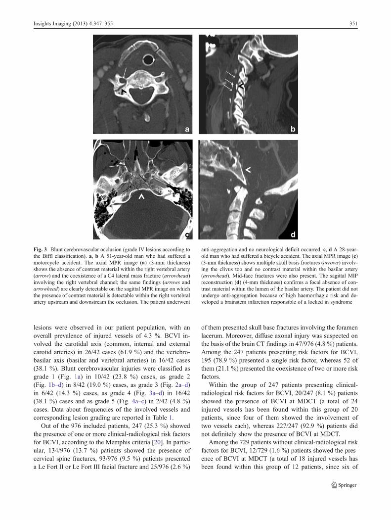
Fig. 3 Blunt cerebrovascular occlusion (grade IV lesions according tothe Biffl classification). a, b A 51-year-old man who had suffered amotorcycle accident. The axial MPR image (a) (3-mm thickness)shows the absence of contrast material within the right vertebral artery(arrow) and the coexistence of a C4 lateral mass fracture (arrowhead)involving the right vertebral channel; the same findings (arrows andarrowhead) are clearly detectable on the sagittal MPR image on whichthe presence of contrast material is detectable within the right vertebralartery upstream and downstream the occlusion. The patient underwent
anti-aggregation and no neurological deficit occurred. c, d A 28-year-old man who had suffered a bicycle accident. The axial MPR image (c)(3-mm thickness) shows multiple skull basis fractures (arrows) involv-ing the clivus too and no contrast material within the basilar artery(arrowhead). Mid-face fractures were also present. The sagittal MIPreconstruction (d) (4-mm thickness) confirms a focal absence of con-trast material within the lumen of the basilar artery. The patient did notundergo anti-aggregation because of high haemorrhagic risk and de-veloped a brainstem infarction responsible of a locked in syndrome
Insights Imaging (2013) 4:347–355 351

them presented the contemporary involvement of two ves-sels each), whereas 717/729 (98.4 %) did not show thepresence of BCVI at MDCT.
The comparison of prevalence of patients affected byBCVI in the group presenting risk factors versus theprevalence of patients affected by BCVI in the groupwithout risk factors demonstrated a significant correlationbetween blunt cerebrovascular injuries and the presenceof clinical-radiological risk factors according to the Mem-phis criteria (p=0.009, χ2-test with Yates’ correction); thestrength of correlation was p<0.0001 for injuries of thevertebro-basilar axis and p=0.025 for carotid arteriesinjuries (both χ2-test with Yates’ correction). Moreover,a statistically significant positive correlation between thenumber of clinical-radiological risk factors present ineach patient and the prevalence of BCVI has been found(p<0.0002, Spearman test with modified standard error[35]): the higher is the number of risk factors, the higheris the probability of having a cerebrovascular injury(p<0.001) (Fig. 6).
Discussion
Our study evaluated over a 7-year period 976 consecutivemulti-trauma patients that underwent a whole-body MDCT
scan according to our multi-trauma protocol within 12 hfrom admission to our emergency department.
Our study showed an extremely high inter-observeragreement (K=0.97) in the classification of carotid, verte-bral and basilar arteries according to the Biffl grading sys-tem; this could be a consequence of the high percentage ofnegative imaging studies (96.3 %) in our patient population.
BCVIs were present in 3.3 % of our multi-trauma pa-tients. The prevalence of BCVI in our patient populationwas higher compared with the values reported in the litera-ture, which range from 0.5 % to 2.7 % according to thedifferent patient populations. This discrepancy can be con-sidered a consequence of the particular characteristics of ourpatient population: indeed, differently from other studies, inour study we included only severely injured patients, clas-sified as multi-trauma at admission to our Emergency De-partment, instead of all blunt-injured patients, and it is wellknown that the probability of suffering from BCVI increaseswith an increase of the Injury Severity Score [1–7].
A total of 42 injured vessels has been found in 32 patients,since ten of them presented the concomitant involvement oftwo vessels each; in nine out of these ten cases the patientshowed symmetrical lesions, regarding the same vessel bilat-erally, i.e. either bilateral involvement of the carotidal axis orbilateral involvement of the vertebro-basilar axis, whereasonly in one out of ten cases carotid and vertebral arteries were
Fig. 4 Blunt cerebrovasculartransection (grade V lesionaccording to the Bifflclassification). A 24-year-oldman who had suffered amotorcycle accident. In theaxial MPR image (a) (3-mmthickness) contrast material isclearly recognisable within theleft vertebral artery (arrow);moreover, active arterialbleeding causing contrastmaterial extravasation isappreciable next to the leftvertebral artery (arrowheads).The same findings arerecognisable on the sagittalMPR image (b) (3-mmthickness) where an anteriorsliding of C1 (arrowhead) overC2 is also appreciable. Thecoronal MPR image (c) (3-mmthickness) demonstrates that theaforementioned alterations aredue to an odontoid process basefracture (type III fractureaccording to the Andersonclassification) (arrowhead).The patient died shortly afterthe CT
352 Insights Imaging (2013) 4:347–355

involved at the same time. This fact might be related to thedifferent trauma dynamics involving carotid and vertebro-basilar arteries; indeed, carotid arteries are usually injured asa result of a stretching mechanism due to hyperextension androtation of the neck, whereas vertebro-basilar arteries, whichare strictly surrounded by bony structures, are more common-ly directly damaged by bone fractures.
A review of the original MDCT radiological reports of thepatients affected by BCVI showed that lesions had beenoriginally missed in 3/32 (9.4 %) patients. The missed injurieswhere one grade 1 lesion of right vertebral artery, one grade 1lesion of left internal carotid artery and one grade 2 lesion ofleft vertebral artery. This result highlights the possible diffi-culties in diagnosing BCVI, particularly in low grade cases.
Our study confirmed the existence of a significant correla-tion between the presence of clinical-radiological risk factors asproposed by the Memphis group and the eventuality of suffer-ing from blunt cerebrovascular injury (p=0.009). To be more
precise, we found a stronger correlation between the presenceof risk factors and vertebro-basilar injuries than between thepresence of risk factors and carotid arteries injuries (p<0.001 vsp=0.025); this result might also be related with the above-mentioned different trauma dynamics involving carotid andvertebro-basilar arteries, the latter usually being directly injuredby bone lesions and, therefore, strongly associated to cervicalspine fractures. The risk factors proposed in the Memphisapproach, however, do not only represent possible direct vas-cular injury pathways, like in case of cervical and skull basefractures, but they alsomay represent an index of the severity ofthe cranio-cervical trauma, like in case of Le Fort II and IIIfractures and diffuse axonal injury; this can explain why carotidarteries injuries are also significantly associated with the above-mentioned risk factors, although not as strongly as vertebralones. Moreover, our study showed the existence of a positivecorrelation between the number of clinical-radiological riskfactors and the probability for a patient of having a blunt
Fig. 5 Possible devastatingoutcomes of untreated BCVIs.A 28-year-old man who hadsuffered a car accident. Thesagittal MIP reconstruction (a)(3-mm thickness) shows aprogressive tapering of the leftinternal carotid artery(arrowheads) that becomesoccluded before the intracranialportion. Latero-lateral digitalsubtraction angiography image(b) shows exactly the samefinding as MDCT (arrowheads)with no contrast material withinthe intracranial portion of leftinternal carotid artery. Thepatient showed no risk factorsfor BCVI and wasneurologically asymptomatic atthe time of MDCT. Anti-aggregation was not undertakenbecause of the presence ofsevere abdominal injuries andhigh bleeding risk. The axialDWI MR image (c) (b=1,000)acquired 2 days later, after theonset of right hemiparesis andaphasia, shows a marked signalhyperintensity in the territory ofthe left mean cerebral arteryrepresenting acute infarction
Table 1 Blunt cerebrovascularinjuries (BCVIs) (42 lesions):number of involved vessels cor-related to the grading of thelesions
Grade 1 Grade 2 Grade 3 Grade 4 Grade 5 Total
Common and internal carotid arteries 6 7 6 7 0 26
Vertebral and basilar arteries 4 1 0 9 2 16
Total 10 8 6 16 2 42
Insights Imaging (2013) 4:347–355 353

cerebrovascular injury: the higher is the number of risk factorsthat are contemporary present in the same time, the higher is theprobability for that patient of having a cerebrovascular injury(p<0.0002) (Fig. 6).
Nevertheless, according to our data 37.5 % of the patientsaffected by BCVI did not show any of the above-mentionedclinical-radiological risk factors. As a result, a screeningexamination for the presence of blunt cerebrovascular inju-ries cannot be exclusively reserved for multi-trauma patientspresenting clinical-radiological risk factors for BCVI, be-cause such an approach would miss an early diagnosis inmore than one-third of positive cases, denying therefore thepossibility of the instauration of a prompt therapy and in-creasing the probability of catastrophic outcomes. There-fore, according to our experience, all multi-trauma patientsshould be screened for the presence of BCVI, despite theabsence of risk factors. With BCVI being relatively uncom-mon, showing an incidence of 3.3 %, the selected screeningmethod should be cost effective and not time consuming orinvasive; in order to achieve this goal, a post-contrast ac-quisition of the neck can be easily integrated into the whole-body MDCT protocol performed in haemodynamically sta-ble multi-trauma patients at admission without additionalcontrast material administration nor an additional dose ex-posure, because it substitutes the plain acquisition of thecervical spine.
The main limitation of our study is due to its retrospec-tive design. Another weakness is the absence of a compar-ison of MDCT with the gold standard, which for BCVI isstill considered to be DSA. Anyway, the recent literature[30, 32] demonstrated that MDCT can be considered anacceptable initial means of screening for BCVI, withoutstatistically significant differences in accuracy comparedwith DSA. In our opinion, an invasive, costly and time
consuming diagnostic investigation like DSA could be re-served to patients with high clinical suspicion of BCVI andnegative MDCT findings.
In conclusion, we strongly suggest to screen all multi-trauma patients for the presence of BCVI by including abolus-timed arterial scan of the neck into the whole-bodyMDCT protocol performed on haemodynamically stablepatients at admission in order to early detect and early treatBCVIs.
Acknowledgments We would like to thank Professor AgostinoTarsitano for his help in the statistical analysis.
Open Access This article is distributed under the terms of the CreativeCommons Attribution License which permits any use, distribution, andreproduction in any medium, provided the original author(s) and thesource are credited.
References
1. Biffl WL, Moore EE, Elliott JP et al (2000) The devastatingpotential of blunt vertebral arterial injuries. Ann Surg 231(5):672–681
2. Mutze S, Rademacher G, Matthes G, Hosten N, Stengel D (2005)Blunt cerebrovascular injury in patients with blunt multiple trau-ma: diagnostic accuracy of duplex Doppler US and early CTangiography. Radiology 237(3):884–892
3. Miller PR, Fabian TC, Bee TK et al (2001) Blunt cerebrovascularinjuries: diagnosis and treatment. J Trauma 51(2):279–285, discus-sion 85–6
4. Schneidereit NP, Simons R, Nicolaou S et al (2006) Utility ofscreening for blunt vascular neck injuries with computed tomo-graphic angiography. J Trauma 60(1):209–215, discussion 15–6
5. Cothren CC, Moore EE, Ray CE Jr et al (2005) Screening for bluntcerebrovascular injuries is cost-effective. Am J Surg 190(6):845–849
6. Sliker CW (2008) Blunt cerebrovascular injuries: imaging withmultidetector CT angiography. Radiographics 28(6):1689–1708,discussion 709–10
7. Stein DM, Boswell S, Sliker CW, Lui FY, Scalea TM (2009) Bluntcerebrovascular injuries: does treatment always matter? J Trauma66(1):132–143, discussion 43–4
8. Fusco MR, Harrigan MR (2011) Cerebrovascular dissections—areview part I: Spontaneous dissections. Neurosurgery 68(1):242–257, discussion 57
9. Fusco MR, Harrigan MR (2011) Cerebrovascular dissections: areview. Part II: blunt cerebrovascular injury. Neurosurgery 68(2):517–530, discussion 30
10. Cogbill TH, Moore EE, Meissner M et al (1994) The spectrum ofblunt injury to the carotid artery: a multicenter perspective. JTrauma 37(3):473–479
11. Rodriguez M, Tyberghien A, Matge G (2001) Asymptomatic ver-tebral artery injury after acute cervical spine trauma. ActaNeurochir (Wien) 143(9):939–945
12. Fabian TC, Patton JH Jr, Croce MA, Minard G, Kudsk KA,Pritchard FE (1996) Blunt carotid injury. Importance of earlydiagnosis and anticoagulant therapy. Ann Surg 223(5):513–522,discussion 22–5
13. Franz RW,Willette PA,WoodMJ,Wright ML, Hartman JF (2012) Asystematic review and meta-analysis of diagnostic screening criteriafor blunt cerebrovascular injuries. J Am Coll Surg 214(3):313–327
Fig. 6 Relative frequency of BCVI correlated with the number of riskfactors. The x-axis shows the number of risk factors characterising eachpatient group, whereas the y-axis reports the probability (in percentage)of having a BCVI for each group. The area of the blue circles iscorrelated with the size of each patient group (729 patients with norisk factors, 195 patients with one risk factor, 41 with two risk factors,10 with three risk factors, 1 with four risk factors)
354 Insights Imaging (2013) 4:347–355

14. Burlew CC, Biffl WL, Moore EE, Barnett CC, Johnson JL,Bensard DD (2012) Blunt cerebrovascular injuries: redefiningscreening criteria in the era of noninvasive diagnosis. JTrauma Acute Care Surg 72(2):330–335, discussion 6–7,quiz 539
15. Mokri B (1990) Traumatic and spontaneous extracranial internalcarotid artery dissections. J Neurol 237(6):356–361
16. Krajewski LP, Hertzer NR (1980) Blunt carotid artery trauma:report of two cases and review of the literature. Ann Surg 191(3):341–346
17. Mayberry JC, Brown CV, Mullins RJ, Velmahos GC (2004)Blunt carotid artery injury: the futility of aggressive screen-ing and diagnosis. Arch Surg 139(6):609–612, discussion12–3
18. DiCocco JM, Fabian TC, Emmett KP et al (2011) Optimal out-comes for patients with blunt cerebrovascular injury (BCVI): tai-loring treatment to the lesion. J Am Coll Surg 212(4):549–557,discussion 57–9
19. Cothren CC, Moore EE, Biffl WL et al (2004) Anticoagulation isthe gold standard therapy for blunt carotid injuries to reduce strokerate. Arch Surg 139(5):540–545, discussion 5–6
20. Miller PR, Fabian TC, Croce MA et al (2002) Prospectivescreening for blunt cerebrovascular injuries: analysis of diagnosticmodalities and outcomes. Ann Surg 236(3):386–393, discussion93–5
21. Harrigan MR, Weinberg JA, Peaks YS et al (2011) Management ofblunt extracranial traumatic cerebrovascular injury: amultidisciplinarysurvey of current practice. World J Emerg Surg 6:11
22. Biffl WL, Ray CE Jr, Moore EE et al (2002) Treatment-relatedoutcomes from blunt cerebrovascular injuries: importance of rou-tine follow-up arteriography. Ann Surg 235(5):699–706, discus-sion 706−7
23. Parodi JC, Schonholz C, Ferreira LM, Bergan J (1999)Endovascular stent-graft treatment of traumatic arterial lesions.Ann Vasc Surg 13(2):121–129
24. Price RF, Sellar R, Leung C, O’Sullivan MJ (1998) Traumaticvertebral arterial dissection and vertebrobasilar arterial thrombosissuccessfully treated with endovascular thrombolysis and stenting.AJNR Am J Neuroradiol 19(9):1677–1680
25. Attigah N, Kulkens S, Zausig N et al (2009) Surgical therapy ofextracranial carotid artery aneurysms: long-term results over a 24-year period. Eur J Vasc Endovasc Surg 37(2):127–133
26. Biffl WL, Ray CE Jr, Moore EE, Mestek M, Johnson JL, Burch JM(2002) Noninvasive diagnosis of blunt cerebrovascular injuries: apreliminary report. J Trauma 53(5):850–856
27. Bromberg WJ, Collier BC, Diebel LN et al (2010) Blunt cerebro-vascular injury practice management guidelines: the Eastern As-sociation for the Surgery of Trauma. J Trauma 68(2):471–477
28. Biffl WL, Cothren CC, Moore EE et al (2009) Western TraumaAssociation critical decisions in trauma: screening for and treat-ment of blunt cerebrovascular injuries. J Trauma 67(6):1150–1153
29. Langner S, Fleck S, Kirsch M, Petrik M, Hosten N (2008) Whole-body CT trauma imaging with adapted and optimized CT angiog-raphy of the craniocervical vessels: do we need an extra screeningexamination? AJNR Am J Neuroradiol 29(10):1902–1907
30. Sliker CW, Shanmuganathan K, Mirvis SE (2008) Diagnosis ofblunt cerebrovascular injuries with 16-MDCT: accuracy of whole-body MDCT compared with neck MDCT angiography. AJR Am JRoentgenol 190(3):790–799
31. Fleck SK, Langner S, Baldauf J, Kirsch M, Kohlmann T,Schroeder HW (2011) Incidence of blunt craniocervical arteryinjuries: use of whole-body computed tomography trauma imag-ing with adapted computed tomography angiography. Neurosur-gery 69(3):615–623, discussion 23–4
32. Rademacher G (2008) Gefaessdissektion der hirnversorgenden Arterien:Screening im Rahmen der Ganzkoerpercomputertomographie. Traumaund Berfufskrankheit 3(10):182–186
33. Biffl WL, Moore EE, Offner PJ et al (1999) Optimizing screeningfor blunt cerebrovascular injuries. Am J Surg 178(6):517–522
34. Hughes KM, Collier B, Greene KA, Kurek S (2000) Traumaticcarotid artery dissection: a significant incidental finding. Am Surg66(11):1023–1027
35. Cook A, Osler T, Gaudet M, Berne J, Norwood S (2011) Bluntcerebrovascular injury is poorly predicted by modeling with otherinjuries: analysis of NTDB data. J Trauma 71(1):114–119
36. Biffl WL, Moore EE, Offner PJ, Brega KE, Franciose RJ, BurchJM (1999) Blunt carotid arterial injuries: implications of a newgrading scale. J Trauma 47(5):845–853
Insights Imaging (2013) 4:347–355 355