bls 2014 – orthopedic emergencies. course objectives 1.identify the structure and function of bone...
TRANSCRIPT
© 2013 Seattle / King County EMS
BLS 2014 – Orthopedic Emergencies
© 2013 Seattle / King County EMS
Course Objectives1. Identify the structure and function of bone2. Describe how to evaluate orthopedic injuries3. Describe energy transmission as it applies to fractures4. Predict injuries based on mechanism of injury5. Describe how to evaluate orthopedic injuries 6. Review splinting principles7. Describe how to choose and apply a splint to treat various orthopedic injuries8. Prioritize splinting in patients who are severely traumatized9. Understand, recognize, and treat shock
© 2013 Seattle / King County EMS
Mechanics
© 2013 Seattle / King County EMS
Bones
Bones are made of calcium, collagen, and living cells Collagen is strong and lightweight,
and provides elasticity Calcium is a mineral that maintains
bone density Bones contain living cells and have
their own blood supply
© 2013 Seattle / King County EMS
Functions of bone
Support Bones are the scaffolding of the body
and provide protection to underlying organs and body systems
Movement Bones provide a framework for the
attachment of muscles, tendons, and ligaments, allowing movement
Physiologic processes Bones produce blood cells and
hormones
© 2013 Seattle / King County EMS
Related StructuresBones & muscles work together to create movement Muscles – attached to bones by tendons Tendons – extension of fascia that cover all skeletal muscles Fascia – sheets or bands of tough, fibrous connective tissue that lie deep under skin form an outer layer of the muscles
Supplied with arteries, veins & nerves.
© 2013 Seattle / King County EMS
Joint Joint – location where two bones come together
Immovable joints – those between the bones of the skull Slightly movable joints – those in the front of the pelvis Movable joints – for example, elbow & knee
© 2013 Seattle / King County EMS
Mechanism of Injury
© 2013 Seattle / King County EMS
Mechanism of Injury
An important aspect of patient care: assess mechanism of injury & determine which forces have been applied to patient's body
Consider signs of blunt or penetrating trauma
Consider which underlying structures may have been impacted by force
© 2013 Seattle / King County EMS
Mechanism of Injury
Significant force is usually required to fracture a bone or dislocate a joint
Many types of forces can cause these injuries Direct – Fall on the tail bone that
cracks coccyx Indirect – Person falling & landing on
feet causing vertebral fracture Twisting – Skiing causes twisting
injuries – can crack ankle or tibia High-energy forces – Car striking
another car
© 2013 Seattle / King County EMS
Trauma and the Elderly Risk of fatality from
multi-system trauma is three times greater at age 70 than age 20
Happens because elderly body does not compensate effectively from trauma
Most trauma deaths in seniors caused by falls & motor vehicle accidents
Consider following factors: Elderly patients may lie
in extreme environments for long periods of time before help arrives leading to hypothermia or hyperthermia
Elderly patients more often dehydrated & malnourished
Chest trauma more likely to cause lung damage because chest wall is less flexible; ribs can break and lacerate lungs
© 2013 Seattle / King County EMS
Osteoporosis Extreme force or transfer of energy is
not always necessary to fracture a bone Osteoporosis – loss of bone density
Usually caused by calcium loss Common in women who have gone through
menopause Insignificant force can easily fracture a
bone weakened by osteoporosis Geriatric patients with osteoporosis
Minor fall, simple twisting injury or even a violent muscle contraction may cause a fracture

© 2013 Seattle / King County EMS
Evaluation
© 2013 Seattle / King County EMS
Arriving at a Trauma Scene
Keep yourself safe! Take appropriate BSI: gloves, gown,
goggles, depending on your assessment of risk
Traffic: is the scene safe for you to enter?
Scenes of violence: is the scene secure?
Initial assessment: mechanism of injury
© 2013 Seattle / King County EMS
Assessment
Start by assessing mechanism of injury Try to determine the forces acting on
the body Patient SICK
ABCs and bleeding control have priority
Orthopedic injuries are secondary Patient NOT SICK:
Perform physical exam and focused history
more time to investigate MOI
© 2013 Seattle / King County EMS
Rapid trauma exam
Assess the patient’s airway Determine level of consciousness Check for life-threatening bleeding Assess perfusion Check for other major injuries
Practice the steps of a rapid trauma exam on every trauma patient!
© 2013 Seattle / King County EMS
Focused history
“What happened?” (mechanism of injury)
Chief complaint Associated complaints Medical history Medications Allergies
© 2013 Seattle / King County EMS
Physical exam
ABCs HEENT (head, eyes, ears, nose) Neck, back Chest Abdomen Extremities Vital signs Skin signs
© 2013 Seattle / King County EMS
Directed orthopedic exam
Notice position of the patient/injured extremity
Inspect for deformity, swelling, bruising
Inspect for open wounds, lacerations, bone fragments
Compare an injured extremity to the uninjured one
Check distal circulation, motor, and sensory function
© 2013 Seattle / King County EMS
CMS
Mnemonic: circulation, motor, & sensory function
Indicators of proper vessel & nerve function
Any extremity with injury or deformity may have underlying damage to important blood vessels & nerves.
Always check CMS of an extremity before & after splinting Note any changes
© 2013 Seattle / King County EMS
CMS – Circulation Upper extremity
injuries check radial pulse & capillary refill Check capillary refill by
gently squeezing & releasing nail bed of a finger
Full color should return within two seconds
These tell you state of perfusion to tissues in extremity
Poor circulation may be caused by shock or damaged blood vessels
Lower extremity injury using posterior tibial or dorsalis pedis pulse
Check capillary refill by blanching nail bed of a toe
© 2013 Seattle / King County EMS
CMS – Motor Function
Ask patient to wiggle his or her fingers (or toes) to check for proper motor function
Lack of movement may reveal tissue or nerve damage

© 2013 Seattle / King County EMS
CMS – Sensory Function
Lightly touch fingers or toes Ask patient to distinguish exact
location of sensation Numbness or tingling distal to
injury may indicate nerve damage
© 2013 Seattle / King County EMS
Reassessment
Continued reassessment of the injured patient is critical Recheck vital signs Ask the patient about increasing
or decreasing pain Ask about changes in sensation
such as tingling or numbness Re-check distal circulation,
motor, and sensory function

© 2013 Seattle / King County EMS
Treatment
© 2013 Seattle / King County EMS
Splinting
Primary reason for applying a splint is to prevent movement of a fractured bone
Proper splinting in field can decrease pain & bleeding which in turn can reduce patient's hospital stay & speed recovery
© 2013 Seattle / King County EMS
Splinting Principles Prioritize ABCs over splinting Immobilize the site of injury Pad the splint well If a bone is fractured, immobilize the
joint above and below the injury If a joint is injured, immobilize the bones
above and below the injury Evaluate distal circulation, motor, and
sensory function before and after splinting
Elevate the extremity if practical
© 2013 Seattle / King County EMS
Specific Injuries and Treatment
Upper BodyLower Body

© 2013 Seattle / King County EMS
Upper Body
© 2013 Seattle / King County EMS
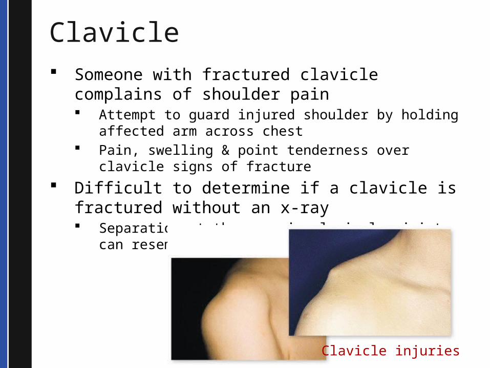
Clavicle Someone with fractured clavicle complains of
shoulder pain Attempt to guard injured shoulder by holding
affected arm across chest Pain, swelling & point tenderness over clavicle signs
of fracture Difficult to determine if a clavicle is fractured
without an x-ray Separation at the acromio-clavicular joint can
resemble a clavicle fracture.
Clavicle injuries

© 2013 Seattle / King County EMS
Fractured – serious injury Bone positioned over major arteries, veins & nerves When fractured…cause nerve & muscular damage
Treatment includes Application of a sling & swathe Transport to medical facility
Clavicle

© 2013 Seattle / King County EMS
Scapula Scapula, also called shoulder blade, less
often injured due to location & protection by large muscles
Fan-shaped bone hard to crack Fractures usually occur from direct blow
For example, baseball bat striking the back
Blunt trauma to scapula
© 2013 Seattle / King County EMS
Scapula Fractures usually are result of significant trauma to back Injury to chest cavity & its components (e.g., the heart and lungs) can accompany injured scapula Examine chest for evidence of other injuries Assess patient's ability to breathe & auscultate breath sounds
© 2013 Seattle / King County EMS
Shoulder Shoulder joint – junction between humerus & scapula Remarkably complex joint Allows us to do many things
Throw a ball Cradle a baby Scratch your back
Because of its complexity, the shoulder is easily injured
© 2013 Seattle / King County EMS
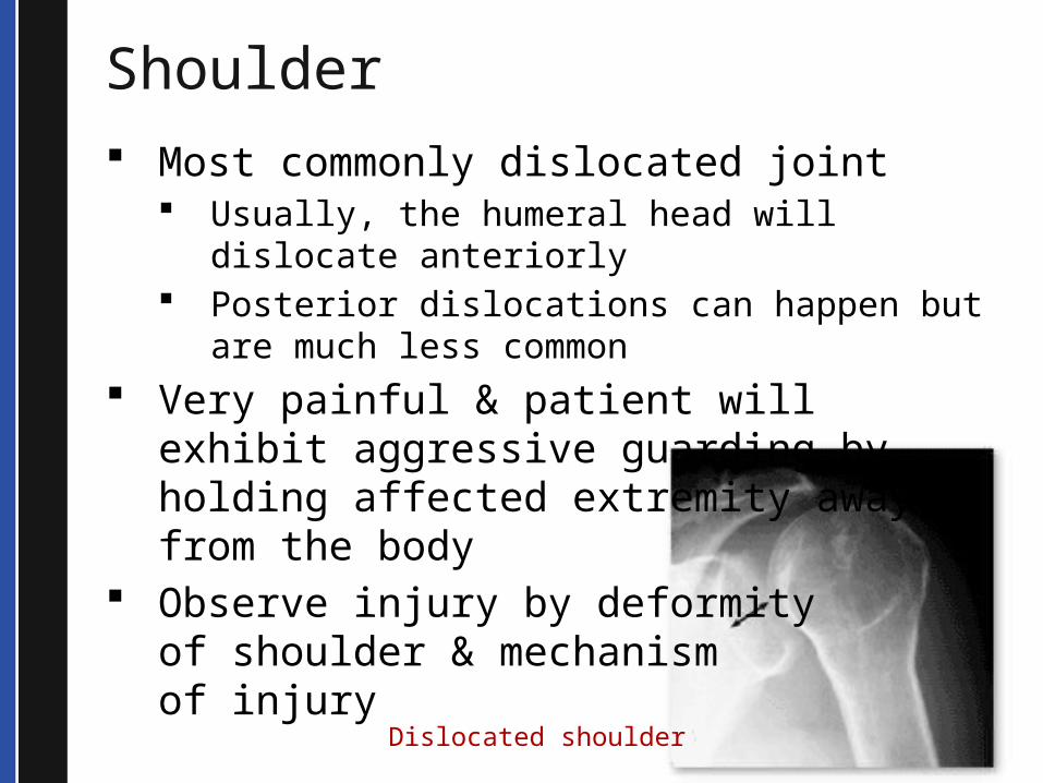
Shoulder Most commonly dislocated joint
Usually, the humeral head will dislocate anteriorly
Posterior dislocations can happen but are much less common
Very painful & patient will exhibit aggressive guarding by holding affected extremity away from the body
Observe injury by deformityof shoulder & mechanism of injury
Dislocated shoulder
© 2013 Seattle / King County EMS
Shoulder
Treatment Application of a sling &
swathe Transport to medical facility
© 2013 Seattle / King County EMS
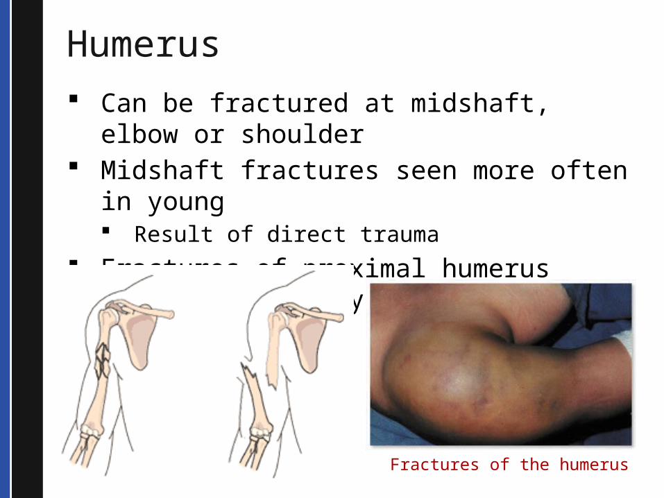
Humerus Can be fractured at midshaft, elbow or
shoulder Midshaft fractures seen more often in
young Result of direct trauma
Fractures of proximal humerus common in elderly patients who have fallen
Fractures of the humerus

© 2013 Seattle / King County EMS
Elbow
Result of a direct force or twisting of arm Elbow dislocations rare—but very
serious injuries Often lead to nerve & vascular
damage Makes olecranon process of ulna
much more prominent Joint usually locked with forearm
moderately flexed on arm This position makes any movement
extremely painful
© 2013 Seattle / King County EMS
Elbow
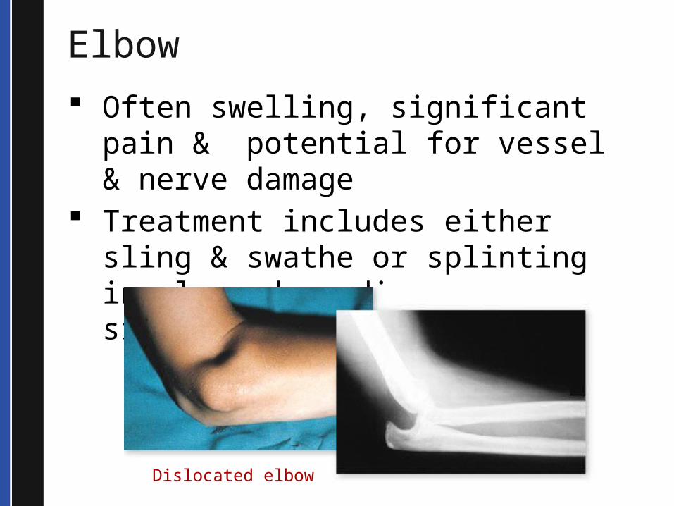
Often swelling, significant pain & potential for vessel & nerve damage
Treatment includes either sling & swathe or splinting in place depending on situation
Dislocated elbow
© 2013 Seattle / King County EMS
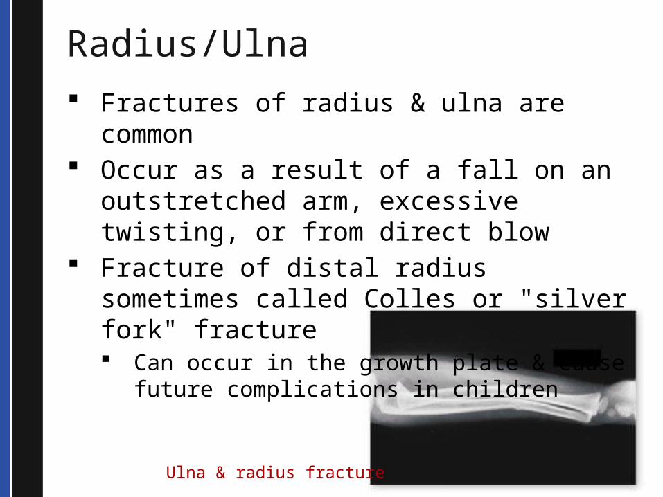
Radius/Ulna Fractures of radius & ulna are common Occur as a result of a fall on an
outstretched arm, excessive twisting, or from direct blow
Fracture of distal radius sometimes called Colles or "silver fork" fracture Can occur in the growth plate & cause
future complications in children
Ulna & radius fracture
© 2013 Seattle / King County EMS
Wrist and Hand Hand & wrist
fractures common & usually result of fall or direct blow
Falls on outstretched hand can crack scaphoid bone (at the base of the thumb)
Fistfight can fracture fourth or fifth metacarpal
Excessive force can dislocate fingers or thumb
Immobilize hand & wrist injuries with rigid splint
Wrist & hand contain many small bones & ligaments
Most injuries will require examination by physician

© 2013 Seattle / King County EMS
Lower Body
© 2013 Seattle / King County EMS
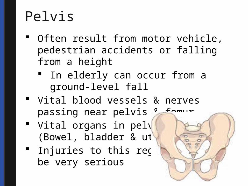
Pelvis Often result from motor vehicle,
pedestrian accidents or falling from a height In elderly can occur from a ground-
level fall Vital blood vessels & nerves passing
near pelvis & femur Vital organs in pelvic area (Bowel,
bladder & uterus) Injuries to this region can
be very serious
© 2013 Seattle / King County EMS
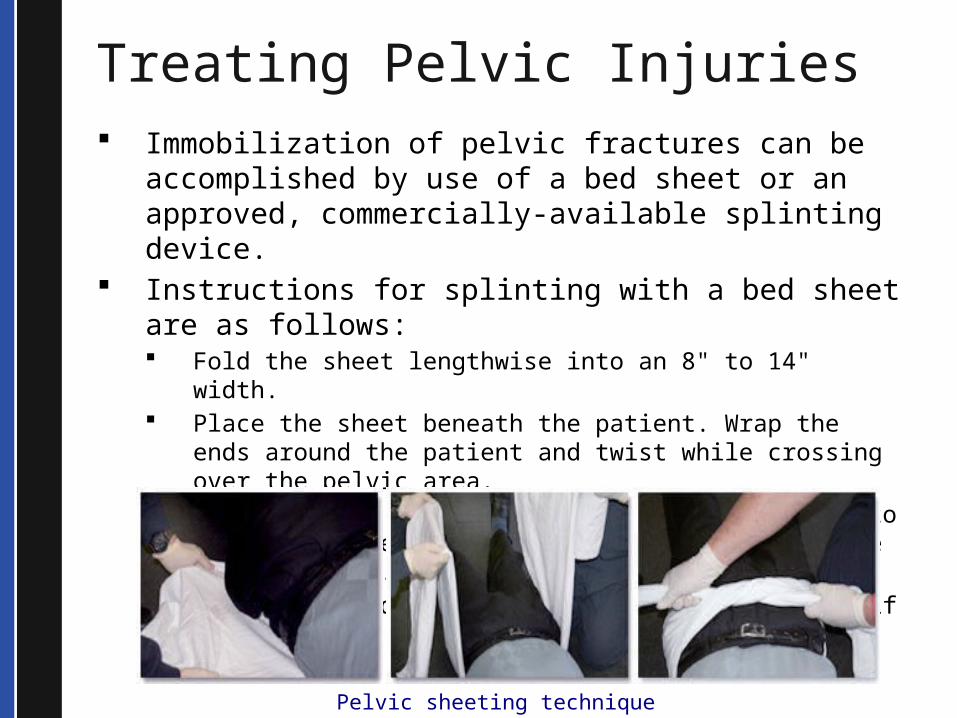
Treating Pelvic Injuries Immobilization of pelvic fractures can be
accomplished by use of a bed sheet or an approved, commercially-available splinting device.
Instructions for splinting with a bed sheet are as follows: Fold the sheet lengthwise into an 8" to 14" width. Place the sheet beneath the patient. Wrap the ends around
the patient and twist while crossing over the pelvic area. Tie the sheet with square knot or plastic ties to apply
moderate pressure around the circumference of the pelvis. Secure the ends of the sheet to the backboard, if possible.
Pelvic sheeting technique
© 2013 Seattle / King County EMS
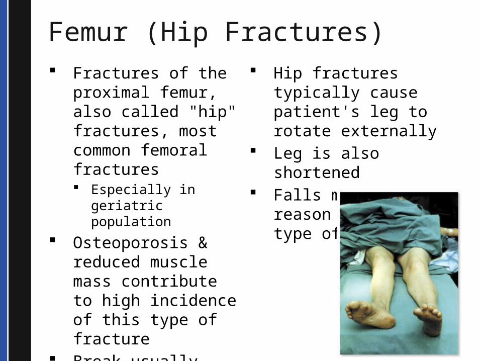
Femur (Hip Fractures) Fractures of the
proximal femur, also called "hip" fractures, most common femoral fractures Especially in geriatric
population Osteoporosis &
reduced muscle mass contribute to high incidence of this type of fracture
Break usually occurs at neck or across proximal shaft
Hip fractures typically cause patient's leg to rotate externally
Leg is also shortened Falls most common
reason for this type of fracture
© 2013 Seattle / King County EMS
Treatment of Hip Fracture
Key points for treating fractured hip: Minimize movement of injured limb Immobilize injured leg in place, if possible Pad generously to immobilize femur
including between legs Pad generously under leg if femur elevated Secure legs together Consider using scoop stretcher to lift to
backboard (padded with blanket) Pad well for comfort Keep patient warm Treat patient gently & minimize movement
© 2013 Seattle / King County EMS
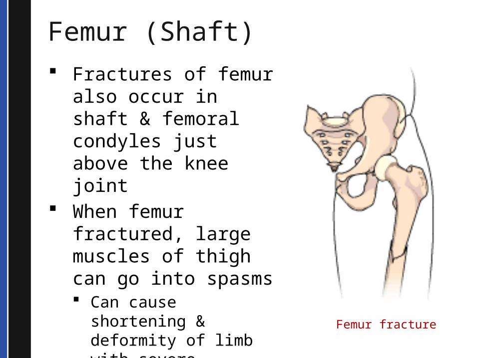
Femur (Shaft) Fractures of femur
also occur in shaft & femoral condyles just above the knee joint
When femur fractured, large muscles of thigh can go into spasms Can cause shortening
& deformity of limb with severe angulation or external rotation at fracture site
Femur fracture
© 2013 Seattle / King County EMS
Femur (Shaft)
Broken ends of femur can pierce skin & cause open fracture
Blood loss can be significant Lead to hypovolemic shock
Bone fragments & deformity can damage important nerves & vessels Long lasting effects Delay recovery
© 2013 Seattle / King County EMS
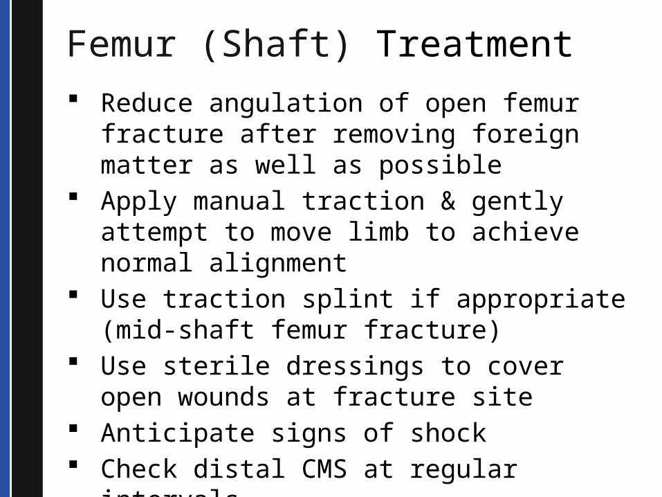
Femur (Shaft) Treatment Reduce angulation of open femur
fracture after removing foreign matter as well as possible
Apply manual traction & gently attempt to move limb to achieve normal alignment
Use traction splint if appropriate (mid-shaft femur fracture)
Use sterile dressings to cover open wounds at fracture site
Anticipate signs of shock Check distal CMS at regular intervals Provide rapid transport
© 2013 Seattle / King County EMS
Traction Splinting
Use traction splint for mid-shaft femur fractures only
Traction splints stabilize bone ends & help reduce muscle spasms in large thigh muscles
Helps prevent further injury to vessels, nerves & tissues
Reduces pain
© 2013 Seattle / King County EMS
Traction Splinting
Contraindications for the use of a traction splint include: Injury close to or involving the
knee Hip injury Pelvis injury Partial amputation or avulsion
with bone separation Lower leg or ankle injury
© 2013 Seattle / King County EMS
Traction Splinting
The key points for applying a traction splint are: Do not apply if there is a destabilizing
injury to hip, knee or ankle Support fracture site when limb is
lifted Apply manual traction & hold until
splint is secured Check CMS before & after apply
splint Video demonstration available at EMS Online:
http://www.emsonline.net/ortho2011/traction.asp
Video demonstration available at EMS Online:
http://www.emsonline.net/ortho2011/traction.asp
© 2013 Seattle / King County EMS
Hip Dislocation
Head & neck of femur, along with the greater trochanter, meet pelvis to form hip
Hip joint is a ball-and-socket joint that is quite strong
Hip dislocations are rare – extremely serious injury
© 2013 Seattle / King County EMS
Hip Dislocation Hip dislocations can
damage large vessels & nerves
Most common cause – motor vehicle accidents Knee strikes dashboard
femur can dislocate backwards
Posterior hip dislocations: leg shortened & rotated internally
Anterior dislocations: leg lengthened & externally rotated
Treatment includes splinting extremity in the position it is found
Do not attempt to reduce a hip dislocation

© 2013 Seattle / King County EMS
Knee Knee joint, like the shoulder joint, is
extremely complex & easily injured Ligament or cartilage damage
commonly seen with twisting injuries Injuries to ligaments of knee range from
mild sprains to complete dislocation of bone ends
Knee injuries
© 2013 Seattle / King County EMS
Knee
Patella (kneecap) susceptible to injury such as fracture or dislocation
Pulseless knee dislocation is a true medical emergency Requires emergent transport to
facility Vascular surgery on hand
© 2013 Seattle / King County EMS
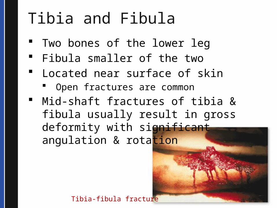
Tibia and Fibula Two bones of the lower leg Fibula smaller of the two Located near surface of skin
Open fractures are common Mid-shaft fractures of tibia & fibula
usually result in gross deformity with significant angulation & rotation
Tibia-fibula fracture
© 2013 Seattle / King County EMS
Tibia and Fibula
Often accompanied by vascular injury
Realigning & splinting limb may restore adequate blood flow to foot
Need to realign an angulated tib/fib fracture Check distal CMS before & after
realignment Rapid transport
© 2013 Seattle / King County EMS
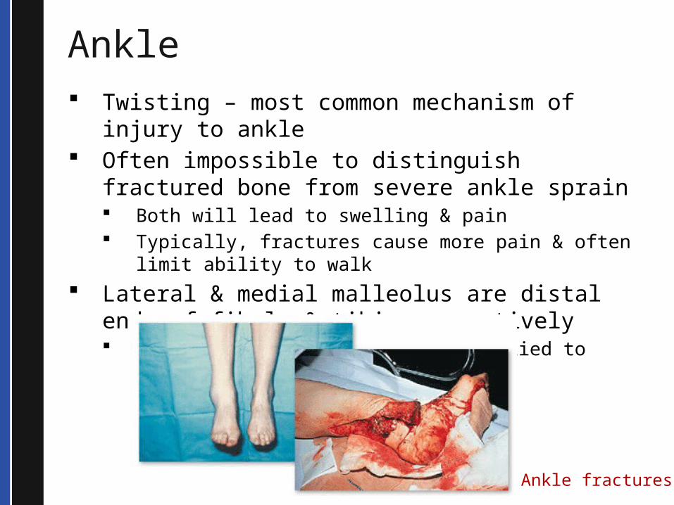
Ankle Twisting – most common mechanism of injury
to ankle Often impossible to distinguish fractured bone
from severe ankle sprain Both will lead to swelling & pain Typically, fractures cause more pain & often limit
ability to walk Lateral & medial malleolus are distal ends of
fibula & tibia respectively Often crack if twisting force applied to ankle is
sufficient
Ankle fractures
© 2013 Seattle / King County EMS
Foot Foot injuries common
Falls from heights Excessive twisting motions
Calcaneus bone (heel bone) may be fractured if patient falls from sufficient height & lands on heels If calcaneus is fractured, there may be
enough force to have other associated fractures such as vertebral fractures
Pain, swelling & ecchymosis may be seen with fractures of foot
© 2013 Seattle / King County EMS
Realignment of Long Bone Fractures You can attempt to realign
fractures of long bones that occur in the middle 1/3 of the bone only
Long bone fractures, which occur in the proximal or distal 1/3, may be realigned only if compromise of distal circulation or nerve function is detected and definitive care is delayed
© 2013 Seattle / King County EMS
Realigning Joint Injuries & Dislocations Splint dislocations or other joint injuries
in position found Exceptions include:
Loss of a distal pulse and neurological function where definitive care is delayed
In these cases: Attempt to straighten into anatomical position until
pulse returns, excessive pain felt, or resistance encountered
Support with blanket, pillow, or well-padded splint Elevate the limb Pack injured area in ice or use ice pack Document attempts to re-align injury
© 2013 Seattle / King County EMS
Compartment Syndrome
Elevation of pressure within fibrous tissue that surrounds & supports muscles & neurovascular structures
Characterized by extreme pain, pain on movement, pulselessness & pallor
Fractures of the forearm or lower leg are the most common injuries that cause compartment syndrome

© 2013 Seattle / King County EMS
Prioritizing
© 2013 Seattle / King County EMS
Prioritize
Orthopedic injuries can be dramatic and distracting – consider the whole patient!
Prioritize ABCs over treatment of orthopedic injuries
Prioritize serious orthopedic injuries (such as pelvic fractures) over more minor injuries (such as a broken ankle)
Anticipate and treat for shock
© 2013 Seattle / King County EMS
Shock Life-threatening condition develops
when circulatory system cannot deliver sufficient blood to body’s tissues
Many causes: Blood loss Cardiac failure Respiratory failure Spinal cord injury
Inadequate tissue perfusion
© 2013 Seattle / King County EMS
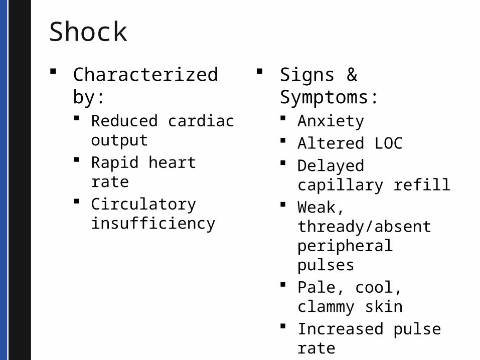
Shock Characterized by:
Reduced cardiac output
Rapid heart rate Circulatory
insufficiency
Signs & Symptoms: Anxiety Altered LOC Delayed capillary
refill Weak,
thready/absent peripheral pulses
Pale, cool, clammy skin
Increased pulse rate
Decreased blood pressure
© 2013 Seattle / King County EMS
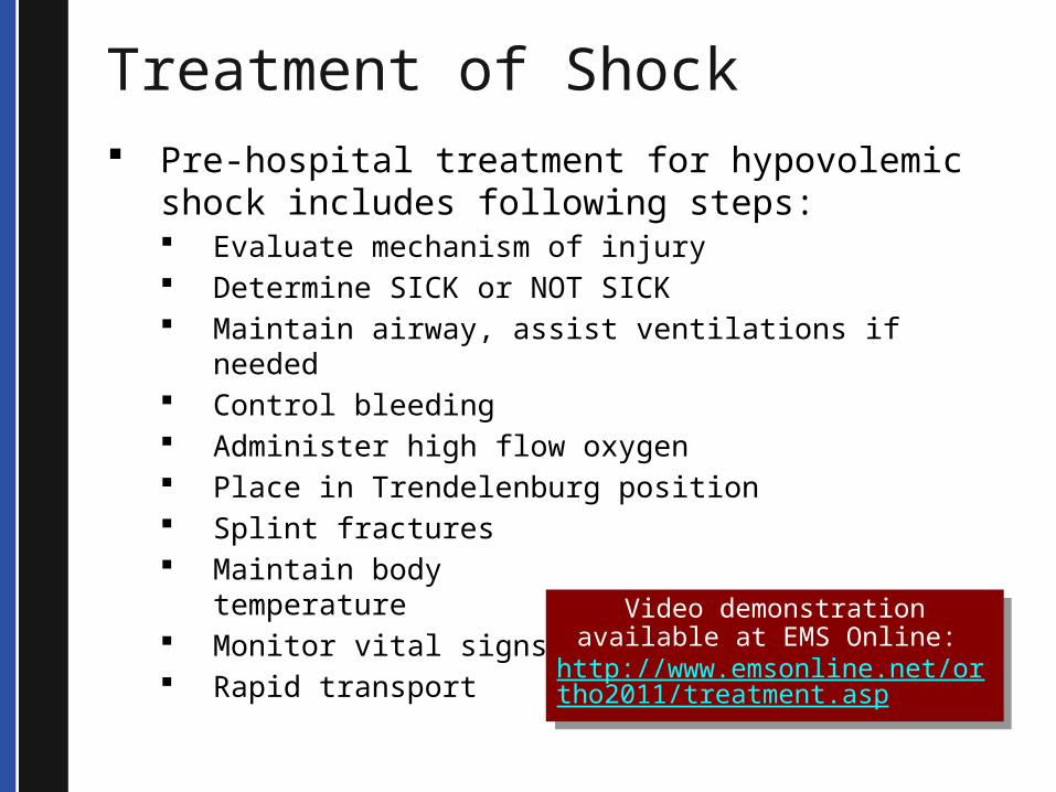
Treatment of Shock Pre-hospital treatment for hypovolemic shock
includes following steps: Evaluate mechanism of injury Determine SICK or NOT SICK Maintain airway, assist ventilations if needed Control bleeding Administer high flow oxygen Place in Trendelenburg position Splint fractures Maintain body
temperature Monitor vital signs Rapid transport
Video demonstration available at EMS Online:
http://www.emsonline.net/ortho2011/treatment.asp
Video demonstration available at EMS Online:
http://www.emsonline.net/ortho2011/treatment.asp
© 2013 Seattle / King County EMS
Case StudiesVideo Case Study #1
http://www.emsonline.net/ortho2011/case1.asp
Video Case Study #2
http://www.emsonline.net/ortho2011/case2.asp
© 2013 Seattle / King County EMS
SummaryPrinciples of splinting are: Support the fracture site Bone fracture - immobilize the joint above
and below the fracture site Joint injury - immobilize the bones above and
below the dislocation Check CMS before and after splinting Pad the splint well Elevate extremity after splinting, if possible
© 2013 Seattle / King County EMS
Summary You can attempt to realign fractures of long
bones that occur in the middle 1/3 of the bone only
Splint dislocations or other joint injuries in position found except in cases of loss of a distal pulse & neurological function where definitive care is delayed
Outcome of most traumatic injuries does not rest with us but in our ability to transport to a Trauma Center in an expeditious fashion
The old adage still applies:
We don't save trauma victims the operating room does!
© 2013 Seattle / King County EMS
Summary Common factor in all types of shock is
inadequate tissue perfusion Perfusion is circulation of blood within
an organ or tissue To maintain adequate perfusion the
body requires four intact components: Pump (heart) Pipes (blood vessels) Fluids (adequate blood volume) Oxygen (adequate oxygenation
© 2013 Seattle / King County EMS
Summary
Signs and symptoms of shock include: Anxiety Altered LOC Delayed capillary refill Weak, thready or absent peripheral pulses Pale, cool, clammy skin Increased pulse rate (an early sign) Decreased blood pressure (a late sign)
© 2013 Seattle / King County EMS
Summary
Treatment of hypovolemic shock includes: Assess the MOI Determine SICK or NOT SICK Maintain airway, assist ventilations if needed Control bleeding Administer high flow oxygen Place in shock position Splint fractures Maintain body temperature Monitor vital signs Rapid transport
© 2013 Seattle / King County EMS
Terms Amputation - removal of a body extremity by trauma
or surgery. As a surgical measure, it is used to control pain or a disease process in the affected limb, such as malignancy or gangrene
Compartment syndrome — Elevation of pressure within fibrous tissue that surrounds & supports muscles and neurovascular structures, characterized by extreme pain, pain on movement, pulselessness, and pallor. It is most frequently seen in fractures below the elbow or knee.
Compensated shock — early stages of shock in which the body is able to compensate for blood loss or injury
Crepitus — Grating or grinding sensation caused by fractured bone ends or joints rubbing together. It also can be caused by rubbing of irregular cartilage tissue or scar tissue.
Dislocation — Disruption of a joint in which ligaments are damaged & the bone ends are completely displaced.
© 2013 Seattle / King County EMS
Terms, continued Distal — The more distant of two or more structures. Fascia — Sheets or bands of fibrous connective tissue
that lie deep under the skin forming the outer layer of a muscle.
Hypotension — Blood pressure that is lower than the normal range — generally a systolic blood pressure less than 90 mmHg in an appropriate clinical setting.
Hypoxia — Condition in which the body tissues and cells do not have enough oxygen.
Ligament — A band of fibrous tissue joining two bones together in a joint.
Osteoporosis — Generalized degenerative bone disease common among postmenopausal women in which there is a reduction of bone mass making the bones fragile and susceptible to injury.
Perfusion — Circulation of blood within an organ or tissue in adequate amounts to meet cellular needs.
© 2013 Seattle / King County EMS
Terms, continued Point tenderness — Tenderness sharply localized at the site of the injury. Found by gently palpating along the bone with the tip of one finger. Proximal — Nearer to a point of reference such as a point of attachment or the midline of the body. Sprain — Joint injury in which there is some partial or temporary dislocation of the bone ends and partial stretching or tearing of the supporting ligaments. Strain — A stretching or tearing of the muscle, causing pain, swelling, and bruising of the soft tissue in the area. Also called a “pulled muscle.” Tendon — Extension of a skeletal muscle that connects the muscle to bone.
© 2013 Seattle / King County EMS
Questions
Dr. Mickey EisenbergMedical DirectorAsk the Doc: http://www.emsonline.net/doc.asp
EMS OnlineGuidelines and Standing Ordershttp://www.emsonline.net/downloads.asp
Susan KolwitzProgram ManagerEmail support: [email protected]