blood grouping in pregnancy, guidelines and foetal genotyping · blood grouping in pregnancy,...
TRANSCRIPT

Blood Grouping in Pregnancy,
Guidelines and Foetal Genotyping
Kasia Ballard 28.02.2017
2
The purpose of the guideline is to make evidence-based recommendations for the application of blood grouping and red cell antibody testing in pregnancy and if possible to prevent, haemolytic disease of the foetus and newborn (HDFN).
The blood group and antibody status of a pregnant woman should be tested at booking and at 28 weeks gestation to identify the ABO group and D status and to detect red cell antibodies that have the potential to be clinically significant.
3
ABO/D typing Screening for re d cell antibodies
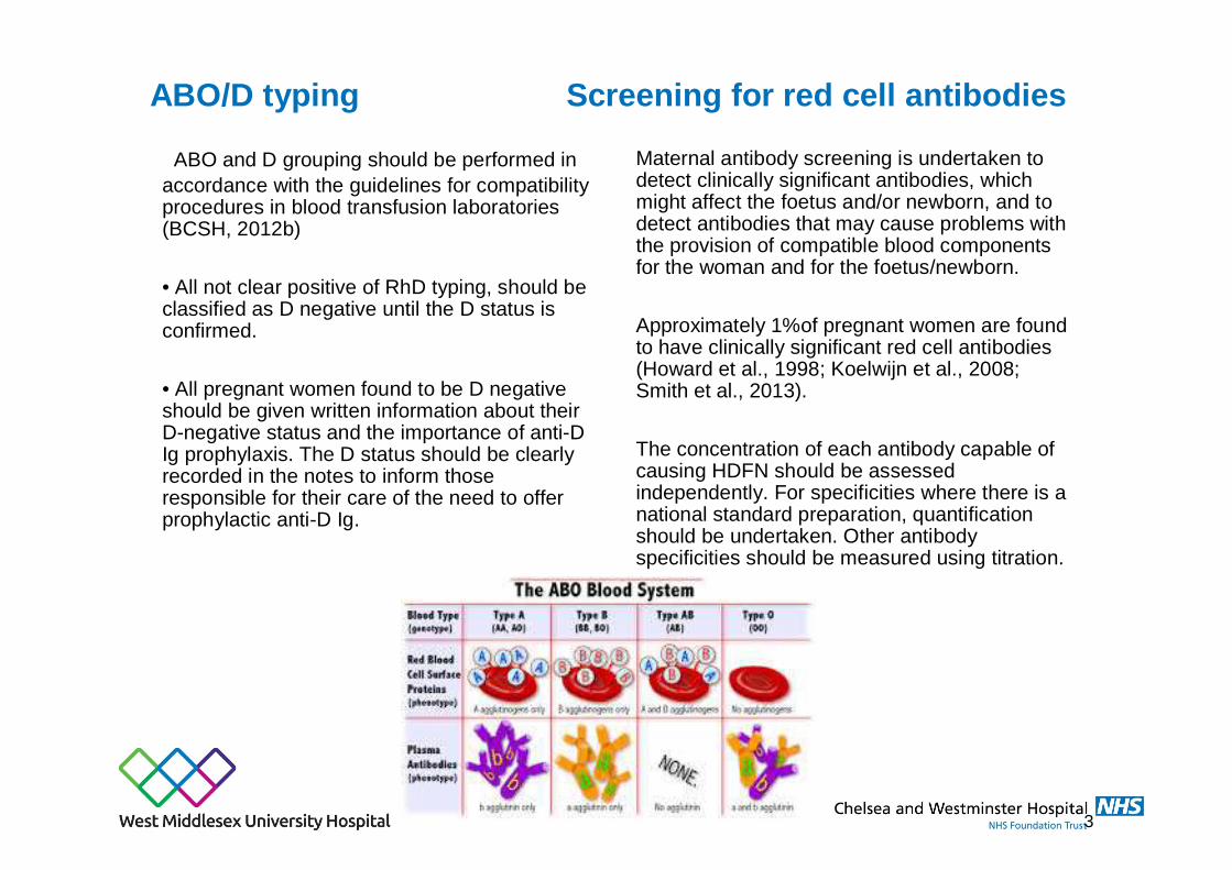
ABO and D grouping should be performed in accordance with the guidelines for compatibility procedures in blood transfusion laboratories (BCSH, 2012b)
• All not clear positive of RhD typing, should be classified as D negative until the D status is confirmed.
• All pregnant women found to be D negative should be given written information about their D-negative status and the importance of anti-D Ig prophylaxis. The D status should be clearly recorded in the notes to inform those responsible for their care of the need to offer prophylactic anti-D Ig.
Maternal antibody screening is undertaken to detect clinically significant antibodies, which might affect the foetus and/or newborn, and to detect antibodies that may cause problems with the provision of compatible blood components for the woman and for the foetus/newborn.
Approximately 1%of pregnant women are found to have clinically significant red cell antibodies (Howard et al., 1998; Koelwijn et al., 2008; Smith et al., 2013).
The concentration of each antibody capable of causing HDFN should be assessed independently. For specificities where there is a national standard preparation, quantification should be undertaken. Other antibody specificities should be measured using titration.

4
Antibody testing
Antibody quantification.
Quantification requires specific equipment and measures antibody concentration against a national standard [National Institute for Biological Standards and Control (NIBSC)].
Anti-D and anti-c are the only antibodies that are currently quantified, and they are reported as IU per millilitre.
Where possible, each sample should be tested in parallel with the previous sample and the results compared to identify significant change
in antibody concentration.
Antibody titration.
Titration is used to assess the concentration of clinically significant red cell antibodies other than anti-D and anti-c.
Doubling dilutions (1 in 2, 1 in 4, etc) of plasma prepared in phosphate-buffered saline are tested by IAT using reagent red cells, where possible, showing heterozygous expression of the corresponding antigen(s).
Sometimes there are more than one antibody specificity is present (including prophylactic anti-D).The concentration of each specificity should be assessed independently, e.g. where anti-K+Fya are present titrate against K−, Fy(a+b+) and K+k+, Fy(a−) cells.
5
Protocol for patients with antibodies
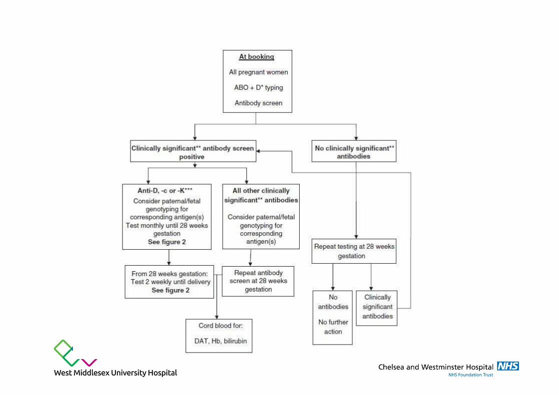
• Samples from pregnant women with immune anti-D or anti-c should be assessed serologically at 4 weekly intervals to 28 weeks gestation and at fortnightly intervals thereafter until delivery. Such cases should be referred to a foetal medicine specialist if the antibody reaches the critical level and/or the level is rising significantly, where assessment of the need for further monitoring will be made.
• Pregnant women with anti-K or other Kell system antibodies (unless the father is confirmed to be negative for the corresponding antigen) should be assessed serologically at monthly intervals to 28 weeks gestation and at fortnightly intervals thereafter until delivery and referred to a foetal medicine specialist when the antibody is first identified
Clinically significant antibodies, other than anti-D, -c or -K, should be excluded or, if present, assessed by titration at the booking appointment and at 28 weeks gestation. If deemed necessary based on a high titre (>32) and/or a past history of HDFN, referral to a specialist in foetal medicine should be made for further assessment.
All babies born to women who have clinically significant antibodies should be closely observed for evidence of HDFN. A DAT should be performed on a cord blood sample, and haemoglobin and bilirubin concentrations should be measured.
A positive DAT is not, in itself, diagnostic of HDFN. Where the DAT is positive and the baby shows signs of HDFN, a red cell eluate may be helpful to confirm the red cell antibody specificity. IgG ABO antibodies occasionally cause severe HDFN, and so, if the baby has a major ABO mismatch with the woman, the eluate should also be tested with A1 and/or B cells, negative for any other antigen against which the woman has made IgG alloantibodies. Regular assessment of bilirubin and haemoglobin concentrations is necessary, and hence, early discharge is not advisable


9
Anti-D immune and prophylaxis
Recommendation
• Blood transfusion laboratories should keep a record of anti-D Ig administration to provide a basis for distinguishing between immune anti-D and prophylaxis anti-D Ig.
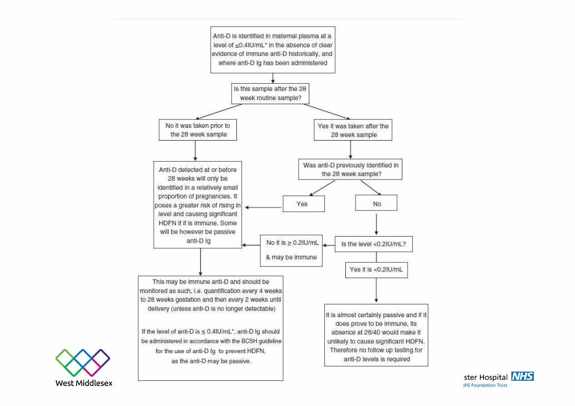
• If anti-D is detected in an antenatal maternal sample (except for that taken immediately prior to delivery), testing should include a measurement of antibody concentration by CFA or similar technique that gives a result that is expressed in or can easily be converted to IU per millilitre of anti-D.
• When there is doubt as to the passive or immune nature of anti-D, the level should be monitored as if it could be immune. In this situation, anti-D Ig prophylaxis should continue to be offered, until the nature of the anti-D is established.

10
Paternal testing
Paternal testing
When a clinically significant antibody capable of causing HDFN is present in a maternal sample, determining the father’s phenotype can provide useful information to predict the likelihood of the foetus expressing the relevant red cell antigen and for counselling the couple regarding future pregnancies.
It should be recognised that in any pregnancy, the partner may not be the biological father (assisted conception with sperm donation from a donor panel, the pregnant woman’s partner will not be the biological father).
It is reasonable to avoid paternal testing and proceed directly to foetal genotyping using cffDNA.
Recommendation
If potentially clinically significant maternal antibodies have been identified, paternal testing should be considered to predict the risk to current and future pregnancies. This may be particularly relevant if non-invasive foetal genotyping is not available for the corresponding red cell antigen.
Foetal genotyping in alloimmunised pregnancies. Foetal genotyping is a useful diagnostic tool when:
a)A pregnant woman has a clinically significant antibody;
b)b) A pregnant woman has a history of HDFN;
c) The father’s antigen status is unknown or he expresses the corresponding antigen.

Taking part in the NHS cffDNA testing
Administer anti-D selectively, only to RhD Negative women with a RhD Positive baby
Improve patient pathway and satisfaction
Focus midwifery / obstetrician time on clinically indicated care
Cost neutral
Potentially save laboratory time

WHY Change the system??Routine Antenatal Anti-D Prophylaxis (RAADP) has been hugely successful in UK:
1930’s RhD antigens identified
1969 First use of anti-D as postnatal prophylaxis
1999 RAADP introduced
Maternal isoimmunisation reduced from 16% to 0.2%
Perinatal mortality 46 to 1.6 / 100000 births

Potentially Sensitising events during pregnancy

Facts16% UK population RhD negative [lower in non Caucasian populations]
~40% of their babies will be RhD Negative = 40,000 women / year receive unnecessary anti D in UK
Introducing cffDNA testing potentially could reduce issuing of proph-D by 30%.
Considering implications:
-financial
-ethical
-safety

2006-11 High throughput foetal RhD testing trials at different gestations:
Highly accurate from 11 +2 weeks gestation
2013/14 Foetal RhD service pilot- North Bristol- UHs Bristol- Weston Area
2002- Recommended further research into foetal RhD typing
BMJ 2014;349:g5243 doi: 10.1136/bmj.g5243DOI: 10.1111/1471-0528.13055 www.bjog.org
1994: Fetal blood groupgenotyping introduced
2001: Fetal D typing on cffDNAExtended to K, C, c, E

Clinical implications: Antenatal
RhD Negative pregnant women tested after 12 weeks’ gestation.
Possible results:
1. Baby Predicted RhD Positive – continue usual RAADP
2. Baby Predicted RhD Negative – no RAADP required;
- no Kleihauer or anti-D for sensitising events and post delivery
3. Inconclusive – treat as if RhD Positive
4. Not tested (‘Rejected’)

Clinical implications: Postnatal
Options include:
1.Continue current policy of cord blood on all babies born to RhD Negative women, with anti-D dose determined by Kleihauer
2. Cord blood only on babies predicted to be RhD Negative
Women delivering babies predicted to be RhD Positive given anti-D, without cord blood group testing.

Setting up the service
‘Self appointed’ multidisciplinary working party:
Obstetrician
Haematologist
Blood Transfusion Manager
IT expert
Experienced Midwife
Guideline and pathway writing; midwife champions identified; patient involvement; business case

Developing Protocol

Concerns to make decisions relating to:
• Identifying the relevant women
• Ensuring sample taken, correctly
• Handling the samples on receipt
• Handling the results
• Availability of results to the clinical teams
• How to audit pathway and get patient feedback
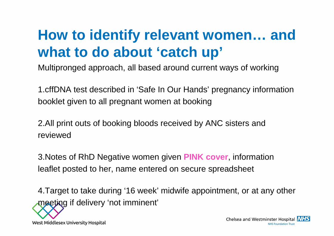
How to identify relevant women… and what to do about ‘catch up’Multipronged approach, all based around current ways of working
1.cffDNA test described in ‘Safe In Our Hands’ pregnancy information booklet given to all pregnant women at booking
2.All print outs of booking bloods received by ANC sisters and reviewed
3.Notes of RhD Negative women given PINK cover , information leaflet posted to her, name entered on secure spreadsheet
4.Target to take during ‘16 week’ midwife appointment, or at any other meeting if delivery ‘not imminent’

Taking the samples, correctly
Departmental education for all staff via monthly Maternity Forum, Newsletters, Team Leaders meetings etc
Midwife champions
HCAs working in ANCs ‘empowered’
Single full PINK top sample
at any clinical point from 14weeks

Handling samples in lab
Received in laboratory via standard pathways from all clinics; sample stable at room temperature
Entered into Laboratory IT system [Winpath]
Sent to NHSBT Colindale on standard daily transport
Transported from Colindale to Bristol on standard NHSBT transport
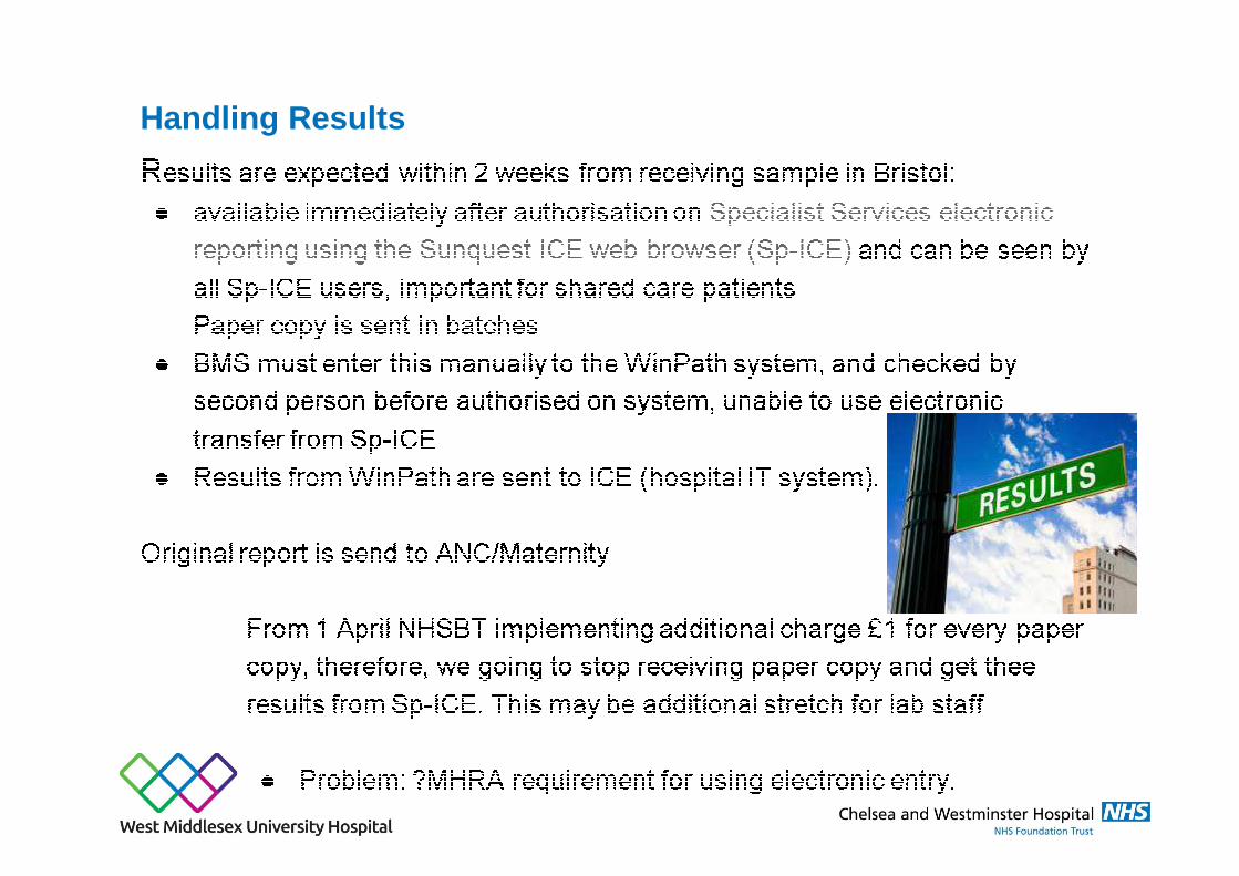
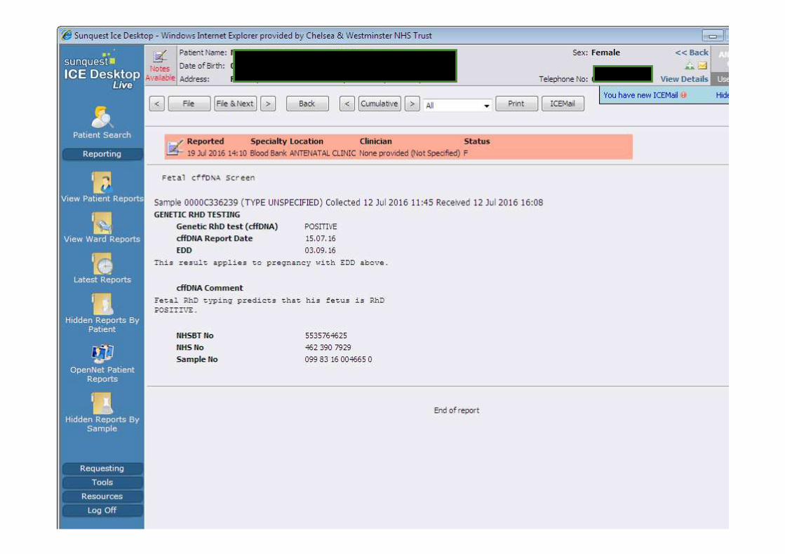
Handling Results

Working process to build the result page on LIMs and Hospital IT systems
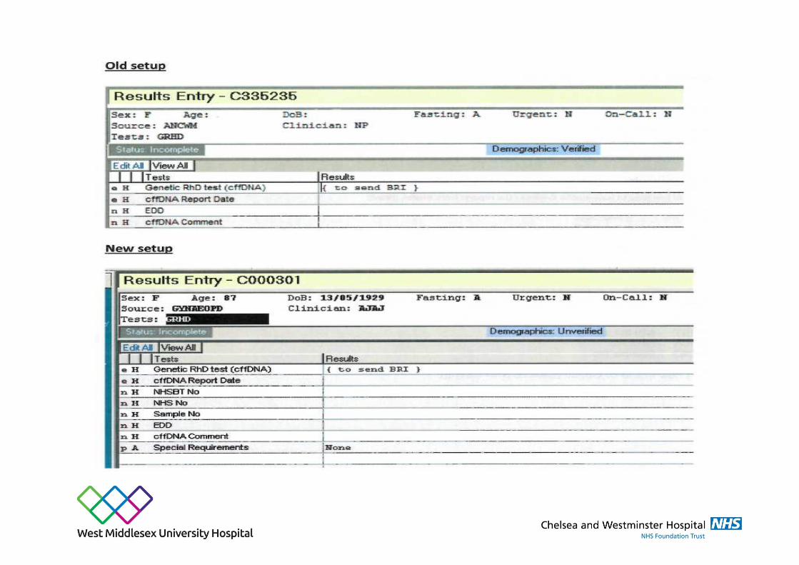
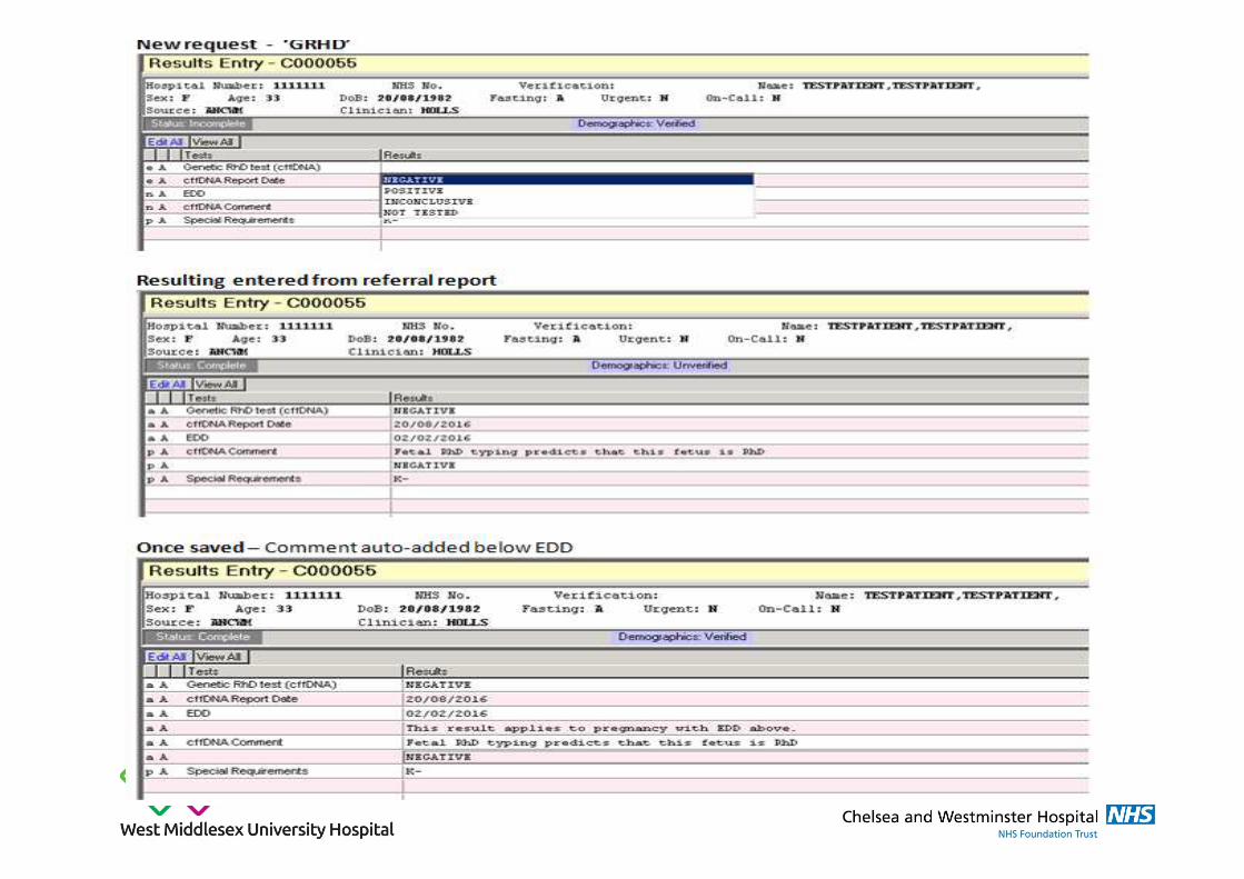
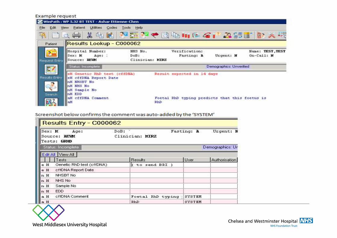

Creating comments

Ongoing audit and patient questionnaire
Started 1st June 2016, in 1st 4 months:
�375 Letters sent to RhD Negative women re testing
�235 Results received back
�87 Babies predicted to be RhD Negative [37%]

Service ProblemsRelatively few…
●Occasional wrong labelling and wrong sample size
●Occasional missed opportunity to take sample
●Likely some cord samples taken unnecessarily
●IT issues were complex to set up, but have been trouble free in clinical practice
●Typing results rather receiving electronically directly from Bristol
●From 1 April 2017, no paper copy (additional charge), could be more challenging for hospital laboratory staff
Service Problems
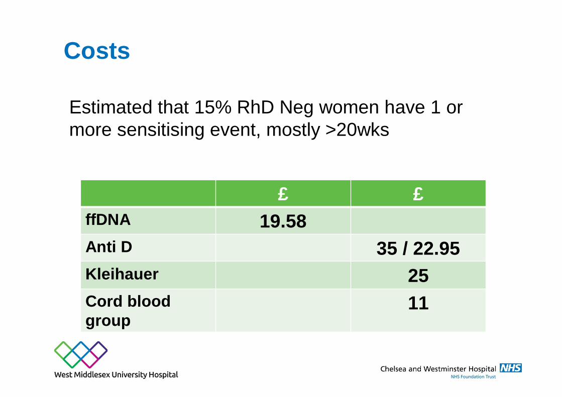
Costs
Estimated that 15% RhD Neg women have 1 or more sensitising event, mostly >20wks
£ £ffDNA 19.58Anti D 35 / 22.95Kleihauer 25Cord blood group
11
Accuracy of the cffDNA results?
Predicted RhD Negative – <1 in 1000 inaccurate
ie actually RhD Positive
check cord blood group for confirmation
1 in 86,000 chance of isoimmunisation
Predicted RhD Positive - 2% inaccurate
ie given anti D un necessarily
Indeterminate - ~8% may fall as technology improves
80-90% actually RhD Positive
Laboratory findings re: discrepant results
• Predicted RhD Positive - confirmed after birth RhD Negative -October 2016
Note: unable to get more discrepant results as the protocol was changed and there is no Cord blood taken for Predicted RhD Positive babies
• Predicted RhD Negative - confirmed after birth RhD Positive -January 2017, investigation in process, sample was sent to Bristol

SummaryImplementation of cffDNA RhD testing is:
•Relatively straightforward
•Cost effective/neutral
•Saving time in midwifery area (less patients in clinic and less worrying if patients has PVB)
•Could save time on KL tests and issuing the anti-D, but BMS spend more time on booking sample for testing, packing, and sending to Bristol
•Also BMS spend more time typing and checking results
•Well received by Maternity staff and patients
