blood, chapter 13 - dr. scott croes' website€¦ · blood, chapter 13 outline of class notes...
TRANSCRIPT

1
Blood, Chapter 13 Outline of class notes for Physiology Objectives: After studying this chapter you should be able to:
1. Describe the functions of blood and briefly describe its composition. 2. Discuss the components of plasma and explain their importance. 3. Explain the difference between plasma and serum. 4. Describe the characteristics and functions of erythrocytes including a description of how
oxygen and carbon dioxide are transported. Be sure to include details of bicarbonate Ions, chloride shift, and the Bohr Effect.
5. Explain the process of hematopoiesis, erythropoiesis, and the life cycle of a RBC. 6. Define hematocrit and describe its components. 7. Describe the characteristics and functions of platelets including the mechanism of clot
formation in a damaged blood vessel. 8. Explain how aspirin and plavix function to prevent blood clots. 9. Explain ABO blood typing and what the “+” and “-“ mean. 10. Discuss the details of hemolytic disease of the newborn (erythroblastosis fetalis). 11. Describe the following disorders: Anemia, sickle-cell disease, hemophilia, and leukemia. 12. Discuss the Clinical Applications from the study guide and assigned Applications to Health.
Cardiovascular System: Blood, Ch 13 • Functions of Blood • Blood:
– Characteristics – Components
• Plasma • Formed Elements
• Blood Cell Formation • Types of Blood cells
– Erythrocytes – Leukocytes – Platelets (thrombocytes)
• Preventing Blood Loss – Clot formation
• Blood Grouping • Diagnostic Blood Tests
The Cardiovascular System: Blood Functions of the Circulatory System
1. Transportation: • Blood gases: O2 from the lungs to the
body cells and CO2 from the body cells to the lungs.
• Nutrients: Absorbed nutrients from the GI tract to the body cells
• Wastes: Cellular waste products are transported to the liver, kidneys, and skin for elimination.
• Regulatory molecules: Hormones from endocrine glands are carried to target cells.

2
2. Regulation: • pH: Stabilizes body pH (acid/base balance) through buffers • Body Temperature: By absorbing and distributing body heat.
• During hot ambient temperatures, blood is diverted from deep to superficial vessels to cool the body
• During cold ambient temperatures, blood is diverted from superficial to deeper vessels to keep the body core warm.
• Based on the absorbing and coolant properties of water • Fluid Volume and water content of cells: Blood osmotic pressure and in conjunction
with the urinary system.
3. Protection: • Clot Formation: Protects against blood loss • Immune system: Circulating antibodies and WBCs protect
against infectious microorganisms and toxins. Physical Characteristics of Blood
• Color depends on oxygen content. – Bright red: oxygen rich – Dark red: oxygen poor.
• Blood is 5x more viscous than water • Normal Blood pH ranges from 7.35 – 7.45.
• Slightly alkaline • Total blood volume:
• Females: 4-5 liters (1.2 gal) • Males: 5-6 liters (1.5 gal) • Makes up ~8% of the total body weight.
Composition of Blood
• Classified as fluid CT • Composed of formed elements (cells and platelets)
suspended in a fluid matrix called plasma. – Formed elements include:
• Erythrocytes (red blood cells; RBCs): Function in oxygen transport.
• Leukocytes (white blood cells; WBCs): Function in immune defense
• Platelets (thrombocytes): Function in blood clotting
Plasma
• Plasma: – Straw-colored liquid that makes up ~55 % of
blood volume. – Consists of:
• ~91% water • ~7% proteins • ~2% other substances such as
enzymes, hormones, metabolites, respiratory gases, nutrients, and electrolytes.

3
• Plasma proteins (7%) divided into 3 classes: Albumins, globulins and clotting proteins • Albumins (~60% of plasma proteins)
– Produced by hepatocytes – Maintain osmotic pressure of the vascular system
• Function to draw water from the surrounding tissue (interstitial) fluid into the capillaries to maintain blood volume and pressure.
• Globulins (~35% of plasma proteins) – Alpha and beta globulins:
• Produced by the liver; function to transport lipids, steroids, fat-soluble vitamins, and metal ions (iron).
– Gamma globulins (immunoglobulins): • Produced by plasma cells (type of leukocyte) • Are antibodies which function in immunity.
• Clotting proteins (~4% of plasma proteins) – Produced by the liver – Two examples are thrombin and fibrinogen – Serum: The yellowish fluid remaining after blood has clotted.
• Similar to plasma but without the clotting proteins and factors. Erythrocytes (Red Blood Cells)
• Round cells shaped like biconcave discs with a diameter of 7-8 µm. – Shape increases surface area for greater gas diffusion.
• Function: O2 transport; also plays a minor role in CO2 transport.
• Quantity: 4.5-5.5 million per cubic millimeter (mm3). – Have about 260 million RBCs/drop of blood.
• Mature RBCs are anucleated and lack mitochondria. – No nucleus = more room for hemoglobin molecules – No mitochondria = the RBC will not use the oxygen it is
transporting. • RBCs generate ATP by anaerobic respiration
Erythrocytes and Hemoglobin • Hemoglobin molecules function in the
transport of oxygen and to a smaller extent, carbon dioxide
– A hemoglobin molecule consists of: 4 polypeptide (protein) chains called globins and 4 iron containing pigment molecules called hemes.
• Of the 4 polypeptide chains, 2 are composed of identical alpha chains, each 141 amino acids long, and two identical beta chains, each 146 amino acids long.
• Each protein chain is bound to one heme group • A heme group consists of a porphyrin ring that holds 1 atom of iron. An iron
atom can reversibly bind to one oxygen molecule (O2). • One hemoglobin molecule can combine with 4 molecules of oxygen • Each red blood cell contains ~280 million hemoglobin molecules and can
thus carry over a billion molecules of oxygen

4
• Hemoglobin gives blood its red color • Hemoglobin transports 98.5% of blood oxygen with the remaining oxygen
transported in the plasma • Depending on what is bound to hemoglobin will determine its name.
• Oxyhemoglobin : when iron binds to oxygen
• Deoxyhemoglobin : when the oxygen is released
• Carboxyhemoglobin : when iron binds to carbon monoxide.
• Bond between hemoglobin and carbon monoxide is about 210 Xs stronger than the bond with oxygen and can be fatal.
Carbon Dioxide Transport
• Carbon dioxide is transported in the blood in three forms: 1. As dissolved CO2 in the plasma, ~10% 2. As carbaminohemoglobin (HbCO2), ~20%. CO2 is attached to an amino acid in the
globin portion. 3. As bicarbonate ion, ~70%
Overview of the Transport of Bicarbonate Ions and the Chloride Shift • In order to increase the amount of CO2 transport, RBCs convert the CO2 to bicarbonate and
H+ at the systemic capillaries. • So what does the RBC do with the bicarbonate and H+?
– H+ binds to hemoglobin – Bicarbonate diffuses out the RBC as Cl- diffuses onto the RBC. This process is known
as the chloride shift and allows bicarbonate to move out of the RBC. – In the pulmonary capillaries this process is reversed (reversed chloride shift)

5
Bicarbonate Ions and the Chloride Shift • In systemic capillaries, carbon dioxide combines with water to form
carbonic acid, but at a slow rate. – In RBCs, the enzyme carbonic anhydrase greatly speeds
this reaction to form carbonic acid (H2CO3). – As carbonic acid builds up it dissociates into hydrogen
ions (H+) and the bicarbonate ions (HCO3-). • Most of the hydrogen ions (H+) remain in the RBC and attach to
hemoglobin (HbH) while most of the bicarbonate ions diffuse out into the plasma by facilitated diffusion and can serve as a buffer.
• The result is that the inside of the RBC gains a net positive charge.
• This attracts chloride ions (Cl-), which move into the RBCs as HCO3- move out.
• Chloride Shift: This exchange of anions, diffusion of Cl- into RBCs as HCO3- diffuses out of the RBCs, is called the chloride shift and occurs in the systemic capillaries.
Question? • How do red blood cells know where to release more
oxygen and where less? Or why do they unload more oxygen at all? Why is O2 released in tissues?
• Answer: The higher the levels of CO2 and thus H+ as occurs in actively metabolizing cells, the greater the release of oxygen from hemoglobin in what is known as the Bohr Effect.
Bohr Effect • Bohr Effect: The unloading of oxygen
from oxyhemoglobin is increased by the bonding of H+ (released from carbonic acid) due to a decrease in pH.
– It results in increased conversion of oxyhemoglobin to deoxyhemoglobin liberating more O2 in the tissues.
– Thus, carbon dioxide increases oxygen unloading – Bohr Effect: The effect of decreased pH
promotes the release of oxygen from oxyhemogobin
Reverse Chloride Shift • In the pulmonary capillaries oxygen binds to
deoxyhemoglobin and becomes oxyhemoglobin. • H+ doesn’t bind well to oxyhemoglobin and H+ is
released within RBCs • The H+ attracts bicarbonate (HCO3-) from the plasma,
which combines with H+ to form carbonic acid and Cl- moves out of the RBC into the plasma.
• Under low CO2 concentrations, as occurs in the lungs, carbonic anhydrase converts carbonic acid to H2O and CO2 which is exhaled.

6
Hematopoiesis • Hematopoiesis (hemopoiesis) is the generation of new blood cells and occurs in the red
bone marrow – Hemocytoblast: Stem cell that gives rise to all blood cells.
• Hematopoiesis Includes: – Leukopoiesis: Formation of leukocytes (white blood cells) – Erythropoiesis: Formation of erythroctes (red blood cells) – Thrombopoiesis: Formation of platelets (thrombocytes) – technically not a cell.
Erythropoiesis
• Erythropoiesis is the formation of RBCs – Occurs in the bone marrow at a rate of ~ 2 million/second to
replace the won out RBCs that are continuously destroyed by the spleen and liver.
– During the process, hemoglobin is synthesized and levels are built up within the future RBC.
• Each erythrocyte consists of ~280 million molecules of hemoglobin
– Near the end of the developmental sequence the nucleus is ejected and the cell becomes a reticulocyte which still some organelles such as mitochondria, ribosomes, and endoplasmic reticulum .
• Loss of the nucleus causes the center of the cell to indent giving it a biconcave shape.
• Reticulocytes are then released into the circulation, loose their mitochondria, ribosomes, and endoplasmic reticulum and develop into erythrocytes within 1-2 days
Clinical Significance of Reticulocytes
• Reticulocytes normally make up about 1 to 2% of the RBCs in the
human body. A reticulocyte count or Retic Count measures the % of reticulocytes within a whole blood sample.
• A reticulocyte percentage that is higher or lower than "normal" can help in the determination of the type of anemia and/or health of the bone marrow because it represents recent production.

7
• Increased reticulocyte count: Can occur when there is an increased production of red blood cells to overcome chronic or severe loss of red blood cells, such as in a hemolytic anemia or hemorrhage.
• Decreased reticulocyte count: Can be attributed to anemia due to poor RBC production as in aplastic anemia or pernicious anemia. Can occur do to chemotherapy, bone marrow malignancies, and various vitamin or mineral deficiencies (B9, B12, iron),
Control of Erythropoiesis
• Normally RBC production and destruction proceed at the same pace. • Erythropoietin (EPO): Hormone
produced by the kidneys that stimulates erythropoiesis
– EPO production is increased by:
• Hypoxia due to decreased RBC count
• Decreased availability of oxygen to blood
• Increased tissue demands for oxygen.
• What would happen if oxygen content increased?
Question? • Why is fecal (Laymen term: poop) matter brown and urine (Laymen term: pee) yellow?
Answer: Breakdown products of RBCs – specifically the heme group Life Cycle of a Typical RBC
• RBCs have a lifespan of ~120 days • Formed in the red bone marrow by erythropoiesis. • Old and damaged RBCs are engulfed by macrophages in the spleen, liver, and bone
marrow. • Hemoglobin is disassembled into heme and globin portions.
• The globin proteins are broken down into amino acids for reuse. • Heme units are striped of their iron and the iron is reused in erythropoiesis. • The remainder of the heme is converted into a pigment called biliverdin (green color)
and then to bilirubin (yellow color) • Most of the bilirubin his excreted by the liver into the bile and the remainder by the
kidneys • Bilirubin and urobillins gives urine its characteristic yellow color.
• Inside the large intestine, bacterial convert the bilirubin into other forms that give feces its characteristic brown color.

8
Hematocrit • Hematocrit (Hct): The
percentage of RBC’s in a blood sample.
– Average normal Hct in females is 42 ± 5% and in males is 47 ± 5%.
• Hematocrit determination: A blood sample is centrifuged; blood separates into two layers.
– Bottom Layer (~45%): Composed of formed elements
• RBCs make up 99% of the formed elements
• WBCs and Platelets make up less than 1% of the formed elements and form a thin layer (buffy coat) between the RBCs and plasma.
– Top Layer (~55%): Plasma; straw-colored layer.
Test Your Knowledge If Arthur has a hematocrit of 49%, then approximately what % of his blood is composed of plasma? a. 49% b. 51% c. 90% d. Cannot be determined

9
Details of Leukocytes will be covered during the immune system. Platelets
• Platelets (thrombocytes) are cell fragments produced by the megakaryocyte in the bone marrow.
– Thrombopoiesis: Process of platelet synthesis.
• Function in clot formation and blood vessel repair. – Contain chemicals vital to coagulation
(blood clots). Mechanism of Clot Formation in a Damaged Blood Vessel
• Blood loss from a damaged vessel involves three steps: 1) vascular spasm 2) formation of a platelet plug, and 3) Production of a fibrin web - clot formation.
1) Vascular spasm: – A cut or torn vessel immediately constricts and reduces blood flow through the vessel.
2) Formation of a platelet plug: – Breakage of the endothelial lining of a vessel exposes collagen fibers from the
basement membrane of the endothelium. – Collagen fibers activate platelets to change shape (spiky processes) and become
sticky which causes them to bind with the collagen fibers and other platelets to form a platelet plug.
– Endothelial cells in the injured area produce a protein called von Willebrand’s factor which binds to both collagen and platelets holding them together.
– Activated platelets release several chemicals from their storage granules including adenosine diphosphate (ADP) and thromboxane A2 (TxA2). Both of which activate other platelets.
– Thromboxane A2 (a prostaglandin) is also a powerful vasoconstrictor which reinforces vascular spasms.
– Platelets also release chemicals to stimulate fibrin formation in the blood clot.

10
3) Production of a fibrin web - clot formation: Blood clotting is the transformation of blood from a liquid into a solid structure that strengthens the platelet plug.
– The final step in clot formation is the conversion of fibrinogen, a soluble protein produced by the liver and normally present in the blood to fibrin, an insoluble threadlike molecule. Actually fibrin monomers attach to produce a fibrin polymer.
• Fibrin molecules adhere to the damaged blood vessels surface forming a netlike meshwork that traps blood cells – the resulting mass of platelets, blood cells, and fibrin becomes the blood clot.
– Thrombin is the enzyme that converts fibrinogen to fibrin at the injury site. • Prothrombin, produced by the liver and normally present in the blood, is the
inactive form of thrombin
• Prothrombin is converted to thrombin by factor X (prothrombinase), which was activated by a cascade of a number of clotting factors including thromboplastin and Ca2+.

11
Dissolution of Clots • During the healing process the fibrin meshwork is dissolved by a fibrin splitting enzyme
called plasmin. – Plasminogen, the inactive form of plasmin, is activated by a number of plasminogen
activators fond within the body including urokinase, kallikerin and tissue plasminogen activator (TPA).
– Clinically, TPA and urokinase, produced by genetically engineered bacteria, and streptokinase, a natural bacterial product, can be injected into the blood stream to dissolve blood clots such as in pulmonary embolism, myocardial infarction, stroke, and thrombus formation.
• TPA, urokinase and Streptokinase activate the conversion of plasminogen to plasmin which dissolves the blood clot.
Blood Clot Prevention - Aspirin and Plavix
• Thromboxane A2 is a prostaglandin required for platelet aggregation and is a potent vasocontrictor. The formation of thromboxane A2 is catalyzed by the cyclooxygenase-1 (COX-1) enzyme.
– Aspirin is a COX-1 and COX-2 inhibitor and thus reduces platelet aggregation by inhibiting thromboxane A2 production.
– Since platelets are not compete cells, they cannot regenerate new enzymes and thus the COX-1 enzyme is inhibited for the life of the platelet (~10 days). Note: aspirin can significantly increase bleeding times.
• Plavix inhibits platelet activation by blocking the receptors for ADP on the platelet plasma membrane.
12
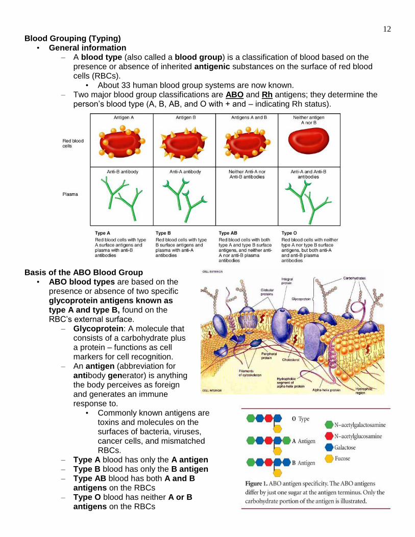
Blood Grouping (Typing) • General information
– A blood type (also called a blood group) is a classification of blood based on the presence or absence of inherited antigenic substances on the surface of red blood cells (RBCs).
• About 33 human blood group systems are now known. – Two major blood group classifications are ABO and Rh antigens; they determine the
person’s blood type (A, B, AB, and O with + and – indicating Rh status).
Basis of the ABO Blood Group • ABO blood types are based on the
presence or absence of two specific glycoprotein antigens known as type A and type B, found on the RBC’s external surface.
– Glycoprotein: A molecule that consists of a carbohydrate plus a protein – functions as cell markers for cell recognition.
– An antigen (abbreviation for antibody generator) is anything the body perceives as foreign and generates an immune response to.
• Commonly known antigens are toxins and molecules on the surfaces of bacteria, viruses, cancer cells, and mismatched RBCs.
– Type A blood has only the A antigen – Type B blood has only the B antigen – Type AB blood has both A and B
antigens on the RBCs – Type O blood has neither A or B
antigens on the RBCs
13
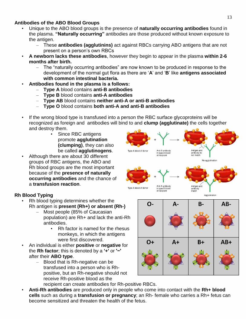
Antibodies of the ABO Blood Groups • Unique to the ABO blood groups is the presence of naturally occurring antibodies found in
the plasma. “Naturally occurring” antibodies are those produced without known exposure to the antigen.
– These antibodies (agglutinins) act against RBCs carrying ABO antigens that are not present on a person’s own RBCs
• A newborn lacks these antibodies, however they begin to appear in the plasma within 2-6 months after birth.
– The “naturally occurring antibodies” are now known to be produced in response to the development of the normal gut flora as there are ‘A’ and ‘B’ like antigens associated with common intestinal bacteria.
• Antibodies found in the plasma is a follows: – Type A blood contains anti-B antibodies – Type B blood contains anti-A antibodies – Type AB blood contains neither anti-A or anti-B antibodies – Type O blood contains both anti-A and anti-B antibodies
• If the wrong blood type is transfused into a person the RBC surface glycoproteins will be
recognized as foreign and antibodies will bind to and clump (agglutinate) the cells together and destroy them.
• Since RBC antigens promote agglutination (clumping), they can also be called agglutinogens.
• Although there are about 30 different groups of RBC antigens, the ABO and Rh blood groups are the most important because of the presence of naturally occurring antibodies and the chance of a transfusion reaction.
Rh Blood Typing • Rh blood typing determines whether the
Rh antigen is present (Rh+) or absent (Rh-) – Most people (85% of Caucasian
population) are Rh+ and lack the anti-Rh antibodies.
• Rh factor is named for the rhesus monkeys, in which the antigens were first discovered.
• An individual is either positive or negative for the Rh factor; this is denoted by a '+' or '−' after their ABO type.
– Blood that is Rh-negative can be transfused into a person who is Rh-positive, but an Rh-negative should not receive Rh-positive blood as the recipient can create antibodies for Rh-positive RBCs.
• Anti-Rh antibodies are produced only in people who come into contact with the Rh+ blood cells such as during a transfusion or pregnancy; an Rh- female who carries a Rh+ fetus can become sensitized and threaten the health of the fetus.

14
Universal Blood Donors and Recipients • Type O- is the universal donor
– Since type O- individuals have no A B, or Rh antigens so no other blood type will reject it.
• Type AB+ is the universal recipient – Since type AB+ individuals lack both anti-A, anti-B and anti-Rh antibodies and can
receive they can accept donor blood of any other blood type. Transfusion Reactions
• Transfusion reactions involve the clumping and hemolysis (rupture) of the donated (transfused) RBCs.
– Clumped RBCs can plug up small blood vessels preventing the delivery of oxygen and removal of waste products.
– Hemolysis cause the release of hemoglobin which if high enough, will precipitate in the kidneys and block the urine-forming structures leading to acute kidney failure.
• The most serious consequences arise from the effect of antibodies in the recipient’s plasma on the incoming door RBCs.
– The effect of the donor’s antibodies on the recipient’s RBCs is minor unless a large amount of blood is transfused, because the donor’s antibodies are diluted by the recipient’s plasma.
– Note: Unless the person needs a large amount of blood, the transfused blood is in the form of “packed red blood cells” which is whole blood minus the plasma portion.
Identification of ABO Blood Groups
• RBC clumping (agglutination) in the typing serum determines the antigens present on the RBC

15
Percentage of Blood types in the United States • Each person’s blood belongs to one of four groups. The following population percentages are
from the United States. • Type A blood has antigen A and anti-B antibodies in its plasma
– About 42% of people have type A blood, with 6% having A-negative (A-) blood and 36% having A-positive (A+) blood.
• Type B blood has antigen B and anti-A antibodies in its plasma – About 10% of people have type B blood, with 2% having B-negative (B-) blood and 8%
having B-positive (B+) blood. • Type AB blood has both antigen A and antigen B and lacks both anti-A and anti-B antibodies.
– Is the rarest, about 4% of people have type AB blood, with 1% having AB-negative (AB-) blood and 3% having AB-positive (AB+) blood.
– AB+ is called the universal recipient because they may receive blood from any other ABO blood type
• Type O blood lacks both A and B antigens and contains both anti-A and anti-B antibodies. – Most common, about 44% of people have type O blood, with 7% having O-negative
(O-) blood and 37% having O-positive (O+) blood – O- is called the universal donor because their blood may be given to any other ABO
type
O O, A, B, AB Both Anti-A and Anti-B
Neither A nor B
Type O
A, B, AB, O AB Neither Anti-A nor Anti-B
A, B Type AB
B, O B, AB Anti-A B Type B
A, O A, AB Anti-B A Type A
Blood Type Antigens Antibodies in plasma
Can donate blood to
type
Can receive blood from type
16
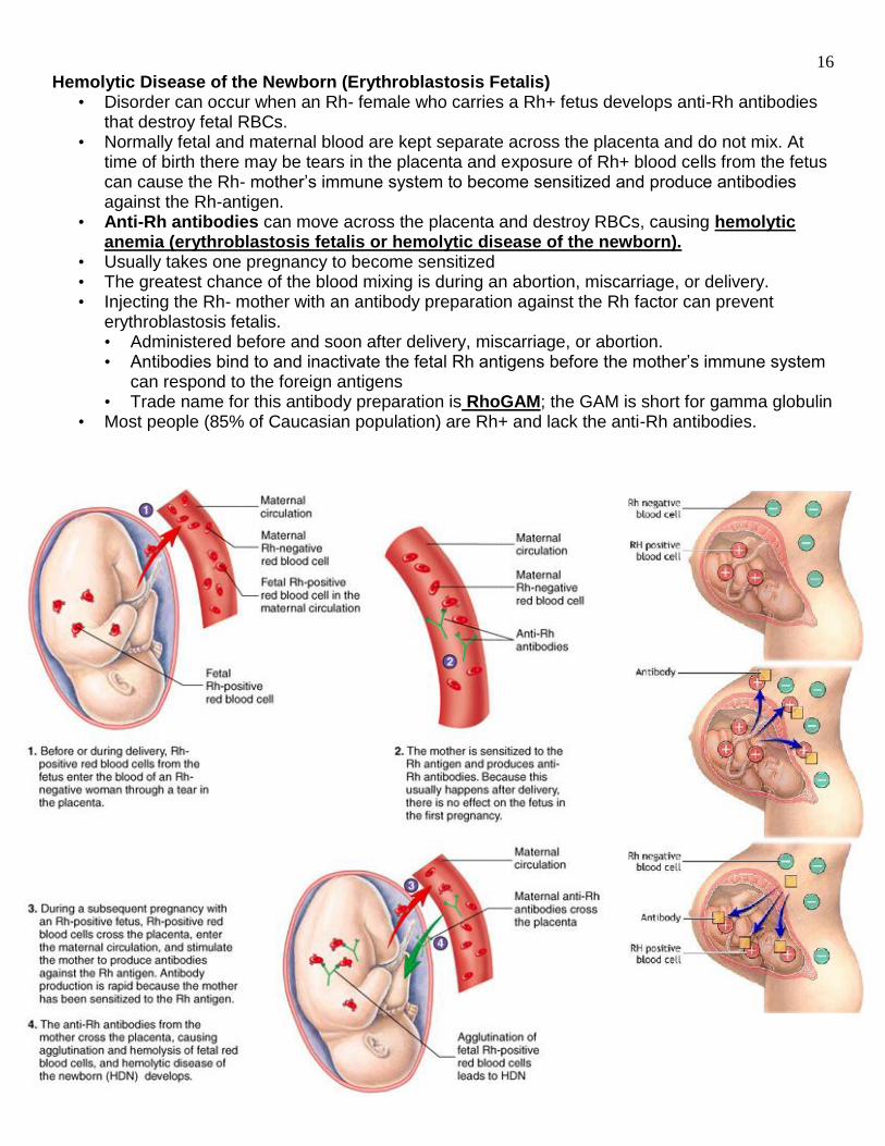
Hemolytic Disease of the Newborn (Erythroblastosis Fetalis) • Disorder can occur when an Rh- female who carries a Rh+ fetus develops anti-Rh antibodies
that destroy fetal RBCs. • Normally fetal and maternal blood are kept separate across the placenta and do not mix. At
time of birth there may be tears in the placenta and exposure of Rh+ blood cells from the fetus can cause the Rh- mother’s immune system to become sensitized and produce antibodies against the Rh-antigen.
• Anti-Rh antibodies can move across the placenta and destroy RBCs, causing hemolytic anemia (erythroblastosis fetalis or hemolytic disease of the newborn).
• Usually takes one pregnancy to become sensitized • The greatest chance of the blood mixing is during an abortion, miscarriage, or delivery. • Injecting the Rh- mother with an antibody preparation against the Rh factor can prevent
erythroblastosis fetalis. • Administered before and soon after delivery, miscarriage, or abortion. • Antibodies bind to and inactivate the fetal Rh antigens before the mother’s immune system
can respond to the foreign antigens • Trade name for this antibody preparation is RhoGAM; the GAM is short for gamma globulin
• Most people (85% of Caucasian population) are Rh+ and lack the anti-Rh antibodies.