blood ans 215 physiology and anatomy of domesticated animals · blood ans 215 physiology and...
TRANSCRIPT
BloodANS 215
Physiology and Anatomy ofDomesticated Animals
I. IntroductionA. Evolved to provide for the transport of nutrients to cells and waste from cells.B. Additional functions relating to its role in maintaining fluid balance, pH
equilibrium, and immune function.C. Also developed means to prevent blood loss
II. Composition of BloodA. Composed of cells and plasmaB. Cells of blood are:
1. Erythrocytes2. Leukocytes3. Platelets
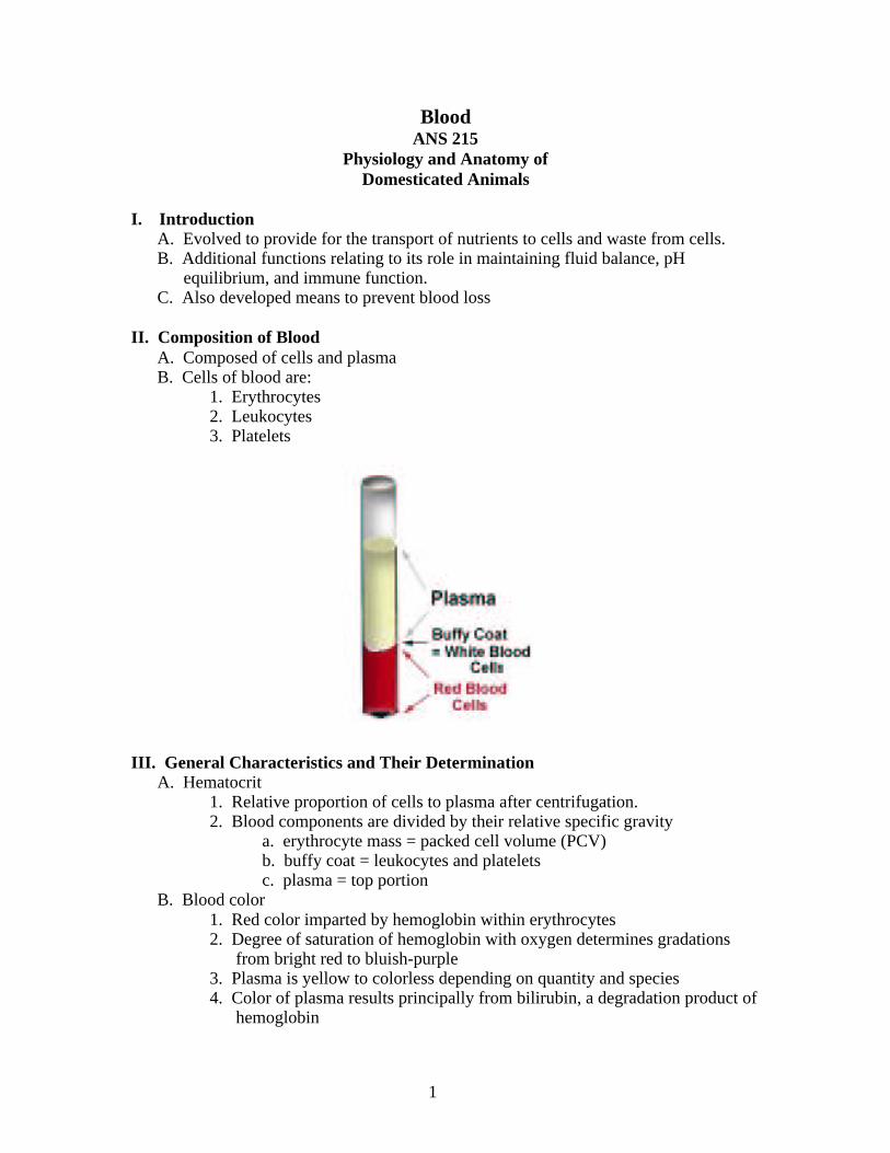
III. General Characteristics and Their DeterminationA. Hematocrit
1. Relative proportion of cells to plasma after centrifugation.2. Blood components are divided by their relative specific gravity
a. erythrocyte mass = packed cell volume (PCV)b. buffy coat = leukocytes and plateletsc. plasma = top portion
B. Blood color1. Red color imparted by hemoglobin within erythrocytes2. Degree of saturation of hemoglobin with oxygen determines gradations
from bright red to bluish-purple3. Plasma is yellow to colorless depending on quantity and species4. Color of plasma results principally from bilirubin, a degradation product of
hemoglobin
1
C. Blood Volume1. Function of the lean body weight and is generally 8 – 10% of body weight2. Ratio of the plasma volume (PV) to the blood volume is the same as
hematocrit ratioa. PV/BV = 1 – PCV/1b. e.g. 1000kg cow has blood volume of 8% of body weight or 80L.
If the hematocrit shows 60% is plasma then the plasma volume should be 48kg or 48L.
D. Blood pH1. Blood has a pH of about 7.42. Venous blood is slightly more acidic than arterial blood
a. Higher CO2 in venous blood3. The pH symbol is the chemical notation for the logarithm of the reciprocal
of the hydrogen ion concentration (H+) in gram atoms per liter of solution.
IV. LeukocytesA. Classified as either granulocytes or agranulocytes
1. Three types of granulocytes based on uptake of staina. neutrophils – take up red and blue dyeb. basophils – take up blue dyec. eosinophils – take up red dye
2. There are two types of agranulocytesa. monocytesb. lymphocytes
B. Granulocytes and monocytes are produced in bone marrow from myeloblasts.C. Lymphocytes originate in a lymphoid cell in lymph tissue.D. After their development, leukocytes circulate in blood until they leave the
circulation to perform their extravascular function.E. Generally, there are more erythrocytes than leukocytes in blood.
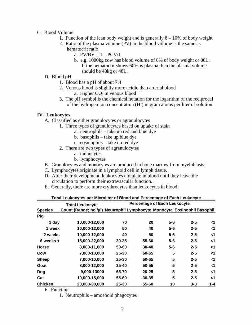
Total Leukocytes per Microliter of Blood and Percentage of Each Leukocyte
SpeciesTotal Leukocyte
Count (Range; no./µl)
Percentage of Each Leukocyte
Neutrophil Lymphocyte Monocyte Eosinophil BasophilPig
1 day 10,000-12,000 70 20 5-6 2-5 <11 week 10,000-12,000 50 40 5-6 2-5 <1
2 weeks 10,000-12,000 40 50 5-6 2-5 <16 weeks + 15,000-22,000 30-35 55-60 5-6 2-5 <1
Horse 8,000-11,000 50-60 30-40 5-6 2-5 <1Cow 7,000-10,000 25-30 60-65 5 2-5 <1
Sheep 7,000-10,000 25-30 60-65 5 2-5 <1Goat 8,000-12,000 35-40 50-55 5 2-5 <1
Dog 9,000-13000 65-70 20-25 5 2-5 <1Cat 10,000-15,000 55-60 30-35 5 2-5 <1
Chicken 20,000-30,000 25-30 55-60 10 3-8 1-4F. Function
1. Neutrophils – amoeboid phagocytes
2
2. Monocytes – largest leukocyte, macrophage, numbers increase in chronic infection.
a. some monocytes fixed in tissues, others migrate3. Eosinophils – dampen inflammatory reactions – increase during certain
types of parasitism, are reduced during stress by cortisol, increase in eosinophils can also indicate allergic response
4. Basophils – enhance inflammatory reaction in response to IgE5. Lymphocytes – involved in immune response
a. T-cells – preprocessed in thymusi. cytotoxic T-cells – killer cells
ii. helper T-cells – assist in activation of killer cells, suppressor T-cells and B-cells
iii. suppressor T-cells – suppress action of cytotoxic and helper T-cells
b. B-cells – preprocessed in bone marrowi. produce antibodies – humeral immunityii. Antibodies inactivate antigens by agglutination and
precipitation.iii. Antibodies are also involved in complement system in
which antibodies bind to foreign cells which then attract phagocytes and killer cells.
G. Plasma cells and megakaryocytes1. Plasma cells are found in lymphoid tissue and produce antibodies.2. Megakaryocytes are found in bone marrow and give rise to blood platelets,
which are involved in clotting.
3
V. Diagnostic Procedures
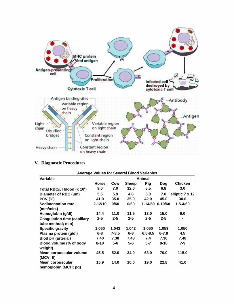
Average Values for Several Blood VariablesVariable Animal Horse Cow Sheep Pig Dog Chicken
Total RBC/µl blood (x 106) 9.0 7.0 12.0 6.5 6.8 3.0
Diameter of RBC (µm) 5.5 5.9 4.8 6.0 7.0 elliptic 7 x 12PCV (%) 41.0 35.0 35.0 42.0 45.0 30.0Sedimentation rate(mm/min.)
2-12/10 0/60 0/60 1-14/60 6-10/60 1.5-4/60
Hemoglobin (g/dl) 14.4 11.0 11.5 13.0 15.0 9.0Coagulation time (capillarytube method; min)
2-5 2-5 2-5 2-5 2-5 -
Specific gravity 1.060 1.043 1.042 1.060 1.059 1.050Plasma protein (g/dl) 6-8 7-8.5 6-8 6.5-8.5 6-7.8 4.5Blod pH (arterial) 7.40 7.38 7.48 7.4 7.36 7.48Blood volume (% of bodyweight)
8-10 5-6 5-6 5-7 8-10 7-9
Mean corpuscular volume(MCV; fl)
45.5 52.0 34.0 63.0 70.0 115.0
Mean corpuscularhemoglobin (MCH; pg)
15.9 14.0 10.0 19.0 22.8 41.0
4
Mean corpuscularhemoglobin concentration(MCHC; %)
35.0 33.0 32.5 32.0 34.0 29.0
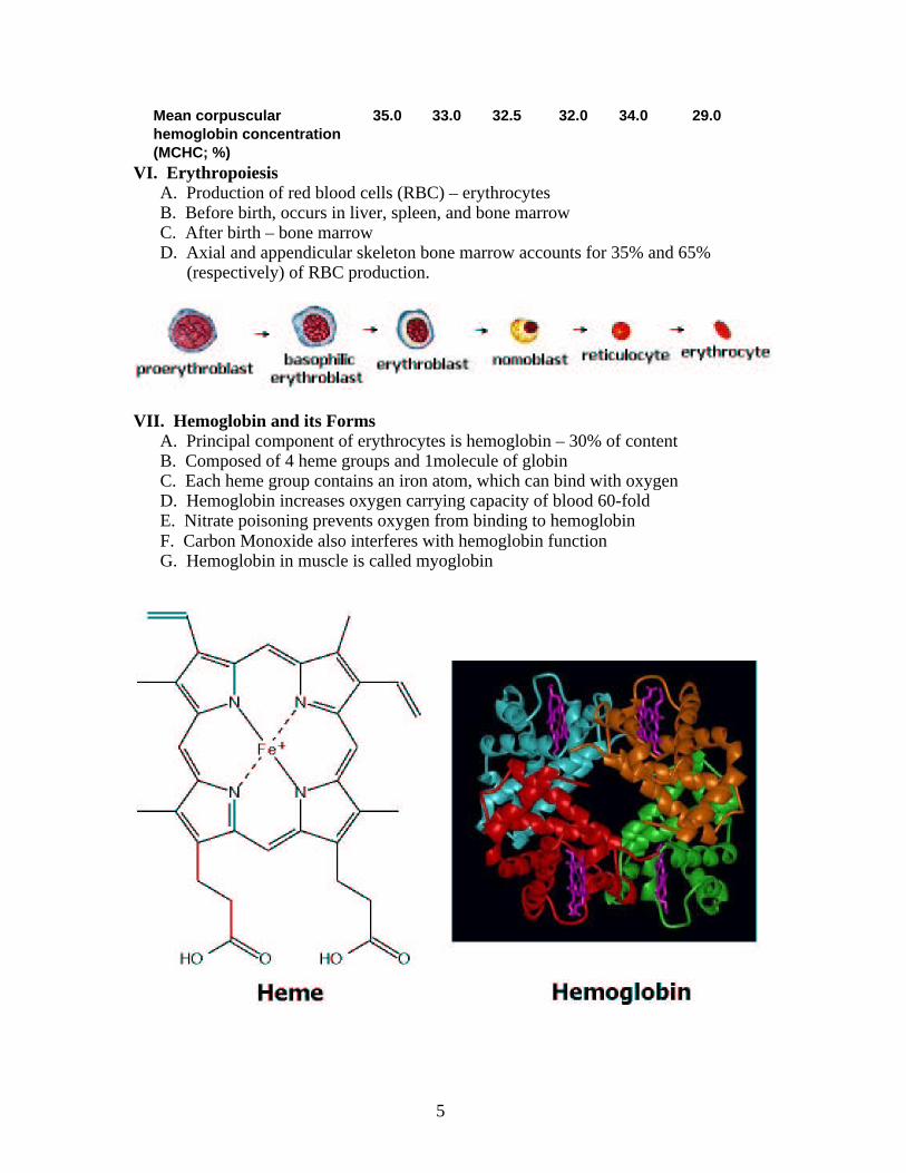
VI. ErythropoiesisA. Production of red blood cells (RBC) – erythrocytesB. Before birth, occurs in liver, spleen, and bone marrowC. After birth – bone marrowD. Axial and appendicular skeleton bone marrow accounts for 35% and 65%
(respectively) of RBC production.
VII. Hemoglobin and its FormsA. Principal component of erythrocytes is hemoglobin – 30% of contentB. Composed of 4 heme groups and 1molecule of globinC. Each heme group contains an iron atom, which can bind with oxygenD. Hemoglobin increases oxygen carrying capacity of blood 60-foldE. Nitrate poisoning prevents oxygen from binding to hemoglobinF. Carbon Monoxide also interferes with hemoglobin functionG. Hemoglobin in muscle is called myoglobin
5
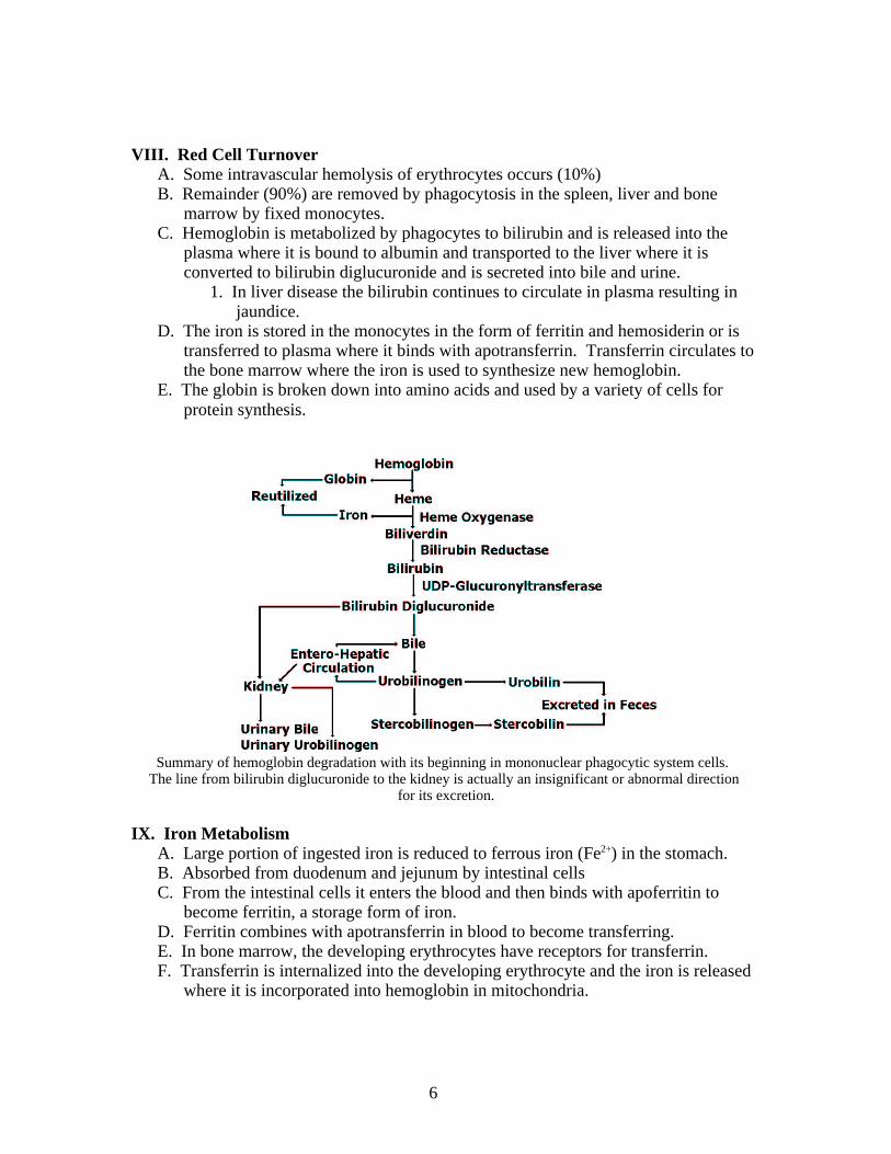
VIII. Red Cell TurnoverA. Some intravascular hemolysis of erythrocytes occurs (10%)B. Remainder (90%) are removed by phagocytosis in the spleen, liver and bone
marrow by fixed monocytes.C. Hemoglobin is metabolized by phagocytes to bilirubin and is released into the
plasma where it is bound to albumin and transported to the liver where it is converted to bilirubin diglucuronide and is secreted into bile and urine.
1. In liver disease the bilirubin continues to circulate in plasma resulting in jaundice.
D. The iron is stored in the monocytes in the form of ferritin and hemosiderin or is transferred to plasma where it binds with apotransferrin. Transferrin circulates to the bone marrow where the iron is used to synthesize new hemoglobin.
E. The globin is broken down into amino acids and used by a variety of cells for protein synthesis.
Summary of hemoglobin degradation with its beginning in mononuclear phagocytic system cells. The line from bilirubin diglucuronide to the kidney is actually an insignificant or abnormal direction
for its excretion.
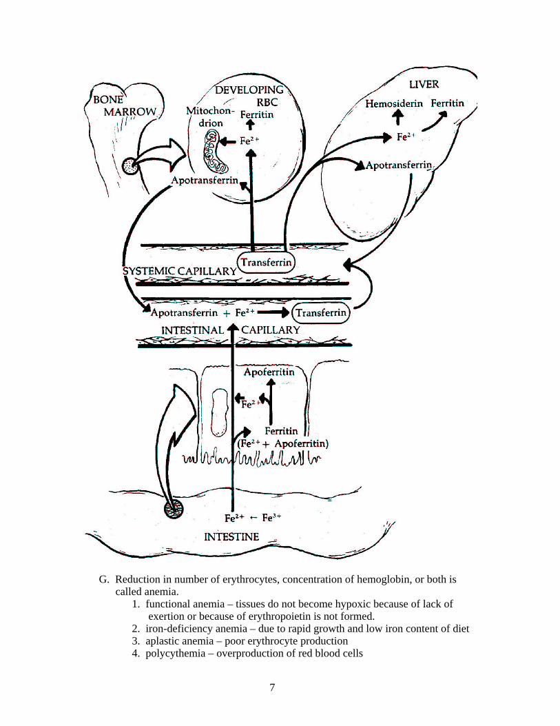
IX. Iron MetabolismA. Large portion of ingested iron is reduced to ferrous iron (Fe2+) in the stomach.B. Absorbed from duodenum and jejunum by intestinal cellsC. From the intestinal cells it enters the blood and then binds with apoferritin to
become ferritin, a storage form of iron.D. Ferritin combines with apotransferrin in blood to become transferring.E. In bone marrow, the developing erythrocytes have receptors for transferrin.F. Transferrin is internalized into the developing erythrocyte and the iron is released
where it is incorporated into hemoglobin in mitochondria.
6
G. Reduction in number of erythrocytes, concentration of hemoglobin, or both is called anemia.
1. functional anemia – tissues do not become hypoxic because of lack of exertion or because of erythropoietin is not formed.
2. iron-deficiency anemia – due to rapid growth and low iron content of diet3. aplastic anemia – poor erythrocyte production4. polycythemia – overproduction of red blood cells
7
X. Prevention of Blood Loss
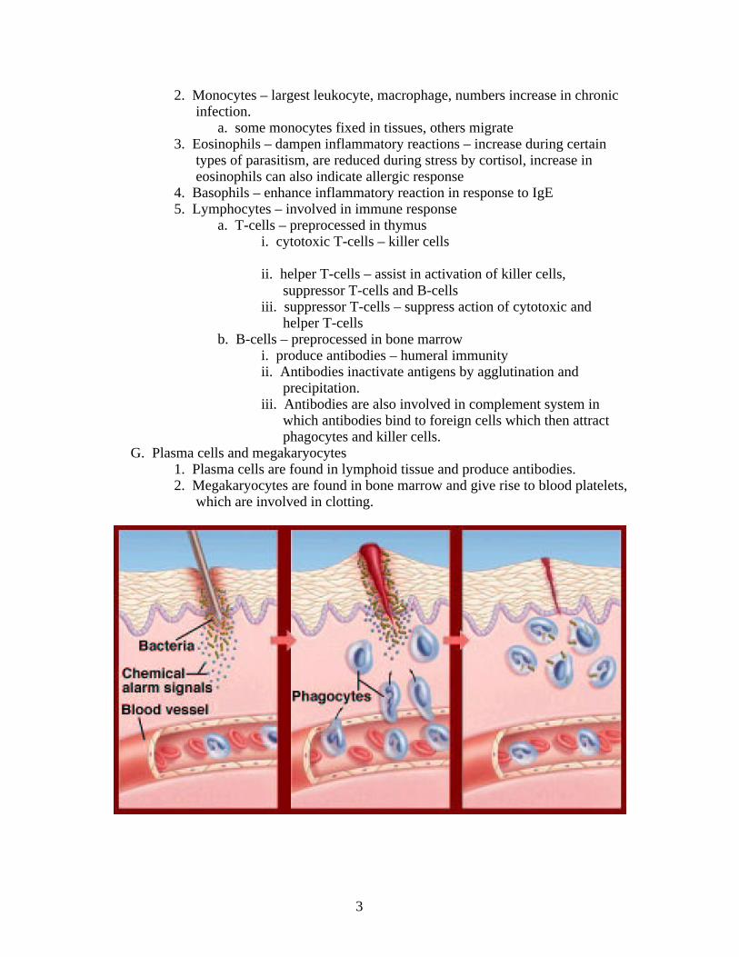
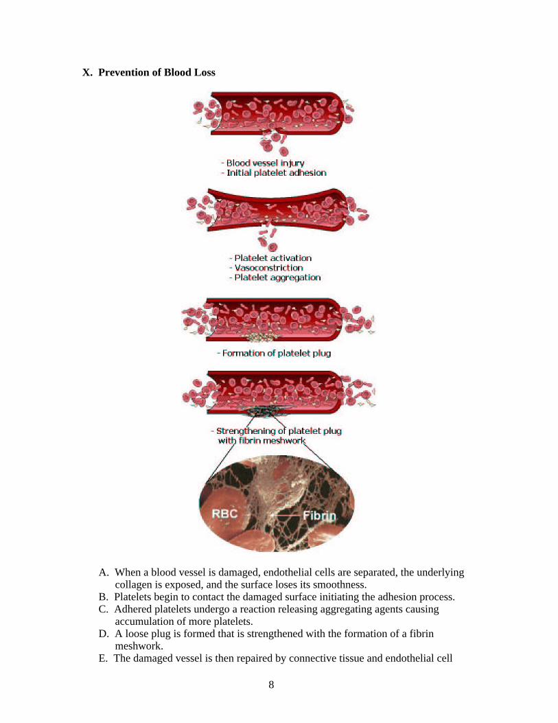
A. When a blood vessel is damaged, endothelial cells are separated, the underlying collagen is exposed, and the surface loses its smoothness.
B. Platelets begin to contact the damaged surface initiating the adhesion process.C. Adhered platelets undergo a reaction releasing aggregating agents causing
accumulation of more platelets. D. A loose plug is formed that is strengthened with the formation of a fibrin
meshwork.E. The damaged vessel is then repaired by connective tissue and endothelial cell
8
growth.F. The platelet-fibrin complex and cell debris is then removed by phagocytes.G. Vascular endothelium lines the entire cardiovascular system.
1. 1 cell layer thick2. Regardless of location it is underlaid by collagen3. Collagen is a potent platelet activator4. The subendothelial tissue also contains fibronectin and Von Willebrand
factor, which both enhance aggregation of platelets.H. Properties of endothelium that inhibit aggregation are:
1. Negative charge on endothelium surface2. Synthesis of inhibitors of aggregation (e.g. prostacyclin) and fibrin
formation (thrombomodulin)3. Generation of activators of fibrin degradation (e.g. tissue plasminogen
activator, TPA)
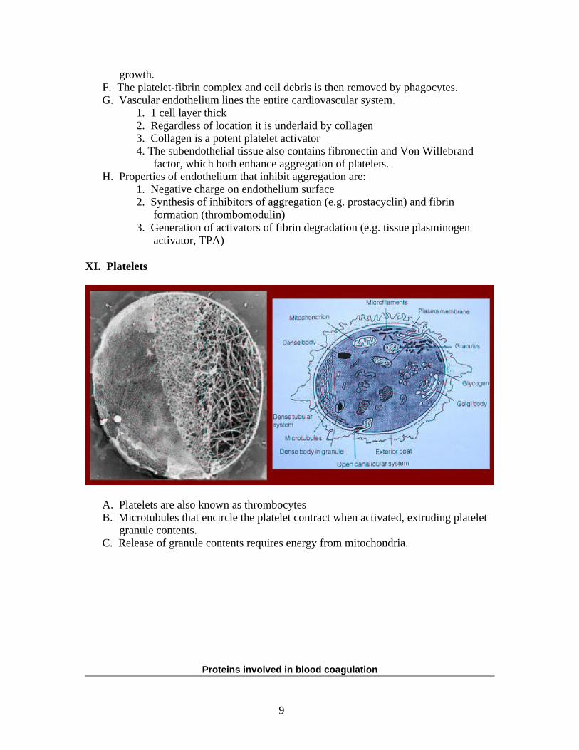
XI. Platelets
A. Platelets are also known as thrombocytesB. Microtubules that encircle the platelet contract when activated, extruding platelet
granule contents.C. Release of granule contents requires energy from mitochondria.
Proteins involved in blood coagulation
9
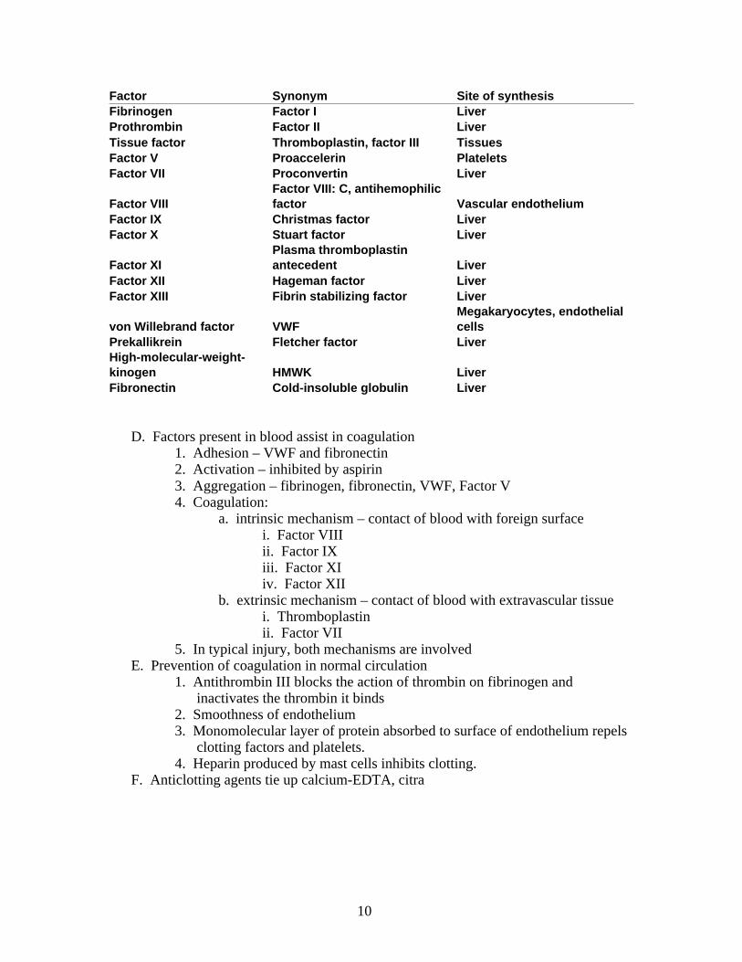
Factor Synonym Site of synthesisFibrinogen Factor I LiverProthrombin Factor II LiverTissue factor Thromboplastin, factor III TissuesFactor V Proaccelerin PlateletsFactor VII Proconvertin Liver
Factor VIIIFactor VIII: C, antihemophilicfactor Vascular endothelium
Factor IX Christmas factor LiverFactor X Stuart factor Liver
Factor XIPlasma thromboplastinantecedent Liver
Factor XII Hageman factor LiverFactor XIII Fibrin stabilizing factor Liver
von Willebrand factor VWFMegakaryocytes, endothelialcells
Prekallikrein Fletcher factor LiverHigh-molecular-weight-kinogen HMWK LiverFibronectin Cold-insoluble globulin Liver
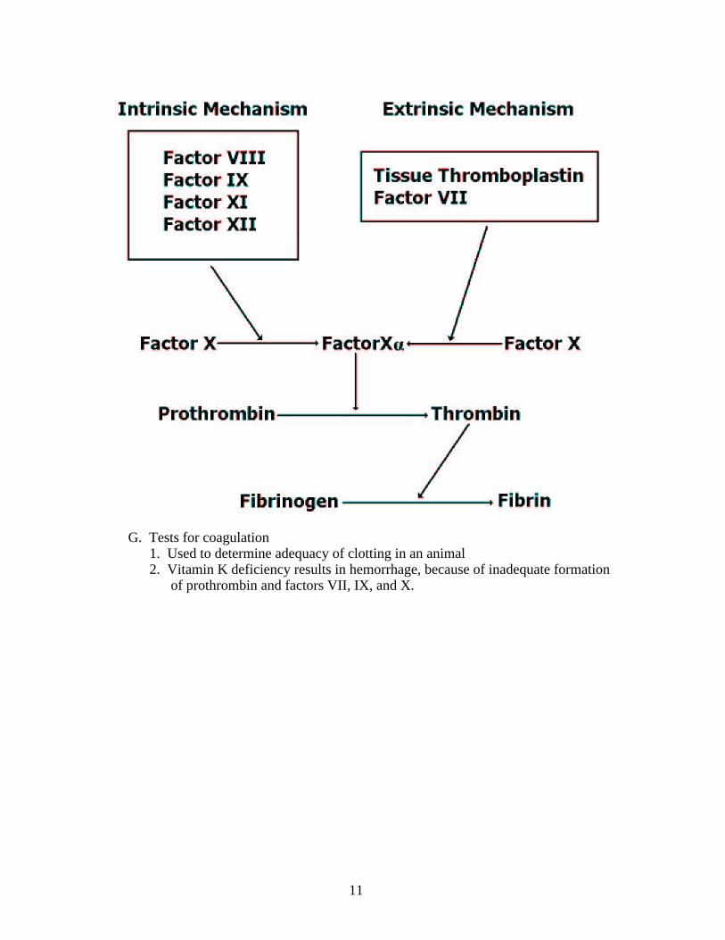
D. Factors present in blood assist in coagulation1. Adhesion – VWF and fibronectin2. Activation – inhibited by aspirin3. Aggregation – fibrinogen, fibronectin, VWF, Factor V4. Coagulation:
a. intrinsic mechanism – contact of blood with foreign surfacei. Factor VIIIii. Factor IXiii. Factor XIiv. Factor XII
b. extrinsic mechanism – contact of blood with extravascular tissuei. Thromboplastinii. Factor VII
5. In typical injury, both mechanisms are involvedE. Prevention of coagulation in normal circulation
1. Antithrombin III blocks the action of thrombin on fibrinogen and inactivates the thrombin it binds
2. Smoothness of endothelium3. Monomolecular layer of protein absorbed to surface of endothelium repels
clotting factors and platelets.4. Heparin produced by mast cells inhibits clotting.
F. Anticlotting agents tie up calcium-EDTA, citra
10
G. Tests for coagulation1. Used to determine adequacy of clotting in an animal2. Vitamin K deficiency results in hemorrhage, because of inadequate formation
of prothrombin and factors VII, IX, and X.
11