biotechnology & you - · 2 biotechnology & you c o n t e n t s table of...
TRANSCRIPT

I n v e s t i g a t i n g
Volume 6, Issue No. 1
BIOTECHNOLOGY & YOU
a magazine of biotechnology applications in healthcare, agriculture, the environment, and industry

2
BIOTECHNOLOGY & YOU C O N T E N T STABLE OF○
○
○
○
○
○
○
○
○
○
○
○
○
○
○
○
○
○
○
○
○
○
○
○
○
○
○
○
○
○
○
○
○
○
○
○
○
○
○
○
○
○
○
○
○
○
○
○
○
○
○
○
○
○
○
○
○
○
○
○
○
○
○
○
○
○
○
○
○
○
○
○
○
○
○
○
○
○
○
○
○
○
○
○
○
3
Volume 6, Issue No. 1
Your World/Our World describes the application ofbiotechnology to problems facing our world. Wehope that you find it an interesting way to learnabout science and engineering.
Development by:The Pennsylvania Biotechnology Association,The PBA Education Committee, andSnavely Associates, Ltd.
Editing by:The Writing Company, Cathryn M. Delude andKenneth W. Mirvis, Ed.D.
Design by:Snavely Associates, Ltd.
Education Advisors:Linda L. Hubler, Pennbrook Middle SchoolNancy E. Guidotti, Penndale Middle School
Science Writer and Advisor:Jill M. Roberts-Lewis, Ph.D., BioDiligence Partners, Inc.
Publication Contributors:Anthony P. Green, Ph.D., Puresyn, Inc.Michael E. Lewis, Ph.D., BioDiligence Partners, Inc.William Graney, M.D., Cephalon, Inc.John L. Tedesco, Brandywine Consultants, Inc.
Special Thanks:The PBA is grateful to the members of the EducationCommittee for their contributions:
Keith Buckingham, Friends Central School
Ceil Ciociola, PRIME, Inc.
Stephen R. Collins, JRH Bioscience
Roberta Cook, Ph.D., The Franklin Institute
Jeff Davidson, Pennsylvania BiotechnologyAssociation
Alan Gardner, Ph.D., SmithKline Beecham
Cynthia Gawron-Burke, Ph.D.
Barbara Handelin, Handelin & Associates
M. Kay Oluwole, DVM, MPS
Daniel M. Keller, Ph.D., Keller Broadcasting
Kodzo Gbewonyo, Ph.D., Dustin Landis andAlthea Talento, Merck & Co., Inc.
Barbara McHale, Gwynedd Mercy College
Robert Pearson, Rhone-Poulenc Rorer
Lois Peck, Ed.D., Philadephia College ofPharmacy & Science
Kamal Rashid, Ph.D., Pennsylvania State University
Jean Scholz, University of Pennsylvania
Lisa Speicher, Wyeth-Ayerst Research
Laurence A. Weinberger, Esq., Committe Chair
If you would like to make suggestions or commentsabout Your World/Our World, drop us a line at:CompuServe: 73150,1623Internet: [email protected] you have questions about the material presented inthis issue of Your World/Our World, please drop Dr.Jill M. Roberts-Lewis an e-mail [email protected] Biotechnology Association1524 W. College Avenue, Suite 206State College, Pennsylvania 16801
Copyright 1996, PBA. All rights reserved.
The Brain in a Nutshell
46
Neurons: An ElectrifyingKind of Cell
Synapses: Getting the MessageAcross
7 Drugs and the Brain: Miracle orMenace?
8 Sandra Moon: Neuroanatomist
9 Alzheimer’s Disease:Fading Memories
12 Mapping the Mind
10 Wanted: Medical Breakthroughs
14 Getting the Point
15 Ethical Issues in Neuroscience
On the Cover: Seeing the Brain in ActionComputerized PET scan images show which regions of the brain are used for different tasks:hearing words (top) and speaking words (bottom). Different colors represent different levels ofbrain activity. Blue = low activity; Green = intermediate activity; Yellow = high activity; Red =intense activity; White = maximum activity.
Photo credit: Marcus E. Raichle, M.D.; Washington University School of Medicine
P R O F I L E
E X P E R I M E N T
In the past few decades scientists havemade incredible advances in studyingthe brain. This issue of Your World/Our
World provides:
• a glimpse of what we know about theway normal brains work and aboutbrain disorders,
• how research and technology help uslearn about the fundamental molecularworkings of the brain,
• how this understanding can lead to thediscovery of new medicines to treatbrain disorders.

3
BrainF I R E ! ! !F I R E ! ! !Imagine you are gazing out thewindow. Suddenly you see flamesshooting out of the roof of thebuilding next door! This image – apattern of light, shape, and move-ment – falls on neurons (nerve cells)at the back of your eyes that aresensitive to light. Signals race fromneuron to neuron along a pathwaythat leads through the thalamus and
on to the occipital lobes in theback of the brain. Here,
other neuronsreassemble the image
by responding tospecific features ofthe fire, such asits movementor its shape.Theseneurons, inturn, sendmessages toother partsof the brain,such as thehippocampus,
which isinvolved in
rememberingwhat a fire is andthe injury it can
cause; theamygdala, which
triggers the feeling offear; the frontal lobe,which formulates theword “fire”; and the
action you will take(shout! run!) and finally,
the motor neurons, whichsend signals down the
spinal cord to stimulate themuscles that will propel you
from your seat, franticallypointing out the window and
screaming, “Fire!”▼
Investigating the Brain!Over 40 years ago, Henry M. sufferedfrom epileptic seizures so severe thathe could not work. To stop hisseizures, surgeons removed a brainstructure called the hippocampus.The results of this surgery were bothtragic and astonishing: Henry cannotremember anything new. He caneasily recall events from before theoperation, but nothing that hashappened since. Henry says his worldis “like waking from a dream.” Hedoes not know how old he is, nor canhe recognize people he has beenseeing for years. To this day, helives from moment to moment,a 70 year old man who isforever 27 years old “inmemory.”
Your brain, a three-pound cluster of cells isthe most complexliving structure thatwe know of in theuniverse. It allowsyou to see, smell,taste, hear, feel, andrespond to the worldaround you. It enablesyou to think, learn,remember, speak,feel emotions and haveappetite, and it plays animportant role infighting disease.
In short, our brains define us. Bystudying the brain, we can learn moreabout who we are, both as individualsand as a species. Everything we learnabout the brain can help us prevent orcure the many devastating braindisorders that kill, cripple, or depriveus of our personality, intelligence, andmemories. ■
■ Frontal lobe: long-term planning,social behavior, speech, movement
■ Parietal lobe: read, write,mathematical and spatial abilities,body sensations
■ Temporal lobe: hear, smell,understand speech, form memoriesand emotions
■ Occipital lobe: vision■ Cerebellum: motor control, balance■ Thalamus: (in center) relay station
for sensory signals■ Hippocampus: (two projections in
center) memory processing■ Amygdala: (at end of projections)
emotional state
This map of the human brainshow the basic arrangementof some of its parts. Seen hereare the two major hemi-spheres, left and right, and theinternal brain structuresmentioned in the storiesabove. The brain structuresare colored to correspond tothe colored words in the text.

4
Neurons:The brain is made of billions of
cells called neurons. In manyways, neurons are like all other
animal cells. They are cytoplasm-filledsacks with such parts as nuclei,ribosomes, and mitochondria. Unlikeother cells, though, a typical neuronhas many branches called dendritesextending from its cell body and along tail called an axon. The axonsspan distances as short as a fraction ofa millimeter, or as long as a meter ormore! Axons connect neurons toother neurons in amazingly intricateways. As shown in the illustrationbelow of three typical neurons, axonsmay connect many times to thedendrites and cell bodies of otherneurons.
The total number of neurons in ourbodies is roughly equivalent to thenumber of stars in our galaxy! Eachof these billions of neurons thenconnects to other neurons hundredsof times so that the total number ofinterconnections is truly astronomi-cal. These trillions of highly orga-nized interconnections are respon-sible for the extraordinary happeningwe call consciousness.
An individual neuron is like atelephone that receives and sendsinformation from one cell to the next.It does this through a uniquecombination of electricity andchemistry. When a neuron receives asignal, it passes it along its length
Axon
Synapse(Detail on page 6)
4

5
Nucleus
A neuron from the cerebral cortexmagnified 600 times. The neuron hasbeen stained with a special dye thatallows us to see the cell body and itsmany branching dendrites.
Ron
ald
F. M
ervi
s, P
h.D
.: N
euro
Met
rix
Res
earc
h, In
c.
An Electrifying Kind of Cellelectrically. To help speed thiselectrical signal, the axons of someneurons are surrounded by a specialinsulating sheath of cells calledmyelin. These electrical signals areeasily detected and recorded. (Seepage 12.)
Then, at the end of the axon, theneuron transmits the signal to thenext neuron by releasing a tinyamount of a chemical–called aneurotransmitter–into the gap, calledthe synapse, between neurons. Whenthe neurotransmitter is released intothe synapse, it is captured by a specialprotein, called a receptor, on the nextneuron. The receptor then tells theneuron to start transmitting an
electrical signal. There are manydifferent neurotransmitters used byneurons and there is a differentreceptor for each neurotransmitter.Each receptor recognizes only onetype of neurotransmitter, like a keyfitting only one lock. The neurotrans-mitters dopamine, serotonin, andglutamate are discussed in followingarticles.
Later in this issue, youwill learn how theevents occurringduring signalingacross the synapseplay a big role inthe workings ofour brain.
Dendrite
Cell Body
Myelin Sheath5

6
Resting state Action potential Receptor stimulation Neurotransmitter removal
A moment in the life of a neuron
Resting state Action potential Receptor stimulation Neurotransmitter removal
This illustration shows theend of an axon whereit meets the next neuron–the
synapse–as a signal is passed betweenthe neurons.
Initially, neurons have an electrical“charge”–like a battery. This charge iscaused by the accumulation ofdifferent ions (atoms with a positiveor negative charge) on the inside andoutside of the neuron. When neuronsare in a resting state (not sending asignal), there are more negative ionson the inside of the neuron and morepositive ions on the outside. Whenenough neurotransmitter crosses thesynapse and attaches to receptors, achemical reaction is triggered thatlets ions move through openings orchannels in the membrane. Positiveions flood into the neurons, causing asudden reversal of the charge insidethe neuron. This reversal of chargetriggers nearby channels to open sothat the opening of channels thenflows along the length of the axon likea wave–much like a wave in a sportsstadium. This has already happenedto the top neuron.
In the illustration, this wave, called anaction potential, has reached the endof the axon, where it has caused theneurotransmitter to be released intothe synapse. Some of the neurotrans-mitter has already bound to receptorson the next neuron stimulating thenext neuron to “fire” electrically. Thisprocess continues from neuron to
Synapses: Getting the Message AcrossIons
Ion Channel
Transporter
Neurotransmitter
Receptor
Drug that blockstransporter
neuron,reaching themany parts of thebrain involved in interpret-ing and reacting to these signals.
Neurotransmitter which is not boundto receptors may be taken back into theneuron through a protein called atransporter. Once the transporterremoves the neurotransmitter from thesynapse, the receptors are no longerexposed to the neurotransmitter.
Scientists who study the brain mayuse their knowledge of how these
signals travel from neuron to neuronto design drugs. For example, theymight design a drug that blocks ionchannels and prevents the actionpotential so the sensation of pain isnot transmitted to the brain; that ishow a local anesthetic works.
Next you will read about two ex-amples of drugs that enhance signalsby blocking the transporter soneurotransmitters have more chanceto stimulate receptors.▼

7
?
The Pleasure Center
of this drug to reduce depression,neuroscientists figured out that theneurons in the brain containingserotonin or serotonin receptors playa very important role in maintaininga healthy balance of emotions.
The Bad NewsWorking with a rehab counselor, Tomrecalled how he had slipped into apattern of substance abuse that hadruined his future. His childhood hadbeen happy and comfortable. Heexcelled in soccer and earned goodgrades in middle school.
When he was fourteen, he startedsneaking drinks of alcohol and hesoon tried other drugs. By the time
Many drugs work by changingthe way the synapse func-tions. For example, the drug
may bind to a part of the synapsesuch as a receptor, channel ortransporter. Other drugs affect otherparts of the neuron. If a drug binds toa brain cell, it can affect the brainfunctions controlled by that neuron.Sometimes the results of this aregood, and sometimes they are bad.
The Good NewsAshley, a high school student,couldn’t remember when she had lastfelt good about herself. She had beena busy, energetic teenager involved inthe school yearbook, Drama Club,and year-round competitive swim-ming. She aced most of her coursesand maintained an active social life.
Gradually, her energy disappeared, andit now seemed impossible to have everfelt so vibrant and happy. She nolonger cared about school or swim-ming, and she thought her friends nolonger cared about her. Her gradesslipped to Cs.
Ashley’s mother scheduled a doctor’sappointment. The doctor diagnosed“clinical depression” and prescribeda drug to combat depression calledProzac®. Within a month, Ashley feltlike herself again and renewed heractivities with enthusiasm.
What happened? Ashley’s depressionprobably resulted from a small shiftin the biochemical activity of a regionof her brain involved in emotion andmotivation. Prozac® treats this effectby increasing the amounts of theneurotransmitter serotonin in herbrain. It does this by blocking theserotonin transporter, so that theserotonin the neurons release has alonger time to stimulate theirreceptors. (See the discussion of thesynapse on page 6.) From the ability
Scientists found evidence of adopamine “pleasure center” bystudying laboratory rats. The ratswere trained to push a lever thatelectrically stimulated a part oftheir brains where dopamine isproduced. The rats repeatedlypressed the lever for hours,ignoring food and water, tocontinue the stimulation. (Inthe same way, people addicted todrugs will neglect their health tostay high.) These findings led tothe concept of “reward circuits”in the brain. Scientists havefound that many different abusedsubstances (such as cocaine,amphetamine, nicotine, andheroin) may directly or indirectlyincrease dopamine release in thispart of the brain. Thus, these
dopamine neurons may provide acommon link to understanding thebasis of dependence to a variety ofdrugs. You can read about moremethods used to study brainfunctions on pages 12-13.▼
he was a senior in high school, he wasusing marijuana and cocaine regu-larly. On the night of his seniorprom, he lost control of his car andslammed into a telephone pole. Thecrash killed his girlfriend and left himparalyzed from the waist down. Tomtested positive for cocaine.
What happened? Abused drugs affectreceptors or other proteins in thebrain, just as medications do.Cocaine increases the levels of theneurotransmitter dopamine in muchthe same way that Prozac increasesthe level of serotonin, by blocking thetransporter. One cluster of dopamineneurons in the brain enhances thepleasure we experience when we dosomething important to our survival,such as eating. When drugs likecocaine over-stimulate the release ofdopamine in this region of the brain,the “user” can be overcome with theurge to pursue the pleasurablesensation and thus become depen-dent on the drug. ■
A rat with a stimulating electrode implanted intoits brain. The rat is pressing a lever that deliversa mild electrical current to a part of the brainthat generates a pleasurable sensation.
Elli
ot S
. Val
enst
ein,
Ph.
D.;U
nive
rsit
y of
Mic
higa
n
MiracleorMenace
DRUGS AND THE BRAIN

8
We will always depend on thenew ideas and methods thatyoung scientists contribute. Inits study of the brain, sciencereturns to one of humanity’soldest questions – Who are we?In seeking answers to thisquestion, the tools of biotech-nology are now playing acritical role.
○
○
○
○
○
○
○
○
○
○
○
○
○
○
○
○
○
○
○
○
○
○
○
○
○
○
○
○
○
○
○
○
○
○
○
○
○
○
○
○
○
○
○
○
○
○
○
○
○
○
○
○
○
○
BrainCareers
P L U S
People from many fields areinvolved in brain research,which has come to be known
as neuroscience. The brain can bestudied at every level, using almostevery discipline from physics andmathematics to chemistry andpsychology. If you are interested inone or more of the following areas,you could someday make animportant contribution to the studyof the brain.
• Molecular biology• Biochemistry• Medicine• Endocrinology• Physiology• Pharmacology• Psychology• Experimental Psychology• Clinical Psychology• Cell biology• Physics• Computers• Electronics• Imaging
Profile of Sandra Moon,NeuroanatomistSandra Moon knows that her profession,
neuroanatomy or “anatomy of the brain,” needsa bit of explanation. “If you want to know how a
complex machine works, it helps to have a blueprintof it. It’s the same with the brain. A neuroanatomisthelps put together a blueprint of the brain.”
Sandy earned her Ph.D. from M.I.T. and now works at Bristol-Myers Squibb inWallingford, CT. Her department develops new medications for diseases anddisorders of the nervous system. Over the past twelveyears, she has worked on medications for anxiety,schizophrenia, depression, and learning and memoryimpairment. More recently, her lab is researchingstrokes. (See page10.) “The brain is a very hungryorgan,” Sandy explains. “It consumes 20% of all theoxygen in the blood. During a stroke, the source ofoxygen is blocked and the brain suffers. Our ability tosave injured brain cells will depend not only on themedications we are developing, but also on what partof the brain is damaged.” That’s where neuroanato-mists come in. They help determine where a newchemical works in the brain so they can know whether itcould become an effective medication.
Sandy loves her job for several reasons. “First, it requires good manual skills justlike my other hobbies: playing the piano, painting, and making paper. In addition,brain cells are very aesthetically pleasing. When I look at their shapes through themicroscope, they remind me of a kaleidoscope. I also like the intellectual challengeof trying to figure out how the brain works. And last but not least, it’s so exciting toknow that my research will have an impact on people’s lives in just a few years.”■
Brain BogglersNeuroscientists are seeking answers tothese questions:
1. How are memories formed andstored in the brain?
2. In what ways does learningchange the brain?
3. In what ways do drugs changethe brain?
4. What causes addiction tocertain drugs?
5. What causes mental illness?
6. What causes aggressive orviolent behavior?
7. Why do we sleep and dream?
8. Why and how do neurons die?
9. How can neurons be stimulatedto regenerate after injury?
10. How does the brain give rise tothought and consciousness?
Did you
know
that...?
Neuropharmacology is the studyof how certain drugs affect thebrain and other parts of thenervous system. A “drug” is asubstance taken into the bodythat interacts with cells andchanges the way they function.

9
Memories
One of the major groups of neurons thatdie in Alzheimer’s disease contains theneurotransmitter acetylcholine. Onecurrent treatment is the drug Cognex®,which increases the levels of acetylcho-line by blocking the enzyme thatnormally degrades this neurotransmit-ter. Cognex® temporarily improvesmental function for some patients, but
does not slow the progress ofthe disease. Anotherapproach being exploredinvolves using nerve growthfactor, a protein that helpsneurons containingacetylcholine survive.Although this protein wouldnot eliminate the cause ofcell death in Alzheimer’sdisease, it might allow manyof the affected neurons tosurvive. ■
Ronald Reagan’s words broughthome the realization that thedevastating brain disease called
Alzheimer’s can happen to anyone,even a two-term president of theUnited States. Chances are you willknow someone who has Alzheimer’s.There is presently no effectivetreatment for Alzheimer’s disease, butas researchers learn more about it,future treatments appear possible.In Alzheimer’s disease, neurons in thecortex and hippocampus that controlhigher mental functions such aslearning, memory, and problemsolving continually die. When theydie, the connections between neuronsare lost. These losses destroy theperson’s ability to reason andremember, and they cause changesin personality and emotion.
The brains of Alzheimer’s patients areriddled with large deposits (calledplaques) of an abnormal proteinknown as amyloid. Amyloid plaquesaccumulate around the neuronsand within the walls of the bloodvessels serving the brain.Researchers believe that theseabnormal plaques may cause theneurons to die, so they are tryingto learn exactly how amyloid ismade and deposited aroundthese brain cells. If they couldunderstand the chemistryinvolved in this process, theymight be able to design drugs toblock the process that causesamyloid protein deposits to form. Amyloid plaques in the cerebral cortex of a
patient who had Alzheimer’s disease.
Rob
ert S
iman
, Ph.
D.;
Cep
halo
n, In
c.
I have recently been told that I am one of themillions of Americans who will be afflicted withAlzheimer’s disease . . . I now begin the journeythat will lead me into the sunset of my life.
President Ronald Reagan,November 5, 1994
Scientists are learning more about thechemical processes involved in thedisease by analyzing the genes inAlzheimer’s patients. Many cases ofAlzheimer’s disease clearly are inher-ited, and more than one gene defectseems to be related to the disease. Thissuggests that Alzheimer’s disease iscaused by several gene defects.
“”
Alzheimer’s Disease:Fading
9
Memories

10
Brain and SpinalCord Injuries:Accidents CanHappen To Us All
I n 1995, actor Christopher Reevewas thrown from a horse,crushing his spinal cord. During
the assassination attempt on Presi-dent Ronald Reagan in 1981, PressSecretary James Brady was shot inthe head. Despite the best availablemedical care, Christopher Reeve isparalyzed from the neck down,and James Brady slurs hisspeech and has limited useof his arms and legs. Whywere these injuries sosevere, and what canresearchers do to help?
More than two millionAmericans suffer brainor spinal cord injurieseach year. About100,000 of theseinjuries lead to life-longdisabilities. Most ofthese victims are young,and many of them wereinjured in car or bikeaccidents. The good news isthat research is leading toexciting new treatments.
Unlike many kinds of cells, whichmultiply to heal wounds, neurons cannever be replaced. If they are lost, thenthe functions they control, such assensation, movement, speech, orlearning, are lost, too. Even if theneuron itself survives an injury,damaged or severed axons may destroythe communication pathway.
Currently, spinal injuries are treatedwith a drug that reduces the loss ofneurons responsible for sensation andmovement. However, there arepresently no treatments that canrestore the neurons lost to injury.
In July 1996, Swedish researchersannounced an important break-through in treating injuries to thespinal cord. These scientists success-fully restored some leg movement to
Stroke: News ofSuccess Against a
Crippling IllnessNeurons are so sensitive to a lack ofoxygen that interruption in the flowof blood to the brain for as little as 5minutes can cause devastating injury.When this loss of blood flow is causedby blockage of the blood vesselsserving the brain, the injury is knownas a stroke. Each day, 1,200 Ameri-cans suffer strokes, and their injuries
range from mild memory loss toextensive paralysis or death.
A recent test in the U.S. andCanada demonstrated thefirst successful treatmentfor stroke, based on a drugthat disolves blood clots.Stroke patients with bloodclots blocking one of themain arteries of the brainwere given a geneticallyengineered enzyme called
tPA within three hours ofthe first symptoms. This
tPA treatment dissolved theblood clots and significantly
reduced permanent disabilitiesin these patients. Researchers now
hope to combine tPA with drugs thatare being developed to reduce evenmore the damage caused by strokes.
The Synaptic SwitchyardAs researchers learn more aboutneurons and synapses, we can begin touse our understanding of the machin-ery of the synapse to design drugs thatwill let us switch some signals on andoff or reduce their strength to controlundesirable conditions. The synapseillustrated on page 11 shows manypossible sites where we can alter thesignals crossing the synapse.
For instance, strokes and many brainand spinal cord injuries depriveneurons of oxygen. When thishappens, the neurons release largeamounts of the neurotransmitterglutamate. Glutamate causes calciumions to flood into neurons throughcalcium channels.
MedicalBreak-
throughs
Wanted:
rats that were partially paralyzed witha severed spinal cord. The scientistsbridged the gap in the spinal cord withnerve grafts taken from the animal’schest. A growth factor added to thenerves promoted their growth. Overseveral months, the new nerves grewseveral centimeters down the spinalcord and re-established connectionswith some of the motor neurons thatcontrol the legs.
While treatments for these devastatinginjuries will be developed, the bestweapon we have to fight them isprevention! Seat belts, helmets, andbasic firearm safety practices wouldprevent thousands of injuries. For brainand spinal injuries, an ounce ofprevention is worth tons of cure.

11
Sometimes drowning victimspulled from icy waters can berevived. Upon recovery, some ofthese victims escape the devastat-ing brain damage that wouldnormally follow such a long
period without oxygen.How is this possible? Adrop in temperature canstop or slow thebiochemical reactionsthat take place in a cell.If the brain is cooled
down quickly enough, itcould limit the deadly
glutamate chainreaction that is
triggered by lack ofoxygen. Some experi-
ments with strokesin animals show
that cooling thebrain a few
degrees belowbody temperature
can prevent braindamage. ▼
Neurons on Ice
The Death of a Neuron
Resting state Glutamate attack Calcium flood Cell death
A Synapse Deprived of OxygenGlutamate
Glutamate Receptor
Glutamate ReceptorBlocker
Calcium
Calcium Channel
Calcium ChannelBlocker
Enzyme
Enzyme Blocker
Too muchcalcium inside aneuron turns on anumber of chemicalreactions that ultimately kill theneuron. These dying neurons in turnrelease large amounts of glutamate,causing a runaway chain reactionthat spreads to more and moreneurons. As a result, many more cellsare destroyed than were originallydamaged by the lack of oxygen.
Researchers, using animal models, areexploring ways to alter the chemistryof the synapse to prevent the chainreaction. In one approach, they maygive the animals a glutamate receptorblocker - a drug that binds to the
glutamatereceptor-thuspreventing glutamate fromattaching to the receptors and startingthe chain reaction. In anotherapproach, they may give the animals acalcium channel blocker - a drug thatblocks the entry of calcium into theneurons. A third approach is to givethe animals an enzyme blocker - adrug that blocks the enzymereactions triggered by calcium. Allthree approaches are demonstrated inthe figure.
The more we learn about the mecha-nisms that destroy neurons, the morehope scientists have of designingdrugs to rescue neurons after a strokeor injury to the head or spine. Thesesame methods may also help treatother diseases in which neurons die,such as Alzheimer’s disease.(See page 9.)

12
If you had met Phineas Gagebefore his accident in 1848, youwould have liked him. He was asoft-spoken, dependable fellowworking as a constructionforeman until an explosion atwork hurled a thirteen-pound,three-foot-long metal rod intohis left cheek and out the top
of his head.
Incredibly, Phineas was notkilled! Instead, he sat up ashort time later, rubbed hiswound, and asked where hisrod went.
Phineas lived another twelveyears, but as a different man. He
was no longer quiet and reliable, butloud-mouthed, obscene, and anaimless drifter. This dramaticpersonality change was probablycaused by massive damage to hisfrontal lobe, the part of the brainthat helps us make and carry outplans, and behaveappropriately insocial situations.
In the yearsfollowingPhineas’accident,scientists beganto develop a“functional
map” of the brain thatidentified which parts ofthe brain controlledspecific functions. Today,scientists continue tolearn more details aboutwhat parts of the braindo. Here are some of thetechniques they use to“map the brain.”
Brain LesionsLesions are places where atiny part of the brain hasbeen removed ordestroyed. By making
surgical lesions in anesthetized animalsand then studying the changes in theanimals’ behavior, scientists can learnabout the function of different brainareas. Scientists also use chemicalscalled neurotoxins that destroy specifictypes of neurons. What do you thinkwould happen to the laboratory rats onpage 7 if a neurotoxin destroyed theneurons that use dopamine as aneurotransmitter?
Electrical RecordingBy recording the electrical currentflowing through individual neuronsin the brain in active animals,scientists can study the brain’s firingpatterns as they happen. That’s howscientists learned that differentneurons respond to different smallfeatures of a visual scene. It is thecombination of all these millions offeatures that recreate the scene in ourbrains and allow us to see the worldaround us. (See “Fire!” on page 3.)
Did you
know
that...?
... Brain tissue is insensitive topain. In some cases, patients
undergoing brain surgery mayhave only local anesthetics, sothey can stay awake and talk
with the surgeon duringthe operation.
Phineas Gage was never the same after a rodpassed through his head, damaging his frontal lobe.

13
Diagnostic Brain ImagingPositron emission tomography (orPET), provides a way of measuringbrain function in awake people. First,patients are injected with a radioactivesubstance that concentrates in themost active parts ofthe brain. A brainscanning machine anda computer convertthis information into acolor-coded picture ofbrain activity, with thebrightened colorsshowing the areas ofthe brain that aremost active. The coverof this magazineshows PET scans oftwo people perform-ing different tasks.
Neurotransmitterand Receptor MappingTo find where a specific neurotrans-mitter is located in the brain, scientistsmake antibodies to that neurotrans-mitter. (An antibody is a protein thatbinds to one specific molecule. In ourbodies, antibodies block disease-causing microorganisms by bindingto them.) Scientists spread thecustom-made antibodies on a thinslice of the brain, and the antibodiesattach to their matching neurotrans-mitters in the slice. Then, scientistsadd a specially treated protein thatbinds to the antibody, making a kind oftriple-decker sandwich: protein onantibody on neurotransmitter. Beforeadding this protein, scientists treat itso it will glow or cause a color changeafter binding to the antibody. Whenthey look at the brain slice under amicroscope, scientists can see wherethe neurotransmitters are by thelocation of the glow or color. Scientistscan use a similar process to locatespecific receptors (the proteins that“receive” neurotransmitters). (Seepage 5.)
The brain “ex-presses” (turnson) more genesthan any other
tissue in the body.Why?
THIN
K
ABOUT THIS!! Gene “Knock Outs”
A modern version of the “lesion”experiment involves the use ofgenetically engineered mice. Bymaking a selective mutation in theDNA within a fertilized mouse egg,scientists can now breed mice thatare missing one particular gene.This deletion is call a knockout. A“knockout” experiment allowsresearchers to evaluate the effectsproduced by the absence of onespecific protein (the one normallymade by the gene that has beenknocked out).
In a dramatic example of thistechnique, scientists deleted thegene for the dopamine transporter,the protein responsible for remov-ing dopamine from the synapse andshuttling it back into the neuronthat released it. Cocaine attaches tothe dopamine transporter andincreases the amount of dopaminein the synapse by preventing itsremoval through the transporter.(See page 7.) Normal mice injectedwith cocaine show a burst ofhyperactivity caused by the in-creased levels of dopamine in thesynapse. However, mice lacking thedopamine transporter are hyperac-tive all the time – because theexcess dopamine stays in thesynapse and continues to stimulatethe dopamine receptors. Whenthese animals are injected withcocaine, they show no responsewhatsoever! Why do you think thishappens? ▼
Jam
es H
. Mea
dor-
Woo
druf
f, M
.D.;U
nive
rsit
y of
Mic
higa
n
▼
Probing the BrainScientists have identified genes (theDNA sequence) for many proteins,such as neurotransmitter receptorsand transporters, that influence brainfunctions. Using the DNA sequence,
scientists can make a “probe”-a pieceof DNA or RNA-that will attach to the“messenger” RNA (mRNA) responsiblefor making a specific protein inside acell. They treat the probe with achemical that makes it glow, andspread a solution containing manycopies of the probe on a thin slice ofthe brain. Scientists can then deter-mine if a particular gene is “turnedon” (actively making the proteinencoded by it) by finding the glowingprobe on the slice. Using this tech-nique, neuroscientists have learnedthat certain genes are turned on andoff at different times in neurons, suchas when the brain is being formedduring fetal development, or when ananimal learns something new.■
. . . If the connections betweenthe two halves of the brain arecut, each hemisphere appears tofunction independently of theother, with its own distinctconsciousness and abilities!
Did you
know
that...?
A probe shows the mRNA for the dopamine receptor in a region of thehuman brain containing many dopamine neurons. The arrow points to a partof the brain that is associated with pleasurable sensations.(See “The PleasureCenter” on page 7.)

14
EXPERIMENT:EXPERIMENT:
QuestionsHave you ever noticed that someareas of your skin are more sensitiveto touch than other parts? Whatcould account for such differences?
Can you determine differences in skinsensitivity by testing for the locationof receptors in the skin?
Make a hypothesisPredict which skin areas are mostsensitive to touch.
ExperimentWork in groups of three; each teamconsists of a:
• Subject: Volunteers his/her skinfor the sensitivity tests.
• Measurer: Touches a prepared cardto the subject’s skin surfaces.
• Recorder: Records plus or minussigns onto the data table for all sixareas of the skin.
Materialstape metric rulertoothpicks cut in half blindfoldsnote pad index card
Procedure1 All teams use the following six skin
areas to touch for sensitivity:
a. chin
b. forearm
c. back of hand
2. Tape pairs of toothpicks onto theindex card as shown to the right sothey are 2mm, 6mm, and 12mmapart. Make sure to label each pairof toothpicks. Tape a singletoothpick to one end of the card.
3. Prepare a data table with the sixskin areas listed down the lefthandcolumn and the three toothpickdistances across the top.
4. Blindfold the subject and thengently touch the subject’s chinwith one of the pairs oftoothpicks. Makesure the two pointstouch the skin atthe same time. Askif s/he feels onepoint or two.
5. If the subjectfeels two points,record a plus in thedata table.If s/he feels only one,recorda minus.
6. Repeat this step with theforearm, back of hand,palm, index finger andthumb tip, alternatingbetween the different pairs oftoothpicks in random order.Occasionally use the singletoothpick so the subject won’tassume you are always usingtwo points.
7. Remember to record a plus orminus for each measurement witha pair of toothpicks. (It is notnecessary to record the responseto the single toothpick.)
Results1. Record results from each team on
the board or overhead.
2. Analyze class data:
• What areas of the skin appear to bemost sensitive? Least sensitive?
•What does the data tell you aboutthe distribution of receptors inthe skin?
•How could you measure themagnitude (size) of the differencein sensitivity between differentskin areas?
Conclusion1. How did the test results compare
with your predictions?
2. Why are some skinareas more sensitivethan others? ■
Making Skin Sensory Comparisons
Getting The Point
d. palm of hand
e. index finger
f. thumb tip
Sensory Homunculus(“little man”)A representation of a crosssection of the brain, showinghow much of the cortex isresponsive to sensation fromvarious regions of the body.The relative number of touchreceptors in the skin isrepresented by the size of thebody parts on the figure.
15
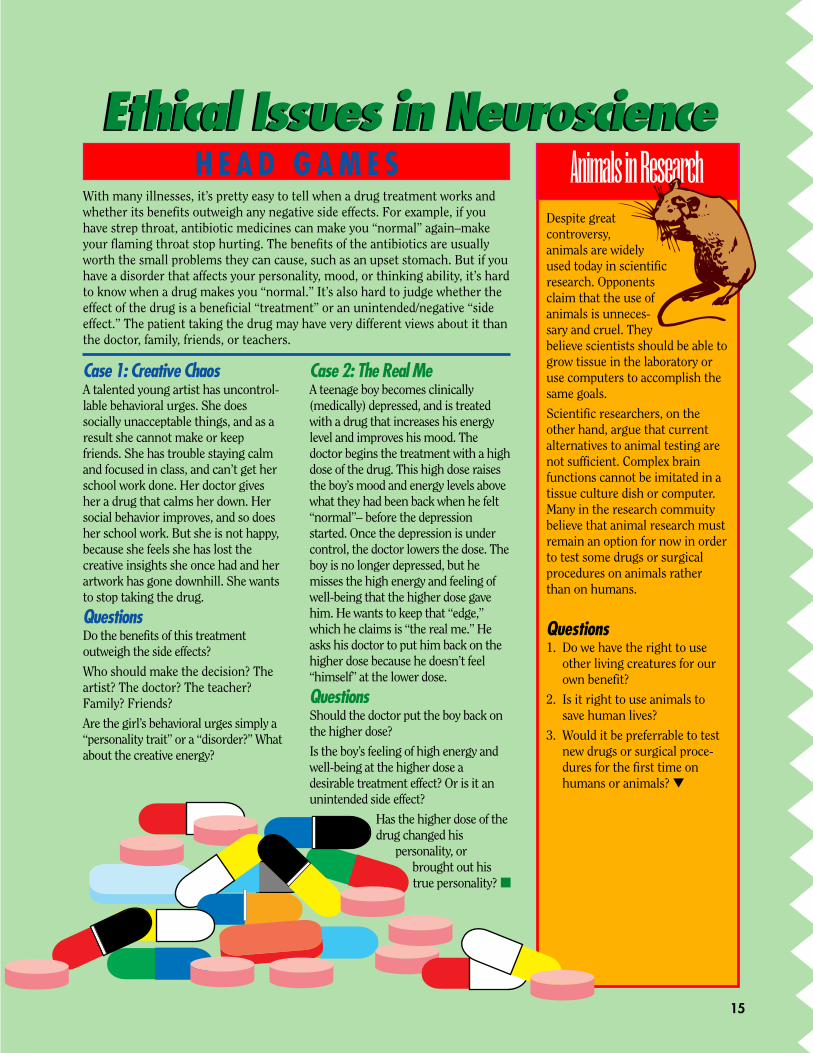
Ethical Issues in NeuroscienceEthical Issues in Neuroscience
Case 1: Creative ChaosA talented young artist has uncontrol-lable behavioral urges. She doessocially unacceptable things, and as aresult she cannot make or keepfriends. She has trouble staying calmand focused in class, and can’t get herschool work done. Her doctor givesher a drug that calms her down. Hersocial behavior improves, and so doesher school work. But she is not happy,because she feels she has lost thecreative insights she once had and herartwork has gone downhill. She wantsto stop taking the drug.
QuestionsDo the benefits of this treatmentoutweigh the side effects?
Who should make the decision? Theartist? The doctor? The teacher?Family? Friends?
Are the girl’s behavioral urges simply a“personality trait” or a “disorder?” Whatabout the creative energy?
Case 2: The Real MeA teenage boy becomes clinically(medically) depressed, and is treatedwith a drug that increases his energylevel and improves his mood. Thedoctor begins the treatment with a highdose of the drug. This high dose raisesthe boy’s mood and energy levels abovewhat they had been back when he felt“normal”– before the depressionstarted. Once the depression is undercontrol, the doctor lowers the dose. Theboy is no longer depressed, but hemisses the high energy and feeling ofwell-being that the higher dose gavehim. He wants to keep that “edge,”which he claims is “the real me.” Heasks his doctor to put him back on thehigher dose because he doesn’t feel“himself” at the lower dose.
QuestionsShould the doctor put the boy back onthe higher dose?
Is the boy’s feeling of high energy andwell-being at the higher dose adesirable treatment effect? Or is it anunintended side effect?
Has the higher dose of thedrug changed his
personality, orbrought out histrue personality? ■
H E A D G A M E S Animals in ResearchDespite greatcontroversy,animals are widelyused today in scientificresearch. Opponentsclaim that the use ofanimals is unneces-sary and cruel. Theybelieve scientists should be able togrow tissue in the laboratory oruse computers to accomplish thesame goals.
Scientific researchers, on theother hand, argue that currentalternatives to animal testing arenot sufficient. Complex brainfunctions cannot be imitated in atissue culture dish or computer.Many in the research commuitybelieve that animal research mustremain an option for now in orderto test some drugs or surgicalprocedures on animals ratherthan on humans.
Questions1. Do we have the right to use
other living creatures for ourown benefit?
2. Is it right to use animals tosave human lives?
3. Would it be preferrable to testnew drugs or surgical proce-dures for the first time onhumans or animals? ▼
With many illnesses, it’s pretty easy to tell when a drug treatment works andwhether its benefits outweigh any negative side effects. For example, if youhave strep throat, antibiotic medicines can make you “normal” again–makeyour flaming throat stop hurting. The benefits of the antibiotics are usuallyworth the small problems they can cause, such as an upset stomach. But if youhave a disorder that affects your personality, mood, or thinking ability, it’s hardto know when a drug makes you “normal.” It’s also hard to judge whether theeffect of the drug is a beneficial “treatment” or an unintended/negative “sideeffect.” The patient taking the drug may have very different views about it thanthe doctor, family, friends, or teachers.
15

16
Read More About It!
World of the Brain by Alvin and VirginiaSilverstein: ISBN 0-688-05777-2
Understanding Your Brain by RebeccaTreays; ISBN 07460 20147
You and Your Body, Brain by DouglasMathers; ISBN 0-8167-2091-6
Know Your Brain available throughNINDS (address below)
Your World/Our World:
Vol.4, No. 2 (Gene Therapy)
Vol. 5, No. 1 (Transgenic Animals)
Vol. 5, No. 2 (Human Genome Project)
Organizations:Society for Neuroscience11 Dupont Circle, N.W. Suite 500Washington, D.C. 20036(202) 462-6688http://www.sfn.org
National Institute of Mental Health(NIMH)Information Resources andInquiries Branch5600 Fishers Lane, Room 7C02Rockville, MD 20857(301) 443-4513http://www.nimh.nih.gov
National Institute of NeurologicalDisorders and Stroke (NINDS)Building 31, Room 8A1631 Center Drive, MSC 2540Bethesda, MD 20892(800) 352-9424http://www.nih.gov/ninds/
Internet Sites:Neuroscience for Kids: http://weber.u.washington.edu/-chudler/neurok.html
Shufflebrain: http://www.indiana.edu/-pietsch/home.html
Addiction: http://idir.net/-irvcohen/index.html
Your World/Our World: http://www.bio.com/pba
E-mail:For questions or comments on this issue:[email protected]
REFERENCES
Dear Students:
We are pleased to provide you with this issue of YourWorld/Our World on the human brain. We hope you find
it an interesting way to learn about biotechnology.Biotechnology can be important to you for two reasons:
1. During your lifetime there will be tremendous discoveries inthis field, and you’ll want to understand what these discoverieswill mean for you, your friends and your family.
2. You can help make those discoveries if you decide to continueto study science and math.
Either way, we hope you join us in discovering the promise ofbiotechnology for the world. We are pleased to acknowledge thesupport of the companies listed.
Sincerely,
Jeff Davidson
Executive DirectorPennsylvania Biotechnology Association
Supporting the national distribution of this issue.
The generosity and support of these sponsors have made the production of YourWorld/Our World possible. Please join us in thanking them:
SponsorsThe Alliance for Science EducationBiotechnology Industry OrganizationCephalon, Inc.Connaught Laboratories, Inc.Merck Institute for Science EducationRhone-Poulenc Rorer GencellTosoHaas
Supporting OrganizationsUtah State University Biotechnology CenterMassachusetts Biotechonology CouncilMaryland BioScience Alliance