biosynthesis of zinc oxide nanoparticles using leaf extract of … · 2017-11-28 · abstract green...
TRANSCRIPT

ORIGINAL ARTICLE
Biosynthesis of zinc oxide nanoparticles using leaf extractof Calotropis gigantea: characterization and its evaluation on treeseedling growth in nursery stage
Sadhan Kumar Chaudhuri1 • Lalit Malodia1
Received: 6 February 2017 / Accepted: 18 August 2017 / Published online: 8 September 2017
� The Author(s) 2017. This article is an open access publication
Abstract Green synthesis of zinc oxide nanoparticles was
carried out using Calotropis leaf extract with zinc acetate
salt in the presence of 2 M NaOH. The combination of
200 mM zinc acetate salt and 15 ml of leaf extract was
ideal for the synthesis of less than 20 nm size of highly
monodisperse crystalline nanoparticles. Synthesized
nanoparticles were characterized through UV–Vis spec-
troscopy, dynamic light scattering (DLS), X-ray diffraction
(XRD), Fourier transform infrared spectroscopy (FTIR),
scanning electron microscopy (SEM), EDX (energy dis-
persive X-ray), and AFM (atomic force microscopy).
Effects of biogenic zinc oxide (ZnO) nanoparticles on
growth and development of tree seedlings in nursery stage
were studied in open-air trenches. The UV–Vis absorption
maxima showed peak near 350 nm, which is characteristic
of ZnO nanoparticles. DLS data showed that single peak is
at 11 nm (100%) and Polydispersity Index is 0.245. XRD
analysis showed that these are highly crystalline ZnO
nanoparticles having an average size of 10 nm. FTIR
spectra were recorded to identify the biomolecules
involved in the synthesis process, which showed absorption
bands at 4307, 3390, 2825, 871, 439, and 420 cm-1. SEM
images showed that the particles were spherical in nature.
The presence of zinc and oxygen was confirmed by EDX
and the atomic % of zinc and oxygen were 33.31 and
68.69, respectively. 2D and 3D images of ZnO nanoparti-
cles were obtained by AFM studies, which indicated that
these are monodisperse having size ranges between 1.5 and
8.5 nm. Significant enhancement of growth was observed
in Neem (Azadirachta indica), Karanj (Pongamia pinnata),
and Milkwood-pine (Alstonia scholaris) seedlings in foliar
spraying ZnO nanoparticles to nursery stage of tree seed-
lings. Out of the three treated saplings, Alstonia scholaris
showed maximum height development.
Keywords Calotropis leaf � ZnO nanoparticle � DLS �XRD � FTIR � SEM with EDS � AFM � Tree seedling
growth
Introduction
In the recent years, the use of metal nanoparticles gained
greater interest due to their diverse applications in the field of
medicine, biology, physics, chemistry, and material sciences
(Kumar et al. 2014). Nanotechnology can be defined as
manipulation of matter through certain chemical and/or
physical processes to create materials with specific properties
which can be used in particular applications. Nanoparticles
can be defined as particles that have at least one dimension less
than 100 nm in size (Thakkar et al. 2014). Unlike bulk
materials, they have unique optical, thermal, electrical,
chemical, and physical properties. Hence, they find a variety
of applications in the areas of medicine, chemistry, environ-
ment, energy, agriculture, information and communication,
heavy industries, and consumer goods (Panigrahi et al. 2004).
In recent years, nanotechnology has emerged as a state-of-the-
art and cutting-edge technology with multifarious applica-
tions in a wide array of fields. It is a very broad area comprising
nanomaterials, nanotools, and nanodevices. Amongst nano-
materials, majority of the research has mainly focused on
nanoparticles as they can be easily prepared and manipulated.
& Sadhan Kumar Chaudhuri
1 Visual Camouflage Group, Camouflage Division, Defence
Laboratory Jodhpur, Defence Research and Development
Organization, Ratanada Palace, Jodhpur, Rajasthan 342011,
India
123
Appl Nanosci (2017) 7:501–512
https://doi.org/10.1007/s13204-017-0586-7
Physical and chemical methods are conventionally used for
the synthesis of nanoparticles; however, due to several limi-
tations of these methods, such as use of toxic compounds,
requirement of very high temperature and pressure, high
expense, and time consumption, research focus has recently
shifted towards the development of clean and eco-friendly
synthesis protocols (Herlekar et al. 2014). It has been
demonstrated that many biological systems including higher
plants and algae, diatoms, bacteria, yeasts, fungi, and human
cells can transform inorganic metal ions into metal nanopar-
ticles via the reductive capacities of the proteins and
metabolites present in these organisms. But synthesis of
nanoparticles using microbes is difficult because it involves
elaborate process of maintaining cell cultures, intracellular
synthesis, and multiple purification steps. Hence, it is signif-
icant to note that the nanoparticle production using plant
displays important advantages over other biological systems
as plants are easily available and the procedure of biogenic
synthesis is cost effective and less tedious as compared to
biosynthesis using fungal source, which requires very long,
tedious, and aseptic culture. The active biological compound
present in plant parts like enzyme itself acts as a reducing and
capping agent, thereby reducing the overall cost of synthesis
process (Jain et al. 2009). In addition to enzymes, other bio-
molecules such as flavanoids, terpenoids, glycosides, alka-
loids, inositols, resins, saponins, terpenes, volatile oil, tannins,
steroids, and quinine also act as reducing and capping agents.
In biogenic synthesis method, small nanoparticles can be
produced during large-scale production (Kajbafna et al. 2009),
and external experimental conditions like high energy and
high pressure are not required causing significant energy
saving (Kajbafna et al. 2012). Zinc oxide is an inorganic
compound with the molecular formula ZnO. It appears as
white powder and is nearly insoluble in water. The powder
ZnO is widely used as an additive in numerous materials and
products including ceramics, glass, cement, rubber, paints,
plastics, lubricants, adhesives, ointments, sealants, pigments,
food, batteries, ferrites, and fire retardants. In the earth crust
ZnO is present as zincite mineral but mostly ZnO used for
commercial purposes is produced synthetically. Nowadays,
the unique properties of nanomaterials have motivated the
researchers to develop many simpler and inexpensive tech-
niques to produce nanostructures of technologically important
materials. Several metal oxide nanoparticles are produced
with possible future applications. Among them, ZnO is con-
sidered to be one of the best exploited at nanodimensions. The
wide band gap and large excitonic binding energy have made
ZnO important both for scientific and industrial applications
(Wang et al. 2004). ZnO nanoparticles have fascinated
research interest and a lot of work has been done using various
plant sources. Leaf extract of Calotropis gigantea using
ZnNO3 salt produced nanoparticles of size 30–35 nm (Vidya
et al. 2013). ZnO nanoparticle of size 100–200 nm were
produced by zinc acetate salt withAcalypha indica leaf extract
(Gnanasangeetha and Thambwani 2013). Parthenium hys-
terophorus leaf extract with ZnNO3 salt produced ZnO
nanoparticles of 16–108 nm size (Sindhura et al. 2014). ZnO
nanoparticles of size ranging from 30 to 35 nm were produced
using Hibiscus rosasinensis with ZnNO3 salt (Devi and
Gayathri 2014). Zinc acetate salt with leaf extract of Azadir-
achta indica produced ZnO nanoparticle of size 25 nm
(Oudhia et al. 2015). Peel extract of Punica granatum with
ZnNO3 salt produced ZnO nanoparticle (Mishra and Sharma
2015). Murraya koengii leaf extract with ZnNO3 salt pro-
duced ZnO nanoparticle of size 50 nm (Divyapriya et al.
2014). Leaf extract of Camellia sinensis with zinc acetate salt
produced ZnO nanoparticle of size 16 nm (Senthilkumar and
Sivakumar 2014). Extensive research is going on for com-
mercializing ZnO nanoparticles throughout the world due to
their unique properties. Olea europa leaf extract with ZnSO4
salt produced ZnO nanoparticles of 20 nm diameter (Awwad
et al. 2014). ZnO nanoparticles of size less than 50 nm were
synthesized using dried sap of roots and shoots of Astragalus
gummifer (Darrudi et al. 2013). Biosynthesis of ZnO
nanoparticle of size ranging from 50 to 200 nm was done
usingCitrus aurantifoliawith zinc acetate salt (Samat and Nor
2013).
Nanofertilizer technology is very innovative but scantly
reported literature is available in scientific journals. Sub-
stituting nanofertilizer for traditional methods of fertilizer
application is a way to release nutrients into the plants in a
gradually and controlled manner, thus preventing autrifi-
cation and pollution of water resources (Naderi and Abedi
2012). There has been a wide use of chemical products
such as fertilizers and minerals which are used to increase
the yield of various products in the field at the same time.
These chemicals result in various side effects in the plants
and soil, and harm the environment in different ways.
Nano-ZnO particle (20 nm), nano-FeO (100 nm), and
nano-CuO (40 nm) at ppm level showed increased shoot
and root length in Mung seedling by foliar spray (Dhoke
et al. 2013). Foliar spray method is more practical for an
agronomic standpoint as plants can absorb essential ele-
ments through their leaves more efficiently compared to
root feeding. Biologically synthesized ZnO nanoparticles
are quickly transported through the plant and included in
the metabolic processes. Mungbean seed germination in
lowest concentration (20 mg) of ZnO suspension solution
showed good shoot and root growth results (Jayarambabu
et al. 2014). Silver nanoparticles synthesized from banana
(Musa balbisiana), neem (Azadirachta indica), and black
tulsi (Ocimum tenuiflorum) showed significant increase in
the root and shoot length of germinated seedlings of Mung
bean (Vigna radiata) and Chickpea (Cicer arietinum)
(Banerjee et al. 2014). Nanofertilizer technology is very
innovative as some reports and patents strongly suggest
502 Appl Nanosci (2017) 7:501–512
123

that there is a vast scope of formulation of nano-fertilizers.
Central Arid Zone Research Institute, Jodhpur, developed
the infrastructure and standardized biosynthesis methods
for synthesis and characterization of large number of
nanonutrients and their effective use in crop improvement.
Significant increase in yield was observed in Pearl Millet
(Pennisetum americanum) due to foliar application of zinc
oxide nanoparticles as fertilizer (Tarafdar et al. 2014).
Currently, research is underway to develop nanocomposite
to supply all the essential nutrients in suitable proportion
through smart delivery system. Preliminary results suggest
that balanced fertilization may be achieved through nan-
otechnology (Tarafdar et al. 2012). Nanofertilizer nutrients
can be encapsulated by nanomaterial cooled with a thin
protective film or delivered as emulsions or nanoparticles.
Nanomaterials could even be used to control the release of
the fertilizer such that the nutrients are only taken up by the
plants and not lost to unintended targets like soil, water, or
microbes. Recent reports indicated that biosynthesized
silver nanoparticles using seed exudates of Spinosa
arvensis showed antifungal (Neofusicoccum parvum)
activities (Khatami et al. 2015) and may be responsible for
increased seed germination.
The plant Calotropis gigantean belonging to the family
Asclepiadaceae, also called as Akra, Shwet akra, and Madara,
is distributed throughout India, especially in dry vast land. The
different parts of the plant are used in Indian traditional
medicine for the treatment of painful muscular spasm,
dysentery, fever, rheumatism, asthma, and as an expectorant
and purgative. Bioactive compounds such as 15b-hydroxy-
cardenolides (1,2) and a 16a-hydroxycalactinic acid methyl
ester (3) along with eleven known compounds including 16a-
hydroxycalotropagenin, coroglaucigenin, 16a-hydrox-
ycalotropin, calactinic acid, calotoxin, desglucouzarin, 12b-
hydroxycoroglaucigenin, frugoside, calotropagenin, dienoic
acid, and mevalonolactone are found in the leaves of this plant
(Singh et al. 2011). Calotropis yields a durable fiber that is
useful for ropes, carpets, fishing nets, and sewing thread.
Floss, obtained from seeds, is used for stuffing purposes.
Extracts of different plant parts such as root, stem, and leaf
affect germination and seedling vigor of many agricultural
crops. It is also used as green manure and improves soil
nutrients and improves moisture binding capacity of soil. The
plant is tolerant of dry and salty conditions and can easily be
established in over-cultivated areas to help improve the soil
conditions and reinvigorate the land (Seeka and Sutthivaiyakit
2010). Moreover, this plant is widely distributed predomi-
nantly in Thar Desert area of Rajasthan. Hence, such medic-
inal and commercial uses of Calotropis plants have increased
our interest and lead us to work on this plant species.
Biosynthesis of nanoparticles is an exciting recent addition to
the large repertoire of nanoparticle synthesis methods and now
nanoparticles have entered a commercial exploration period.
Gold, silver, copper, and zinc have been used mostly for the
synthesis of stable dispersions of nanoparticles, which are
useful in areas such as photolysis, diodes, piezoelectric devi-
ces, fluorescent tubes, laser, sensor, photography, biological
labeling, photonics, and surface-enhanced Raman scattering
detection. Till date, use of ZnO nanoparticles is mainly
restricted in agriculture applications for enhanced productiv-
ity in crop plants. But there is not a single report showing the
use of ZnO nanoparticles in nursery stage for enhanced growth
and development of tree seedlings. Defence Laboratory
Jodhpur is working on arboriculture camouflage for military
applications in the desert area of Rajasthan. In the desert area
of Rajasthan, the growth of trees is very slow due to adverse
climatic conditions (high temperature up to 50 �C, low
humidity, less rainfall, and saline soil). To get enhanced
growth and development, large canopy in short-time foliar
spraying of ZnO nanoparticles may be desirous to overcome
the climatic problems and enhanced growth and development
of seedlings at nursery stage.
In this paper, we reported the biosynthesis of
stable colloidal zinc oxide nanoparticles using leaf extract
of C. gigantea. This plant is an important medicinal plant
and predominantly distributed in Thar Desert area of
Rajasthan. Details of biosynthesis, characterization, and its
effects on enhanced growth and development of tree
seedling in nursery stage are described.
Materials and methods
Reagents
Zinc acetate dihydrate (Lot # MKBQ7110v) was obtained
from Sigma-Aldrich and NaOH from E-Merck, India.
Collection of plant material
Fresh leaves of C. gigantea were collected from Defence
Laboratory, Jodhpur campus (26.2717�N, 73.0378�E).
Collected leaves were thoroughly washed under tap water
and then were washed with Milli-Q water and chopped
with knife. The leaves were kept in oven for drying at
60 �C for 3 days, and dried leaves were powdered using
home mixer blender and stored in an air-tight container at
room temperature till further use.
Preparation of leaf extract
Glass goods and plastic wares were thoroughly cleaned in
chromic acid and washed repeatedly in tap water followed
by final wash in Milli-Q water before drying in hot air
oven. Glass goods, plastic wares, and Milli-Q water
Appl Nanosci (2017) 7:501–512 503
123

required for biosynthesis and characterization of nanopar-
ticles were autoclaved before use. 5 g of dried leaf powder
was transferred to a 250-ml beaker containing 100 ml
Milli-Q water and heated for 15 min at 60 �C. The solution
was kept for cooling at room temperature and then filtered
using Whatman filter paper no. 1. The filtrate was collected
in amber bottle and was stored at 4 �C for further
experiment.
Biosynthesis of ZnO NPs
15 ml of leaf extract of Calotropis was added to 2.195 g of
zinc acetate dihydrate dissolved in 35 ml of distilled water
(overall concentration 200 mM solution). The reaction
mixture was kept on magnetic stirrer for 6 h. After 6 h,
2 M NaOH (4 g of NaOH pellet in 50 ml of Milli-Q water)
was added to the solution and it was placed in incubator at
60 �C with magnetic stirring for overnight. White mixture
was centrifuged at 14,000 rpm for 15 min. Precipitate was
subjected to washing with alcohol and distilled water three
times each. Precipitate was dried in an incubator at
40–50 �C and fine powder was prepared with the help of
ceramic pestle and mortar. Fine powder was used for
characterization with AFM, SEM, FTIR, XRD, EDS, UV–
Vis, and DLS.
Characterization of biosynthesized ZnO NPs
pH analysis
Zinc acetate aqueous solution (200 mM) showed pH 6.58.
pH of aqueous Calotropis leaf extract was 6.23. The
changed pH of reaction mixtures was recorded using digital
pH meter (Eutech Cyberscan pH 300) during the synthesis
of zinc oxide nanoparticles.
UV–Vis spectroscopy
The reduction of zinc ion was monitored by measuring
optical density through UV–Vis spectroscopy of the reac-
tion medium after diluting small aliquots of reaction mix-
ture ten times diluted with Milli-Q water and transferred to
cuvette, and analysis was done using UV–Vis spec-
trophotometer (Ocean Optics, USA).
Dynamic light scattering (DLS) analysis and zeta potential
measurement
Particle size distribution and average size of zinc oxide
nanoparticles were obtained through particle size analyzer.
Liquid sample before centrifugation was diluted ten times
using Milli-Q water and transferred to cuvette, and analysis
was done using DLS (Malvern Zetasizer, Nano Z500 UK).
The sample holder temperature was maintained at 25 �C.
The measurements depend on the size of the particle core,
surface structure, particle concentration, and the type of the
ion in the mixture. The zeta potential of the synthesized
nanoparticles was determined in water as dispersant.
X-ray diffraction
The formation and quality of compounds were investigated
by X-ray diffraction technique. For this purpose, synthe-
sized zinc oxide NPs were centrifuged (1400 rpm; 8 �C)
for 15 min, pellet was washed three times with ethanol and
finally with sterile Milli-Q water for three cycles. The
purified ZnO NP precipitate was dried in oven at 60 �C and
powdered with ceramic mortar–pestle. Powdered sample
was analyzed using X-ray diffractometer (X’Pert PRO-
PAN Analytical, Europe). The scanning was done in the
region of 2h from 20� to 80�.
FTIR analysis
FTIR was used to identify the possible functional groups
involved in the reduction of zinc ion and capping of
reduced zinc oxide nanoparticles. FTIR spectrum was
recorded using Shimadzu, Japan, infrared (IR) double-
beam spectrophotometer. FTIR analysis of dried ZnO
nanoparticles (NPs) was carried out through potassium
bromide (KBr) pellet method in 1:30 ratios (NPs:KBr) and
spectrum was recorded in transmittance mode at a resolu-
tion of 4 cm-1. The peaks (stretching) obtained were
plotted as transmittance in Y-axis and wave number (cm-1)
in X-axis. The spectrum was recorded in the wave number
range 500–4500 cm-1 and analyzed subtracting the spec-
trum of pure KBr.
SEM analysis
Scanning electron microscopy (SEM) analysis was carried
out using Carl Zeiss Japan, model machine. Thin film of
nanoparticle powder sample was prepared on carbon-
coated tape by adhering small amount of dried fine powder
of sample on the grid, excess sample was removed with the
help of blotting paper. The film on the SEM grid was
allowed to dry by putting it under a mercury lamp for
5 min. The SEM analysis was used to determine the sur-
face structure of biogenically synthesized ZnO NPs.
Energy-dispersive X-ray spectroscopy
Sample used for SEM was used as it is and the same
instrument was used. EDX analysis was carried out to
504 Appl Nanosci (2017) 7:501–512
123

determine the chemical purity, elemental composition, and
stoichiometry of the synthesized zinc oxide nanoparticles.
Atomic force microscopy
Freshly cleaved mica sheet was adhered on substrate
using double-sided tape. Diluted sample prepared using
ultrasonicator was placed upon the substrate using drop
technique with autoclaved microtip. This sample was
placed and dried in incubator at 37 �C for 1 h. Analysis
was done using Z-03 scanner in AFM (NT-MDT Solver
Pro, Russia).
Evaluation of ZnO NP effects on tree seedling growth
One-year-old saplings developed in Defence Laboratory,
Jodhpur, nursery were used for the treatment of zinc oxide
(ZnO) nanoparticles (NPs). Six trenches were prepared in a
specific distance within the iron net house in open air.
Hundred saplings of each species (Neem—Azadirachta
indica, Milkwood-pine—Alstonia scholaris, and Tilpapra/
Karanj—Pongamia pinnata) were placed in control and
treated sets (fifty saplings in each trench). Initial heights
were recorded with measuring scale. Spraying of biogenic
ZnO NPs at 30 mg per liter synthesized from Calotropis
leaf extract was carried out through aerosol foliar spraying.
The optimal concentration (30 mg/l) for seedling treatment
was determined in tree seed germination studies (data not
included here) before the nanoparticle foliar spray treat-
ment. Watering of the trenches was carried out in specific
interval in a general practice (twice a week) of nursery
development. After 3 months of treatment, the height of
each sapling was measured and spraying of ZnO NPs at
30 mg per liter was done once again and after 6 months the
final height of each sapling was recorded for statistical data
analysis.
Statistical data analysis
Analytical determination was carried out from the average
mean data of 50 plants in a set (control and treated). All
experimental data were expressed as mean ± standard
error.
Results and discussion
Color change
Reduction of zinc is confirmed by color change of the
reaction mixture from pale yellow to white (Fig. 1;
Table 1).
Variation in pH during biosynthesis of ZnO NPs
UV–vis spectroscopy
Optical properties of ZnO nanoparticles were characterized
using UV–Vis spectrophotometer. The UV–Vis absorption
curve of ZnO nanoparticles is shown in Fig. 2. Zinc oxide
formation was confirmed as the absorption peak (lambda
max) was found near 350 nm. This result correlates with
the already reported results, in which absorption peak was
found at 360 nm (Jayarambabu et al. 2014) (Table 2).
Dynamic light scattering analysis
The size of synthesized zinc oxide nanoparticles when
analyzed by DLS shows the mean size of synthesized ZnO
nanoparticles as 11 nm (100%) in a single peak (Fig. 3)
and Polydispersity Index (PDI) is 0.245.
Zeta potential
Figure 4 shows the measured zeta potential value of
biosynthesized zinc oxide nanoparticles in the colloidal
solution. The nanoparticles possess a negative zeta poten-
tial value of -20.7 mV, which indicated that even after the
storage of 3 months at room temperature these are highly
stable due to electrostatic repulsive force. The high nega-
tive value confirms the repulsion among the particles and
the negative indicates that nanoparticles are stable. Mea-
surement of zeta potential depends on the movement of
nanoparticles under influence of an applied electric field.
This movement depends upon surface charge and the local
environment of the particle.
X-ray diffraction
The dried powdered sample was used to perform XRD for
confirming the size of ZnO nanoparticles. The graph
(Fig. 5) showed main peaks corresponding to 2h values of
32.25�, 34.88�, 36.70�, 47.97�, 56.99�, 63.23�, 66.79�,68.33�, 69.43�, and 77.39�. This result is almost similar to
already reported results in O. europa leaf extracts in which
2h values were obtained at 31.84�, 34.50�, 36.32�, 47.59�,56.63�, 66.89�, 67.98�, 69.09�, and 76.98� (Awwad et al.
2014). The size of nanoparticles was in the range of
8–12 nm and the average size of nanoparticles determined
by XRD was 10 nm (using Debye–Scherrer equation).
FTIR analysis
To determine the functional groups responsible for the
synthesis of ZnO NPs in C. gigantea leaf extract, FTIR
analysis was performed. The FTIR spectrum of C. gigantea
Appl Nanosci (2017) 7:501–512 505
123

leaf extract is shown in Fig. 6, which shows absorption
bands at 4307, 3390, 2825, 871, 439, and 420 cm-1 which
are characteristic of –OH stretching vibration and –CH
stretching vibration. The band at 871 cm-1 is due to
asymmetrical and symmetrical stretching of zinc car-
boxylates resulting in the involvement of carboxylic groups
in protein of Calotropis leaf extract. This result coincides
with the already reported result of biosynthesis of ZnO
nanoparticles using O. europa leaf extract (Awwad et al.
2014). Studies confirmed the presence of phytochemicals
(eicosatrienoic acid methyl ester, hexatriacontaine, tri-
methyl undecatriene, and trifluoroacetic acid), volatile
essential oil (phytol), and flavanoids (varinging, quercitrin,
hesperitin, and kaempferol). It also contains acalyphamide,
2-methylanthraquinone, tri-o-methyl ellagic acid, sitos-
terol, glucoside, stigmasterol, quinine, tannins, resins, and
Fig. 1 a 200 mM zinc acetate solution (transparent), b 35 ml of zinc acetate solution ? 15 ml of leaf extract (yellowish white), c after 6 h (pale
yellow color), d after 24 h (white color intensity increased)
Fig. 2 UV–vis absorption
spectra of zinc acetate solution,
plant extracts and synthesized
ZnO nanoparticles after 24 h
Table 1 Change in color of solution during formation of zinc oxide nanoparticles using Calotropis leaf extract
Solution Before reduction After reduction Color intensity Time
Calotropis leaf extract Light brown
200 mM zinc acetate Transparent Pale yellow
Yellowish white
White
?
??
???
Immediate
After 6 h
After 24 h
506 Appl Nanosci (2017) 7:501–512
123

0
20
40
60
80
100
120
1 11 21 31
Leaf extract-15ml Calotropis
Salt- 35 ml 200 mM Zinc acetate
One Peak- 11 nm (100%)
PDI- 0.245
Size (d.nm)
Inte
nsity
(%)
Fig. 3 Size distribution and
mean particle size of
synthesized zinc oxide
nanoparticles
0
100000
200000
300000
400000
-100 0 100 200
Tota
l Cou
nts
Apparent Zeta Potential (mV)
Zeta Potential Distribution
Fig. 4 Zeta potential
measurement
Table 2 Change in pH of solution during formation of zinc oxide nanoparticles using Calotropis leaf extract
Solution Before reduction (pH) After reduction (pH)
Calotropis leaf extract 6.23 –
200 mM zinc acetate 6.58 –
15 ml of leaf extract ? 35 ml of 200 mM zinc acetate 6.14
11.12 (immediately after adding NaOH)
11.40 (after 6 h)
12.50 (after 24 h)
Appl Nanosci (2017) 7:501–512 507
123

essential oils. Results inveterate that –OH stretching
around 3390 cm-1 and –CH stretching around 2825 cm-1
are responsible for strong capping on the ZnO nanoparti-
cles. This result matches with the already reported result of
biosynthesis of ZnO nanoparticles using Acalypha indica
leaf extract (Gnanasangeetha and Thambwani 2013).
SEM analysis
SEM was employed to analyze the structure of nanoparti-
cles that were formed. SEM image has shown individual
ZnO nanoparticles as well as a number of aggregates. SEM
image in Fig. 7 shows that these are spherical-shaped
nanoparticles. This SEM result coincides with results
already reported, which shows formation of spherical-
shaped nanoparticles and aggregated molecules in Calo-
tropis leaf extract (Vidya et al. 2013).
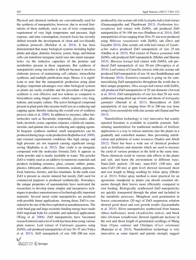
Energy-dispersive X-ray spectroscopy
EDX analysis was carried out to determine the elemental
composition and stereochemistry of the synthesized zinc
oxide nanoparticles. In Fig. 8, zinc and oxygen signals
detected that the synthesized nanoparticles are in pure state
of chemical nature. The single peak of Zn and O is found
between 0 and 2, and two peaks of Zn were found in
between 8 and 10. These results correlate with the already
reported results in which similar peaks have been observed
in ZnO NP synthesis using Acalypha indica leaf extract
(Gnanasangeetha and Thambwani 2013).
Further analysis was done to find weight % and atomic
% of zinc and oxygen elements present in the biogenically
synthesized sample using Calotropis leaf extract (Table 3).
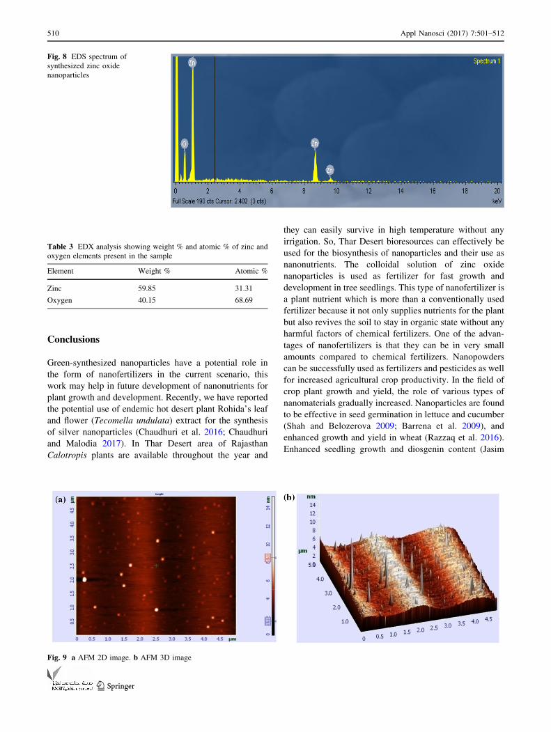
Atomic force microscopy
To validate the surface morphology of biogenic ZnO NPs,
drop-coated two- and three-dimensional AFM images were
taken in noncontact mode. Result showed variability in
morphological features of biosynthesized zinc oxide
nanoparticles (Fig. 9a, b). The sizes range from 1.5 to
8.5 nm and the particles are more or less homogeneous in
size range and monodisperse nature.
Evaluation of ZnO NP effects on tree seedling growth
For studying the effects of ZnO NPs on growth and
development of three important tree seedlings (Azadirachta
indica, Alstonia scholaris, and P. pinnata) in nursery stage,
they were treated with ZnO NPs at a final concentration of
Fig. 5 XRD graph of biosynthesized zinc oxide nanoparticles
508 Appl Nanosci (2017) 7:501–512
123

30 mg/l for each treatment. After treatment, seedling
growth in terms of plant height was measured. Increased
height in all three plant species could be observed
compared to control sets (Fig. 10). Significantly enhanced
growth and development was evident in Alstonia scholaris
compared to other two species (Fig. 11).
Fig. 7 SEM image of zinc
oxide nanoparticles
Fig. 6 FT-IR spectrum of the biosynthesized ZnO nanoparticles
Appl Nanosci (2017) 7:501–512 509
123
Conclusions
Green-synthesized nanoparticles have a potential role in
the form of nanofertilizers in the current scenario, this
work may help in future development of nanonutrients for
plant growth and development. Recently, we have reported
the potential use of endemic hot desert plant Rohida’s leaf
and flower (Tecomella undulata) extract for the synthesis
of silver nanoparticles (Chaudhuri et al. 2016; Chaudhuri
and Malodia 2017). In Thar Desert area of Rajasthan
Calotropis plants are available throughout the year and
they can easily survive in high temperature without any
irrigation. So, Thar Desert bioresources can effectively be
used for the biosynthesis of nanoparticles and their use as
nanonutrients. The colloidal solution of zinc oxide
nanoparticles is used as fertilizer for fast growth and
development in tree seedlings. This type of nanofertilizer is
a plant nutrient which is more than a conventionally used
fertilizer because it not only supplies nutrients for the plant
but also revives the soil to stay in organic state without any
harmful factors of chemical fertilizers. One of the advan-
tages of nanofertilizers is that they can be in very small
amounts compared to chemical fertilizers. Nanopowders
can be successfully used as fertilizers and pesticides as well
for increased agricultural crop productivity. In the field of
crop plant growth and yield, the role of various types of
nanomaterials gradually increased. Nanoparticles are found
to be effective in seed germination in lettuce and cucumber
(Shah and Belozerova 2009; Barrena et al. 2009), and
enhanced growth and yield in wheat (Razzaq et al. 2016).
Enhanced seedling growth and diosgenin content (Jasim
Fig. 8 EDS spectrum of
synthesized zinc oxide
nanoparticles
Table 3 EDX analysis showing weight % and atomic % of zinc and
oxygen elements present in the sample
Element Weight % Atomic %
Zinc 59.85 31.31
Oxygen 40.15 68.69
Fig. 9 a AFM 2D image. b AFM 3D image
510 Appl Nanosci (2017) 7:501–512
123

et al. 2016) were also reported in silver nanoparticle-trea-
ted seeds of fenugreek (Trigonella foenum-graecum L.). In
military camouflage applications, fast growth and large
canopy trees are ideal for natural concealment. In the desert
area of Rajasthan, plant growth is very much slow due to
harsh climatic conditions. The formulation of nanonutrients
using nanoparticles and their foliar spraying in nursery
seedlings may enhance the growth and development, and
stress tolerance to biotic and abiotic factors of identified
saplings in nursery stage, which could be used in military
camouflage package (arboriculture camouflage).
Open Access This article is distributed under the terms of the
Creative Commons Attribution 4.0 International License (http://
creativecommons.org/licenses/by/4.0/), which permits unrestricted
use, distribution, and reproduction in any medium, provided you give
appropriate credit to the original author(s) and the source, provide a
link to the Creative Commons license, and indicate if changes were
made.
Publisher’s Note Springer Nature remains neutral with regard to
jurisdictional claims in published maps and institutional affiliations.
References
Awwad AM, Albiss B, Ahmad AL (2014) Green synthesis, charac-
terization and optical properties of zinc oxide nanosheets using
Olea europa leaf extract. Adv Mater Lett 5:520–524
Banerjee P, Satpathy M, Mukhopadhayay A, Das P (2014) Leaf
extract mediated green synthesis of silver nanoparticles from
widely available Indian plants: synthesis, characterization,
antimicrobial property and toxicity analysis. Bioresourc Biopro-
cess 1:1–10
Fig. 11 Comparison between treated (T) and control (C) saplings of Azadirachta, Alstonia, and Pongamia after 3 months of ZnO nanoparticles
foliar spraying (treatment)
020406080
100120
Control (N) Treated (N) Control (A) Treated(A) Control (T) Treated (T)Heig
ht o
f sas
aplin
gs in
Cm
Height of different saplings: N= Neem (Azadirachta indica );A= Milkwood-pine ( Alstonia scholaris); T= Karanj (Pongamia pinnata)
Effects of ZnO nanopar�cles on seedling gowth in nursery
Ini�al A�er 03 Months A�er 06 Months
Fig. 10 Effects of ZnO nanoparticles (30 mg/l) on growth and development of nursery saplings. Data represent mean of 50 replicates and
vertical bars represent ± standard error
Appl Nanosci (2017) 7:501–512 511
123

Barrena R, Casals E, Colon J, Font X, Sanchez A, Puntes V (2009)
Evaluation of the ecotoxicity of model nanoparticles. Chemo-
sphere 75:850–857
Chaudhuri SK, Malodia L (2017) Phytosynthesis and characterization
of silver nanoparticles synthesized from flower extract of Roheda
(Tecomella undulata G. Don). Defence Life Sci J 2:65–73
Chaudhuri SK, Chandela S, Malodia L (2016) Plant mediated green
synthesis of silver naoparicles using Tecomella undulata leaf
extract and their characterization. Nano Biomed Eng 8:1–8
Darrudi M, Oskuee RK, Kargar H (2013) Sol-gel synthesis, charac-
terization and neurotoxicity effect of zinc oxide nanoparticles
using gum tragacanth. Ceram Int 40:4827–4831
Devi RS, Gayathri R (2014) Green synthesis of zinc oxide nanopar-
ticles by using Hibiscus rosa-sinensis. Int J Curr Eng Technol
4:2444–2446
Dhoke SK, Mahajan P, Kamble R, Khanna A (2013) Effects of
nanoparticles suspension of mung seedlings by foliar spray
method. Nanotechnol Dev 3:1–5
Divyapriya S, Sowmia C, Sasikala S (2014) Synthesis of zinc oxide
nanoparticles and microbial activity of Murraya koeniggi. World
J Pharm Pharm Sci 12:1635–1645
Gnanasangeetha D, Thambwani DS (2013) Biogenic production of
zinc oxide nanoparticles using Acalypha indica. J Chem Biol
Phys Sci 1:238–246
Herlekar H, Barne S, Kumar R (2014) Plant mediated green synthesis
of iron nanoparticles. J Nanoparticles. doi:10.1155/2014/140614
Jain J, Arora S, Rajkumar JM, Khandelwal S, Pahnihar KM (2009)
Silver nanoparticles in therapeutics: development of an antimi-
crobial gel formation for topical use. Mol Pharm 5:1388–1401
Jasim B, Roshmi T, Mathew J, Radhakrishnan EK (2016) Plant
growth and diosgenin enhancement effect of silver nanoparticles
in Fenugreek (Trigonella foenum-graecum L.). Saudi Pharm J.
doi:10.1016/j.jsps.2016.09.012
Jayarambabu N, Sivakumari B, Prabhu YT (2014) Germination and
growth characteristics of mungbean seeds affected by synthe-
sized zinc oxide nanoparticles. Int J Curr Eng Technol
5:3411–3416
Kajbafna A, Shayegh MR, Mazhammi M (2009) Nanostructure sword
like ZnO wires: rapid synthesis and characterization through a
microwave assisted route. J Alloy Compd 1:293–297
Kajbafna A, Ghorham H, Parnikar A, Sambrey JP, Sadrhezhaad SK
(2012) Effects of morphology on photocatalytic performance of
ZnO nanostructures synthesized by rapid microwave irradiation
methods. Superlattices Microstruct 4:512–522
Khatami M, Pourseyedi S, Khatami M, Hamidi M, Zaeifi M, Soltani L
(2015) Synthesis of silver nanoparticles using seed extract of
Spinosa arvensis as a novel bioresource, and evaluation of their
antifungal activity. Bioresourc Bioprocess 2:1–7
Kumar PPNV, Pammi SVN, Kollu P et al (2014) Green synthesis and
characterization of silver nanoparticles using Boerhaavia diffusa
plant extract and their antimicrobial activity. Ind Crops Prod
52:562–566
Mishra V, Sharma R (2015) Green synthesis of zinc oxide nanopar-
ticles using fresh peels extract of Punica granatum and its
antimicrobial activities. Int J Pharma Res Health Sci 3:694–699
Naderi MR, Abedi A (2012) Application of nanotechnology in
agriculture and refinement of environment pollutants. J Nan-
otechnol 1:18–26
Oudhia A, Kulkarni P, Sharma S (2015) Green synthesis of ZnO
nanotubes for bioapplications. Int J Curr Eng Technol 1:280–281
Panigrahi S, Kendu S, Ghosh SK, Nath S, Pal T (2004) General methods
of synthesis of metal nanoparticles. J Nanopart Res 4:411–414
Razzaq A, Ammara R, Jhanzab HM, Mahmood T, Hafeez A, Hussain
S (2016) A novel nanomaterial to enhance growth and yield of
wheat. J Nanosci Technol 2(1):55–58
Samat NA, Nor RM (2013) Sol-gel synthesis of zinc oxide
nanaoparticles using Citrus aurantafolia extracts. Ceram Int
39:545–548
Seeka C, Sutthivaiyakit S (2010) Cytotoxic cardenolides from the
leaves of Calotropis gigantea. Chem Pharm Bull 5:725–728
Senthilkumar SR, Sivakumar T (2014) Green tea Camellia sinensis
mediated synthesis of zinc oxide nanoparticles and studies on
their antimicrobial activities. Int J Pharm Pharm Sci 6:461–465
Shah V, Belozerova I (2009) Influence of metal nanoparticles on the
soil microbial community and germination of lettuce seeds.
Water Air Soil Pollut 197:143–148
Sindhura KS, Prasad TN, Selvam P, Hussain OM (2014) Synthesis,
characterization and evaluation of effect of phytogenic zinc
nanopaticles on soil exoenzymes. Appl Nanosci 4:819–827
Singh RP, Shukla P, Singh PK (2011) Biological approach of ZnO
nanoparticle formation and characterization. Adv Mater Lett
2:313–317
Tarafdar JC, Xiang Y, Wang WN, Dong Q, Biswas P (2012)
Standardization of size, shape and concentration of nanoparticle
for plant application. Appl Biol Res 14:138–144
Tarafdar JC, Raliya R, Mahawar H, Rathore I (2014) Development of
zinc nanofertilizers to enhance crop production in pearl millet
(Pennicetum americanum). Agric Resourc 3:219–226
Thakkar KS, Mhatre SS, Parikh RY (2014) Biological synthesis of
metabolic nanoparticles. Nanomedicine 2:257–262
Vidya C, Hiremath S, Chandraprabha MN, Venugopal I, Jain A,
Bansal K (2013) Green synthesis of ZnO nanoparticle by
Calotropis gigantea. Int J Curr Eng Technol 4:118–120
Wang X, Ding Y, Summers CJ, Wang ZL (2004) Large scale
synthesis of 6 nm wide ZnO nanobelts. J Phys Chem
26:8773–8777
512 Appl Nanosci (2017) 7:501–512
123