biosorption of azo dyes by magnetic microspheres and … abstract raspberry-like fe 3o 4@yeast...
TRANSCRIPT
Loughborough UniversityInstitutional Repository
Biosorption of azo dyes byraspberry-like Fe3O4@yeastmagnetic microspheres andtheir efficient regeneration
using heterogeneousFenton-like catalytic
processes over an up-flowpacked reactor
This item was submitted to Loughborough University's Institutional Repositoryby the/an author.
Citation: SONG, R. ...et al., 2015. Biosorption of azo dyes by raspberry-like Fe3O4@yeast magnetic microspheres and their efficient regeneration usingheterogeneous Fenton-like catalytic processes over an up-flow packed reactor.Reaction Kinetics Mechanisms and Catalysis, 115(2), pp. 547-562.
Additional Information:
• This paper was accepted for publication in the journal Reaction Kinetics,Mechanisms and Catalysis and the definitive published version is availableat http://dx.doi.org/10.1007/s11144-015-0854-z
Metadata Record: https://dspace.lboro.ac.uk/2134/19059
Version: Accepted for publication
Publisher: c© Akad�emiai Kiad�o Zrt
Rights: This work is made available according to the conditions of the Cre-ative Commons Attribution-NonCommercial-NoDerivatives 4.0 International(CC BY-NC-ND 4.0) licence. Full details of this licence are available at:https://creativecommons.org/licenses/by-nc-nd/4.0/

Please cite the published version.
2
1
Biosorption of Azo Dyes by Raspberry-Like Fe3O4@Yeast Magnetic
Microspheres and Their Efficient Regeneration Using Heterogeneous
Fenton-like Catalytic Processes over an Up-Flow Packed Reactor
Rui Song a, Bo Bai*, b, Gianluca Li Puma c, Honglun Wang b, Yourui Suo b
a College of Environmental Science and Engineering, Chang’an University, Xi’an, 710054, P.R.
China;
b Northwest Plateau Institute of Biology, Chinese Academy of Sciences, Xining, 810001, People’s
Republic of China;
c Environmental Nanocatalysis and Photoreaction Engineering, Department of Chemical Engineering,
Loughborough University, Loughborough, LE11 3TU, United Kingdom.
* Corresponding author: Tel: +86 298 233 9052; Fax: +86 298 233 9961; Email: [email protected]
2
Abstract
Raspberry-like Fe3O4@yeast composite microspheres, whose properties integrate the biosorption
features of yeast cells with the excellent magnetic and catalytic properties of Fe3O4 nanoparticles were
synthesized by a simple electrostatic-interaction-driven self-assembly heterocoagulation. They were
successfully applied in an up-flow packed column for the removal of the model water contaminant
methylene blue dye (MB) by consecutive bioadsorption-heterogeneous Fenton oxidation cycles. The
as-synthesized Fe3O4@yeast composites were characterized by field emission scanning electron
microscopy (FE-SEM), energy-dispersive spectroscopy (EDS), powder X-ray diffraction (XRD) and
Fourier transform infrared (FT-IR) spectroscopy. The adsorption process was controlled by the
electrostatic interactions between the adsorbent and contaminant. The adsorbent is suitable for the
adsorption of positively charged compounds at mildly acidic pH, neutral and alkaline pH, with the
highest performance observed at alkaline pH. The experimental breakthrough curves measured at
different influent MB concentration, flow rate, bed height and pH were modeled by the Yoon-Nelson
model. The in-situ regeneration of the contaminant-loaded Fe3O4@yeast microspheres and their reuse
in multiple cycles was demonstrated by triggering the heterogeneous Fenton-like reaction catalyzed
by the supported magnetite. The raspberry-like Fe3O4@yeast magnetic microsphere should be a
promising and practical adsorbent for removal and destruction of positively charged organic
compounds in wastewater.
Keywords: Yeast; Iron oxide; Fixed-bed; Fenton-like reaction; Regeneration; Wastewater
3
1. Introduction
Organic azo dyes represent more than half of all colouring compounds used by the world textile
industry. It has been estimated that 15% of this is released into the environment without an
appropriate treatment [1]. Organic azo dyes in wastewater have been classified as priority pollutants
particularly owing to the toxicity of their microbial metabolites, which can be mutagenic, genotoxic
and carcinogenic [2]. Traditional physico-chemical treatment techniques such as absorption,
oxidization, and flotation are usually ineffective to treat low azo dyes concentrations [3]. Biological
materials such as micro-organisms (including bacteria, fungi, and algae) [4] and biomass can remove
a broad range of azo dyes [5-6]. However, an efficient and sustainable method for pollution abatement
that uses biosorbents requires the regeneration and reuse of the sorbent, the destruction of the
sorbed/desorbed pollutants, and the safe disposal of the sorbent to the environment. Yeast is an
effective biosorbent [7] which meets several green requirements for large-scale pollutant removal
including, high selectivity, efficiency, cost-effectiveness, availability and biocompatibility.
Heterogeneous Fenton processes are powerful method for the destruction of azo dyes in aqueous
solutions [8–10], especially, those catalyzed by nano-sized magnetite (Fe3O4) particles [11-13]. In this
system, the octahedral site of the magnetite structure can easily accommodate both Fe2+ and Fe3+,
allowing the Fe species to be reversibly oxidized and reduced producing •OH radicals when in the
presence of H2O2 [14-15]. Furthermore, Fe3O4 nanoparticles can be easily separated from the reaction
system by a simple magnetic separation procedure.
The treatment of contaminated streams by Fenton oxidation with slurry suspensions of Iron oxide
can be inefficient since uses large volumes of reagents/catalyst, the water matrix can inefficiently
consume •OH radicals and large contact times are often required. In contrast, the efficiency of the
4
heterogeneous Fenton process for the treatment of wastewater can be increased significantly by
applying an enrichment or pre-concentration sorption method prior to the oxidation of the
contaminants by the Fenton process on immobilized magnetite.
The combination of an inorganic catalyst with a biological sorbent into hybrid or composite material
is attracting increasing attention as it enables new functional properties, which are not possible in their
starting components. For example a new class of complex nano-structured hybrid microspheres with a
raspberry-like structure can provide a unique micro/nano-environment which has found new
applications in the material, chemical, medical and environmental sectors [16-20]. Bai et al. have
synthesized novel raspberry-like TiO2@yeast composites, which were effective photocatalysts for the
degradation of dyes [21]. The raspberry-shape provided larger surface area of the supported catalyst.
However, such materials required the use of UV light photons, which restrict their technological
development.
In this study, we investigate a new composite material, made of raspberry-like Fe3O4@yeast
microspheres that integrate the biosorption features of yeast cells with the excellent magnetic and
catalytic properties of Fe3O4 nanoparticles, and its application for the effective removal of
contaminants from wastewater by consecutive biosorption and heterogeneous Fenton
oxidation/regeneration cycles. In this new material, the traditional magnetic Fe3O4 nano-particles were
anchored on the yeast surface by an electrostatic self-assembly process that produced a heterogeneous
Fenton’s catalyst with an unique raspberry-like microstructure. The effectiveness of the raspberry-like
Fe3O4@yeast composite microspheres for the removal of the cationic dye methylene blue (MB), a
model compound used in the standardization of degradation experiments (e.g., ISO 10678:2010), was
investigated in a continuous, up-flow, fixed-bed column system. The modeling of the adsorption
5
phenomena and breakthrough profiles allowed the prediction of the breakthrough time after which the
in-situ regeneration of the composite material and the decomposition of MB was triggered through the
heterogeneous Fenton reaction catalyzed by the Fe3O4 nanoparticles. The robustness of the composite
catalytic biosorbent was assessed for multiple adsorption/regeneration cycles.
2. Materials and methods
2.1. Materials
All chemicals used were of analytical grade and used without further purification. The powdered yeast
was purchased from Angel Yeast Company. Ferric chloride hexahydrate (FeCl3·6H2O), absolute
ethanol (95 wt %), formaldehyde (CH2O), hydrazine hydrate (N2H4·H2O), methylene blue (MB), and
sulfuric acid (H2SO4) were provided by Xi’an Chemical Agent Corp. Double deionized water was
used throughout all the experiments.
2.2. Preparation of Fe3O4@yeast composite microspheres
Fe3O4 nanoparticles were firstly synthesized through a hydrothermal method using FeCl3·6H2O as a
single iron source. Briefly, FeCl3·6H2O (1.20 g), CH2O (2 mL) and N2H4·H2O (5 mL) were dissolved
in deionized water (40 mL) under magnetic stirring for 15 min. The solution was then transferred to a
Teflon-lined stainless-steel autoclave and heated at 120 °C. After 5 h of reaction, the autoclave was
cooled to room temperature. The black Fe3O4 nanoparticles were collected magnetically and washed
with three consecutive cycles of ethanol and distilled water and then dried in vacuum at 80 °C for 1 h.
The dried Fe3O4 nanoparticles were then re-dispersed in 150 mL of distilled water and the pH adjusted
to 5 by adding H2SO4 (1 M). The suspension was stirred for 30 min to promote the de-aggregation of
the Fe3O4 nanoparticles. In a separate vessel, 1.000 g of yeast powder was washed with distilled water
and ethanol three times, respectively. It was dispersed in 150 mL of distilled water and the pH was
adjusted to 5 by adding H2SO4 (1 M). The suspension was magnetically stirred for 30 min to facilitate
6
the dispersion of the yeast particles. Finally, the suspensions of Fe3O4 nanoparticles and yeast were
blended, under continuous stirring for 1 h at room temperature, and then the suspension was left for 3
h without further stirring, during which raspberry-like Fe3O4@yeast microspheres were produced via
electrostatic self-assembly. The products were collected with a magnet, washed with distilled water
and ethanol three times, and then dried at 80 °C for further use.
2.3 Material characterization
The particle size and surface morphology of the samples were observed by a Philips XL 30 field
emission scanning electron microscope (FE-SEM). The elemental composition of the composite
microspheres was determined with the energy-dispersive spectroscopy (EDS) of the FE-SEM. The
crystallographic structures of the samples were identified by powder X-ray diffraction (XRD) using
Cu Kα radiation (λ=0.15418 nm) at a scanning rate of 0.02°/min. Fourier-transform infrared (FT-IR)
spectra of samples were recorded on a Bio-Rad FTS135 spectrometer in the range 500-4000 cm−1
using a KBr wafer technique, to study the formation and ripening mechanism of the samples.
2.4 Up-flow fixed-bed adsorption experiments
The adsorption and regeneration performance of the Fe3O4@yeast composites was investigated in
up-flow fixed-bed columns made from glass (12 cm high, 0.8 cm internal diameter) operated in
continuous flow. A stock solution of MB (1000 mg/L) was prepared and further diluted to the
required feed concentration in each experiment. The effect of the influent concentration (100, 150 and
200 mg/L), flow rate (5, 10, and 15 mL/min), bed depth (1.2, 2.4 and 3.6 cm, corresponding to 0.1,
0.2 and 0.3 g of adsorbent, respectively) and pH (3, 5, 7, 9, 11) on the removal of MB in the columns
was investigated. The pH of the dye solutions was adjusted by adding NaOH and H2SO4, respectively.
Samples collected from the inlet/outlet of the columns at regular intervals were analyzed using a
7
Jenway 6405 UV−vis spectrophotometer at 664 nm. All of the experiments were performed in
triplicate and at room temperature.
2.5 Regeneration experiment
After each adsorption cycle, the exhausted column bed was rinsed by flowing 100 mL double
deionized water in the upward direction at the same flow rate used during the adsorption cycle. Then
the in-situ regeneration of the Fe3O4@yeast bed was triggered by flowing a 10% H2O2 aqueous
solution through the column bed in the upward direction at a flow rate of 1 mL/min for 1 h. The
column was finally rinsed with double deionized water to remove the residual H2O2. After the
completion of regeneration procedure, the bed was reused for the next adsorption-regeneration cycle,
up to three consecutive cycles.
3. Results and discussion
3.1 Characterization and mechanism
Figure 1
Figure 1 shows typical FE-SEM images of the original yeast and the as-obtained Fe3O4@yeast
composite microspheres under different magnifications. The original yeast (Figure 1(a)) shows cells
with a smooth surface and ellipsoidal shape with regular diameter (length 4.0 ± 0.2 µm; width 2.6 ±
0.2 µm). The Fe3O4@yeast composite microspheres (Figure 1(b)) maintained the ellipsoidal shape of
the original yeast with the rough surface morphology and relatively good monodispersity. The
dimensions of the composite microspheres slightly increased (length 4.5 ± 0.2 µm; width 3.0 ± 0.2 µm)
suggesting that the Fe3O4 nanoparticles (on average 20 nm, inset in Figure 1(c)) were successfully
attached onto the surface of yeast. The higher magnification image (Figure 1(c)), shows a rough
surface decorated with numerous Fe3O4 nanoparticles with exposed yeast bare areas. Furthermore,
8
each of the composite microspheres approached a distinct raspberry-like morphology.
The EDS analysis of the composite microspheres is shown in Figure S1of Supporting Information (SI).
The C and in part the O peaks resulted from the original yeast cell wall, the Fe and part of the O
originated from the Fe3O4 supported nanoparticles, and Pt arose from the metal spraying before SEM
studies. No other elements were detected, indicating that the final product was impurities free.
Figure 2
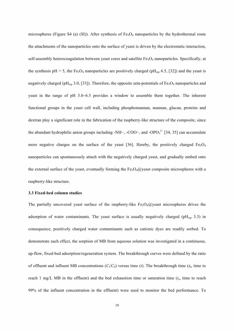
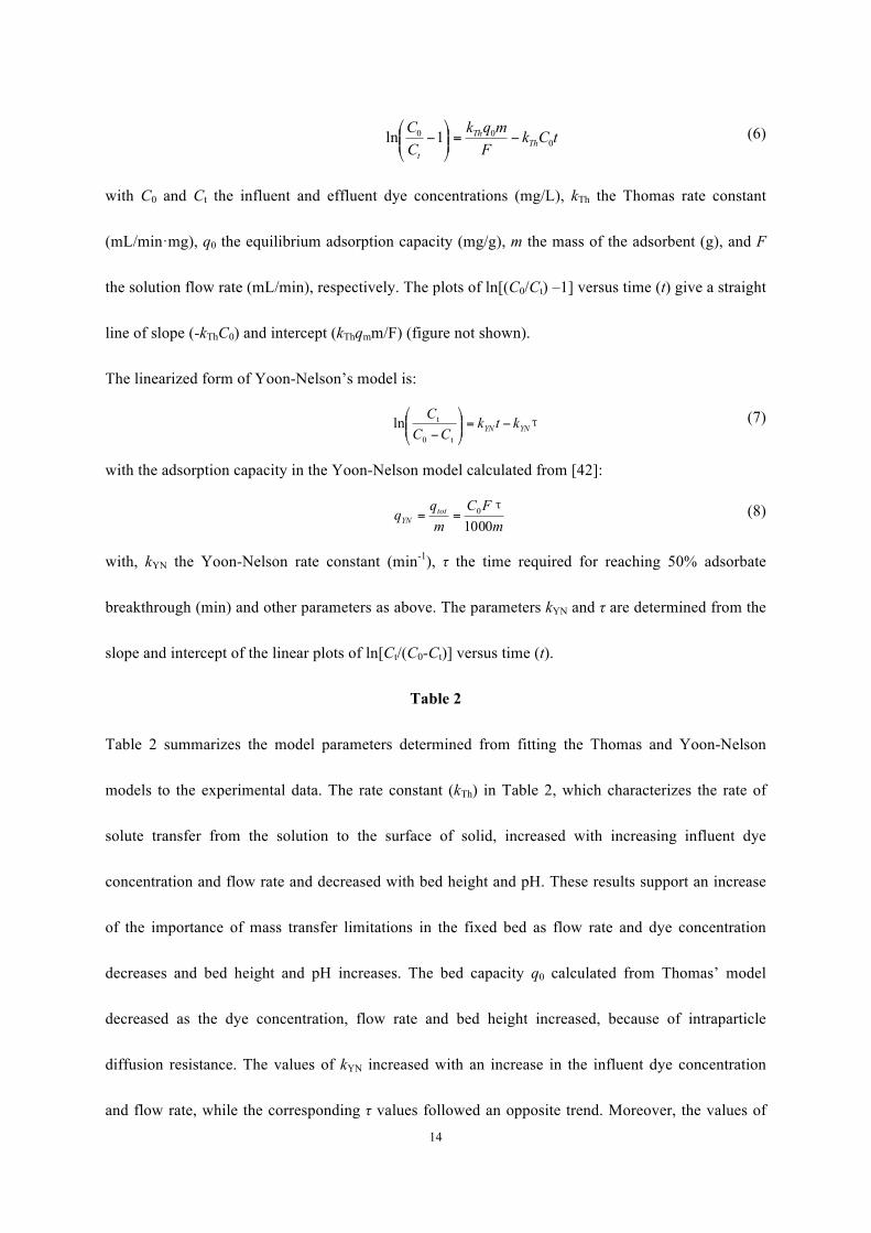
Figure 2 shows the XRD patterns of the original yeast, pure Fe3O4 nanoparticles and the Fe3O4@yeast
composite microspheres, respectively. The amorphous yeast showed only a broad peak at around 2θ =
20o (Figure 2a). The strong and sharp diffraction peaks at 18.4o, 30.3o, 35.6o, 37.3o, 43.2o, 53.4o, 57.2o
and 62.9o in the patterns of the Fe3O4 nanoparticles (Figure 2b) were indexed as (110), (220), (311),
(222), (400), (422), (511) and (440) respectively of the face center-cubic phase of Fe3O4 (JCPDS card
No. 19-0629) which agree with literature [22-23]. The broad peak at 2θ = 20o of the Fe3O4@yeast
composite microspheres (Figure 2c) originated mainly by the amorphous structure of yeast. The
remaining peaks supported the incorporation of the Fe3O4 nanoparticles in the composite and no other
diffraction peaks were found in the range investigated indicating the high purity of the products. The
intensity of the Fe3O4 peaks in the microspheres were lower than those of the pure Fe3O4 nanoparticles,
which may result from the smaller number of Fe3O4 nanoparticles supported on the yeast surface [24,
25] and their good monodispersion surface traits [26,27].
The ferromagnetic properties of the Fe3O4@yeast composite microspheres in aqueous solution are
shown in Figure S2 (SI). The Fe3O4@yeast composite microspheres (40 g/L) were dispersed in
deionized water in a cuvette forming a stable suspension (Figure S2 (a)). Under the influence of a
magnetic field, there was a rapid separation of the Fe3O4@yeast composite particles to the wall of the

9
cuvette, which yielded an almost colorless aqueous solution (Figure S2 (b) (SI)). This indicated that
the Fe3O4 nanoparticles were successfully incorporated onto the surface of the yeast hosts endowing
the yeast cells with magnetic properties allowing easy separation, recovery and reuse. The black
colour imparted by the Fe3O4 nanoparticles to the ivory yeast in the composite further supports the
successful incorporation of Fe3O4 nanoparticles on the yeast surface.
Figure 3
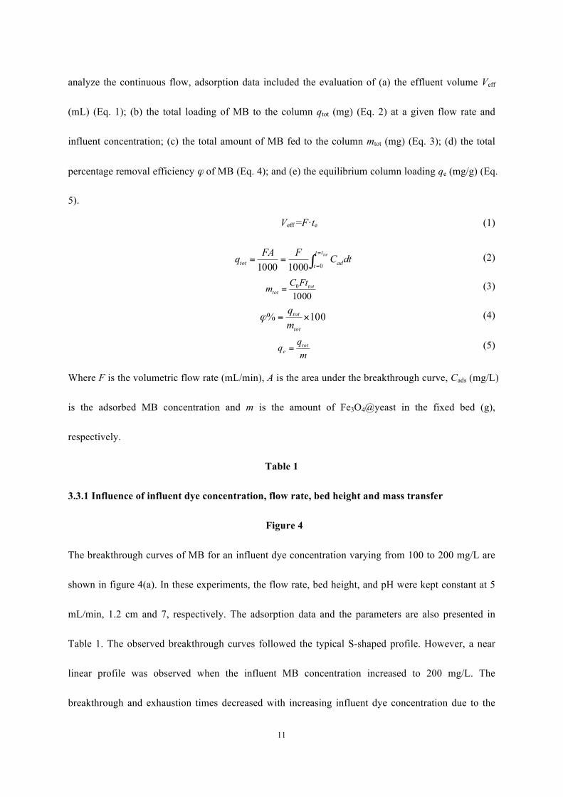
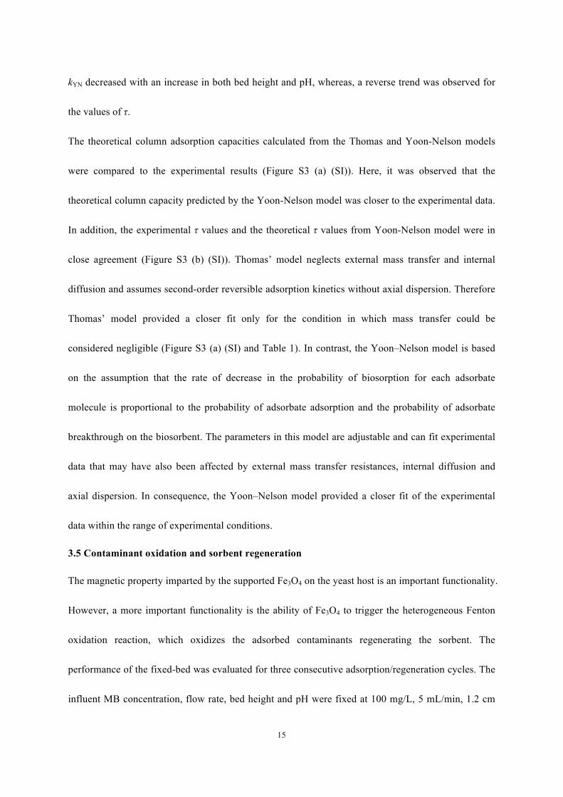
The bonding forces acting between the nanoparticles and the yeast were investigated by FT-IR
(Figure 3). The broad and intense peaks in the region from 550 to 650 cm−1 of the Fe3O4 nanoparticles
(Figure 3(a)) are assigned to the Fe-O band. The peaks at around 3300−3700 cm-1 and 1384 cm−1
belong to OH stretching vibrations and to the H−O−H bending band, suggesting that H2O molecules
adsorbed on the surface of Fe3O4 [28]. The additional broad peaks at 3340, 1461, 1360, 1074 and 894
cm-1 may result from residual hydrazine hydrate remaining from the synthesis process. Regarding the
yeast (Figure 3(b)), the broad and intense peaks at 3292, 2926, 1641, 1384 and 1074 cm−1 are
assigned to the N−H stretching and bending vibration, the CH2 asymmetric and symmetric stretching
vibration, the amide group, C=O stretching vibration, and the P=O stretching vibration, respectively
[29-31]. In the Fe3O4@yeast spectra (Figure 3(c)), except for the characteristic absorption peak of
yeast, a peak appeared in the range of 550 to 650 cm−1. Moreover, the characteristic peaks of yeast at
3292 cm−1, 1641 cm−1 and 1054 cm−1 shifted to 3310 cm−1, 1656 cm−1, and 1064 cm−1 respectively in
the Fe3O4@yeast composite microspheres, suggesting that the amino (–NH2), carboxyl (-COO−) and
phosphate (-OPO32−) groups played important roles in the anchoring of the Fe3O4 nanoparticles to the
yeast surface.
Based on the above analysis, we propose a formation mechanism of Fe3O4@yeast composite
10
microspheres (Figure S4 (a) (SI)). After synthesis of Fe3O4 nanoparticles by the hydrothermal route
the attachments of the nanoparticles onto the surface of yeast is driven by the electrostatic-interaction,
self-assembly heterocoagulation between yeast cores and satellite Fe3O4 nanoparticles. Specifically, at
the synthesis pH = 5, the Fe3O4 nanoparticles are positively charged (pHiep 6.5, [32]) and the yeast is
negatively charged (pHiep 3.0, [33]). Therefore, the opposite zeta-potentials of Fe3O4 nanoparticles and
yeast in the range of pH 3.0~6.5 provides a window to assemble them together. The inherent
functional groups in the yeast cell wall, including phosphomannan, mannan, glucan, proteins and
dextran play a significant role in the fabrication of the raspberry-like structure of the composite, since
the abundant hydrophilic anion groups including -NH−, -COO−, and -OPO32− [34, 35] can accumulate
more negative charges on the surface of the yeast [36]. Hereby, the positively charged Fe3O4
nanoparticles can spontaneously attach with the negatively charged yeast, and gradually embed onto
the external surface of the yeast, eventually forming the Fe3O4@yeast composite microspheres with a
raspberry-like structure.
3.3 Fixed-bed column studies
The partially uncovered yeast surface of the raspberry-like Fe3O4@yeast microspheres drives the
adsorption of water contaminants. The yeast surface is usually negatively charged (pHiep 3.3) in
consequence, positively charged water contaminants such as cationic dyes are readily sorbed. To
demonstrate such effect, the sorption of MB from aqueous solution was investigated in a continuous,
up-flow, fixed-bed adsorption/regeneration system. The breakthrough curves were defined by the ratio
of effluent and influent MB concentrations (Ct/C0) versus time (t). The breakthrough time (tb, time to
reach 1 mg/L MB in the effluent) and the bed exhaustion time or saturation time (te, time to reach
99% of the influent concentration in the effluent) were used to monitor the bed performance. To
11
analyze the continuous flow, adsorption data included the evaluation of (a) the effluent volume Veff
(mL) (Eq. 1); (b) the total loading of MB to the column qtot (mg) (Eq. 2) at a given flow rate and
influent concentration; (c) the total amount of MB fed to the column mtot (mg) (Eq. 3); (d) the total
percentage removal efficiency ϕ of MB (Eq. 4); and (e) the equilibrium column loading qe (mg/g) (Eq.
5).
Veff =F·te (1)
∫=
===
tottt
t adtot dtCFFAq010001000
(2)
10000 tot
totFtCm = (3)
100% ×=tot
tot
mq
ϕ (4)
mqq tot
e = (5)
Where F is the volumetric flow rate (mL/min), A is the area under the breakthrough curve, Cads (mg/L)
is the adsorbed MB concentration and m is the amount of Fe3O4@yeast in the fixed bed (g),
respectively.
Table 1
3.3.1 Influence of influent dye concentration, flow rate, bed height and mass transfer
Figure 4
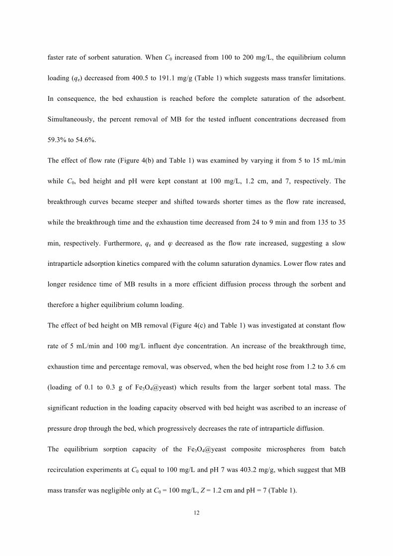
The breakthrough curves of MB for an influent dye concentration varying from 100 to 200 mg/L are
shown in figure 4(a). In these experiments, the flow rate, bed height, and pH were kept constant at 5
mL/min, 1.2 cm and 7, respectively. The adsorption data and the parameters are also presented in
Table 1. The observed breakthrough curves followed the typical S-shaped profile. However, a near
linear profile was observed when the influent MB concentration increased to 200 mg/L. The
breakthrough and exhaustion times decreased with increasing influent dye concentration due to the
12
faster rate of sorbent saturation. When C0 increased from 100 to 200 mg/L, the equilibrium column
loading (qe) decreased from 400.5 to 191.1 mg/g (Table 1) which suggests mass transfer limitations.
In consequence, the bed exhaustion is reached before the complete saturation of the adsorbent.
Simultaneously, the percent removal of MB for the tested influent concentrations decreased from
59.3% to 54.6%.
The effect of flow rate (Figure 4(b) and Table 1) was examined by varying it from 5 to 15 mL/min
while C0, bed height and pH were kept constant at 100 mg/L, 1.2 cm, and 7, respectively. The
breakthrough curves became steeper and shifted towards shorter times as the flow rate increased,
while the breakthrough time and the exhaustion time decreased from 24 to 9 min and from 135 to 35
min, respectively. Furthermore, qe and ϕ decreased as the flow rate increased, suggesting a slow
intraparticle adsorption kinetics compared with the column saturation dynamics. Lower flow rates and
longer residence time of MB results in a more efficient diffusion process through the sorbent and
therefore a higher equilibrium column loading.
The effect of bed height on MB removal (Figure 4(c) and Table 1) was investigated at constant flow
rate of 5 mL/min and 100 mg/L influent dye concentration. An increase of the breakthrough time,
exhaustion time and percentage removal, was observed, when the bed height rose from 1.2 to 3.6 cm
(loading of 0.1 to 0.3 g of Fe3O4@yeast) which results from the larger sorbent total mass. The
significant reduction in the loading capacity observed with bed height was ascribed to an increase of
pressure drop through the bed, which progressively decreases the rate of intraparticle diffusion.
The equilibrium sorption capacity of the Fe3O4@yeast composite microspheres from batch
recirculation experiments at C0 equal to 100 mg/L and pH 7 was 403.2 mg/g, which suggest that MB
mass transfer was negligible only at C0 = 100 mg/L, Z = 1.2 cm and pH = 7 (Table 1).
13
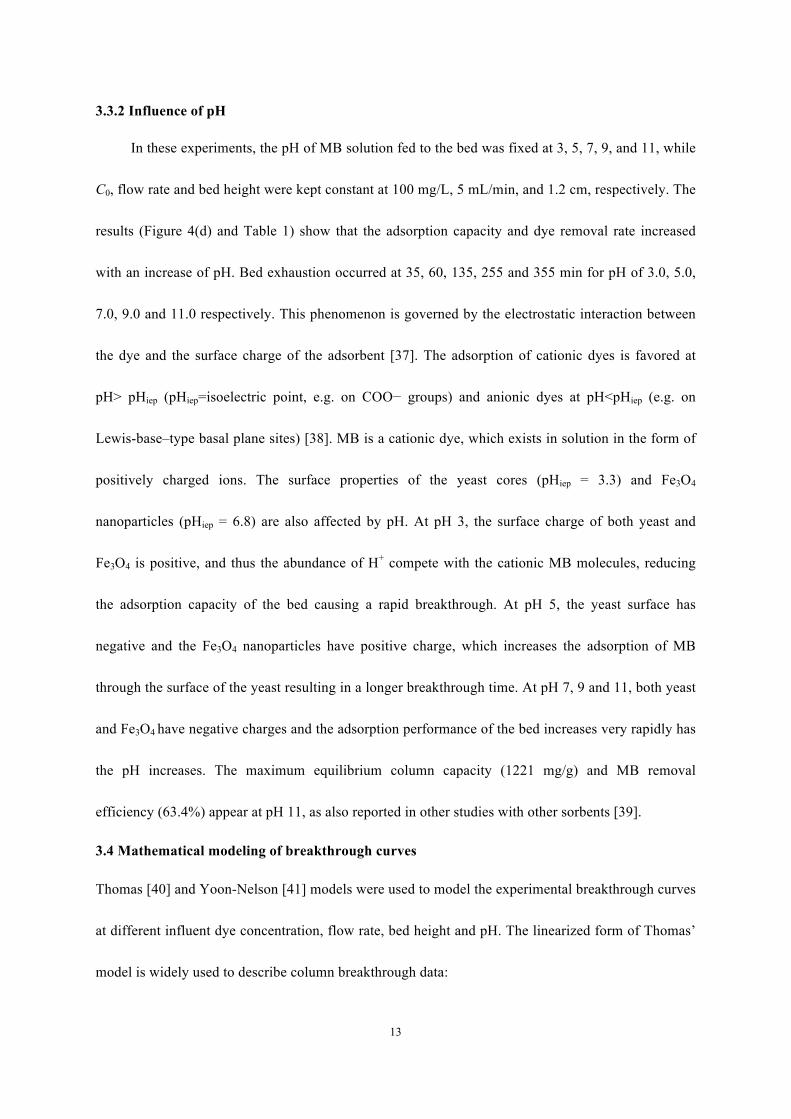
3.3.2 Influence of pH
In these experiments, the pH of MB solution fed to the bed was fixed at 3, 5, 7, 9, and 11, while
C0, flow rate and bed height were kept constant at 100 mg/L, 5 mL/min, and 1.2 cm, respectively. The
results (Figure 4(d) and Table 1) show that the adsorption capacity and dye removal rate increased
with an increase of pH. Bed exhaustion occurred at 35, 60, 135, 255 and 355 min for pH of 3.0, 5.0,
7.0, 9.0 and 11.0 respectively. This phenomenon is governed by the electrostatic interaction between
the dye and the surface charge of the adsorbent [37]. The adsorption of cationic dyes is favored at
pH> pHiep (pHiep=isoelectric point, e.g. on COO− groups) and anionic dyes at pH<pHiep (e.g. on
Lewis-base–type basal plane sites) [38]. MB is a cationic dye, which exists in solution in the form of
positively charged ions. The surface properties of the yeast cores (pHiep = 3.3) and Fe3O4
nanoparticles (pHiep = 6.8) are also affected by pH. At pH 3, the surface charge of both yeast and
Fe3O4 is positive, and thus the abundance of H+ compete with the cationic MB molecules, reducing
the adsorption capacity of the bed causing a rapid breakthrough. At pH 5, the yeast surface has
negative and the Fe3O4 nanoparticles have positive charge, which increases the adsorption of MB
through the surface of the yeast resulting in a longer breakthrough time. At pH 7, 9 and 11, both yeast
and Fe3O4 have negative charges and the adsorption performance of the bed increases very rapidly has
the pH increases. The maximum equilibrium column capacity (1221 mg/g) and MB removal
efficiency (63.4%) appear at pH 11, as also reported in other studies with other sorbents [39].
3.4 Mathematical modeling of breakthrough curves
Thomas [40] and Yoon-Nelson [41] models were used to model the experimental breakthrough curves
at different influent dye concentration, flow rate, bed height and pH. The linearized form of Thomas’
model is widely used to describe column breakthrough data:
14
tCkFmqk
CC
ThTh
t0
00 1ln −=⎟⎟⎠
⎞⎜⎜⎝
⎛− (6)
with C0 and Ct the influent and effluent dye concentrations (mg/L), kTh the Thomas rate constant
(mL/min·mg), q0 the equilibrium adsorption capacity (mg/g), m the mass of the adsorbent (g), and F
the solution flow rate (mL/min), respectively. The plots of ln[(C0/Ct) –1] versus time (t) give a straight
line of slope (-kThC0) and intercept (kThqmm/F) (figure not shown).
The linearized form of Yoon-Nelson’s model is:
τYNYN ktkCC
C−=⎟⎟
⎠
⎞⎜⎜⎝
⎛
− t0
tln (7)
with the adsorption capacity in the Yoon-Nelson model calculated from [42]:
mFC
mqq tot
YN 10000 τ== (8)
with, kYN the Yoon-Nelson rate constant (min-1), τ the time required for reaching 50% adsorbate
breakthrough (min) and other parameters as above. The parameters kYN and τ are determined from the
slope and intercept of the linear plots of ln[Ct/(C0-Ct)] versus time (t).
Table 2
Table 2 summarizes the model parameters determined from fitting the Thomas and Yoon-Nelson
models to the experimental data. The rate constant (kTh) in Table 2, which characterizes the rate of
solute transfer from the solution to the surface of solid, increased with increasing influent dye
concentration and flow rate and decreased with bed height and pH. These results support an increase
of the importance of mass transfer limitations in the fixed bed as flow rate and dye concentration
decreases and bed height and pH increases. The bed capacity q0 calculated from Thomas’ model
decreased as the dye concentration, flow rate and bed height increased, because of intraparticle
diffusion resistance. The values of kYN increased with an increase in the influent dye concentration
and flow rate, while the corresponding τ values followed an opposite trend. Moreover, the values of
15
kYN decreased with an increase in both bed height and pH, whereas, a reverse trend was observed for
the values of τ.
The theoretical column adsorption capacities calculated from the Thomas and Yoon-Nelson models
were compared to the experimental results (Figure S3 (a) (SI)). Here, it was observed that the
theoretical column capacity predicted by the Yoon-Nelson model was closer to the experimental data.
In addition, the experimental τ values and the theoretical τ values from Yoon-Nelson model were in
close agreement (Figure S3 (b) (SI)). Thomas’ model neglects external mass transfer and internal
diffusion and assumes second-order reversible adsorption kinetics without axial dispersion. Therefore
Thomas’ model provided a closer fit only for the condition in which mass transfer could be
considered negligible (Figure S3 (a) (SI) and Table 1). In contrast, the Yoon–Nelson model is based
on the assumption that the rate of decrease in the probability of biosorption for each adsorbate
molecule is proportional to the probability of adsorbate adsorption and the probability of adsorbate
breakthrough on the biosorbent. The parameters in this model are adjustable and can fit experimental
data that may have also been affected by external mass transfer resistances, internal diffusion and
axial dispersion. In consequence, the Yoon–Nelson model provided a closer fit of the experimental
data within the range of experimental conditions.
3.5 Contaminant oxidation and sorbent regeneration
The magnetic property imparted by the supported Fe3O4 on the yeast host is an important functionality.
However, a more important functionality is the ability of Fe3O4 to trigger the heterogeneous Fenton
oxidation reaction, which oxidizes the adsorbed contaminants regenerating the sorbent. The
performance of the fixed-bed was evaluated for three consecutive adsorption/regeneration cycles. The
influent MB concentration, flow rate, bed height and pH were fixed at 100 mg/L, 5 mL/min, 1.2 cm
16
and 7, respectively.
Table 3
The column capacity decreased after each regeneration cycle although MB uptake and removal rate
remained very high (Table 3). The column bed exhibited equilibrium column loading capacities of
over 273.5 mg/g dry beads and removal rate over 57.5% in all three cycles. The reduction in column
capacity should be attributed to the inability of the Fenton reaction to remove MB sorbed deeply into
the yeast cores, since Fe3O4 is primarily supported on the surface. Comparing the parameters qe and
φ% with those reported elsewhere [43-44], the Fe3O4@yeast adsorbent presents the advantage that it
can be easily regenerated in-situ and reused in multiple cycles.
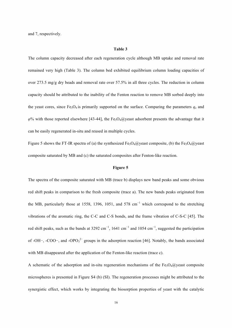
Figure 5 shows the FT-IR spectra of (a) the synthesized Fe3O4@yeast composite, (b) the Fe3O4@yeast
composite saturated by MB and (c) the saturated composites after Fenton-like reaction.
Figure 5
The spectra of the composite saturated with MB (trace b) displays new band peaks and some obvious
red shift peaks in comparison to the fresh composite (trace a). The new bands peaks originated from
the MB, particularly those at 1558, 1396, 1051, and 578 cm−1 which correspond to the stretching
vibrations of the aromatic ring, the C-C and C-S bonds, and the frame vibration of C-S-C [45]. The
red shift peaks, such as the bands at 3292 cm−1, 1641 cm−1 and 1054 cm−1, suggested the participation
of -OH−, -COO−, and -OPO32− groups in the adsorption reaction [46]. Notably, the bands associated
with MB disappeared after the application of the Fenton-like reaction (trace c).
A schematic of the adsorption and in-situ regeneration mechanisms of the Fe3O4@yeast composite
microspheres is presented in Figure S4 (b) (SI). The regeneration processes might be attributed to the
synergistic effect, which works by integrating the biosorption properties of yeast with the catalytic
17
oxidation properties of the supported Fe3O4 nanoparticles. Specifically, the MB molecules are firstly
removed from aqueous solution by biosorption and pre-concentrated on the bare areas of the
Fe3O4@yeast composite microspheres. Then, the enriched MB molecules are decomposed by the
Fe3O4/H2O2 Fenton-like oxidation reaction. The H2O2 introduced to the MB saturated column is
catalytically decomposed by the supported Fe3O4 nanoparticles yielding •OH radicals, which then
attack the adsorbed MB molecules regenerating the sorbent. The targeted localized reaction at or in
the close proximity of the composite surface is much more efficient than the same Fenton-like
reaction carried out by Fe3O4 particles in suspensions in slurry reactors. Less catalyst is used, •OH
scavenging reactions species in solution are minimized (e.g., carbonates and not targeted species) and
also the reaction acts on a much higher pollutant load, which increases the contaminant removal rate
(i.e., first-order contaminant removal). In return, the regenerated surfaces of the yeast core can
provide continuous adsorption sites for the biosorption of contaminants in repeated cycles.
4. Conclusion
In this study we have investigated a new composite material, which integrates the biosorption
features of yeast cells with the magnetic and catalytic properties of Fe3O4 nanoparticles, for the
effective removal and oxidative destruction of contaminants in water and wastewater. The
raspberry-like Fe3O4@yeast composite microspheres, were fabricated via a simple electrostatic
self-assembly approach using inexpensive and abundant raw materials. These composites exhibited a
ferromagnetic property that allows them to be easily separated/recovered by use of an external
magnetic field. The adsorption and regeneration performance by a Fenton-like reaction of the
microspheres was investigated for the continuous removal of the cationic dye methylene blue (MB) in
an up-flow packed column. The adsorption process was controlled by the electrostatic interactions
18
between the adsorbent and the contaminant. The adsorbent is suitable for the adsorption of positively
charged compounds at mildly acidic pH, neutral and alkaline pH, with the highest performance
observed at alkaline pH. The raspberry-like Fe3O4@yeast composite material can be easily
regenerated by applying a Fenton-like reaction and reused. Considering the facile method of
fabrication of the composite from abundant and inexpensive raw materials, the easy recovery and
separation in water, the superior adsorption performance, the simple and effective way of regeneration
and the robustness of the adsorbent for consecutive adsorption/regeneration cycles, we conclude that
the raspberry-like Fe3O4@yeast magnetic microsphere should be a promising and practical adsorbent
for removal and destruction of positively charged organic compounds in wastewater. The embedding
of Fe3O4 onto the yeast surface can be extended to the simple fabrication of other Fe3O4
nanoparticles/bio-macromolecules or other magnetic oxides/yeast materials with similar structure.
Associated Content
Supporting Information
Additional information as noted in the text. This material is available free of charge via the Internet at
http://link.springer.com.
Acknowledgments
This work was supported by China Postdoctoral Science Special Foundation, Scientific Research
Foundation for the Returned Overseas Chinese Scholars, National Natural Science Foundation of
China (No.21176031) and Fundamental Research Funds for the Central Universities (No.
2013G2291015).
19
References
[1] R.C. Wu, J.H. Qu, Water Environ. Res. 2004, 76, 2637-2642.
[2] P. Kovacic, R. Somanathan, J. Appl. Toxicol. 2014, 34, 825-834.
[3] P. Drogui, J.F. Blais, Recent Patents on Engineering. 2007, 1, 257-272.
[4] M. Solís, A. Solís, H.I. Pérez, N. Manjarrez, M. Flores, Process Biochem. 2012, 47, 1723-1748.
[5] V. Janaki, K. Vijayaraghavan, A.K. Ramasamy, K.J. Lee, B.T. Oh, S. Kamala-Kannan, J. Hazard.
Mater. 2012, 241-242, 110-117.
[6] A.H. Chen, S.M. Chen, J. Hazard. Mater. 2009, 172, 1111-1121.
[7] J.X. Yu, R.A. Chi, X.Z. Su, Z.Y. He, Y.F. Qi, Y.F. Zhang, J. Hazard. Mater. 2010, 177, 222-227.
[8] M.L. Rache, A.R. García, H.R. Zea, A.M.T. Silva, L.M. Madeira, J.H. Ramírez, Appl. Catal. B:
Environ. 2014, 146, 192-200.
[9] L. Gu, S. Huang, N. Zhu, D. Zhang, H. Yuan, Z. Lou, J. Hazard. Mater. 2013, 263, 450-457.
[10] A.D.L. Maria, S. Marta, B. Juan, React Kinet. Mech. Cat. 2013, 110, 101–117.
[11] X. Liang, Y. Zhong, S. Zhu, J. Zhu, P. Yuan, H. He, J. Zhang, J. Hazard. Mater. 2010, 181,
112-120.
[12] S. Xavier, R. Gandhimathi, P.V. Nidheesh, S.T. Ramesh, Desalination and Water Treatment.
DOI:10.1080/19443994.2013.844083.
[13] R.C. Wu, J.H. Qu, Water Environ. Res. 2004, 76, 2637–2642.
[14] S.H. Kong, R.J. Watts, J. H. Choi, Chemosphere. 1998, 37, 1473–1482.
[15] B.W. Tyre, R.J. Watts, G.C. Miller, J. Environ. Qual. 1991, 20, 832–838.
[16] S.J. Guo, S.J. Dong, E.K. Wang, J. Phys. Chem. C. 2009, 113, 5485–5492.
[17] L. Zhang, Y. Li, J.Q. Sun, J.C. Shen, Langmuir. 2008, 24, 10851–10857.
20
[18] H.L. Liu, D. Wang, X.L. Yang, Colloids Surf. A: Physicochem. Eng. Asp. 2012, 397, 48–58.
[19] X. Wang, T. Akagi, M. Akashi, B. Masanori, Mini-Rev. Org. Chem. 2007, 4, 51–59.
[20] H.J. Tsai, Y.L. Lee, Langmuir. 2007, 23, 12687–12692.
[21] B. Bai, N. Quici, Z.Y. Li, G.L. Puma, Chem. Eng. J. 2011, 170, 451–456.
[22] S.X. Zhang, X.L. Zhao, H.Y. Niu, Y.L. Shi, Y.Q. Cai, G.B. Jiang, J. Hazard. Mater. 2009, 167,
560−566.
[23] T. Zhu, J.S. Chen, X.W. Lou (David), J. Phys. Chem. C. 2011, 115, 9814–9820.
[24] L.F. Duan, S.S. Jia, Y.J. Wang, J. Chen, L.J. Zhao, J Mater. Sci. 2009, 44, 4407–4412.
[25] N.S. Kim, J.D. Kim, J. Ind. Eng. Chem. 2012, 18, 1721–1729.
[26] X.D. Su, J.Z. Zhao, Y.L. Li, Y.C. Zhu, X.K. Ma, F. Sun, Z.C. Wang, Colloids Surf. A:
Physicochem. Eng. Asp. 2009, 349, 151–155.
[27] M.S. Hassan, T. Amna, O.B. Yang, H.C. Kim, M.S. Khil, Ceram. Int. 2012, 38, 5925–5930.
[28] Z.P. Li, L. Gao, S. Zheng, Appl. Catal. A: Gen. 2002, 236, 163–171.
[29] S. Kumar, T. Surendar, B. Kumar, A. Baruah, V. Shanker, J. Phys. Chem. C. 2013, 117,
26135−26143.
[30] J.J. Cui, W. He, H.T. Liu, S.J. Liao, Y.Z. Yue, Colloids Surf. B. 2009, 74, 274–278.
[31] K.C. Blakeslee, A. Robert, S.R. Condrate, J. Am. Ceram. Soc. 1971, 54, 559–563.
[32] S.J. Joris, C.H. Amberg, J. Phys. Chem. 1971, 75, 3172–3178.
[33] Y.C. Chang, D.H. Chen, J. Colloid Interf. Sci. 2005, 283, 446–451.
[34] M. Mercier-Bonin, K. Ouazzani, P. Schmitz, S. Lorthois, J. Colloid Interf. Sci. 2004, 271,
342–350.
[35] J.L. Wang, C. Chen, Biotechnol. Adv. 2009, 27, 195–226.
21
[36] S. Mann, D.D. Archibald, J.M. Didymus, T. Douglas, B.R. Heywood, F.C. Meldrum, N.J. Reeves,
Science. 1993, 261, 1286–1292.
[37] L.H. Ai, H.Y. Huang, Z.L. Chen, Wei, X.; Jiang, J. Chem. Eng. J. 2010, 156, 243–249.
[38] M.A.M. Salleh, D.K. Mahmoud, W.A.W.A. Karim, A. Idris, Desalination. 2011, 280, 1–13.
[39] M. Doğan, M. Alkan, Chemosphere. 2003, 50, 517-528.
[40] H.C. Thomas, J. Am. Chem. Soc. 1944, 66, 1664–1666.
[41] Y.H. Yoon, J.H. Nelson, Am. Ind. Hyg. Assoc. J. 1984, 45, 509-516.
[42] S.H. Hasan, D. Ranjan, M. Talat, J. Hazard. Mater. 2010, 181, 1134-1142.
[43] D. Kavitha, C. Namasivayam, Bioresour. Technol. 2007, 98, 14–21.
[44] L. Chen, B. Bai, Ind. Eng. Chem. Res. 2013, 52, 15568–15577.
[45] L.H. Ai, C.Y. Zhang, F. Liao, Y. Wang, M. Li, L.Y. Meng, J. Jiang, J. Hazard. Mater. 2011, 198,
282– 290.
[46] S.V. Mohan, S.K. Mohan, M.J. Kathikeyan, J. Sci. Ind. Res. 2001, 60, 410– 415.
Figure captions
Figure1. FE-SEM images of (a) the originalyeast, (b and c) Fe3O4@yeast microspheres
observed under different magnifications.
Figure 2.XRD patterns of (a) the original yeast,(b) Fe3O4 nanoparticles, and (c)Fe3O4@yeast
compositemicrospheres.
Figure 3.FT-IR spectrum of (a) the Fe3O4 NPs, (b) the pristine of yeast and (c) Fe3O4@yeast
composite microspheres.
Figure 4. The breakthrough curves for MB adsorption by Fe3O4@yeast composites (a) at
different influent dye concentration (flow rate = 5 mL/min, bed height =1.2 cm, and pH = 7.0),
(b) at different flow rate (influent dye concentration = 100 mg/L, bed height =1.2 cm, and pH
= 7.0), (c) at different bed height (influent dye concentration = 100 mg/L,flow rate = 5
mL/min, and pH = 7.0) and (d) at different pH (influent dye concentration = 100 mg/L, flow
rate = 5 mL/min, and bed height =1.2 cm).
Figure 5. FT-IR spectra of (a) Fe3O4@yeast composites, (b) the Fe3O4@yeast composites
saturated by MB and (c) the saturated composites after Fenton-like reaction.
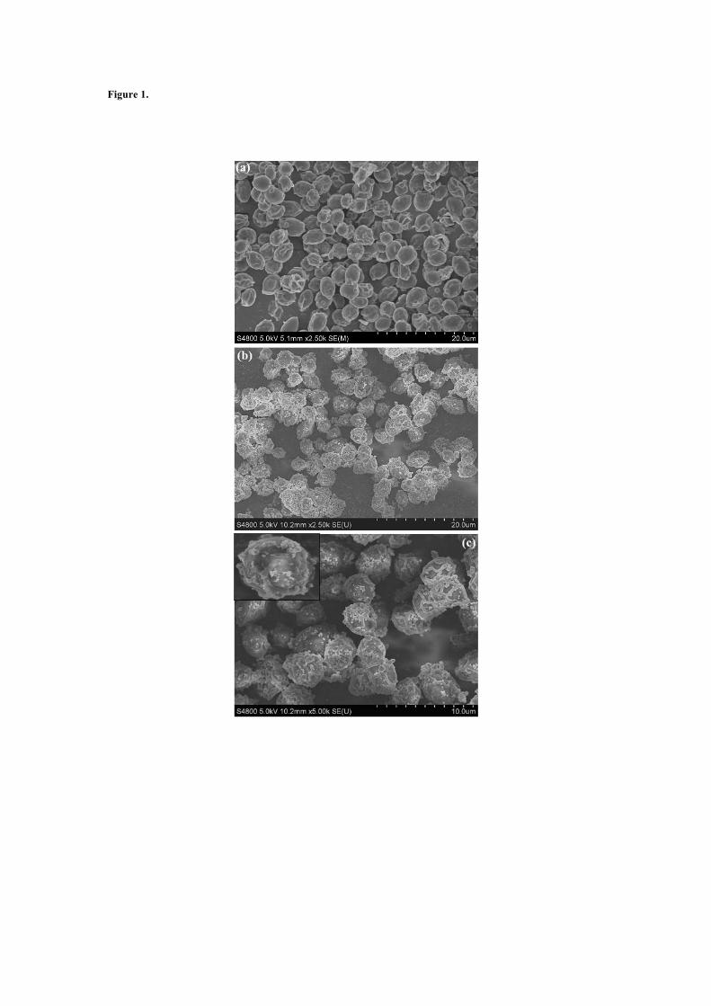
Figure 1.
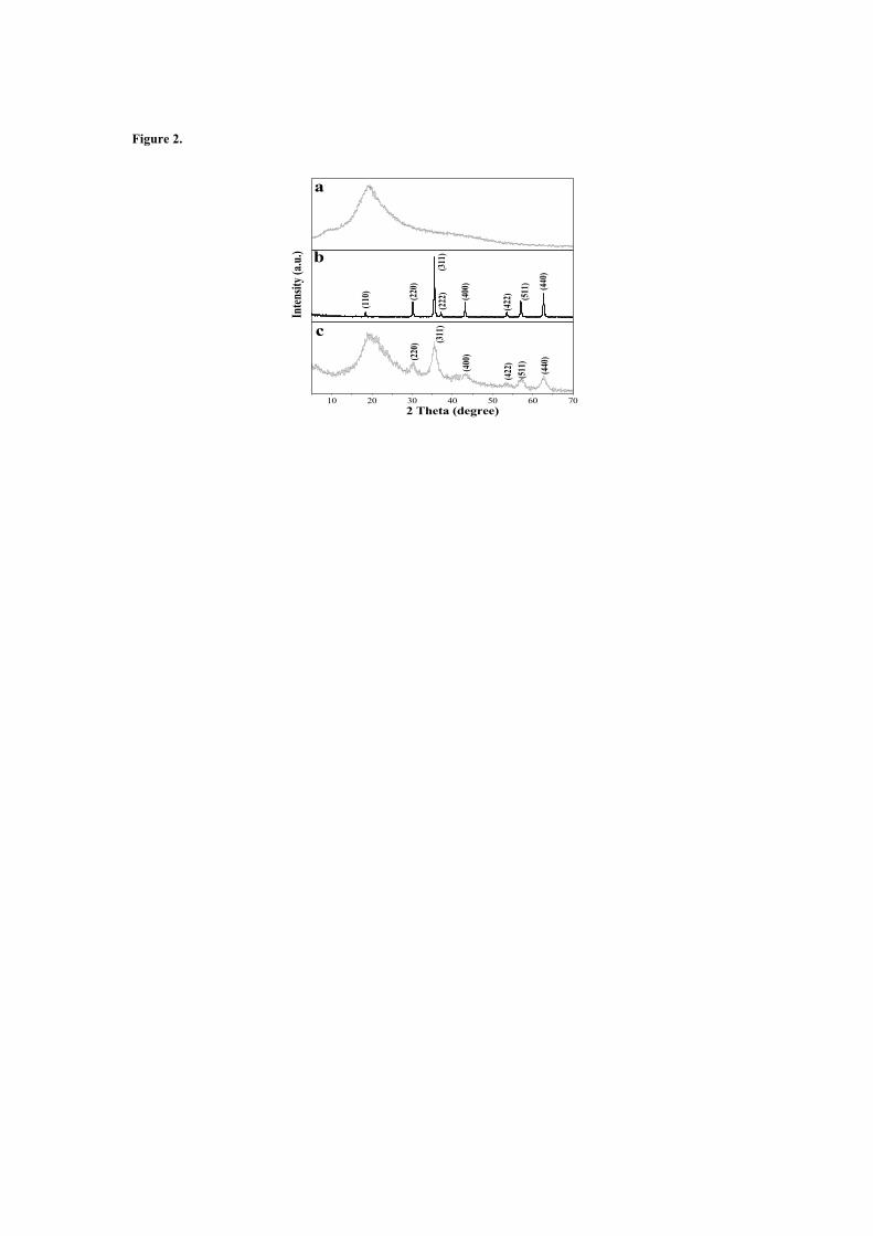
Figure 2.
10 20 30 40 50 60 70
a
(220
) (311
)
(400
)
(422
)(5
11)
(440
)
c
(222
)
b
(110
)
(422
)(220
)
(311
)
(400
)
(511
)
2 Theta (degree)
Inten
sity (
a.u.)
(440
)
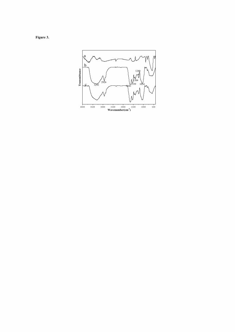
Figure 3.
4000 3500 3000 2500 2000 1500 1000 500
1054
1240
13841539
16412926
3292c
b
Transmittance
Wavenumber(cm-1)
a
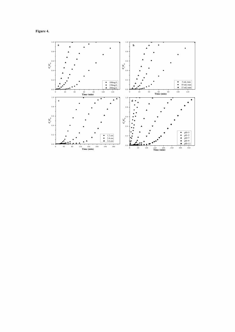
Figure 4.
0 20 40 60 80 100 1200.0
0.2
0.4
0.6
0.8
1.0
Ct/C
0
Time (min)
100mg/L 150mg/L 200mg/L
a
0 20 40 60 80 100 1200.0
0.2
0.4
0.6
0.8
1.0
b
Ct/C
0
Time (min)
5 mL/min 10 mL/min 15 mL/min
0 40 80 120 160 200 240 2800.0
0.2
0.4
0.6
0.8
1.0
c
Ct/C
0
Time (min)
1.2 cm 2.4 cm 3.6 cm
0 50 100 150 200 250 300 3500.0
0.2
0.4
0.6
0.8
1.0
d
Ct/C
0
Time (min)
pH=3 pH=5 pH=7 pH=9 pH=11
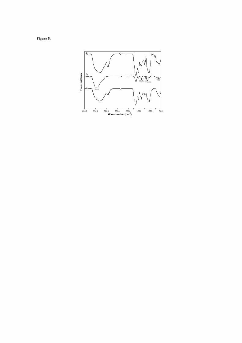
Figure 5.
4000 3500 3000 2500 2000 1500 1000 500
1545
598
c
b1186
Wavenumber(cm-1)
Transmittance
11331098
a
3444
1396652

Table captions
Table 1.Column data and parameters obtained at different conditions.
Table 2. Parameters predicted from the Thomas and Yoon-Nelson models.
Table 3. Column data and parameters obtained after Fenton-like regeneration.
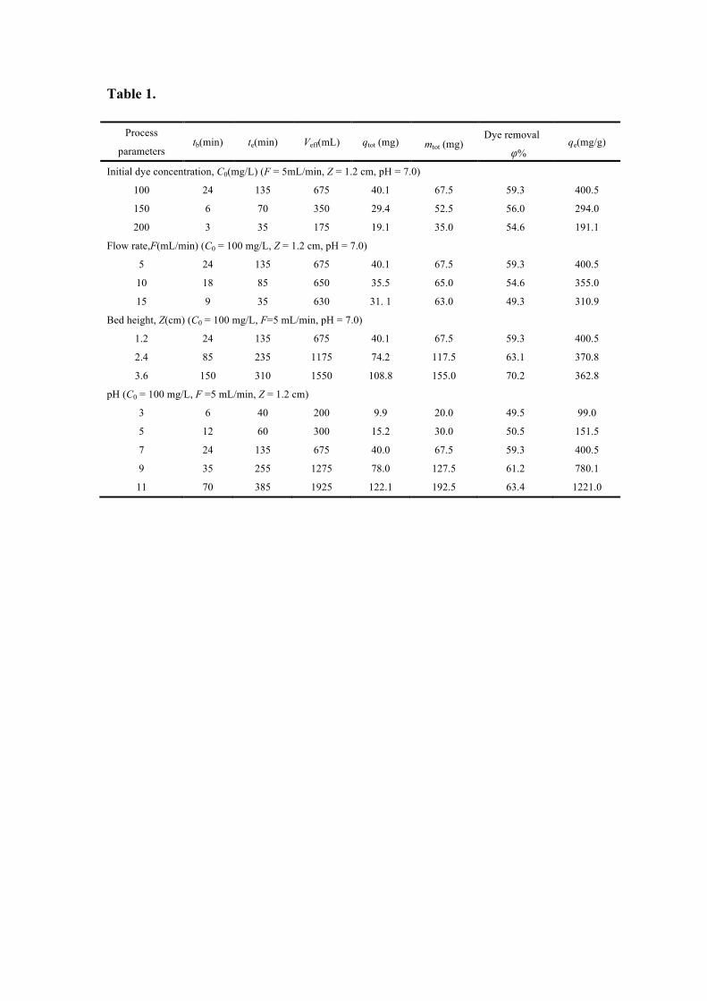
Table 1.
Process
parameters tb(min) te(min) Veff(mL) qtot (mg) mtot (mg)
Dye removal
φ% qe(mg/g)
Initial dye concentration, C0(mg/L) (F = 5mL/min, Z = 1.2 cm, pH = 7.0)
100 24 135 675 40.1 67.5 59.3 400.5
150 6 70 350 29.4 52.5 56.0 294.0
200 3 35 175 19.1 35.0 54.6 191.1
Flow rate,F(mL/min) (C0 = 100 mg/L, Z = 1.2 cm, pH = 7.0)
5 24 135 675 40.1 67.5 59.3 400.5
10 18 85 650 35.5 65.0 54.6 355.0
15 9 35 630 31. 1 63.0 49.3 310.9
Bed height, Z(cm) (C0 = 100 mg/L, F=5 mL/min, pH = 7.0)
1.2 24 135 675 40.1 67.5 59.3 400.5
2.4 85 235 1175 74.2 117.5 63.1 370.8
3.6 150 310 1550 108.8 155.0 70.2 362.8
pH (C0 = 100 mg/L, F =5 mL/min, Z = 1.2 cm)
3 6 40 200 9.9 20.0 49.5 99.0
5 12 60 300 15.2 30.0 50.5 151.5
7 24 135 675 40.0 67.5 59.3 400.5
9 35 255 1275 78.0 127.5 61.2 780.1
11 70 385 1925 122.1 192.5 63.4 1221.0
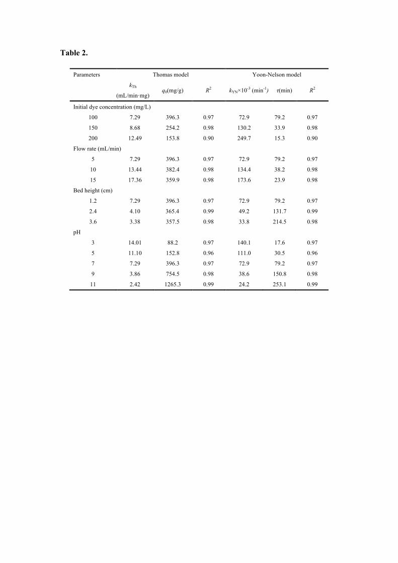
Table 2.
Parameters Thomas model Yoon-Nelson model
kTh
(mL/min·mg) q0(mg/g) R2 kYN×10-3 (min-1) τ(min) R2
Initial dye concentration (mg/L)
100 7.29 396.3 0.97 72.9 79.2 0.97
150 8.68 254.2 0.98 130.2 33.9 0.98
200 12.49 153.8 0.90 249.7 15.3 0.90
Flow rate (mL/min)
5 7.29 396.3 0.97 72.9 79.2 0.97
10 13.44 382.4 0.98 134.4 38.2 0.98
15 17.36 359.9 0.98 173.6 23.9 0.98
Bed height (cm)
1.2 7.29 396.3 0.97 72.9 79.2 0.97
2.4 4.10 365.4 0.99 49.2 131.7 0.99
3.6 3.38 357.5 0.98 33.8 214.5 0.98
pH
3 14.01 88.2 0.97 140.1 17.6 0.97
5 11.10 152.8 0.96 111.0 30.5 0.96
7 7.29 396.3 0.97 72.9 79.2 0.97
9 3.86 754.5 0.98 38.6 150.8 0.98
11 2.42 1265.3 0.99 24.2 253.1 0.99

Table 3.
Cycle time tb te Veff(mL) qtot(mg) mtot(mg)
Dye removalφ%
qe(mg/g)
1th 24 135 675 40.1 67.5 59.3 400.5
2th 18 115 500 33.8 57.5 58.7 337.5
3th 12 95 300 27.4 47.5 57.6 273.5
Supporting Information of Figures
Figure S1. EDS spectra of Fe3O4@yeast composite microspheres.
Figure S2. Photographs of the dispersion and separation processes of Fe3O4@yeast: (a)
without external magnetic field, and (b) with external magnetic field.
Figure S3.Models fitting of experimental (a) column capacity q (mg/g) and (b) the 50%
adsorbate breakthrough time τ (min).
Figure S4.Proposed mechanism of (a) formation and (b) the in situ heterogeneous Fenton-like
regeneration of Fe3O4@yeast composites.
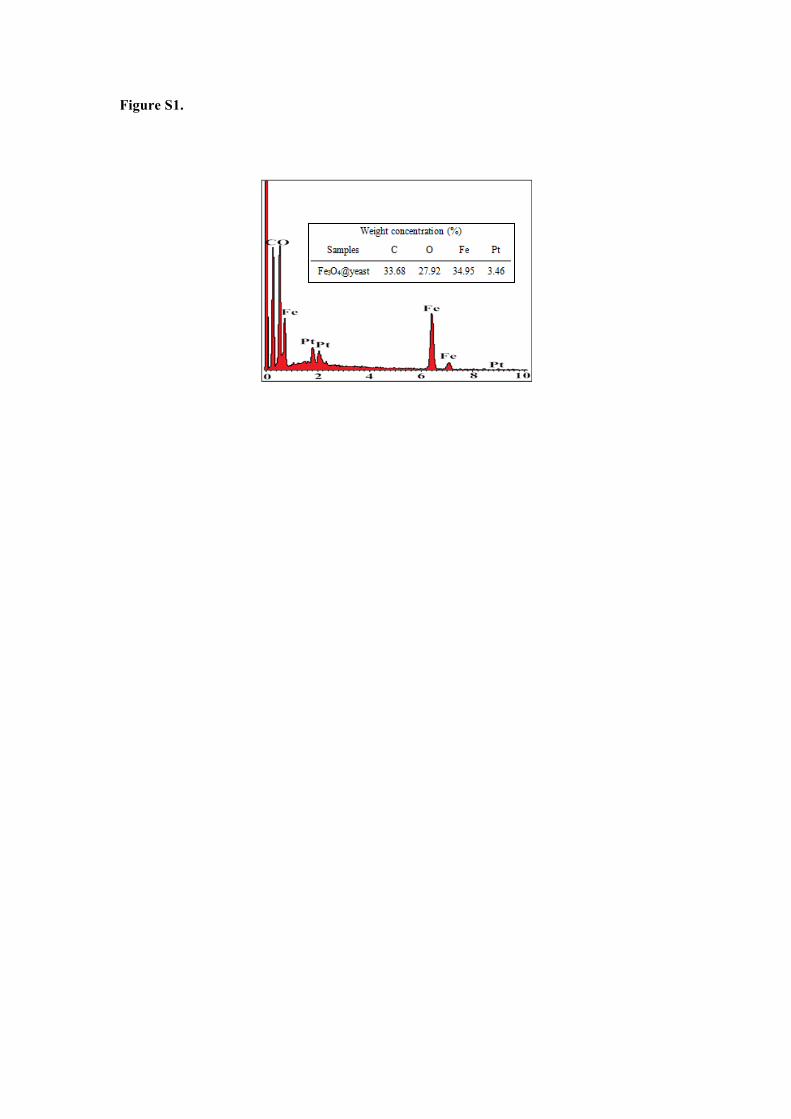
Figure S1.
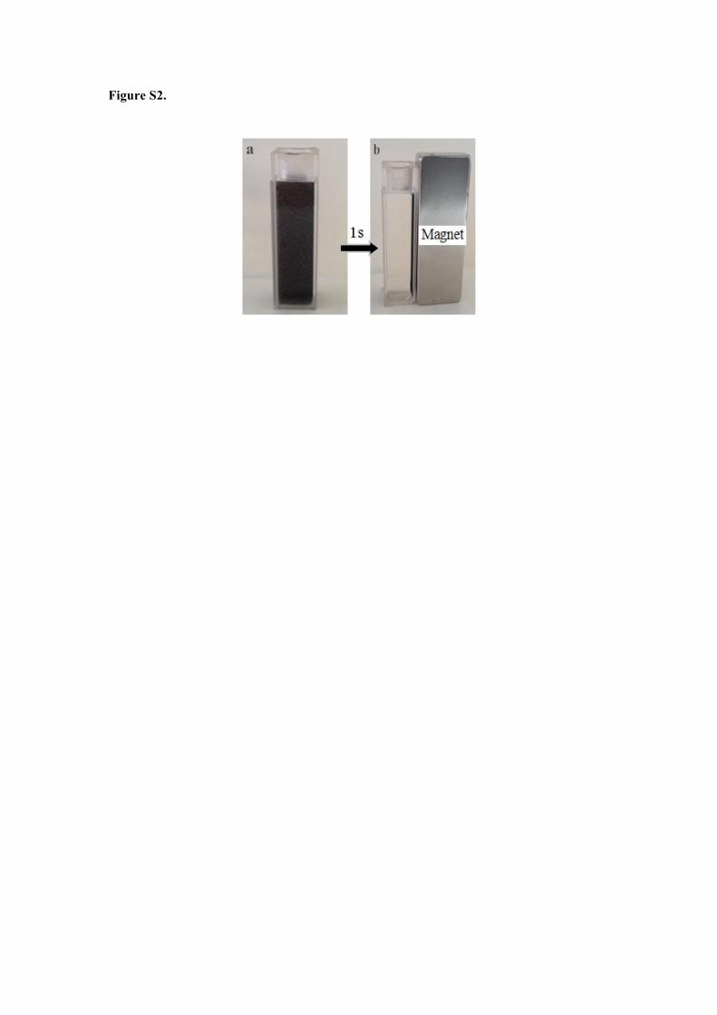
Figure S2.

Figure S3.
100 150 200
100
200
300
400
5 10 15 1.2 2.4 3.6 11 9 7 5 3
200
400
600
800
1000
1200
1400(a)
experimental Thomas model Yoon-Nelson model
pHBed height(cm)
F(mL·min-1)C0(mg·L-1)
q(m
g·g-1
)
q(m
g·g-1
)
100 150 200
20
40
60
80
5 10 15 1.2 2.4 3.6 3 5 7 9 110
50
100
150
200
250(b)
C0(mg·L-1)
50%
bre
akth
roug
h Ti
me(
min
)
experimental theoretical
50%
bre
akth
roug
h Ti
me(
min
)
pHBed height(cm)F(mL·min-1)
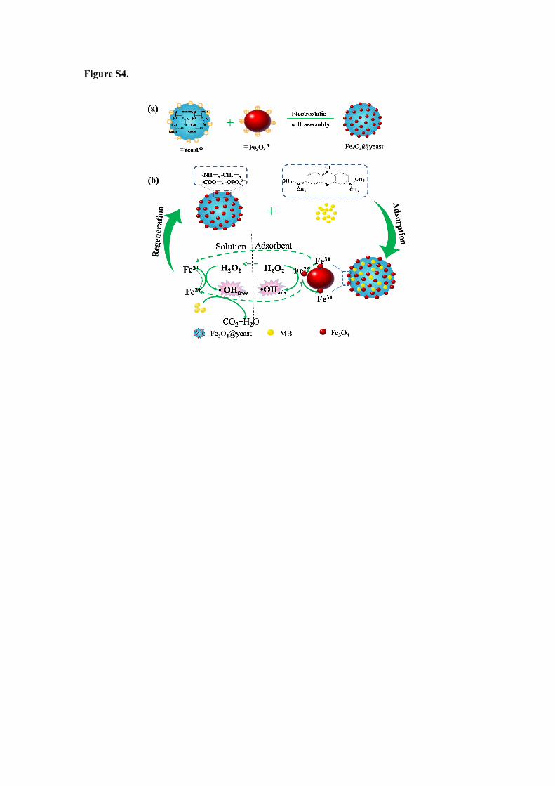
Figure S4.