biopsy of equine embryos with the eppendorf piezoxpert
TRANSCRIPT

Nuno Costa-Borges, Instituto Valenciano de Infertilidad (IVI), Barcelona, Spain, [email protected] Sanchez Arbouin and Miguel Blanco, Assisted Reproductive Techniques, Paul Schockemöhle Stud Lewitz, Neustadt-Glewe, Germany
No 047 | February 2012 - revised August 2012
Userguide
Biopsy of equine embryos at the early/expanded blastocyst stage is a rather complex procedure, essentially due to the presence of a distinctive capsule in the perivitelline space that is extraordinarily resistant and difficult to pen-etrate using conventional micromanipulation methods. In addition, this embryonic capsule has been considered essential for the embryo survival and, until recently, many doubts persisted about whether it could be breached without impairing the viability of the biopsied embryo. Recent studies, however, suggested that encapsulated equine blastocysts could be biopsied using a piezo-actuated micropipette that allows creating a small hole in the capsule to collect trophectoderm cells for genetic analysis without compromising their further viability. In this Userguide, the biopsy procedure of encapsulated equine embryos is described using the new device from Eppendorf for piezo- assisted manipulation, the Eppendorf PiezoXpert, in combination with the Eppendorf TransferMan NK 2 microma-nipulation workstation.
Biopsy of Equine Embryos with the Eppendorf PiezoXpert® for Preimplantation Genetic Diagnosis
Establishing an efficient clinical method for preimplantation genetic diagnosis (PGD) in the horse could represent a very valuable tool for the equine breeding industry, as it would allow the elimination of devastating genetic diseases or the selection of the desired gender, coat color, or other genetic traits in the offspring produced (Figure 1) [1-4]. Tradition-ally, the most popular method to collect equine embryos has been performed by uterine flush of inseminated mares on Day 6 - Day 8 after ovulation, when the embryos are generally at the early/expanded blastocyst stages, respec-tively [5,6]. In this sense, to make a PGD program practical, the flushed embryos would have to be biopsied at these developmental stages, vitrified, and later transferred into synchronized recipients, once the results from the genetic analysis had been assessed.
Fig. 1: Mare with foal.
Abstract
Introduction
• Binocular stereo microscope (SZH, Olympus or similar)• Inverted microscope with up to 40x, Modulation contrast (IX 71, Olympus or similar)• Microinjector (CellTram Oil, Eppendorf)• Microinjector (CellTram vario, Eppendorf)• 2x Micromanipulators (TransferMan NK 2, Eppendorf) • Piezo-drill unit (Eppendorf PiezoXpert, Eppendorf)• Borosilicate Pasteur pipettes for embryo manipulation• Hamilton microliter syringe (Hamilton Company)• Piezo Drill Tip ES (ID 15µm, angle 25°, blunt end, Eppendorf)• VacuTip (ID 15µm, OD 100µM, angle 35°, Eppendorf)• Petri culture dishes 90 mm (Nunc)• Holding medium (Minitub)• Mercury (215477, Sigma-Aldrich)• Mineral oil (Nidoil, Nidacon)• Polyvinylpyrrolidone (PVP; PVP360, Sigma-Aldrich)• Perfluor (FC770, Sigma-Aldrich)
Userguide No 047 | Page 2
Equipment and Material
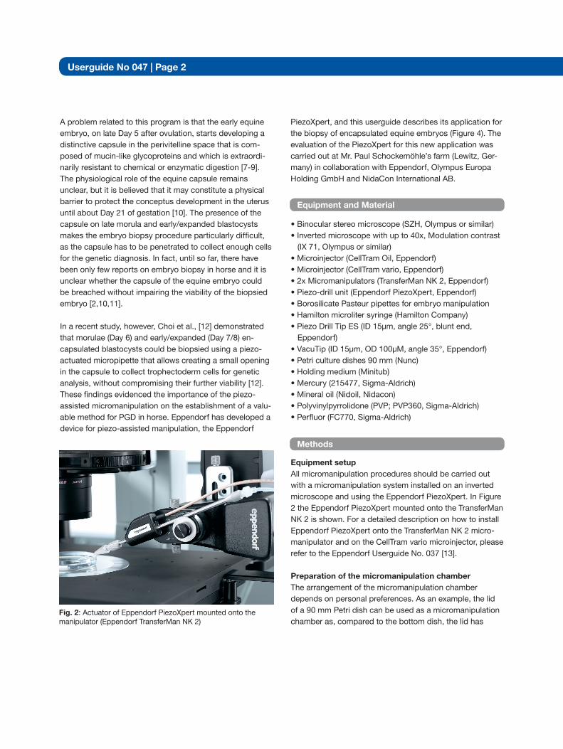
Fig. 2: Actuator of Eppendorf PiezoXpert mounted onto the manipulator (Eppendorf TransferMan NK 2)
Equipment setupAll micromanipulation procedures should be carried out with a micromanipulation system installed on an inverted microscope and using the Eppendorf PiezoXpert. In Figure 2 the Eppendorf PiezoXpert mounted onto the TransferMan NK 2 is shown. For a detailed description on how to install Eppendorf PiezoXpert onto the TransferMan NK 2 micro-manipulator and on the CellTram vario microinjector, please refer to the Eppendorf Userguide No. 037 [13]. Preparation of the micromanipulation chamberThe arrangement of the micromanipulation chamber depends on personal preferences. As an example, the lid of a 90 mm Petri dish can be used as a micromanipulation chamber as, compared to the bottom dish, the lid has
A problem related to this program is that the early equine embryo, on late Day 5 after ovulation, starts developing a distinctive capsule in the perivitelline space that is com-posed of mucin-like glycoproteins and which is extraordi-narily resistant to chemical or enzymatic digestion [7-9]. The physiological role of the equine capsule remains unclear, but it is believed that it may constitute a physical barrier to protect the conceptus development in the uterus until about Day 21 of gestation [10]. The presence of the capsule on late morula and early/expanded blastocysts makes the embryo biopsy procedure particularly difficult, as the capsule has to be penetrated to collect enough cells for the genetic diagnosis. In fact, until so far, there have been only few reports on embryo biopsy in horse and it is unclear whether the capsule of the equine embryo could be breached without impairing the viability of the biopsied embryo [2,10,11]. In a recent study, however, Choi et al., [12] demonstrated that morulae (Day 6) and early/expanded (Day 7/8) en-capsulated blastocysts could be biopsied using a piezo-actuated micropipette that allows creating a small opening in the capsule to collect trophectoderm cells for genetic analysis, without compromising their further viability [12]. These findings evidenced the importance of the piezo-assisted micromanipulation on the establishment of a valu-able method for PGD in horse. Eppendorf has developed a device for piezo-assisted manipulation, the Eppendorf
Methods
PiezoXpert, and this userguide describes its application for the biopsy of encapsulated equine embryos (Figure 4). The evaluation of the PiezoXpert for this new application was carried out at Mr. Paul Schockemöhle’s farm (Lewitz, Ger-many) in collaboration with Eppendorf, Olympus Europa Holding GmbH and NidaCon International AB.
strongly recommended to assign a reference number to each collected embryo to guarantee the identification of the corresponding biopsied cells throughout all steps of the PGD. Preparation of microcapillariesHolding and biopsy microcapillaries can be purchased from Eppendorf. Whenever possible, the holding capillary should have an outside diameter (OD) slightly smaller than that of the embryo, an inner diameter (ID) of about 20 µm, and an angle of approximately 20 degrees at the tip. If the OD of the holding capillary is too small in propor-tion to the diameter of the embryo, it can cause the embryo to move when piezo impulses are applied and reduce the efficiency of the drilling. The biopsy capillary should have a blunt-end, an ID of about 15 µm at the tip and an angle identical to that of the holding pipette. Before connecting the biopsy capillary to the Eppendorf PiezoXpert, the capil-lary should be backfilled with a small amount of mercury (approximately 2 µL, i.e. 4 mm inside the capillary) using a Hamilton Syringe. This is an important step to enhance the transmission of the piezo impulses and to ensure an optimal performance when drilling the zona pellucida and the capsule. As an alternative to mercury, Perfluor can be used. Please con-sult [13] for a detailed description on how to connect the biopsy capillary to the Eppendorf PiezoXpert actuator. Eppendorf PiezoXpert parametersPre-installation of Eppendorf PiezoXpert parameters (inten-sity, speed and number of piezo impulses) can significantly speed up the biopsy procedure and at the same time shorten the time-period in which the embryos are exposed to stressful conditions. Thus, this will improve the final results. Up to 3 sets of optimized programs can be stored in the Eppendorf PiezoXpert. Each program consists of parameter sets A and B, see Figure 5. Usually, parameter set A is used with strong parameters for the penetration of the zona pellucida and breaching of the capsule. Set B is used afterwards in exceptional situations when cells to be biopsied cannot be isolated from the embryos by gentle suction with the biopsy capillary: In these rare cases, ad-ditional gentle piezo impulses can help achieve successful cell isolation. Both sets A and B can be triggered via either the button on the control unit or the foot control.
Userguide No 047 | Page 3
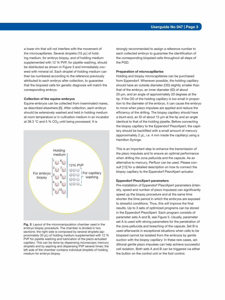
Fig. 3: Layout of the micromanipulation chamber used in the embryo biopsy procedure. The chamber is divided in two sections: the right side is composed by several droplets (ap-proximately 20 µL) of holding medium supplemented with 12 % PVP for pipette washing and lubrication of the piezo-actuated capillary. This can be done by dispensing microscopic mercury droplets and by aspiring and dispensing PVP several times; the left side of the chamber contains individual droplets of holding medium for embryo biopsy.
Holdingmedium
12% PVP
For embryobiopsy
For capillarywashing
1 6
7
8
9
10
2
3
4
5
a lower rim that will not interfere with the movement of the microcapillaries. Several droplets (10 µL) of hold-ing medium, for embryo biopsy, and of holding medium supplemented with 12 % PVP, for pipette washing, should be distributed as shown in Figure 3 and immediately cov-ered with mineral oil. Each droplet of holding medium can then be numbered according to the reference previously attributed to each embryo after collection, to guarantee that the biopsied cells for genetic diagnosis will match the corresponding embryo. Collection of the equine embryosEquine embryos can be collected from inseminated mares, as described elsewhere [6]. After collection, each embryo should be extensively washed and held in holding medium at room temperature or in cultivation medium in an incubator at 38.3 °C and 5 % CO2 until being processed. It is
Userguide No 047 | Page 4
Parameter
Morula Early Blastocyst Expanded Blastocyst
Set A Set B Set A Set B Set A Set B
Intensity 10 1 15 - 20 1 25 - 30 1
Speed 4 1 8 1 10 1
Pulse ∞ 1 ∞ 1 ∞ 1
Table 1: Parameter settings using a capillary backfilled with mercury for biopsy of equine embryos at morula, early- and expanded-blastocysts stages.
medium where the embryo to be biopsied was previously placed. Embryo biopsy can then be performed by applica-tion of piezo impulses (see Table 1) combined with gentle suction in the biopsy capillary to allow the drilling of a small hole in the zona pellucida and the embryonic capsule, and thus facilitate the access of the blunt-end pipette to the trophectoderm cells, as explained in detail in Figure 4. An acceptable number (5 to 10) of trophectoderm cells should be removed as quickly as possible by applying gentle suc-tion with the biopsy capillary after penetrating the cap-sule. In some cases, when simple suction with the biopsy pipette is not efficient to isolate the cells from the embryo, a few weak piezo impulses facilitate the access of the blunt-end pipette to the trophectoderm cells, as explained in detail in Figure 4. After the biopsy, the removed cells should be released from the capillary in the vicinity of the biopsied embryo and then aspirated and placed in a minimum vol-ume (5 µL) of phosphate buffered saline (PBS) in a 0.2 mL PCR tube using a fine-bore glass pipette. The tubes with the biopsied cells can then be kept at 4 °C or be frozen directly in liquid nitrogen until the genetic analysis is assessed by PCR, as described elsewhere [1,14]. Post-embryo biopsyAfter the biopsied cells have been obtained, the biopsied embryo should be quickly removed from the micromanipula-tion chamber and placed in a droplet of culture medium in a humidified 5 % CO2 incubator at 38.3 °C. This step allows the embryo to recover from the biopsy procedure while keeping it under controlled conditions. Meanwhile, the biop-sied cells can be processed for the genetic analysis. The results obtained in the PGD should then be carefully interpreted before deciding whether the biopsied embryo should be transferred or not into a synchronized recipient.
Optimization of parameters:1. Set the parameters for speed, intensity and number of piezo impulses to 1.2. Gradually increase the value for intensity until the piezo impulses are strong enough to penetrate the zona pellucida and the capsule.3. Fine tune the speed and pulse parameters. 4. Use the lowest parameter settings that work. Usually, the parameter settings for Perfluor may be slightly higher than for mercury. NOTE: Users should optimize the parameters for their own experiments as the settings always depend on individual laboratory protocols. As a guideline, if non-cryopreserved embryos are used, the Eppendorf PiezoXpert parameters using mercury recommended for morula, early and ex-panded blastocysts are shown in Table 1. These param-eters can be saved in the Eppendorf PiezoXpert control unit as different programs according to the embryonic develop-mental stage of the embryos to be biopsied. Embryo biopsy procedureAfter all equipment has been set-up, a single embryo should be moved to a droplet of holding medium in the micromanipulation chamber under a stereo microscope. The chamber should then be placed onto the inverted microscope and the embryo should be morphologically inspected at 20x and 40x magnifications. The embryo should be fixed for biopsy with the holding capillary by gentle suction. The biopsy capillary should next be washed and lubricated in the PVP containing medium droplets, and then moved back to the droplet of holding
Userguide No 047 | Page 5
A B
C D
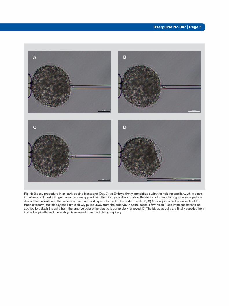
Fig. 4: Biopsy procedure in an early equine blastocyst (Day 7). A) Embryo firmly immobilized with the holding capillary, while piezo impulses combined with gentle suction are applied with the biopsy capillary to allow the drilling of a hole through the zona pelluci-da and the capsule and the access of the blunt-end pipette to the trophectoderm cells. B, C) After aspiration of a few cells of the trophectoderm, the biopsy capillary is slowly pulled away from the embryo. In some cases a few weak Piezo impulses have to be applied to detach the cells from the embryo before the pipette is completely removed. D) The biopsied cells are finally expelled from inside the pipette and the embryo is released from the holding capillary.

[ 1] Peippo J, Huhtinen M, Kotilainen T. Sex diagnosis of equine preimplantation embryos using the polymerase chain reaction. Theriogenology 1995. 44: 619-627. [ 2] Huhtinen M, Peippo J, Bredbacka P. Successful transfer of biopsied equine embryos. Theriogenology 1997 48: 361–367.[ 3] Finno CJ, Spier SJ, Valberg SJ. Equine diseases caused by known genetic mutations. Vet J. 2009 179:336-347 [ 4] Tryon RC, Penedo MC, McCue ME, Valberg SJ, Mickelson JR, Famula TR, Wagner ML, Jackson M, Hamilton MJ, Nooteboom S, Bannasch DL. Evaluation of allele frequencies of inherited disease genes in subgroups of American Quarter Horses. J Am Vet Med Assoc. 2009 234: 120-125.[ 5] Betteridge KJ, Eaglesome MD, Mitchell D, Flood PF, Beriault R. Development of horse embryos up to twenty two days after ovulation: observations on fresh specimens. J Anat. 1982 135: 191–209.[ 6] Squires EL, McCue PM, Vanderwall D. The current status of equine embryo transfer. Theriogenology 1999 51:91-104.[ 7] Oriol JG, Betteridge KJ, Clarke AJ, Sharom FJ x Mucin-like glycoproteins in the equine embryonic capsule. Mol Reprod Dev. 1993a 34 255–265. [ 8] Oriol JG, Sharom FJ, Betteridge KJ. Developmentally regulated changes in the glycoproteins of the equine embryonic capsule. J Reprod Fertil. 1993b 99: 653–664. [ 9] Albihn A, Waelchli RO, Samper J, Oriol JG, Croy BA, Betteridge KJ. Production of capsular material by equine trophoblast transplanted into immunodeficient mice. Reproduction 2003 125: 855–863.[10] Stout TA, Meadows S, Allen WR. Stage-specific formation of the equine blastocyst capsule is instrumental to hatching and to embryonic survival in vivo. Anim Reprod Sci. 2005 87: 269–281.[11] Skidmore J, Boyle MS, Cran D, Allen WR. Micromanipulation of equine embryos to produce monozygotic twins. Equine Veterinary Journal 1989 8: 126–128.[12] Choi YH, Gustafson-Seabury A, Velez IC, Hartman DL, Bliss S, Riera FL, Roldán JE, Chowdhary B, Hinrichs K. Viability of equine embryos after puncture of the capsule and biopsy for preimplantation genetic diagnosis. Reproduction 2010 140: 893-902. [13] Hajarian H and KwanMor K. Piezo-actuated Mouse ICSI (intracytoplasmic sperm injection) using the Eppendorf PiezoXpert®. Eppendorf Userguide 037. www.eppendorf.com[14] Ogilvie CM, Braude PR, Scriven PN. Preimplantation genetic diagnosis--an overview. J Histochem Cytochem 2005 53:255-260.
References
Fig. 5: Setting of parameter sets A and B
Userguide No 047 | Page 6

Userguide No
Your local distributor: www.eppendorf.com/worldwideEppendorf AG · 22331 Hamburg · Germany · Tel: +49 40 53801-0 · Fax: +49 40 538 01-556 · E-mail: [email protected]
Eppendorf North America, Inc. · 102 Motor Parkway · Hauppauge, N.Y. 11788-5178 · USATel: +1 516 334 7500 · Toll free phone: +1 800-645-3050 · Fax: +1 516 334 7506 · E-mail: [email protected]
Application Support Europe, International: Tel: +49 1803 666 789 (Preis je nach Tarif im Ausland; 9 ct/min aus dem dt. Festnetz; Mobilfunkhöchstpreis 42 ct/min) · E-mail: [email protected]
North America: Tel: +1 800 645 3050 · E-mail: [email protected] Pacific: Tel: +60 3 8023 6869 · E-mail: [email protected]
047
Trad
emar
ks o
r re
gist
ered
trad
emar
ks o
f oth
er m
anuf
actu
rers
or
dist
ribut
ors
are
ackn
owle
dged
as
the
prop
erty
of t
heir
resp
ectiv
e ow
ners
. Tra
dem
arks
as
men
tione
d in
this
pub
licat
ion
are
incl
uded
. O
lym
pus
is a
regi
ster
ed tr
adem
ark
of O
lym
pus
Cor
pora
tion.
Nun
c is
a re
gist
ered
trad
emar
k of
Nun
c A
/S, S
igm
a-A
ldric
h is
a re
gist
ered
trad
emar
k of
Sig
ma-
Ald
rich
Co.
LLC
., H
amilt
on is
a re
gist
ered
trad
emar
k of
Ham
ilton
Med
ical
AG
. ep
pend
orf®
, Epp
endo
rf P
iezo
Xpe
rt®
, Fem
toJe
t®, T
rans
ferM
an®
, Cel
lTra
m®
and
Inje
ctM
an®
are
regi
ster
ed tr
adem
arks
of E
ppen
dorf
AG
. • G
alax
y® is
a re
gist
ered
trad
emar
k of
New
Bru
nsw
ick
Sci
entif
ic In
c., U
SA
. M
icro
load
er™
, Tra
nsfe
rMan
NK
2™
, Inj
ectM
an N
I 2™
and
Fem
totip
II™
are
trad
emar
ks o
f Epp
endo
rf A
GO
rder
-No.
AU
04 7
WW
020
/GB
2/0T
/081
2/C
RE
A •
All
right
s re
serv
ed, i
nclu
ding
gra
phic
s an
d im
ages
• C
opyr
ight
© 2
012
by E
ppen
dorf
AG
, Ham
burg
, Ger
man
y.
Ordering information
Product Description Order no. International
Order no. North America
Eppendorf PiezoXpert® Basic device incl. Actuator, Food control and Spacer plate* 5194 000.016 5194000024
TransferMan® NK 2** Micromanipulator with proportional movement control for suspension cells 5188 000.012 920000011
CellTram® Air** Manual pressure device for the reliable holding of suspension cells 5176 000.017 920002021
CellTram® Oil** Manual hydraulic pressure device for the reliable holding of suspension cells 5176 000.025 920002030
CellTram® vario** Manual hydraulic pressure device, with gears for coarse and fine control 5176 000.033 920002111
VacuTip™ **, *** 25 glass capillaries for holding large cells (e.g. eggs), sterilized, tip angle 35° 5175 108.000 930001015
Piezo Drill Tips ES™ 25 glass capillaries for the piezo-assisted transferof embryonal stem cells, angle 25° 5175 250.001 930001104
Microloader Capillary tip for filling microinjection capillaries, set of 2x 96 pcs. 5242 956.003 930001007
Microscope Adapter Adapter for any inverse microscopes Available on request
Available on request
Galaxy® 14 S (230 V) **** “Personal” sized CO2 incubator offering 14L of CO2 Incubation with an LED display CO14S- 2300000
Galaxy® 14 S (230 V) **** Incubator with active 1-19 % O2 control CO14S- 2300200
* For mounting the Eppendorf PiezoXpert onto TransferMan NK 2 or PatchMan NP 2** This product is registered in Europe as a medical device (according to Medical Device Directive MDD/93/42/EDD). This product is not registered in the U.S. as a medical device and does not have a 510(k) registration. For resarch use only. Not for use in human medical applications. *** Proven non-cytotoxicity by the mouse embryo development test **** New Brunswick CO2 Incubators have been designed for research use only. New Brunswick CO2 Incubators in general are not certified for any human IVF/medical application