bioprospecting potential of endophytic bacteria from ...€¦ · isolation and preliminary...
TRANSCRIPT
Int.J.Curr.Microbiol.App.Sci (2017) 6(10): 1718-1730
1718
Original Research Article https://doi.org/10.20546/ijcmas.2017.610.208
Bioprospecting Potential of Endophytic Bacteria from Leaves of
Gossypium hirsutum
Azba A. Shaikh1*
, P.R. Parmar2, B.K. Rajkumar
2, D.H. Patel
2,
H.R. Desai2 and B.G. Solanki
2
1Shree Ramkrishna Institute of Computer Education and Applied Sciences,
M.T.B College Campus, Surat-395 001, Gujarat, India 2Main Cotton Research Station (MCRS), NAU, Athwa Farm, GhodDod Road,
Surat – 395007, Gujarat, India *Corresponding author
A B S T R A C T
Introduction
Plant growth promoting microbes is an
attractive way to replace chemical or
synthetic compounds in agriculture field
because of its eco-friendly and economical
feasibility (Bhattacharyya et al., 2012; Kirti et
al., 2016). The ‘biopropecting’ word
describes the collection and screening of
biological material for commercial purpose.
Every naturally propagated plant is colonized
by divert communities of microbes referred to
as ‘endophytes’. Endophytes defined as
microorganisms that can be isolated from
surface-disinfected plant tissues or extracted
from within plants and that do not harm the
host plants (Hallmann et al., 1998). Recent
studies have indicated that endophytes play
many important beneficial roles in the
metabolism and physiology of the host plant
International Journal of Current Microbiology and Applied Sciences ISSN: 2319-7706 Volume 6 Number 10 (2017) pp. 1718-1730 Journal homepage: http://www.ijcmas.com
A total of 19 bacterial endophytes were isolated from leaves of cotton (Gossypium
hirsutum) and characterized for plant growth promoting traits viz., siderophores
production, phosphate solubilization, IAA and ammonia production; and preliminarily
screened for chitinase, gelatinase, protease and lipase. Quantitative analysis of
siderophores production and phosphate solubilization indicated that six endophyteseach
viz., EB11, EB4, EB3, EB5, EB8 and EB9 were able to produce siderophores ranging
between 80.26 to 92.58 % and EB9, EB11, EB14, EB17, EB4 and EB2 solubilized
phosphorus ranging 10.24-13.58mg/ml within 48 hrs. After 72 hrs incubation, seven
isolates EB3, EB13, EB8, EB11, EB15, EB2 and EB17 produced IAA in range of 15-28
µg/ml. Within 48 hrs, the ammonia production ranged from 0.8 to 1.6 (µg/ml). Primary
screening of enzymes showed that chitinase was produced by EB5 (1.9 mm) and EB3 (1.1
mm); protease by EB15, EB12, EB11, EB1 and EB4 (11.80-16.60mm); gelatinase by EB1,
EB9, EB1, EB12 and EB16 (10.00 -17.00mm); and lipase by EB10, EB13, EB17, EB14,
EB12 and EB11 (10.00-15.00mm). Results suggested all endophytic bacterial strains
studied are potential for the one or other parameter studied thus further study will help to
make consortium as biofertilizer or bioprotectant to enhance plant productivity and
protection.
K e y w o r d s
Gossypium
hirsutum,
Endophytic bacteria,
PGPR, Bioprospect.
Accepted:
17 September 2017
Available Online:
10 October 2017
Article Info

Int.J.Curr.Microbiol.App.Sci (2017) 6(10): 1718-1730
1719
via direct and indirect way. Direct way
includes production of phytohormones,
phosphorous solubilisation, nitrogen fixation,
and siderophore production as an iron
chelators etc., while indirect way includes
suppression of plant diseases by elevating
plant resistance mechanisms or by producing
various enzymes or metabolites (Bakker and
Schippers, 1987; DéFago et al., 1990;
Kachhap et al., 2014). In last few decades a
large array of endophytic bacterial species of
Pseudomonas, Azospirillum, Azotobacter,
Klebsiella, Enterobacter, Alcaligenes,
Arthobacter, Burkholderia, Bacillus and
Serratia have been reported to enhance plant
growth (Malfanova et al., 2013).
Biochemically cotton plant is the source of
valuable compounds such as terpenes,
phenolics, fatty acids, lipids, carbohydrates
and proteins. Especially, leaves of cotton
plants contain camphene, limonene, myrcene,
sabinene and other compounds (Egbuta et al.,
2017). Therefore, cotton leaf may harbor
endophytes that are potential in plant
protection, provide nutrients to the plants, and
withstand abiotic stresses. Due to high
diversity of soil bacteria and its high
contribution to plant growth, traditional
microbiological approaches are widely
focused on root endophytic bacteria
(Compantet al., 2010). On the other hand, leaf
endophytic bacteria have not received much
attention so far. The present study aims to
bridge this gap. Leaf endophytic bacteria are
the collection of selective phyllosphere
bacteria and they reside in the leaves and
maintain endophytic symbiotic relationship
with the host plant as well other microbe lives
within the leaf. Every microbe within the leaf
may have some beneficial function in terms of
plant protection as well as to provide nutrients
to the plants (Neilands, 1981; Lindowet al.,
2003). Studies indicated the predominance of
endophytic bacteria in cotton viz., Erwinia
sp., Bacillus pumilus, B. brevis, Clavibacter
species, Xanthomonas sp (Misaghi and
Donndelinger 1990), Agrobacterium sp.,
Serratia sp., Burkholderia sp., Bacillus sp.,
Staphylococcus sp., Rhizobium, Variovorax
sp., Pseudomonas sp., Acenitobacter sp.,
Artharobacter sp. And Enterobacter species
(McInroy and Kloepper 1995a and b;
Mussonet al., 1995). Some endophytes such
as Pseudomonas fluorescence 89B-61 found
as a controlling agent of Fusarium wilt of
cotton caused by Fusarium oxysporum f. sp.
vasinfectum (Chen et al., 1995). McGee
(2002) reported that due to presence of
endophytes in leaves, plant is protected from
fungal pathogens and pest. For example,
methanol extract of endophytes isolated from
the cotton leaf reduced the larval growth of
insect pest. In another study significant
reduction of damping off disease by R. solani
was observed with the talc-based
bioformulation with chitin that containe
dendophytic bacteria such as Bacillus
EPCO102, Bacillus EPCO16 and
Pseudomonas fluorescence Pf1 strains
(Rajendran et al., 2008). Therefore, the
objective of this study is to isolate bacterial
endophytes from cotton leaf and characterize
their plant growth promoting traits viz.,
siderophores production, phosphate
solubilization, IAA and ammonia production;
and preliminarily screening for chitinase,
gelatinase, protease and lipase in order to bio-
prospect the potential of these endophytic
bacteria as biofertilizer or bioprotectant.
Materials and Methods
Collection of leaf sample and its surface
sterilization for isolation of endophytes
Healthy leaf sample of cotton was collected
from the farm at Main Cotton Research
Station, Athwa farm, Surat, Gujarat. The
collected leaf were taken to the laboratory and
preserved at 4°C in sealed plastic bags and
subjected to isolate endophytic bacteria.

Int.J.Curr.Microbiol.App.Sci (2017) 6(10): 1718-1730
1720
Surface sterilization of leaf samples was
performed according to the method of
Musson et al., (1995). Healthy cotton leaf
sample was washed by running tap water for
1-2 min followed by 1 min wash with 75%
alcohol to remove the soil particles
completely.
Then the leaf sample was washed with sterile
1% (v/v) sodium hypochlorite embedded with
0.05% (v/v) Triton X-100 for3 min followed
by four sequential wash with sterile phosphate
buffer (pH 7.0). At this point, 0.1 ml of final
phosphate buffer washed solution was spread
onthe nutrient agar (Musson et al., 1995).
Isolation and preliminary characterization
of endophytic bacteria
After surface sterilization, leaf sample cut into
5 mm square pieces with sterile blade. Each
of the leaf pieces embedded carefully on the
surface of different media viz., Nutrient Agar,
R2A Agar and Soil Extract Agar (Musson et
al., 1995).
Four leaf pieces per plate were placed on each
medium. Plating was done in triplicate and all
plates were incubated at room temperature
(27oC) for three to four days.
After incubation, suspension of the bacteria
which grown surrounding the leaf sample was
prepared. Further, isolation and purification of
endophytic bacteria was carried out using
quadrant method (Sanders, 2012). Each of
purified colonies than transferred to nutrient
agar slants and were stored at 4oC.
Bergey’s manual of determinative
bacteriology was used for recording
preliminary cultural characteristics such as
size, shape, margin, elevation, consistency,
opacity, pigment and gram reaction of
isolated bacterial endophytes (MacFaddin,
2000).
Evaluation of plant growth promoting
traits of the endophytic isolates
Siderophores detection assay
Siderophores produced by the endophytic
isolates were determined using qualitative and
quantitative assay as described by Schwyn
and Neilands (1985). For quantitative assay,
test isolates were grown in iron free sterile
succinic acid medium (Hi-media) for 48 hrs at
room temperature at 120 rpm. Cell
supernatant (0.5 ml) after centrifugation at
15,000 rpm for 5 min was mixed with 0.5ml
of CAS solution (1:1) to observe the colour
change from blue to orange or yellow.
Optimal density was measured at 630 nm and
the percentage of siderophore production was
calculated as per formula [(Ar-As)/Ar]
×100=% siderophore units, where Ar =
absorbance of reference (succinic acid media
+ CAS assay solution), As = absorbance of
sample. As a reference sterile succinic acid
medium free from inoculates was used. For
qualitative analysis, 48 hrs old cultures of
endophytic bacteria grown in succinic acid
medium were spot inoculated on CAS agar
plates and incubated at 27ºC for 48 hrs.
Positive reaction was indicated by a color
change of the CAS reagent from blue to
orange surround the colony shows the
siderophores production. Zones index was
calculated by dividing yellow/orange zone on
CAS agar by growth diameter of spot
inoculants (Louden et al., 2011).
Assessment of phosphate solubilization
ability
Quantitative estimation of phosphate
solubilization activity of isolates was
performed using method described by King
(1932). For that, bacterial endophytes
inoculated into the sterile Pikovskaya’s
medium (Pikovskaya, 1948) kept for 48 hrs at
room temperature at 120 rpm. Culture
Int.J.Curr.Microbiol.App.Sci (2017) 6(10): 1718-1730
1721
supernatant after centrifugation used to
estimate the concentration of soluble
phosphate using stannous chloride method
(King, 1932). For that, 1.0ml of the
supernatant was mixed with 10 ml of 0.5M
NaHCO3, 10 ml of 1.5% ammonium
molybdate, 1 ml of working stannous chloride
and the final volume to 50ml using distilled
water. Optimal density measured at 660nm
after 15 min of incubation. The concentration
of phosphate was estimated using standard
curve of tricalcium phosphate in the range of
0.04-0.4 mg/ml.
Qualitative estimation of tricalcium phosphate
solubilization was performed using the
method of Pikovskaya (1948). Each
endophytic cell suspensions were spot
inoculated on Pikovskaya’s agar plates and
incubated at 27±2℃ for 48 hrs to observe the
zone of solubilisation. Phosphate
solubilisation index was calculated by
dividing phosphate solubilisation zone on
pikovskaya’s agar by growth diameter of spot
inoculants.
Indole Acetic Acid (IAA) production assay
The ability of bacterial endophytes to produce
IAA was measured using colorimetric method
described byLoper and Scroth (1986). For
each of endophytic bacterial isolates was
grown in sterile nutrient broth embedded with
0.2 ml of 1% L-tryptophan for 48 hrs. Further,
1.0 ml supernatant of each endophytic
bacterial isolates mixed with 1.0ml of
Salkowski’s reagent to develop the pink
colour for the positive result of IAA
production and measured at 530 nm.
Concentration of IAA produced was
estimated against standard curve of IAA in
the range of 10-100 µg/ml.
Production of ammonia
The ability of endophytic bacterial strains to
produce ammonia was assessed as described
by Cappuccino and Sherman (1992). In this
method, each isolate inoculated into the 5.0ml
of sterile peptone water broth and incubated at
room temperature for 48 hrs at 120 rpm. After
incubation, 1.0 ml of culture supernatant was
mixed with 1.0 ml of Nesseler’s reagent.
Color change from brown to yellow was
measured spectrophotometrically at 450nm.
Concentration of ammonia produced was
estimated against standard curve of
ammonium nitrate in the range of 0.1–1.0
µg/ml.
Preliminary screening of endophytic
bacterial strains for the chitinase, protease,
lipase and gelatinaseproduction
Preliminary study of different enzyme
production viz., chitinase, protease, lipase and
gelatinase was performed using plate agar
assay on the principle of zone of
solubilization of the substrate. Solubilization
index was calculated by dividing the
respective substrate solubilisation zone on
agar plate by growth diameter of spot
inoculants.
Chitinase activity of bacterial isolates was
determined by using chitin agar plates
according to the method described by Kuddus
and Ahmad (2013). Each bacterial isolate was
spot inoculated and incubated for several days
at 27 ± 2°C to study the zone of
solubilisation.
Protease activity was performed using casein
agar plate according to the method described
by Olajuyigbe and Ajele (2005). Spot
inoculation of each bacterial cell suspension
was carried out on casein agar plate, kept for
48 hrs at room temperature to observe the
zone of solubilization. Determination of
lipase activity was performed using
tributyrene agar plate according to method of
Sirisha et al., (2010). Endophytic cell
suspension was spot inoculated on tributyrene
agar plates and incubated for 48-72 hrs at

Int.J.Curr.Microbiol.App.Sci (2017) 6(10): 1718-1730
1722
room temperature for the formation of zone of
solubilization.
Presence of gelatinase was confirmed using
preparation of gelatin agar plate according
method described by Smith and Goodner
(1958). Gelatin agar plates which were spot
inoculated by each of the strains were flooded
by Frazier’s reagent to observe the zone of
solubilization after 72 hrs.
Results and Discussion
Isolation of Endophytic bacterial strains
from the leaf of G. hirsutum and their
preliminary characteristics
Before isolation, surface sterilization of the
plant tissue is a crucial step to ensure the
isolation of endophytes. Thus, confirmation of
proper surface sterilization of leaf carried out
by inoculating last phosphate buffer wash into
the nutrient broth and on nutrient agar plate.
Absence of any growth after three days
incubation indicated the proper surface
sterilization of the leaf sample. The series of
washes with tap water, 75% alcohol followed
with sequential wash with sodium
hypochlorite reduces 90% surface
contaminants from the plant tissues and
ensure proper sterilization (Musson et al.,
1995).
To get the diversity of endophytic bacteria, it
is necessary to provide different nutrient
conditions. Thus, the bacterial endophytes
were isolated on three different media viz.,
Nutrient agar, R2A agar and soil extract agar.
As nutrient medium is known for all types of
microbes, R2A for oligotrophs and soil
extract agar provides nutrients same as
present within the soil. Figure 1 shows the
growth of bacteria surrounding the leaf
sample.
Initially a total of 23 colonies were isolated
from the leaf of cotton plant. Of the 23
colonies, eight each were observed on
Nutrient and R2A medium; and the other
seven on soil extract agar. Further, all these
colonies when grown on nutrient medium
reduced to 19 colonies based on their similar
colony characteristics. Basic cultural
characteristics and gram reaction of all
endophytic bacterial isolates are presented in
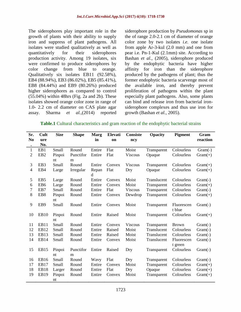
Table 1. Out of 19 isolates, ten were gram
positive and nine were gram negative rods.
All the isolates were colourless except EB9
pigmented as fluorescent green; EB14 as
fluorescent blue and EB 11 was brown
pigmented. Further, all colonies were found to
be round shaped with entire margin except
EB2 and EB4 were punctiform and irregular
in shape, respectively; while, EB4 showed
repand margin. All the isolated bacterial
endophytes were transparent except EB2 and
EB4, they were opaque. The results are
further in confirmation with the findings of
Arunachalam and Gayathri (2010) who also
reported the variation for cultural
characterization of endophytic bacteria
isolated from Andrographis paniculata.
Plant growth promoting traits of the
endophytic bacterial isolates
Endophytes are known to provide the plant
nutrients, plant health benefits along with
protection against pest and disease. Therefore
use of endophytes as an alternative eco-
friendly method for the sustainable
environment. Thus, an attempt has been made
to study the endophytic bacteria from the leaf
of G. hirsutum for their ability to produce
plant growth promoting traits viz.,
siderophores, indole acetic acid, ammonia and
ability to solubilize phosphate. Besides these,
preliminary study of enzyme production viz.,
chitinase, protease, gelatinase and lipase were
carried out because of their role in plant
protection. Table 2 summarizes the presence
of plant growth promoting traits and enzyme
activities of all 19 endophytic bacterial
isolates.
Int.J.Curr.Microbiol.App.Sci (2017) 6(10): 1718-1730
1723
The siderophores play important role in the
growth of plants with their ability to supply
iron and suppress of plant pathogens. All
isolates were studied qualitatively as well as
quantitatively for their siderophores
production activity. Among 19 isolates, six
were confirmed to produce siderophores by
color change from blue to orange.
Qualitatively six isolates EB11 (92.58%),
EB4 (88.94%), EB3 (86.02%), EB5 (85.41%),
EB8 (84.44%) and EB9 (80.26%) produced
higher siderophores as compared to control
(55.04%) within 48hrs (Fig. 2a and 2b). These
isolates showed orange color zone in range of
1.0- 2.2 cm of diameter on CAS plate agar
assay. Sharma et al.,(2014) reported
siderophore production by Pseudomonas sp in
the of range 2.0-2.1 cm of diameter of orange
color zone by two isolates i.e. one isolate
from apple Ar-3-kul (2.0 mm) and one from
pear i.e. Pn-1-Kul (2.1mm) site. According to
Bashan et al., (2005), siderophore produced
by the endophytic bacteria have higher
affinity for iron than the siderophore
produced by the pathogens of plant; thus the
former endophytic bacteria scarvenge most of
the available iron, and thereby prevent
proliferation of pathogens within the plant
especially plant pathogens. Also, some plants
can bind and release iron from bacterial iron-
siderophore complexes and thus use iron for
growth (Bashan et al., 2005).
Table.1 Cultural characteristics and gram reaction of the endophytic bacterial strains
Sr.
No
.
Cult
ure
No.
Size Shape Marg
in
Elevati
on
Consiste
ncy
Opacity Pigment Gram
reaction
1 EB1 Small Round Entire Flat Moist Transparent Colourless Gram(-)
2 EB2 Pinpoi
nt
Punctifor
m
Entire Flat Viscous Opaque Colourless Gram(+)
3 EB3 Small Round Entire Convex Viscous Transparent Colourless Gram(+)
4 EB4 Large Irregular Repan
d
Flat Dry Opaque Colourless Gram(+)
5 EB5 Large Round Entire Convex Moist Translucent Colourless Gram(-)
6 EB6 Large Round Entire Convex Moist Transparent Colourless Gram(+)
7 EB7 Small Round Entire Flat Viscous Transparent Colourless Gram(-)
8 EB8 Pinpoi
nt
Round Entire Convex Dewdrop Transparent Colourless Gram(+)
9 EB9 Small Round Entire Convex Moist Transparent Fluorescen
t blue
Gram(-)
10 EB10 Pinpoi
nt
Round Entire Raised Moist Transparent Colourless Gram(+)
11 EB11 Small Round Entire Convex Viscous Transparent Brown Gram(-)
12 EB12 Small Round Entire Raised Moist Translucent Colourless Gram(-)
13 EB13 Small Round Entire Raised Moist Translucent Colourless Gram(-)
14 EB14 Small Round Entire Convex Moist Translucent Fluorescen
t green
Gram(-)
15 EB15 Pinpoi
nt
Punctifor
m
Entire Raised Dry Transparent Colourless Gram(-)
16 EB16 Small Round Wavy Flat Dry Transparent Colourless Gram(-)
17 EB17 Small Round Entire Convex Moist Transparent Colourless Gram(+)
18 EB18 Larger Round Entire Flat Dry Opaque Colourless Gram(+)
19 EB19 Pinpoi
nt
Round Entire Convex Moist Transparent Colourless Gram(+)

Int.J.Curr.Microbiol.App.Sci (2017) 6(10): 1718-1730
1724
Table.2 Data on plant growth promoting traits and the enzyme production of all isolated
endophytic bacteria from leaf of G. hirsutam
Isolates Siderophore
production
Phosphate
Solubilization
IAA
Production
Ammonia
Production
Protease
Production
Lipase
Production
Gelatinase
Production
Chitinase
Production
EB1 - - + + + + + -
EB2 - - + + - - - -
EB3 + - + + - + - +
EB4 + + + + + + - -
EB5 + - + + - + + +
EB6 - - + + + + - -
EB7 - - + + - + + -
EB8 - - + + - - - -
EB9 + + + + + + + -
EB10 + - + + - + - -
EB11 + + + + + + + -
EB12 - - + + + + + -
EB13 - + + + + + - -
EB14 - + + + + + + -
EB15 - - + + + + + -
EB16 - - + + + + + -
EB17 - + + + - + - -
EB18 - - + + + + + -
EB19 - - + + + + - -
Total 06 06 19 19 12 17 10 02
(a)
(b)
(c)
Fig.1 Growth of endophytic bacteria from leaf sample ofG. hirsutum on (a) nutrient agar, (b)
Soilextract agar and (c) R2A agar medium

Int.J.Curr.Microbiol.App.Sci (2017) 6(10): 1718-1730
1725
Siderophore production
0
20
40
60
80
100
120E
B1
EB
2
EB
3
EB
4
EB
5
EB
6
EB
7
EB
8
EB
9
EB
10
EB
11
EB
12
EB
13
EB
14
EB
15
EB
16
EB
17
EB
18
EB
19
% S
idero
ph
ore p
ro
du
cti
on
0
0.5
1
1.5
2
2.5
Zo
ne I
nd
ex
(m
m)
Endophytic bacteria
2(a)
2(b)
Fig.2(a) Graphical data represents the
qualitative and quantitative
production of siderophores by
endophytic bacteria
Fig.2(b) Confirmation of siderophores production by
color change from blue to orange quantitatively
andqualitatively by CAS assay
Phosphate solubilizaiton
0
2
4
6
8
10
12
14
16
18
EB
1
EB
2
EB
3
EB
4
EB
5
EB
6
EB
7
EB
8
EB
9
EB
10
EB
11
EB
12
EB
13
EB
14
EB
15
EB
16
EB
17
EB
18
EB
19
Ph
osp
ha
te s
olu
bil
iza
tio
n (
mg
/ml)
0
0.5
1
1.5
2
2.5
3
3.5
4
4.5
Zo
ne
Ind
ex (
mm
)
Endophytic bacteria 3(a)
3(b)
Fig.3(a) Graphical data represents the
qualitative and quantitative
production of phosphate
solubilization by endophytic bacteria
Fig.3(b) Solubilization of phosphate on Pikovkya’s
agar plate by EB9
Production of IAA(µg/ml)
0
5
10
15
20
25
30
35
EB
1
EB
2
EB
3
EB
4
EB
5
EB
6
EB
7
EB
8
EB
9
EB
10
EB
11
EB
12
EB
13
EB
14
EB
15
EB
16
EB
17
EB
18
EB
19
IAA
(µg
/ml)
pro
du
cti
on
Endophytic bacteria
4(a)
4(b)
Fig.4(a) Quantitative estimation of
IAA(µg/ml)
Fig.4(b) Detection of IAA production by
development of pink color
Int.J.Curr.Microbiol.App.Sci (2017) 6(10): 1718-1730
1726
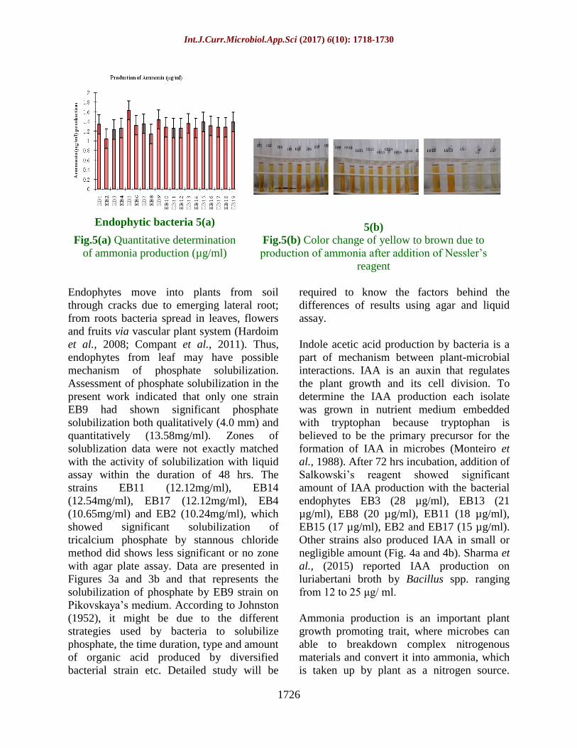
Endophytic bacteria 5(a)
5(b) Fig.5(a) Quantitative determination
of ammonia production (µg/ml)
Fig.5(b) Color change of yellow to brown due to
production of ammonia after addition of Nessler’s
reagent
Endophytes move into plants from soil
through cracks due to emerging lateral root;
from roots bacteria spread in leaves, flowers
and fruits via vascular plant system (Hardoim
et al., 2008; Compant et al., 2011). Thus,
endophytes from leaf may have possible
mechanism of phosphate solubilization.
Assessment of phosphate solubilization in the
present work indicated that only one strain
EB9 had shown significant phosphate
solubilization both qualitatively (4.0 mm) and
quantitatively (13.58mg/ml). Zones of
solublization data were not exactly matched
with the activity of solubilization with liquid
assay within the duration of 48 hrs. The
strains EB11 (12.12mg/ml), EB14
(12.54mg/ml), EB17 (12.12mg/ml), EB4
(10.65mg/ml) and EB2 (10.24mg/ml), which
showed significant solubilization of
tricalcium phosphate by stannous chloride
method did shows less significant or no zone
with agar plate assay. Data are presented in
Figures 3a and 3b and that represents the
solubilization of phosphate by EB9 strain on
Pikovskaya’s medium. According to Johnston
(1952), it might be due to the different
strategies used by bacteria to solubilize
phosphate, the time duration, type and amount
of organic acid produced by diversified
bacterial strain etc. Detailed study will be
required to know the factors behind the
differences of results using agar and liquid
assay.
Indole acetic acid production by bacteria is a
part of mechanism between plant-microbial
interactions. IAA is an auxin that regulates
the plant growth and its cell division. To
determine the IAA production each isolate
was grown in nutrient medium embedded
with tryptophan because tryptophan is
believed to be the primary precursor for the
formation of IAA in microbes (Monteiro et
al., 1988). After 72 hrs incubation, addition of
Salkowski’s reagent showed significant
amount of IAA production with the bacterial
endophytes EB3 (28 µg/ml), EB13 (21
µg/ml), EB8 (20 µg/ml), EB11 (18 µg/ml),
EB15 (17 µg/ml), EB2 and EB17 (15 µg/ml).
Other strains also produced IAA in small or
negligible amount (Fig. 4a and 4b). Sharma et
al., (2015) reported IAA production on
luriabertani broth by Bacillus spp. ranging
from 12 to 25 μg/ ml.
Ammonia production is an important plant
growth promoting trait, where microbes can
able to breakdown complex nitrogenous
materials and convert it into ammonia, which
is taken up by plant as a nitrogen source.

Int.J.Curr.Microbiol.App.Sci (2017) 6(10): 1718-1730
1727
Also, formation of ammonia leads to alkaline
condition, which suppresses the growth of
certain pathogenic fungi (Jha et al., 2012). In
our study, peptone was used as a nitrogenous
compound which breakdown leads to
formation of ammonia; it was detected by
color change using Nessler’s reagent. In the
present study, it was observed that all
endophytic isolates showed ammonia
production in the range of 1.6-0.8 (µg/ml)
within 48 hrs (Fig. 5a and 5b). Significant
differences were not recorded for ammonia
production among all 19 isolates.
Preliminary assessment of enzymes
production of endophytic bacteria
On leaves of plant, limited sites are available
where the pathogen can attack the plant.
Bacteria capable of multiplying within the
leaf can compete with pathogens by
producing enzymes or such chemical
compounds as a metabolic product. Diverse
group of microbes as an endophytes lives
within the plant tissues. From that, such
microbes are able to synthesize and secret
hydrolytic enzymes. Many microbes produce
and excrete lytic enzymes such as chitinase,
protease, gelatinase and lipase, which can
hydrolysepolymeric compounds viz., chitin,
proteins and lipids. Secretion of these
enzymes by different microbes results in the
suppression of plant pathogenic activity
directly (Pal et al., 2006). For example, fungal
cell wall made up of chitin, which might be
breakdown by endophytic microbial chitinase
(Bashan et al., 2005). Plate agar assay was
used for the preliminary study of enzyme
production by the isolates. Among 19 isolates,
two bacterial strains EB5 (1.9mm) and EB3
(1.1mm) showed zone of solubilization with
chitin agar plate after 7 days. Protease
production was shown by six isolates, EB15
(16.60mm), EB12 (14.00mm), EB11
(13.60mm), EB1 (12.60mm) and EB4
(11.80mm). Gelatinase activity was recorded
in five strains EB1 (17.00mm), EB9
(12.50mm), EB11 (15.50mm), EB12 (10.00)
and EB16 (15.70mm) on gelatin agar plate.
Significant lipase activity on tributyrene agar
plate was recorded with EB10 (15.00mm),
EB13 (13.70mm), EB17 (12.80mm) EB14
(12.20mm), EB12 (11mm) and EB11
(10mm). Rajendran (2006) reported that
application of endophytes such as Bacillus
species and Pseudomonas species to cotton
plant induced the expression of chitinase and
other enzymes which reduced the disease
severity caused by Xanthomonas axonopodis
pv. malvacearum. Similarly, Khianngam et
al., (2013) isolated and screened twenty
endophytic bacteria from mangrove plants in
Thailand for the presence of hydrolytic
enzymes viz., proteases, lipases, amylases or
cellulases.
From our study, it can be concluded that all
the 19 endophytic bacterial strains studied are
potential strains for the one or other plant
growth promoting traits and hydrolytic
enzymes involved in protection against pest
and disease. These potential isolates could be
used to make consortium as biofertilizer or
bioprotectant to enhance plant productivity
and protection. Among 19 isolates, EB9 and
EB11 showed all activities except chitinase
production, thus these two are the candidates
endophytic bacterial strains to be studied at
molecular level along with their effects on
plant growth under pot and field conditions
that would help us to understand the plant
microbe interaction in detail.
References
Arunachalam, C., and Gayathri, P.
2010.Studies on bioprospecting of
endophytic bacteria from the medicinal
plant of Andrographis paniculata for
their antimicrobial activity and
antibiotic susceptibility pattern. Int. J.
Cur. Pharm. Res. 2(4): 63-68.

Int.J.Curr.Microbiol.App.Sci (2017) 6(10): 1718-1730
1728
Bakker, A. W., and Schippers, B.
1987.Microbial cyanide production in
the rhizosphere in relation to potato
yield reduction and Pseudomonas spp.
mediated plant growth-stimulation. Soil
Biol. Biochem. 19(4):451-457.
Bashan, Y., and De Bashan, L. E. 2005. Plant
growth promotingIn encyclopedia of
soils in the environment edited by: D.
Hillel. American Society of
Microbiology: Washington DC. pp. 607-
654.
Bhattacharyya, P. N., and Jha, D. K. 2012.
Plant growth-promoting rhizobacteria
(PGPR): emergence in
agriculture. World J. Microbiol.
Biotechol. 28(4): 1327-1350.
Cappuccino, J. C., and Sherman, N. 1992. In:
Microbiology: A Laboratory Manual.
Edited by:: Benjamin/cummings.
Pub.Co. New York. pp. 125-179.
Chen, C., Bauske, E. M., Musson, G.,
Rodriguezkabana, R. and Kloepper, J.
W. 1995.Biological control of Fusarium
wilt on cotton by use of endophytic
bacteria. Biol. Control. 5(1): 83-91.
Compant, S., Clément, C. and Sessitsch, A.
2010.Plant growth-promoting bacteria
in the rhizo-and endosphere of plants:
their role, colonization, mechanisms
involved and prospects for
utilization. Soil Biol. Biochem. 42(5):
669-678.
Compant, S., Mitter, B., Colli-Mull, J. G.,
Gangl, H. and Sessitsch, A.
2011.Endophytes of grapevine flowers,
berries, and seeds: identification of
cultivable bacteria, comparison with
other plant parts, and visualization of
niches of colonization. Microb.
Ecol. 62(1): 188-197.
Defago, G., and Haas, D. 1990.
Pseudomonads as antagonists of
soilborne plant pathogens: modes of
action and genetic analysis. In Soil
biochemistry Edited by: J.M. Bollag
and G. Stotzky. Marcel Dekker, Inc.,
New York. pp. 249–291.
Egbuta, M.A., McIntosh, S., Waters, D.L.,
Vancov, T. and Liu, L. 2017.Biological
importance of cotton by-products
relative to chemical constituents of the
cotton plant. Molecules. 22(1): 93.
Hallmann, J., Quadt-Hallmann, A.,
Rodrıguez-Kábana, R. and Kloepper,
J.W. 1998.Interactions between
Meloidogyne incognita and endophytic
bacteria in cotton and cucumber. Soil
Biol. Biochem. 30(7): 925-937.
Hardoim, P. R., van Overbeek, L. S. and van
Elsas, J. D. 2008. Properties of bacterial
endophytes and their proposed role in
plant growth. Trends Microbiol. 16(10):
463-471.
Jha, C. K., Patel, B. and Saraf, M.
2012.Stimulation of the growth of
Jatropha curcas by the plant growth
promoting bacterium Enterobacter
cancerogenus MSA2. World J.
Microbiol. Biotechnol. 28(3): 891-899.
Johnston, H.W., 1952. The solubilization of
phosphate: The action of various
organic compounds on dicalcium and
tricalcium phosphates. N. Z. J. Sci.
Technol. 436-446.
Kachhap, S., Chaudhary, A. and Singh, S. D.
2015. Response of plant growth
promoting rhizobacteria (pgpr) in
relation to elevated temperature
conditions in groundnut (Arachis
hypogaea L.). The Ecoscan. 9(3and4):
771-778.
Khianngam, S., Techakriengkrai, T.,
Raksasiri, B.V., Kanjanamaneesathian,
M. and Tanasupawat, S. 2013. Isolation
and screening of endophytic bacteria for
hydrolytic enzymes from plant in
mangrove forest at Pranbur i,
PrachuapKhiri Khan, Thailand. In
Endophytes for plant protection: the
state of the art Edited by: Schneider C,
Leifert C, Feldmann F. Proc 5th

Int.J.Curr.Microbiol.App.Sci (2017) 6(10): 1718-1730
1729
IntSymp Plant Protect Plant Health
Europe. Deutsche Phytomedizinische
Gesellschaft, Berlin.pp 279–284.
King, E. J., 1932.The colorimetric
determination of phosphorus. Biochem.
J. 26(2): 292.
Kirti, S., Dipta, B., Bhardwaj, S., Pawar, R.
and Kaushal, R. 2016. Screening and
characterization of plant growth
promoting rhizobacteria associated with
cherry (Prunusavium
L.). Screening. 11(4): 2111-2115.
Kuddus, M., and Ahmad, I.Z. 2013.Isolation
of novel chitinolytic bacteria and
production optimization of extracellular
chitinase. J. Genetic Eng. Biotechnol.
11(1): 39-46.
Lindow, S. E., and Brandl, M. T.
2003.Microbiology of the
phyllosphere. Appl. Environ.
Microbiol. 69(4): 1875-1883.
Loper, J.E., and Schroth, M.N., 1986.
Influence of bacterial sources of indole-
3-acetic acid on root elongation of sugar
beet. Phytopathology. 76(4): 386-389.
Louden, B.C., Haarmann, D. and Lynne, A.
M. 2011.Use of blue agar CAS assay
for siderophore detection. J. Microbiol.
Biol. Edu. 12(1): 51.
MacFaddin, J. F., 2000. Biochemical tests for
identification of medical bacteria.
Williams and Wilkins, London.
Malfanova, N., Kamilova, F., Validov, S.,
Chebotar, V. and Lugtenberg, B.
2013.Is L-arabinose important for the
endophytic lifestyle of Pseudomonas.
Arch. Microbiol. 195: 9-17.
McGee, P.A., 2002. Reduced growth and
deterrence from feeding of the insect
pest Helicoverpa armigera associated
with fungal endophytes from
cotton. Anim. Prod. Sci.42 (7): 995-999.
McInroy, J. A., and Kloepper, J. W. 1995a.
Population dynamics of endophytic
bacteria in field-grown sweet corn and
cotton. Can. J. Microbiol.41: 895-901.
McInroy, J.A., and Kloepper, J. W.
1995b.Survey of indigenous bacterial
endophytes from cotton and sweet corn.
Plant Soil, 173: 337-342.
Misaghi, I. J., and Donndelinger, C.R.
1990.Endophytic bacteria in symptom-
free cotton plants. Phytopathology.
80(9): 808-811.
Monteiro, A.M., Crozier, A. and Sandberg, G.
1988.Endogenous hormones,
germination and early seedling growth
of Dalbergia dolicbopetala. J. Pl.
Physiol. 132(6): 762-765.
Musson, G., McInroy, J. A. and Kloepper, J.
W. 1995.Development of delivery
systems for introducing endophytic
bacteria into cotton. Biocontrol Sci.
Technol. 5(4): 407-416.
Neilands, J. B., 1981. Iron absorption and
transport in microorganisms. Annu. Rev.
nutr. 1(1): 27-46.
Olajuyigbe, F.M. and Ajele, J.O. 2005.
Production dynamics of extracellular
protease from Bacillus species. Afr. J.
Biotechnol. 4(8): 776.
Pal, K. K., and Gardener, B. M.
2006.Biological control of plant
pathogens. The Plant Health
Instructor. 2: 1117-1142.
Pikovskaya, R. I., 1948. Mobilization of
phosphorus in soil in connection with
vital activity of some microbial
species. Mikrobiologiya. 17(362): 370.
Rajendran, L., Saravanakumar, D.,
Raguchander, T.G. and Samiyappan, R.
2006. Endophytic bacterial induction of
defence enzymes against bacterial blight
of cotton. Phytopathol.Mediterr.45 (3):
203-214.
Rajendran, S., and Sriranjini, V. 2008.Plant
products as fumigants for stored-
product insect control. J. Stored Prod.
Res. 44(2): 126-135.
Sanders, E.R., 2012. Aseptic laboratory
techniques: plating methods. J. Vis.
Exp. (63): e3064.

Int.J.Curr.Microbiol.App.Sci (2017) 6(10): 1718-1730
1730
Schwyn, B., and Neilands, J. B.
1987.Universal chemical assay for the
detection and determination of
siderophores. Analytical Biochem.
160:47–56.
Sharma, R., Walia, A., Chauhan, A. and
Shirkot, C. K. 2015.Multitrait plant
growth promoting bacteria from tomato
rhizosphere and evaluation of their
potential as bioinoculants. App. Biol.
Res.17: 1-12.
Sharma, S., Kaur, M. and Prashad, D.
2014.Isolation of fluorescent
Pseudomonas strain from temperate
zone of himachalpradesh and their
evaluation as plant growth promoting
rhizobacteria (PGPR). The Bioscan.
9(1): 323-328.
Sirisha, E., Rajasekar, N. and Narasu, M. L.
2010. Isolation and optimization of
lipase producing bacteria from oil
contaminated soils. Adv. Biol. Res. 4(5):
249-252.
Smith Jr, H.L., and Goodner, K.
1958.Detection of bacterial gelatinases
by gelatin-agar plate methods. J.
Bacteriol. 76(6): 662.
How to cite this article:
Azba A. Shaikh, P.R. Parmar, B.K. Rajkumar, D.H. Patel, H.R. Desai and Solanki B.G. 2017.
Bioprospecting Potential of Endophytic Bacteria from Leaves of Gossypium hirsutum.
Int.J.Curr.Microbiol.App.Sci. 6(10): 1718-1730. doi: https://doi.org/10.20546/ijcmas.2017.610.208