biophysical methods in early drug discovery
TRANSCRIPT

doi: 10.5599/admet.733 222
ADMET & DMPK 7(4) (2019) 222-241; doi: http://dx.doi.org/10.5599/admet.733
Open Access : ISSN : 1848-7718
http://www.pub.iapchem.org/ojs/index.php/admet/index Review
Biophysical methods in early drug discovery
Geoffrey Holdgate*1, Kevin Embrey2, Alexander Milbradt2 and Gareth Davies1 1Hit Discovery, Discovery Sciences, R&D, AstraZeneca, Alderley Park, UK
2Structure, Biophysics and FBLG, Discovery Sciences, R&D, AstraZeneca, Cambridge, UK
*Corresponding Author: E-mail:[email protected]; Tel.: +44 (0)1625 234218
Received: September 30, 2019; Revised: November 19, 2019; Published: December 11, 2019
Abstract
Biophysical methods such as mass spectrometry, surface plasmon resonance, nuclear magnetic resonance, and both differential scanning isothermal titration calorimetry are now well established as key components of the early drug discovery process. These approaches are used successfully for a range of activities, including assay development, primary screening, hit confirmation and detailed mechanistic characterisation of compound binding. Matching the speed, sensitivity and information content of the various techniques to the generation of critical data and information required at each phase of the drug discovery process has been key. This review describes the framework by which these methods have been applied in the drug discovery process and provides case studies to exemplify the impact.
Keywords
Affinity selection mass spectrometry; surface plasmon resonance; nuclear magnetic resonance;
differential scanning fluorimetry; isothermal titration calorimetry; microscale thermophoresis
Introduction
Biophysical methods, which can encompass a wide range of techniques focussed on measuring the
structure, properties, dynamics or function of biomolecules, have been increasingly employed in the drug
discovery process since their first introduction in the early 1990s. It was during this time that commercial
instruments such as the first isothermal titration calorimeter (ITC) [1] and the first surface plasmon
resonance (SPR) [2] instruments became available and their use exemplified using biochemical systems.
Alongside this, existing biophysical approaches were used in novel ways to identify and characterise
protein-ligand interactions, for example the first report of the use of affinity selection, coupled to detection
by mass spectrometry, for the identification of molecules binding to a macromolecule [3], and the use of
nuclear magnetic resonance (NMR) to identify fragments that could subsequently be optimised and linked
to form more potent compounds [4]. The development of these biophysical methods coincided with the
advent of high-throughput screening (HTS), leading from natural product screening of a few hundred
compounds each week in the late 1980s, through to HTS hits being responsible for starting matter for
almost half of drug companies’ portfolios in the mid-1990s [5,6]. This allowed the valuable combination of
biophysics with HTS to contribute to the establishment of high quality, high-throughput assays through the
characterisation of protein and tool ligands, as well as the evaluation of HTS output through orthogonal
application of biophysical methods to screen for true target engagement [7]. More recently, biophysical

ADMET & DMPK 7(4) (2019) 222-241 Biophysical methods in early drug discovery
doi: 10.5599/admet.733 223
methods have matured, throughput has increased, and sensitivity improved, such that some of these
methods can also now be employed in primary screening, not only for fragments, which typically screen
lower numbers of low molecular weight compounds [8], but for screening libraries comprising many
thousands of compounds [9]. The high-fidelity nature of many biophysical methods, coupled to the specific
information content they can access, has also meant that they are used increasingly to characterise the
mode of action of hits and leads. Some methods provide access to kinetic data [10], whilst others enable
thermodynamic characterisation of ligand binding [11]. Others may allow structural insights into binding
mode and binding site [12,13]. Our understanding of the strengths and weaknesses of the methods, as well
as a better appreciation of the impact that results from appropriate positioning within the drug discovery
process has contributed significantly to the increased use of biophysical methods observed today. This
review will provide an overview of the benefit that can be gained from the incorporation of biophysical
methods within areas of the drug discovery process and will provide case studies to exemplify their impact.
Assay development
The capability to design, build and implement assays that are specific, robust and sensitive enough to
identify and characterise potential new drug molecules is fundamental to drug discovery. In developing a
new assay there are several factors that must be considered. These include: the nature of the reagents,
such as their identity, purity, concentration, functionality and stability; the features of the detection
system, such as the sensitivity, dynamic range, potential for interference and reproducibility; the analysis of
the data; and the subsequent statistical examination of assay performance. The application of biophysical
methods early during assay development can help to understand some of these features and to ensure that
high-fidelity, fit for purpose assays are developed. Of course, this extra resource needs to be considered
during project planning, but the benefits of applying this early outweighs the resource that would need to
be applied to rescue projects which have been misled or have failed due to spurious activity from poorly
characterised reagents or assays.
Reagent quality control
Early application of biophysical methods often focusses on understanding the quality of the reagents
available, both in terms of the suitability of the target protein and of the behaviour of the known tool
compounds. For a protein to be deemed suitable to be used in subsequent drug discovery assays it should
fit criteria associated with the group of characteristics highlighted in the paragraph above. The protein
should be the right protein, so its identity should be confirmed, otherwise invalid and misleading results
may be obtained. The purity of the protein is also important as impurities may have similar activities or bind
test compounds. The concentration of the protein should be measured so that considerations around
concentration dependent effects can be understood. The functionality of the protein should also be
investigated. This may require measurements of ligand binding, the catalytic activity (in the case of
enzymes) or the ability to carry out other functions or interactions. Finally, the stability of the protein
should be assessed, both in terms of its thermodynamic and kinetic stability, as well as its ability to
withstand certain conditions such as freeze-thawing. It also may be necessary to characterise the binding of
other molecules such as substrates, cofactors, binding partners or competing probe molecules, to
understand their requirement and concentration range desired and permitted within the assay
methodologies under consideration. These attributes of the target protein may be investigated using a
variety of approaches and biophysical methods can help to provide confidence in the target protein, by
being employed alongside or instead of standard biochemical methods for protein characterisation. Table 1
shows several possible methods that may be employed in such characterisation and it highlights the
Holdgate et al. ADMET & DMPK 7(4) (2019) 222-241
224
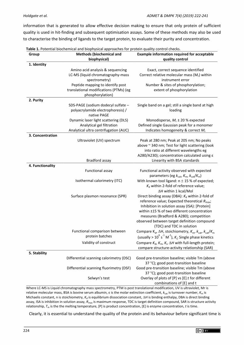
information that is generated to allow effective decision making to ensure that only protein of sufficient
quality is used in hit-finding and subsequent optimisation assays. Some of these methods may also be used
to characterise the binding of ligands to the target protein, to evaluate their purity and concentration.
Table 1. Potential biochemical and biophysical approaches for protein quality control checks.
Group Methods (biochemical and biophysical)
Example information required for acceptable quality control
1. Identity Amino acid analysis & sequencing Exact, correct sequence identified LC-MS (liquid chromatography-mass
spectrometry) Correct relative molecular mass (Mr) within
instrument error Peptide mapping to identify post
translational modifications (PTMs) (eg phosphorylation)
Number & sites of phosphorylation; extent of phosphorylation
2. Purity SDS-PAGE (sodium dodecyl sulfate –
polyacrylamide electrophoresis) / native PAGE
Single band on a gel; still a single band at high loading
Dynamic laser light scattering (DLS) Monodisperse, Mr ± 20 % expected Analytical gel filtration Defined single Gaussian peak for a monomer Analytical ultra centrifugation (AUC) Indicates homogeneity & correct Mr
3. Concentration Ultraviolet (UV) spectrum Peak at 280 nm; Peak at 205 nm; No peaks
above ~ 340 nm; Test for light scattering (look into ratio at different wavelengths eg
A280/A230); concentration calculated using ε Bradford assay Linearity with BSA standards
4. Functionality Functional assay Functional activity observed with expected
parameters (eg kcat, Km, kcat/Km) Isothermal calorimetry (ITC) With known tool ligand: n 15 % of expected;
Kd within 2-fold of reference value;
H within 1 kcal/Mol Surface plasmon resonance (SPR) Direct binding assay (DBA): Kd within 2-fold of
reference value; Expected theoretical Rmax; Inhibition in solution assay (ISA): [Protein]
within ±15 % of two different concentration measures (Bradford & A280); competition
observed between target definition compound (TDC) and TDC in solution
Functional comparison between protein batches
Compare Kd, H, stoichiometry, Km, kcat, kcat/Km
(usually > 106 s
-1 M
-1), Ki; Single phase kinetics
Validity of construct Compare Kd, Km, Ki, H with full-length protein; compare structure-activity relationship (SAR)
5. Stability
Differential scanning calorimetry (DSC) Good pre-transition baseline; visible Tm (above 37 °C); good post-transition baseline
Differential scanning fluorimetry (DSF) Good pre-transition baseline; visible Tm (above 37 °C); good post-transition baseline
Selwyn’s test Overlay of plots of [P] vs [E].t for different combinations of [E] and t
Where LC-MS is Liquid chromatography mass spectrometry, PTM is post translational modification, UV is ultraviolet, Mr is relative molecular mass, BSA is bovine serum albumin, ε is the molar extinction coefficient, kcat is turnover number, Km is
Michaelis constant, n is stoichiometry, Kd is equilibrium dissociation constant, H is binding enthalpy, DBA is direct binding assay, ISA is inhibition in solution assay, Rmax is maximum response, TDC is target definition compound, SAR is structure activity relationship, Tm is the the melting temperature, [P] is product concentration, [E] is enzyme concentration, t is time.
Clearly, it is essential to understand the quality of the protein and its behaviour before significant time is
ADMET & DMPK 7(4) (2019) 222-241 Biophysical methods in early drug discovery
doi: 10.5599/admet.733 225
spent developing assays and certainly before bulk amounts of protein are made for activities such as high-
throughput screening and X-ray crystallography. In this respect it is useful to remember that the target
protein goes into every well of the experiment, and so issues with the performance of this reagent has the
potential to compromise the entire experiment. This can be contrasted with efforts to ensure individual
compound purity and activity. Although individual compound integrity is important to ensure that the
identification of useful chemical start points from any active hit compound is not missed, it does not have
the same propensity to compromise the whole hit identification process, as would be the case for a protein
of questionable integrity.
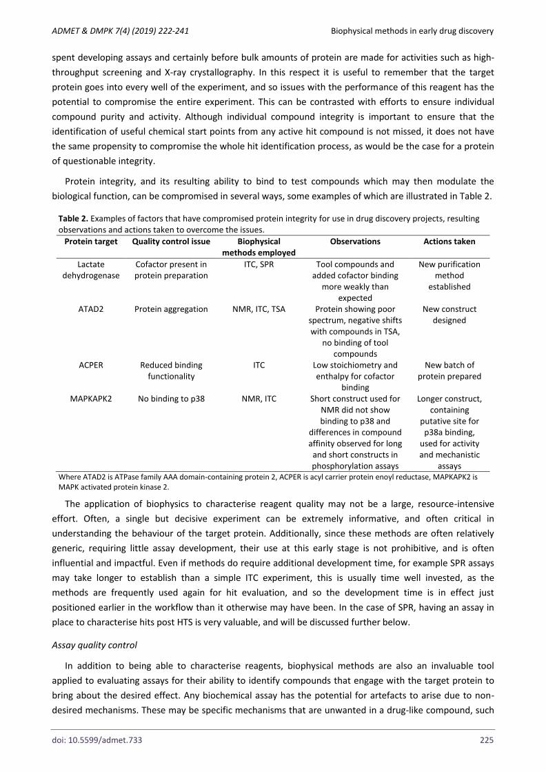
Protein integrity, and its resulting ability to bind to test compounds which may then modulate the
biological function, can be compromised in several ways, some examples of which are illustrated in Table 2.
Table 2. Examples of factors that have compromised protein integrity for use in drug discovery projects, resulting observations and actions taken to overcome the issues.
Protein target Quality control issue Biophysical methods employed
Observations Actions taken
Lactate dehydrogenase
Cofactor present in protein preparation
ITC, SPR Tool compounds and added cofactor binding
more weakly than expected
New purification method
established
ATAD2 Protein aggregation NMR, ITC, TSA Protein showing poor spectrum, negative shifts with compounds in TSA,
no binding of tool compounds
New construct designed
ACPER Reduced binding functionality
ITC Low stoichiometry and enthalpy for cofactor
binding
New batch of protein prepared
MAPKAPK2 No binding to p38 NMR, ITC Short construct used for NMR did not show binding to p38 and
differences in compound affinity observed for long
and short constructs in phosphorylation assays
Longer construct, containing
putative site for p38a binding,
used for activity and mechanistic
assays Where ATAD2 is ATPase family AAA domain-containing protein 2, ACPER is acyl carrier protein enoyl reductase, MAPKAPK2 is MAPK activated protein kinase 2.
The application of biophysics to characterise reagent quality may not be a large, resource-intensive
effort. Often, a single but decisive experiment can be extremely informative, and often critical in
understanding the behaviour of the target protein. Additionally, since these methods are often relatively
generic, requiring little assay development, their use at this early stage is not prohibitive, and is often
influential and impactful. Even if methods do require additional development time, for example SPR assays
may take longer to establish than a simple ITC experiment, this is usually time well invested, as the
methods are frequently used again for hit evaluation, and so the development time is in effect just
positioned earlier in the workflow than it otherwise may have been. In the case of SPR, having an assay in
place to characterise hits post HTS is very valuable, and will be discussed further below.
Assay quality control
In addition to being able to characterise reagents, biophysical methods are also an invaluable tool
applied to evaluating assays for their ability to identify compounds that engage with the target protein to
bring about the desired effect. Any biochemical assay has the potential for artefacts to arise due to non-
desired mechanisms. These may be specific mechanisms that are unwanted in a drug-like compound, such
Holdgate et al. ADMET & DMPK 7(4) (2019) 222-241
226
as reactivity (for example thiol reactivity), redox cycling, colloidal aggregation, heavy metal contamination,
protein unfolding, protein denaturation (so called pan assay interference compounds or PAINs [14]) or
biological system or technology interferences, such as coupled enzyme inhibitors, fluorescent compounds
or quenchers. Understanding the liability of a biochemical assay to these types of compounds helps to
understand the potential output from high-throughput screens (HTS) utilising that assay approach. At
AstraZeneca, HTS development includes testing a small library of around 1000 compounds with unwanted
mechanism of inhibition (the uMOI set) and around 7000 compounds (the validation set), which are meant
to represent the diversity present in the full screening set of around 2 million compounds. This allows the
assay to be assessed in terms of its susceptibility to PAINs, as well as providing an assessment of
reproducibility, likely hit rate and to highlight potential artefacts and propensity for false positives and
negatives. Biophysical methods such as SPR or NMR are employed following this early screening activity to
characterise the hits, so that the knowledge arising from an understanding of the reasons behind false
positives may be used to further optimise the assay to avoid these types of hits in the full screen. Table 3
exemplifies several assays, across different target classes, where biophysical characterisation post
validation set testing, influenced the subsequent hit identification strategy or tactics.
This workflow therefore allows decisions to be made based on any specific issues that arise due to the
nature of the assay, that may be mitigated or avoided by modifying the screening cascade. For example,
knowing that heavy metals contaminants or redox cycling compounds may be hits in the biochemical assay,
but the compounds are not true binders to the target protein in a high-fidelity biophysical method allows
the primary screen to be modified to reduce the liability to such effects. In these examples, the use of metal
chelating agents and investigating different reducing agents may lead to changes to the assay protocol.
Alternatively, post screen triage may involve introducing additional assays that allow compounds
functioning by these undesirable mechanisms to be identified and deprioritised. Thus, the use of
biophysical methods in this way represents a valuable investment to increase the probability that high-
quality hits will be identified during the primary screen. As can be seen from Table 3, in some cases, the
impact of the biophysical testing was taking a decision not to run the primary screen at all, but to pursue
alternative approaches, such as fragment-based lead generation (FBLG). These decisions, although difficult
at the time, due to the previously committed investment, ultimately may result in substantial cost savings,
firstly from not committing to HTS (at an average cost of around $90k) and secondly from not following up
spurious hits. Figure 1 shows the interplay that is required between the assay methodology, the use of
high-quality reagents and the role that biophysics has in helping to characterise these aspects to facilitate
the implementation of valid screening assays.
Primary screening
There are few biophysical methods that can be applied to primary screening as usually defined in the
high-throughput setting, which often refers to the testing of 1 million or more compounds, and usually
these methods are more frequently applied for secondary hit evaluation. The predominate reasons are the
amount of protein required and the throughput required to achieve primary screening in a reasonable
timeframe. To address these issues compound mixtures are often used to facilitate reductions in reagents
and time, since many compounds may be screened from a single well. Often, mixtures have no adverse
effects on the protein, or on the ability to detect binding, but sometimes problems are experienced if there
are compound-compound interactions or if the compound organic load or the compound solvent
concentration [DMSO] is too high.

ADMET & DMPK 7(4) (2019) 222-241 Biophysical methods in early drug discovery
doi: 10.5599/admet.733 227
Table 3. Impact of biophysical evaluation of hits identified during HTS development. Protein target
Assay methodology
Number of compounds tested
Biophysical methods
employed
Observations Actions taken
KEAP1 HTRF 180 NMR, SPR No genuine hits identified HTS was stopped and FBLG approach
used instead MALT1 Fluorescence
intensity following proteolytic
cleavage
60 NMR 17% of hits showed specific binding. 38% showed redox cycling behaviour. 27% were
not soluble
Incorporation of a redox-artefact assay
in the cascade reduced the number
of redox-active compounds reaching the NMR assay from
38% to 5% ERRγ FRET 180 NMR FRET assay suggested that hit
rate would be low. NMR suggested that 90% were
false positives
HTS in this format was not run
TTBK1 ADP-glo 250 SPR 69 verified hits, then profiled versus phosphorylated and
non-phosphorylated protein Large number of reactive
compounds identified
ADP-glo assay was not run
ACPER Fluorescence intensity following substrate turnover
Tool compounds and 630 fragments
SPR, ITC Characterisation demonstrated that
compounds showing several different mechanisms of inhibition could be found
Project view on needing a cofactor
competitive inhibitor was changed and
assays configured to find all mechanisms
LTC4S HTRF, RapidFire Total of 50 selected from actives in one
or both assays
NMR, SPR 77% of the total hits shown to bind and also to displace
tool ligand. Confirmation rate was 90% for RapidFire hits,
40% for HTRF hits
RapidFire assay prioritised for full
HTS
aPC 4 different assays: 1) Chromogenic
cleavage 2) Peptide
cleavage coupled assay
3) Peptide cleavage RapidFire 4) Fibrin clot assay
Total of 250 identified from the assays as follows:
1) 90 2) 50 3) 10
4) 100
190 selected for biophysical testing (90 from assays 1-3
and total output from 4
SPR, NMR Numbers of confirmed hits originating from each assay
approach: 1) 8
2) 16 3) 9
4) 13
Fibrin clot assay was shown to identify compounds
binding at a site distal from the active site
Fibrin clot assay was selected for HTS,
based on the ability to identify novel, exo
site binders.
Where KEAP1 is Kelch Like ECH Associated Protein 1, MALT1 is Mucosa-associated lymphoid tissue lymphoma translocation protein 1, ERRγ is Estrogen-related receptor gamma, TTBK1 is Tau tubulin kinase 1, ACPER is acyl carrier protein enoyl reductase, LTC4S is Leukotriene C4 synthase and aPC is activated protein c.
Two biophysical methods that are often capable of providing throughputs approaching that of
traditional HTS are affinity selection mass spectrometry (AS-MS) [15] and thermal shift assays (TSA) [16].
These methods utilise mass detection of ligands bound to a target protein and the ligand-induced increase
in thermal stability respectively to identify hits. The benefit of these approaches in primary screening is that
the assay is generic and can rapidly be optimised for the target of interest. The disadvantage in biophysical
primary screening is that identified hits may bind to the target protein but may not have the desired
biological effect.
An alternative use of biophysical methods in primary screening is in testing a much smaller library of low
molecular weight compounds in so-called fragment-based screening [17]. This was first described

Holdgate et al. ADMET & DMPK 7(4) (2019) 222-241
228
employing NMR screening to identify fragments that could subsequently be linked and optimised to make
higher affinity compounds [4]. More recently, fragment screening has been described by other biophysical
methods including both SPR [18] and TSA [19].
Figure 1. Interplay between reagents, assay methodology and biophysical quality control in the development of valid assays.
High-throughput screening – affinity selection mass spectrometry (AS-MS)
The application of mass spectrometry (MS) in the drug discovery process has been well established for
many years. It has been applied both in the characterisation of target proteins, where it has been primarily
focussed on quality control as well as target identification and validation. For small molecules, the
technique has been used for metabolism and pharmacokinetics studies as compound identification. More
recently, MS approaches have been developed to detect and characterise protein-small molecule
complexes.
Affinity selection mass spectrometry (AS-MS) is now the predominant biophysical method used for
primary screening. It was first introduced in the late 1990s [20] and various formats have been described,
evaluated and implemented since then [21]. However, the basic premise of all these methods is the
detection of compounds that bind to the target protein using mass detection of the bound ligand. This
simple binding assay has the benefit of reduced interference, since there is no requirement for functional
activity which often entails more complex assays and therefore often increased probability of artefacts
arising due to effects on the read-out, rather than from true target engagement. Another advantage is that
this screening approach can be applied to orphan genomic targets and targets for which functional assays
cannot be developed. The major disadvantage, alongside the requirement, mentioned above, for assessing
functional activity post-screen, is that these methods are not commercially available and systems, expertise
and sometimes even the software solutions required for data analysis have to be developed in house.
Although there are subtle differences in the way AS-MS methods may be employed, the principle of the
different approaches is essentially the same. It involves incubating a library of small molecule compound
mixtures with the target protein, separating the bound small molecules from non-binders and detecting
those binders using mass spectrometry, Figure 2.
The relatively slow MS detection still limits the throughput of these methods and even with the
development of acoustic mist ionisation approaches [22], which have increased the speed of mass
spectrometry detection, large compound mixtures are still required with mixture sizes ranging from around

ADMET & DMPK 7(4) (2019) 222-241 Biophysical methods in early drug discovery
doi: 10.5599/admet.733 229
100 to almost 3000 compound per well. Consideration of the composition of these mixtures is important as
these mixtures have potential for introducing solubility issues, compound-compound reactivity and
instability. Smaller mixtures are clearly less disposed to these types of problems but may impose
restrictions on the size of the compound library that be screened. Whilst this method is not dependent
upon protein function, MS has a low tolerance to detergents present in biological buffers. This is primarily
due to the propensity for large aggregates to form which may interfere with binding and detection. Thus,
the application is limited to screening soluble proteins as the detergents required for membrane protein
preparations are not compatible.
Figure 2. Schematic representation of the workflow for AS-MS primary screening. Mixtures of test compound are incubated with the target protein before separation of bound ligands from free ligands by size plate-based exclusion chromatography. Bound ligands, which are eluted with the target protein, are subsequently detected by mass spectrometry.
Even with these limitations, AS-MS still represents an important addition to the high-throughput
screening toolbox. The sensitivity means that relatively low amounts of target protein are required, of
course there is no requirement for labelling of reagents and the process can be automated, all features that
contribute to the quality, speed and cost considerations required when deciding upon a primary screening
approach.
High-throughput screening – thermal shift assay (TSA)
The thermal shift assay (TSA) also known as Thermofluor or differential scanning fluorimetry has been
used for several years to study protein stability [23]. It is a rapid and simple method that allows the melting
temperature, Tm (temperature at which 50 % of the protein is unfolded), to be determined under different
conditions. It has extensively been used to optimise buffer conditions for X-ray crystallographic studies on
soluble proteins, such that those conditions yielding the highest stability may be used as start points for
crystallisation trials. The addition of compounds that bind to the target protein, thermodynamically
stabilise the protein relative to controls in the absence of ligand, and this stabilisation can be used to
identify binders from non-binders.
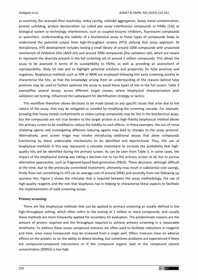
The TSA experiment involves incubating the target protein with test compounds in the presence of a dye
that binds to hydrophobic regions of the protein. The temperature is then increased uniformly, and the
fluorescence of dye monitored with temperature. As the protein unfolds more hydrophobic regions are

Holdgate et al. ADMET & DMPK 7(4) (2019) 222-241
230
exposed, there is increased dye binding and the fluorescence intensity increases. Compounds that increase
the thermal stability of the protein may be identified as those giving an increased Tm, resulting from a shift
in the unfolding curve to higher temperatures, Figure 3(a).
0 10 20 30 40 50 60 700
1000
2000
3000
4000
5000
Temperature °C
RF
U
0 20 40 60 80
-100
0
100
200
300
400
Temperature °C
R
FU
/T
Figure 3. (a) Typical unfolding curve in a TSA. Squares / solid line show the protein unfolding in the absence of ligand. Triangles / dotted line show unfolding in the presence of a compound that stabilises by 5 °C. The red
arrow indicates the shift in Tm caused by the addition of compound; (b) First derivative of the data in (a).
The advantages of TSA as a primary screening method include the simplicity of the approach, the cost
effectiveness and the potential to access a wide range of binding affinities. The method requires little assay
development, which is in effect limited to adjusting [protein] and [dye] to give a suitable signal. The reagent
requirements are protein and dye only, meaning that the costs associated with expensive biochemical
reagents are avoided. The method does not require specialised instrumentation and is carried out using
standard thermocyclers often used for real-time polymerase chain reactions (RT-PCR). Protein quantities
can potentially limit the method, since screening 500,000 single wells can require around 0.5 g of a 40 kDa
target protein. Whilst there is no direct correlation between Tm and Kd [24], due to differences in enthalpic
and entropic contributions to binding affinity having a differing effect on Tm, it is possible to observe
stabilisation conferred by ligands covering a wide range of affinities from mM to very tight-binding.
Analysis of TSA data typically involves determining the Tm for each curve, which can be achieved by
fitting an appropriate equation to the data, or for simple 2 state transitions, simply by taking the first
derivative of the fluorescence versus temperature data, Figure 3 (b). For high-throughput analysis Genedata
Screener® (Genedata, Basel, Switzerland) includes a module that can robustly and efficiently analyse the
many temperature curves created, which significantly reduces the analysis time.
Fragment-based screening
Fragment-based drug discovery (FBDD) is now a well-established approach with FBDD having delivered 2
marketed medicines (Vemurafenib and Venetoclax) and around 35 compounds currently in clinical trials
[25]. Identifying fragment hits is a challenge in traditional biochemical assays, since they are likely to bind
weakly to the target protein, and the apparent affinity may be weaker still, due to competition with
substrates. Biophysical methods are well suited to fragment-based screening [25], as they are sufficiently
sensitive to detect weak interactions, and reliable enough to avoid false hits. Historically, techniques
including NMR, SPR and X-ray crystallography were applied to fragment screening, but more recently
methods such as microscale thermophoresis (MST) [26] and TSA have also been used.
Compared to traditional HTS, the throughput of fragment screening is generally lower, and the
(a) (b)

ADMET & DMPK 7(4) (2019) 222-241 Biophysical methods in early drug discovery
doi: 10.5599/admet.733 231
biophysical methods employed often require larger quantities of protein. Fortunately, this issue is
overcome by the ability to screen fewer molecules, but to cover a much larger proportion of chemical space
using fragment screening libraries. For example, screening 1 million compounds out of the estimated 1030
compounds that could potentially be synthesised with 36 heavy atoms (around 500 Da) is 1019-fold less
efficient than screening 1000 compounds from the 108 potential compounds with 12 heavy atoms (around
160 Da).
Another way of considering this is to reframe the coverage of chemical space by considering how many
compounds need to be screened in order to obtain a sufficient number of hits against the target of interest.
Hann, et al [27] found that hit rates decreased as the complexity of ligands increased. Of course, smaller
ligands will generally bind less tightly and so the apparent hit rate depends upon the sensitivity of the
detection method. Thus, the probability of detecting binding for ligands of different sizes is expected to be
low for very small ligands (due to detection sensitivity) high for small ligands and decreases with size for
larger ligands (due to the increasing probability of steric clashes between the ligand and protein). This
suggests that focusing on fragments with lower heavy atom counts with the most sensitive biophysical
screening methods provides the highest probability of success.
NMR is well-suited for fragment screening, as it can detect binding for fragments having millimolar Kd
values. Two general approaches may be applied for proton NMR-based screening, which monitor either
differences in the spectra of the small molecules or the protein.
Ligand-based screening methods are often used for medium-sized proteins but work better with larger
target proteins. No isotope labelling is needed, and the quantity of protein required is relatively small.
Another advantage is that when ligand-observed screens are undertaken, knowledge of the chemical shift
pattern for each ligand avoids the necessity for deconvolution. Disadvantages of direct-detection ligand-
based screens are that ligand-based screening does not provide information on the binding site, and often
false positive rates may be greater than with protein-observed methods, as it is sometimes difficult to
discriminate between promiscuous, non-specific binding due to compound aggregation and site-specific
binding. Finally, and almost uniquely for biophysical methods, ligand-based NMR screening becomes
challenging when binding is too tight. These issues can be overcome by using a reporter or “spy-molecule”
in the NMR experiment. However, this requires that such a ligand is available when the assay is developed
and has the disadvantage that compounds binding at a non-overlapping site may be missed.
Probably the most robust fragment screening method is protein-observed NMR, where changes in
chemical shift for an isotopically labelled protein are monitored. The advantage of using this method is that
not only can hits be detected, but affinities can be determined, and binding sites identified if the protein
signals have been assigned. This method is suitable for proteins of around 10 – 50 kDa providing that
uniformly 15N-labeled protein can be obtained. A detailed description of the application of different ligand
and protein observed NMR methods in fragment-based screening is beyond the scope of this article, but a
valuable overview is given by Harner et al [28].
Since fragments usually have weak binding affinities, they are almost always in the fast exchange
regime, and Kd values can be calculated from changes in chemical shifts with increasing fragment
concentration. However, if fragment binding affinity is higher or fragment optimisation leads to compounds
that have improved affinity (for example Kds of the order of 10 - 50 μM) then intermediate exchange of
resonances can become a problem, with resonances broadening and disappearing and NMR is no longer
useful for Kd determinations. At this point, other biophysical techniques such as ITC and SPR are often used
to measure affinity.

Holdgate et al. ADMET & DMPK 7(4) (2019) 222-241
232
SPR can also be applied to primary fragment screening, and many of the technical practical challenges
including working with low-molecular weights compounds with limited solubility and showing low-affinity
interactions in high refractive index solvents such as DMSO have been addressed through appropriate assay
design and control experiments. Improvements in instrumentation and data analysis procedures have also
helped to position SPR alongside NMR in the consideration of methods for delivering fragment screening
hits [29].
Immobilisation of the target protein in a functional manner is still a key factor, and several different
strategies may need to be explored. Our experience suggests that use of an AvitagTM (Avidity LLC) [30] with
coupling via biotinylation of the tagged protein and capture on a streptavidin chip often produces a suitable
surface for fragment screening, and subsequent characterisation. Issues with potential fragment binding to
the streptavidin are usually overcome by blocking with suitable biotin analogues. Other tagging approaches
for immobilisation have been used successfully to capture membrane proteins, providing the possibility of
using FBDD versus members of this important class of drug targets, such as G-protein coupled receptors
(GPCRs) [31].
As mentioned previously, fragment screening using TSA has also proved to be a useful approach, and
has been exemplified as a primary fragment screen, with detailed biophysical follow up, for identifying
fragment hits with the potential for disrupting protein-protein interactions (PPIs) [32]. This method, whilst
not being suitable for membrane proteins, has the advantage of not requiring immobilisation or labelling of
the protein, and so may be more widely applicable to proteins that may be difficult to work with in NMR or
SPR.
Microscale thermophoresis (MST) [33] is a developing technique that has also been applied for fragment
screening [34]. MST detects the movement of fluorescent molecules in a microscopic temperature gradient
created by focusing an infra-red laser beam on a section of a microliter-volume capillary. Binding of ligands
typically changes the size, charge, and/or hydration shell of the target protein, producing a change in the
thermophoretic movement of the protein. MST requires that the position of one binding partner can be
fluorescently monitored, so for screening purposes this is most likely the target protein. Whilst this can
potentially introduce artefacts, and the low protein requirement the absence of a need for immobilisation
can position this method as useful approach if NMR or SPR cannot be used.
Fragment screening is clearly amenable to a range of biophysical methods, due in large part to the
reduced numbers of compounds that are routinely tested. At AstraZeneca the fragment library consists of a
soluble set of around 14000 compounds. Within that is contained a core set of 3456 compounds which are
routinely used for biophysical screening. This library is further divided into a soluble set used for SPR
screening of 3072 compounds and a simple set of 1152 compounds, which are often screened by NMR.
There are 768 compounds that are common to both the soluble and simple sets. The design and usage
principles behind these sets were that all compounds should have a heavy atom count of less than 20, have
molecular weight less than 275, a calculated log P of less than 3 and have a predicted aqueous solubility of
greater than 100 µM. Additionally, the soluble set was designed for SPR screening, so contained
compounds that were larger (potentially important for the indirect mass detection), more complex, and
had previously been checked for issues of aggregation / interaction with the dextran matrix by ‘clean-
screening’ [35]. The overlapping 768 compounds formed a ‘ligandability’ set that were of intermediate
complexity, soluble and ’clean’ and could be used to assess ligandability by any appropriate biophysical
method, Figure 4. Using these sets of compounds enables an efficient workflow for both initial ligandability
assessment and primary fragment screening by NMR and SPR.

ADMET & DMPK 7(4) (2019) 222-241 Biophysical methods in early drug discovery
doi: 10.5599/admet.733 233
Figure 4. Description of the AstraZeneca core fragment library.
Hit confirmation
To ensure that hits identified via HTS are valid before subsequent time and resource are invested in
optimisation activities several steps are applied in the workflow shown in Figure 5. This involves re-testing
actives in the same assay format as the primary screen to confirm that the activity is reproducible.
Concentration response assays are then employed to measure the concentration of test compound that
brings about 50 % of the maximal effect (EC50). A range of counter screens may be used to determine
whether potential hit compounds demonstrate artefactual behaviour, if they possess unwanted
mechanisms of action or whether they also affect a number of related targets whose modulation should be
avoided for selectivity reasons. Typically, compounds are then grouped into clusters, representing the
active compounds from each interesting chemical series. This allows a small number of representative
compounds from each cluster to be tested in a suitable biophysical technique. Positioning biophysical
testing early in this way allows focus on those compounds that are confirmed by a biophysical method and
de-prioritising those that may likely be false positives. In this context, biophysical methods provide a means
of selecting compounds based on positive selection criteria such as target engagement, mode of action, and
for some methods even functional activity (MS, NMR and even ITC can for instance, be used to monitor
inhibition of substrate conversion for enzymatic reactions), rather than the negative criteria associated with
filtering compounds for unwanted mechanism of action.
Figure 5. The combination of approaches that are used to triage from primary screening actives to validated hits.

Holdgate et al. ADMET & DMPK 7(4) (2019) 222-241
234
As with other application areas of biophysical methods, each has its own strengths and weaknesses in
hit confirmation and the choice of method will depend upon the throughput required, the amount of
protein available, the affinity range expected and the information content desired, Table 4. If possible, the
hit confirmation strategy may include several techniques to provide increased confidence and deliver a
deeper understanding of the binding interaction. Positioning one or more biophysical methods will depend
upon what is known about the target protein, what is required in terms of setting up each biophysical
method and the information desired.
An important consideration is how biophysics might be used in the confirmation of hits resulting from
cell-based screening. Of course, for isolated protein-based biochemical assays, the same or similar protein
constructs may be used for the biophysical triage step, and so the physiological relevance (or lack thereof)
of each is at least consistent. This is different for the case of cell screens, where the biophysical approach
may be considered less physiologically relevant than the cell assay. Biophysical methods still have value in
confirming binding and providing additional information, but extra caution needs to be applied in
comparing the results and in making decisions about fate of compounds based on similarities or
differences. In this situation, there is additional value in the use of tool compounds which may have similar
modes of action to HTS hits. Confidence can be gained for those HTS hits that show similar behaviour to the
tool compounds during the biophysics confirmation stage. It may also be prudent to explore additional
(biophysical) methods that allow interrogation of target engagement in cells such as CETSA (cellular thermal
shift assay) [36].
Table 4. Comparison of the some of the most common methods used for hit confirmation.
Technique Specific requirements Protein consumption
Throughput Dynamic range Information content
NMR 15
N labelling for 2D protein observed NMR
High Medium mM - µM High (binding site)
SPR Suitable immobilisation
Low High mM - pM High (kinetics)
ITC Protein and ligand in identical buffer
High Low mM - nM High (thermodynamics)
MST Fluorescent labelling of one partner
Low Medium mM - pM Medium (affinity)
Mechanistic characterisation
Biophysical methods are extremely valuable in helping to carry out in depth characterisation of protein-
ligand binding interactions. They often provide a simpler way of complementing biochemical approaches in
providing kinetic, thermodynamic and mode of action information. For example, kinetic binding
information can be obtained directly using SPR, whereas traditional enzyme kinetics experiments are
required to access rate constants for slow-binding interactions. This often entails establishing time-courses
under suitable concentrations of substrate(s) for which the control is linear and then observing the slow
decrease in enzymatic rate as the test compound equilibrates with the target protein. These types of
experiments are time-consuming and often can be difficult to analyse to extract the relevant rate constants
controlling ligand binding. A further example is discerning order of addition of substrates to the enzyme in
the reaction mechanism. This can be assessed using traditional enzyme kinetic experiments where the rate
is measured whilst varying one substrate concentration in the presence of a fixed concentration of the
other substrate. This can also be extended to test compounds. However, it can be complemented relatively
straightforwardly by using ITC to determine whether the presence of one ligand (for example substrate) is
required or competes with binding of another (a second substrate, or analogue, or a test compound).

ADMET & DMPK 7(4) (2019) 222-241 Biophysical methods in early drug discovery
doi: 10.5599/admet.733 235
ITC can also provide the values of the thermodynamic contributions (enthalpy, H and entropy, S) to
the binding affinity directly, and from the temperature dependence of the Kd values, van’t Hoff values can
be calculated from any of the other biophysical methods. The same temperature dependent studies for the
association and dissociation rate constants measured by SPR lead to transition state energies for the
association and dissociation steps of the binding reaction which enables construction of detailed
thermodynamic reaction pathway models for protein-ligand binding. These approaches were used to
provide structural and dynamic insights into the binding of different compounds to FGFR1 kinase, to
understand the energetics required for movement of the activation loop [37]. Although, it can be difficult to
predict these values or to use this type of thermodynamic data directly to design new compounds during
lead optimisation, having the ability to dissect the contributions to binding and transition state free
energies can provide valuable understanding that will ultimately lead to a more thorough appreciation of
the features that contribute to high affinity binding interactions.
More interest, perhaps rightly, seems to have been focussed on kinetic parameters compared to
thermodynamic data and the value of combining knowledge of target-compound residence times with
pharmacokinetic (PK) and pharmacodynamic (PD) data seems to be gaining awareness [38]. The ability to
combine kinetic data from SPR on isolated proteins with data from cell washout experiments provides an
extra level of information during lead optimisation. For example, the utility of PK/PD modelling, which
attempts to describe the kinetics of the effects of compound following administration, is likely to be
enhanced by direct measurements of the kinetics of compound binding and target turnover.
Case study 1
Biophysical characterisation of PHGDH fragment hits
The enzyme 3-phosphohydroxyglycerate dehydrogenase (PHGDH) utilises oxidised ninotinamide
dinucleotide (NAD+) and catalyses the conversion of 3-phosphohydroxyglycerate to 3-
phosphohydroxypyruvate in humans. The conversion of 3-phosphohydroxyglycerate to 3-
phosphohydroxypyruvate is the first, and rate-limiting step, in synthesis of the amino acid serine. Studies
have linked PHGDH to the in vivo tumourigenesis in aggressive breast tumours and functional genomics
reveal that the serine synthesis pathway is essential in breast cancer [39]. Thus, the druggability of the
target was explored with small molecules. To this end a fragment screen of 384 fragments was undertaken
using crystal cocktail soaking (mixtures of 4) against the NAD binding domain of PHGDH and 34 hits were
identified. The binding affinities of these hits were determined in a 2D NMR binding assay using 15N-
labeled NAD binding domain, Figure 6a. These data, in conjunction with X-ray crystallography data, Figure
6b, were used to identify fragments with the greatest potential for development, Scheme 1. A simple
analogue of an initial crystallography hit (compound 1), the 5-fluoroindole-2-carboxamide (compound 2)
bound at a resolution of 1.95 Å in the adenine pocket of the NAD binding site (Figure 6b). The indole NH
forms a water-bridged hydrogen bond network with Ser-211, and the 5-fluoro substituent fits nicely into a
lipophilic cleft in the binding pocket. In addition, the carboxamide of compound 2 makes a water-bridged
interaction with Asp-174 and the crystal structure shows favourable vectors to grow the fragment into the
phosphate binding site. In this endeavour, compounds were made that should displace the water mediating
the hydrogen bond interaction between the carboxamide of compound 2 and Asp-174. The synthesis of the
α-hydroxymethylbenzylamide, compound 4, led to improved affinity to the single digit µM. A crystal
structure of compound 4 (Figure 6c) showed clear opportunities to further explore the phosphate binding
pocket by installing additional functionality on the phenyl ring of the benzyl amide and this led to the
synthesis of compound 5 with a further 10-fold improvement in affinity.

Holdgate et al. ADMET & DMPK 7(4) (2019) 222-241
236
Figure 6 (a) 1H15N-TROSY- HSQC spectra illustrating binding of compound 2, compound concentrations used are shown as is the plot of the chemical shift change versus compound concentration used to determine the
affinity (Kd = 980 ± 60 µM); (b) crystal structure of compound 2 bound at a resolution of 1.95 Å in the adenine pocket of the NAD binding site showing a water-bridged interaction from the carboxamide to Asp-174 (c) crystal structure of compound 4 shows displacement of the water mediated interaction with Asp-174 and
opportunities to add additional functionality on the phenyl ring of the benzyl amide, which led to the synthesis of compound 5.
Despite the abundant 2D NMR and crystallography data clearly demonstrating that the NAD binding
domain of PHGDH was a folded and stable protein capable of binding ligands, further biophysical data
comparing the function of this domain with the full-length protein highlighted differences. A combination
of ITC and SPR measurements, Figure 7a-c, clearly demonstrate that NADH binds approximately 20-fold
more tightly to the full-length protein. Considering this knowledge, a 1D NMR competition assay with NAD+
as the reference compound was developed. This screen employed the more physiologically relevant full
length PHGDH protein, to provide continuity for affinity measurement during the subsequent analoguing
phase. It is noteworthy that fragments and many of the synthesised analogues had no measurable activity
in the human PHGDH protein NAD fluorescence intensity biochemical assay. Whilst the biophysical
methods allowed a wide dynamic range of weak mM to sub µM affinities be determined, the biochemical
assay could only be used to provide affinities that could drive SAR once those affinities were sub
micromolar. In summary, a fragment crystal cocktail screen, supported with biophysical affinity
measurements, provided the first known small molecule known nanomolar inhibitors of PHGDH and
valuable tools for interrogating the biology of this target.
Scheme 1. Initially identified fragment binder (compound 1) and key compounds (2 – 5) that were made in the search for a cell active compound to probe the biology associated with PHGDH as a drug target.

ADMET & DMPK 7(4) (2019) 222-241 Biophysical methods in early drug discovery
doi: 10.5599/admet.733 237
Figure 7 (a) ITC titration data for the binding of NADH to full length PHGDH, Kd = 0.33 ± 0.08 µM, N = 0.78 ± 0.01. (b) SPR titration data for NADH binding to full length PHGDH, Kd = 0.4 ± 0.03 µM determined from a
steady state fit, with the kinetic fit curve shown in the inset. (c) SPR titration data for NADH binding to NAD binding domain of PHGDH, Kd = 6.7 ± 0.5 µM determined from a steady state fit, with the kinetic fit curve
shown in the inset.
Case study 2
Biophysical characterisation of hits from DNA Encoded Library (DEL) screening
DNA-encoded library (DEL) technologies [40] offer an alternative to traditional high-throughput
screening and have the unique ability to interrogate very large compound libraries (of around 109–1011
small molecules). Whereas traditional HTS methods often rely on activity-based isolated protein or cell-
based assays, the DEL screening method is based on affinity selection versus an immobilised protein target,
is rapid and requires only microgram quantities of protein. Targets can be screened under multiple
different experimental conditions in parallel. For example, different protein concentrations, addition of
different cofactors or competing inhibitors, including proteins which represent important selectivity targets
could all represent selection conditions that represent useful ways for identifying novel start points, with
potentially differentiated modes of action. In a DEL screen versus the acyl carrier protein enoyl reductase
InhA, a pool of 11 DNA-encoded libraries consisting of more than 65 billion on-DNA compounds were
tested and subsequent selections made utilising different forms of InhA including the apo protein,
InhA.NAD+, and InhA.NADH complexes. Analysis of output highlighted four general profiles of hits: (i)
enriched only versus apo InhA, (ii) enriched only versus InhA.NAD+ complex, (iii) enriched only versus InhA:
NADH complex but not in the presence of an added inhibitory small molecule, and (iv) enriched in the
presence of InhA.NAD+ and InhA.NADH but not in the presence of an added inhibitory small molecule.
Compounds, which were subsequently synthesised off-DNA were tested in in vitro enzyme assays as well as
profiled biophysically using SPR. Compounds were injected either alone, in the presence of 2 mM NAD+, or
in the presence of 100 μM NADH to monitor binding to different forms of the protein. Where required,
excess cofactor was included in the compound injections to ensure that the protein remained saturated
with cofactor. Compounds selected from these different conditions could then be easily profiled against
apo, NADH or NAD+-bound forms of InhA and their respective affinities determined from SPR experiments,
Table 5, allowing potentially differentiated profiles to be investigated. The DEL technology allowed the
identification of multiple classes of InhA inhibitors, some of which had cell-based activity directly from the
primary screen. Compounds were identified as cofactor-specific binders of InhA with often with higher
affinity for the NADH bound form. Compounds similar to compound 7, Figure 8, were demonstrated to
inhibit bacterial growth in Mycobacterium tuberculosis minimum inhibitory concentration (MIC) assays and

Holdgate et al. ADMET & DMPK 7(4) (2019) 222-241
238
to kill Mycobacterium tuberculosis infected human THP-1 cells.
Table 5. Example of SPR profiling of DEL screening hits
Compound Kd µM
Apo InhA InhA.NAD+ InhA.NADH
6 >100 13.4 ± 4.3 0.3 ± 0.2 7 >100 46.7 ± 11.6 0.09 ± 0.06 8 >100 >100 0.3 ± 0.1 9 >100 >100 36.8 ± 3.3 10 >100 49.0 ± 2.9 0.06 ± 0.03 11 12.4 ± 1.4 0.3 ± 0.1 5.0 ± 1.2 12 >100 >100 6.3 ± 2.6
SO
N
N
N
N
O
O
O
N
NN N
N
O
OO
N
N
N
OF
N
N
N
O
NN
NN
N
N
O
O
ON
NN
N
NN
O
O
O
O
NN
N
F
FF
NN
Br
OOO
N
N
N
N
O
O
O
Compound 6 Compound 7 Compound 8 Compound 9
Compound 10 Compound 11 Compound 12
Figure 8. Compound structures identified via DEL screening and profiled using SPR.
Discussion
There are a range of biophysical methods that are available for application at many points during early
drug discovery. Each method has its own advantages and disadvantages that lead to different applications
dependent upon the reagents available and the information content desired. For example, ASMS is most
suited to higher throughput screening, whereas SPR is positioned to deliver kinetic data. NMR can provide
structural insights and is the preferred approach for primary fragment screening and ITC provides a rapid
thermodynamic characterisation of the binding event. The decision about which method or combination of
methods to employ can be complex and subject to change depending upon particular projects, even if they
are following similar hit identification and lead generation processes. The choice should ultimate focus on
what information is required, the numbers of compounds required from which data needs to be generated
to provide this information and the timescale required to deliver this data. Of course, consideration of the
target itself and the nature of the binding site(s) being targeted may also influence this decision. Ultimately,
practical limitations around protein availability, and costs will also need to be taken into account. However,
projects often benefit from the impact that a combination of biophysical and biochemical assay derived
information can provide, and biophysical methods should be an integral part of any protein centric drug
discovery project.

ADMET & DMPK 7(4) (2019) 222-241 Biophysical methods in early drug discovery
doi: 10.5599/admet.733 239
Conclusions
Biophysical methods have evolved from being employed to address isolated issues with tool compounds
or target proteins, experienced in some early stage projects [41], to now being an essential and integral
part of the workflow positioned to establish and pursue hit identification and characterisation across the
whole portfolio. Over the last 5 years or so, this progression has been driven by the impact these methods
have had, an increased throughput for some methods, and the recognition that a better understanding of
the reagents, tools, assays and that mechanistic characterisation and differentiation of hits yields a more
efficient early stage drug discovery process. This has led to a more focussed use of biophysics alongside the
more traditional approaches, such as enzyme and cell-based assays, which has increased the quality of
these early hit-finding assays. Biophysical methods also are increasingly used as primary hit finding
approaches, and no longer just for small, fragment-based screens, but also for screening increasingly large
compound libraries. The recognition that compound binding, can be placed ahead of activity-based
screening, in an orthogonal dyad has been embraced by screening groups and it may be especially useful
for novel targets, whose functional activity is unknown or difficult to assay. As a result, biophysical methods
will remain a key facet for increasing drug discovery success.
Conflict of interest: The authors are all employees of AstraZeneca Pharmaceuticals
References
[1] T. Wiseman, S. Williston, J.F. Brandts, L.N. Lin. Rapid measurement of binding constants and heats of binding using a new titration calorimeter. Anal. Biochem. 179 (1989) 131-137.
[2] L.G. Fägerstam, A. Frostell, R. Karlsson, M. Kullman, A. Larsson, M. Malmqvist, H. Butt. Detection of antigen-antibody interactions by surface plasmon resonance. Application to epitope mapping. J. Mol. Recognit. 3 (1990) 208-214.
[3] R.N. Zuckermann, J.M. Kerr, M.A. Siani, S.C. Banville, D.V. Santi. Identification of highest-affinity ligands by affinity selection from equimolar peptide mixtures generated by robotic synthesis. Proc. Natl. Acad. Sci. U.S.A. 89 (1992) 4505-4509.
[4] S.B. Shuker, P.J. Hajduk, R.P. Meadows, S.W. Fesik. Discovering high-affinity ligands for proteins: SAR by NMR. Science 274 (1996) 1531-1534.
[5] R.M. Burch, D.J. Kyle. Mass receptor screening for new drugs. Pharm. Res. 8 (1991) 141-147.
[6] D.A. Pereira, J.A. Williams. Origin and evolution of high throughput screening. Br. J. Pharmacol. 152 (2007) 53-61.
[7] J.R. Miller, V. Thanabal, M.M. Melnick, M. Lall, C. Donovan, R.W. Sarver, D.Y. Lee, J. Ohren, D. Emerson. The use of biochemical and biophysical tools for triage of high-throughput screening hits - A case study with Escherichia coli phosphopantetheine adenylyltransferase. Chem. Biol. Drug. Des. 75 (2010) 444-454.
[8] S.J. Pfaff, M.S. Chimenti, M.J. Kelly, M.R. Arkin. Biophysical methods for identifying fragment-based inhibitors of protein-protein interactions. Methods Mol. Biol. 1278 (2015) 587-613.
[9] T.N. O'Connell, J. Ramsay, S.F. Rieth, M.J. Shapiro, J.G. Stroh. Solution-based indirect affinity selection mass spectrometry--a general tool for high-throughput screening of pharmaceutical compound libraries. Anal. Chem. 86 (2014) 7413-7420.
[10] P.J. Tonge. Drug-Target Kinetics in Drug Discovery. ACS Chem. Neurosci. 9 (2018) 29-39.
[11] G. Klebe. Applying thermodynamic profiling in lead finding and optimization. Nat. Rev. Drug. Discov. 14 (2015) 95-110.
[12] M.G. Carneiro, E. Ab, S. Theisgen, G. Siegal. NMR in structure-based drug design. Essays Biochem. 61 (2017) 485-493.

Holdgate et al. ADMET & DMPK 7(4) (2019) 222-241
240
[13] G.R. Masson, M.L. Jenkins, J.E. Burke. An overview of hydrogen deuterium exchange mass spectrometry (HDX-MS) in drug discovery. Expert Opin. Drug Discov. 12 (2017) 981-994.
[14] J.Baell, M. A. Walters. Chemical con artists foil drug discovery. Nature 513 (2014) 481-483.
[15] M.W. Pantoliano, E.C. Petrella, J.D. Kwasnoski, V.S. Lobanov, J. Myslik, E. Graf, T. Carver, E. Asel, B.A. Springer, P. Lane, F.R. Salemme. High-density miniaturized thermal shift assays as a general strategy for drug discovery. J. Biomol. Screen. 6 (2001) 429-440.
[16] T.N. O'Connell, J. Ramsay, S.F. Rieth, M.J. Shapiro, J.G. Stroh. Solution-based indirect affinity selection mass spectrometry--a general tool for high-throughput screening of pharmaceutical compound libraries. Anal. Chem. 86 (2014) 7413-7420.
[17] B. Lamoree, R.E. Hubbard. Current perspectives in fragment-based lead discovery (FBLD). Essays Biochem. 61 (2017) 453-464.
[18] I. Navratilova, A.L. Hopkins. Fragment screening by surface plasmon resonance. ACS Med. Chem. Lett. 1 (2010) 44-48.
[19] J.K. Kranz, C. Schalk-Hihi. Protein thermal shifts to identify low molecular weight fragments. Meth. Enzymol. 493 (2011) 277-298.
[20] R.B. van Breemen, C.R. Huang, D. Nikolic, C.P. Woodbury, Y.Z. Zhao, D.L. Venton. Pulsed ultrafiltration mass spectrometry: a new method for screening combinatorial libraries. Anal. Chem. 69 (1997) 2159-2164.
[21] T.N. O'Connell, J. Ramsay, S.F. Rieth, M.J. Shapiro, J.G. Stroh. Solution-based indirect affinity selection mass spectrometry--a general tool for high-throughput screening of pharmaceutical compound libraries. Anal. Chem. 86 (2014) 7413-7420.
[22] I. Sinclair, M. Bachman, D. Addison, M. Rohman, D.C. Murray, G. Davies, E. Mouchet, M.E. Tonge, R.G. Stearns, L. Ghislain, S.S. Datwani, L. Majlof, E. Hall, G.R. Jones, E. Hoyes, J. Olechno, R.N. Ellson, P.E. Barran, S.D. Pringle, M.R. Morris, J. Wingfield. Acoustic Mist Ionization Platform for Direct and Contactless Ultrahigh-Throughput Mass Spectrometry Analysis of Liquid Samples. Anal. Chem. 91 (2019) 3790-3794.
[23] T.E. Carver, B. Bordeau, M.D. Cummings, E.C. Petrella, M.J. Pucci, L.E. Zawadzke, B.A. Dougherty, J.A. Tredup, J.W. Bryson, J. Yanchunas, M.L. Doyle, M.R. Witmer, M.I. Nelen, R.L. DesJarlais, E.P. Jaeger, H. Devine, E.D. Asel, B.A. Springer, R. Bone, F.R. Salemme, M.J. Todd. Decrypting the biochemical function of an essential gene from Streptococcus pneumoniae using ThermoFluor technology. J. Biol. Chem. 280 (2005) 11704-11712.
[24] M. Redhead, R. Satchell, C. McCarthy, S. Pollack, J. Unitt. Thermal Shift as an Entropy-Driven Effect. Biochemistry 56 (2017) 6187-6199.
[25] D.A. Erlanson, S.W. Fesik, R.E. Hubbard, W. Jahnke, H. Jhoti. Twenty years on: the impact of fragments on drug discovery. Nat. Rev. Drug Discov. 15 (2016) 605-619.
[26] P. Linke, K. Amaning, M. Maschberger, F. Vallee, V. Steier, P. Baaske, S. Duhr, D. Breitsprecher, A. Rak. An Automated Microscale Thermophoresis Screening Approach for Fragment-Based Lead Discovery. J. Biomol. Screen. 21 (2016) 414-421.
[27] A.R. Leach, M.M. Hann. Molecular complexity and fragment-based drug discovery: ten years on. Curr. Opin. Chem. Biol. 15 (2011) 489-496.
[28] M.J. Harner, A.O. Frank, S.W. Fesik. Fragment-based drug discovery using NMR spectroscopy. J. Biomol. NMR 56 (2013) 65-75.
[29] A.M. Giannetti. From experimental design to validated hits a comprehensive walk-through of fragment lead identification using surface plasmon resonance. Meth. Enzymol. 493 (2011) 169-218.
[30] D. Beckett, E. Kovaleva, P.J. Schatz. A minimal peptide substrate in biotin holoenzyme synthetase-catalyzed biotinylation. Protein Sci. 8 (1999) 921-929.
[31] C.A. Shepherd, A.L. Hopkins, I. Navratilova. Fragment screening by SPR and advanced application to GPCRs. Prog. Biophys. Mol. Biol. 116 (2014) 113-123.

ADMET & DMPK 7(4) (2019) 222-241 Biophysical methods in early drug discovery
doi: 10.5599/admet.733 241
[32] D.E. Scott, M.T. Ehebauer, T. Pukala, M. Marsh, T.L. Blundell, A.R. Venkitaraman, C. Abell, M. Hyvönen. Using a fragment-based approach to target protein-protein interactions. Chembiochem 14 (2013) 332-342.
[33] M. Jerabek-Willemsen, C.J. Wienken, D. Braun, P. Baaske, S. Duhr. Molecular interaction studies using microscale thermophoresis. Assay Drug Dev. Technol. 9 (2011) 342-353.
[34] P. Linke, K. Amaning, M. Maschberger, F. Vallee, V. Steier, P. Baaske, S. Duhr, D. Breitsprecher, A. Rak. An Automated Microscale Thermophoresis Screening Approach for Fragment-Based Lead Discovery. J Biomol. Screen. 21 (2016) 414-421.
[35] C.C. Genick, D. Barlier, D. Monna, R. Brunner, C. Bé, C. Scheufler, J. Ottl. Applications of Biophysics in High-Throughput Screening Hit Validation. J. Biomol. Screen. 19 (2014) 707-714.
[36] D. Martinez Molina, P. Nordlund. The Cellular Thermal Shift Assay: A Novel Biophysical Assay for In Situ Drug Target Engagement and Mechanistic Biomarker Studies. Annu. Rev. Pharmacol. Toxicol. 56 (2016) 141-161.
[37] T. Klein, N. Vajpai, J.J. Phillips, G. Davies, G.A. Holdgate, C. Phillips, J.A. Tucker, R.A. Norman, A.D. Scott, D.R. Higazi, D. Lowe, G.S. Thompson, A.L. Breeze. Structural and dynamic insights into the energetics of activation loop rearrangement in FGFR1 kinase. Nat. Commun. 6 (2015) 7877.
[38] P.J. Tonge. Drug-Target Kinetics in Drug Discovery. ACS Chem. Neurosci. 9 (2018) 29-39.
[39] R. Possemato, K.M. Marks, Y.D. Shaul, M.E. Pacold, D. Kim, K. Birsoy, S. Sethumadhavan, H.K. Woo, H.G. Jang, A.K. Jha, W.W. Chen, F.G. Barrett, N. Stransky, Z.Y. Tsun, G.S. Cowley, J. Barretina, N.Y. Kalaany, P.P. Hsu, K. Ottina, A.M. Chan, B. Yuan, L.A. Garraway, D.E. Root, M. Mino-Kenudson, E.F. Brachtel, E.M. Driggers, D.M. Sabatini. Functional genomics reveal that the serine synthesis pathway is essential in breast cancer. Nature 476 (2011) 346-350.
[40] D. Neri, R.A. Lerner. DNA-Encoded Chemical Libraries: A Selection System Based on Endowing Organic Compounds with Amplifiable Information. Annu. Rev. Biochem. 87 (2018) 479-502.
[41] G. Holdgate, S. Geschwindner, A. Breeze, G. Davies, N. Colclough, D. Temesi, L. Ward. Biophysical methods in drug discovery from small molecule to pharmaceutical. Methods Mol. Biol. 1008 (2013) 327-355.
©2019 by the authors; licensee IAPC, Zagreb, Croatia. This article is an open-access article distributed under the terms and conditions of the Creative Commons Attribution license (http://creativecommons.org/licenses/by/3.0/)