biophysical journal volume 109 november 2015 2067–2078 ... · to phosphatidylethanolamine (pe),...
TRANSCRIPT

Biophysical Journal Volume 109 November 2015 2067–2078 2067
Article
The Molecular Mechanism Underlying Recruitment and Insertion of Lipid-Anchored LC3 Protein into Membranes
Lipi Thukral,1,* Durba Sengupta,2 Amrita Ramkumar,1 Divya Murthy,1 Nikhil Agrawal,1 and Rajesh S. Gokhale1,*1CSIR-Institute of Genomics and Integrative Biology (IGIB), New Delhi, India; and 2CSIR-National Chemical Laboratory (NCL), Pune, India
ABSTRACT Lipid modification of cytoplasmic proteins initiates membrane engagement that triggers diverse cellular pro-cesses. Despite the abundance of lipidated proteins in the human proteome, the key determinants underlying membrane recog-nition and insertion are poorly understood. Here, we define the course of spontaneous membrane insertion of LC3 proteinmodified with phosphatidylethanolamine using multiple coarse-grain simulations. The partitioning of the lipid anchor chains pro-ceeds through a concerted process, with its two acyl chains inserting one after the other. Concurrently, a conformational rear-rangement involving the a-helix III of LC3, especially in the three basic residues Lys65, Arg68, and Arg69, ensures stable insertionof the phosphatidylethanolamine anchor into membranes. Mutational studies validate the crucial role of these residues, andfurther live-cell imaging analysis shows a substantial reduction in the formation of autophagic vesicles for the mutant proteins.Our study captures the process of water-favored LC3 protein recruitment to the membrane and thus opens, to our knowledge,new avenues to explore the cellular dynamics underlying vesicular trafficking.
INTRODUCTION
The membrane localization of many proteins is dependenton lipid modifications that impart distinct attributes to itsfunctionality (1). Such lipidated proteins can convert be-tween membrane-free and membrane-associated states byattachment of lipids with a particular chemical composition.These lipid attachments, mostly consisting of either myris-tate, palmitate, farnesyl, or geranylgeranyl moieties, directproteins to various cell membranes. In recent years, proteinlipidation reactions and their structural characterizationshave been a major focus of interest (2–6). Although therole of covalent lipid modifications in initiating keysignaling is well established, the mechanism of transitionfrom the cytoplasmic protein to the membrane-associatedstate is poorly understood.
Early reports on the association of Ras protein with mem-branes gave impetus to this field, providing initial insightsinto trafficking and signaling of these lipid-modifiedproteins (7,8). These studies triggered a multitude ofexperimental and computational studies examining thefunctioning of Ras proteins and their association withmembranes (3–6,9–20). The role of hydrophobic chains inpromoting membrane binding, as well as specific mem-brane targeting, is well established. However, there is adebate as to whether the lipid chain alone is sufficient toprovide stable membrane binding (21), or whether acombination of protein-mediated interactions and the acyl
Submitted June 15, 2015, and accepted for publication September 18, 2015.
*Correspondence: [email protected] or [email protected]
Lipi Thukral and Durba Sengupta contributed equally to this work.
Editor: Markus Deserno.
� 2015 by the Biophysical Society
0006-3495/15/11/2067/12
chain is required for insertion of the protein into membranes(22–24).
Here, we study a unique covalent modification of themicrotubule-associated protein light chain 3 (LC3), a keyprotein required during initiation of the autophagy process(25). The cytosolic form of LC3 is conjugated reversiblyto phosphatidylethanolamine (PE), resulting in lipid-modi-fied LC3 that stably associates with the autophagosomemembrane (Fig. 1, a and b) (26). In many LC3 homologs,including yeast Atg8, the lipid anchor attaches to the proteinvia a conserved Gly120 residue, as shown in Fig. 1 c. Twofeatures of LC3 are of particular interest: 1) its role in mem-brane biogenesis through lipidation (27), and 2) its associa-tion with the autophagosome membrane (28). Membranebiogenesis involves de novo the formation of a crescent-shaped double-membrane structure called the isolationmembrane (or phagophore) that sequesters the cargo beforeforming a vesicle (the autophagosome). The final stage re-quires fusion of the autophagosomewith lysosomes and sub-sequent degradation of the cargo. During the initial stages ofphagophore formation, LC3 is known to interact with otherautophagic proteins from the endoplasmic reticulum mem-branes, which aid in the formation of a mature double-mem-brane autophagosome (29). Additionally, LC3 plays animportant role in autophagosome transport through therecruitment of specific motor proteins, which culminates inhemifusion of the autophagosome and lysosome mem-branes. Given the importance of posttranslationally attachedPE in controlling LC3 behavior, dynamic monitoring of theassociation of lipidated LC3 with the membrane can revealmechanistic insights into the formation of autophagosomeassembly.
http://dx.doi.org/10.1016/j.bpj.2015.09.022

FIGURE 1 Structural properties of LC3-PE and its spontaneous membrane insertion. (a) The structure of the LC3 protein shown in CG representation,
with the PE chain covalently attached to its C-terminus. The secondary structural elements are comprised of four a-helices and five b-sheets shown in green.
(b) A schematic representation of the LC3-PE protein linked with the lipid anchor chain (blue) and Gly120 at the C-terminus (red) highlighted for clarity.
(c) Amino acid alignment of LC3 with its homolog yeast protein Atg8, showing the conserved Gly120 residue. (d) Time evolution of the distance between
the PE chain of LC3 and the POPC bilayer along the 15 trajectories. The protein inserts in trajectories 1–8 and 10–15. In trajectory 9, the PE chain insertion is
not observed. (e) Snapshots of LC3-PE insertion. The structures were extracted from trajectory 3, one of the representative productive trajectories. The LC3-
PE transition from an aqueous state (water not shown for clarity) to the membrane-bound and finally to the membrane-inserted state is shown. To see this
figure in color, go online.
2068 Thukral et al.
We report here the partitioning of the lipid anchorof LC3 into the bilayer, with the starting structureplaced in the aqueous phase. We performed multiple inde-pendent molecular dynamics (MD) trajectories, with acombined simulation time of 600 ms, which correspondsto 2.4 ms of effective time. We used the MARTINIcoarse-grain (CG) model, which has been applied withremarkable success in the study of lipid domains and pro-tein-lipid interactions (30–33). Spontaneous insertionevents of the lipid anchor were observed in >10 simula-tions, reproducing the observed membrane-inserted stateof LC3-PE (26,34).
Subsequently, we decipher the molecular mechanism ofprotein insertion into the bilayer with high statistical reli-ability. Combining computational predictions and experi-mental analysis of live-cell imaging, we were able tocharacterize crucial residues underlying membrane recogni-
Biophysical Journal 109(10) 2067–2078
tion of the lipidated LC3 protein. Further, we discuss the im-plications of perturbations introduced within the protein(through mutations) and the membrane (by introducingnegatively charged lipids), which allowed us to study therelative contributions from fundamental hydrophobic andelectrostatic interactions.
MATERIALS AND METHODS
We performed 15 simulations of lipidated protein interacting with 1-palmi-
toyl-2-oleoyl-sn-glycero-3-phosphocholine (POPC) bilayers. In addition,
we simulated the lipidated LC3 protein with a heterogeneous bilayer. To
comprehensively explore the LC3-protein-membrane dynamics, we inves-
tigated the effect of mutations in the LC3 protein and performed control
simulations where the LC3 protein was not lipidated. The details of prepa-
ration of the starting structures, the bilayer compositions, and the MD pro-
tocol are provided below. A comprehensive list of simulations is given in
Table S1 in the Supporting Material.

Membrane Insertion of Lipidated Protein 2069
Starting structure
The LC3 protein structure was taken from the Protein Data Bank database
(PDB: 1UGM (25)). The three missing residues (Thr118, Phe119, and
Gly120) in the crystal structure at the C-terminus constituting the hyper-
variable region were modeled as a random coil, whereas the missing
residues at the N-terminus (1–4) were not included. The atomistic struc-
ture was equilibrated in water and mapped onto a CG model based on
the MARTINI force field (35,36) using the martinize script (37). The stan-
dard MARTINI CG parameters (version 2.2) were used to describe the
protein, together with an elastic network to define the tertiary structure
(35,36). A standard implementation of the elastic network was used
with a force constant of 500 kJ/mol. The distance constraints of all sec-
ondary structural elements were set to be constant, including the terminal
residues.
The PE lipid anchor was attached to the C-terminal glycine residue
(Gly120) and the parameters were derived from already existing 1-palmi-
toyl-2-oleoly-sn-phosphoethanolamine (POPE) lipid parameters of the
MARTINI force field. Extra bonds were added between the terminal pro-
tein residues and the headgroup of the lipid anchor, but not between the
lipid tail and the protein to allow full flexibility of the lipid anchor. Two
mutant proteins were generated by mutating all the charged residues of
a-helix III, i.e., Lys65, Arg68, Arg69, and Arg70, to either all Ala residues
in one mutant or all Ile residues in the other. The parameters for the Ala
mutant were generated using the martinize script (37). The Ile mutant
was generated by replacing the Arg or Lys side chain with Ile. Dummy
atoms were attached to the Ile side chain to keep the total number of
side-chain beads constant.
Lipid bilayers
The zwitterionic 1-palmitoyl-2-oleoly-sn-phosphocholine (POPC) mem-
brane was built by self-assembly of 284 randomly placed lipid molecules.
The self-assembled bilayer, consisting of a total of 1136 POPC molecules,
was then replicated in the x and y directions and equilibrated for 10 ns.
The negatively charged heterogeneous membrane was generated by self-
assembly CG MD simulations. In these simulations, POPC, POPE, and
cardiolipin (CL) were randomly placed in a simulation box (without pro-
tein). Recently, it was reported that LC3 contains CL binding sites and
associates with the mitochondrial membrane (38). Thus, the ratios of lipids
in the heterogeneous membrane patch were chosen to represent a mitochon-
drial membrane patch (39). In total, the bilayer contained 568 POPC,
284 POPE, and 284 CL lipids to maintain a ratio of 2:1:1. Subsequently,
a production run was performed for 200 ns. After z100 ns of the simula-
tion, the bilayer was formed with equal distribution of lipids in the two
leaflets.
System set-up
In each of the simulations, the protein was initially placed in the aqueous
phase, at a distance of z8 nm from the bilayer center. Each simulation
box contained a single lipidated LC3 protein and 1136 lipid molecules in
both the charged and uncharged membranes. To allow free diffusion of
the LC3 protein in the aqueous layer, the distance between the periodic im-
ages of the membrane was chosen to be ~15 nm in height, leading to a final
box size of z18 � 20 � 20 nm and 21 � 20 � 20 nm for the POPC and
charged membranes, respectively. In the POPC membrane, 52,290 water
beads and two negative counterions (Cl�) were added by replacing two
water molecules so as to produce a neutral system, totalling to 67,346
atoms. In the charged membrane, 57,455 water beads and 282 positive
counterions (Naþ) were added by replacing the solvent molecules, totalling
to 76,199 atoms. Additional simulations of two mutant proteins, nonlipi-
dated LC3 molecules with membranes, with a pure POPC bilayer were
also performed.
MD simulations
The MD simulations were performed using the program GROMACS,
version 4.5.5 (40). The MARTINI force field, version 2.2, was used to
describe the protein, lipids, and water (35,36). Simulations were performed
at 310 K using the Berendsen thermostat (41) with a coupling time of
0.1 ps. The pressure was also coupled (coupling time 1.0 ps, compressibility
5 � 10�5 bar�1) using a semiisotropic coupling scheme where the lateral
and perpendicular pressures are coupled independently to maintain a con-
stant pressure of 1 bar (41). The nonbonded interactions were treated
with a switch function from 0.0 to 1.2 nm for the Coulomb interactions
and 0.9 to 1.2 for the LJ interactions (pair-list update frequency of once
per 10 steps). Periodic boundary conditions were used and the time step
used to integrate Newton’s equations of motion was 20 fs.
Analysis of trajectories
The trajectories were analyzed using a number of order parameters that cap-
ture principal aspects of protein and membrane dynamics.
Membrane thickness and density
These parameters were calculated using analysis tools developed previously
(42). The translational motion of the protein was removed (i.e., the position
of the center of mass of the protein was constant) before calculating the dis-
tance-dependent membrane-thickness profile. The thickness was calculated
as the average distance between two phosphate beads (PO4) in the head-
group of POPC. To calculate the thickness, different timepoints during
the course of the simulations were determined and the values of thickness
from the given time point to 100 ns were averaged.
Pressure profiles
The pressure tensor was calculated over the entire system by dividing it into
grids of 0.1 nm.For a systemwith planar symmetry, such as a lipid bilayer, the
local pressure can be divided into planar, PL ¼ ðPxx þ PyyÞ=2, and normal
(along the membrane normal direction), PN ¼ Pzz, components. The lateral
pressure profile, p(z) is then defined as a difference between the lateral and
the normal components of the pressure tensor, that is, pðzÞ ¼ PL � PN. The
lateral pressure profiles were calculated as described in previous work
(43,44) after removal of the center-of-mass motion of the protein.
Spontaneous curvature
The spontaneous monolayer curvature, c0, was calculated from the first
moment of the stress (44):
c0 ¼ 1
km
Zd0
z pðzÞ dz; (1)
where, km is the monolayer bending modulus, equal to half the bilayer
bending modulus ðkb=2Þ, z is the distance across the membrane relative
to the center of the bilayer ðz ¼ 0Þ, and d is the thickness of the monolayer.
Since the expression is not independent of the choice of definition of local
pressure, it should be used as a qualitative measure. Positive values for the
radius of spontaneous curvature reflect an increased preference for posi-
tively curved surfaces (such as in micelles and pores), and increasingly
negative values correspond to an increasing preference for negatively
curved surfaces (such as in stalks and inverted hexagonal phase) (44).
The standard error of the mean quantifies the precision ofthe mean
It is a measure of how far the sample mean is likely to be from the true
population mean. It takes into account both the standard deviation and
the sample size. Error bars were also calculated for the residue-wise
Biophysical Journal 109(10) 2067–2078

2070 Thukral et al.
distribution histogram and the lateral pressure profiles. The statistical error
on the above parameters was estimated through the standard error of its
mean as follows:
s ¼ Pn
i¼ 1ðai � aÞ2ðn� 1Þn
!1=2
(2)
Pnai
a ¼ i¼ 1
n; (3)
where ai is the generic parameter evaluated in the ith subset. Here, n corre-
sponds to 14 independent trajectories.
Other parameters
The probability of contact formation was calculated by defining a contact
when the distance between each protein residue in a-helix III and the mem-
brane is <0.8 nm. Protein localization with respect to the membrane (see
Fig. 5) is categorized into three phases depending on the distance of the pro-
tein from the center of mass of the bilayer: the water-associated (WA)
phase, at R6 nm, the membrane-associated (MA) phase, at R3 and
%6 nm, and the membrane-inserted (MI) phase, at %3 nm.
Experimental protocol
Plasmids and antibodies
LC3B mutants were cloned in pmCherry-C1 vector (Clonetech, Mountain
View, CA). The mutants for LC3 were generated using two-step-overlap
polymerase chain reaction with flanking end primers. Primers for cloning
and mutation are listed in Table S2. Anti-mCherry antibody was purchased
from Abcam (ab167453, Cambridge, United Kingdom). For immunoblot
analysis, cells were lysed and analyzed by SDS/PAGE and Western blot ac-
cording to standard protocols. b-actin (ab20272) was used as a normalizing
control.
Cell culture, transfections, and Western blot
Hela and HEK cells were grown in Dulbecco’s modified Eagle’s medium
supplemented with 10% fetal bovine serum (heat inactivated) at 60–80%
confluence at 5% CO2 levels. Cells were transfected to 60% confluency us-
ing Lipofectamine 2000 (11668019, Invitrogen,:Carlsbad, CA) for expres-
sion of plasmid according to the manufacturer’s instructions. Transfected
cells were analyzed 48 h after transfection. For live imaging, cells were
grown in two-chambered Nunc chamber slides (Lab Tek chambered cover-
glass, 155380, Thermo Fisher Scientific, Waltham, MA).
Confocal microscopy and data processing of mCherry-LC3puncta
Cells transiently expressing the indicated mCherry fusion proteins were
treated under normal conditions. Fluorescent live samples were visualized
on a Zeiss LSM 710 confocal system (built around a Zeiss Axiovert
200 M inverted microscope), using a 63�, 1.3 NA oil objective. Data were
collected as z-stacks with approximately 25 planes and 0.5–0.6 mM spacing
between each plane. The merged image was created using the maximum-in-
tensity projection software built into the Zeiss system. The formation of
punctate structureswas estimated usingVolocity software. The average num-
ber of puncta per cell was calculated and plotted using GraphPad.
All data were analyzed with Prism software (GraphPad, San Diego, CA)
using the two-tailed unpaired Student t-test. All values are expressed as the
mean 5 SD. Error bars represent the standard deviation. Each experiment
was replicated at least three times as independent biological replicates, as
indicated in the figure legends. Differences were considered significant at
*p % 0.05, **p % 0.01, and ***p % 0.001, respectively.
Biophysical Journal 109(10) 2067–2078
RESULTS
Insertion of the covalently attached PE chain ofLC3 into the membrane
To explore the unbiased insertion of the protein into thebilayer, we performed 15 independent simulations withthe starting structure of LC3-PE placed z8 nm from thebilayer center composed of zwitterionic POPC lipids. Inour simulations, no apriori contacts or restraints wereapplied between the protein and the membrane. The timeevolution of the conformational transitions of lipid chainbound to LC3 is displayed in Fig. 1 d using the distance be-tween the PE and the membrane as an order parameter.Spontaneous insertion of the hydrophobic anchor of proteininto the membrane (dark blue, dz 0.5 nm) is observed in 14of 15 trajectories (1–8,10–15). In these 14 productive cases,once the PE inserts into the hydrophobic core, the interac-tion with the lipid bilayer is highly stable. Most of the trajec-tories lead to insertion within 4 ms in the simulations.However, in the single nonproductive ninth trajectory, inser-tion of the PE is not observed.
Fig. 1 e demonstrates the progress of LC3-PE insertionduring one of the representative trajectories. The startingstructure was placed in the aqueous phase (Fig. 1 e, upperleft) and the diffusion of LC3 toward the membrane wasobserved (Fig. 1 e, upper right to lower right). All simula-tions where insertion has been achieved follow a similarcourse of molecular interactions. The initial event of proteinrecruitment to the lipid involves docking to the surface ofthe membrane, which is crowded with polar headgroups. Af-ter the protein is completely bound, a sequential stepwiseinsertion of the acyl chains commences in a concerted pro-cess, with the two acyl chains inserting one after the other(Fig. 1 e, lower right). Finally, the lipid anchor of the proteinis completely buried in the membrane along with the acylchain of POPC lipids. Previous computational studies withother lipidated proteins employed preformed protein-mem-brane contacts, and the simulations were performed using anatomistic force field (11,12). However, unbiased docking ofprotein onto the lipid membrane using atomistic simulationsis difficult due to currently accessible timescales. Ourapproach, using CG simulations of the full-length proteinplaced far from the bilayer successfully permits us to tracethe MI state of the lipidated LC3 protein, as expected to pro-ceed experimentally (26,34).
The membrane targeting mechanism oflipidated LC3
Deciphering the membrane contact sites of LC3
A critical question that arises is, how does the proteinachieve the MI state? Since lipid modification of proteins in-creases their affinity for membranes, the partitioning of lipidchains into the membrane is deterministic to the function of

Membrane Insertion of Lipidated Protein 2071
the protein. Fig. 2 a plots the distance of each amino acidresidue from the center of mass of the bilayer. This illus-trates the interaction of the LC3 polypeptide chain withthe membrane after insertion of the PE chain. Residues inthe segment from a-III, b-IV, and b-V exhibit close contactwith the membrane in all simulations (see error bars). Struc-turally, this region corresponds to a rather large contactinterface, containing several basic residues, as highlightedin blue in Fig. 2 b. Before insertion, it appears that the pro-cess of protein interaction with the membrane is primarilydriven by a random diffusion process, since all the regionsof the protein show an equal propensity to approach themembrane interface (see Fig. S1).
Further, to determine the protein regions driving the inser-tion process, we calculated the time evolution of secondarystructural elements of the LC3. Fig. 2 c shows that the a-IIIcontacts the membrane before b-IV, and b-V in one of therepresentative productive trajectories. Since LC3 is doublylipidated in the form of PE, we also monitored the timeoccurrence of each acyl chain insertion and found that in9 of 14 productive simulations, the two acyl chains insertone after another (Fig. S2).
Further, as a measure of validation, the probability of con-tact formation of each residue in a-III with the membranewas measured for all 14 productive simulations andcompared to a single nonproductive simulation (Fig. 2 d).The productive trajectories revealed that most of the resi-dues in a-III form a strong association with the membrane(contact probability z0.5). Interestingly, in the singlenonproductive simulation, these residues form few contacts
with the membrane (contact probability %0.006). Most ofthe residues populating this cluster are basic in nature,including Arg68. Arginine-rich patches are often the mostcommon scaffold of the cell-penetrating peptides that effi-ciently enter into cells (45). In a recent study, it was sug-gested that Arg68 plays an essential role in the autophagicactivity of LC3 (46).
Taken together, these data suggest a possible mechanismfor LC3 insertion into membranes. The dynamic changesin the lipidated LC3 protein are induced by specific pro-tein-lipid interactions. A cluster of basic amino acids inthe a-helix III region determines and facilitates the insertionof the PE anchor by electrostatically driven protein-membrane association. This occurs in all productive trajec-tories but is absent in the one nonproductive simulation.Similar findings have been reported previously for myris-toylated C-kinase substrate, where a cluster of basicresidues is essential for membrane insertion with the myris-toyl chain (23).
In silico mutation of a cationic patch weakens membraneinteractions
Our data suggest a model where the positively charged patchin a-helix III of LC3 directly binds to the membrane,thereby assisting in the LC3-PE insertion process, as shownin the pre- and postinsertion stages in Fig. 3 a. To test thishypothesis, all basic residues of a-helix III, i.e., Lys65,Arg68, Arg69, and Arg70, were replaced with Ala and Ile res-idues in the two corresponding mutants. Trajectories ofthese Ala and Ile mutants were run on timescales similar
FIGURE 2 Localization of protein onto the
membrane. (a) The residue-wise distribution of
the distance of the protein from the membrane.
The secondary structural elements are mentioned
at the top. The highlighted box shows the structural
interface that makes close contact with the mem-
brane in all simulations. Error bars show the stan-
dard error calculated for the period of the
trajectory subsequent to insertion, for all simula-
tions combined. In addition, see Fig. S1 for the pre-
insertion-phase residue-wise distribution. (b) LC3
displays a remarkably asymmetric surface charge
distribution with a contiguous segment of posi-
tively charged surface (dark blue) and the a-helix
III region highlighted in light blue. (c) Time evolu-
tion of the distance of a-helix III, b-sheet IV, and
b-sheet V from the membrane in one of the repre-
sentative productive simulations. For details of the
time kinetics of individual acyl chain insertion
events, see Fig. S2. (d) The probability of contact
formation of a-helix III residues with the mem-
brane. Data on the left are calculated from all pro-
ductive LC3-PE simulations of LC3 after insertion
and those on the right from the single nonproduc-
tive trajectory 9. The error bars shown represent
the mean 5 SE of all simulations. A distance cut-
off of 0.8 nm between each protein residue and the
membrane was used. To see this figure in color,
go online.
Biophysical Journal 109(10) 2067–2078
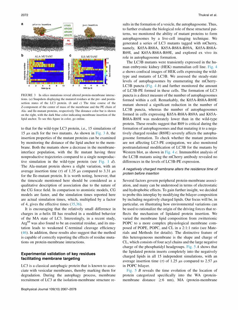
FIGURE 3 In silico mutations reveal altered protein-membrane interac-
tions. (a) Snapshots displaying the mutated residues at the pre- and postin-
sertion states of the LC3 protein. (b and c) The time course of the
Z-component of the center of mass of the membrane and the PE chain of
Ala- and Ile-mutant proteins, respectively The distance color bar is shown
on the right, with the dark blue color indicating membrane insertion of the
lipid anchor. To see this figure in color, go online.
2072 Thukral et al.
to that for the wild-type LC3 protein, i.e., 15 simulations of15 ms each for the two mutants. As shown in Fig. 3 b, theinsertion properties of the mutant proteins can be examinedby monitoring the distance of the lipid anchor to the mem-brane. Both the mutants show a decrease in the membrane-interface population, with the Ile mutant having threenonproductive trajectories compared to a single nonproduc-tive simulation in the wild-type protein (see Fig. 1 d).The Ala-mutant protein shows a slight variation, with anaverage insertion time (t) of 1.35 ms compared to 3.31 msfor the Ile-mutant protein. It is worth noting, however, thatthe timescale mentioned here should be considered as aqualitative description of association due to the nature ofthe CG force field. In comparison to atomistic models, CGmodels are faster, and the simulation times reported hereare actual simulation times, which, multiplied by a factorof 4, gives the effective times (35,36).
It is encouraging that the relatively small difference incharges in a-helix III has resulted in a modified behaviorof the MA state of LC3. Interestingly, in a recent study,Arg68 was also found to be an essential residue, and its mu-tation leads to weakened C-terminal cleavage efficiency(46). In addition, these results also suggest that the methodis capable of correctly reporting the effects of residue muta-tions on protein-membrane interactions.
Experimental validation of key residuesfacilitating membrane targeting
LC3 is a classical autophagic protein that is known to asso-ciate with vesicular membranes, thereby marking them fordegradation. During the autophagy process, membranerecruitment of LC3 at the isolation-membrane structure re-
Biophysical Journal 109(10) 2067–2078
sults in the formation of a vesicle, the autophagosome. Thus,to further evaluate the biological role of these structural pat-terns, we monitored the ability of mutant proteins to formautophagosomes by a live-cell imaging technique. Wegenerated a series of LC3 mutants tagged with mCherry,namely, K65A-R68A, K65A-R68A-R69A, K65A-R68A-R69I, and K65A-R68A-R69E, and explored ex vivo itsrole in autophagosome formation.
The LC3B mutants were transiently expressed in the hu-man embryonic kidney (HEK) mammalian cell line. Fig. 4a shows confocal images of HEK cells expressing the wild-type and mutants of LC3B. We assessed the steady-statelevels of autophagosomes by enumerating the mCherry-LC3B puncta (Fig. 4 b) and further monitored the amountof LC3B-PE formed in these cells. The formation of LC3puncta is a direct measure of the number of autophagosomesformed within a cell. Remarkably, the K65A-R68A-R69Emutant showed a significant reduction in the number ofLC3B puncta, whereas the number of autophagosomesformed in cells expressing K65A-R68A-R69A and K65A-R68A-R69I was moderately lower than in the wild-typeprotein. These results suggest that R69 is critical during theformation of autophagosomes and that mutating it to a nega-tively charged residue (R69E) severely affects the autopha-gosome formation. To check whether the mutant proteinsare not affecting LC3-PE conjugation, we also monitoredposttranslational modification of LC3B for the mutants byWestern blot, as shown in Fig. 4 c. Immunoblot analysis ofthe LC3B mutants using the mCherry antibody revealed nodifferences in the levels of LC3B-PE expression.
A negatively charged membrane alters the residence time ofprotein before insertion
Several factors govern peripheral protein-membrane associ-ation, and many can be understood in terms of electrostaticand hydrophobic effects. To gain further insight, we decidedto probe this interplay by modifying the bilayer compositionby including negatively charged lipids. Our focus will be, inparticular, on illustrating how environmental variations canbe used to rationalize the origin of the driving forces that re-flects the mechanism of lipidated protein insertion. Wevaried the membrane lipid composition from zwitterionicPOPC to a more complex physiological membrane com-posed of POPE, POPC, and CL in a 2:1:1 ratio (see Mate-rials and Methods for details). The distinctive feature ofthis heterogeneous membrane is the shape and charge ofCL, which consists of four acyl chains and the large negativecharge of the phosphatidyl headgroups. Fig. 5 A shows thatthe lipidated protein inserts completely into the negativelycharged lipids in all 15 independent simulations, with anaverage insertion time (t) of 1.25 ms compared to 2.57 msin POPC bilayer.
Fig. 5 B reveals the time evolution of the location ofprotein categorized specifically into the WA (protein-membrane distance R6 nm), MA (protein-membrane

FIGURE 4 Experimental validation of howmutation of residues crucial to membrane targeting affects autophagosome formation. (a) Immunofluorescence
micrographs of HEK cells cotransfected with mCherry-LC3mutants along with wild-type LC3 and a vector control. The formation of punctate structures was
notably reduced in the LC3-K65A-R68A-R69E mutant. Scale bars, 5mm. (b) Quantitation of the number of puncta in cells transfected with wild-type LC3
and mutants using Volocity software (nR10 from three independent experiments). Bars represent the mean5 SD across replicates. *p% 0.05; **p% 0.01;
and ***p% 0.001. (c) Western blot analysis for monitoring LC3 flux. HEK cell lysates expressing mCherry-LC3 and LC3 mutants were subjected to immu-
noblotting with anti-mCherry and anti-b-actin antibodies. The presence of both nonlipidated and lipidated LC3 was observed. To see this figure in color,
go online.
Membrane Insertion of Lipidated Protein 2073
distance R3 nm and %6 nm), and MI (protein-membranedistance %3 nm) states of four representative trajectories(trajectories 3, 4, 14, and 15). The colored bars in the figureindicate the different states (maroon, yellow, and dark bluefor the WA, MA, and MI states, respectively). The exis-tence of these states in all 15 trajectories is shown inFig. S3. We found that, contrary to what would be ex-pected, the protein is able to form stable binding at thewater-membrane interface (Z z 5 nm (Fig. S3, yellow))before insertion in most of the simulations. Also, the mo-lecular signature of the protein on the membrane is indeedassociated with residues involved in the a-helix III region(data not shown). Together, these results suggest thatprotein-membrane docking is governed by electrostatic in-teractions, whereby anionic lipids orient and steer the pro-tein toward the membrane surface, followed by the finalstage of the MI state, which is driven by hydrophobicforces of acyl chains.
Control simulations of the nonlipidated LC3protein
The lipidated protein clearly inserts into a zwitterionicbilayer in 14 of 15 trajectories (Fig. 1 d) and into a nega-tively charged bilayer in all trajectories (Fig. 5 A). Further,to analyze the contributions of the hydrophobic lipid anchor
of the protein, control simulations of nonlipidated protein inthe membrane environment were performed (see Materialsand Methods). First, the nonlipidated protein favors theaqueous water phase, with the protein distance rangingfrom 4.5 nm to 8.5 nm from the center of the bilayer, asshown in Fig. 6. Second, the protein undergoes fast-time-scale membrane association-dissociation events. Clearly,the hydrophobic anchor attached to the protein contributesactively in the insertion process of the protein. Since thenonlipidated protein was designed as a control to mimicthe in vivo cytosolic LC3 protein, these results furtherstrengthen our hypothesis that interplay between the lipidanchor and the cluster of basic residues is crucial for proteininsertion.
Bilayer perturbations
The membrane-perturbing propensity of LC3 protein wasdetermined by measuring the density and thickness of thebilayer (Fig. S4). At the point of PE insertion, localizedchanges in the density of the acyl chains and the membranethickness are observed, as shown in Fig. S4, A and B, respec-tively. However, during the course of the simulations, themembrane perturbations equilibrate (Fig. S4, C and D).Further, a comparison with the control simulation of thepure POPC bilayer reveals only marginal differences in
Biophysical Journal 109(10) 2067–2078

FIGURE 5 Role of electrostatistics in protein-membrane interaction.
(A) Time evolution of the distance between the PE chain of the LC3 and
the negatively charged bilayer along the 15 trajectories. The lipid anchor
of the protein inserts in all trajectories. (B) Localization of the protein dur-
ing four representative trajectories. The bars represent the existence of WA
(maroon), MA (yellow), and MI (dark blue) states. See Materials and
Methods for details regarding the definition of the different parameters.
To see this figure in color, go online.
2074 Thukral et al.
the bilayer properties (Fig. S4, E and F). Although a singleLC3 is not able to induce large perturbations, we believethat the effect of LC3 on the reorganization of membrane
FIGURE 6 The nonlipidated protein. Shown is a time series of the dis-
tance between the nonlipidated LC3 protein and the center of mass of the
membrane across five independent simulations. The protein tends to be in
the aqueous phase (distanceR 6 nm), with fast-timescale membrane disso-
ciation events. The color gradient reflects the distance (in nanometers) for
the MA (green) and WA (orange/red) states. To see this figure in color,
go online.
Biophysical Journal 109(10) 2067–2078
may be additive and that at higher protein/lipid ratios, thevariations measured above could be higher.
Fig. 7 depicts the lateral-pressure profiles of the LC3-anchored membrane and the pure bilayer in black and red,respectively. The pressure profiles were calculated afterinsertion of the POPC chain and are averaged over the mem-brane lateral area and all productive trajectories, based on aprevious implementation (43) (see Materials and Methodsfor details). After the insertion of LC3-PE, the lateral pres-sure near the lipid tails is decreased by 100 bar. However,the variations near the center of the bilayer were minimal.Counterintuitively, the pressure profiles appeared to be sym-metric at both bilayer leaflets despite the insertion of thePOPC chain. To test the effect of the lipid chain, we calcu-lated the pressure profile within 1 nm of the protein (seeFig. S5) for a few representative simulations. Comparisonof the two leaflets clearly shows the deviations betweenthem. It thus appears that the large bilayer used in our sim-ulations appears to equilibrate the effect of the lipid chainover the entire bilayer. Proteins docking with lipid surfacescan induce changes in the local spontaneous curvature,causing deviations from the lamellar phase (47).
The pressure profiles of membranes can be correlatedwith elastic properties such as local spontaneous curvature,which is calculated from the first moment of the pressureprofile (48); for an extended discussion, see (49). The pres-sure profile calculated over the entire bilayer was used tocalculate the monolayer spontaneous curvature. Since themembrane tension is calculated to be zero, the bilayermidplane was used as the Gibbs dividing surface of themonolayer. Within these assumptions, the LC3-bound mem-brane was calculated to have a spontaneous curvature closeto 0.0 nm�1, in comparison with the pure POPC bilayer,which has a curvature of �0.15 nm�1 (31). We thus seean increase in the local spontaneous curvature upon LC3insertion. In general, negative values for the radius of
FIGURE 7 Pressure profiles. Comparison of the lateral pressure profiles
of the productive simulations (black) and the pure POPC bilayer without
protein (red) as control simulations. The pressure profiles were calculated
after the insertion of the lipid anchor for the productive simulations. The
error bars shown represent the standard error between the simulations.
For further details, see Materials and Methods. To see this figure in color,
go online.

Membrane Insertion of Lipidated Protein 2075
spontaneous curvature are indicative of a preference forconcave-shaped curved surfaces (inverted phases, stalks)and positive values indicate a propensity toward convex-shaped curved surfaces (such as in vesicle budding) (50).The increase in spontaneous curvature of the bilayer arisesfrom the decrease in negative pressure arising from theacyl chain region of the bilayer, as seen in Fig. 7. The valuesreported should be considered to be qualitative, sincediscrepancies have been observed with calculation of thelipid spontaneous curvature, and especially the Gaussiancurvature modulus, from this method and others (51). Dur-ing autophagy, membrane recruitment of LC3 at the isola-tion membrane results in the formation of a vesicle, theautophagosome. The finding on spontaneous curvaturecalculated from our simulations suggests a possible initi-ating role of LC3 in stabilizing highly curved organelleslike autophagosomes.
DISCUSSION
Several problems have impeded our understanding of lipid-modified proteins and their interaction with phospholipidbilayers, in particular, the difficulty in obtaining proteincrystals with the lipid anchor (52). Computer simulationshave now started to reveal the molecular details of theirconformational dynamics and provided putative con-tributions of both protein and membrane lipids (11–13,16,20,53,54). Among the earliest reports, atomistic MDsimulations of membrane-bound H-ras protein predictedtwo modes of membrane binding. In GTP-bound conforma-tions, the a4 helix of the G domain facilitates membraneassociation, whereas GDP-bound structures favored electro-static interactions between basic residues of the C-terminalregion and the membrane (12). In all previous reports, sim-ulations begin from a well-defined membrane-boundconformer, and therefore, details of key transition eventssuch as membrane binding are not known. In this work,we have demonstrated spontaneous insertion of aqueous-phase lipidated LC3 into the membrane using unbiased mul-tiple independent microsecond-long simulations performedat physiological temperature. Examination of the eventsbefore, during, and after insertion of PE reveals a stagewiseprocess involving contributions from both the lipid anchorand the protein.
Due to the complex nature of lipid anchor insertion, weobserve highly stable conformations of the LC3-PE MIphase. This overstabilization of interactions increases theroughness of the energy landscape and hence hinders therapid sampling of reversible transitions. This is likely whyit is extremely difficult to obtain spontaneous reversibletransitions of complex processes, even though reasonableagreement between simulations and experiments regardingthermodynamics has been demonstrated for a range of pro-teins (16,55). In this work, we only used symmetric bilayers,and therefore, protein binding to both the leaflets evolves in
a similar manner. Biological membranes contain a heteroge-neous mixture of phospholipids, and thus, they can act asguiding forces for molecular recognition. To investigatethis phenomenon, we performed multiple LC3 simulationswith a lipid bilayer consisting of POPC, POPE, and CL ina 2:1:1 ratio. Permeation of the lipid anchor occurs in allsimulations, and interestingly, the insertion time, t, isgreatly reduced. The presence of negatively charged CLsignificantly alters the kinetics of the first step involvingprotein-membrane docking. However, the second step ofinsertion, driven by the hydrophobic anchor, occurs on aslow timescale, as the protein residence time on the mem-brane is increased.
Consequently, the reactions must be guided by a recogni-tion process, where specific residues determine specificity.Based on several spontaneous insertion events using com-puter simulations, wewere able to identify key residues facil-itating the LC3 membrane-targeting process. Our hypothesiswas substantiated by a combined in silico and in vitroapproach whereby we mutated the basic residue patch con-sisting of Lys65, Arg68, and Arg69 in the a-helix III region.The lipidated form of LC3 stably inserts itself into autopha-gosome membranes, where it is crucial during phagophoreelongation and cargo recognition. Using live-cell imaging,we monitored the presence of the autophagosome andobserved a significant decrease in puncta formation in oneparticular mutant, i.e., K65A-R68A-R69E (Fig. 4). The spe-cific mutant is based on a complete-charge-reversal strategyand severely hampers membrane targeting by the LC3 pro-tein. These data are in concordance with several previousexperimental and computational approaches where posi-tively charged residues close to the binding site are respon-sible for the protein penetration into the membrane (56).Owing to the underlying complexity of such interactions,side-chain orientations of these residues (K65-R68-R69),might also play a major role in membrane association. How-ever, the CG simulations performed in this work provide thedetermining factors for the progression of a lipidated proteinmembrane insertion. Future work involving large-scaleatomistic simulations might clarify the range in which suchside-chain orientations are crucial for guiding the final inser-tion stage of the lipid anchor.
As with this work, it is also imperative to discuss thepossible limitations of the CG MARTINI model, and theconclusions should be carefully interpreted. First, the sec-ondary structure of the protein is restrained during the entiresimulation, and therefore, the conformational changeswithin the protein induced by PE cannot be determined.Second, it is unclear how the water model influences theprotein-membrane interactions as the accuracy of electro-static screening by the water model is not adequate (37).
Although LC3 may have many cellular functions, it isbest characterized as the master regulator of autophagy, aprocess by which the cell forms a specialized vesicle, the au-tophagosome, to degrade the cellular waste. It is a multistep
Biophysical Journal 109(10) 2067–2078

FIGURE 8 Schematic representation of the
mechanism of lipidated-LC3 membrane insertion.
The left part of the diagram shows an unproductive
insertion event, with the protein’s hydrophobic an-
chor not inserted into the membrane. Both the lipid
anchor (control simulations) and key residues in the
protein (experimental mutations) are necessary for
insertion of the lipidated protein. These dynamic
changes within the lipidated protein are induced
by specific protein-lipid interactions. This speci-
ficity determines and facilitates PE insertion, as
shown at the right of the diagram. To see this figure
in color, go online.
2076 Thukral et al.
process where the lipidated form of LC3 is involved in theassembly of the isolation membrane and further assists toform a vesicle, the autophagosome. To probe the membraneperturbations induced upon LC3 binding in our simulations,we calculated various parameters, including membranethickness, density, and lateral pressure. During PE insertion,minor changes were observed in density and thickness,which equilibrate during the course of the simulation. How-ever, an increase in the local spontaneous curvature uponLC3 insertion was found that is indicative of the curva-ture-inducing properties of LC3. Our results are consistentwith a recent study on a lipid-anchored oligomer, whichcauses membrane perturbations by altering the lateral pres-sure (55). Our curvature results also highlight the underlyingbiological significance of the phagophore in autophagosomeformation during autophagy. The lipidation process of LC3family proteins is known to be sensitive to membrane curva-ture and lipid packing. A recent review on autophagic phag-ophore expansion suggests that additional anchor proteinsinvolved during the initiation step (such as Atg3) add anadditional level of spatial and temporal specificity to thelipid membrane insertion (57).
CONCLUSIONS
Macroautophagy is a conserved cellular recycling processessential for homeostasis and cell survival during stress. Inresponse to stress, cellular components are sequesteredinto a growing phagophore that closes to form double mem-brane vesicles. The key to this process is the covalent conju-gation of phosphatidylethanolamine (PE) to the LC3 proteinthat plays an important role in the formation of the autopha-gosome (28). The aim of this work was to reveal a reliableand robust mechanism of transition of cytoplasmic to mem-brane-inserted LC3-PE. This was accomplished by exam-ining the events before, during, and after insertion of PEin silico. We have performed independent, unbiased CGMD simulations of LC3-PE, starting from aqueous-phaseconformations placed far from the membrane. In this report,we explore lipidated LC3 dynamics with both zwitterionicphosphatidylcholine and a complex negatively chargedbilayer composed of CL. Although prior experimental
Biophysical Journal 109(10) 2067–2078
studies show that the PE attachment is required for the bio-logical activity of the lipidated LC3 protein (28), dynamiccharacterization of the translocation of the cytoplasmic lipi-dated protein to the membrane-inserted form is challenging.
We propose that a basic patch of amino acid residues ina-helix III orients and steers the protein toward the mem-brane, acting as a specific determinant of membrane interac-tion. The final stage in reaching the membrane-inserted stateinvolves hydrophobic forces of the lipid anchor to cross themembrane interface. Our data support the dual-recognition-mode hypothesis (56), with both protein and membranelipids driving the permeation process (Fig. 8). In the sce-nario presented here, our findings thus provide a structuralbasis for the rational design of autophagic modulators tar-geting LC3 and possibly other lipidated proteins.
SUPPORTING MATERIAL
Five figures and two tables are available at http://www.biophysj.org/
biophysj/supplemental/S0006-3495(15)00992-3.
AUTHOR CONTRIBUTIONS
L.T., D.S., and R.S.G. designed the study, analyzed the results, and drafted
the manuscript. A.R. and D.M. perfomed live cell-based imaging experi-
ments. N.A. performed heterogeneous bilayer simulations.
ACKNOWLEDGMENTS
We are indebted to CSIR-Fourth Paradigm Institute (4PI) for computational
resources.
L.T. is funded by an INSPIRE Faculty Fellowship from the Department of
Science and Technology. D.S. is funded by a Ramalingaswami Fellowship
from the Department of Biotechnology. This work was supported by project
BSC0302 to R.S.G. from the Council of Scientific and Industrial Research
(CSIR) of India.
REFERENCES
1. Casey, P. J. 1995. Protein lipidation in cell signaling. Science.268:221–225.
2. Hang, H. C., and M. E. Linder. 2011. Exploring protein lipidation withchemical biology. Chem. Rev. 111:6341–6358.
Membrane Insertion of Lipidated Protein 2077
3. Wittinghofer, A., and I. R. Vetter. 2011. Structure-function relation-ships of the G domain, a canonical switch motif. Annu. Rev. Biochem.80:943–971.
4. Thapar, R., J. G. Williams, and S. L. Campbell. 2004. NMRcharacterization of full-length farnesylated and non-farnesylatedH-Ras and its implications for Raf activation. J. Mol. Biol.343:1391–1408.
5. Kotting, C., J. Guldenhaupt, and K. Gerwert. 2012. Time-resolvedFTIR spectroscopy for monitoring protein dynamics exemplified byfunctional studies of Ras protein bound to a lipid bilayer. Chem.Phys. 396:72–83.
6. Hannoush, R. N., and J. Sun. 2010. The chemical toolbox for moni-toring protein fatty acylation and prenylation. Nat. Chem. Biol.6:498–506.
7. Willumsen, B. M., A. Christensen, ., D. R. Lowy. 1984. The p21 rasC-terminus is required for transformation and membrane association.Nature. 310:583–586.
8. Hancock, J. F., A. I. Magee, ., C. J. Marshall. 1989. All ras proteinsare polyisoprenylated but only some are palmitoylated. Cell. 57:1167–1177.
9. Vogel, A., C. P. Katzka, ., D. Huster. 2005. Lipid modifications of aRas peptide exhibit altered packing and mobility versus host membraneas detected by 2H solid-state NMR. J. Am. Chem. Soc. 127:12263–12272.
10. Brunsveld, L., J. Kuhlmann,., H. Waldmann. 2006. Lipidated ras andrab peptides and proteins–synthesis, structure, and function. Angew.Chem. Int. Ed. Engl. 45:6622–6646.
11. Gorfe, A. A., R. Pellarin, and A. Caflisch. 2004. Membrane localizationand flexibility of a lipidated ras peptide studied by molecular dynamicssimulations. J. Am. Chem. Soc. 126:15277–15286.
12. Gorfe, A. A., M. Hanzal-Bayer, ., J. A. McCammon. 2007. Structureand dynamics of the full-length lipid-modified H-Ras protein in a1,2-dimyristoylglycero-3-phosphocholine bilayer. J. Med. Chem.50:674–684.
13. Gorfe, A. A., A. Babakhani, and J. A. McCammon. 2007. H-ras proteinin a bilayer: interaction and structure perturbation. J. Am. Chem. Soc.129:12280–12286.
14. Abankwa, D., A. A. Gorfe, ., J. F. Hancock. 2010. Ras membraneorientation and nanodomain localization generate isoform diversity.Proc. Natl. Acad. Sci. USA. 107:1130–1135.
15. Brunsveld, L., H. Waldmann, and D. Huster. 2009. Membrane bindingof lipidated Ras peptides and proteins—the structural point of view.Biochim. Biophys. Acta. 1788:273–288.
16. Gorfe, A. A., A. Babakhani, and J. A. McCammon. 2007. Free energyprofile of H-ras membrane anchor upon membrane insertion. Angew.Chem. Int. Ed. Engl. 46:8234–8237.
17. Huster, D., A. Vogel, ., K. Arnold. 2003. Membrane insertion of alipidated ras peptide studied by FTIR, solid-state NMR, and neutrondiffraction spectroscopy. J. Am. Chem. Soc. 125:4070–4079.
18. Reuther, G., K.-T. Tan, ., D. Huster. 2006. The lipidated membraneanchor of full length N-Ras protein shows an extensive dynamics asrevealed by solid-state NMR spectroscopy. J. Am. Chem. Soc.128:13840–13846.
19. Weise, K., S. Kapoor,., R. Winter. 2011. Membrane-mediated induc-tion and sorting of K-Ras microdomain signaling platforms. J. Am.Chem. Soc. 133:880–887.
20. Vogel, A., G. Reuther, ., D. Huster. 2010. Backbone conformationalflexibility of the lipid modified membrane anchor of the human N-Rasprotein investigated by solid-state NMR and molecular dynamics simu-lation. Biochim. Biophys. Acta. 1798:275–285.
21. Schroeder, H., R. Leventis, ., J. R. Silvius. 1997. S-Acylation andplasma membrane targeting of the farnesylated carboxyl-terminal pep-tide of N-ras in mammalian fibroblasts. Biochemistry. 36:13102–13109.
22. Sigal, C. T., W. Zhou, ., M. D. Resh. 1994. Amino-terminal basicresidues of Src mediate membrane binding through electrostatic inter-
action with acidic phospholipids. Proc. Natl. Acad. Sci. USA.91:12253–12257.
23. McLaughlin, S., and A. Aderem. 1995. The myristoyl-electrostaticswitch: a modulator of reversible protein-membrane interactions.Trends Biochem. Sci. 20:272–276.
24. Gerlach, H., V. Laumann, ., M. Geyer. 2010. HIV-1 Nef membraneassociation depends on charge, curvature, composition and sequence.Nat. Chem. Biol. 6:46–53.
25. Sugawara, K., N. N. Suzuki,., F. Inagaki. 2004. The crystal structureof microtubule-associated protein light chain 3, a mammalian homo-logue of Saccharomyces cerevisiae Atg8. Genes Cells. 9:611–618.
26. Ichimura, Y., T. Kirisako,., Y. Ohsumi. 2000. A ubiquitin-like systemmediates protein lipidation. Nature. 408:488–492.
27. Nakatogawa, H., Y. Ichimura, and Y. Ohsumi. 2007. Atg8, a ubiquitin-like protein required for autophagosome formation, mediates mem-brane tethering and hemifusion. Cell. 130:165–178.
28. Kabeya, Y., N. Mizushima,., T. Yoshimori. 2000. LC3, a mammalianhomologue of yeast Apg8p, is localized in autophagosome membranesafter processing. EMBO J. 19:5720–5728.
29. Uemura, T., M. Yamamoto, ., S. Waguri. 2014. A cluster of thintubular structures mediates transformation of the endoplasmic reticu-lum to autophagic isolation membrane.Mol. Cell. Biol. 34:1695–1706.
30. Schafer, L. V., D. H. de Jong, ., S. J. Marrink. 2011. Lipid packingdrives the segregation of transmembrane helices into disordered lipiddomains in model membranes. Proc. Natl. Acad. Sci. USA.108:1343–1348.
31. Sengupta, D., and A. Chattopadhyay. 2012. Identification of cholesterolbinding sites in the serotonin1A receptor. J. Phys. Chem. B.116:12991–12996.
32. Arnarez, C., J.-P. Mazat,., X. Periole. 2013. Evidence for cardiolipinbinding sites on the membrane-exposed surface of the cytochrome bc1.J. Am. Chem. Soc. 135:3112–3120.
33. Marrink, S. J., and D. P. Tieleman. 2013. Perspective on the Martinimodel. Chem. Soc. Rev. 42:6801–6822.
34. Kirisako, T., Y. Ichimura, ., Y. Ohsumi. 2000. The reversible modifi-cation regulates the membrane-binding state of Apg8/Aut7 essentialfor autophagy and the cytoplasm to vacuole targeting pathway.J. Cell Biol. 151:263–276.
35. Marrink, S. J., H. J. Risselada,., A. H. de Vries. 2007. The MARTINIforce field: coarse grained model for biomolecular simulations. J. Phys.Chem. B. 111:7812–7824.
36. Monticelli, L., S. K. Kandasamy, ., S.-J. Marrink. 2008. TheMARTINI coarse-grained force field: extension to proteins. J. Mol.Model. 4:819–834.
37. de Jong, D. H., X. Periole, and S. J. Marrink. 2012. Dimerization ofamino acid side chains: lessons from the comparison of different forcefields. J. Chem. Theory Comput. 8:1003–1014.
38. Chu, C. T., J. Ji, ., V. E. Kagan. 2013. Cardiolipin externalization tothe outer mitochondrial membrane acts as an elimination signal for mi-tophagy in neuronal cells. Nat. Cell Biol. 15:1197–1205.
39. Karo, J., P. Peterson, and M. Vendelin. 2012. Molecular dynamics sim-ulations of creatine kinase and adenine nucleotide translocase in mito-chondrial membrane patch. J. Biol. Chem. 287:7467–7476.
40. Pronk, S., S. Pall, ., E. Lindahl. 2013. GROMACS 4.5: a high-throughput and highly parallel open source molecular simulation tool-kit. Bioinformatics. 29:845–854.
41. Berendsen, H. J., J. P. M. Postma, ., J. Haak. 1984. Molecular dy-namics with coupling to an external bath. J. Chem. Phys. 81:3684–3690.
42. Castillo, N., L. Monticelli, ., D. P. Tieleman. 2013. Free energy ofWALP23 dimer association in DMPC, DPPC, and DOPC bilayers.Chem. Phys. Lipids. 169:95–105.
43. Ollila, O. H., H. J. Risselada,., S. J. Marrink. 2009. 3D pressure fieldin lipid membranes and membrane-protein complexes. Phys. Rev. Lett.102:078101.
Biophysical Journal 109(10) 2067–2078

2078 Thukral et al.
44. Sengupta, D. 2012. Cholesterol modulates the structure, bindingmodes, and energetics of caveolin-membrane interactions. J. Phys.Chem. B. 116:14556–14564.
45. Patil, K. M., R. J. Naik, ., V. A. Kumar. 2012. Highly efficient(R-X-R)-type carbamates as molecular transporters for cellular deliv-ery. J. Am. Chem. Soc. 134:7196–7199.
46. Liu, C., H. Ma, ., L. Yu. 2013. Arginine68 is an essential residue forthe C-terminal cleavage of human Atg8 family proteins. BMC CellBiol. 14:27.
47. Zimmerberg, J., and M. M. Kozlov. 2006. How proteins producecellular membrane curvature. Nat. Rev. Mol. Cell Biol. 7:9–19.
48. Helfrich, W. 1981. Amphiphilic mesophases made of defects. InPhysique des defauts (Physics of defects). R. Balian, M. Kleman,and J.-P. Poirier, editors. North-Holland, Amsterdam, pp. 716–755.
49. Deserno, M. 2015. Fluid lipid membranes: from differential geometryto curvature stresses. Chem. Phys. Lipids. 185:11–45.
50. Marrink, S. J., A. H. de Vries, and D. P. Tieleman. 2009. Lipids on themove: simulations of membrane pores, domains, stalks and curves.Biochim. Biophys. Acta. 1788:149–168.
51. Hu, Y., S. Ou, and S. Patel. 2013. Free energetics of arginine perme-ation into model DMPC lipid bilayers: coupling of effective counterion
Biophysical Journal 109(10) 2067–2078
concentration and lateral bilayer dimensions. J. Phys. Chem. B.117:11641–11653.
52. Ahearn, I. M., K. Haigis,., M. R. Philips. 2012. Regulating the regu-lator: post-translational modification of RAS. Nat. Rev. Mol. Cell Biol.13:39–51.
53. Jensen, M. Ø., O. G. Mouritsen, and G. H. Peters. 2004. Simulations ofa membrane-anchored peptide: structure, dynamics, and influence onbilayer properties. Biophys. J. 86:3556–3575.
54. Vogel, A., K.-T. Tan, ., D. Huster. 2007. Flexibility of ras lipid mod-ifications studied by 2H solid-state NMR and molecular dynamics sim-ulations. Biophys. J. 93:2697–2712.
55. Li, H., and A. A. Gorfe. 2014. Membrane remodeling by surface-boundprotein aggregates: insights from coarse-grained molecular dynamicssimulation. J. Phys. Chem. Lett. 5:1457–1462.
56. Lumb, C. N., J. He, ., M. S. Sansom. 2011. Biophysical and compu-tational studies of membrane penetration by the GRP1 pleckstrin ho-mology domain. Structure. 19:1338–1346.
57. Johansen, T., and T. Lamark. 2014. Selective autophagy goes exclusive.Nat. Cell Biol. 16:395–397.