biopharmaceutical classification of poorly soluble drugs ... · • users may download and print...
TRANSCRIPT

Syddansk Universitet
Biopharmaceutical classification of poorly soluble drugs with respect to “enablingformulations”Buckley, Stephen; Frank, Kerstin ; Fricker, Gert; Brandl, Martin
Published in:European Journal of Pharmaceutical Sciences
Publication date:2013
Document versionFinal published version
Citation for pulished version (APA):Buckley, S., Frank, K., Fricker, G., & Brandl, M. (2013). Biopharmaceutical classification of poorly soluble drugswith respect to “enabling formulations”. European Journal of Pharmaceutical Sciences, 50(1), 8-16.
General rightsCopyright and moral rights for the publications made accessible in the public portal are retained by the authors and/or other copyright ownersand it is a condition of accessing publications that users recognise and abide by the legal requirements associated with these rights.
• Users may download and print one copy of any publication from the public portal for the purpose of private study or research. • You may not further distribute the material or use it for any profit-making activity or commercial gain • You may freely distribute the URL identifying the publication in the public portal ?
Take down policyIf you believe that this document breaches copyright please contact us providing details, and we will remove access to the work immediatelyand investigate your claim.
Download date: 05. Feb. 2017
Accepted Manuscript
Biopharmaceutical classification of poorly soluble drugs with respect to “ena‐
bling formulations”
Stephen Timothy Buckley, Kerstin Julia Frank, Gert Fricker, Martin Brandl
PII: S0928-0987(13)00129-2
DOI: http://dx.doi.org/10.1016/j.ejps.2013.04.002
Reference: PHASCI 2733
To appear in: European Journal of Pharmaceutical Sciences
Received Date: 29 January 2013
Revised Date: 2 April 2013
Accepted Date: 3 April 2013
Please cite this article as: Buckley, S.T., Frank, K.J., Fricker, G., Brandl, M., Biopharmaceutical classification of
poorly soluble drugs with respect to “enabling formulations”, European Journal of Pharmaceutical Sciences (2013),
doi: http://dx.doi.org/10.1016/j.ejps.2013.04.002
This is a PDF file of an unedited manuscript that has been accepted for publication. As a service to our customers
we are providing this early version of the manuscript. The manuscript will undergo copyediting, typesetting, and
review of the resulting proof before it is published in its final form. Please note that during the production process
errors may be discovered which could affect the content, and all legal disclaimers that apply to the journal pertain.

Biopharmaceutical classification of poorly soluble drugs with respect to
“enabling formulations”
Stephen Timothy Buckley a,d,*, Kerstin Julia Franka,b,c*, Gert Frickerb and Martin
Brandla
a Dept. of Physics, Chemistry and Pharmacy, University of Southern Denmark, Campusvej 55, DK‐5230
Odense M, Denmark
b Institute of Pharmacy and Molecular Biotechnology, University of Heidelberg, Im Neuenheimer Feld 366,
D‐69120 Heidelberg, Germany
c current affiliation: Boehringer Ingelheim Pharma GmbH & Co. KG, Development Germany
d current affiliation: Novo Nordisk A/S, ADME Department, Novo Nordisk Park DK‐2760 Måløv Denmark
*K.J. Frank and S.T. Buckley have contributed equally to this work, this review article is derived from their
research work done at University of Southern Denmark prior to joining their current employers.
Correponding author:
Martin Brandl
Email [email protected]
Phone +45 6550 2525
Keywords : solubility, permeability, solubilization, micelle,
Abstract
The large number of drug candidates with poor dissolution characteristics seen in the past decade, has
fostered interest in so‐called “enabling formulations”, i.e., formulations which shall make such drugs bio‐
available. Development of enabling formulations is currently being guided by the following (simplified)
hypothesis: If a poorly soluble drug (BCS class II drug) can be transferred into a solubilized state, one can
achieve an absorption profile close to that of a soluble drug (BCS class I drug). Thus, formulation
development typically endeavours to achieve the most robust solubility enhancement.
Here we critically review both common in vitro approaches and experimental data available in literature
pertaining to the solubility and permeability of poorly soluble drugs from enabling formulations, and
discuss their interplay. Recent in‐vitro data indicate, that commonly employed surfactants as well as
endogenous surfactants present in the intestine, although enhancing drug solubility, mostly hamper drug
permeation. Mechanistic studies demonstrate a direct correlation between passive transcellular diffusion
and the concentration of molecularly dissolved drug. The latter may be reduced due to partitioning into
micelles or other solubilizing carriers, but enhanced in supersaturating formulations.
We conclude thus that biopharmaceutical assessment approaches that rely on the amount of molecularly
dissolved drug should guide us towards successful enabling formulations.
Abbreviations:
BCS Biopharmaceutical Classification Scheme
GI Gastrointestinal
p‐GP p‐Glycoprotein
MRP multidrug resistance‐related protein
BCRP breast cancer resistant protein
CMC critical micelle concentration
CsA Cyclosporin A
TEER transepithelial elctrical resistance
SNEDDS self nanoemulsifying drug delivery system
SSDS supersaturating drug delivery systems

FaSSIF fasted state simulated intestinal fluid
FeSSIF fed state simulated intestinal fluid
HBSS Hank’s buffered salt solution
API active pharmaceutical ingredient
ASD amorphous solid dispersion
Introduction
Drug bioavailability prediction relies on the principles originally laid down in the Biopharmaceutical
Classification Scheme (BCS, Amidon et al., 1995). In the initial phase, a combination of solubility testing and
in vitro transport studies (Artursson et al. 2001) are typically employed as a prognostic tool to predict oral
absorption. At later stages in vitro dissolution testing (Dressman et al., 1998) as well other permeation
approaches (Ungell, 2004) are often added to the tool‐box.
During the past decade, the proportion of drug candidates with relatively poor biopharmaceutical
properties, mainly candidates with poor dissolution characteristics, has grown significantly. Accordingly, so‐
called “enabling formulations”, i.e., formulations which shall make such drugs bio‐available, have
increasingly gained attention. As a result, biopharmaceutical classification is shifting focus from drug
candidate‐assessment to formulation assessment.
Standard measures aimed at enhanced oral bioavailability of poorly water soluble drugs include pH
adjustment, salt formation and reduction of particle size. Enabling formulation‐approaches include the use
of co‐solvents, complexing agents, (non‐ionic) surfactants, self‐(micro‐ or nano‐) emulsifying drug delivery
systems, liposomes, amorphous solid dispersions and mesoporous carriers. For recent reviews see (Rahman
et al., 2013; Singh et al., 2011; Van Hoogevest et al., 2011).
Irrespective of the formulation principle employed, development of solubility‐enhancing formulations is
currently being guided by the following (simplified) hypothesis: If a poorly soluble drug (BCS class II drug)
can be transferred into a solubilized state, one can achieve an absorption profile close to that of a soluble
drug (BCS class I drug). Any drug precipitation occurring during GI‐passage may compromise drug
bioavailability ‐ or in other words ‐ formulation development aims at achieving the most robust
solubilization (Cui et al., 2009; Gursoy & Benita, 2004; Holm et al., 2006; Li et al., 2012; Pouton, 2006;
Pouton & Porter, 2008; Stillhart et al., 2012; Tønsberg et al., 2010).
Interestingly, substantial absorption enhancement in vivo through solubilizing formulations remains
challenging, as illustrated by the very limited number of successful examples currently on the market. At
the same time it is common knowledge that the enhancement in bioavailability achieved in vivo rarely
reaches a comparable magnitude to that which solubility enhancement is observed in vitro (Araya et al.,
2005; Barakat, 2010; Mellaerts et al., 2008; Singh et al., 2013).
Thus, the motivation for the current work was to critically review both common in vitro approaches and
experimental data available in literature pertaining to solubility and permeability of poorly soluble drugs,
and to discuss their interplay. To this end, we examine the effects of surfactants that are commonly

employed in enabling formulations; while also extending this discussion to include enabling formulations
which are not based on surfactants. In the second section, we address the influence of endogenous
surfactants present in the intestine on solubility and permeation. In this context, the concept of
supersaturation is discussed in the third section. Finally, we conclude regarding which biopharmaceutical
assessment approaches appear most promising in terms of successfully guiding towards development of
enabling formulations.
Influence of nonionic surfactants on the solubility and absorption/permeability of poorly soluble drugs
It is generally accepted that surfactants effectively enhance the apparent solubility of poorly soluble drugs
in aqueous media through micellar solubilization, though, there are a limited number of cases where the
drug is hardly incorporated in micelles due to lack of solubility in both polar and apolar environments. To
predict the impact of surfactants on oral drug absorption or drug permeability in vitro is more of a
challenge. This is due to a complex scenario of events, the mechansims of which have only recently been
revealed in greater detail, such as: (1) The influence of micelle association of drugs on passive transcellular
diffusion. (2) The importance of direct actions, which surfactants may exert on the intestinal barrier such as
blockage of exsorptive drug transporters [p‐GP (ABCB1) , BCRP (ABCG2) or MRPs (ABCCs)] or metabolising
enzymes (CYP 450) as well as the opening of tight junctions, all of which are expected to enhance uptake.
Already in 1997, Borchardt and co‐workers investigated the impact of non‐ionic surfactants on Caco‐2
transport of a model peptide (at low peptide concentrations) (Nerurkar et al., 1997). With increasing
surfactant concentrations, the apical to basolateral (A‐to‐B) transport of the peptide was found initially
enhanced but then reduced. They postulated an interaction of active and passive transport pathways,
where surfactants at rather low concentrations (i.e., below CMC) were assumed to inhibit carrier‐mediated
exsorptive transport, while at unusually high surfactant concentrations (i.e., 20x CMC) a weak interaction of
the peptide with surfactant micelles would reduce its effective thermodynamic activity and thus limit
passive absorptive uptake.
In 2001, Polli and colleagues (Rege et al., 2001) observed that A‐to‐B transport of certain drug compounds
across Caco‐2 monolayers was enhanced at pharmaceutically relevant concentrations of polysorbate 80,
namely those that were substrates of p‐GP or MRP‐like efflux systems. In contrast, permeation of a non‐
substrate (benzoic acid) remained unaffected by the surfactant.
In 2003, Amidon and coworkers (Chiu et al., 2003) reported that the presence of 0.2% (w/v) of the non‐
ionic surfactant polyethoxylated castor oil (Cremophor EL®) decreased A‐to‐B flux of the hydrophobic cyclic
peptide cyclosporin A (CsA) in Caco‐2 monolayers almost 3‐fold compared to that in the absence of
surfactant. Although they did not study drug/surfactant‐interactions experimentally, they assumed that
micellar solubilization of CsA would reduce its thermodynamic activity and its permeability. The surfactant
employed is known to inhibit p‐GP efflux, while CsA is a known p‐GP‐substrate in these models. Collectively,
this should increase the permeability of CsA. However, they hypothesized that micellar solubilization
effects would outweigh potential inhibition of p‐GP mediated exsorptive transport, resulting in a net overall
reduction in CsA permeability.
Other reports postulating such interplay between permeability reduction due to micelle association and
permeability enhancement due to p‐GP blockage refer to vitamin E‐TGPS (Varma and Panchagnula, 2005)
Shaik (Shaik et al., 2008) demonstrated that efflux inhibition of two protease inhibitors by poloxamer P85
was due to direct p‐GP‐interaction rather than alteration in the expression of the MDR‐1 gene.
Today, there is increasing evidence that a broad variety of non‐ionic surfactants has an inhibitory effect not
only on p‐GP but also on other ABC‐transporters, such as BCRP and MRPs (Cuestas et al, 2011; Guo et al.,
2010; Yamagata et al., 2009), although the exact mechanism of interaction has still to be elucidated.
Porter and co‐workers (Katneni et al, 2006) quantified the extent of micellar solubilization of poorly soluble
drugs from equilibrium solubility studies in the presence of non‐ionic surfactants (polysorbate 80 and
polyethoxylated castor oil) and suggested to subtract the fraction of micelle‐bound drug to correct
apparent permeability values obtained from in vitro intestinal tissue transport studies. For a range of drugs
with varying degrees of lipophilicity, their data supported the hypothesis that micelle‐bound drug is
“inactive” in terms of permeation.
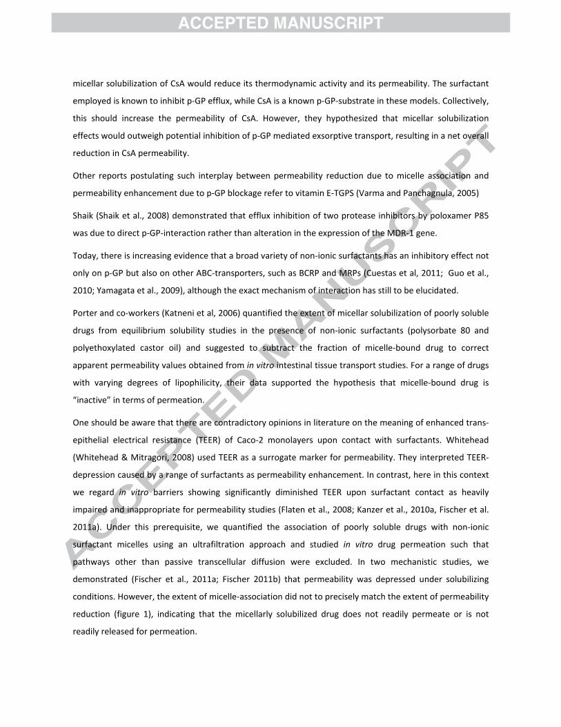
One should be aware that there are contradictory opinions in literature on the meaning of enhanced trans‐
epithelial electrical resistance (TEER) of Caco‐2 monolayers upon contact with surfactants. Whitehead
(Whitehead & Mitragori, 2008) used TEER as a surrogate marker for permeability. They interpreted TEER‐
depression caused by a range of surfactants as permeability enhancement. In contrast, here in this context
we regard in vitro barriers showing significantly diminished TEER upon surfactant contact as heavily
impaired and inappropriate for permeability studies (Flaten et al., 2008; Kanzer et al., 2010a, Fischer et al.
2011a). Under this prerequisite, we quantified the association of poorly soluble drugs with non‐ionic
surfactant micelles using an ultrafiltration approach and studied in vitro drug permeation such that
pathways other than passive transcellular diffusion were excluded. In two mechanistic studies, we
demonstrated (Fischer et al., 2011a; Fischer 2011b) that permeability was depressed under solubilizing
conditions. However, the extent of micelle‐association did not to precisely match the extent of permeability
reduction (figure 1), indicating that the micellarly solubilized drug does not readily permeate or is not
readily released for permeation.

In the same year Miller and coworkers (Miller et al., 2011) suggested a theoretical approach taking into
account the effects of micellar solubilization on both the membrane permeability and the unstirred water
layer permeability, modelling the interplay between solubilization and permeation.
Besides the described effects of micellar solubilization with subsequently reduced transcellular diffusion
and inibition of p‐GP‐mediated efflux, surfactants may cause other effects: Uihelyi (Uihelyi et al., 2012)
described enhanced paracellular uptake across Caco‐2 monolayers in the presence of polysorbates or
labrasol. Since poorly soluble drugs typically follow the transcellular pathway, this observation is of limited
relevance here. Mudra (Mudra & Borchardt, 2010) described influences of various non‐ionic surfactants on
CYP3A, which may elevate or diminish plasma levels of CYP3A‐mediated metabolites
Finally, surfactants may influence supersaturation. A thorough discussion of this aspect is found in the
section on supersaturation below.
Overall, in recent years we are seeing accumulating evidence from in vitro studies that surfactants, while
enhancing the apparent solubility of drugs via micellarization, also inhibit passive (transcellular) drug
permeation in a concentration‐dependent manner. Conflicting evidence regarding absorption
enhancement by surfactants may be the result of a significant impairment of the permeation screen. Thus,
stringent controls on barrier integrity as well as cell toxicity are essential.
Influence of other enabling formulations on the solubility and absorption/permeability of poorly soluble
drugs
When looking at other enabling formulations, similar observations were recently reported in literature.
Dahan demonstrated that when using cyclodextrins as pharmaceutical solubilizers, a trade‐off exists
between apparent solubility increase and permeability decrease (Dahan et al., 2010; Miller et al., 2012a). At
the same time re‐crystallization of drug from supersaturated solutions may be retarded (see section on
supersaturation below; Brewster et al., 2008)
Even in co‐solvent systems decreased permeability with increased solubility was observed for progesterone
in combination with propylene glycol and polyethylene glycol (Miller et al., 2012b) in a test where drug
concentration was maintained at 75% saturation solubility level. In this case constant thermodynamic
activity has to be assumed as well as the absence of drug binding to colloidal structures. A similar
observation is reported by Beig et al. 2012.
Very recently, Müllertz and co‐workers (Larsen et al., 2013) observed in a dog study that self
nanoemulsifying drug delivery systems (SNEDDS), where the content of drug was close to the saturation
limit, showed absorption to a higher extent than more robust formulations, where the drug content was
well below the saturation limit. They concluded that the traditional optimization strategy of SNEDDS‐
formulations towards a high solubility of the drug compound in vitro may lead to a lower bioavailability.
Influence of FaSSIF/bile salts/phospholipids (i.e., mixed micelles) on the solubility and permeability of
poorly soluble drugs
According to current paradigms, efficient absorption of drug molecules is dependent on their solubilization
(Jones et al., 2006). In this regard, the solubilization of poorly soluble drugs is highly dependent on the
composition of the intestinal milieu as they transit through the gut. Intestinal fluids are comprised of a
complex lipid mixture containing bile acids and lyso‐phospholipids. Notably, these components closely
associate and interact with drug molecules in the intestinal tract and in this way can markedly impact their
dissolution behavior.
To address these issues, biorelevant media have been increasingly employed in in vitro solubility and
dissolution studies. Reflecting both fasted and fed states (FaSSIF and FeSSIF, respectively), these media
have facilitated careful examination of the impact of micellar encapsulation on the solubility and
dissolution of a variety of poorly soluble compounds. Although these media represent a simplification of
the luminal composition, they have been shown to accurately estimate the dissolution process in vivo (e.g,
Galia et al., 1998). In particular, they have permitted a broader mechanistic understanding of the potential
influence these interactions have on the behavior of low solubility drugs upon oral ingestion.
Bile salt concentrations increase following ingestion of a meal (from 4‐6 mmol/L to 10‐20 mmol/L) (Jones et
al., 2006). In the case of poorly soluble drugs, this typically results in an increase in their solubility and
dissolution rate. Solubility tests using biorelevant media have shown that the solubility of poorly soluble
drugs can be markedly increased. Since it has been demonstrated that biorelevant media under certain
prerequisites may also be employed for in vitro permeability screening (Ingels et al., 2002), accumulating
evidence suggests that enhanced solubility does not necessarily translate into a correspondingly higher
proportion of drug available for absorption. Rather, that fraction solubilized by bile salt/lecithin mixed
micelles remains encapsulated and thus potentially unavailable for passage across the epithelial barrier.
In vivo bile salts serve as endogenous surfactants, capable of solubilizing lipophilic drugs. Indeed, non‐
native surfactants have been widely employed as excipients in formulations of poorly soluble drugs to
improve their aqueous solubility. However, while the amalgam of different bile salts contained within
intestinal fluids can act to improve the apparent solubility of these compounds, they concomitantly reduce
the free fraction of drug. Consequently, this can significantly diminish the proportion of drug which is
available to permeate across the intestinal membrane. In this environment, drug compounds are in an
equilibrium state between free and micelle‐bound. In effect, for micelle‐bound drug to traverse the

epithelial barrier, it must first partition out of the micelles (i.e., be subsumed into the free fraction). Only
then is it available for membrane permeation. It is also important to note that formulations may
significantly change the inherent properties of a compound and therefore the impact of intestinal fluid
constituents on their absorption (see section on supersaturation).
In the presence of bile salts and lipoidal components, evidence suggests that the thermodynamic activity of
drugs in solution is diminished on account of their solubilization within mixed micelles (Poelma et al., 1991).
They have examined the influence of mixed micelles on drug transport rates from a luminal solution across
the unstirred water layer (UWL) and intestinal wall of chronically isolated rat intestinal loops. Measuring
the loss of drug compound from the intestine, they observed that the disappearance rate of the lipophilic
drugs dantrolene, griseofulvin and ketoconazole was reduced in the presence of micelles. Simultaneous
solubility measurements in micellar solutions illustrated that the fraction solubilised was significantly
augmented. In part, this was ascribed to the decreased fraction of free drug in solution as a result of its
micellar solubilization.
Subsequent studies have provided further support to this hypothesis. Investigations by Ingels (Ingels et al.,
2004) have shown that the transport of various lipophilic drugs across Caco‐2 monolayers was decreased in
the presence of a FaSSIF buffer system. They attributed these differences to micellar encapsulation of the
API by mixed micelles, thus limiting the free fraction of drug available for permeation. Observations by
Kataoka (Kataoka et al., 2006) showed that the permeability of both ketoprofen and metoprolol was
reduced in FeSSIF relative to that measured in FaSSIF. It was speculated that these drugs were taken up in
bile salt/lecithin mixed micelles to a greater extent, thus reducing the fraction of free drug in the apical
chamber. The flux of estradiol has also been shown to be diminished in the presence of SIFs due to micellar
encapsulation (Lind et al., 2007), while similar observations have also been made in the case of metoprolol
(Patel et al., 2006) and amprenavir (Brouwers et al., 2006).
Notably, in addition to its effects on drug absorption via encapsulation in mixed micelles, in vitro studies
indicate that the presence of bile salts may also impact the functionality of membrane‐bound active
carriers. Sodium taurocholate (present in FaSSIF buffer) has been shown to inhibit p‐GP activity in a
concentration‐dependent manner, as illustrated by increased absorptive and decreased secretory transport
of cyclosporin A, a p‐GP substrate (Ingels et al., 2002) (Figure 2). Interestingly, subsequent investigations
(Ingels et al., 2004) revealed that although the transport of talinolol, digoxin and doxorubicin (all p‐GP
substrates) was diminished in the secretory direction, no impact was observed on absorptive transport.
These differences may be explained in part by variations in the relative affinity for p‐GP of these
compounds, and the extent to which they are affected by micellar encapsulation. Thus, the interplay
between micelle‐association (i.e., permeation inhibition) and p‐GP modulation (i.e., absorption
enhancement) requires careful scrutiny in the case of poorly water soluble compounds which concurrently
exhibit affinity for active carriers such as p‐GP. In contrast, comparable studies by Fossati et al., 2008, failed
to detect any significant p‐GP inhibitory effects in the presence of FaSSIF.
When considering the permeability of poorly water‐soluble drugs in intestinal fluids, particular regard must
be given for the thermodynamics of the solute in this environment. Here, permeation across the membrane
is not solely a function of its concentration. On account of micellar‐ interactions with bile salts and lecithins,
their activity and thus membrane permeability can be dramatically altered. Work by Yano (Yano et al.,
2010) has eloquently illustrated this. Mathematically, they separated the contribution of both free and
micelle‐bound drug to overall drug permeability into discrete terms (Pfree and Pmicelle). Thus,
determination of the relative impact of both free form of the drug and that encapsulated within mixed
micelles could be readily calculated. For three compounds (hexylparaben, heptylparaben and troglitazone)
the Pfree values were notably higher than their Pmicelle values in either FaSSIF or FeSSIF, emphasizing that
API encapsulated within micelles exhibits a relatively diminished activity in respect of membrane
permeation. Furthermore, in accordance with previous findings by Poelma (Poelma et al., (1991), the
permeability of griseofulvin was found to be strongly related to its free fraction.
More recently, we have provided a closer examination of the mechanism(s) underlying the actions of
micelle encapsulation of poorly soluble drugs on drug absorption in vitro (Frank et al., 2012a). Analysis of
the transport of suspensions of ABT‐102, a poorly water‐soluble, yet well permeating compound, in FaSSIF
and Hank’s buffered salt solution (HBSS) buffers systems revealed that although its solubility in FaSSIF was
increased 30 times relative to HBSS, the permeation rate across Caco‐2 monolayers remained unaltered. Of
note, the concentration employed in the donor compartment was above the drug’s solubility limits; thus,
the API was present in multiple forms (i.e., molecularly dissolved, in micelles, suspension). By means of
dialysis, the amount of molecularly dissolved drug was assessed. Interestingly, while increases in apparent
solubility were observed in FaSSIF, the concentration of molecularly dissolved drug remained unchanged
(relative to HBSS). Collectively, these data add further credence to the hypothesis that increased apparent
solubility due to micellarization does not necessarily lead to an increase of the amount of drug available for
passive diffusion across a cell monolayer.
Analogous results have also been recently reported by Holmstock et al., (Holmstock et al., 2013). Despite a
6‐fold increase in the solubility of indinavir in fed state human intestinal fluid relative to the fasted state,
the free fraction of drug was 11‐fold lower on account of significantly higher micellar encapsulation. In
terms of drug permeation, this translated into a marked decrease in intestinal permeability (22‐fold).

Work by Berginc (Berginc et al., 2012) points to a potential concentration‐dependent effect. At lower
concentrations of bile salts and lecithins they observed a predominantly enhancing effect on the
permeation of a range of drugs, which they attributed to their surface active actions on the cell membrane.
However, at higher concentrations (i.e., above the CMC) where micellar encapsulation of API was more
marked, decreases in both free concentration of drug and permeability were noted. Crucially, despite the
fact that many of the compounds tested were known substrates for active transporters, no attempt was
made to differentiate between the influence of sodium taurocholate via mixed micelles and its effects on
active drug carriers (e.g., p‐GP). In order to avoid ambiguity regarding the true impact of bile salts on drug
permeability (i.e., micelle encapsulation, transporter inhibition or both), non‐cellular models can be
effectively employed. In this regard, the phospholipid vesicle‐based permeation assay (PVPA) model has
been shown to be compatible with biorelevant media (Fischer et al., 2012c). In such an arrangement, which
is devoid of active transports and whereby only passive diffusion mechanisms are at play, the precise
impact of micelle‐mediated reductions in drug transport can be carefully examined.
The inherent properties of fluid within the intestinal tract show a high degree of dynamicity. For example,
its physicochemical properties (e.g., composition, pH, volume, metabolic activity) will depend on
physiological state and vary from one segment of the intestine to another. Significantly, these factors can
influence the behaviour of formulations and their co‐formulated drugs. Using biorelevant media, in vitro
estimations of the impact of these factors can be estimated.
While cumulative data suggests that lipophilic APIs are transported to the epithelial surface via the aqueous
phase, the contribution of other phases cannot be wholly excluded. Emerging evidence indicates that
uptake of fatty acids may proceed via vesicular‐mediated mechanisms (Ehehalt et al., 2006); although it
remains to be demonstrated if this process occurs in the intestine (Porter et al., 2007). Nevertheless, recent
investigations by Vertzoni and colleagues (Vertzoni et al., 2012) provide evidence to suggest a potential
contribution of the lipid phase in the transport of lipophilic compounds. They observed that aspirates
containing coarse lipid particles were primarily responsible for delivering danazol to the cell surface for
absorption, rather than corresponding micellar phases. However, the precise impact of the lipid phase is
likely dependent on the drug’s physicochemical properties and the dose administered.
Collectively, intestinal fluids are comprised of a variety of components (e.g., bile salts, phospholipids)
capable of triggering faster drug dissolution and enhancing drug solubility by means of formation of mixed
micelles. Above their critical micelles concentration (CMC) mixed micelles are generated and interact with
drug compounds resulting in their encapsulation. Significantly, the magnitude of these interactions is
governed in part by the physicochemical characteristics of the drug (e.g., solubility, log P, pKa etc.,) (Palm et

al., 1997; Schwarz et al., 1996), with poorly soluble drugs (i.e., BCS Class II) most notably affected.
Nevertheless, while these components exert apparent enhancement in drug solubility, the increased
encapsulation of API within mixed micelles potentially limits the free fraction available for absorption.
While an increasing number of studies provide substantive proof to this hypothesis, evidently, further
investigations are necessary to more precisely elucidate the dynamics underlying this phenomenon.
The influence of supersaturation on permeation
The influence of an enabling formulation on the solubility and dissolution rate of an API is typically assessed
by the shake flask method or the USP dissolution methods. In both cases, the concentration of the API in
solution upon dispersion of the formulation is quantified, either online by fiber optic probe or UV
measurement or by sampling and offline measurements. For the offline quantification, the API in solution is
separated from non‐dissolved matter by membrane‐filtration (0.2 or 0.45 µm pore size) or bench top
centrifugation.
As discussed above, most enabling formulations contain or generate micellar or other solubilized states of
the drug. If the concentration of API in solution is assessed with the methods described above, an increased
concentration of the API in solution is detected, because online measurements, as well as sampling with
filtration or bench top centrifugation are sensitive to both, free (molecularly dissolved) API and API in
solubilizing vehicles. Furthermore, several cases of spontaneous formation of drug nanoparticles in
dissolution medium are described in literature. An amorphous solid dispersion of ritonavir (or ritonavir and
lopinavir) was found to form API‐containing nano‐particles with a size starting at 40 nm (Tho et al., 2010;
Kanzer et al., 2010b). Formation of 12 nm thick itraconazole‐nanofibers was detected in simulated
intestinal fluid (Mellaerts et al., 2010). Such nanoparticles may stay in the supernatant and/or slip through
filter pores (at least in part) such that they may interfere with both UV‐and ‐ quantification.
We suggest screening of the aqueous dispersions of enabling formulations for the presence and
morphology of supramolecular structures in the nanometer range, such as micelles, nanoparticles, oil
droplets, complexes etc. This can be done by dynamic light scattering (DLS) and scanning electron
microscopy (SEM) (Buch et al., 2010a) or by size exclusion chromatography or asymmetrical field flow
fraction in combination with an RI, UV or light scattering detector (Kanzer 2010b, Frank 2012b).
Unfortunately, in literature, the ability of so‐called supersaturating drug delivery systems (SSDDS) to
provoke enhanced solubility of the API (which is called supersaturation in the cited studies) is typically
assessed by bench top centrifugation (e.g., Linn et al. 2012) or filtration through filters between 0.22 µm
(e.g., Schwebel et al., 2011) and 0.45 µm (e.g., Lindfors et al. 2008 and Do et al. 2011). Due to the presence
of co‐solvents and/or solubilizing additives in most supersaturating drug delivery systems (Brouwers 2009),
most likely the observed enhanced solubility is due to micellarization or complexation of the API rather
than an increased concentration of molecularly dissolved API (“true” supersaturation).
Very few studies have tried to differentiate experimentally between solubilized and molecularly dissolved
drug. Overhoff et al. utilized filters with a pore size of 0.02 µm in an attempt to separate supramolecular
assemblies from molecularly dissolved API from aqueous dispersions of amorphous solid dispersions (ASDs)
containing tacrolimus in combination with the excipients sodium dilaurylsulfate, polyvinylacetate and
poloxamer 407 (Overhoff et al., 2008). However, micellar structures or complexes might be smaller than 20
nm and therefore, the use of ultracentrifugation or dialysis membranes is regarded as more precise.
Initially, we employed asymmetric flow‐field flow fractionation in order to isolate both micellar and
nanoparticulate species from aqueous dispersions of ritonavir‐/lopinavir melt extrudates and quantified the
drug content in these (Kanzer et al., 2010b). Unfortunately, recoveries were too poor to draw firm
quantitative conclusions. Subsequently, we utilised a reverse dialysis set‐up with a dialysis membrane (cut‐
off of 3.5 kDa) to determine supersaturation in aqueous dispersions of another ASD, which contained,
besides the API, a hydrophilic polymer and three different surfactants. This approach allowed quantitattive
differentiation between an enhanced concentration of molecularly dissolved API (“true” supersaturation)
and enhanced concentration of micellar and polymer‐bound drug (“apparent” supersaturation; Frank et al.,
2012b). The absence of supramolecular structures in the dialysate was proven by DLS (Frank, 2012).
Regarding the impact of supersaturation on permeability, so far only a few studies have been published, in
which the influence of supersaturation on the permeability of poorly soluble APIs across screens that mimic
the epithelial barrier in the small intestine was investigated. Crucially, these studies generally do not
differentiate between molecularly dissolved and solubilized drug. However, when comparing various
formulations, the flux (or permeation rate) is used rather than the apparent permeability (flux divided by
starting concentration) that is employed typically to assess the ability of a molecule to pass the membrane.
The comparison of the flux has become the state of the art, because the API might be present in various
forms in the donor (nano‐particles, micelles…), which might lead to deceptive results when the flux is
divided by the concentration of API in solution.
Miller et al. investigated the impact of an ASD (binary system of progesterone and HPMC‐AS) on the flux of
progesterone across the parallel artificial membrane permeation assay (PAMPA) system and the rat
intestinal perfusion model (Miller et al., 2012c). Here, they observed a dependency between the extent of
“supersaturation” (assessed by bench top centrifugation) and flux. Khan et al. observed a linear
dependency between enhanced solubility (determined by filtration through 0.45 µm filters) and flux (across
Caco‐2 cell monolayers) from various ASDs containing containing PEG 8000 (Khan et al. 2010).

Buch et al. used a dissolution/permeation system (Buch et al., 2010b) to evaluate both dissolution on the
donor side and permeation across a Caco‐2 cell monolayer of five “supersaturating formulations”
containing fenofibrate, polymers and various surfactants. In their data set, fraction dissolved (filtrated 0.2
μm) showed only moderate correlation with in vitro permeability. In these studies, it was not distinguished
between solubilized or molecularly dissolved API.
In a recent study (Frank et al. 2012c) we demonstrated that enhanced flux of ABT‐102 (a BCS class 2 drug)
across Caco‐2 cells effectively proceeds in parallel with an increased concentration of molecularly dissolved
API (“true” supersaturation), as determined by reverse by dialysis (3.5 kDa). In contrast, an even higher
increase in apparent solubility due to micellarization (assessed by bench top centrifugation) neither
affected the concentration of molecularly dissolved ABT‐102 nor its permeation rate.
Although this is a single observation and further experimental evidence is needed, we hypothesize that a
positive effect on permeation rate occurs only if an enabling formulation induces an increased
concentration of molecularly dissolved API (as compared to the thermodynamic equilibrium solubility of
the crystalline API). To our understanding, only such an (temporarily) enhanced concentration of
molecularly dissolved API should be called supersaturation. In contrast, any apparent solubility
enhancement, which reflects an increase in solubility evoked by solubilizing agents is regarded less likely (if
not unlikely) to enhance permeability. Interestingly, very recently Anby et al., 2012, reported a case of lipid
digestion‐triggered “supersaturation”.
A further point to consider is the physical instability of “truly” supersaturated solutions. As described in a
review by Brouwers et al., (Brouwers et al, 2009), true supersaturation is a physically unfavourable state
and it is common to add precipitation inhibitors to supersaturating drug delivery systems to prolong this
state and to prevent precipitation. At the same time, many precipitation inhibitors show a high affinity for
the drug in solution and thus potentially form supramolecular assemblies, which again may affect drug
permeation.
In general, precipitation and specifically re‐crystallization of the API is considered as highly unfavorable for
the bioavailability of an API. Thomas et al. (Thomas et al. 2012) recently reported a case, where
precipitation of the API did not proceed in parallel with its crystallization. In their study, the precipitate
formed upon lipolysis of a lipid based drug delivery system was found to be amorphous and to exhibit a fast
dissolution rate. Interestingly, in our lab, the precipitation of amorphous micro‐particles from dispersions of
an ASD was recently found mandatory for maintenance of a supersaturated state, inducing an up to four
fold enhanced flux of the API across Caco‐2 cells over prolonged periods of time (Frank et al., in press).
Accordingly, for poorly soluble and well permeating drugs, enhanced concentration of molecularly
dissolved API appears to go in parallel with enhanced permeation rate. Many SSDDS contain solubilizing
agents. Due to lack of discriminative data, it is not currently possible to judge whether their good
bioavailability is due to true supersaturation or increased apparent solubility (or both).
Conclusions
Traditional biopharmaceutical classification is based on a static and a kinetic factor; whether the drug is
sufficiently soluble in the intestinal (i.e., solubility) and permeation rate. In the current review it is proposed
that such an approach is inappropriate in cases where the drug is solubilized.
Solubility in its most widespread definition, i.e., as determined in pharmacopeial dissolution tests,
comprises both molecularly dissolved drug species, species that are solubilized (e.g., micelle, cyclodextrin
etc.,) and even sub‐micron particulate species that are small enough to pass filters or remain in the
supernatant during bench top centrifugation. Recent research indicates that these different drug species do
not equally sustain the permeation process.
Even when only taking into account the amount of molecularly dissolved drug, classical BCS‐classification
may be erroneous because it does not account for the amount of molecularly dissolved drug that may
spontaneously arise during the permeation experiment. In cases where the “naked” drug is not sufficiently
soluble in mere aqueous medium for carrying out in vitro permeability tests, it is essential to take into
account the influence of the additives.
Future perspectives
We propose three alternative types of approaches for biopharmaceutical drug formulation assessment:
1) A simple approach would follow the classical combination of in vitro solubility and permeability, but
defining molecularly dissolved drug as the only permeating species. To this end, one would need to
quantify the concentration of molecularly dissolved drug in an aqueous dispersion of the enabling
formulation, e.g., by inverse dialysis as described in (Frank et al., 2012c). Hereby, true solubility
(i.e., true supersaturation) is the key factor of biopharmaceutical performance assessment of
enabling formulations. To predict oral bioavailability, this true solubility value is combined with the
apparent in vitro permeability of the “naked” API as determined by Caco‐2 (or other) permeability
screens in the absence of any co‐solvents or solubilizers. Such an approach still does not take into
account dynamic changes occurring during GI passage, such as dilution effects, changes of both
solubilizing agent and drug concentration due to absorption and/or enzymatic cleavage etc.,
2) Combined dissolution/permeation approaches as suggested in (Buch et al., 2009; Buch et al.,
2010ba; Frank et al., 2012; Kanzer et al., 2010b; Kataoka et al., 2006; Kataoka et al., 2012) may
represent a promising short‐cut in terms of circumventing the experimental difficulties associated
with quantification of the molecularly dissolved fraction of the drug. Two‐phase dissolution
(Grundy et al., 1997) represents an alternative approach, where the dissolved drug partitions into
an organic phase. Its suitability for assessment of enabling formulations is under investigation
(Philips et al., 2012; Vangani et al., 2009).
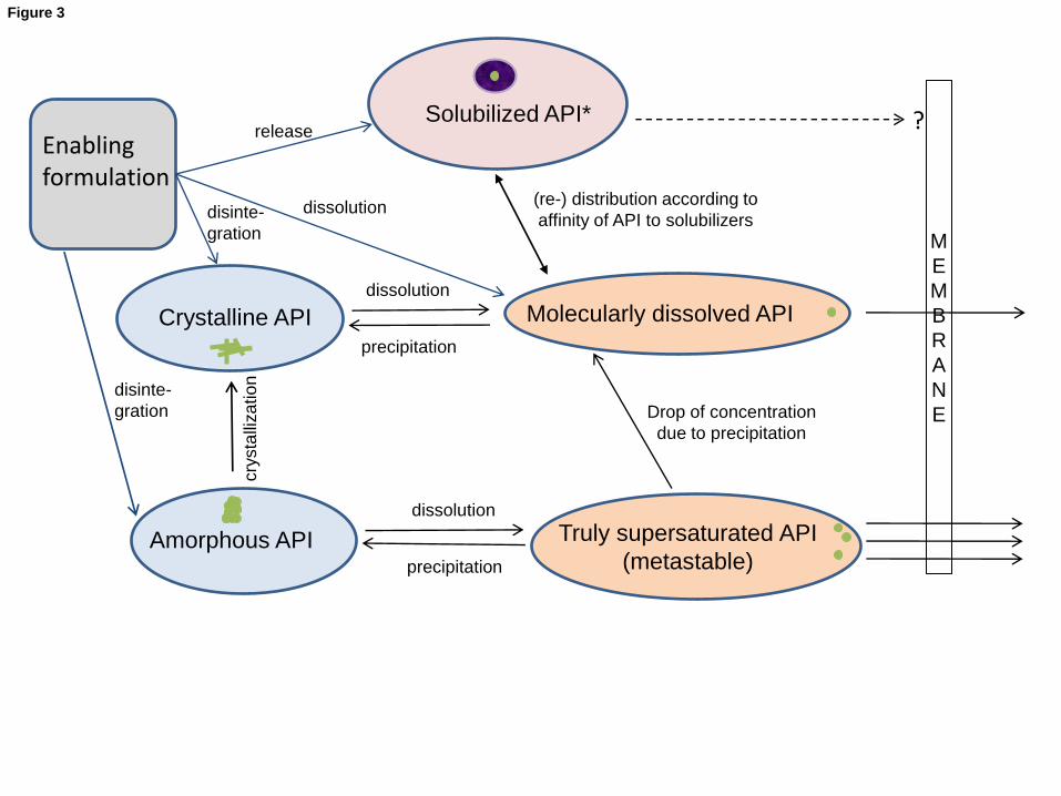
3) For detailed mechanistic studies, however, we would suggest to investigate the kinetics of all the
inter‐related processes (figure 3) separately (Van Speybroeck et al., 2012) to avoid misleading
assumptions (Grassi et al., 2002):
Drug dissolution rate: amount of molecularly dissolving drug over time from the solid state
Drug release rate(s): amount of molecularly dissolved drug released from each of the other states
such as solubilized or nanoparticulate states.
Ideally, such a kinetic investigation of dissolution and release processes should take into account
intraluminal volume changes, digestion of excipients etc., in order to accurately reflect the in vivo
situation.
Barrier flux: amount of drug permeating across the barrier over time
Finaly, one should emphasize that it is unclear for the time being if refined in vitro studies, as suggested
here, are appropriate to yield better in vitro in vivo correlations than the currently employed methods.
References
Amidon, G.L., Lennernas, H., Shah, V.P., Crison, J.R., 1995. A theoretical basis for a biopharmaceutic drug classification: The correlation of in vitro drug product dissolution and in vivo bioavailability Pharm. Res., 12 (3), 413‐420.
Anby, M. U., Williams, H. D., McIntosh, M., Benameur, H., Edwards, G. A., Pouton, C. W., Porter, C. J. H., 2012. Lipid digestion as a trigger for supersaturation: Evaluation of the impact of supersaturation stabilization on the in vitro and in vivo performance of self‐emulsifying drug delivery systems. Mol. Pharm. 9(7), 2063‐2079.
Araya, H., Tomita, M., Hayashi, M., 2005. The novel formulation design of O/W microemulsion for improving the gastrointestinal absorption of poorly water soluble compounds. Int. J. Pharm., 305(1‐2), 61‐74.
Artursson, P., Palm, K., Luthman, K. , 2001. Caco‐2 monolayers in experimental and theoretical predictions of drug transport. Adv. Drug Del. Rev., 46(1‐3), 27‐43.
Barakat, N.S., 2010. Enhanced oral bioavailability of etodolac by self‐emulsifying systems: In‐vitro and in‐vivo evaluation. J. Pharm. Pharmacol., 62 (2), 173‐180.
Beig, A., Miller, J. M., Dahan, A., 2012. Accounting for the solubility‐permeability interplay in oral formulation development for poor water solubility drugs: The effect of PEG‐400 on carbamazepine absorption. Eur. J. Pharm. Biopharm., 81(2), 386‐391.
Berginc, K., Trontelj, J., Kristl, A., 2012. Bio‐relevant media to assess drug permeability: Sodium taurocholate and lecithin combination or crude bile? Int. J. Pharm., 429(1‐2), 22‐30.
Brewster, M. E., Vandecruys, R., Peeters, J., Neeskens, P., Verreck, G., & Loftsson, T., 2008. Comparative interaction of 2‐hydroxypropyl‐β‐cyclodextrin and sulfobutylether‐β‐cyclodextrin with itraconazole: Phase‐solubility behavior and stabilization of supersaturated drug solutions. Eur. J. Pharm. Sci., 34(2‐3), 94‐103.
Brouwers, J., Tack, J., Lammert, F. & Augustijns, P. 2006, Intraluminal drug and formulation behavior and integration in in vitro permeability estimation: A case study with amprenavir, J. Pharm. Sci. 95(2), 372‐383.
Brouwers, J., Brewster, M. E., & Augustijns, P., 2009. Supersaturating drug delivery systems: The answer to solubility‐limited oral bioavailability? J. Pharm. Sci., 98(8), 2549‐2572.
Buch, P., Langguth, P., Kataoka, M., Yamashita, S., 2009. IVIVC in oral absorption for fenofibrate immediate release tablets using a dissolution/permeation system J. Pharm. Sci., 98 (6), 2001‐2009.
Buch, P., Holm, P., Thomassen, J.Q., Scherer, D., Branscheid, R., Kolb, U., Langguth, P., 2010a. IVIVC for fenofibrate immediate release tablets using solubility and permeability as in vitro predictors for pharmacokinetics J. Pharm. Sci., 99 (10), 4427‐4436.
Buch, P., Holm, P., Thomassen, J.Q., Scherer, D., Kataoka, M., Yamashita, S., Langguth, P., 2010b. IVIVR in oral absorption for fenofibrate immediate release tablets using dissolution and dissolution permeation methods. Pharmazie, 65 (10), 723‐728. .
Chiu, Y. ‐., Higaki, K., Neudeck, B. L., Barnett, J. L., Welage, L. S., & Amidon, G. L., 2003. Human jejunal permeability of cyclosporin A: Influence of surfactants on P‐glycoprotein efflux in caco‐2 cells. Pharm. Res., 20(5), 749‐756.
Cuestas M.L., Sosnik A., Mathet V.L., 2011. Poloxamines display a multiple inhibitory activity of ATP‐binding cassette (ABC) transporters in cancer cell lines. Mol. Pharm. 8:1152‐64.
Cui, J., Yu, B., Zhao, Y., Zhu, W., Li, H., Lou, H., & Zhai, G., 2009. Enhancement of oral absorption of curcumin by self‐microemulsifying drug delivery systems. Int. J. Pharm., 371(1‐2), 148‐155.
Dahan, A., Miller, J. M., Hoffman, A., Amidon, G. E., & Amidon, G. L., 2010. The solubility‐permeability interplay in using cyclodextrins as pharmaceutical solubilizers: Mechanistic modeling and application to progesterone. J. Pharm. Sci., 99(6), 2739‐2749.
Do, T.T., Van Speybroeck, M., Mols, R., Annaert, P., Martens, J., Van Humbeeck, J., Vermant, J., (...), Van Den Mooter, G., 2011. The conflict between in vitro release studies in human biorelevant media and the in vivo exposure in rats of the lipophilic compound fenofibrate. Int. J. Pharm., 414 (1‐2), 118‐124.
Dressman, J. B., Amidon, G. L., Reppas, C., & Shah, V. P., 1998. Dissolution testing as a prognostic tool for oral drug absorption: Immediate release dosage forms. Pharm. Res., 15(1), 11‐22.
Ehehalt, R., Füllekrug, J., Pohl, J., Ring, A., Herrmann, T., Stremmel W., 2006. Translocation of long chain fatty acids across the plasma membrane‐lipid rafts and fatty acid transport proteins. Mol. Cell. Biochem., 284(1‐2),135‐40
Fischer, S.M., 2012. In Vitro Permeability Screening of Poorly Soluble Drugs: Comparison of the Phospholipid Vesicle‐Based Permeation Assay and the Caco‐2 Model with Oral Bioavailability. PhD‐thesis University of Southern Denmark, Odense Denmark.

Fischer, S. M., Brandl, M., & Fricker, G., 2011a. Effect of the non‐ionic surfactant poloxamer 188 on passive permeability of poorly soluble drugs across caco‐2 cell monolayers. Eur. J. Pharm. Biopharm., 79(2), 416‐422.
Fischer, S. M., Flaten, G. E., Hagesæther, E., Fricker, G., & Brandl, M., 2011b. In‐vitro permeability of poorly water soluble drugs in the phospholipid vesicle‐based permeation assay: The influence of nonionic surfactants. J. Pharm. Pharmacol., 63(8), 1022‐1030.
Fischer, S.M., Buckley, S.T., Kirchmeyer, W., Fricker, G., Brandl, M., 2012c. Application of simulated intestinal fluid on the phospholipid vesicle‐based drug permeation assay. Int J Pharm. 422(1‐2), 52‐58.
Flaten, G. E., Luthman, K., Vasskog, T., & Brandl, M., 2008. Drug permeability across a phospholipid vesicle‐based barrier. 4. the effect of tensides, co‐solvents and pH changes on barrier integrity and on drug permeability. Eur. J. Pharm. Sci., 34(2‐3), 173‐180.
Fossati, L., Dechaume, R., Hardillier, E., Chevillon, D., Prevost, C., Bolze, S., Maubon, N., 2008. Use of simulated intestinal fluid for Caco‐2 permeability assay of lipophilic drugs. Int. J. Pharm. 360, 148‐155.
Frank, K.J., 2012. Investigations into Melt Extrudates: Particles, Solubility and Permeability. PhD‐thesis University of Southern Denmark, Odense Denmark.
Frank, K. J., Westedt, U., Rosenblatt, K. M., Hölig, P., Rosenberg, J., Mägerlein, M., Brandl, M., Fricker, G., 2012a. Impact of FaSSIF on the solubility and dissolution‐/permeation rate of a poorly water‐soluble compound. Eur. J. Pharm. Sci., 47(1), 16‐20.
Frank, K. J., Westedt, U., Rosenblatt, K. M., Hölig, P., Rosenberg, J., Mägerlein, M., Fricker, G., Brandl, M., 2012b. The amorphous solid dispersion of the poorly soluble ABT‐102 forms nano/microparticulate structures in aqueous medium: Impact on solubility. Int. J. Nanomedicine, 7, 5757‐5768.
Frank, K. J., Rosenblatt, K. M., Westedt, U., Hölig, P., Rosenberg, J., Mägerlein, M., Fricker, G., Brandl, M., 2012c. Amorphous solid dispersion enhances permeation of poorly soluble ABT‐102: True supersaturation vs. apparent solubility enhancement. Int. J. Pharm., 437(1‐2), 288‐293.
Galia, E., Nicolaides, E., Hörter, D., Löbenberg, R., Reppas, C., & Dressman, J. B., 1998. Evaluation of various dissolution media for predicting in vivo performance of class I and II drugs. Pharm. Res., 15(5), 698‐705.
Grassi, M., Coceani, N. & Magarotto, L. 2002, Modelling partitioning of sparingly soluble drugs in a two‐phase liquid system. Int. J. Pharm. 239(1‐2), 157‐169.
Grundy, J.S., Anderson, K.E., Rogers, J.A. & Foster, R.T. 1997, "Studies on dissolution testing of the nifedipine gastrointestinal therapeutic system. I. Description of a two‐phase in vitro dissolution test", Journal of Controlled Release, vol. 48, no. 1, pp. 1‐8.
Guo S, Zhang X, Gan L, Zhu C, Gan Y., 2010) Effect of poly (ethylene oxide)‐poly (propylene oxide)‐poly (ethylene oxide) micelles on pharmacokinetics and intestinal toxicity of irinotecan hydrochloride: potential involvement of breast cancer resistance protein (ABCG2). J Pharm Pharmacol. 62:973‐84
Gursoy, R. N., & Benita, S., 2004. Self‐emulsifying drug delivery systems (SEDDS) for improved oral delivery of lipophilic drugs. Biomedicine and Pharmacotherapy, 58(3), 173‐182.
Holm, R., Jensen, I. H. M., & Sonnergaard, J., 2006. Optimization of self‐microemulsifying drug delivery systems (SMEDDS) using a D‐optimal design and the desirability function. Drug Dev. Ind. Pharm., 32(9), 1025‐1032.
Holmstock, N., De Bruyn, T., Bevernage, J., Annaert, P., Mols, R. Tack, J., Augustijns, P., 2013. Exploring food effects on indinavir absorption with human intestinal fluids in the mouse intestine. Eur. J. Pharm. Sci. in press

Ingels, F., Deferme, S., Destexhe, E., Oth, M., Van Den Mooter, G., Augustijns, P., 2002. Simulated intestinal fluid as transport medium in the Caco‐2 cell culture model. Int. J. Pharm., 232 (1‐2), 183‐192.
Ingels, F., Beck, B., Oth, M., & Augustijns, P., 2004. Effect of simulated intestinal fluid on drug permeability estimation across caco‐2 monolayers. Int. J. Pharm., 274(1‐2), 221‐232.
Jones, H.M., Parrott, N., Ohlenbusch, G., Lavé, T., 2006. Predicting pharmacokinetic food effects using biorelevant solubility media and physiologically based modelling. Clinical Pharmacokinetics, 45 (12), 1213‐1226.
Kanzer, J., Tho, I., Flaten, G.E., Hölig, P., Fricker, G., Brandl, M., 2010a. In‐vitro permeability screening of melt extrudate formulations containing poorly water‐soluble drug compounds using the phospholipid vesicle‐based barrier. J. Pharm. Pharmacol., 62 (11), 1591‐1598.
Kanzer, J., Hupfeld, S., Vasskog, T., Tho, I., Hölig, P., Mägerlein, M., Fricker, G. & Brandl, M., 2010b. In situ formation of nanoparticles upon dispersion of melt extrudate formulations in aqueous medium assessed by asymmetrical flow field‐flow fractionation. J. Pharm. Biomed. Analysis, 53(3), 359‐365
Kataoka, M., Masaoka, Y., Sakuma, S., Yamashita, S., 2006. Effect of food intake on the oral absorption of poorly water‐soluble drugs: In vitro assessment of drug dissolution and permeation assay system. J. Pharm. Sci., 95 (9), 2051‐2061.
Kataoka, M., Sugano, K., Da Costa Mathews, C., Wong, J. W., Jones, K. L., Masaoka, Y., Yamashita, S., 2012. Application of dissolution/permeation system for evaluation of formulation effect on oral absorption of poorly water‐soluble drugs in drug development. Pharm. Res., 29(6), 1485‐1494.
Katneni, K., Charman, S.A., Porter, C.J.H., 2006. Permeability assessment of poorly water‐soluble compounds under solubilizing conditions: The reciprocal permeability approach. J. Pharm. Sci., 95 (10), 2170‐2185.
Khan, M.S., 2010. Solid dispersions: Formulation, characterisation, permeability and genomic evaluationPhD‐thesis, Aston University, Aston, UK.
Larsen, A. T., Ohlsson, A. G., Polentarutti, B., Barker, R. A., Phillips, A. R., Abu‐Rmaileh, R., Müllertz, A., 2013. Oral bioavailability of cinnarizine in dogs: Relation to SNEDDS droplet size, drug solubility and in vitro precipitation. Eur. J. Pharm. Sci., 48(1), 339‐350.
Li, S., Pollock‐Dove, C., Dong, L. C., Chen, J., Creasey, A. A., & Dai, W., 2012. Enhanced bioavailability of a poorly water‐soluble weakly basic compound using a combination approach of solubilization agents and precipitation inhibitors: A case study. Molecular Pharmaceutics, 9(5), 1100‐1108.
Lind, M.L., Jacobsen, J., Holm, R., Müllertz, A., 2007. Development of simulated intestinal fluids containing nutrients as transport media in the Caco‐2 cell culture model: Assessment of cell viability, monolayer integrity and transport of a poorly aqueous soluble drug and a substrate of efflux mechanisms. Eur. J. Pharm. Sci., 32 (4‐5), 261‐270.
Lindfors, L., Forssén, S., Westergren, J., & Olsson, U., 2008. Nucleation and crystal growth in supersaturated solutions of a model drug. J. Colloid Interf. Sci., 325(2), 404‐413.
Linn, M., Collnot, E.‐M., Djuric, D., Hempel, K., Fabian, E., Kolter, K., Lehr, C.‐M., 2012. Soluplus ® as an effective absorption enhancer of poorly soluble drugs in vitro and in vivo. Eur. J. Pharm. Sci., 45 (3), 336‐343.
Mellaerts, R., R. Mols, J. A. G. Jammaer, C. A. Aerts, P. Annaert, J. Van Humbeeck, G. Van den Mooter, P. Augustijns, and J. A. Martens. 2008. Increasing the Oral Bioavailability of the Poorly Water Soluble Drug Itraconazole with Ordered Mesoporous Silica. Eur. J. Pharm. Biopharm. 69 (1), 223‐230.
Mellaerts, R., Aerts, A., Caremans, T. P., Vermant, J., Van Den Mooter, G., Martens, J. A., & Augustijns, P., 2010. Growth of itraconazole nanofibers in supersaturated simulated intestinal fluid. Mol. Pharm. 7(3), 905‐913.
Miller, J.M., Beig, A., Krieg, B.J., Carr, R.A., Borchardt, T.B., Amidon, G.E., Amidon, G.L., , Dahan, A., 2011. The solubility‐permeability interplay: Mechanistic modeling and predictive application of the impact of micellar solubilization on intestinal permeation. Mol. Pharm., 8 (5), 1848‐1856.
Miller, J. M., & Dahan, A. , 2012a. Predicting the solubility‐permeability interplay when using cyclodextrins in solubility‐enabling formulations: Model validation. Int. J. Pharm., 430(1‐2), 388‐391.
Miller, J. M., Beig, A., Carr, R. A., Webster, G. K., & Dahan, A., 2012b. The solubility‐permeability interplay when using cosolvents for solubilization: Revising the way we use solubility‐enabling formulations. Mol. Pharm. 9(3), 581‐590.
Miller, J.M., Beig, A., Carr, R.A., Spence, J.K., Dahan, A., 2012c. A win‐win solution in oral delivery of lipophilic drugs: Supersaturation via amorphous solid dispersions increases apparent solubility without sacrifice of intestinal membrane permeability. Mol. Pharm. 9 (7), 2009‐2016.
Mudra, D.R., Borchardt, R.T., 2010. Absorption barriers in the rat intestinal mucosa. 3: Effects of polyethoxylated solubilizing agents on drug permeation and metabolism. J. Pharm. Sci., 99 (2), 1016‐1027.
Nerurkar, M. M., Ho, N. F. H., Burton, P. S., Vidmar, T. J., & Borchardt, R. T. (1997). Mechanistic roles of neutral surfactants on concurrent polarized and passive membrane transport of a model peptide in caco‐2 cells. J. Pharm. Sci., 86(7), 813‐821.
Overhoff, K.A., McConville, J.T., Yang, W., Johnston, K.P., Peters, J.I., Williams III, R.O., 2008. Effect of stabilizer on the maximum degree and extent of supersaturation and oral absorption of tacrolimus made by ultra‐rapid freezing. Pharm. Res., 25 (1), 167‐175.
Palm, K., Stenberg, P., Luthman, K., & Artursson, P., 1997. Polar molecular surface properties predict the intestinal absorption of drugs in humans. Pharm. Res., 14(5), 568‐571.
Patel, N., Forbes, B., Eskola, S., Murray, J., 2006. Use of simulated intestinal fluids with Caco‐2 cells and rat ileum. Drug Dev. Ind. Pharm., 32 (2), 151‐161.
Phillips, D.J., Pygall, S.R., Brett Cooper, V. & Mann, J.C., 2012. Toward biorelevant dissolution: Application of a biphasic dissolution model as a discriminating tool for HPMC matrices containing a model BCS class II drug. Dissolution Technologies19(1), 25‐34.
Poelma, F.G.J., Breas, R., Tukker, J.J., Crommelin, D.J.A., 1991. Intestinal absorption of drugs. The influence of mixed micelles on the disappearance kinetics of drugs from the small intestine of the rat. J. Pharm. Pharmacol., 43 (5), 317‐324. Pouton, C. W., & Porter, C. J. H., 2008. Formulation of lipid‐based delivery systems for oral administration: Materials, methods and strategies. Adv. Drug Delivery Rev., 60(6), 625‐637.
Pouton, C.W., 2000. Lipid formulations for oral administration of drugs: Non‐emulsifying, self‐emulsifying and 'self‐microemulsifying' drug delivery systems. Eur. J. Pharm. Sci., 11 (SUPPL. 2), S93‐S98.
Porter, C.J., Trevaskis, N.L., Charman, W.N., 2007. Lipids and lipid‐based formulations: optimizing the oral delivery of lipophilic drugs. Nat. Rev. Drug. Discov., 6, 231‐48.
Rahman, M. A., Hussain, A., Hussain, M. S., Mirza, M. A., & Iqbal, Z. , 2013. Role of excipients in successful development of self‐emulsifying/ microemulsifying drug delivery system (SEDDS/SMEDDS). Drug Dev. Ind. Pharm., 39(1), 1‐19.
Rege, B.D., Yu Lawrence, X., Hussain, A.S., Polli, J.E., 2001. Effect of common excipients on Caco‐2 transport of low‐permeability drugs. J. Pharm. Sci., 90 (11), 1776‐1786.

Schwarz M.A., Neubert R.H., Dongowski G., 1996. Characterization of interactions between bile salts and drugs by micellar electrokinetic capillary chromatography. Part I. Pharm Res. 13(8), 1174‐80.
Schwebel, H.J., Van Hoogevest, P., Leigh, M.L.S., Kuentz, M., 2011. The apparent solubilizing capacity of simulated intestinal fluids for poorly water‐soluble drugs. Pharm. Dev. Technology, 16 (3), 278‐286.
Shaik, N., Pan, G., Elmquist, W.F., 2008. Interactions of pluronic block copolymers on P‐GP efflux activity: Experience with HIV‐1 protease inhibitors. (2008) J. Pharm. Sci., 97 (12), 5421‐5433.
Singh, A., Worku, Z.A., Van Den Mooter, G., 2011. Oral formulation strategies to improve solubility of poorly water‐soluble drugs Expert Opinion on Drug Delivery, 8 (10), 1361‐1378.
Singh, B., Singh, R., Bandyopadhyay, S., Kapil, R., & Garg, B., 2013. Optimized nanoemulsifying systems with enhanced bioavailability of carvedilol. Coll. Surf. B: Biointerfaces, 101, 465‐474.
Stillhart, C., Cavegn, M., & Kuentz, M., 2012. Study of drug supersaturation for rational early formulation screening of surfactant/co‐solvent drug delivery systems. J. Pharm. Pharmacol.,
Tho, I., Liepold, B., Rosenberg, J., Maegerlein, M., Brandl, M. & Fricker, G., 2010. Formation of nano/micro‐dispersions with improved dissolution properties upon dispersion of ritonavir melt extrudate in aqueous media. Eur. J. Pharm. Sci., 40, 25‐32.
Thomas, N., Holm, R., Müllertz, A., & Rades, T., 2012. In vitro and in vivo performance of novel supersaturated self‐nanoemulsifying drug delivery systems (super‐SNEDDS). J. Contr. Rel., 160(1), 25‐32.
Tønsberg, H., Holm, R., Boll, J.B., Jacobsen, J., Müllertz, A., 2010. Effects of polysorbate 80 on the in‐vitro precipitation and oral bioavailability of halofantrine from polyethylene glycol 400 formulations in rats. J. Pharm. Pharmacol., 62 (1), 63‐70.
Ujhelyi, Z., Fenyvesi, F., Váradi, J., Fehér, P., Kiss, T., Veszelka, S., Bácskay, I., 2012. Evaluation of cytotoxicity of surfactants used in self‐micro emulsifying drug delivery systems and their effects on paracellular transport in caco‐2 cell monolayer. Eur. J. Pharm. Sci., 47(3), 564‐573.
Ungell, A. 2004. Caco‐2 replace or refine? Drug Discovery Today. 1(4), 423‐430
Van Hoogevest, P., Liu, X., & Fahr, A., 2011. Drug delivery strategies for poorly water‐soluble drugs: The industrial perspective. Expert Opinion on Drug Delivery, 8(11), 1481‐1500.
Van Speybroeck, M., Mellaerts, R., Mols, R., Thi, T.D., Martens, J.A., Van Humbeeck, J., Annaert, P., Augustijns, P., 2010. Enhanced absorption of the poorly soluble drug fenofibrate by tuning its release rate from ordered mesoporous silica. Eur. J. Pharm. Sci., 41 (5), 623‐630.
Vangani, S., Li, X., Zhou, P., Del‐Barrio, M.‐A., Chiu, R., Cauchon, N., Gao, P., Jasti, B., 2009. Dissolution of poorly water‐soluble drugs in biphasic media using USP 4 and fiber optic system. Clinical Research and Regulatory Affairs, 26 (1‐2), 8‐19.
Varma, M.V.S., Panchagnula, R., 2005. Enhanced oral paclitaxel absorption with vitamin E‐TPGS: Effect on solubility and permeability in vitro, in situ and in vivo. Eur. J. Pharm. Sci., 25 (4‐5), 445‐453.
Vertzoni, M., Markopoulos, C., Symillides, M., Goumas, C., Imanidis, G., Reppas, C., 2012. Luminal lipid phases after administration of a triglyceride solution of danazol in the fed state and their contribution to the flux of danazol across Caco‐2 cell monolayers. Mol. Pharm., 9, 1189‐98.
Whitehead, K., & Mitragotri, S., 2008. Mechanistic analysis of chemical permeation enhancers for oral drug delivery. Pharm. Res., 25(6), 1412‐1419.

Yamagata T, Morishita M, Kusuhara H, Takayama K, Benameur H, Sugiyama Y., 2009. Characterization of the inhibition of breast cancer resistance protein‐mediated efflux of mitoxantrone by pharmaceutical excipients. Int. J. Pharm. 370:216‐9.
Yano, K., Masaoka, Y., Kataoka, M., Sakuma, S., Yamashita, S., 2010. Mechanisms of membrane transport of poorly soluble drugs: Role of micelles in oral absorption processes. J. Pharm. Sci., 99 (3), 1336‐1345.

Legends to figures
Figure 1
a) Ketoprofen permeation across phospholipid vesicle based permeation assay (PVPA) barriers in the
absence and presence of surfactant poloxamer (P‐188), uncorrected and corrected (for the freely dissolved
fraction), (mean ± SD, n=6). Reprinted from Fischer, 2012, with permission
b) Nadolol permeation across phospholipid vesicle based permeation assay (PVPA) barriers in the absence
and presence of surfactant poloxamer (P‐188), uncorrected and corrected (for the freely dissolved
fraction), (mean ± SD, n=6). Reprinted from Fischer, 2012, with permission
c) Fraction of molecularly dissolved drug at different surfactant poloxamer (P‐188) concentrations, given as
percentages of the solution in phosphate buffered saline (PBS), (mean ± SD, n=3). Reprinted from Fischer,
2012, with permission
Figure 2
Transport of CsA in the apical to basolateral (A‐B) or in opposite direction (B‐A) after addition of Cyclosporin
A (CsA 1 µM) to the donor side in transport medium (TM), faste state simulated intestinal fluid (FASSIF) or
dilutions of FASSIF. Bars represent average Papp value±SD (n=3). Reprinted from Ingels et al. 2002, figure 4,
with permisssion.
Figure 3
Schematic overview over different states, in which the active pharmaceutical ingredient (API) may occur in
the gastro‐intestinal tract and their role in terms of permeability across the intestinal barrier. Solid state
comprises both, crystalline and amorphous forms; dissolved state comprises both, solubilized state,
molecularly dissolved state and truly supersaturated state.

Figure 1a
0 10 20 503.0
3.5
4.0
4.5
5.0
5.5
6.0
6.5
*
uncorrected corrected
Pa
pp
ket
opro
fen
(10-6
cm/s
)
P-188 concn (mg/ml)
*
Figure 1b
0 10 20 500.4
0.6
0.8
1.0
1.2
1.4
1.6 uncorrected corrected
Pa
pp
nad
olol
(10-6
cm/s
)
P-188 concn (mg/ml)
*
Figure(s)
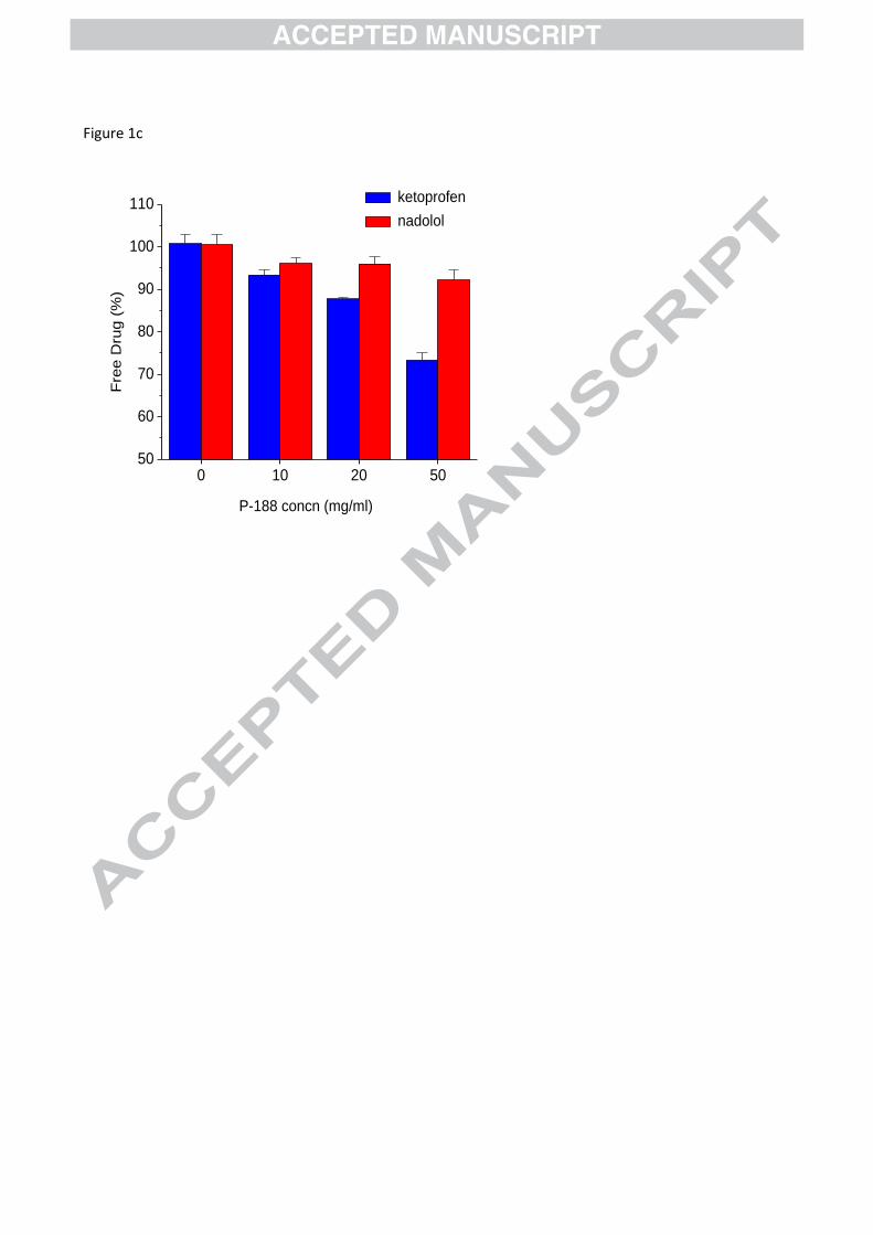
Figure 1c
0 10 20 5050
60
70
80
90
100
110
Fre
e D
rug
(%)
P-188 concn (mg/ml)
ketoprofen nadolol

Figure 2
Figure(s)
MEMB R A N E
Solubilized API*
Truly supersaturated API (metastable)
Molecularly dissolved API
Amorphous API
Crystalline API
precipitation
dissolution
(re-) distribution according to affinity of API to solubilizers
Drop of concentration due to precipitation
crys
talliz
atio
n
precipitation
dissolution
Enabling formulation
release
dissolution disinte-gration
disinte-gration
?
Figure 3