biomembrane protocols volume 27 || crystallization of membrane proteins for x-ray analysis
TRANSCRIPT
Crystallization of Membrane Proteins for X-Ray Analysis
Brian J. Sutton and Maninder K Sohi
1. Introduction 1.1. General Principles
In order to determine the structure of a protein by X-ray crystal- lography, well ordered three-dimensional crystals are required. How- ever, despite the wealth of experience accumulated in the course of the crystallization and structural analyses of several hundred soluble globular proteins and their complexes, the process of crystallization still remains something of an art, and is often the rate limiting step of any analysis. For membrane proteins that present an additional challenge by virtue of their amphipathic nature, experience is considerably more limited, and the first three-dimensional crystals suitable for X-ray analysis were only reported in 1980 (I,2). Many membrane proteins form two-dimensional arrays in situ, and these may be studied by electron microscopy and electron diffraction of tilted specimens to determine their three-dimensional structure, but only in the pioneer- ing study of bacteriorhodopsin has the resolution of the structural analysis approached that obtainable by X-ray crystallography (3). The formation of two-dimensional crystalline arrays will not be consid- ered in this chapter. The first membrane protein crystal structure, the bacterial photosynthetic reaction center complex, was solved in 1985 (4); this was followed by the second reaction center structure in 1986 (5,6), and more recently porin in 1991 (7). Since these pioneering
From Methods m Molecular Biology, Vol 27’ Blomembrane Protocols Ii Architecture and Funcbon Edlted by. J. M. Graham and J. A. Higgins
Copynght 01994 Humana Press Inc , Totowa, NJ
1

2 Sutton and Sohi
studies, increasing numbers of membrane protein crystallizations have been reported. For recent reviews, see refs. 8-11.
Protein crystals grow from solutions that have reached a state of supersaturation as a result of the addition of a precipitant, such as ammonium sulfate (AS) or polyethylene glycol (PEG). However, ini- tial solubilization of a membrane protein requires the presence of detergent, and experience has also shown that the growth of crystals of membrane proteins is often promoted by the addition of small deter- gent-like amphiphilic molecules. Thus, although the crystallization of soluble proteins commonly occurs from a four-component system (protein/precipitant/buffer/water), for membrane proteins at least a six-component system (protein/detergent/additive/precipitant/buffer/ water) is involved. The relative concentrations of these components and the nature of the detergent, additive, and precipitant must all be established, in addition to other variables, such as pH and temper- ature; crystallization is almost always found to occur under very pre- cisely defined conditions. For a few membrane proteins, the phase diagram of the system has been investigated in some detail (9,11), and certain general principles have emerged. For example, the addi- tion of precipitant to or decrease in temperature of a system contain- ing detergent and protein usually leads to separation of a detergent-rich and a detergent-depleted phase. It appears that conditions most suit- able for crystal growth are often found close to, and on the single-phase side of this phase separation boundary (9), although crystallization occasionally does occur after phase separation (2). Knowledge of the phase diagram for the system under trial is clearly helpful.
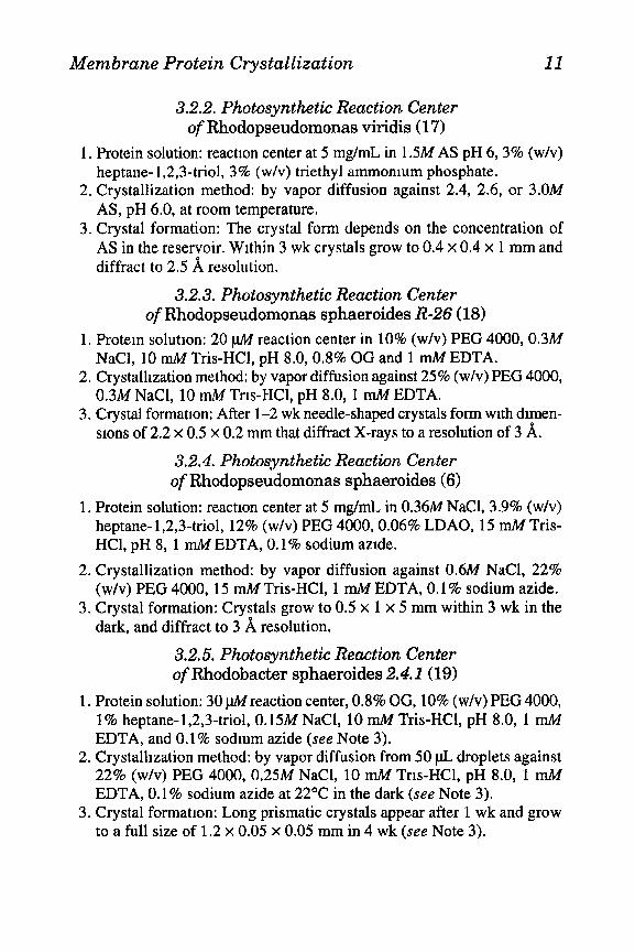
The solubilization and crystallization of membrane proteins is pre- sumed to occur as shown in Fig. 1. The amphipathic detergent molecules alone will form micelles above the critical micellar concentration (CMC), but in the presence of protein will form mixed micelles. The detergent molecules in effect form a belt around the central hydro- phobic region of the protein that is normally buried in the membrane. In crystals of membrane proteins it was supposed that contacts occur between the hydrophilic regions, and this is indeed found to be so in the structures solved to date. Michel (8) proposed that two types of crystal might occur (Fig. 1). Type I crystals essentially consist of stacked layers of two-dimensional membrane-like arrays of molecules.

Membrane Protein Crystallization 3
Fig. 1. The formatron of two types of three-drmensional crystals from mixed mrcelles of membrane protein and detergent molecules (after Mrchel [S]) The deter- gent molecules (not to scale) interact with the hydrophobic regions of the protein (indicated by broken lines), and crystal contacts are formed between the hydro- philic regions (solid lines).
Type II crystals consist of three-dimensional lattices with extensive regions of solvent that presumably accommodate the detergent mol- ecules in their micellar structure. Crystals of both types have been found, and the recent report of various crystal forms of porin exemplify both types (12). However, in no X-ray analysis to date have detergent molecules been sufficiently ordered to be seen in the structure. In one of the crystal forms of porin (12) it appears that direct, specific con- tact exists between hydrophobic regions of adjacent protein molecules, not mediated by detergent molecules at all. The nature of intermo- lecular contacts in membrane protein crystals may be found to be yet more diverse.

4 Sutton and Sohi
1.2. Operational Requirements
1.2.1. Detergents The detergents used for crystallization must be both mild and of a
high purity, and since crystal growth may take weeks or months, the protein must be completely stable in their presence. It may therefore be necessary to first remove the detergent(s) used in the initial isola- tion and purification of the protein. The detergents that have been used most successfully for crystallization are of the nonionic type, such as n-octyl-P-o-glucopyranoside (OG) and related alkyl deriva- tives, or of the zwitterionic type, such as ZVJV-dimethyl-dodecylamine oxide (lauryl dimethyl amineoxide, LDAO). A comprehensive list of detergent molecules used for crystallization is given in the review by Ktihlbrandt (10). Both OG and LDAO are small, and this may in part account for their success. The diameter of a micelle of OG or LDAO is approx 4 nm, similar to the thickness of the lipid bilayer (4.5 nm), and thus a membrane protein molecule in a mixed micelle with these detergents will have all of its hydrophilic regions available for form- ing intermolecular crystal contacts. Larger protein complexes may require larger detergent molecules.
The ratio of detergent-to-protein concentration is also crucial. The detergent concentration must be sufficient to allow solubilization of all of the protein molecules, yet not produce too many detergent-only micelles, since these may impede crystallization. Too little detergent may lead to aggregation of the protein. Often the best detergent:protein molar ratio is about 200, which corresponds to about 2.5 micelles per protein molecule for OG or LDAO. However, very different ratios (from 20 to 5000) have been used successfully.
1.2.2. Additives Some membrane proteins require the addition of small amphiphilic
molecules, such as heptane-1,2,3-trio1 or glycerol, for crystallization. Michel proposed that these smaller molecules may fit into the cavities in the hydrophobic surface regions of the protein, and/or substitute for detergent molecules in the micelles, reducing their size and allowing for better packing in the crystal lattice (8). He termed this the “small amphiphile concept.” In a recent study it has been shown that heptane- 1,2,3-trio1 does indeed reduce the mass and radius of micelles formed
Membrane Protein Crystallization 5
with LDAO (13). The presence of additives also affects the phase sepa- ration properties of a system, and often prevents separation.
1.2.3. Precipitants Once the membrane protein is solubilized, the strategies for crystalli-
zation and choice of precipitants is essentially the same as for the crys- tallization of soluble globular proteins (see ref. 14 for recent review). Low-mol-wt PEG has been used most extensively, but AS and other salts have been used, as well as 2-methyl-2,4-pentanediol (MPD).
1.2.4. Crystallization Techniques A number of techniques have been developed for the crystallization of
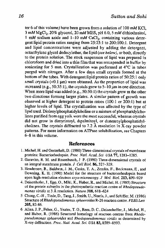
soluble proteins, and all are also suitable for membrane proteins. Detailed descriptions of these techniques may be found elsewhere (15,16), but all involve slowly changing the conditions within a sample of the protein to achieve supersaturation. The most common method for initial screening of conditions, and one that uses very small aliquots of the protein, is vapor diffusion in hanging drops. In this method, a drop of solution containing protein, buffer, detergent, additive, and pre- cipitant at a concentration below that required for precipitationkrys- tallization, is suspended from a siliconized glass cover slip above a sealed reservoir containing buffer and precipitant at a concentration slightly above that required for precipitation/crystallization. The arrangement is shown in Fig. 2A. Equilibration between drop and reservoir occurs through the vapor phase, and the precipitant concen- tration (and that of other components) in the drop increases. The rate of change of precipitant concentration in the protein solution may be controlled by the concentration difference between the drop and the reservoir, and their relative volumes. The drops are conveniently set up below glass cover slips sealed over the wells of a tissue culture plate, and the volume of each drop may be as little as l-2 l.tL.
In another vapor diffusion arrangement, the drops of protein solu- tion lie in depression wells on glass slides, sealed within a volume that also contains the reservoir solution (Fig. 2B). Diffusion across a dialysis membrane may also be used to alter conditions in the protein solution, and perspex microdialysis “buttons” of vol 5-100 pL are available for these experiments (Fig. 2C). A dialysis membrane is stretched over the cavity containing the protein solution and secured

6 Sutton and Sohi
I
LiTI I3 J’ D c 9 p D
Fig. 2. Schematic diagrams of apparatus for micro-scale protein crystallization P: protein solution, R: reservoir solution (with whrch protem solution equrhbrates); D: dialysis membrane. A: Vapor diffusion in hanging drops. B: Vapor diffusion in sitting drops. C: Microdialysis “buttons.” D: Dialysis in capillary tubes,
with an O-ring, and the button is then placed in the reservoir solution. Alternatively, a dialysis membrane may be used to seal one end of a capillary tube containing the protein solution that is then placed in the reservoir solution (Fig. 2D). Equilibration across a dialysis membrane is considerably more rapid than via the vapor phase. Other methods involve free diffusion across the interface between two solutions layered one upon the other in a sealed capillary tube, or across a thin liquid bridge between two drops, one of protein, the other of precipitant.
Finally, robotic systems for protein crystallization are now avail- able and under development. These involve automated micropipeting of solutions for vapor diffusion or free interface diffusion, and have the advantages of ease of use and reproducibility.
1.2.5. General Strategy The initial step in crystallization strategy is to scan ranges of protein,
detergent, additive, and precipitant concentrations, at different pH and temperatures, to establish first the solubility properties of the system
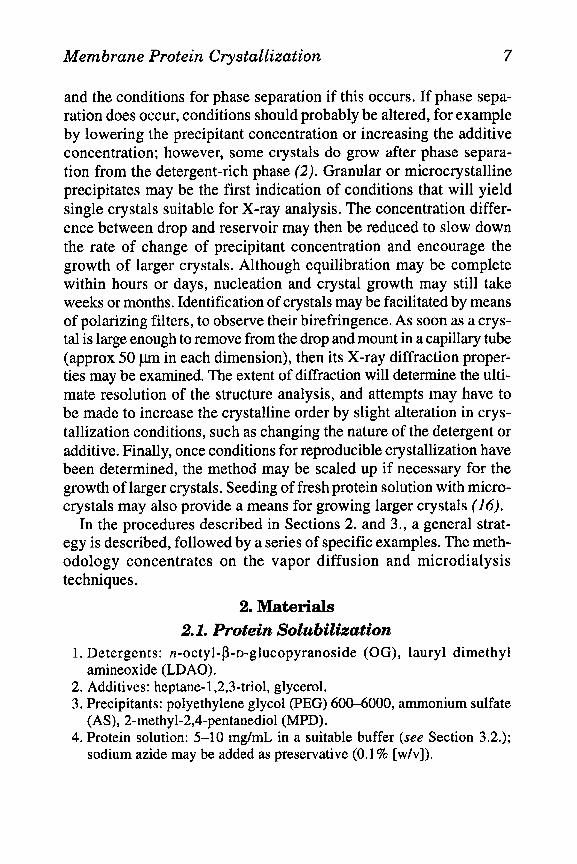
Membrane Protein Crystallization 7
and the conditions for phase separation if this occurs. If phase sepa- ration does occur, conditions should probably be altered, for example by lowering the precipitant concentration or increasing the additive concentration; however, some crystals do grow after phase separa- tion from the detergent-rich phase (2). Granular or microcrystalline precipitates may be the first indication of conditions that will yield single crystals suitable for X-ray analysis. The concentration differ- ence between drop and reservoir may then be reduced to slow down the rate of change of precipitant concentration and encourage the growth of larger crystals. Although equilibration may be complete within hours or days, nucleation and crystal growth may still take weeks or months. Identification of crystals may be facilitated by means of polarizing filters, to observe their birefringence. As soon as a crys- tal is large enough to remove from the drop and mount in a capillary tube (approx 50 l.trn in each dimension), then its X-ray diffraction proper- ties may be examined. The extent of diffraction will determine the ulti- mate resolution of the structure analysis, and attempts may have to be made to increase the crystalline order by slight alteration in crys- tallization conditions, such as changing the nature of the detergent or additive. Finally, once conditions for reproducible crystallization have been determined, the method may be scaled up if necessary for the growth of larger crystals. Seeding of fresh protein solution with micro- crystals may also provide a means for growing larger crystals (16).
In the procedures described in Sections 2. and 3., a general strat- egy is described, followed by a series of specific examples. The meth- odology concentrates on the vapor diffusion and microdialysis techniques.
2. Materials 2.1. Protein Solubilization
1. Detergents: n-octyl-fi-n-glucopyranoside (OG), lauryl dimethyl amineoxide (LDAO).
2. Additives: heptane-1,2,3-triol, glycerol. 3. Precipitants: polyethylene glycol (PEG) 600-6000, ammonium sulfate
(AS), 2-methyl-2,6pentanediol (MPD). 4. Protein solution: 5-10 mg/mL in a suitable buffer (see Section 3.2.);
sodium azide may be added as preservative (0.1% [w/v]).
8 Sutton and Sohi
2.2. Vapor Diftfision 1. For small scale work use 24-well tissue culture plates (Linbro, Flow
Laboratories, Inc., McLean, VA), or Crystalplates for protein crystalli- zation (Flow Laboratories, Inc.).
2. For large scale vapor diffusion in sitting drops: PyrexTM glass depres- sion spot plates, 3 or 9 well (Corning Glass Works; J. Bibby Science Products, Ltd., Stone, UK).
3. Microscope cover slips. 4. Dimethyldichlorosilane solution for silicomzation. 5. Microscope and glass slides.
2.3. Microdialysis 1. Perspex microdialysis buttons, 5-100 pL vol (Cambridge Repetition
Engineers, Ltd., Cambridge, UK). 2. Thick-walled glass capillary tubes; internal diameter 1 mm, external
diameter 7 mm, length 20-30 mm. 3. Dialysis membrane. 4. Plastic tubing to fit the capillary tubes (make sure it fits tightly).
2.4. Protein Solution Concentration 1. Vacuum desiccator with phosphorus pentoxide. 2. Ultrafiltration apparatus.
3. Methods 3.1. General Strategy
A range of different detergents and detergent concentrations should be investigated first, and the effect of the addition of small amphiphiles tested subsequently. Experiments should be set up at various pHs. Tem- perature variation may also be investigated, as well as alternative buffer systems, and the effect of addition of NaCl. The extent of the initial screen of conditions will depend on the quantity of protein available (see Note 1).
3.1.1. Vapor Difusion Before setting up hanging or sitting drops, obtain an indication of
potential crystallization conditions from simple preliminary experi- ments (see steps l-3 and Note 2).
1. Layer 10 pL drops of protein solution (with buffer and detergent) on 10 pL drops of precipitant solution on a microscope slide.

Membrane Protein Crystallization 9
2. Place a cover slip over the drops and seal m a container with the reser- voir solution (see step 5) to maintain humidity.
3. Observe under the microscope for signs of phase separation and/or micro- crystalline precipitate.
4. Prepare glass cover slips: Silicomze by coating with dimethyldtchloro- silane and allow to dry in a fume cupboard. Then wash with distilled water and dry in oven. Ensure they are dust free.
5. Place reservoir solutions (200-500 pL precipitant in buffer at chosen concentrattons) in the wells of the tissue culture plate or in depressions of a spot plate (leaving the other depressions for the protein solution).
6. Add detergent (OG or LDAO) to desired concentration (typical range 0.25-l% [w/v]) and small amphiphile additive (e.g., heptane-1,2,3-triol) to desired concentration (typically l-5% [w/v]) to the protein solution.
7. Add precipitant (PEG 600, 2000, 4000, 6000; AS; MPD) to desired concentratton (PEG typically 5-15% [w/v]; AS typically l-2M; MPD typically 5-20% [v/v]). The concentration of precipitant should be lower than that in the reservoir.
8. Centrifuge the protein solution to clarify and suspend 2-5 pL from glass cover slips.
9. Check that the drops are clear and seal cover slips over wells with vis- cous oil or vacuum grease (see Fig. 2A).
10. Alternatively place 5-50 pL clarified protein solution in each of the remaining depressions of a spot plate and seal in a clear plastic box (see Fig. 2B).
11. Store trays or plates at constant, defined temperature. 12. Observe after several hours, and regularly thereafter. The results ~111
dictate the choice of conditions for subsequent trial conditions.
3.1.2. Microdialysis in Specially Designed “Buttons” 1. Place the protein solution containing detergent, additive, and precipr-
tant in the perspex button (~015-100 pL) so that the meniscus is raised above the level of the rim of the well. Refer to Section 3.1.1. for details of typical concentration ranges.
2. Stretch a piece of presoaked dialysis membrane over the button and hold it in place with the O-ring, taking care that no au bubbles are trapped under it (as shown m Fig. 2C).
3. Place the button in a sealed glass bottle containmg sufficient reservoir solution to cover the button. The reservoir solutton should contam deter- gent, additive, and precipitant, the latter at a higher concentration than that m the protein solution.
4. Store at a constant temperature.

10 Sutton and Sohi
5. Monitor the contents of the button for crystal formation under an inverted microscope (without removing it from the reservoir solution).
6. If necessary, change the dialysis conditions by transferring the button to different reservoir solutions.
3.1.3. Microdialysis in Capillary Tubes 1. Place a piece of dialysis membrane over one end of a capillary tube and
secure it with a ring of plastic tubing. 2. Cut the plastic tubing (see Fig. 2D) to provide a support for the capil-
lary tube and to allow access of the reservoir solution to the dialysis membrane.
3. Place protein solution (typically 5-20 pL) containing detergent, additive, and precipitant in the capillary, ensuring that no air bubbles are intro- duced. Refer to Section 3.1.1. for details of typical concentration ranges.
4. Seal the other end of the tube with a wax plug or Parafilm. 5. Place the capillary tube m a sealed glass bottle containing sufficient
reservoir solution to cover the tube. See Section 3.1.2. for composition of reservoir solution.
6. Store at a constant temperature. 7. At intervals, briefly remove the tube for exammation under the micro-
scope. 8. Change dialysis conditions if necessary by transferring the capillary to
different reservoir solution(s).
3.2. Specific Crystallization Procedures
3.2.1. Bacteriorhodopsin from Purple Membrane of Halobacterium halobium RIMI (1)
1. Protein solution: Suspend purple membranes (0.2 pm01 bacteriorhodop- sin) in 5 mL of 20 mM sodium phosphate or AS pH 5.0, with 1% OG, and stir overnight at 33°C. After centrifugation at 150,OOOg for 1 h, adjust the concentration of AS or sodium phosphate in the supernatant to 0.5M by adding a concentrated solutton at pH 5.0, and concentrate the solu- tion four-fold in a desiccator containing phosphorus pentoxide. Remove any bacteriorhodopsin precipitate by centrifugation at 8000g for 2 min.
2. Crystallization method: by vapor diffusion against a large reservoir of 2.8M sodium phosphate pH 4.8, or 2.5M AS (unbuffered), at 23OC for up to 1 wk.
3. Crystal formation: The crystals formed should diffract X-rays to a reso- lution of about 8 A.
Membrane Protein Crystallization 11
3.2.2. Photosynthetic Reaction Center of Rhodopseudomonas viridis (17)
1. Protein solution: reaction center at 5 mg/mL in 1.5M AS pH 6, 3% (w/v) heptane- 1,2,3-triol, 3% (w/v) triethyl ammomum phosphate.
2. Crystallization method: by vapor diffusion against 2.4, 2.6, or 3.OM AS, pH 6.0, at room temperature.
3. Crystal formation: The crystal form depends on the concentration of AS in the reservoir. Within 3 wk crystals grow to 0.4 x 0.4 x 1 mm and diffract to 2.5 8, resolution.
3.2.3. Photosynthetic Reaction Center of Rhodopseudomonas sphaeroides R-26 (18)
1. Protein solutron: 20 pM reaction center in 10% (w/v) PEG 4000,0.3M NaCl, 10 rrM Tris-HCl, pH 8.0,0.8% OG and 1 mM EDTA.
2. Crystallization method: by vapor diffusion against 25% (w/v) PEG 4000, 0.3M NaCl, 10 mM Trrs-HCl, pH 8,0, 1 mM EDTA.
3. Crystal formatron: After l-2 wk needle-shaped crystals form wrth drmen- srons of 2.2 x 0.5 x 0.2 mm that diffract X-rays to a resolution of 3 A.
3.2.4. Photosynthetic Reaction Center of Rhodopseudomonas sphaeroides (6)
1, Protein solution: reaction center at 5 mg/mL in 0.36M NaCl, 3.9% (w/v) heptane- 1,2,3-triol, 12% (w/v) PEG 4000,0.06% LDAO, 15 mZi4 Tris- HCl, pH 8, 1 n&I EDTA, 0.1% sodium azrde.
2. Crystallization method: by vapor diffusion against 0.6M NaCl, 22% (w/v) PEG 4000, 15 r&f Tris-HCl, 1 mM EDTA, 0.1% sodium azide.
3. Crystal formation: Crystals grow to 0.5 x 1 x 5 mm within 3 wk in the dark, and diffract to 3 8, resolution.
3.2.5. Photosynthetic Reaction Center of Rhodobacter sphaeroides 2.4. I (19)
1. Protein solution: 30 wreaction center, 0.8% OG, 10% (w/v) PEG 4000, 1% heptane-1,2,3-triol, 0.15M NaCl, 10 m&I Tris-HCl, pH 8.0, 1 mM EDTA, and 0.1% sodmm azide (see Note 3).
2. Crystallrzation method: by vapor diffusion from 50 pL droplets against 22% (w/v) PEG 4000, 0.25M NaCl, 10 mM Trrs-HCl, pH 8.0, 1 mM EDTA, 0.1% sodium azide at 22OC in the dark (see Note 3).
3. Crystal formation: Long prismatic crystals appear after 1 wk and grow to a full size of 1.2 x 0.05 x 0.05 mm in 4 wk (see Note 3).
12 Sutton and Sohi
3.2.6. Photosynthetic Light-Harvesting Pigment-Protein Complex BBOO-850 of Rhodopseudomonas capsulata (20)
1. Protein solution: in a buffer containing 0.08% LDAO and 140-200 mM NaCl. Concentrate the complex by Amicon ultrafiltration to 10 mg/mL. Add 6% (w/v) heptane-1,2,3-trio1 and 10% (w/v) hexyldimethylamine oxide, then mix 5 pL of a solution of 15% (w/w) PEG 4000 and 1.56M NaCl with an equal volume of the protein solution m a depression of a spot plate.
2. Crystalhzatron method: by vapor diffusion against at least 100 pL of reservoir solutron containing 12.5% (w/w) PEG 4000 and 1.3M NaCl placed in the remaining depressions at 22°C m the dark.
3. Crystal formation: Crystals of length 0.5 mm that diffract X-rays to a resolution of about 10 8, appear after l-2 wk.
3.2.7. Light-Harvesting Chlorophyll a/b Protein Complex from Pea Chloroplasts (21)
1. Protein solutron: Adjust the concentration of KC1 in the fraction con- taining the complex (see Note 4) to 300 nuI4 by adding solid KCl. After 5-10 mm at room temperature, pellet the silky crystallme precipitate by centrifugation at 25,000g for 5 min. Wash the pellet twice with 100 rniI4 KCl; once with distilled water, by resuspension and recentrifugatron and finally dissolve the pellet rn 0.7-0.9% (w/v) n-nonyl-P-n-gluco- pyranoside. Keep this stock solution containing 34 mg/mL of chloro- phyll in the dark at 4°C. MIX a small volume of this stock solution with half a volume of 75 m&I KCl, 30 mM Na/K phosphate buffer, pH 6.0, and centrifuge for 2 min in a microcentrrfuge.
2. Crystallization method: by vapor diffusion in hangmg or sitting drops against 80-l 20 rniI4 Na/K phosphate at 12-18OC.
3. Crystal formation: Hexagonal plates 150 pm across and 2-3 pm m thick- ness form within several days m drops containing 2.3 miI4 chlorophyll and a molar ratio of detergent to chlorophyll of 8-10. Dark green octa- hedral crystals of size 0.3-0.7 mm grow wrthm l-2 wk from solutions contaming 2.5 mM chlorophyll and detergent to chlorophyll ratios of 7-8.5, when equilibrated against 140 rrUV Na/K phosphate. The octahe- dral crystals diffract X-rays to about 20 8, resolution,
3.2.8. Porin from E. coli Outer Membrane (2)
1. Protein solution: 20 mg/mL protein in 20 mI14 sodium phosphate, 3 mM sodium azrde, pH 7.0 with 1% (w/v) n-octyl-a-o-glucopyranosrde (a-OG), O.lM NaCl (see Notes 2,5, and 6).
Membrane Protein Crystallization 13
2. Crystallization method: by microdialysis in capillary tubes (50 x 3 mm internal diameter) sealed at one end by a dialysis membrane. Dialyze 200~pL aliquots against 20 mM sodium phosphate, 3 rnM sodium azide, pH 7.0 containing 12% PEG 6000 and 1% a-OG at 37°C. Exchange this solution for one containing additionally 1M NaCl (see Notes 5 and 6).
3. Crystal formation: After 2 d a microcrystalline shower appears; larger crystals appear 4 d later that diffract to 3.8 8, resolution (see Note 5 and 6).
3.2.9. Porin from E. coli Outer Membrane (22) 1. Protein solution: Prepare the protein in 20 rnM sodium phosphate pH
7.0, O.lM NaCl, containing 3 mM sodium azide, 3 mM dithiothreitol, 1% (w/v) OG.
2. Crystallization method: by large scale vapor diffusion. Place 200 pL of the crystallization solution containing 5 mg/mL protein, 3.6% (w/v) PEG 4000 and 0.5% OG in the phosphate-NaCl buffer in small plastic or glass cups within a sealed vapor diffusion chamber (see step 10, Section 3.1.1.). The reservoir solution is 18% PEG 4000, 0.5M NaCl, O.lM sodium phosphate pH 7.0 (see Notes 5 and 6).
3. Crystal formation: Large crystals of dimension 0.2-0.7 mm appear after 3-6 wk at room temperature.
3.2.10. Porin from Rhodobacter capsulatus (23)
1. Protein solutton: 5-7 mg/mL in a buffer containing 20 mM Tris-HCl, pH 7.2,300 mMLiCl,3 mM sodium azide, 0.6% (w/v) n-octyltetraoxy- ethylene and 7-10% (w/v) PEG 600.
2. Crystallization method: by vapor diffusion in hanging drops at 20°C equilibrated against a 23-30% PEG 600 solution.
3. Crystal formation: Crystals of dimensions 0.5 x 0.5 x 0.5 mm grow within 5-10 d, which diffract X-rays to at least 1.8 8, resolution.
3.2.11. Maltoporin (Protein 1amB) from E. coli Outer Membrane (24)
1. Protein solution: Stock solution contams 20 mg protein/ml m O.lM NaCl, 20 rnM sodium phosphate buffer pH 7.0, 3 mit4 sodium azide, 1 mM dithiothreitol, 1% octyl polyoxyethylene (Cs-POE).
2. Crystallization method: by vapor diffusion in sitting drops from a pro- tein solution at 5 mg/mL m stock buffer with 3.6% (w/v) PEG 4000, 0.25% (w/v) Cs-POE, and 0.25% (w/v) OG against at least a 20-fold volume excess of reservoir buffer containing 25.2% (w/v) PEG 4000, 0.7M NaCl, 0.14M sodium phosphate and 3 mM sodmm azrde.

14 Sutton and Sohi
3. Crystal formation: Phase separation precedes the appearance of crys- tals that are large, and diffract X-rays to 8 8, resolution. Addition of ethylene glycol ethyl ether results in an increase in the size of the crys- tals without improving their quality. If PEG 4000 is replaced by polyvinylpyrrolidone 10,000, better-ordered crystals are produced that diffract to a resolution of 4 A.
3.2.12. Maltoporin (Protein 1amB) from E. coli Outer Membrane (25)
1, Protein solution: 7 mg/mL protein, 20 mMN-2-hydroxyethylpiperazine- N’-Zethane sulfonic acid, pH 7.0,0.4% P-decylmaltoside, 0.1% dodecyl- nonaoxyethylene, 0.1MMgC12, 3 mA4sodium azide and 7.5% PEG 2000.
2. Crystallization method: by microdialysis of 50 pL of the solution against the same buffer containing PEG 2000 at 15-18%.
3. Crystal formation: Crystals of dimensions 0.25 x 0.25 x 0.4 mm grow within a few days at room temperature, and diffract to 3 8, resolution.
3.2.13. Phosphoporin from E. coli Outer Membrane (26) 1. Protein solution: 8-10 mg protein/ml in 50 &phosphate buffer, pH 7.6,
100 mM NaCl, 7% PEG 4000,0.8% OG and 0.1% octyltetraoxyethylene. 2. Crystalhzatron method: by microdialysis against the same buffer con-
taining 14% PEG 4000. 3. Crystal formation: C stals grow to a size of 0.5 x 0.5 x 0.08 mm in 2 -3
wk, and diffract to 3 x resolution.
Some other procedures are given in Notes 6-9.
4. Notes 1. The extent of X-ray diffraction from crystals may vary from very low
to very high resolution, and considerable effort may have to be directed toward increasing not only the size but also the order within the crys- tals, once they have been produced.
2. An example of a preliminary experiment (2): Layer a 10 pL drop con- taining 10 mg/mL porin protein in buffer (20 mM sodium phosphate, 3 mM sodium azide, pH 7.0) with 1% (w/v) OG, O.lM NaCl, on a 10 pL drop contammg 25% (w/v) PEG 4000 in O.lM NaCl and buffer. Under the microscope spontaneous phase separation can be observed, and microcrystals appear within 15-30 mm. See Section 3.2.8. for further Information.
3. When the concentrations in the droplet were 20 pM reaction center, 0.8% OG, 8% (w/v) PEG 4000, 1% heptane-1,2,3-triol, 0.25M NaCl, 10 mit4 Tris-HCl, pH 8.3, and those in the reservoir 25% PEG 4000,

Membrane Protein Crystallization 15
1 .OM NaCl, square plates with dimensions 0.15 x 0.15 x 0.05 mm were obtained that diffracted to a resolution of 3.7 A.
4. Thylakoid membranes were solubilized with Triton X-100 and the com- plex purified by sucrose gradient centrifugation (21).
5. The solubilizatron procedure used in Section 3.2.9 has also been used for microdialysis in capillary tubes (22). A protein concentration of 20 mg/mL was dialyzed against 12-18% PEG 6000 and 1% OG m buffer. When equilibrium was reached, the open-ended capillary was closed with a plastic film and the reservoir solution exchanged for one con- taining l-l .5M NaCl. A spontaneous phase separation was followed by the growth of crystals that diffracted to 2.9 8, resolution.
6. The production of E. coli porin by microdialysis has also been per- formed (27) using a protein concentration of 10 mg/mL m 0.05M Tris- HCl, pH 9.8 with 0.6% (w/w) n-octyl-2-hydroxyethylsulfoxide, 0.1% octyl polyoxyethylene, 0.7M MgC12 and 6% (w/w) PEG 2000. Samples of 50 pL were equilibrated with 3.5 mL of buffer containing 8.5-10.5% PEG 2000. Crystals 0.25 mm in all dimensions grew within 2-3 wk, and diffracted to 2.7 A resolution.
7. Crystals of phospholipase A from E. coli outer membrane (28) have been grown by liquid-interface diffusion from 2.5 pL of protein solu- tion at 12 mg/mL (50 lipid molecules/protein molecule), 10 n-&f N-N- bis[2-hydroxyethyll-2-aminoethane sulfonic acid (BES)-NaOH, pH 6.8, 0.9% OG, mixed with 1 pL of 15 or 30 mIt4 CaC&. The precipitant solution was 2.5 pL of 10 mJ4 BES-NaOH, pH 6.8 containing 20% (v/v) MPD. The crystals, of dimensions 0.3 x 0.3 x 0.01 mm, diffracted to 2.7 8, resolution.
8. Cytochrome c oxidase from bovine heart mitochondria (29) is isolated with either Tween 20 or Brij 35 as the detergent and dialyzed against 10 m&I phosphate buffer, pH 7.4. The enzyme was concentrated at 4°C by Amicon ultrafiltration, and crystals (co.02 mm) appeared at a pro- tein concentration of about 1.5 mM. These crystals were pelleted by centrifugation and dissolved in 0.3-l .O m&f sodium phosphate buffer, pH 7.4. The solution was dialyzed against the same buffer for 15 h at 4’C, and small brpyramrdal crystals were obtained. Crystals obtained rapidly in this way were always small, but when 1 mL of 0.75 mA4 protein was concentrated for 5-10 min less than the time required to obtain the inmal small crystals, and then stored in quartz tubes (2 x 20 mm) for at least 1 wk at OOC, large crystals (0.3 x 0.5 x 0.7 mm) were obtained that diffracted X-rays to a resolution of 8 A.
9. A number of membrane proteins need lipid for proper crystallizatron. Crystals of Ca-ATPase from sarcoplasmic reticulum (30; see also Chap-
16 Sutton and Sohi
ter 6 of this volume) have been grown from a solution of 100 mM KCl, 3 mM MgCl,, 20% glycerol, 20 mM MES, pH 6.0,5 mM dithiothreitol, 1 r&f sodium azide and l-10 nuI4 CaC12, containing various deter- gent:lipid:protein ratios ranging from 25:25: 1 to 200: 100: 1. Detergent and lipid concentrations were adjusted by adding the detergent, octaethylene glycol dodecylether, the lipid (see below), or both, directly to the protein solution. The stock suspensron of lipid was prepared in chloroform and dried into a thin film that was resuspended in buffer by sonicating for 5 mm. Crystallization was performed at 4OC in tubes purged with nitrogen. After a few days small crystals formed at the bottom of the tubes. With detergent:lipid:protein ratios of 50:25: 1 only small crystals (co.1 pm) were obtained. As the proportion of lipid was increased (e.g., 50:35: l), the crystals grew to 5-10 pm in one directron. When more lipid was added (e.g., 50:50:1) the crystals grew m the other two directions forming larger plates. A similar pattern of growth was observed at higher detergent to protein ratios (100: 1 or 200: 1) but at higher levels of lipid. The crystallization was affected by the type of lipid used. Dioleoylphosphatidylcholine or a mixture of phosphatidylcho- lines purified from egg yolk were the most successful, whereas crystals did not grow in dimyristoyl, drpalmitoyl, or drstearoylphosphatidyl- cholines. The crystals diffracted to 7.2 8, resolution in X-ray powder patterns. For more information on ATPase solubilization, see Chapters 6-8 in this volume.
References 1. Michel, H. and Oesterhelt, D. (1980) Three-dimensional crystals of membrane
proteins: Bacteriorhodopsin. Proc Natl. Acad. Sci USA 77, 1283-1285. 2. Garavito, R. M. and Rosenbusch, J P. (1980) Three-drmensional crystals of
an integral membrane protein, J Cell Biol. 86,327-329. 3. Henderson, R., Baldwin, J. M., Ceska, T. A., Zemlin, F., Beckmann, E , and
Downing, K. H. (1990) Model for the structure of bacterlorhodopsin based upon high-resolution electron cryo-microscopy. J Mol. Biol. 213, 899-929
4. Deisenhofer, J., Epp, O., Miki, K., Huber, R., and Michel, H. (1985) Structure of the protein subunits in the photosynthetic reaction centre of Rhodopseudo- monas viridis at 3 A resolution. Nature 318,618-624
5 Chang, C.-H , Tiede, D., Tang, J., Smith, U., Norris, J., and Schiffer, M. (1986) Structure of Rhodopseudomonas sphaeroides R-26 reaction centre. FEBS Lett 205,82-86.
6 Allen, J. P., Feher, G., Yeates, T. O., Rees, D. C., Deisenhoffer, J., Michel, H., and Huber, R. (1986) Structural homology of reaction centres from Rhodo- pseudomonas sphaeroides and Rhodopseudomonas viridis as determined by X-ray diffraction. Proc. Nat1 Acad. Sci. USA 83,8589-8593.

Membrane Protein Crystallization 17
7 Weiss, M. S., Kreusch, A., Schiltz, E., Nestel, U., Welte, W., Weckesser, J., and Schulz, G E. (1991) The structure of porin from Rhodobacter capsulatus at 1.8 8, resolution. FEBS Lett 280,379-382.
8. Mlchel, H. (1983) Crystalbsatlon of membrane protems. Trends Biochem. Sci. 8,56-59.
9. Garavito, R. M., Marcovic-Hously, Z., and Jenkins, J. (1986) The growth and characterisation of membrane protein crystals. J. Crystal Growth 76,701-709.
10 Kiihlbrandt, W. (1988) Three-dimensional crystalhsatron of membrane pro- teins. Q. Rev. Biophys 21,429-477.
11 Michel, H. (1991) Crystallisation of Membrane Proteins CRC, Boca Raton, FL 12. Pauptit, R. A., Schirmer, T., Jansonius, J. N., Rosenbusch, J. P., Parker, M.
W., Tucker, A. D , Tsernoglou, D., Weiss, M S., and Schulz, G. E. (1991) A common channel-forming motif in evolutronarily distant porins. J. Struct. Biol. 107,136-145.
13. Tlmmms, P. A , Hauk, J., Wacker, T., and Welte, W. (1991) The influence of heptane- 1,2,3-trio1 on the size and shape of LDAO micelles. Implications for the crystalhsatlon of membrane proteins. FEBS Lett. 280, 115-120.
14. McPherson, A. (1990) Current approaches to macromolecular crystallisation. Eur J Btochem. 189, l-23
15. McPherson, A. (1982) Preparation and Analysis of Protein Crystals. Wiley, New York.
16 Wyckoff, H. W., Hus, C. H. W., and Timasheff, S. N. (eds.) (1985) Section II: Crystallisation and treatment of crystals, in Methods m Enzymology, vol. 114, Part A, Academic, London
17 Mlchel, H (1982) Three-dimensional crystals of a membrane protein com- plex. The photosynthetic reaction centre from Rhodopseudomonas virtdts. J. Mol. Biol. X8,567-572
18. Chang, C -H., Schiffer, M., Tiede, D., Smith, U., and Norris, J (1985) Characterisation of bacterial photosynthetic reaction centre crystals from Rhodopseudomonas sphaeroides R-26 by X-ray diffraction. J. Mol. Biol 186, 201-203.
19 Frank, H A , Taremi, S S., and Knox, J. R. (1987) Crystallisatlon and prelim- inary X-ray and optical spectroscopic characterisation of the photochemical reaction centre from Rhodobacter sphaeroides strain 2 4.1. J. Mol. Biol. 198, 139-141
20. Welte, W., Wacker, T., Leis, M., Kreutz, W., Shiozawa, J., Gad’on, N., and Drews, G. (1985) Crystallisation of the photosynthetic light-harvesting pig- ment-protein complex B800-850 of Rhodopseudomonas capsulata. FEBS Lett. 182,260-264.
21 Kuhlbrandt, W. (1987) Three-dimensional crystals of the light-harvestmg chlo- rophyll a/b protein complex from pea chloroplasts. J. Mol. Biol. 194, 757- 762.
22 Garavlto, R. M , Jenkins, J , Jansonms, J N , Karlsson, R , and Rosenbusch, J. P. (1983) X-ray diffraction analysis of matrix porin, an integral membrane protein from Escherichia coli outer membranes J. Mol. Biol 164,3 13-327.
18 Sutton and Sohi
23. Kreusch, A , Weiss, M. S., Welte, W , Weckesser, J., and Schulz, G. E. (1991) Crystals of an integral membrane protein diffracting to 1.8 A resolution. .I. Mol Blol 217,9-10.
24. Garavito, R. M., Hmz, U., and Neuhaus, J.-M. (1984) The crystallisation of outer membrane proteins from Escherichia coli. Studres on LamB and ompA gene products. J. Biol. Chem. 259,4254-4257.
25 Stauffer, K. A., Page, M G. P., Hardmeyer, A., Keller, T A, and Pauptit, R A. (1990) Crystalhsatron and preliminary X-ray characterisation of maltoporm from Escherichia cob J Mol. Biol. 221,297-299.
26. Tucker, A. D., Jackman, S., Parker, M. W., and Tsernoglou, D (1991) Crys- tallisation and prelimmary X-ray analysis of phosphoporm from the outer mem- brane of Escherlchia ~011. J. Mol. Biol. 222,881-884.
27. Pauptit, R. A., Zhang, H., Rummel, G., Schrrmer, T., Jansonius, J N., and Rosenbusch, J. P. (1991) Trigonal crystals of porm from Escherichia ~011. J. Mol Biol. 218,505-507.
28. Gros, P., Groendijk, H , Drenth, J., and HOI, W. G J. (1988) Experiments m membrane protein crystallisation. J. Crystal Growth 90, 193-200.
29. Yoshikawa, S., Tera, T., Takahashi, Y., Tsukrhara, T , and Caughey, W. S. (1988) Crystallme cytochrome c oxrdase of bovine heart mitochondrial mem- brane: Composrtion and X-ray diffraction studies. Proc. Nat1 Acad Sci. USA S&1354-1358.
30. Stokes, D. L. and Green, N. M. (1990) Three-dimensronal crystals of CaATPase from sarcoplasmic reticulum: Symmetry and molecular packing. Biophys J. 57,1-14