biomechanics of locomotion in sharks, rays, and chimeras
TRANSCRIPT

139
0-8493-1514-X/04/$0.00+$1.50© 2004 by CRC Press LLC
5
Biomechanics of Locomotion in Sharks, Rays,
and Chimeras
Cheryl A.D. Wilga and George V. Lauder
CONTENTS
5.1 Introduction .................................................................................................................................. 1395.1.1 Approaches to Studying Locomotion in Chondrichthyans ............................................ 1395.1.2 Diversity of Locomotory Modes in Chondrichthyans.................................................... 1415.1.3 Body Form and Fin Shapes ............................................................................................ 141
5.2 Locomotion in Sharks .................................................................................................................. 1425.2.1 Function of the Body during Steady Locomotion and Maneuvering ............................ 1425.2.2 Function of the Caudal Fin during Steady Locomotion and Maneuvering................... 1445.2.3 Function of the Pectoral Fins during Locomotion ......................................................... 148
5.2.3.1 Anatomy of the Pectoral Fins.......................................................................... 1485.2.3.2 Role of the Pectoral Fins during Steady Swimming ...................................... 1495.2.3.3 Role of the Pectoral Fins during Vertical Maneuvering ................................. 1525.2.3.4 Function of the Pectoral Fins during Benthic Station-Holding...................... 1545.2.3.5 Motor Activity in the Pectoral Fins................................................................. 155
5.2.4 Synthesis.......................................................................................................................... 1565.3 Locomotion in Skates and Rays .................................................................................................. 1575.4 Locomotion in Holocephalans ..................................................................................................... 1595.5 Future Directions.......................................................................................................................... 160Acknowledgments.................................................................................................................................. 161References ....................................................................................................................................................
5.1 Introduction
The body form of sharks is notable for the distinctive heterocercal tail with external morphologicalasymmetry present in most taxa, and the ventrolateral winglike pectoral fins extending laterally fromthe body (Figure 5.1). These features are distinct from the variation in body form present in actinop-terygian fishes (Lauder, 2000) and have long been of interest to researchers wishing to understand thefunctional design of sharks (Garman, 1913; Magnan, 1929; Grove and Newell, 1936; Harris, 1936;Aleev, 1969; Thomson, 1971).
5.1.1 Approaches to Studying Locomotion in Chondrichthyans
Historically, many attempts have been made to understand the function of the median and paired finsin sharks and rays, and these studies have included work with models (Harris, 1936; Affleck, 1950;Simons, 1970), experiments on fins removed from the body (Daniel, 1922; Harris, 1936; Alexander,1965; Aleev, 1969), and quantification of body form and basic physical modeling (Thomson, 1976;
1514_C05.fm Page 139 Friday, February 13, 2004 10:58 AM

140
Biology of Sharks and Their Relatives
Thomson and Simanek, 1977). More recently, direct quantification of fin movement using videographyhas allowed a better understanding of fin conformation and movement (Ferry and Lauder, 1996; Fishand Shannahan, 2000; Wilga and Lauder, 2000), although such studies have to date been limited torelatively few species. Obtaining high-resolution three-dimensional (3D) data on patterns of shark finmotion is a difficult task, and these studies have been confined to a highly controlled laboratoryenvironment where sharks swim in a recirculating flow tank. Although locomotion of sharks and raysunder these conditions does not allow the range of behaviors seen in the wild, the ability to obtain datafrom precisely controlled horizontal swimming as well as specific maneuvering behaviors has beenvital to both testing classical hypotheses of fin function and to discovery of new aspects of locomotorymechanics. A key general lesson learned from recent experimental kinematic and hydrodynamic anal-yses of shark locomotion is the value of understanding the 3D pattern of fin movement, and therequirement for experimental laboratory studies that permit detailed analyses of fin kinematics andhydrodynamics.
Two new laboratory-based approaches in recent years have been particularly fruitful in clarifying thebiomechanics of shark locomotion. Chief among these has been the use of two-camera high-speed videosystems to quantify patterns of fin motion in 3D (e.g., Ferry and Lauder, 1996; Wilga and Lauder, 2000).Two-dimensional (2D) analyses are subject to very large errors when motion occurs in 3D, and theorientation of a planar surface element in 3D can be opposite to the angle appearing in a single 2D view;an example of this phenomenon relevant to the study of shark tails is given in Lauder (2000). The useof two simultaneous high-speed video cameras permits determination of the
x
,
y
, and
z
locations ofindividual tail points and hence the 3D orientation of fin and body surface elements. Using two separatesynchronized cameras greatly increases the resolution in each view, as opposed to using a mirror to splita single camera image into two views, which produces low-resolution images of each view.
The second new approach to studying shark locomotor biomechanics has been the application of flowvisualization techniques from the field of fluid mechanics. Briefly, the technique of digital particle imagevelocimetry (DPIV) (Willert and Gharib, 1991; Krothapalli and Lourenco, 1997) allows direct visual-ization of water flow around the fins of swimming sharks and quantification of the resulting body andfin wake (e.g., Lauder and Drucker, 2002; Wilga and Lauder, 2002; Lauder et al., 2003). We now have
FIGURE 5.1
Propulsion mechanisms in chondrichthyans. Numbers indicate body groups (see text). E = epicaudal lobe;H = hypochordal lobe; S = subterminal lobe. (Based on Webb, 1984; Webb and Blake, 1985.)
1514_C05.fm Page 140 Friday, February 13, 2004 10:58 AM

Biomechanics of Locomotion in Sharks, Rays, and Chimeras
141
the ability to understand the hydrodynamic significance of different fin and body shapes, and to measureforces exerted on the water as a result of fin motion (Lauder and Drucker, 2002). This represents a realadvance over more qualitative previous approaches such as injection of dye to gain an impression ofhow the fins of fishes function. Finally, more traditional experimental techniques such as electromyo-graphy to quantify the timing of muscle activation, in combination with newer techniques such assonomicrometry (Donley and Shadwick, 2003), are revealing new aspects of shark muscle functionduring locomotion.
5.1.2 Diversity of Locomotory Modes in Chondrichthyans
Sharks, rays, and chimeras have had a long evolutionary history leading to the locomotor modes observedin extant forms (Carroll, 1988). Chondrichthyans have a remarkable diversity of body forms andlocomotor modes for a group containing so few species (Figure 5.1). All sharks swim using continuouslateral undulations of the axial skeleton. However, angel sharks, which are dorsoventrally depressed,may supplement axial propulsion with undulations of their enlarged pectoral fins. Four modes of axialundulatory propulsion have been described, based on decreasing proportion of the body that is undulatedduring locomotion, which form a continuum from anguilliform to thunniform (Webb and Keyes, 1982;Webb and Blake, 1985; Donley and Shadwick, 2003). In anguilliform swimmers, the entire trunk andtail participate in lateral undulations where more than one wave is present. This mode is characteristicof many elongate sharks such as orectolobiforms,
Chlamydoselachus,
and more benthic carcharhiniformsharks like scyliorhinids. More pelagic sharks, such as squaliforms, most carcharhiniforms, and somelamniforms, are carangiform swimmers (Breder, 1926; Gray, 1968; Lindsey, 1978; Donley and Shadwick,2003), where undulations are mostly confined to the posterior half of the body with less than one wavepresent. The amplitude of body motion increases markedly over the posterior half of the body (Webband Keyes, 1982; Donley and Shadwick, 2003). Only the tail and caudal peduncle undulate in thunniformswimmers, which is a distinguishing feature of lamniform sharks, most of which are high-speed cruisers.
Most batoids (skates and rays) have short, stiff head and trunk regions with slender tails and thereforemust swim by moving the pectoral fins. There are two modes of appendage propulsion exhibited bybatoids: undulatory and oscillatory (Webb, 1984) (Figure 5.1). Similar to axial swimmers, undulatoryappendage propulsors swim by passing undulatory waves down the pectoral fin from anterior to posterior(Daniel, 1922). Most batoids are undulatory appendage propulsors. However, some myliobatiforms, suchas eagle and manta rays, swim by flapping their pectoral fins up and down in a mode known as oscillatoryappendage propulsion. Holocephalans are appendage propulsors and utilize a combination of flappingand undulation of the pectoral fins for propulsion and maneuvering, much like many teleost fishes.
5.1.3 Body Form and Fin Shapes
Most species of sharks have a fusiform-shaped body that varies from elongate in species such as bamboosharks to the more familiar torpedo shape of white sharks. However, angelsharks and wobbegong sharksare dorsoventrally depressed. There is great variability in the morphology of the paired and unpairedfins. Four general body forms have been described for sharks that encompass this variation (Thomsonand Simanek, 1977), with two additional body forms that include batoids and holocephalans.
Sharks with body type 1 have a conical head, a large deep body, large pectoral fins, a narrow caudalpeduncle with lateral keels, and a high aspect ratio tail (high heterocercal angle) that is externallysymmetrical. These are typically fast-swimming pelagic sharks such as
Carcharodon
,
Isurus
, and
Lamna
.As is typical of most high-speed cruisers, these sharks have reduced pelvic, second dorsal, and analfins, which act to increase streamlining and reduce drag. However,
Cetorhinus
and
Rhincodon
, whichare slow-moving filter feeders, also fit into this category. In these sharks, the externally symmetricaltail presumably results in more efficient slow cruising speeds in large-bodied pelagic sharks and alsoaligns the mouth with the center of mass and the center of thrust from the tail and probably increasesfeeding efficiency.
Sharks with body type 2 have a more flattened ventral head and body surface, a less deep body, largepectoral fins, a lower heterocercal tail angle and lack keels. These are more generalized, continental
1514_C05.fm Page 141 Friday, February 13, 2004 10:58 AM

142
Biology of Sharks and Their Relatives
swimmers such as
Alopias
,
Carcharias
,
Carcharhinus
,
Galeocerdo
,
Negaprion
,
Prionace
,
Sphyrna,Mustelus,
and
Triakis
.
Alopias
is similar to these sharks despite the elongate pectoral and caudal fins.Similarly, hammerheads, with the exception of the cephalofoil, also fit into this category. These sharksprobably have the greatest range of swimming speeds. They also retain moderately sized pelvic, seconddorsal, and anal fins and therefore remain highly maneuverable over their swimming range.
Sharks with body type 3 have relatively large heads, blunt snouts, more anterior pelvic fins, moreposterior first dorsal fins, a low heterocercal tail angle with a small to absent hypochordal lobe and alarge subterminal lobe. These sharks are slow-swimming epibenthic, benthic, and demersal sharks suchas
Scyliorhinus
,
Ginglymostoma
,
Chiloscyllium
,
Galeus
,
Apristurus
,
Psudeotriakis,
and Hexanchiformes.Pristiophoriforms and pristiforms may fit best into this category. Although the body morphology ofhexanchiform sharks is most similar to these, they have only one dorsal fin that is positioned moreposterior on the body than the pelvic fins.
Body type 4 is united by only a few characteristics and encompasses a variety of body shapes. Thesesharks lack an anal fin and have a large epicaudal lobe. Only squalean or dogfish sharks are representedin this category. Most of these species are deep-sea sharks and have slightly higher pectoral fin insertions,i.e.,
Squalus
,
Isistius
,
Centroscymus
,
Centroscyllium
,
Dalatius
,
Echinorhinus
,
Etmopterus,
and
Somnio-sus
. However,
Squalus
also frequents continental waters and have higher aspect tails similar to those intype 2.
A fifth body type can be described based on dorsoventral flattening of the body, enlarged pectoralfins, and a reduction in the caudal half of the body. This type would include batoids, except for pristiforms,pristiophoriforms, and angelsharks. These chondrichthyans are largely benthic, but also include thepelagic myliobatiform rays. Rajiforms and myliobatiforms locomote by undulating the pectoral fins,whereas torpediniforms undulate the tail and rhinobatiforms undulate both the pectoral fins and tail.
Holocephalans or chimeras represent the sixth body type. They resemble teleosts in that they arelaterally compressed and undulate the pectoral fins rather than the axial body in steady horizontalswimming. Tail morphology ranges from a long and tapering (leptocercal) to distinctly heterocercal.
5.2 Locomotion in Sharks
5.2.1 Function of the Body during Steady Locomotion and Maneuvering
The anatomy of the various components of shark fin and body musculature and skeleton has recentlybeen reviewed elsewhere (Bone, 1999; Compagno, 1999; Kemp, 1999; Liem and Summers, 1999), andis not covered again here, where our focus is the biomechanics of fin and body locomotion. However,it is worth noting that there are very few detailed studies of the musculature and connective tissue withinfins, and knowledge of how myotomal musculature is modified at the caudal peduncle and how myotomaland skin connective tissue elements insert within the tail is poor at best (Reif and Weishampel, 1986;Wilga and Lauder, 2001). Such studies will be particularly valuable for understanding how muscularforces are transmitted to paired and median fins.
One of the most important factors in shark locomotion is the orientation of the body, as this is theprimary means by which the overall force balance (considered in detail below) is achieved duringswimming and maneuvering. When sharks are induced to swim horizontally so that the path of any pointon the body is at all times parallel to the
x
(horizontal) axis with effectively no vertical (
y
) motion, thebody is tilted up at a positive angle of attack to oncoming flow (Figure 5.2). This positive body angleoccurs even though sharks are swimming steadily and not maneuvering, and are maintaining their verticalposition in the water. This positive body angle ranges from 11
∞
to 4
∞
in
Triakis
and
Chiloscyllium,
respectively, at slow swimming speeds of 0.5
l
/s. The angle of body attack varies with speed, decreasingto near zero at 2
l
/s swimming speed (Figure 5.2). During vertical maneuvering in the water column,the angle of the body is altered as well (Figure 5.3). When leopard sharks rise so that all body pointsshow increasing values along the
y
-axis, the body is tilted to a mean angle of 22
∞
into the flow. Duringsinking in the water, the body is oriented at a negative angle of attack averaging
-
11
∞
in
Triakis
(Figure
1514_C05.fm Page 142 Friday, February 13, 2004 10:58 AM

Biomechanics of Locomotion in Sharks, Rays, and Chimeras
143
5.3). These changes in body orientation undoubtedly reflect changes in lift forces necessary either tomaintain body position given the negative buoyancy of most sharks or to effect vertical maneuvers.
Locomotor kinematics of the body in sharks at a variety of speeds has been studied by Webb andKeyes (1982). Recently, Donley and Shadwick (2003) have presented electromyographic recordings ofbody musculature to correlate activation patterns of red myotomal fibers with muscle strain patterns andbody movement. Donley and Shadwick (2003) noted that red muscle fibers in the body myotomes of
Triakis
are activated to produce the body wave at a consistent relative time all along the length of thebody. The onset of muscle activation always occurred as the red fibers were lengthening, and these fiberswere deactivated consistently during muscle shortening. Donley and Shadwick (2003) concluded thatthe red muscle fibers along the entire length of the body produce positive power, and hence contribute
FIGURE 5.2
Plot of body angle vs.
flow speed to show the decreasing angle of the body with increasing speed. Each symbolrepresents the mean of five body angle measurements (equally spaced in time) for five tail beats for four individuals. Imagesshow body position at the corresponding flow speeds in
l
/s, where
l
is total body length (flow direction is left to right). Atall speeds, sharks are holding both horizontal and vertical position in the flow, and not rising or sinking in the water column.Body angle was calculated using a line drawn along the ventral body surface from the pectoral fin base to the pelvic fin baseand the horizontal (parallel to the flow). A linear regression (
y
= 15.1
-
7.4
x
, adjusted
r
2
= 0.43;
P
< 0.001) was significantand gives the best fit to the data. (From Wilga, C.D. and G.V. Lauder. 2000.
J. Exp. Biol
. 203:2261
-
2278.)
FIGURE 5.3
Plot of body angle vs.
behavior during locomotion at 1.0
l
/s. Circles indicate holding behavior, trianglesshow rising behavior and squares reflect sinking behavior. Body angle was calculated as in Figure 5.2. Each point representsthe mean of five sequences for each of four individuals. To the right are representative images showing body position duringrising, holding, and sinking behaviors. Body angle is significantly different among the three behaviors (ANOVA,
P
=0.0001). (From Wilga, C.D. and G.V. Lauder. 2000.
J. Exp. Biol
. 203:2261
-
2278.)
1514_C05.fm Page 143 Friday, February 13, 2004 10:58 AM

144
Biology of Sharks and Their Relatives
to locomotor thrust generation, in contrast to some previous hypotheses, which suggested that locomotionin fishes is powered by anterior body muscles alone.
During propulsion and maneuvering in sharks, skates, and rays both median fins (caudal, dorsal, andanal) as well as paired fins (pectoral, pelvic) play an important role. In this chapter, however, we focuson the caudal fin and pectoral fins as virtually nothing quantitative is known about the function of dorsal,anal, and pelvic fins. Only Harris (1936) has conducted specific experiments designed to understand thefunction of multiple fins, and these studies were performed on model sharks placed in an unnatural bodyposition in a wind tunnel. The role of the dorsal, anal, and pelvic fins during locomotion in elasmobranchsis a key area for future research on locomotor mechanics.
5.2.2 Function of the Caudal Fin during Steady Locomotion and Maneuvering
Motion of the tail is a key aspect of shark propulsion, and the heterocercal tail of sharks moves in acomplex 3D manner during locomotion. Ferry and Lauder (1996) used two synchronized high-speedvideo cameras to quantify the motion of triangular segments of the leopard shark tail during steadyhorizontal locomotion. Sample video frames from that study are shown in Figure 5.4, which illustratestail position at six times during a half tail stroke. One video camera viewed the tail laterally giving the
x
and
y
coordinates of identified locations on the tail, while a second camera aimed at a mirror downstreamof the tail provided a posterior view giving
z
and
y
coordinates for those same locations. Tail markerlocations were connected into triangular surface elements (Figure 5.5A and B) and their orientation wastracked through time. This approach is discussed in more detail by Lauder (2000). Analysis of surfaceelement movement through time showed that for the majority of the tail beat cycle the caudal fin surfacewas inclined at an angle greater than 90
∞
to the horizontal (Figure 5.5), suggesting that the downwashof water from the moving tail would be directed posteroventrally. These data provided kinematiccorroboration of the classical model of shark heterocercal tail function, which hypothesized that the
FIGURE 5.4
Composite video sequence of the tail beating from the leftmost extreme (A), crossing the midline of the beat(B, C, and D), and beating to the rightmost extreme or maximum lateral excursion (reached in E and F). In F, the tail hasstarted its beat back to the left. Times for each image are shown at the top with the last three digits indicating elapsed timein milliseconds. Each panel contains images from two separate high-speed video cameras, composited into a split-screenview. (From Ferry, L.A. and G.V. Lauder. 1996.
J. Exp. Biol
. 199:2253–2268.)
1514_C05.fm Page 144 Friday, February 13, 2004 10:58 AM
Biomechanics of Locomotion in Sharks, Rays, and Chimeras
145
shark caudal fin would generate both thrust and lift by moving water posteriorly and ventrally (Groveand Newell, 1936; Alexander, 1965; Lauder, 2000).
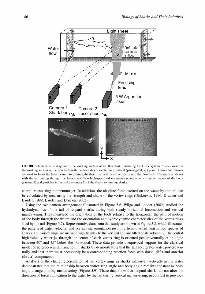
Although kinematic data provide strong evidence in support of the classical view of heterocercal tailfunction in sharks, they do not address what is in fact the primary direct prediction of that model: thedirection of water movement. To determine if the heterocercal tail of sharks functions hydrodynamicallyas expected under the classical view, a new technique is needed that permits direct measurement of waterflow. DPIV is such a technique and a schematic diagram of this approach as applied to shark locomotionis illustrated in Figure 5.6. Sharks swim in a recirculating flow tank, which has been seeded with small(12-
m
m mean diameter) reflective hollow glass beads. A 5 to 10 W laser is focused into a light sheet 1to 2 mm thick and 10 to 15 cm wide and this beam is aimed into the flow tank using focusing lensesand mirrors (Figure 5.6). Sharks are induced to swim with the tail at the upstream edge of the light sheetso that the wake of the shark passes through the light sheet as this wake is carried downstream. A secondsynchronized high-speed video camera takes images of the shark body so that orientation and movementsin the water column can be quantified.
Analysis of wake flow video images proceeds using standard DPIV processing techniques, and furtherdetails of DPIV as applied to problems in fish locomotion are provided in a number of recent papers(Drucker and Lauder, 1999; Wilga and Lauder, 1999, 2000, 2001, 2002; Lauder, 2000; Lauder andDrucker, 2002; Lauder et al., 2002, 2003). Briefly, cross-correlation of patterns of pixel intensity betweenhomologous regions of images separated in time is used to generate a matrix of velocity vectors, whichreflect the pattern of fluid flow through the light sheet. Sample DPIV data are presented in Figure 5.8.From these matrices of velocity vectors the orientation of fluid accelerated by the tail can be quantified,and any rotational movement measured as fluid vorticity. Recent research on fish caudal fin function hasshown that the caudal fin of fishes sheds momentum in the form of vortex loops as the wake rolls upinto discrete torus-shaped rings with a central high-velocity jet flow (Drucker and Lauder, 1999; Lauderand Drucker, 2002). By quantifying the morphology of these wake vortex rings, we can determine thedirection of force application to the water by the heterocercal tail by measuring the direction of the
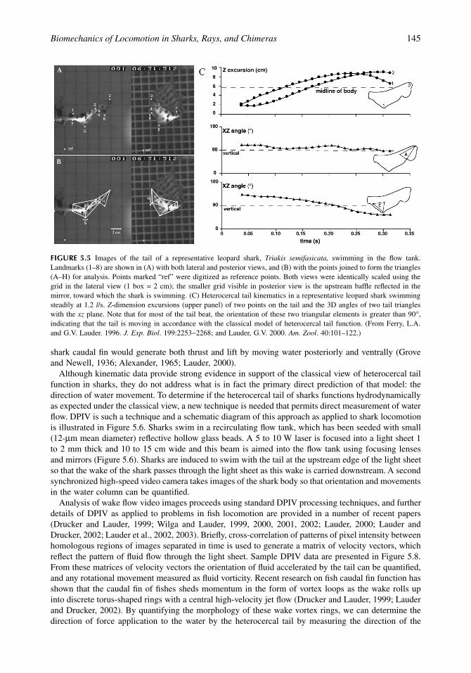
FIGURE 5.5
Images of the tail of a representative leopard shark,
Triakis semifasicata,
swimming in the flow tank.Landmarks (1–8) are shown in (A) with both lateral and posterior views, and (B) with the points joined to form the triangles(A–H) for analysis. Points marked “ref” were digitized as reference points. Both views were identically scaled using thegrid in the lateral view (1 box = 2 cm); the smaller grid visible in posterior view is the upstream baffle reflected in themirror, toward which the shark is swimming. (C) Heterocercal tail kinematics in a representative leopard shark swimmingsteadily at 1.2
l
/s.
Z
-dimension excursions (upper panel) of two points on the tail and the 3D angles of two tail triangleswith the
xz
plane. Note that for most of the tail beat, the orientation of these two triangular elements is greater than 90
∞
,indicating that the tail is moving in accordance with the classical model of heterocercal tail function. (From Ferry, L.A.and G.V. Lauder. 1996.
J. Exp. Biol
. 199:2253
-
2268; and Lauder, G.V. 2000.
Am. Zool
. 40:101–122.)
1514_C05.fm Page 145 Friday, February 13, 2004 10:58 AM
146
Biology of Sharks and Their Relatives
central vortex ring momentum jet. In addition, the absolute force exerted on the water by the tail canbe calculated by measuring the strength and shape of the vortex rings (Dickinson, 1996; Drucker andLauder, 1999; Lauder and Drucker, 2002).
Using the two-camera arrangement illustrated in Figure 5.6, Wilga and Lauder (2002) studied thehydrodynamics of the tail of leopard sharks during both steady horizontal locomotion and verticalmaneuvering. They measured the orientation of the body relative to the horizontal, the path of motionof the body through the water, and the orientation and hydrodynamic characteristics of the vortex ringsshed by the tail (Figure 5.7). Representative data from that study are shown in Figure 5.8, which illustratesthe pattern of water velocity, and vortex ring orientation resulting from one tail beat in two species ofsharks. Tail vortex rings are inclined significantly to the vertical and are tilted posterodorsally. The centralhigh-velocity water jet through the center of each vortex ring is oriented posteroventrally at an anglebetween 40
∞
and 45
∞
below the horizontal. These data provide unequivocal support for the classicalmodel of heterocercal tail function in sharks by demonstrating that the tail accelerates water posteroven-trally and that there must necessarily be a corresponding reaction force with dorsal (lift) and anterior(thrust) components.
Analysis of the changing orientation of tail vortex rings as sharks maneuver vertically in the waterdemonstrates that the relationship between vortex ring angle and body angle remains constant as bodyangle changes during maneuvering (Figure 5.9). These data show that leopard sharks do not alter thedirection of force application to the water by the tail during vertical maneuvering, in contrast to previous
FIGURE 5.6
Schematic diagram of the working section of the flow tank illustrating the DPIV system. Sharks swam inthe working section of the flow tank with the laser sheet oriented in a vertical (parasagittal,
xy
) plane. Lenses and mirrorsare used to focus the laser beam into a thin light sheet that is directed vertically into the flow tank. The shark is shownwith the tail cutting through the laser sheet. Two high-speed video cameras recorded synchronous images of the body(camera 1) and particles in the wake (camera 2) of the freely swimming sharks.
1514_C05.fm Page 146 Friday, February 13, 2004 10:58 AM

Biomechanics of Locomotion in Sharks, Rays, and Chimeras
147
FIGURE 5.7
Schematic summary illustrating body and wake variables measured relative to the horizontal: body angle,from a line drawn along the ventral body surface; path of motion of the center of mass; tail angle between the caudalpeduncle and dorsal tail lobe; ring axis angle from a line extending between the two centers of vorticity; and mean vortexjet angle. Angle measurements from the variables of interest (dotted lines) to the horizontal (dashed line) are indicated bythe curved solid lines. Angles above the horizontal are considered positive and below the horizontal negative. Ring axisangle was measured from 0
∞
to 180
∞
(From Wilga, C.D. and G.V. Lauder. 2002.
J. Exp. Biol
. 205:2365–2374.)
FIGURE 5.8
DPIV analysis of the wake of the tail of representative (A)
Triakis semifasciata
and (B)
Chiloscylliumpunctatum
sharks during steady horizontal locomotion at 1.0
l
/s. On the left is a tracing depicting the position of the tailrelative to the shed vortex ring visible in this vertical section of the wake. The plot to the right shows fluid vorticity withthe matrix of black velocity vectors representing the results of DPIV calculations based on particle displacements super-imposed on top. A strong jet, indicated by the larger velocity vectors, passes between two counterrotating vorticesrepresenting a slice through the vortex ring shed from the tail at the end of each beat. The black dashed line represents thering axis angle.
Note:
Light gray color indicates no fluid rotation, the dark gray color reflects clockwise fluid rotation, andmedium gray color indicates counterclockwise fluid rotation. To assist in visualizing jet flow, a mean horizontal flow of
U
= 19 and
U
= 24 cm/s was subtracted from each vector for
T. semifasciata
and
C. punctatum,
respectively. (From Wilga,C.D. and G.V. Lauder. 2002.
J. Exp. Biol
. 205:2365–2374.)
1514_C05.fm Page 147 Friday, February 13, 2004 10:58 AM

148
Biology of Sharks and Their Relatives
data from sturgeon that demonstrated the ability to actively alter tail vortex wake orientation as theymaneuver (Liao and Lauder, 2000).
5.2.3 Function of the Pectoral Fins during Locomotion
5.2.3.1 Anatomy of the Pectoral Fins —
There are two distinct types of pectoral fins in sharksbased on skeletal morphology. In aplesodic fins, the cartilaginous radials are blunt and extend up to 50%into the fin with the distal web supported only by ceratotrichia. In contrast, plesodic fins have radialsthat extend more than 50% into the fin to stiffen it and supplement the support of the ceratotrichia(Compagno, 1988) (Figure 5.10). The last row of radials tapers to a point distally in plesodic fins.Plesodic fins appear in Lamniformes, hemigaleids, carcharhinids, sphyrnids, and batoids except forpristids; other groups have aplesodic fins (Shirai, 1996). The restricted distribution of plesodic pectoralfins in extant sharks, the different morphology in each group, and their occurrence in more derivedmembers (by other characters) of each group strongly suggest that plesodic pectorals are derived andhave evolved independently from aplesodic pectorals (Zangerl, 1973; Compagno, 1973, 1988; Bendix-Almgreen, 1975). The decreased skeletal support of aplesodic pectoral fins over plesodic fins allows
FIGURE 5.9
Plot of body angle
vs. (A) tail angle, (B) jet angle, and (C) ring axis angle in leopard sharks,
Triakissemifasciata
while swimming at 1.0
l
/s. Solid lines indicate a significant linear regression, and the dotted line representsthe predicted relationship. The lack of significance of the tail vs. body angle regression (
P
= 0.731,
r
2
= 0.003) indicatesthat the sharks are not altering tail angle as body angle changes, but instead are maintaining a constant angular relationshipregardless of locomotor behavior. Jet angle decreases with increasing body angle (P < 0.001, r2 = 0.312, y = –17 – 1.087x)at the same rate as the predicted parallel relationship, indicating that the vortex jet is generated at a constant angle to thebody regardless of body position. Ring axis angle increases with body angle at the same rate as the predicted perpendicularrelationship (P < 0.001, r2 = 0.401, y = 107 + 1.280x). Circles, triangles, and squares represent holds, rises, and sinks,respectively. (From Wilga, C.D. and G.V. Lauder. 2002. J. Exp. Biol. 205:2365–2374.)
1514_C05.fm Page 148 Friday, February 13, 2004 10:58 AM
Biomechanics of Locomotion in Sharks, Rays, and Chimeras 149
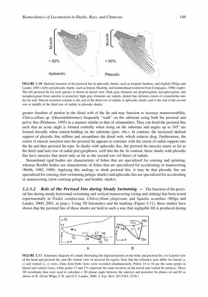
greater freedom of motion in the distal web of the fin and may function to increase maneuverability.Chiloscyllium sp. (Orectolobiformes) frequently “walk” on the substrate using both the pectoral andpelvic fins (Pridmore, 1995) in a manner similar to that of salamanders. They can bend the pectoral finssuch that an acute angle is formed ventrally when rising on the substrate and angles up to 165∞ areformed dorsally when station-holding on the substrate (pers. obs.). In contrast, the increased skeletalsupport of plesodic fins stiffens and streamlines the distal web, which reduces drag. Furthermore, theextent of muscle insertion into the pectoral fin appears to correlate with the extent of radial support intothe fin and thus pectoral fin type. In sharks with aplesodic fins, the pectoral fin muscles insert as far asthe third (and last) row of radial pterygiophores, well into the fin. In contrast, those sharks with plesodicfins have muscles that insert only as far as the second row (of three) of radials.
Streamlined rigid bodies are characteristic of fishes that are specialized for cruising and sprinting,whereas flexible bodies are characteristic of fishes that are specialized for accelerating or maneuvering(Webb, 1985, 1988). Applying this analogy to shark pectoral fins, it may be that plesodic fins arespecialized for cruising (fast-swimming pelagic sharks) and aplesodic fins are specialized for acceleratingor maneuvering (slow-cruising pelagic and benthic sharks).
5.2.3.2 Role of the Pectoral Fins during Steady Swimming — The function of the pecto-ral fins during steady horizontal swimming and vertical maneuvering (rising and sinking) has been testedexperimentally in Triakis semifasciata, Chiloscyllium plagiosum, and Squalus acanthias (Wilga andLauder, 2000, 2001, in prep.). Using 3D kinematics and fin marking (Figure 5.11), these studies haveshown that the pectoral fins of these sharks are held in such a way that negligible lift is produced during
FIGURE 5.10 Skeletal structure of the pectoral fins in aplesodic sharks, such as leopard, bamboo, and dogfish (Wilga andLauder, 2001) (left) and plesodic sharks, such as lemon, blacktip, and hammerhead (redrawn from Compagno, 1988) (right).The left pectoral fin for each species is shown in dorsal view. Dark gray elements are propterygium, mesopterygium, andmetapterygium from anterior to posterior; light gray elements are radials; dotted line delimits extent of ceratotrichia intothe fin web. Muscle insertion extends to the end of the third row of radials in aplesodic sharks and to the end of the secondrow or middle of the third row of radials in plesodic sharks.
FIGURE 5.11 Schematic diagram of a shark illustrating the digitized points on the body and pectoral fin. (A) Lateral viewof the head and pectoral fin, and (B) ventral view of pectoral fin region. Note that the reference axes differ for lateral (x,y) and ventral (x, z) views. Data from both views were recorded simultaneously. Points 14 to 16 are the same points inlateral and ventral views, while points 17 and 17v represent the same location on the dorsal and ventral fin surfaces. These3D coordinate data were used to calculate a 3D planar angle between the anterior and posterior fin planes (a and b) asshown in B. (From Wilga, C.D. and G.V. Lauder. 2000. J. Exp. Biol. 203:2261-2278.)
1514_C05.fm Page 149 Friday, February 13, 2004 10:58 AM

150 Biology of Sharks and Their Relatives
steady horizontal locomotion. The pectoral fins are cambered with an obtuse dorsal angle between theanterior and posterior regions of the fin (mean 190∞ to 191∞) (Figure 5.12). Thus, the planar surface ofthe pectoral fin is held concave downward relative to the flow during steady swimming (Figure 5.13) aswell as concave mediolaterally.
The posture of the pectoral fins relative to the flow during steady horizontal swimming in these sharkscontrasts markedly to those of the wings in a cruising passenger aircraft. The anterior and posteriorplanes of the pectoral fins in these sharks during steady horizontal swimming are at negative and positiveangles, respectively, to the direction of flow (Figure 5.13). When both planes are considered together,the chord angle is -4∞ to -5∞ to the flow. Conversely, the wings of most cruising passenger aircraft havea positive attack angle to the direction of oncoming air, which generates positive lift.
The planar surface of the pectoral fins of these sharks is held at a negative dihedral (fin angle relativeto the horizontal) angle from –6∞ (C. plagiosum) to -23∞ (T. semifasciata) during steady horizontalswimming (Figure 5.14). The pectoral fins are destabilizing in this position (Smith, 1992; Simons, 1994;Wilga and Lauder, 2000) and promote rolling motions of the body, such as those made while maneuveringin the water column. For example, in a roll, the fin with the greatest angle to the horizontal meets theflow at a greater angle of attack, resulting in a greater force (Fx) directed into the roll, while the angleof attack of the more horizontally oriented fin is reduced by the same amount. The more horizontal fintherefore possesses a smaller force (Fx) opposing the roll while the more inclined fin has greater forcedirected into the roll, thereby contributing to the rolling motion. This is in direct contrast to previousstudies suggesting that the pectoral fins of sharks are oriented to prevent rolling, as in the keel of a ship(Harris, 1936, 1953). Wings that are tilted at a positive angle with respect to the horizontal have a positivedihedral angle, as in passenger aircraft, and are self-stabilizing in that they resist rolling motions of thefuselage (Figure 5.14) (Smith, 1992; Simons, 1994). When a passenger aircraft rolls, the more horizontallyoriented wing generates a greater lift force than the inclined wing (Smith, 1992; Simons, 1994). Thus,a corrective restoring moment arises from the more horizontal wing, which opposes the roll, and theaircraft is returned to the normal cruising position. Interestingly, the negative dihedral wings of fighteraircraft, which are manufactured for maneuverability, function similarly to that of shark pectoral fins.
The flow of water in the wake of the pectoral fins during locomotion in these three species wasquantified using DPIV, to estimate fluid vorticity and the forces exerted by the fin on the fluid (seeDrucker and Lauder, 1999; Wilga and Lauder, 2000). These results further corroborate the conclusionfrom the 3D kinematic data that the pectoral fins generate negligible lift during steady horizontal
FIGURE 5.12 Graph of 3D pectoral fin angle vs. body angle for rising, holding, and sinking behaviors at 1.0 l/s in leopardsharks. Symbols are as in Figure 5.3. Body angle was calculated using the line connecting points 12 and 13 (see Figure5.11) and the horizontal (parallel to the flow). Each point represents the mean of five sequences for each of four individuals.Images to the right show sample head and pectoral fin positions during each behavior. Pectoral fin angles equal to 180∞indicate that the two fin triangles (see Figure 5.11) are coplanar; angles less than 180∞ show that the fin surface is concavedorsally; angles greater than 180∞ indicate that the fin surface is concave ventrally. The 3D internal pectoral fin angle issignificantly different among the three behaviors (ANOVA, P = 0.0001). The least-squares regression line is significant(slope 0.41, P < 0.001; adjusted r2 = 0.39). (From Wilga, C.D. and G.V. Lauder. 2000. J. Exp. Biol. 203:2261-2278.)
1514_C05.fm Page 150 Friday, February 13, 2004 10:58 AM

Biomechanics of Locomotion in Sharks, Rays, and Chimeras 151
swimming. There was virtually no vorticity or downwash detected in the wake of the pectoral fins duringsteady horizontal swimming, which shows that little or no lift is being produced by the fins (Figure5.15). According to Kelvin’s law, vortices shed from the pectoral fin must be equivalent in magnitudebut opposite in direction to the theoretical bound circulation around the fin (Kundu, 1990; Dickinson,1996). Therefore, the circulation of the shed vortex can be used to estimate the force on the fin. Meandownstream vertical fluid impulse calculated in the wake of the pectoral fins during steady horizontalswimming was not significantly different from zero. This indicates that the sharks are holding theirpectoral fins in such a way that the flow speed and pressure are equivalent on the dorsal and ventralsurfaces of the fin. Furthermore, if the pectoral fins were generating lift to counteract moments generatedby the heterocercal tail, there would necessarily be a downwash behind the wing to satisfy Kelvin’s law.The lack of an observable and quantifiable downwash indicates clearly that, during holding behavior,pectoral fins generate negligible lift.
These results showing that the pectoral fins of these sharks do not generate lift during steady forwardswimming stand in stark contrast to previous findings on sharks with bound or amputated fins (Daniel,1922; Harris, 1936; Aleev, 1969). Although the results of such radical experiments are difficult to evaluate,it is likely that the lack of pectoral fin motion prevented the sharks from initiating changes in pitch andtherefore limited their ability to achieve a horizontal position and adjust to perturbances in oncomingflow. Lift forces measured on the pectoral fins and body of a plaster model of Mustelus canis in a windtunnel also suggested that the pectoral fins generated upward lift while the body generated no lift (Harris,1936). However, the pectoral fins were modeled as rigid flat plates (2D) and tilted upward 8∞ to the flowwhile the longitudinal axis of the body was oriented at 0∞ to the flow. Although it is possible that M.canis locomotes with the body and pectoral fins in this position, the results of current studies on live
FIGURE 5.13 Orientation of the two pectoral fin planes (a and b) in 3D space during pelagic holding in bamboo sharks,Chiloscyllium plagiosum (leopard and dogfish sharks show similar conformations). Panels show (A) lateral, (B) ventrolateral,and (C) posterior views of the fin planes. Points defining the fin triangles correspond to the following digitized locationsin Figure 5.11: A, anterior, point 14, black circle; L, point 15, black square; P, posterior, point 16; M, medial, point 17.Chord angle to the flow is given in the lateral view, camber and internal fin angle between planes a and b are given in theventrolateral view, and the dihedral angle is shown in the posterior view (note that in the posterior view the angles aregiven as acute to the xy plane). (From Wilga, C.D. and G.V. Lauder. 2001. J. Morphol. 249:195–209.)
1514_C05.fm Page 151 Friday, February 13, 2004 10:58 AM

152 Biology of Sharks and Their Relatives
freely swimming and closely related T. semifasciata, as described herein, which has a very similar bodyshape, show a radically different orientation of the body and pectoral fins.
Three-dimensional kinematic analyses of swimming organisms are crucial to deriving accurate hypoth-eses about the function of the pectoral fins and body (Wilga and Lauder, 2000). The 2D angle of theanterior margin of the pectoral fin as a representation of the planar surface of the pectoral fin in sharksis extremely misleading. Although the pectoral fin appears to be oriented at a positive angle to the flowin lateral view, 3D kinematics reveals that the fin is actually concave downward with a negative dihedral.When viewed laterally, this negative-dihedral concave-downward orientation of the pectoral fin createsa perspective that suggests a positive angle of attack when the angle is, in fact, negative.
5.2.3.3 Role of the Pectoral Fins during Vertical Maneuvering — Triakis semifasciata,Chiloscyllium plagiosum, and Squalus acanthias actively adjust the angle of their pectoral fins tomaneuver vertically in the water column (Wilga and Lauder, 2000, 2001, in prep.). Rising in the water
FIGURE 5.14 Schematic diagram of the dihedral orientation of the pectoral fins in a shark during holding, rising andsinking behaviors. Forces during a roll are illustrated below for the pectoral fins of a shark and the wings of an airplane.The body and fin are represented as a cross section at the level of plane a of the pectoral fin (see Figure 5.11). Thin graydouble-headed arrows represent the dihedral angle between the plane a (dotted line) and pectoral fin. Thick arrows showthe direction of movement of the body and fins or wing during a roll. Note that positive dihedrals (such as those used inaircraft design) are self-stabilizing, while fins oriented at a negative dihedral angle, as in sharks, are destabilizing in rolland tend to amplify roll forces. Fx, horizontal force; Fy, vertical force; FL, resultant force. (From Wilga, C.D. and G.V.Lauder. 2000. J. Exp. Biol. 203:2261-2278.)
1514_C05.fm Page 152 Friday, February 13, 2004 10:58 AM

Biomechanics of Locomotion in Sharks, Rays, and Chimeras 153
FIGURE 5.15 DPIV data from leopard shark pectoral fins during (top) holding vertical position, (middle) sinking, and(bottom) rising behaviors at 1.0 l/s (patterns for bamboo and dogfish sharks are similar). The video image (on the left) isa single image of a shark with the left pectoral fin located just anterior to the laser light sheet. Note that the ventral bodymargin is faintly visible through the light sheet. The plot on the right shows fluid vorticity with velocity vectors withconventions as in Figure 5.8. In the holding position, note that the fin is held in a horizontal position, and that the vorticityplot shows effectively no fluid rotation. Hence, the pectoral fins in this position do not generate lift forces. During sinking,note that there is a clockwise vortex (dark gray region of rotating fluid to the right) that resulted from the upward fin flip(curved white arrow) to initiate the sinking event. During rising, note that the fin has flipped ventrally (curved white arrow)to initiate the rising event, and that a counterclockwise vortex (medium gray region of rotating fluid to the right) has beenshed from the fin. To assist in visualizing the flow pattern, a mean horizontal flow of U = 33 cm/s was subtracted fromeach vector. (Modified from Wilga and Lauder, 2001.)
1514_C05.fm Page 153 Friday, February 13, 2004 10:58 AM

154 Biology of Sharks and Their Relatives
column is initiated when the posterior plane of the fin is flipped downward to produce mean obtusedorsal fin angles around 200∞, while the leading edge of the fin is rotated upward relative to the flow.This downward flipping of the posterior plane of the fin increases the chord angle to +14, and as a resultthe shark rises in the water. In contrast, to sink in the water the posterior plane of the pectoral fin isflipped upward relative to the anterior plane, which produces a mean obtuse dorsal fin angle of 185∞.At the same time, the leading edge of the fin is rotated downward relative to the flow such that the chordangle is decreased to -22∞, and the shark sinks in the water.
The dihedral angle of shark pectoral fins changes significantly during vertical maneuvering in thewater column (Figure 5.14). The dihedral angle increases to -35∞ during rising and decreases to -5∞during sinking. This may be due to a need for greater stability during sinking behavior because theheterocercal tail generates a lift force that tends to drive the head ventrally. Holding the pectoral fins ata low dihedral angle results in greater stability during sinking compared to rising. The greater negativedihedral angle increases maneuverability and allows rapid changes in body orientation during rising.
These angular adjustments of the pectoral fins are used to maneuver vertically in the water columnand generate negative and positive lift forces, which then initiate changes in the angle of the body relativeto the flow. As the posterior plane of the pectoral fin is flipped down to ascend, a counterclockwisevortex, indicating upward lift force generation, is produced and shed from the trailing edge of the finand pushes the head and anterior body upward (Figure 5.15). This vortex is readily visible in the wakeas it rolls off the fin and is carried downstream. The opposite flow pattern occurs when sharks initiatea sinking maneuver in the water column. A clockwise vortex, indicating downward lift force generation,is visualized in the wake of the pectoral fin as a result of the dorsal fin flip and pulls the head andanterior body of the shark downward (Figure 5.15).
Lift forces produced by altering the planar surface of the pectoral fin to rise and sink appear to be amechanism to reorient the position of the head and anterior body for maneuvering. Changing the orientationof the head will alter the force balance on the body as a result of interaction with the oncoming flow andwill induce a change in vertical forces that will move the shark up or down in the water column. Forcesgenerated by the pectoral fins are significantly greater in magnitude during sinking than during rising. Thismay be due to the necessity of reorienting the body through a greater angular change to sink from thepositive body tilt adopted during steady swimming. A shark must reposition the body from a positive bodytilt of 8∞ (mean holding angle) down through the horizontal to a negative body tilt of –11∞ (mean sinkingangle), a change of 19∞. In contrast, to rise a shark simply increases the positive tilt of the body by 14∞(mean rise – hold difference), which should require less force given that the oncoming flow will assist thechange from a slightly tilted steady horizontal swimming position to a more inclined rising body position.
5.2.3.4 Function of the Pectoral Fins during Benthic Station-Holding — Chiloscylliumplagiosum have a benthic lifestyle and spend much of their time resting on the substrate on and aroundcoral reefs where current flows can be strong. To maintain position on the substrate during significantcurrent flow, these sharks shift their body posture to reduce drag (Wilga and Lauder, 2001). The sharksreorient the longitudinal axis of the body to the flow with the head pointing upstream during currentflow, but do not orient when current flow is negligible or absent. Body angle steadily decreases from 4∞at 0 l/s to 0.6∞ at 1.0 l/s as they flatten their body against the substrate with increasing flow speed. Thisreduces drag in higher current flows thereby promoting station-holding. This behavior is advantageousin fusiform benthic fishes that experience a relatively high flow regime, such as streams where salmonparr are hatched (Arnold and Webb, 1989) and inshore coral reefs where bamboo sharks dwell(Compagno, 1984).
Chiloscyllium plagiosum also reorient the pectoral fins to generate negative lift, increase friction, andoppose downstream drag during station-holding in current flow (Wilga and Lauder, 2001). They hold thepectoral fins in a concave upward orientation, similar to that in sinking, which decreases from a meanplanar angle of 174∞ at 0 l/s to a mean of 165∞ at 1.0 l/s. At the same time, the chord angle steadilydecreases from a mean of 2.7∞ at 0 l/s to a mean of –3.9∞ at 1.0 l/s. Flattening the body against the substratelowers the anterior edge of the fin, whereas elevating the posterior edge of the fin to decrease the planarangle significantly decreases the chord angle (Figure 5.16). In this orientation, water flow is deflected upand over the fin and produces a clockwise vortex that is shed from the fin tip. The clockwise vortex
1514_C05.fm Page 154 Friday, February 13, 2004 10:58 AM

Biomechanics of Locomotion in Sharks, Rays, and Chimeras 155
produces significant negative lift (mean –0.084 N) directed toward the substrate that is eight times greaterthan that generated during sinking. As the clockwise vortex shed from the fin rotates just behind the fin,flow recirculates upstream and pushes against the posterior surface of the fin, which opposes downstreamdrag. These movements generate negative lift that is directed toward the substrate and acts to increase totaldownward force and friction force, thereby promoting station-holding as predicted by previous studies(Arnold and Webb, 1991; Webb and Gerstner, 1996), as well as a novel mechanism leading to vortexshedding that opposes downstream drag to further aid benthic station-holding (Wilga and Lauder, 2001).
5.2.3.5 Motor Activity in the Pectoral Fins — Movement of the posterior plane of the pec-toral fin during sinking and rising is actively controlled by Triakis semifasciata. At the beginning of arise, the pectoral fin depressors (ventral fin muscles, adductors) are active to depress the posterior portionof the pectoral fin (Figure 5.17). Small bursts of activity in the lateral hypaxialis, protractor, and levatormuscles are sometimes present during rising, probably to stabilize pectoral fin position. In contrast, thepectoral fin levators (dorsal fin muscles, adductors), as well as the cucullaris and ventral hypaxialis, arestrongly active during elevation of the posterior portion of the fin at the beginning of sinking. Virtually
FIGURE 5.16 DPIV data from the pectoral fins of a representative bamboo shark Chiloscyllium plagiosum while holdingstation on the substrate. The video image on the left shows a shark with the left pectoral fin located in the anterior end ofthe laser light sheet; other conventions as in Figures 5.8 and 5.15. Note that the fin is held at a negative chord angle to theflow. A clockwise vortex (negative vorticity) was produced in the wake of the pectoral fins, which continued to rotate justbehind the fin for several seconds until it was carried downstream by the flow (as seen here), after which a new vortexforms in the wake of the fin. (Modified from Wilga and Lauder, 2001.)
FIGURE 5.17 Electromyographic data from selected pectoral fin and body muscles during locomotion in Triakis semi-fasciata at 1.0 l/s for four behaviors: holding position at 1.0 and 1.5 l/s and sinking and rising at 1.0 l/s. Note the nearabsence of fin muscle activity while holding position at 1.0 l/s and recruitment of body and fin muscles at 1.5 l/s. Thehypaxialis was implanted in both lateral (mid-lateral dorsal and posterior to pectoral fin base) and ventral (posterior tocoracoid bar) positions. All panels are from the same individual. Scale bar represents 500 ms.
1514_C05.fm Page 155 Friday, February 13, 2004 10:58 AM

156 Biology of Sharks and Their Relatives
no motor activity is present in the pectoral fin muscles while holding position at 0.5 and 1.0 l/s, indicatingthat the pectoral fins are not actively held in any particular position during steady horizontal locomotion.However, at higher flow speeds (1.5 l/s), recruitment of epaxial and hypaxial muscles occurs with slightactivity in the pectoral fin muscles that may function to maintain stability.
Epaxial or hypaxial muscles are recruited to elevate or depress the head and anterior body duringrising or sinking, respectively. At the initiation of rising behavior, simultaneously with the head pitchingupward, a strong burst of activity occurs in the cranial epaxialis, while it is virtually silent during holdingand sinking. Similarly, a strong burst of activity occurs in the ventral hypaxialis during the initiation ofsinking behavior, again with virtually no activity during holding and rising. This shows that the head isactively elevated or depressed to rise or sink, respectively, and that conformational changes in the anteriorbody assist the forces generated by the pectoral fins to accomplish vertical maneuvers. Finally, antago-nistic pectoral fin muscles become active as rising or sinking slows or during braking (i.e., the levatorsare active as rising stops and the depressors are active as sinking stops).
5.2.4 Synthesis
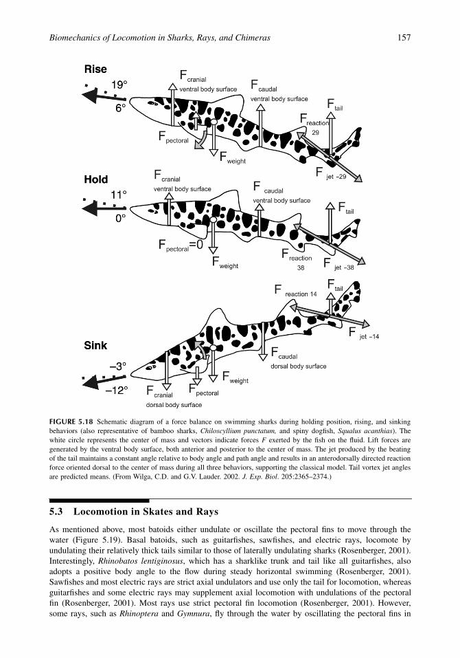
The data presented above on pectoral and caudal fin function and body orientation in the shark speciesstudied permit construction of a new model of the overall force balance during swimming (Figure 5.18).It is useful to discuss separately the vertical force balance and the rotational (torque) balance. Duringsteady horizontal locomotion, when sharks are holding vertical position, body weight is balanced by liftforces generated by the heterocercal tail and ventral body surface. The ventral surface generates lift bothanterior and posterior to the center of body mass by virtue of its positive angle of attack to the oncomingwater. Sharks adjust their body angle to modulate the total lift force produced by the body, and can thuscompensate for changes in body weight over both short and longer time frames.
Rotational balance is achieved by balancing the moments of forces around the center of mass. It hasnot been generally appreciated that the ventral body surface generates both positive and negative torquescorresponding to the location of the ventral surface anterior and posterior to the center of mass. Waterimpacting the ventral body surface posterior to the center of mass will generate a counterclockwisetorque of the same sign as that generated by the heterocercal tail. In contrast, water impacting the ventralbody anterior to the center of mass will generate a clockwise torque, which is opposite in sign to thatgenerated by the ventral body and tail posterior to the center of mass. As a result of experimental datademonstrating that shark pectoral fins do not generate lift during steady horizontal locomotion (Wilgaand Lauder, 2000, 2001) as a result of their orientation relative to the flow, no role in generating eitherlift or torque is attributed to the pectoral fins during horizontal locomotion. This stands in contrast tothe textbook depiction of shark locomotion in which the pectoral fins play a central role in controllingbody position during horizontal locomotion. In our view, experimental kinematic and hydrodynamicdata obtained over the last 5 years demonstrate that control of body orientation is the key to modulatinglift and torques during horizontal swimming while the pectoral fins are not used for balancing forcesduring horizontal swimming.
However, during maneuvering the pectoral fins do play a key role in generating both positive and negativelift forces and hence torques about the center of mass (Figure 5.18). To rise in the water, sharks rapidlymove the trailing pectoral fin edge ventrally, and a large vortex is shed, generating a corresponding liftforce. This force has a clockwise rotational moment about the center of mass pitching the body up, increasingthe angle of the body, and hence the overall lift force. As a result, sharks move vertically in the water evenwhile maintaining horizontal position via increased thrust produced by the body and caudal fin.
To stop this vertical motion or to maneuver down (sink) in the water the trailing pectoral fin edge israpidly elevated, shedding a large vortex, which produces a large negative lift force (Figure 5.18). Thisgenerates a counterclockwise torque about the center of mass, pitching the body down, exposing thedorsal surface to incident flow, and producing a net sinking motion. Pectoral fins thus modulate body pitch.
Overall, the force balance on swimming sharks is maintained and adjusted by small alterations inbody angle and this in turn is achieved by elevation and depression of the pectoral fins. Pectoral finsthus play a critical role in shark locomotion by controlling body position and facilitating maneuvering,but they do not function to balance tail lift forces during steady horizontal locomotion.
1514_C05.fm Page 156 Friday, February 13, 2004 10:58 AM
Biomechanics of Locomotion in Sharks, Rays, and Chimeras 157
5.3 Locomotion in Skates and Rays
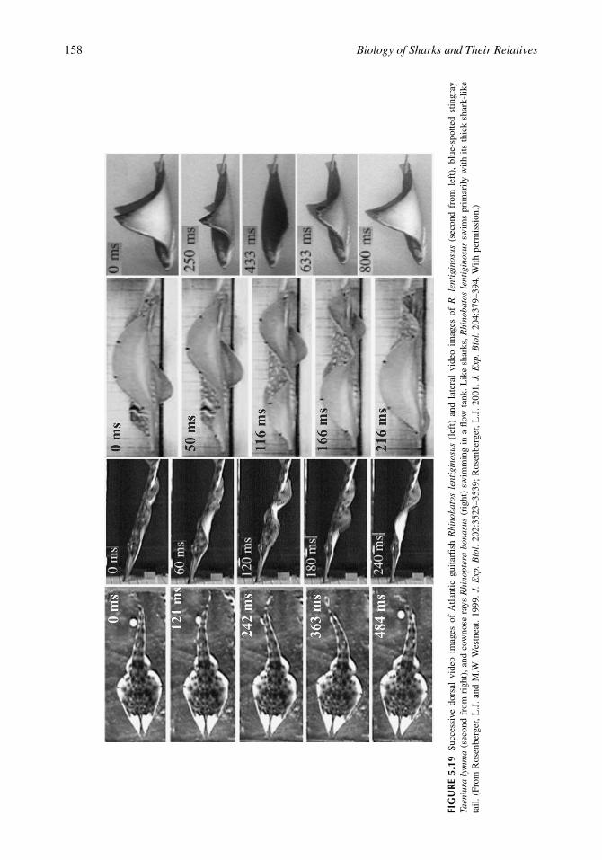
As mentioned above, most batoids either undulate or oscillate the pectoral fins to move through thewater (Figure 5.19). Basal batoids, such as guitarfishes, sawfishes, and electric rays, locomote byundulating their relatively thick tails similar to those of laterally undulating sharks (Rosenberger, 2001).Interestingly, Rhinobatos lentiginosus, which has a sharklike trunk and tail like all guitarfishes, alsoadopts a positive body angle to the flow during steady horizontal swimming (Rosenberger, 2001).Sawfishes and most electric rays are strict axial undulators and use only the tail for locomotion, whereasguitarfishes and some electric rays may supplement axial locomotion with undulations of the pectoralfin (Rosenberger, 2001). Most rays use strict pectoral fin locomotion (Rosenberger, 2001). However,some rays, such as Rhinoptera and Gymnura, fly through the water by oscillating the pectoral fins in
FIGURE 5.18 Schematic diagram of a force balance on swimming sharks during holding position, rising, and sinkingbehaviors (also representative of bamboo sharks, Chiloscyllium punctatum, and spiny dogfish, Squalus acanthias). Thewhite circle represents the center of mass and vectors indicate forces F exerted by the fish on the fluid. Lift forces aregenerated by the ventral body surface, both anterior and posterior to the center of mass. The jet produced by the beatingof the tail maintains a constant angle relative to body angle and path angle and results in an anterodorsally directed reactionforce oriented dorsal to the center of mass during all three behaviors, supporting the classical model. Tail vortex jet anglesare predicted means. (From Wilga, C.D. and G.V. Lauder. 2002. J. Exp. Biol. 205:2365–2374.)
1514_C05.fm Page 157 Friday, February 13, 2004 10:58 AM
158 Biology of Sharks and Their Relatives
FIG
UR
E 5.
19Su
cces
sive
dor
sal
vide
o im
ages
of
Atla
ntic
gui
tarfi
sh R
hino
bato
s le
ntig
inos
us (
left
) an
d la
tera
l vi
deo
imag
es o
f R
. le
ntig
inos
us (
seco
nd f
rom
lef
t),
blue
-spo
tted
stin
gray
Taen
iura
lym
ma
(sec
ond
from
rig
ht),
and
cow
nose
ray
s R
hino
pter
a bo
nasu
s (r
ight
) sw
imm
ing
in a
flow
tan
k. L
ike
shar
ks, R
hino
bato
s le
ntig
inos
us s
wim
s pr
imar
ily w
ith i
ts t
hick
sha
rk-l
ike
tail.
(Fr
om R
osen
berg
er,
L.J
. an
d M
.W. W
estn
eat.
1999
. J.
Exp
. B
iol.
202:
3523
–353
9; R
osen
berg
er,
L.J
. 20
01.
J. E
xp.
Bio
l. 20
4:37
9–39
4. W
ith p
erm
issi
on.)
1514_C05.fm Page 158 Friday, February 13, 2004 10:58 AM

Biomechanics of Locomotion in Sharks, Rays, and Chimeras 159
broad up- and downstrokes in a manner that would provide vertical lift similar to that of aerial bird flight(Rosenberger, 2001). Although skates undulate the pectoral fins to swim when in the water column, theyhave enlarged muscular appendages on the pelvic fins that are modified for walking or “punting” offthe substrate (Koester and Spirito, 1999) in a novel locomotor mechanism.
Some rays are able to vary the mechanics of the pectoral fins during locomotion (Rosenberger, 2001).There appears to be a trade-off between the amplitude of undulatory waves and fin beat frequency: thosethat have higher wave amplitudes have fewer waves, and vice versa (Rosenberger, 2001). This phenom-enon appears to be correlated with lifestyle. Fully benthic rays and skates that are mostly sedentary,such as Daysatis sabina and D. say, have low-amplitude waves with high fin beat frequencies, permittinghigh maneuverability at low speeds, which is more suited for swimming slowly along the substrate tolocate food items (Rosenberger, 2001). Fully pelagic rays are able to take advantage of the 3D environ-ment of the water column and oscillate the pectoral fins using high-amplitude waves and low fin beatfrequencies (Rosenberger, 2001). Rays and skates that have both benthic and pelagic lifestyles, such asRaja sp. and D. violacea and D. americana, are typically more active and have intermediate values ofamplitude and frequency (Rosenberger, 2001).
However, oscillatory appendage propulsors that feed on benthic mollusks and crustaceans, such ascownose and butterfly rays, do not extend the fins below the ventral body axis during swimming,presumably so that they can use the lateral line canals to detect prey and also to avoid contact with thesubstrate (Rosenberger, 2001). In contrast, oscillatory appendage propulsors that feed in the watercolumn, i.e., filter feeders such as manta and mobulid rays extend the pectoral fins equally above andbelow the body axis during swimming (Rosenberger, 2001). Some batoids are capable of modifying theswimming mechanism dependent on habitat; Gymnura undulates the pectoral fins when swimming alonga substrate and oscillates them when swimming in the water column (Rosenberger, 2001). Undulatorymechanisms are efficient at slow speeds, have reduced body and fin drag, and are highly maneuverable(Blake, 1983a,b; Lighthill and Blake, 1990; Walker and Westneat, 2000; Rosenberger, 2001). In contrast,oscillatory mechanisms are efficient at fast cruising and generate greater lift, but are less well suited formaneuvering (Chopra, 1974; Blake, 1983b; Cheng and Zhaung, 1991; Rosenberger, 2001).
Different strategies are employed to increase swimming speed in various batoid species (Rosenberger,2001). Most Dasyatis species increase fin beat frequency, wave speed, and stride length to increaseswimming speed, while amplitude is held constant (Rosenberger and Westneat, 1999; Rosenberger,2001). However, Taeniura lymma and D. americana increase fin beat frequency and wave speed butdecrease wave number while holding amplitude constant to increase speed (Rosenberger and Westneat,1999; Rosenberger, 2001). Similarly, Raja elganteria increases wave speed and decreases wave numberto swim faster (Rosenberger and Westneat, 1999; Rosenberger, 2001). Oscillatory propulsors, Rhinopteraand Gymnura, increase wave speed in addition to fin-tip velocity to increase swimming speed (Rosen-berger and Westneat, 1999; Rosenberger, 2001). Interestingly, Gymnura pauses between each fin beatat high flow speeds (Rosenberger and Westneat, 1999; Rosenberger, 2001), similar to the burst and glideflight mechanisms of aerial birds.
As expected, the dorsal and ventral fin muscles are alternately active during undulation of the pectoralfin from anterior to posterior (Figure 5.20) (Rosenberger and Westneat, 1999). The intensity of musclecontraction is increased to swim faster in T. lymma (Rosenberger and Westneat, 1999). The ventralmuscles are also active longer than the respective dorsal muscles indicating that the downstroke is themajor power-producing stroke (Rosenberger and Westneat, 1999). Chondrichthyans are negatively buoy-ant; thus lift must be generated to counter the weight of the fish as well as for locomotion. Interburstduration is decreased in T. lymma at higher swimming speeds with the fin muscles firing closer together(Rosenberger and Westneat, 1999).
5.4 Locomotion in Holocephalans
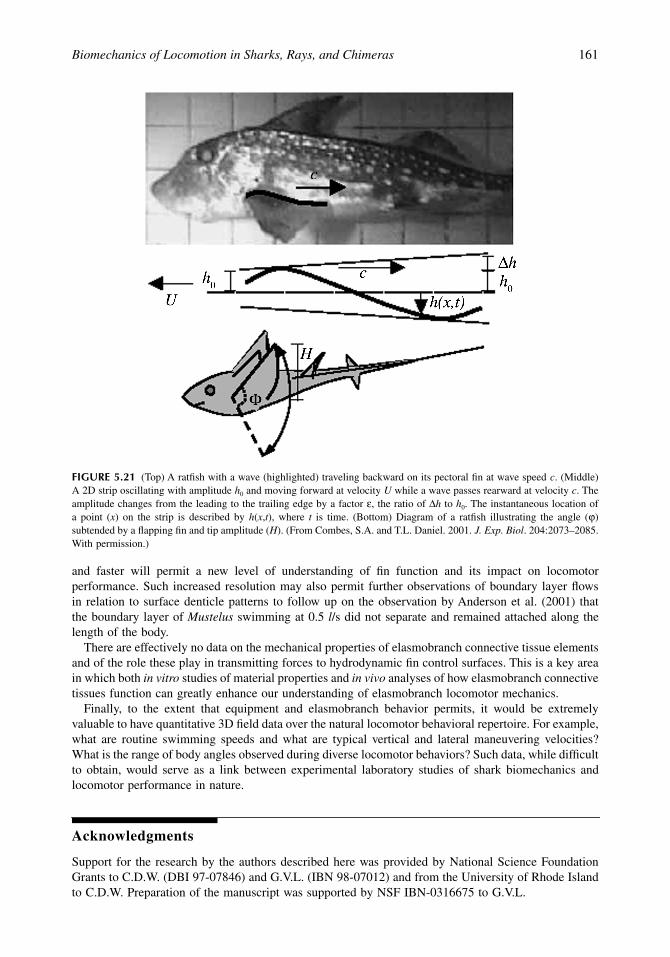
Chimeras have large flexible pectoral fins that have been described as both undulatory and oscillatory.The leading edge of the pectoral fin is flapped, which then passes an undulatory wave down the pectoralfin to the trailing edge (Combes and Daniel, 2001) (Figure 5.21). As expected, adult chimeras had a
1514_C05.fm Page 159 Friday, February 13, 2004 10:58 AM

160 Biology of Sharks and Their Relatives
larger-amplitude wave that was generated at a lower frequency than juvenile chimeras (Combes andDaniel, 2001). Interestingly, there is no net chordwise bend in the pectoral fin which averages a 0∞ angleof attack to the flow over a stroke cycle (Combes and Daniel, 2001). Potential flow models based onkinematic and morphological variables measured on the chimeras for realistic flexible fins and theoreticalstiff fins emphasize the importance of considering flexion in models of animal locomotion (Combes andDaniel, 2001). Significantly higher values for thrust were calculated when the fin was assumed to bestiff rather than flexible as in reality (Combes and Daniel, 2001).
5.5 Future Directions
The diversity of shark species for which we have even basic functional data on locomotor mechanics isextremely limited. Most papers to date have focused on leopard (Triakis) and bamboo (Chiloscyllium)sharks swimming under controlled laboratory conditions. A high priority for future studies of locomotionin sharks, skates, and rays is to expand the diversity of taxa studied, especially for analyses of sharkmechanics. The data obtained by Rosenberger (2001) on batoid locomotion are exemplary for theirbroadly comparative character, but studies like this are rare, perhaps necessarily so when detailedfunctional data must be obtained for a variety of behaviors.
Experimental studies of kinematics and hydrodynamics would benefit from increased spatial andtemporal resolution so that a more detailed picture could be obtained of patterns of fin deformation andthe resulting hydrodynamic wake, especially during unsteady maneuvering behaviors. New high-reso-lution digital video systems with greater than 1024 by 1024 pixels per frame operating at 500 frames/s
FIGURE 5.20 Electromyographic (EMG) data illustrating the muscle activity for the pectoral fin undulation of blue-spottedstingrays Taeniura lymma at a low speed of 1.2 disk length/s (A) and at a higher speed of 3.0 disk length/s (B). The electroderecordings are taken from the following muscles: anterior dorsal, mid-anterior dorsal, mid-posterior dorsal, posterior dorsal,anterior ventral, middle ventral, posterior ventral. The arrows below the EMG activity indicate the point during the fin-beatcycle at which the anterior, middle, and posterior fin markers are at their maximum (peak upstroke) and minimum (peakdownstroke) excursion. (From Rosenberger, L.J. and M.W. Westneat. 1999. J. Exp. Biol. 202:3523–3539; Rosenberger, L.J.2001. J. Exp. Biol. 204:379-394. With permission.)
1514_C05.fm Page 160 Friday, February 13, 2004 10:58 AM
Biomechanics of Locomotion in Sharks, Rays, and Chimeras 161
and faster will permit a new level of understanding of fin function and its impact on locomotorperformance. Such increased resolution may also permit further observations of boundary layer flowsin relation to surface denticle patterns to follow up on the observation by Anderson et al. (2001) thatthe boundary layer of Mustelus swimming at 0.5 l/s did not separate and remained attached along thelength of the body.
There are effectively no data on the mechanical properties of elasmobranch connective tissue elementsand of the role these play in transmitting forces to hydrodynamic fin control surfaces. This is a key areain which both in vitro studies of material properties and in vivo analyses of how elasmobranch connectivetissues function can greatly enhance our understanding of elasmobranch locomotor mechanics.
Finally, to the extent that equipment and elasmobranch behavior permits, it would be extremelyvaluable to have quantitative 3D field data over the natural locomotor behavioral repertoire. For example,what are routine swimming speeds and what are typical vertical and lateral maneuvering velocities?What is the range of body angles observed during diverse locomotor behaviors? Such data, while difficultto obtain, would serve as a link between experimental laboratory studies of shark biomechanics andlocomotor performance in nature.
Acknowledgments
Support for the research by the authors described here was provided by National Science FoundationGrants to C.D.W. (DBI 97-07846) and G.V.L. (IBN 98-07012) and from the University of Rhode Islandto C.D.W. Preparation of the manuscript was supported by NSF IBN-0316675 to G.V.L.
FIGURE 5.21 (Top) A ratfish with a wave (highlighted) traveling backward on its pectoral fin at wave speed c. (Middle)A 2D strip oscillating with amplitude h0 and moving forward at velocity U while a wave passes rearward at velocity c. Theamplitude changes from the leading to the trailing edge by a factor e, the ratio of Dh to h0. The instantaneous location ofa point (x) on the strip is described by h(x,t), where t is time. (Bottom) Diagram of a ratfish illustrating the angle (j)subtended by a flapping fin and tip amplitude (H). (From Combes, S.A. and T.L. Daniel. 2001. J. Exp. Biol. 204:2073–2085.With permission.)
1514_C05.fm Page 161 Friday, February 13, 2004 10:58 AM

162 Biology of Sharks and Their Relatives
References
Affleck, R. J. 1950. Some points in the function, development, and evolution of the tail in fishes. Proc. Zool.Soc. Lond. 120:349–368.
Aleev, Y. G. 1969. Function and Gross Morphology in Fish, trans. from the Russian by M. Raveh. Keter Press,Jerusalem.
Alexander, R. M. 1965. The lift produced by the heterocercal tails of Selachii. J. Exp. Biol. 43:131–138.Anderson, E. J., W. McGillis, and M. A. Grosenbaugh. 2001. The boundary layer of swimming fish. J. Exp.
Biol. 204:81–102.Arnold, G. P. and P. W. Webb. 1991. The role of the pectoral fins in station-holding of Atlantic salmon parr
(Salmo salar L.). J. Exp. Biol. 156:625–629.Bendix-Almgreen, S. E. 1975. The paired fins and shoulder girdle in Cladoselache, their morphology and
phyletic significance. Colloq. Int. C. N. R. S. (Paris) 218:111–123.Blake, R. W. 1983a. Fish Locomotion. Cambridge University Press, Cambridge, U.K.Blake, R. W. 1983b. Median and paired fin propulsion, in Fish Biomechanics. P. W. Webb and D. Weihs, Eds.,
Praeger, New York, 214–247.Bone, Q. 1999. Muscular system: microscopical anatomy, physiology, and biochemistry of elasmobranch
muscle fibers, in Sharks, Skates, and Rays: The Biology of Elasmobranch Fishes. W. C. Hamlett, Ed.,Johns Hopkins University Press, Baltimore, 115–143.
Breder, C. M. 1926. The locomotion of fishes. Zool. N.Y. 4:159–256.Carroll, R. L. 1988. Vertebrate Paleontology and Evolution. W. H. Freeman, New York.Cheng, J. and L. Zhaung. 1991. Analysis of swimming three-dimensional waving plates. J. Fluid Mech.
232:341–355.Chopra, M. G. 1974. Hydrodynamics of lunate-tail swimming propulsion. J. Fluid Mech. 64:375–391.Combes, S. A., and T. L. Daniel. 2001. Shape, flapping and flexion: wing and fin design for forward flight.
J. Exp. Biol. 204:2073–2085.Compagno, L. J. V. 1973. Interrelationships of living elasmobranchs, in Interrelationships of Fishes. P. H.
Greenwood, R. S. Miles, and C. Patterson, Eds., Zool. J. Linn. Soc., Suppl. 1.Compagno, L. J. V. 1984. Sharks of the World. United Nations Development Program, Rome.Compagno, L. J. V. 1988. Sharks of the Order Carcharhiniformes. Princeton University Press, Princeton, NJ.Compagno, L. J. V. 1999. Endoskeleton, in Sharks, Skates, and Rays: The Biology of Elasmobranch Fishes.
W. C. Hamlett, Ed., Johns Hopkins University Press, Baltimore, 69–92.Daniel, J. F. 1922. The Elasmobranch Fishes. University of California Press, Berkeley.Dickinson, M. H. 1996. Unsteady mechanisms of force generation in aquatic and aerial locomotion. Am. Zool.
36:537–554.Donley, J. and R. Shadwick. 2003. Steady swimming muscle dynamics in the leopard shark Triakis semifas-
ciata. J. Exp. Biol. 206:1117–1126.Drucker, E. G. and G. V. Lauder. 1999. Locomotor forces on a swimming fish: three-dimensional vortex wake
dynamics quantified using digital particle image velocimetry. J. Exp. Biol. 202:2393–2412.Ferry, L. A. and G. V. Lauder. 1996. Heterocercal tail function in leopard sharks: a three-dimensional kinematic
analysis of two models. J. Exp. Biol. 199:2253–2268.Fish, F. E. and L. D. Shannahan. 2000. The role of the pectoral fins in body trim of sharks. J. Fish Biol.
56:1062–1073.Garman, S. 1913. The Plagiostoma (sharks, skates, and rays). Mem. Mus. Comp. Zool. Harvard Coll. 36.Gray, J. 1968. Animal Locomotion. Weidenfeld and Nicolson, London.Grove, A. J. and G. E. Newell. 1936. A mechanical investigation into the effectual action of the caudal fin of
some aquatic chordates. Ann. Mag. Nat. Hist. 17:280–290.Harris, J. E. 1936. The role of the fins in the equilibrium of the swimming fish. I. Wind tunnel tests on a
model of Mustelus canis (Mitchell). J. Exp. Biol. 13:476–493.Harris, J. E. 1953. Fin patterns and mode of life in fishes, in Essays in Marine Biology. S. M. Marshall and
A. P. Orr, Eds., Oliver and Boyd, Edinburgh, 17–28.Kemp, N. E. 1999. Integumentary system and teeth, in Sharks, Skates, and Rays: The Biology of Elasmobranch
Fishes. W. C. Hamlett, Ed., Johns Hopkins University Press, Baltimore, 43–68.
1514_C05.fm Page 162 Friday, February 13, 2004 10:58 AM
Biomechanics of Locomotion in Sharks, Rays, and Chimeras 163
Koester, D. M. and C. P. Spirito. 1999. Pelvic fin locomotion in the skate, Leucoraja erinacea. Am. Zool.39:55A.
Krothapalli, A. and L. Lourenco. 1997. Visualization of velocity and vorticity fields, in Atlas of Visualization,Vol. 3. Y. Nakayama and Y. Tanida, Eds., CRC Press, Boca Raton, FL, 69–82.
Kundu, P. 1990. Fluid Mechanics. Academic Press, San Diego.Lauder, G. V. 2000. Function of the caudal fin during locomotion in fishes: kinematics, flow visualization,
and evolutionary patterns. Am. Zool. 40:101–122.Lauder, G. V. and E. Drucker. 2002. Forces, fishes, and fluids: hydrodynamic mechanisms of aquatic loco-
motion. News Physiol.. Sci. 17:235–240.Lauder, G. V., J. Nauen, and E. G. Drucker. 2002. Experimental hydrodynamics and evolution: function of
median fins in ray-finned fishes. Integ. Comp. Biol. 42:1009–1017.Lauder, G. V., E. G., Drucker, J. Nauen, and C. D. Wilga. 2003. Experimental hydrodynamics and evolution:
caudal fin locomotion in fishes, in Vertebrate Biomechanics and Evolution. V. Bels, J.-P. Gasc, and A.Casinos, Eds., Bios Scientific Publishers, Oxford, 117–135.
Liao, J. and G. V. Lauder. 2000. Function of the heterocercal tail in white sturgeon: flow visualization duringsteady swimming and vertical maneuvering. J. Exp. Biol. 203:3585–3594.
Liem, K. F. and A. P. Summers. 1999. Muscular system, in Sharks, Skates, and Rays: The Biology ofElasmobranch Fishes. W. C. Hamlett, Ed., Johns Hopkins University Press, Baltimore, 93–114.
Lighthill, J. and R. Blake. 1990. Biofluid dynamics of balistiform and gymnotiform locomotion. Part 1.Biological background and analysis by elongated-body theory. J. Fluid Mech. 212:183–207.
Lindsey, C. C. 1978. Form, function, and locomotory habits in fish, in Fish Physiology, Vol. 7. Locomotion.W. S. Hoar and D. J. Randall, Eds., Academic Press, New York, 1–100.
Magnan, A. 1929. Les charactéristiques géométriqes et physiques des poissons. Ann. Sci. Nat. Zool. 10:1–132.Pridmore, P. A. 1995. Submerged walking in the epaulette shark Hemiscyllium ocellatum (Hemiscyllidae) and
its implications for locomotion in rhipidistian fishes and early tetrapods. Zoology 98:278–297.Reif, W.-E. and D. B. Weishampel. 1986. Anatomy and mechanics of the lunate tail in lamnid sharks. Zool.
Jahrb. Anat. 114:221–234.Rosenberger, L. 2001. Pectoral fin locomotion in batoid fishes: undulation versus oscillation. J. Exp. Biol.
204:379–394.Rosenberger, L. J. and M. W. Westneat. 1999. Functional morphology of undulatory pectoral fin locomotion
in the stingray Taeniura lymma (Chondrichthyes: Dasyatidae). J. Exp. Biol. 202:3523–3539.Shirai, S. 1996. Phylogenetic interrelationships of neoselachians (Chondrichthyes: Euselachii), in Interrela-
tionships of Fishes. M. Stiassny, L. Parenti, and G. D. Johnson, Eds., Academic Press, San Diego, 9–34.Simons, J. R. 1970. The direction of the thrust produced by the heterocercal tails of two dissimilar elasmo-
branchs: the Port Jackson shark, Heterodontus portusjacksoni (Meyer), and the piked dogfish, Squalusmegalops (Macleay). J. Exp. Biol. 52:95–107.
Simons, M. 1994. Model Aircraft Aerodynamics. Argus Books, Herts, England.Smith, H. C. 1992. The Illustrated Guide to Aerodynamics. TAB Books, New York.Thomson, K. S. 1971. The adaptation and evolution of early fishes. Q. Rev. Biol. 46:139–166.Thomson, K. S. 1976. On the heterocercal tail in sharks. Paleobiology 2:19–38.Thomson, K. S. and D. E. Simanek. 1977. Body form and locomotion in sharks. Am. Zool. 17:343–354.Walker, J. A. and M. W. Westneat. 2000. Mechanical performance of aquatic rowing and flying. Proc. R. Soc.
Lond. B 267:1875–1881.Webb, P. W. 1984. Form and function in fish swimming. Sci. Am. 251:72–82.Webb, P. W. 1988. Simple physical principles and vertebrate aquatic locomotion. Am. Zool. 28:709–725.Webb, P. W. and R. W. Blake. 1985. Swimming, in Functional Vertebrate Morphology. M. Hildebrand, D. M.
Bramble, K. F. Liem, and D. B. Wake, Eds., Harvard University Press, Cambridge, MA, 110–128.Webb, P. W. and C. L. Gerstner. 1996. Station-holding by the mottled sculpin, Cottus bairdi (Teleostei:
Cottidae), and other fishes. Copeia 1996:488–493.Webb, P. W. and R. S. Keyes. 1982. Swimming kinematics of sharks. Fish. Bull. 80:803–812.Wilga, C. D. and G. V. Lauder. 1999. Locomotion in sturgeon: function of the pectoral fins. J. Exp. Biol.
202:2413–2432.Wilga, C. D. and G. V. Lauder. 2000. Three-dimensional kinematics and wake structure of the pectoral fins
during locomotion in leopard sharks Triakis semifasciata. J. Exp. Biol. 203:2261–2278.
1514_C05.fm Page 163 Friday, February 13, 2004 10:58 AM

164 Biology of Sharks and Their Relatives
Wilga, C. D. and G. V. Lauder. 2001. Functional morphology of the pectoral fins in bamboo sharks, Chilos-cyllium plagiosum: benthic versus pelagic station holding. J. Morphol. 249:195–209.
Wilga, C. D. and G. V. Lauder. 2002. Function of the heterocercal tail in sharks: quantitative wake dynamicsduring steady horizontal swimming and vertical maneuvering. J. Exp. Biol. 205:2365–2374.
Willert, C. E. and M. Gharib. 1991. Digital particle image velocimetry. Exp. Fluids 10:181–193.Zangerl, R. 1973. Interrelationships of Early Chondrichthyans. Academic Press, London.
1514_C05.fm Page 164 Friday, February 13, 2004 10:58 AM