biology – premed windsor university school of medicine premed i – biology january 2015
TRANSCRIPT

Biology – Premed Windsor University School of
Medicine
Premed I – Biology
January 2015

Pre Med – Biology Chapter 12
EXCRETION, HOMEOSTASIS &
OSMOREGULATION

EXCRETION, HOMEOSTASIS & OSMOREGULATION

DEFINITIONS• Metabolism – All chemical changes occurring in
a cell or living organism. Includes rxns that build up or breaks down substances.
• Excretion – Removal of unwanted products from the body such as:– Waste products of chemical rxns e.g. CO2, urea,
nitrogenous waste– Excess salts & water taken in with diet– Spent/used hormones and– Drugs or other foreign substances taken in the
alimentary canal and absorbed by the blood

DEFINITIONS
• Defecation – Removal of feces/undigested food from the body
• NOTE: Secretion is different from excretion; as secretion is the release of a useful substance such as an enzyme or hormone from cells

WHY IS EXCRETION IMPORTANT?
• Because if these waste products remain in the organism they will become toxic or poisonous to the organism by damaging its tissues.
• Also, excretion is necessary to maintain a balance in the body (homeostasis).

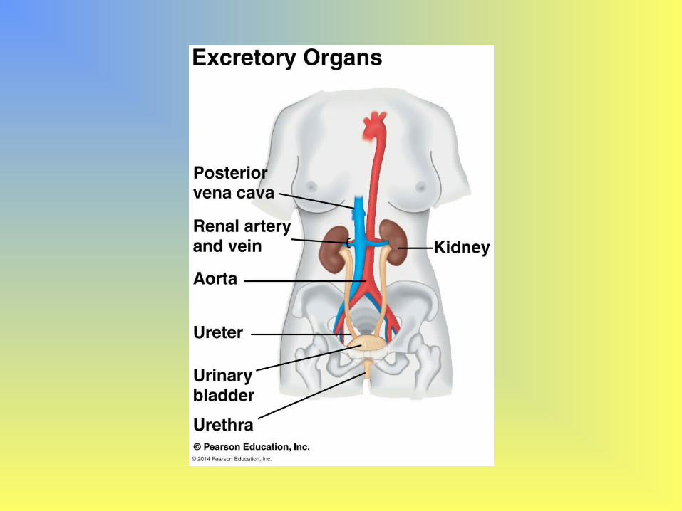
EXCRETORY ORGANS IN ANIMALS
• Lungs: Remove CO2
• Kidneys: Remove urea, other nitrogenous waste from the blood, excess water, salts, hormones & drugs
• Liver: Yellow-green pigment from the breakdown of Hb is excreted with bile into small intestine & excreted with feces this pigment gives feces its brown color. Also produces urea
• Skin: Salt & urea are excreted in sweat as a response to an increase in temperature


PARTS OF THE EXCRETORY SYSTEM
• Kidney – main organ
• Size – each is 12 cm long by 7 cm wide
• The left kidney is slightly higher than the right
• Bean-shaped and red located at the back of the abdominal cavity behind the intestines.
• Put hand on hips – thumbs indicate location of kidneys just below diaphragm

• Kidneys receive “dirty” blood from renal artery while the renal vein takes cleansed/filtered blood away from kidneys to the heart.
• Nitrogenous & other waste flow down through the ureter to the bladder.
• The ureter connects each kidney to the bladder (muscular bag @ bottom of kidney)


• The bladder is drained by the urethra to the opening of penis/vagina.
• At the top of the urethra are 2 sets of sphincter muscles which controls the release of urine. The lower sphincter muscle is controlled voluntarily while the upper muscle is controlled involuntarily.
• It automatically relaxes when the bladder is full.• When the bladder is full the stretching stimulates
sensory nerve endings in its wall which sends nerve impulses to the brain causing urine to be released – This is called urination.
• Babies can’t control their voluntary sphincter muscles

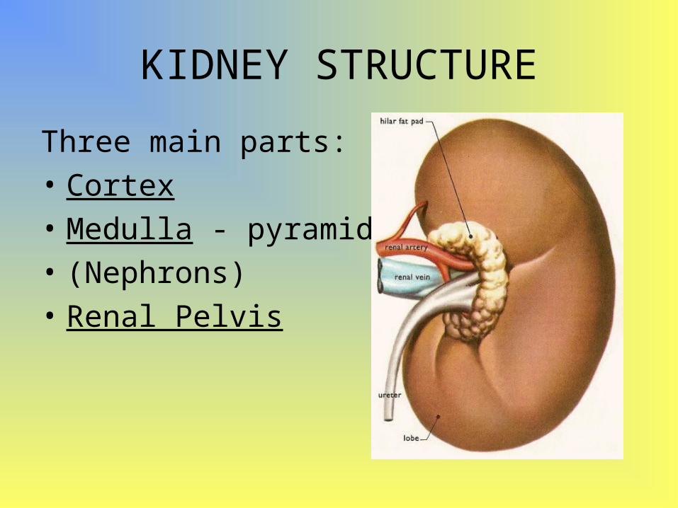
KIDNEY STRUCTURE
Three main parts:
• Cortex
• Medulla - pyramids
• (Nephrons)
• Renal Pelvis

• The outer region of the kidney = CORTEX
• 2. Inner region = MEDULLA
• 3. The area where all of the urine collects is called the PELVIS OF THE KIDNEY
• PELVIS --- Also where blood supply enters and exits the kidney through Renal Artery & Renal Vein
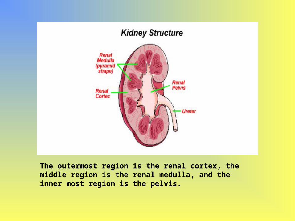
The outermost region is the renal cortex, the middle region is the renal medulla, and the inner most region is the pelvis.

KIDNEY STRUCTURE
• Contains about 160km of blood vessels & more than a million nephrons/kidney tubules
• NEPHRON ==== Functional unit of Kidney
• Two areas: darker outer region – cortex; paler inner region – medulla
• Cortex contains many tiny blood vessels which branch from renal artery.
• Cortex also contains nephrons which run through the medulla.

• Medulla is connected to ureter
• Medulla consists of several cone-shaped areas called pyramids which point inwards towards the concave side of the kidney i.e. into the pelvis
• Urine drains from tips of pyramids (at the pelvis) into funnel-shaped spaces formed by the top of ureter
• Ureter carries urine to the bladder


STRUCTURE OF A NEPHRON
• Basic unit of the kidney
• Glomerulus – Made of entwined glomerular capillaries surrounded by Bowman’s capsule
• Bowman’s capsule - Cup-like structure with a closed end at the beginning of the nephron is located in the cortex.


• Afferent Arteriole The afferent arteriole receives blood rich in oxygen from the renal artery. This blood is transported to the glomerulus of the nephron where it is pressure filtered.
• Efferent Arteriole: Carries blood away from the glomerulus and form the peritubular capillaries surrounding proximal and distal tubules

• Proximal convoluted tubule - The first twisted region after the Bowman's capsule; it's in the cortex.
• Works by actively (using ATP) transporting nutrients such as glucose, amino acid, and salts back into the capillaries
• Loop of Henle - A long, hairpin loop after the proximal tubule, it extends from the cortex down into the medulla and back.– Descending Limb ( Permeable only to water)– Ascending Limb ( Impermeable to water, but actively
pumps out sodium and chloride making medulla “Salty”

• Distal convoluted tubule - This second twisted portion of the nephron after the loop of Henle is located in the cortex.
• Secretion (K+ and H+) and re-absorption (Na+) and Calcium occurs through secondary active transport
• Collecting duct - This long straight portion after the distal tubule that is the open end of the nephron extends from the cortex down through the medulla.
• Receives filtrate from several nephrons and down to the pelvis of the kidney
• CD can remove water from the filtrate, making urine concentrated
• Aquaporin channels in the collecting duct allow water molecules to cross the epithelium


How the kidney concentrates urine?


STRUCTURE OF A NEPHRON• Kidney has millions of nephrons/kidney tubules
– 3cm long• They begin in the cortex, loops down into the
medulla, back to cortex, down through the medulla to the pelvis where they join with the ureter
• The 1st part of the nephron is the Bowman’s capsule which is a round, cup-shaped object which is ~ 0.2mm in diameter.
• Each capsule encloses a ball of finely divided and inter-twined blood capillaries called the glomerulus (plural glomeruli).

• The glomeruli are supplied with blood containing N2 and other wastes products to be cleansed.
• Each tubule/nephron emerges from the Bowman’s capsule on the side opposite to the glomerulus, goes into a series of loops, then joins the wider collecting duct.
• These ducts collect urine from the nephrons and transport it straight through the medulla to the tips of the kidneys

NOTE: Blood supply to nephrons• Oxygenated blood comes to the kidneys at
a high pressure through the renal artery from the main aorta.
• Inside the kidney, the renal artery divides into arterioles (afferent arteriole) which carry blood to glomeruli capillaries with a small drop in pressure
• The blood vessel (efferent arteriole) which leaves each glomerulus branches into a capillary network around the coiled and looped part of each nephron, before joining the renal vein.

• The afferent arteriole has a larger diameter than the efferent arteriole to withstand the pressure of blood entering it.
• This difference in arteriole diameters helps to raise the blood pressure in the glomerulus.

FORMATION OF URINE
• Urine is formed in 2 stages:1. Blood is filtered into the kidney tubules to
form a clear liquid (filtrate) which contains the waste urea and many useful substances which the body can’t lose. This stage is called filtration.
2. These useful substances are reabsorbed from the filtrate back into the blood leaving urea and other useless substances in the kidney tubules. This stage is called re-absorption.

1. Pressure/Ultra-filtration
• Filtration of the blood occurs in the Bowman’s capsule through 2 layers of membranes: the capillary wall of each glomerulus and the inner wall of each Bowman’s capsule.
• These membranes have a surface area of 1 m2

• Blood enters the Bowman’s capsule at a high pressure due to the difference in diameters of the afferent & efferent arterioles.
• This pressure allows small molecules such as water, glucose, AA, vitamins, hormones, salts and urea to pass into the Bowman’s capsule.
• Together they form the liquid glomerular filtrate.
• Larger molecules – plasma proteins & blood cells remain in the blood outside the Bowman’s capsule.

• The filtering process in kidneys is similar to the formation of tissue fluid as both processes involve blood being forced at high pressure through capillary walls where proteins and blood cells (erythrocytes & leucocytes) are filtered out leaving a clear liquid.
• The glomerular filtrate formed in the Bowman’s capsule drains into another part of the nephron: the proximal convoluted tubule.
• Kidneys produce 125cm3 of g. filtrate a minute

2. Selective Re-absorption• This occurs via three mechanisms:• Osmosis • Diffusion, and • Active Transport. • Occurs in the proximal convoluted tubule.• Most of the volume of the filtrate solution
is reabsorbed in the proximal convoluted tubule (PCT)
• This prevents dehydration as useful substances are reabsorbed into the capillaries that surround each nephron.

• Glucose, water and some salts need to be kept in the blood.
• Why is glucose reabsorbed?• Glucose is used for energy.• Most of the energy consumed by the kidneys is
used in the reabsorption of sodium ions (Na+), which are solutes - that is, they are dissolved in the water component of the filtrate solution. As the concentration of Na+ in the filtrate solution is high (about the same as the concentration of Na+ in blood plasma), Na+ moves from the tubular fluid into the cells of the PCT

All glucose, amino acids and 85% of mineral ions are reabsorbed by active transport.
Small proteins are reabsorbed. 80% of water is absorbed back into the blood by osmosis.
The proximal convoluted tubule cells have many mitochondria to provide ATP for active transport and microvilli to increase surface area for absorption.
Other such substances that are reabsorbed with Na+ via active transport include glucose, amino acids, lactic acid, and bicarbonate ions (HCO3-).
These then move on through cells via diffusion and/or other transport processes

Homeostatic Regulation of the Kidney

• Combination of Nervous & Hormonal controls maintain the osmoregulatory function of the mammalian kidney!

OSMOREGULATION• This is the regulation of body fluids.• Another role of kidneys• Kidneys can control the concentration of
urine & thus regulate the water content of the blood.
• What will the kidneys do if one were to drink a litre of water all at once?
• The kidneys will respond to this imbalance by producing a larger volume of dilute urine.

• What will happen if the blood is concentrated? • The kidneys will produce a smaller volume of
urine.• How can the blood become concentrated?• Diet – salty, a lot of sugar.• These changes are controlled by a hormone
produced by the pituitary gland at the base of the brain.
• Hormone: anti-diuretic hormone/ADH or vasopressin.

• ADH begins to work when your body loses too much water e.g. sweating or not replacing water loss via drinking.
• Loss of water = increase of concentration blood.• This is detected by special cells in the
hypothalamus which is a region of the brain.• These cells are sensitive to the solute
concentration of blood which causes the pituitary gland to release more ADH.
• ADH travels through the blood stream to the kidney.
• ADH causes the walls of the collecting ducts to become more permeable to water to allow more water to be absorbed back into the blood..

• This makes the urine concentrated so the body loses less water, but makes the blood dilute.
• When the water content of blood returns to normal – ADH is no longer released.
• Kidney tubules reabsorb less water.• If a person drinks too much water, blood is too
dilute – less ADH is secreted• Leads to the kidney tubules becoming less
permeable to water – more water passes out urine.
• Hence ADH ensures that the internal environment is kept constant.


ADH response pathway in the Collecting Duct
1. ADH binds to membrane receptor
2. Receptor triggers signal cascade
3. Vesicles with aquaporin water channels are inserted into membrane lining of Collecting Duct
4. Aquaporin channels enhance reabsorption of water from Collecting Duct

ADH & Negative feedback
• Internal environment
• Change in internal environment
• Detector senses change
• Correcting action
• Internal environment returns to normal
Neg
ativ
e fe
edba
ck lo
op
switc
hes
off c
ause
of
corr
ectin
g ac
tion
Renin-Angiotensin-Aldosterone System(RAAS)
• Aldosterone is a hormone produced in the adrenal cortex that maintains salt balance in the blood
• Aldosterone is released in response to low blood volume and low blood pressure within the kidney
• When blood pressure drops in the kidneys, there is a drop in the amount of pressure filtration at the glomerulus causing the juxtaglomerular apparatus to secrete the enzyme renin.

RAAS
• Renin initiates a sequence of steps that cleave a plasma protein called Angiotensinogen yielding a peptide Angiotensin II
• Angiotensin II raises blood pressure by constricting arterioles, decreasing blood flow
• Angiotensin II also stimulates the adrenal glands to release a hormone called Aldosterone
• Aldosterone causes the nephron’s distal tubules and CD to reabsorb more Na+ and water, therefore increasing blood volume and pressure


Coordination of ADH & RAAS
• Both increase water reabsorption but they regulate different osmoregulatory problems:
ADH release Response to an increase in blood osmolarity (eg. Dehydration)
RAAS Response to drop in blood volume and blood pressure (eg. major wound or severe diarrhea)
ADH & RAAS are partners in homeostasis

• Another hormone, Atrial Natriuretic Peptide (ANP) opposes the RAAS
• Wall of atria of the heart release ANP in response to an increase in blood volume and pressure
• ANP Inhibits the release of renin from JGA, inhibits NaCl reabsorption and reduces aldosterone release, therefore lowering blood volume and blood pressure
