biology for engineers: cellular and systems neurophysiology christopher fiorillo bis 521, fall 2009...
TRANSCRIPT
Biology for Engineers: Cellular and Systems Neurophysiology
Christopher Fiorillo
BiS 521, Fall 2009
042 350 4326, [email protected]
Part 5: Neurotransmitters, Receptors, and Signal Transduction
Reading: Bear, Connors, and Paradiso
Chapter 6
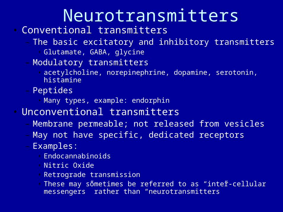
Neurotransmitters• Conventional transmitters
– The basic excitatory and inhibitory transmitters• Glutamate, GABA, glycine
– Modulatory transmitters• acetylcholine, norepinephrine, dopamine, serotonin, histamine
– Peptides• Many types, example: endorphin
• Unconventional transmitters– Membrane permeable; not released from vesicles– May not have specific, dedicated receptors– Examples:
• Endocannabinoids• Nitric Oxide• Retrograde transmission• These may sometimes be referred to as “inter-cellular messengers” rather
than “neurotransmitters”
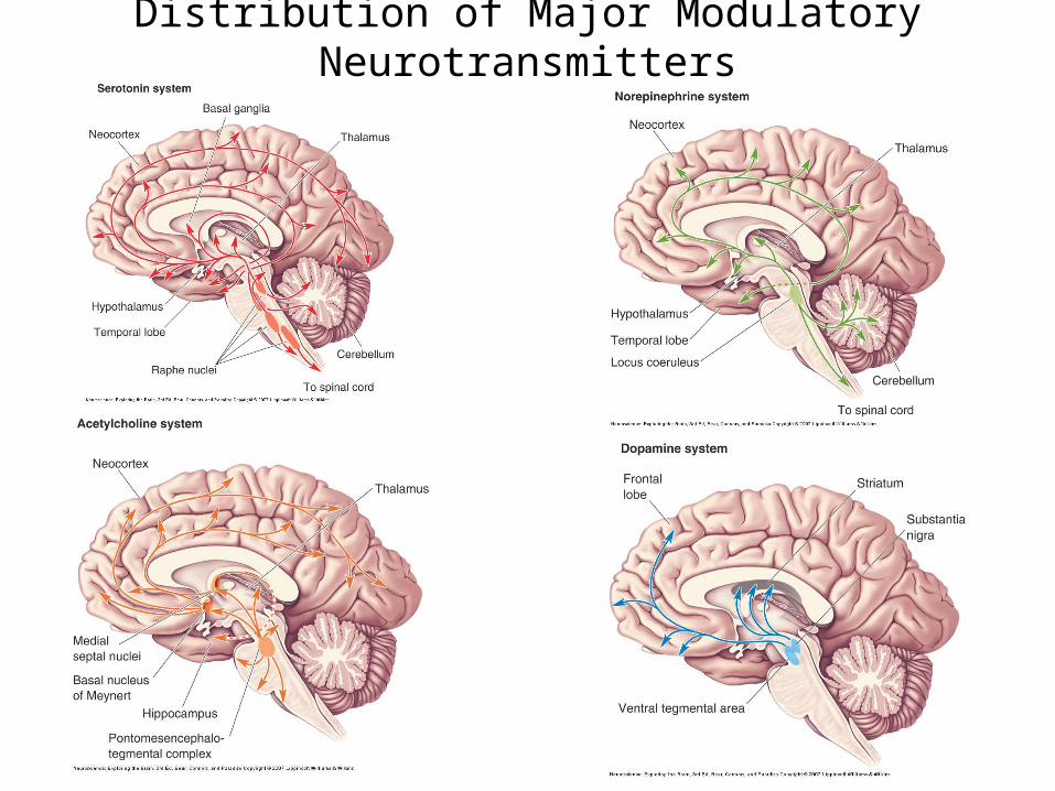
Distribution of Major Modulatory Neurotransmitters

Peptide Neurotransmitters• Peptides are often cotransmitters: they are released together with a
small transmitter• Release of peptides typically requires a high-frequency train of
stimuli• Peptides act on slow metabotropic receptors. There are not
peptide-gated ion channels• There are a great divesity of peptides
– Examples:• Opioid peptides
– Endorphin, enkephalin, dynorphin
• Substance P• Orexin
• The functions of peptides are generally not well understood– They can have excitatory or inhibitory effects– They are best thought of as modulatory
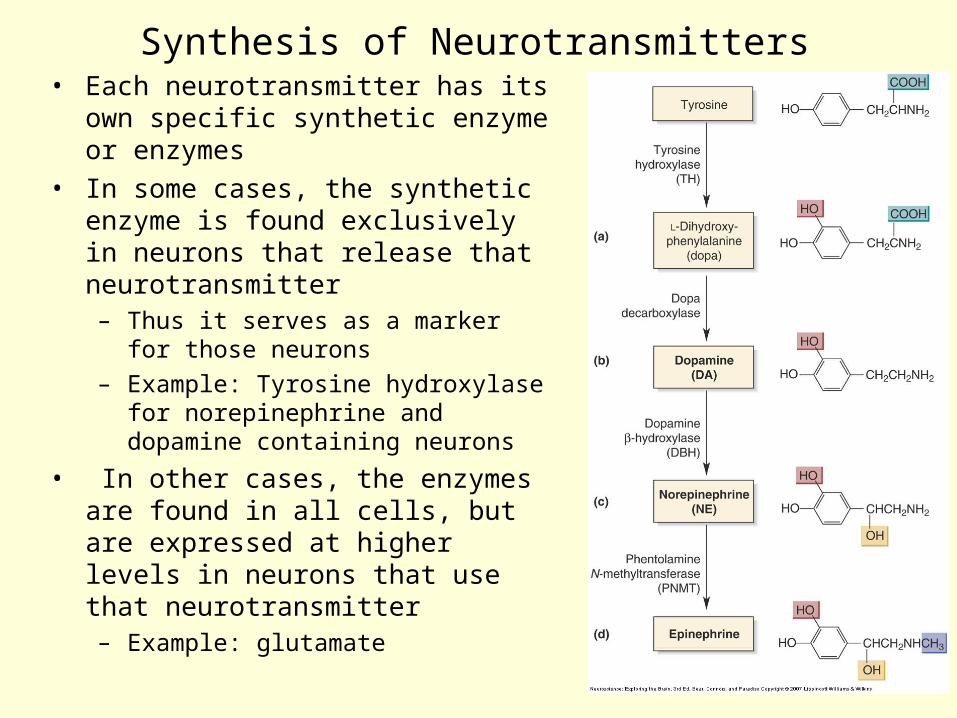
Synthesis of Neurotransmitters• Each neurotransmitter has its own specific
synthetic enzyme or enzymes
• In some cases, the synthetic enzyme is found exclusively in neurons that release that neurotransmitter– Thus it serves as a marker for those
neurons
– Example: Tyrosine hydroxylase for norepinephrine and dopamine containing neurons
• In other cases, the enzymes are found in all cells, but are expressed at higher levels in neurons that use that neurotransmitter– Example: glutamate

Receptor Pharmacology• Ligands are molecules that bind to a receptor
– Natural or artificial
• Agonists bind and activate a receptor• Antagonists bind to a receptor and prevent its activation
by agonists

Major Receptor Classifications for Major Neurotransmitters
• Glutamate– AMPA, NMDA, kainate– mGluR1 - mGluR8
• GABA– GABAA
– GABAB
• Norepinephrine– Alpha1, Alpha2, Beta
• Dopamine– D1 - D5
• Serotonin (5-HT)– 5-HT3
– 5-HT1 5-HT2 5-HT4 5-HT5
• Acetylcholine– Nicotinic– Muscarinic (M1 - M5)
Ionotropic, ligand-gated ion channelsMetabotropic, G-protein coupled receptors
• These are only major divisions. Finer distinctions have been made.
• All of the ionotropic receptors have multiple types of subunits, and different types of subunits combine to make a receptor / ion channel.

Ligand-gated ion channels• Most are pentamers
– Glutamate receptors are tetramers
• Most require two transmitter molecules to bind in order to open the channel
• Some have binding sites for modulators– GABAA is modified by several commonly used classes of drugs
– These drugs enhance the effect of GABA, and reduce anxiety
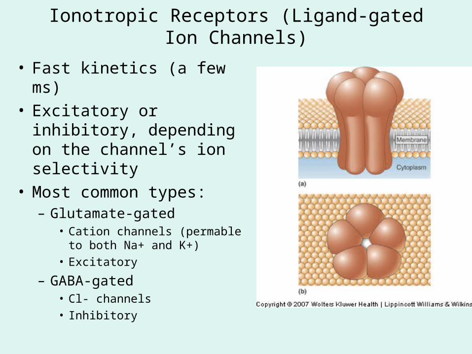
Ionotropic Receptors (Ligand-gated Ion Channels)
• Fast kinetics (a few ms)• Excitatory or inhibitory,
depending on the channel’s ion selectivity
• Most common types:– Glutamate-gated
• Cation channels (permable to both Na+ and K+)
• Excitatory
– GABA-gated• Cl- channels
• Inhibitory

Glutamate EPSCs
Fast Excitatory and Inhibitory Postsynaptic Potentials• Mediated by Ionotropic Receptors (ligand-gated ion channels)
• Fast GABA IPSPs (~30 ms) typically last longer than fast glutamate EPSPs (~5 ms) (contrary to the drawing below and in the textbook)
• PSCs are faster than PSPs due to the membrane capacitance (which usually has a time constant of 1-30 ms).

Metabotropic Receptors (G-protein-coupled receptors)• Modifies “effectors” though G-proteins
– G-proteins metabolize GTP to GDP. Since they use energy, these receptors are called “metabotropic”
• Often modulatory rather than simply excitatory or inhibitory
• One receptor may alter multiple types of ion channels and other effectors
• Slow kinetics (> 100 ms)
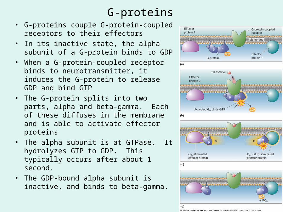
G-proteins• G-proteins couple G-protein-coupled receptors
to their effectors
• In its inactive state, the alpha subunit of a G-protein binds to GDP
• When a G-protein-coupled receptor binds to neurotransmitter, it induces the G-protein to release GDP and bind GTP
• The G-protein splits into two parts, alpha and beta-gamma. Each of these diffuses in the membrane and is able to activate effector proteins
• The alpha subunit is at GTPase. It hydrolyzes GTP to GDP. This typically occurs after about 1 second.
• The GDP-bound alpha subunit is inactive, and binds to beta-gamma.
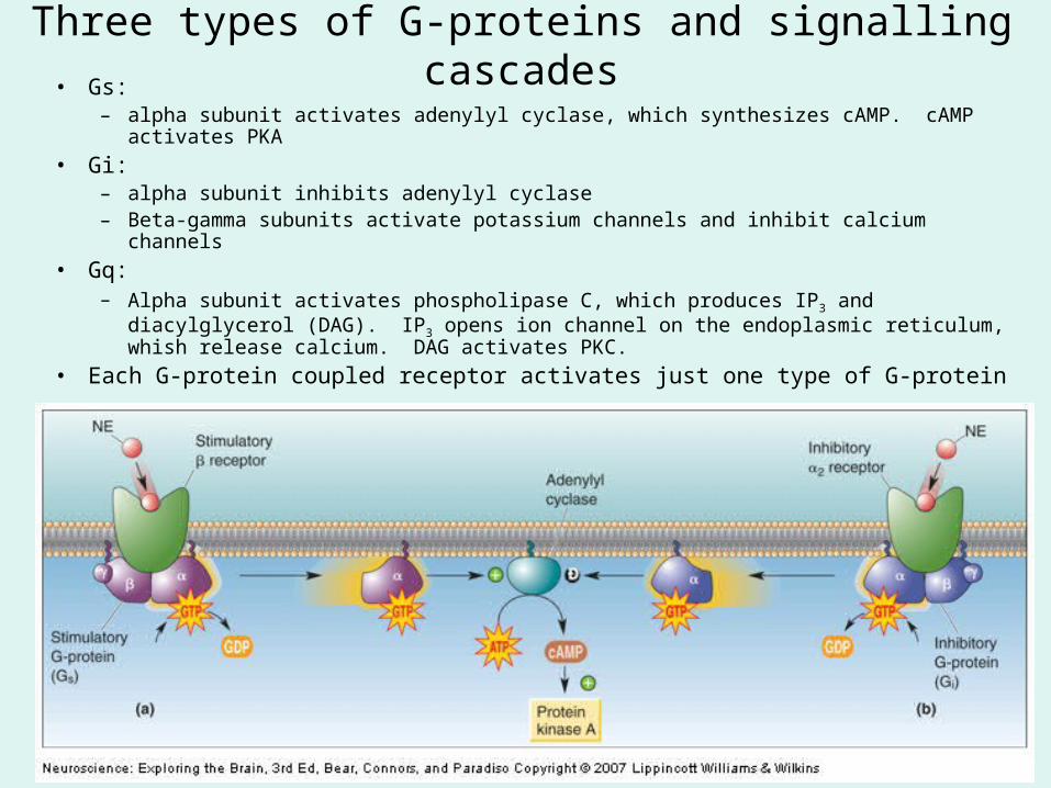
Three types of G-proteins and signalling cascades• Gs:
– alpha subunit activates adenylyl cyclase, which synthesizes cAMP. cAMP activates PKA
• Gi: – alpha subunit inhibits adenylyl cyclase– Beta-gamma subunits activate potassium channels and inhibit calcium channels
• Gq:– Alpha subunit activates phospholipase C, which produces IP3 and diacylglycerol (DAG). IP3
opens ion channel on the endoplasmic reticulum, whish release calcium. DAG activates PKC.
• Each G-protein coupled receptor activates just one type of G-protein

Direct activation of K+ channels by G-proteins • Gi Beta-gamma subunits activate potassium channels• This is the basis for the GABAB IPSP• They also inhibit Ca2+ channels
GABAB IPSP
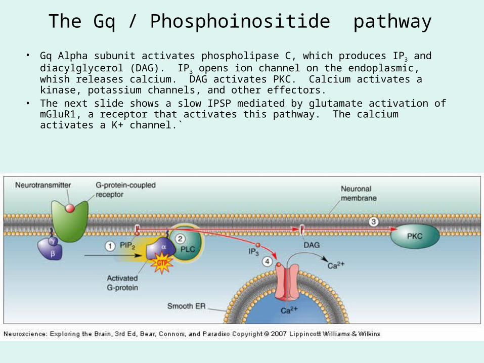
The Gq / Phosphoinositide pathway
• Gq Alpha subunit activates phospholipase C, which produces IP3 and diacylglycerol (DAG). IP3 opens ion channel on the endoplasmic, whish releases calcium. DAG activates PKC. Calcium activates a kinase, potassium channels, and other effectors.
• The next slide shows a slow IPSP mediated by glutamate activation of mGluR1, a receptor that activates this pathway. The calcium activates a K+ channel.`

Slow Postsynaptic Potentials Mediated by Metabotropic Receptors
• Metabotropic PSPs typically require multiple stimuli at high frequencies– 10 stimuli at 66 Hz (15 ms
intervals) in these examples
• PSPs start after about 100-300 ms and last for a second or longer
• There are many other effects of metabotropic receptors besides PSPs– Modification of ion channels
– Modification of gene expression
GABAB IPSP
Inhibition by glutamate (mediated by mGluR1 and Ca2+ activated K+ channels)
Inhibition and excitation by the same glutamate receptor (mGluR1)
Second Messengers• The “first” messenger is the neurotransmitter, which mediates
intercellular communication between cells• A second messenger is a small molecule that carries information within
a cell (through diffusion). It mediates intracellular communication.• Examples:
– cAMP– cGMP– IP3
– DAG– Calcium
• Calcium– The most common second messenger– Three main sources
• Voltage-gated calcium channels• NMDA receptors (gated by glutamate)• Intracellular calcium stores in the endoplasmic reticulum
– Calcium is released when IP3 or calcium opens channels in the ER
Protein Kinases and Phosphatases• The function of proteins (including ion channels) is
modulated through phosphorylation– A phosphate group is attached to a serine, threonine or tyrosine
residue
• Kinases add phosphate groups; phosphatases remove them
• Best known kinases:– Protein kinase A (PKA)– Protein kinase C (PKC)– Calcium-calmodulin-dependent protein kinase (cam-kinase)
• Each kinase or phosphatase modifies a variety of different proteins (effectors)– The set of effector proteins depends on the kinase /
phosphatase