biology - carnes ap bio · the cell from the external environment. ... – isotonic solution:...
TRANSCRIPT

Copyright © 2008 Pearson Education, Inc., publishing as Pearson Benjamin Cummings
PowerPoint® Lecture Presentations for
Biology
Eighth Edition
Neil Campbell and Jane Reece
Lectures by Chris Romero, updated by Erin Barley with contributions from Joan Sharp
BIG IDEA II Biological systems utilize free energy and molecular building blocks
to grow, to reproduce and to maintain dynamic homeostasis.
Enduring Understanding 2.B
Growth, reproduction and dynamic homeostasis
require that cells create and maintain internal environments
that are different from their external environments.
Essential Knowledge 2.B.1
Cell membranes are selectively permeable due to their structure.

Copyright © 2008 Pearson Education, Inc., publishing as Pearson Benjamin Cummings
Essential Knowledge 2.B.1: Cell membranes are selectively permeable due to their structure.
• Learning Objectives:
– (2.10) The student is able to use representations and
models to pose scientific questions about the
properties of cell membranes and selective
permeability based on molecular structure.
– (2.11) The student is able to construct models that
connect the movement of molecules across
membranes with membrane structure and function.

Copyright © 2008 Pearson Education, Inc., publishing as Pearson Benjamin Cummings
Cell membranes separate the internal environment of the cell from the external environment.
• The cell or plasma membrane is selectively permeable; that is, it allows
some substances to cross it more easily than others.
• The structure of the cell membrane results in its selectively permeable
properties.
Copyright © 2008 Pearson Education, Inc., publishing as Pearson Benjamin Cummings
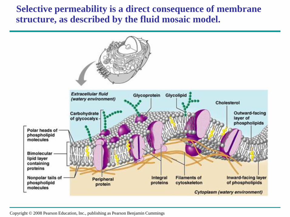
Selective permeability is a direct consequence of membrane structure, as described by the fluid mosaic model.

Fig. 7-2
Hydrophilic head
WATER
Hydrophobic tail
WATER

Fig. 7-3
Phospholipid
bilayer
Hydrophobic regions of protein
Hydrophilic regions of protein

Fig. 7-9
(a) Transport
ATP
(b) Enzymatic activity
Enzymes
(c) Signal transduction
Signal transduction
Signaling molecule
Receptor
(d) Cell-cell recognition
Glyco-
protein
(e) Intercellular joining (f) Attachment to
the cytoskeleton
and extracellular
matrix (ECM)

Copyright © 2008 Pearson Education, Inc., publishing as Pearson Benjamin Cummings
Selective Permeability of the Cell Membrane

Copyright © 2008 Pearson Education, Inc., publishing as Pearson Benjamin Cummings
Selective Permeability of the Cell Membrane

Copyright © 2008 Pearson Education, Inc., publishing as Pearson Benjamin Cummings
Types of Transport Proteins
• Transport proteins allow passage of hydrophilic
substances across the membrane:
1. Some transport proteins, called channel proteins, have a
hydrophilic channel that certain molecules or ions can use
as a tunnel.
2. Channel proteins called aquaporins facilitate the passage of
water.
3. Other transport proteins, called carrier proteins, bind to
molecules and change shape to shuttle them across the
membrane.
• A transport protein is specific for the substance it moves –
i.e. FORM FITS FUNCTION!!!
Copyright © 2008 Pearson Education, Inc., publishing as Pearson Benjamin Cummings

Copyright © 2008 Pearson Education, Inc., publishing as Pearson Benjamin Cummings
Cell walls provide a structural boundary, as well as a permeability barrier for some substances to the internal environment.
• The cell wall is the tough, usually flexible but sometimes fairly
rigid layer that surrounds some types of cells. Cell walls provide
a structural boundary, as well as a permeability barrier for some
substances to the internal environments.
– It is located outside the cell membrane and provides these cells
with structural support and protection, in addition to acting as a
filtering mechanism.
– A major function of the cell wall is to act as a pressure vessel,
preventing over-expansion when water enters the cell.
– Cell walls are found in plants, bacteria, fungi, algae, and some
archaea. Animals and protozoa do not have cell walls.
– The material in the cell wall varies between species, and can also
differ depending on cell type and developmental stage.

Copyright © 2008 Pearson Education, Inc., publishing as Pearson Benjamin Cummings
Plant Cell Walls

Copyright © 2008 Pearson Education, Inc., publishing as Pearson Benjamin Cummings
Fungi Cell Walls
Copyright © 2008 Pearson Education, Inc., publishing as Pearson Benjamin Cummings
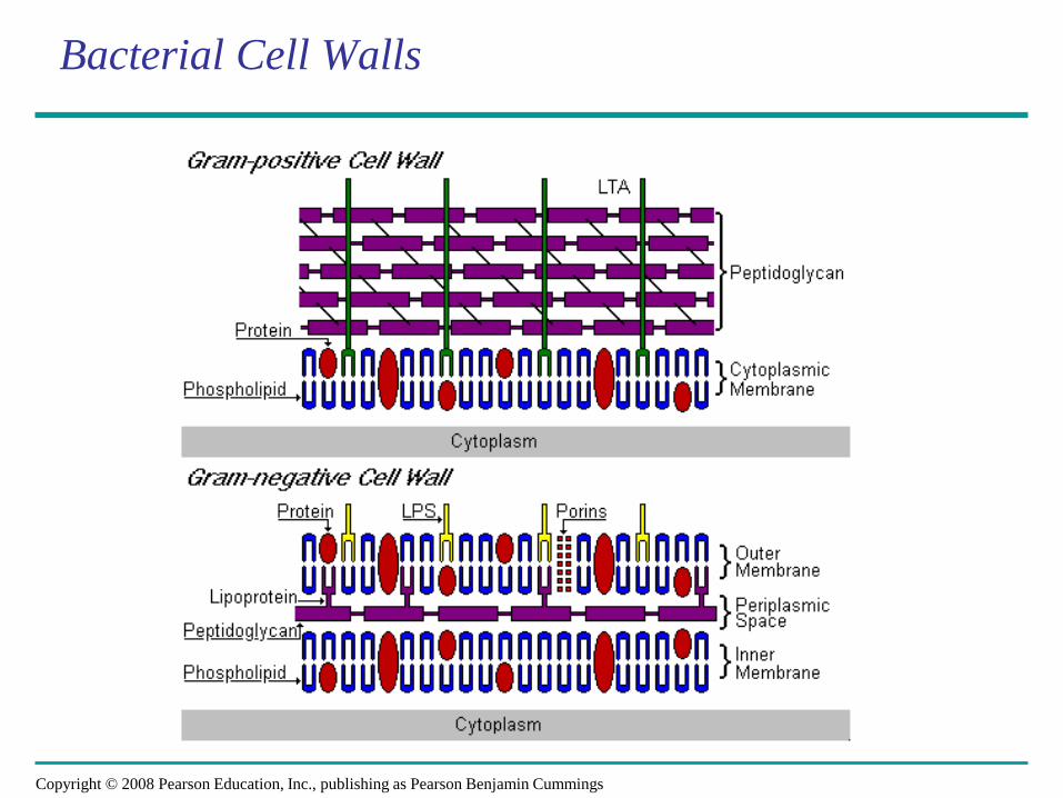
Bacterial Cell Walls
Copyright © 2008 Pearson Education, Inc., publishing as Pearson Benjamin Cummings
PowerPoint® Lecture Presentations for
Biology
Eighth Edition
Neil Campbell and Jane Reece
Lectures by Chris Romero, updated by Erin Barley with contributions from Joan Sharp
BIG IDEA II Biological systems utilize free energy and molecular building blocks
to grow, to reproduce and to maintain dynamic homeostasis.
Enduring Understanding 2.B Growth, reproduction and dynamic homeostasis require that cells create and
maintain internal environments that are different from their external environments.
Essential Knowledge 2.B.2 Growth and dynamic homeostasis are maintained by the
constant movement of molecules across membranes.

Copyright © 2008 Pearson Education, Inc., publishing as Pearson Benjamin Cummings
Essential Knowledge 2.B.2: Growth and dynamic homeostasis
are maintained by the constant movement of molecules across membranes.
• Learning Objectives:
– (2.12) The student is able to use representations and
models to analyze situations or solve problems
qualitatively and quantitatively to investigate
whether dynamic homeostasis is maintained by the
active movement of molecules across membranes.

Copyright © 2008 Pearson Education, Inc., publishing as Pearson Benjamin Cummings
Passive transport does not require the input of metabolic energy; the net movement of molecules is from high concentration to low concentration.
• Passive Transport is the diffusion of a substance across a
semi-permeable membrane from an area of high
concentration to an area of low concentration WITHOUT
the use of energy.
• Passive transport plays a primary role in the import of
resources and the export of wastes.
– Diffusion is the tendency for molecules to spread out evenly into
the available space.
– Although each molecule moves randomly, diffusion of a
population of molecules may exhibit a net movement in one
direction.
– At dynamic equilibrium, as many molecules cross one way as
cross in the other direction.
Copyright © 2008 Pearson Education, Inc., publishing as Pearson Benjamin Cummings

Fig. 7-11 Molecules of dye Membrane (cross section)
WATER
Net diffusion Net diffusion Equilibrium
(a) Diffusion of one solute
Net diffusion
Net diffusion
Net diffusion
Net diffusion
Equilibrium
Equilibrium
(b) Diffusion of two solutes

Copyright © 2008 Pearson Education, Inc., publishing as Pearson Benjamin Cummings
• Substances diffuse down their concentration
gradient, the difference in concentration of a
substance from one area to another.
• No work must be done to move substances down
the concentration gradient.
• The diffusion of a substance across a biological
membrane is passive transport because it
requires no energy from the cell to make it
happen.
Copyright © 2008 Pearson Education, Inc., publishing as Pearson Benjamin Cummings
Passive Transport & Concentration Gradients

Copyright © 2008 Pearson Education, Inc., publishing as Pearson Benjamin Cummings
DIFFUSION EXISTS IN TWO FORMS: Dialysis and Osmosis
• Dialysis: the PASSIVE movement of particles across a semi-permeable membrane from an area of high concentration to an area of low concentration (no energy).
• Osmosis: the PASSIVE movement of water molecules across a semi-permeable membrane from an area of high concentration to an area of low concentration (no energy).

Copyright © 2008 Pearson Education, Inc., publishing as Pearson Benjamin Cummings
Effects of Osmosis on Water Balance
• Osmosis is the diffusion of water across a
selectively permeable membrane.
• Water diffuses across a membrane from the
region of lower solute concentration to the region
of higher solute concentration.
• THIS MEANS THAT WATER ALWAYS
ATTEMPTS TO DILUTE!!!!
Copyright © 2008 Pearson Education, Inc., publishing as Pearson Benjamin Cummings

Lower
concentration of solute (sugar)
Fig. 7-12
H2O
Higher
concentration of sugar
Selectively permeable
membrane
Same concentration
of sugar
Osmosis

Copyright © 2008 Pearson Education, Inc., publishing as Pearson Benjamin Cummings
Water Balance of Cells Without Walls
• Tonicity is the ability of a solution to cause a cell
to gain or lose water:
– Isotonic solution: Solute concentration is the
same as that inside the cell; no net water
movement across the plasma membrane
– Hypertonic solution: Solute concentration is
greater than that inside the cell; cell loses
water
– Hypotonic solution: Solute concentration is
less than that inside the cell; cell gains water
Copyright © 2008 Pearson Education, Inc., publishing as Pearson Benjamin Cummings

Fig. 7-13
Hypotonic solution
(a) Animal
cell
(b) Plant
cell
H2O
Lysed
H2O
Turgid (normal)
H2O
H2O
H2O
H2O
Normal
Isotonic solution
Flaccid
H2O
H2O
Shriveled
Plasmolyzed
Hypertonic solution

Copyright © 2008 Pearson Education, Inc., publishing as Pearson Benjamin Cummings
• Hypertonic or hypotonic environments create
osmotic problems for organisms.
• Osmoregulation, the control of water balance, is
a necessary adaptation for life in such
environments.
• The protist Paramecium, which is hypertonic to its
pond water environment, has a contractile vacuole
that acts as a pump to regulate its water balance.
Copyright © 2008 Pearson Education, Inc., publishing as Pearson Benjamin Cummings
Osmoregulation

Fig. 7-14
Filling vacuole 50 µm
(a) A contractile vacuole fills with fluid that enters from a system of canals radiating throughout the cytoplasm.
Contracting vacuole
(b) When full, the vacuole and canals contract, expelling
fluid from the cell.

Copyright © 2008 Pearson Education, Inc., publishing as Pearson Benjamin Cummings
Water Balance of Cells with Walls
• Cell walls help maintain water balance.
• A plant cell in a hypotonic solution swells until the
wall opposes uptake; the cell is now turgid (firm).
• If a plant cell and its surroundings are isotonic,
there is no net movement of water into the cell; the
cell becomes flaccid (limp), and the plant may
wilt.
Copyright © 2008 Pearson Education, Inc., publishing as Pearson Benjamin Cummings

Copyright © 2008 Pearson Education, Inc., publishing as Pearson Benjamin Cummings
• In a hypertonic environment, plant cells lose
water; eventually, the membrane pulls away from
the wall, a usually lethal effect called plasmolysis.
Copyright © 2008 Pearson Education, Inc., publishing as Pearson Benjamin Cummings
Water Balance of Cells with Walls
Copyright © 2008 Pearson Education, Inc., publishing as Pearson Benjamin Cummings
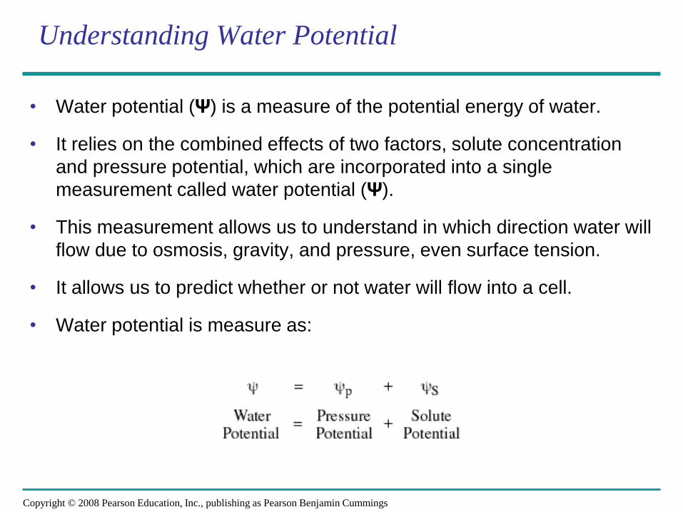
Understanding Water Potential
• Water potential (Ψ) is a measure of the potential energy of water.
• It relies on the combined effects of two factors, solute concentration
and pressure potential, which are incorporated into a single
measurement called water potential (Ψ).
• This measurement allows us to understand in which direction water will
flow due to osmosis, gravity, and pressure, even surface tension.
• It allows us to predict whether or not water will flow into a cell.
• Water potential is measure as:

Copyright © 2008 Pearson Education, Inc., publishing as Pearson Benjamin Cummings
Understanding Water Potential
• The standard for measuring
water potential is pure water.
• Pure water has a water potential
of ZERO bars.
• Water potential is measured on
either side of a membrane.
• Water flows from an area of high
water potential to an area of
lower water potential.

Copyright © 2008 Pearson Education, Inc., publishing as Pearson Benjamin Cummings
Understanding Solute Potential
• Solute potential is also called osmotic
potential because solutes affect the
direction of osmosis.
• Solute potential DECREASES with
increasing solute CONCENTRATION; and
this decrease causes a DECREASE IN
WATER POTENTIAL.
• This is because solutes bind to water
molecules reducing the number of free
water molecules to move and therefore
perform work.
• This means that INCREASING solute
concentration DECREASES water
potential!

Copyright © 2008 Pearson Education, Inc., publishing as Pearson Benjamin Cummings
Effect of Solute on Water Potential

Copyright © 2008 Pearson Education, Inc., publishing as Pearson Benjamin Cummings
Calculating Solute Potential (ΨS)
• Once you know a solute concentration, you can calculate solute potential
using the following formula: (ΨS ) = – iCRT
• Pure water has a solute potential (Ψs) of zero. Solute potential can never be positive.
• Adding solute to pure water is a negative experience; thus the solute potential becomes negative.
– i = The ionization constant (two what degree will the substance produce ions in water):
• for NaCl this would be 2;
• for sucrose or glucose, this number is 1
– C = Molar concentration (experimentally determined)
• Iso-osmolar molarity: would allow you to place a potato in the solution and get no movement of water.
• Point at which the line crosses 0 on the graph.
– R = Pressure constant = 0.0831 liter bar/mole K
– T = Temperature in degrees Kelvin = 273 + °C of solution

Copyright © 2008 Pearson Education, Inc., publishing as Pearson Benjamin Cummings
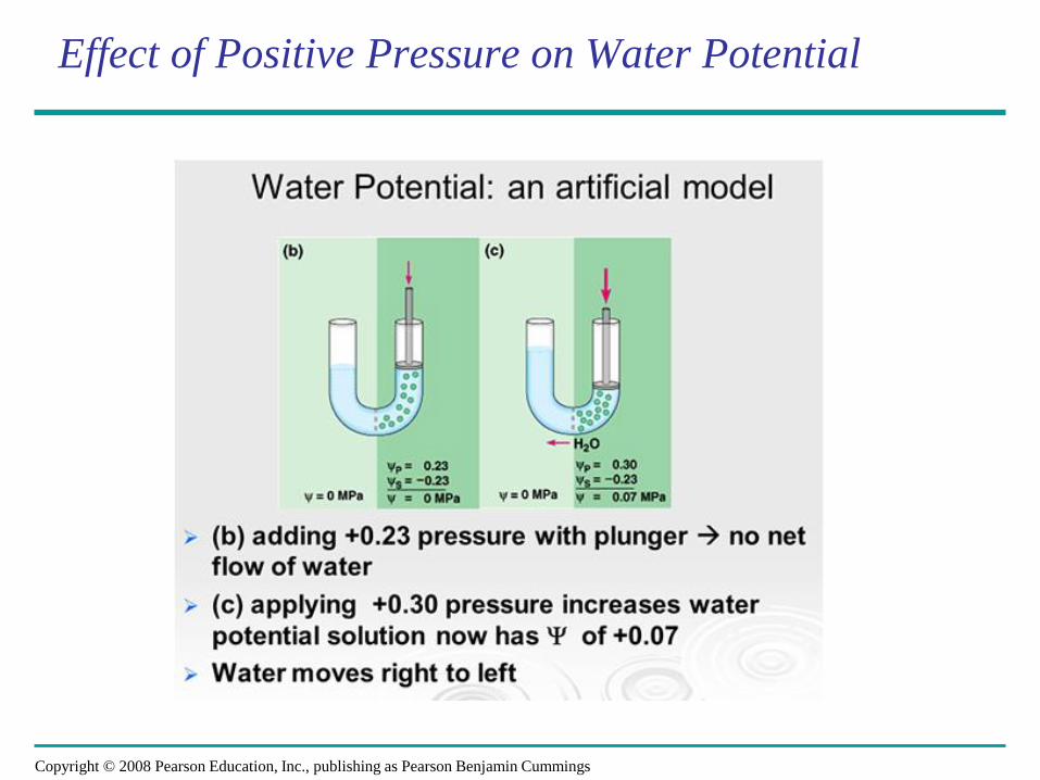
Pressure Potential (ΨP)
• Pressure potential is the sum of all pressure on water. It is measured in units
of MPa or Bars.
– Turgor pressure is the force caused by the cell membrane pushing against
the cell wall. Wall pressure is an equal and opposite force exerted by the
cell wall. It counteracts the movement of water due to osmosis.
– Pressure potential can be NEGATIVE as in water tension (transpiration in
the xylem of a plant).
– Pressure potential can be POSITIVE as in turgor pressure (water in living
cells under positive pressure).
– Pure water in an open container has a water potential of zero at one
atmospheric pressure.
– If physical pressure is applied to a solution, then its water potential (the
potential for the water to move and do work) will be affected. How it is
affected depends upon the direction of the pressure.
– Increased pressure raises water potential!
Copyright © 2008 Pearson Education, Inc., publishing as Pearson Benjamin Cummings
Effect of Positive Pressure on Water Potential

Copyright © 2008 Pearson Education, Inc., publishing as Pearson Benjamin Cummings
Effect of Negative Pressure on Water Potential

Copyright © 2008 Pearson Education, Inc., publishing as Pearson Benjamin Cummings
Water Potential in Plant Cells - Turgid

Copyright © 2008 Pearson Education, Inc., publishing as Pearson Benjamin Cummings
Water Potential in Plant Cells - Plasmolysis

Copyright © 2008 Pearson Education, Inc., publishing as Pearson Benjamin Cummings
Key Points to Remember
• Water always moves from [hi] to [low] concentration, or from [hi]
to [low] potential. This means that water always moves from
hypotonic to hypertonic.
• [Solute] is related to osmotic potential (the potential for water
to move across a semi-permeable membrane from high to low
concentration).
– Increased solutes decreases water potential.
• [Pressure] is related to pressure potential.
– Increased pressure raises water potential.
• Always use ZERO for pressure potential in animal cells and
open containers.
– 1 bar of pressure = 1 atmosphere.

Copyright © 2008 Pearson Education, Inc., publishing as Pearson Benjamin Cummings
Facilitated Diffusion
• In facilitated diffusion, transport proteins speed
the passive movement of molecules across the
plasma membrane.
• Channel proteins provide corridors that allow a
specific molecule or ion to cross the membrane -
facilitated diffusion is still passive because the
solute moves down its concentration gradient.
• Channel proteins include:
– Aquaporins, for facilitated diffusion of water
– Ion channels that open or close in response to a
stimulus (gated channels)
Copyright © 2008 Pearson Education, Inc., publishing as Pearson Benjamin Cummings

Fig. 7-15
EXTRACELLULAR FLUID
Channel protein
(a) A channel protein
Solute CYTOPLASM
Solute Carrier protein
(b) A carrier protein

Copyright © 2008 Pearson Education, Inc., publishing as Pearson Benjamin Cummings
Active transport requires free energy to move molecules from regions of low concentration to regions of high concentration.
• Active Transport uses free energy to move
molecules AGAINST their concentration gradients
via proteins imbedded in the cell membrane.
– Active transport is a process where free energy (often
provided by ATP) is used by proteins embedded in the
membrane to “move” molecules and/or ions across the
membrane and to establish and maintain concentration
gradients.
– Membrane proteins are NECESSARY for active
transport.
– Active transport allows cells to maintain concentration
gradients that DIFFER from their surroundings.
Copyright © 2008 Pearson Education, Inc., publishing as Pearson Benjamin Cummings

Fig. 7-17 Passive transport
Diffusion Facilitated diffusion
Active transport
ATP

Copyright © 2008 Pearson Education, Inc., publishing as Pearson Benjamin Cummings
Figure 8.15 The sodium-potassium pump: a specific case of active transport

Copyright © 2008 Pearson Education, Inc., publishing as Pearson Benjamin Cummings
How Ion Pumps Maintain Membrane Potential
• Membrane potential is the voltage difference
across a membrane:
– Voltage is created by differences in the distribution
of positive and negative ions.
• Two combined forces, collectively called the
electrochemical gradient, drive the diffusion of
ions across a membrane:
– A chemical force (the ion’s concentration gradient)
– An electrical force (the effect of the membrane
potential on the ion’s movement)
Copyright © 2008 Pearson Education, Inc., publishing as Pearson Benjamin Cummings

Copyright © 2008 Pearson Education, Inc., publishing as Pearson Benjamin Cummings
• An electrogenic pump is a transport protein that generates voltage across a
membrane:
– For example, the sodium-potassium pump is the major electrogenic
pump of animal cells.
• The main electrogenic pump of plants, fungi, and bacteria is a proton pump.
Copyright © 2008 Pearson Education, Inc., publishing as Pearson Benjamin Cummings
Cellular Pumps

Copyright © 2008 Pearson Education, Inc., publishing as Pearson Benjamin Cummings
Cotransport
• Cotransport is coupled transport by a membrane protein. It occurs
when the active transport of a solute indirectly drives the transport of
another solute:
– For example, plants commonly use the gradient of hydrogen ions
generated by proton pumps to drive the active transport of
nutrients into the cell.
Copyright © 2008 Pearson Education, Inc., publishing as Pearson Benjamin Cummings

Copyright © 2008 Pearson Education, Inc., publishing as Pearson Benjamin Cummings
The processes of endocytosis and exocytosis move large molecules from the external environment to the internal environment and vice versa.
• Bulk transport across the plasma membrane
occurs by exocytosis and endocytosis.
– Small molecules and water enter or leave the
cell through the lipid bilayer or by transport
proteins, but large molecules, such as
polysaccharides and proteins, cross the
membrane in bulk via vesicles.
– Bulk transport requires energy and is therefore
a type of ACTIVE transport.
Copyright © 2008 Pearson Education, Inc., publishing as Pearson Benjamin Cummings

Copyright © 2008 Pearson Education, Inc., publishing as Pearson Benjamin Cummings
Exocytosis
• In exocytosis, transport vesicles migrate to the
membrane, fuse with it, and release their contents.
– Many secretory cells use exocytosis to export
their products (proteins).
– In what part of the cell were these proteins
made? The ER or cytoplasm?
Copyright © 2008 Pearson Education, Inc., publishing as Pearson Benjamin Cummings

Copyright © 2008 Pearson Education, Inc., publishing as Pearson Benjamin Cummings
Endocytosis
• In endocytosis, the cell takes in macromolecules
by forming vesicles from the plasma membrane.
• Endocytosis is a reversal of exocytosis, involving
different proteins.
• There are three types of endocytosis:
– Phagocytosis (“cellular eating”)
– Pinocytosis (“cellular drinking”)
– Receptor-mediated endocytosis
Copyright © 2008 Pearson Education, Inc., publishing as Pearson Benjamin Cummings

Copyright © 2008 Pearson Education, Inc., publishing as Pearson Benjamin Cummings
• In phagocytosis a cell engulfs a particle in a
vacuole. The vacuole fuses with a lysosome to
digest the particle.
• In pinocytosis, molecules are taken up when
extracellular fluid is “gulped” into tiny vesicles.
• In receptor-mediated endocytosis, binding of
ligands to receptors triggers vesicle formation:
– A ligand is any molecule that binds specifically to a
receptor site of another molecule.
Copyright © 2008 Pearson Education, Inc., publishing as Pearson Benjamin Cummings
Phagocytosis & Pinocytosis

Fig. 7-20 PHAGOCYTOSIS
EXTRACELLULAR
FLUID
CYTOPLASM
Pseudopodium
“Food”or other particle
Food vacuole
PINOCYTOSIS
1 µm
Pseudopodium
of amoeba
Bacterium
Food vacuole
An amoeba engulfing a bacterium
via phagocytosis (TEM)
Plasma membrane
Vesicle
0.5 µm
Pinocytosis vesicles forming (arrows) in a cell lining a small
blood vessel (TEM)
RECEPTOR-MEDIATED ENDOCYTOSIS
Receptor
Coat protein
Coated vesicle
Coated pit
Ligand
Coat protein
Plasma membrane
A coated pit
and a coated vesicle formed during receptor- mediated endocytosis (TEMs)
0.25 µm

Copyright © 2008 Pearson Education, Inc., publishing as Pearson Benjamin Cummings
PowerPoint® Lecture Presentations for
Biology
Eighth Edition
Neil Campbell and Jane Reece
Lectures by Chris Romero, updated by Erin Barley with contributions from Joan Sharp
BIG IDEA II Biological systems utilize free energy and molecular building blocks
to grow, to reproduce and to maintain dynamic homeostasis.
Enduring Understanding 2.B
Growth, reproduction and dynamic homeostasis
require that cells create and maintain internal environments
that are different from their external environments.
Essential Knowledge 2.B.3
Eukaryotic cells maintain internal membranes
that partition the cell into specialized regions.

Copyright © 2008 Pearson Education, Inc., publishing as Pearson Benjamin Cummings
Essential Knowledge 2.B.3: Eukaryotic cells maintain internal
membranes that partition the cell into specialized regions.
• Learning Objectives:
– (2.13) The student is able to explain how internal
membranes and organelles contribute to cell functions.
– (2.14) The student is able to use representations and
models to describe differences in prokaryotic and
eukaryotic cells.

Copyright © 2008 Pearson Education, Inc., publishing as Pearson Benjamin Cummings
Overview: The Fundamental Units of Life
• All organisms are made of cells.
• The cell is the simplest collection of matter
that can live.
• Cell structure is correlated to cellular function.
• All cells are related by their descent from earlier
cells.

Copyright © 2008 Pearson Education, Inc., publishing as Pearson Benjamin Cummings
Types of Cells
• The basic structural and functional unit of every
organism is one of two types of cells: prokaryotic
or eukaryotic.
• Eukaryotic cells have internal membranes that
compartmentalize their functions:
– Protists, fungi, animals, and plants all consist
of eukaryotic cells.
• Only organisms of the domains Bacteria and
Archaea consist of prokaryotic cells.

Copyright © 2008 Pearson Education, Inc., publishing as Pearson Benjamin Cummings
Comparing Prokaryotic and Eukaryotic Cells
• Basic features of all cells:
– Plasma membrane
– Semifluid substance called cytosol
– Chromosomes (carry genes)
– Ribosomes (make proteins)

Copyright © 2008 Pearson Education, Inc., publishing as Pearson Benjamin Cummings
Prokaryotes vs. Eukaryotes
• Prokaryotes:
– Archaea and bacteria generally LACK internal membranes and organelles but have a cell wall!
– NO NUCLEUS, but do have nucleoid region with DNA present
– Small and Simple
– Have cell membranes and cytoplasm
• Ex. Bacteria and Archaea
• Eukaryotes:
– Contain nuclei
– Contains organelles that perform specialized functions
– Unicellular or multicellular
• Ex. Plants, animals, protists, fungi

Copyright © 2008 Pearson Education, Inc., publishing as Pearson Benjamin Cummings
Fig. 6-6
Fimbriae
Nucleoid
Ribosomes
Plasma membrane
Cell wall
Capsule
Flagella
Bacterial chromosome
(a) A typical rod-shaped bacterium
(b) A thin section through the bacterium Bacillus coagulans (TEM)
0.5 µm

Copyright © 2008 Pearson Education, Inc., publishing as Pearson Benjamin Cummings
• Eukaryotic cells are characterized by having:
– DNA in a nucleus that is bounded by a
membranous nuclear envelope
– Membrane-bound organelles
– Cytoplasm in the region between the plasma
membrane and nucleus
• Eukaryotic cells are generally much larger than
prokaryotic cells.
Eukaryotic Cells

Copyright © 2008 Pearson Education, Inc., publishing as Pearson Benjamin Cummings
A View of the Eukaryotic Cell
• A eukaryotic cell has internal membranes that
partition the cell into organelles.
• Plant and animal cells have most of the same
organelles-
– Not in Animal Cells:
• Chloroplasts | central vacuole | tonoplast | cell wall | plasmodesmata
– Not in Plant Cells:
• Lysosomes | centrioles | flagella

Fig. 6-9a
ENDOPLASMIC RETICULUM (ER)
Smooth ER Rough ER Flagellum
Centrosome
CYTOSKELETON:
Microfilaments
Intermediate filaments
Microtubules
Microvilli
Peroxisome
Mitochondrion Lysosome
Golgi apparatus
Ribosomes
Plasma membrane
Nuclear envelope
Nucleolus
Chromatin
NUCLEUS

Fig. 6-9b
NUCLEUS
Nuclear envelope
Nucleolus
Chromatin
Rough endoplasmic reticulum
Smooth endoplasmic reticulum
Ribosomes
Central vacuole
Microfilaments
Intermediate filaments
Microtubules
CYTO- SKELETON
Chloroplast
Plasmodesmata
Wall of adjacent cell
Cell wall
Plasma membrane
Peroxisome
Mitochondrion
Golgi apparatus

Copyright © 2008 Pearson Education, Inc., publishing as Pearson Benjamin Cummings
Organelles: Emergent Properties
• All biological systems are composed of parts that interact
with each other. These interactions result in
characteristics not found in the individual parts alone. In
other words, “THE WHOLE IS GREATER THAN THE SUM
OF ITS PARTS.”
– This phenomenon is referred to as emergent properties.
• Biological systems from the molecular level to the
ecosystem level exhibit properties of biocomplexity and
diversity.
• Together, these two properties provide robustness to
biological systems, enabling greater resiliency and
flexibility to tolerate and respond to changes in the
environment.

Copyright © 2008 Pearson Education, Inc., publishing as Pearson Benjamin Cummings
Internal membranes facilitate cellular processes by minimizing competing interactions and by increasing surface area where reactions can occur.

Copyright © 2008 Pearson Education, Inc., publishing as Pearson Benjamin Cummings
Membranes and membrane-bound organelles in eukaryotic cells localize (compartmentalize) intracellular metabolic processes and specific enzymatic reactions.
• Subcellular Structures that Function in Control:
– Nucleus (plant and animal)
– Centrosome (plant and animal)
• Subcellular Structures that Function in Assembly, Transport, and Storage:
– Endoplasmic reticulum (plant and animal)
– Ribosomes (plant and animal)
– Golgi apparatus (plant and animal)
– Vacuoles (plant -1 large, and animal - many)
– Lysosomes (animal)
– Leucoplasts (plant only)
• Subcellular Structures that Function in Energy Transformations:
– Chloroplasts and Chromoplasts (plant only)
– Mitochondria (plant and animal)

Copyright © 2008 Pearson Education, Inc., publishing as Pearson Benjamin Cummings
Fig. 6-10 – The Nucleus
Nucleolus
Nucleus
Rough ER
Nuclear lamina (TEM)
Close-up of nuclear envelope
1 µm
1 µm
0.25 µm
Ribosome
Pore complex
Nuclear pore
Outer membrane Inner membrane
Nuclear envelope:
Chromatin
Surface of nuclear envelope
Pore complexes (TEM)

Copyright © 2008 Pearson Education, Inc., publishing as Pearson Benjamin Cummings
The Endomembrane System
• The endomembrane system regulates protein traffic and
performs metabolic functions in the cell. These membranes
serve to minimize competing interactions and increase the
surface area where reactions can occur.
• Components of the endomembrane system:
– Nuclear envelope
– Endoplasmic reticulum
– Golgi apparatus
– Lysosomes
– Vacuoles
– Plasma membrane
• These components are either continuous or connected via
transfer by vesicles.

Figure 7.16: relationships among organelles of the endomembrane system

Copyright © 2008 Pearson Education, Inc., publishing as Pearson Benjamin Cummings
Lysosomes: Digestive Compartments
• A lysosome is a membranous sac of hydrolytic
enzymes that can digest macromolecules.
• Lysosomal enzymes can hydrolyze proteins, fats, polysaccharides, and nucleic acids.
• Lysosomes use enzymes to recycle the cell’s own organelles and macromolecules, a process called autophagy.
– Some types of cells can engulf other cells by phagocytosis; this forms a food vacuole.
– When this happens, a lysosome fuses with the food vacuole and digests the molecules.

Figure 7.14 The formation and functions of lysosomes (Layer 1)

Figure 7.14 The formation and functions of lysosomes (Layer 2)

Figure 7.14 The formation and functions of lysosomes (Layer 3)

Fig. 6-14
Nucleus 1 µm
Lysosome
Digestive
enzymes Lysosome
Plasma
membrane
Food vacuole
(a) Phagocytosis
Digestion
(b) Autophagy
Peroxisome
Vesicle
Lysosome
Mitochondrion
Peroxisome
fragment
Mitochondrion
fragment
Vesicle containing
two damaged organelles 1 µm
Digestion
In phagocytosis, large substances
are taken up by a cell and digested
by lysosome enzymes.
In autophagy, lysosomes also use
enzymes to recycle the cell’s own
organelles and macromolecules.

Copyright © 2008 Pearson Education, Inc., publishing as Pearson Benjamin Cummings
Apoptosis – Programmed Cell Death
• Programmed destruction of cells (apoptosis) by
their own lysosomal enzymes is important in the
development of many multicellular organisms
(such as tadpoles into frogs).
• This even occurs in the hands of human embryos
(which are webbed until lysosomes digest the
tissue between the fingers).

Copyright © 2008 Pearson Education, Inc., publishing as Pearson Benjamin Cummings
Lysosomal Disorders
• A variety of inherited disorders called lysosomal
storage diseases affect lysosomal metabolism.
– In Pompe’s disease, the liver is damaged by
an accumulation of glycogen due to the
absence of a lysosomal enzyme needed to
break down that polysaccharide.
– In Tay-sacs disease, a lipid-digesting enzyme
is missing or inactive, and the brain becomes
impaired by an accumulation of lipids in the
cells.

Copyright © 2008 Pearson Education, Inc., publishing as Pearson Benjamin Cummings
The Golgi Complex
cis face
(“receiving” side of
Golgi apparatus) Cisternae
trans face
(“shipping” side of
Golgi apparatus) TEM of Golgi apparatus
0.1 µm

Copyright © 2008 Pearson Education, Inc., publishing as Pearson Benjamin Cummings
Fig. 6-11: Ribosomes
Cytosol
Endoplasmic reticulum (ER)
Free ribosomes
Bound ribosomes
Large subunit
Small subunit
Diagram of a ribosome TEM showing ER and ribosomes
0.5 µm

Fig. 6-12
Smooth ER
Rough ER Nuclear envelope
Transitional ER
Rough ER Smooth ER
Transport vesicle
Ribosomes Cisternae
ER lumen
200 nm

Copyright © 2008 Pearson Education, Inc., publishing as Pearson Benjamin Cummings
Functions of Smooth ER
• The smooth ER
– Synthesizes lipids
– Metabolizes carbohydrates
– Detoxifies poison
– Stores calcium

Copyright © 2008 Pearson Education, Inc., publishing as Pearson Benjamin Cummings
Functions of Rough ER
• The rough ER
– Functions to compartmentalize the cell, serves as
mechanical support, provides site-specific protein
synthesis with membrane-bound ribosomes.
– Has bound ribosomes, which secrete glycoproteins
(proteins covalently bonded to carbohydrates)
– Distributes transport vesicles, proteins surrounded by
membranes
– Is a membrane factory for the cell

Copyright © 2008 Pearson Education, Inc., publishing as Pearson Benjamin Cummings
Pathway of Protein-Based Secretions through Cells
ribosome on rough ER vesicle cis Golgi trans Golgi vesicle plasma membrane exocytosis

Copyright © 2008 Pearson Education, Inc., publishing as Pearson Benjamin Cummings
Mitochondria & Chloroplasts
• Mitochondria and chloroplasts change free energy
from one form to another.
• Mitochondria are the sites of cellular respiration,
a metabolic process that generates ATP.
• Chloroplasts, found in plants and algae, are the
sites of photosynthesis.
• Mitochondria and chloroplasts are NOT part of the
endomembrane system of cells:
– Both have a double membrane, have proteins made by
free ribosomes, and contain their own DNA.

Copyright © 2008 Pearson Education, Inc., publishing as Pearson Benjamin Cummings
Fig. 6-17
Free ribosomes in the mitochondrial matrix
Intermembrane space
Outer membrane
Inner membrane
Cristae
Matrix
0.1 µm

Fig. 6-18
Ribosomes
Thylakoid
Stroma
Granum
Inner and outer membranes
1 µm

Copyright © 2008 Pearson Education, Inc., publishing as Pearson Benjamin Cummings
The Cell: A Living Unit is Greater than the Sum of It’s Parts