biology 250: human anatomy - kroosita2's...
TRANSCRIPT
Anatomi Manusia
Digestive System 1
Alimentary System (Canal)
• Also Gastrointestinal (GI) Tract
• Continuous muscular digestive tube
• Digestion and absorption
• Mouth, pharynx, esophagus, stomach, intestines
Accessory Digestive System
• Contribute to the digestion of food
• Teeth, tongue, gallbladder, liver, pancreas
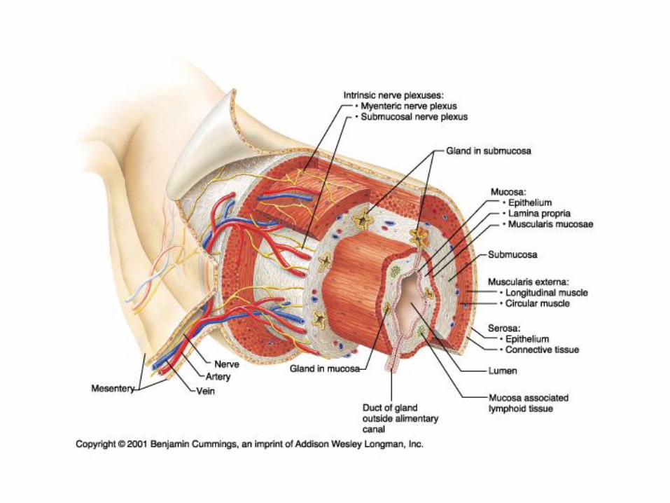
• Lumen: central canal where food is transported
• Mucosa (Mucous Membrane):
– Innermost layer; moist epithelial membrane
– Secretion, absorption, protection
• Submucosa: just external to mucosa
– Contains blood vessels, nerve fibers
– Gives walls its elastic characteristic
• Muscularis Externa: external to submucosa
– Contains smooth muscle
• Inner circular layer, Outer Longitudinal Layer
– Responsible for peristalsis & segmentation
• Serosa: protective outermost layer
– Composed of connective tissue & epithelium
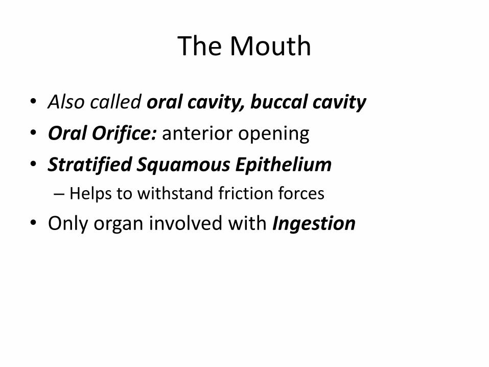
The Mouth
• Also called oral cavity, buccal cavity
• Oral Orifice: anterior opening
• Stratified Squamous Epithelium
– Helps to withstand friction forces
• Only organ involved with Ingestion

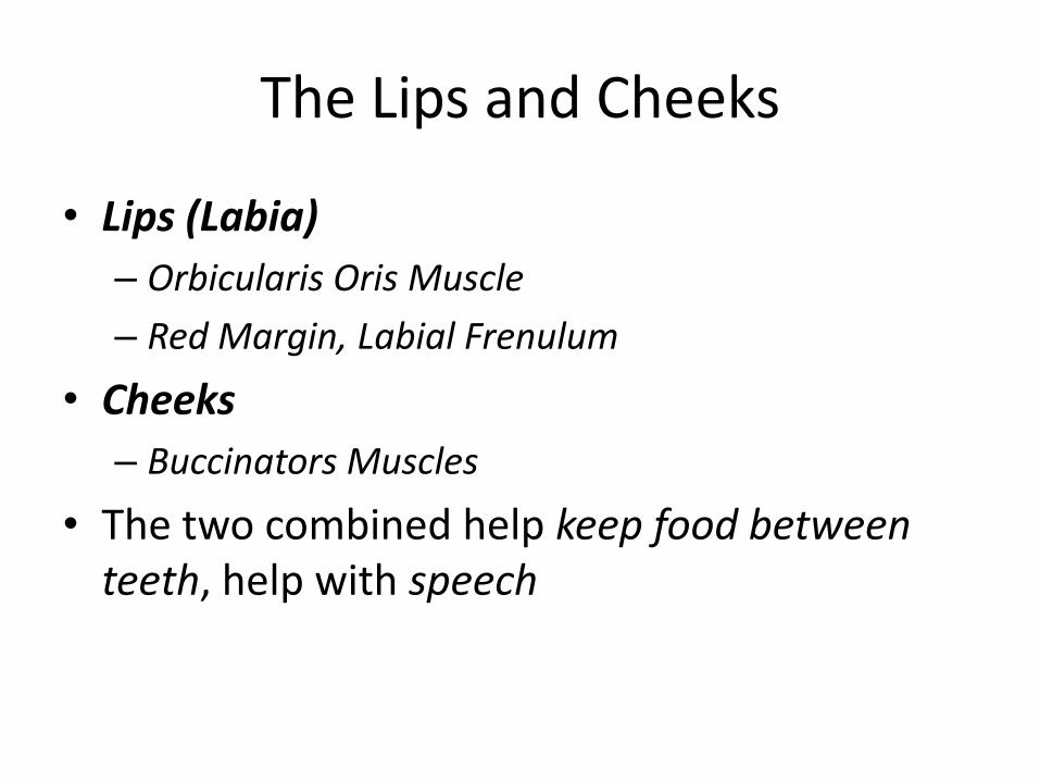
The Lips and Cheeks
• Lips (Labia)
– Orbicularis Oris Muscle
– Red Margin, Labial Frenulum
• Cheeks
– Buccinators Muscles
• The two combined help keep food between teeth, help with speech
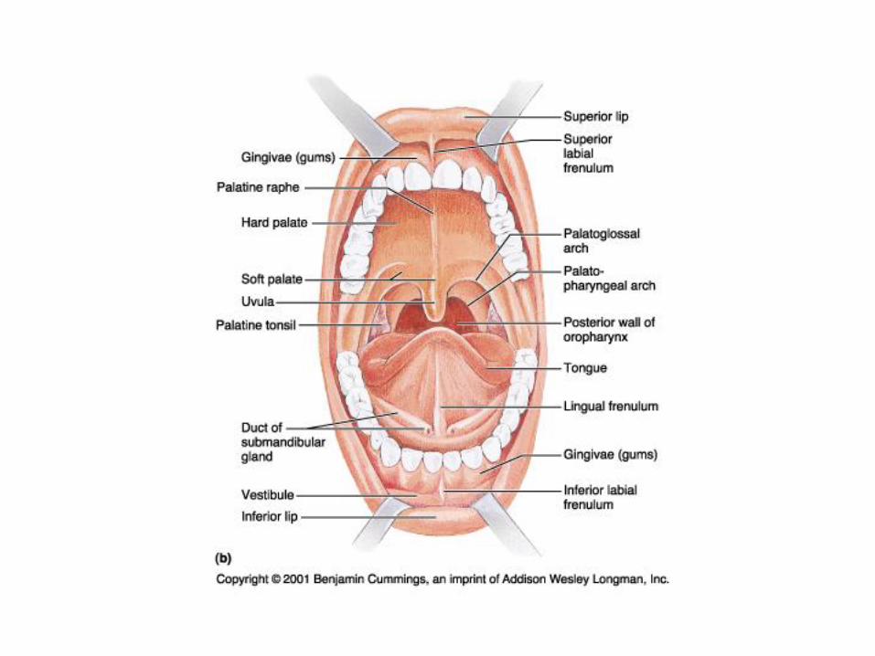
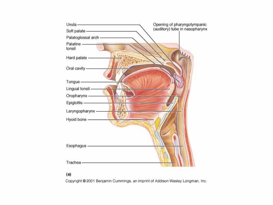
The Palate
• Forms the roof of the mouth
• Hard Palate
– Food forced against it by the tongue
• Soft Palate
– Uvula: prevents food from traveling to the nasal cavity
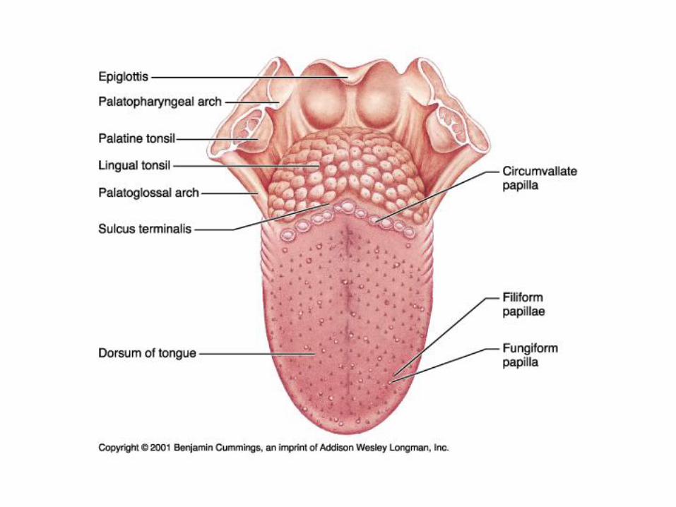
The Tongue
• Occupies the floor of the mouth
• Helps to reposition food between teeth
• Occupies the taste buds
• Intrinsic Muscles
– Changes shape of tongue, not position
• Extrinsic Muscles
– Changes position of tongue, not shape
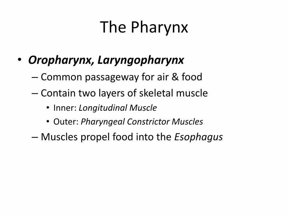
The Pharynx
• Oropharynx, Laryngopharynx
– Common passageway for air & food
– Contain two layers of skeletal muscle
• Inner: Longitudinal Muscle
• Outer: Pharyngeal Constrictor Muscles
– Muscles propel food into the Esophagus
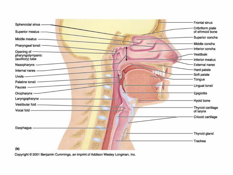
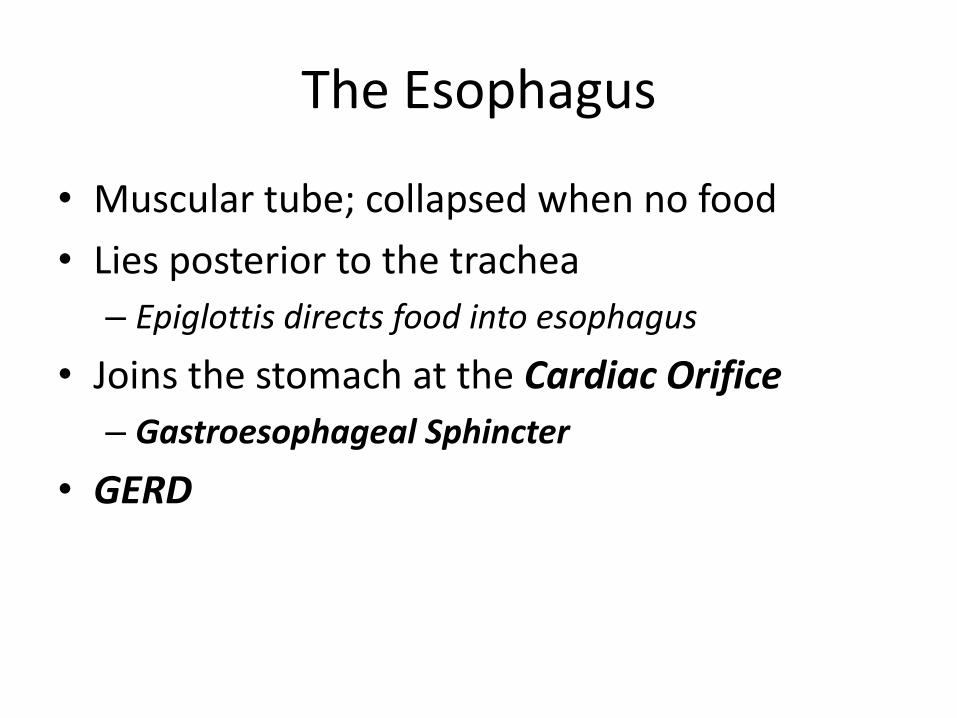
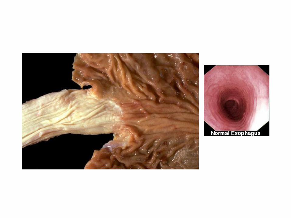
The Esophagus
• Muscular tube; collapsed when no food
• Lies posterior to the trachea
– Epiglottis directs food into esophagus
• Joins the stomach at the Cardiac Orifice
– Gastroesophageal Sphincter
• GERD

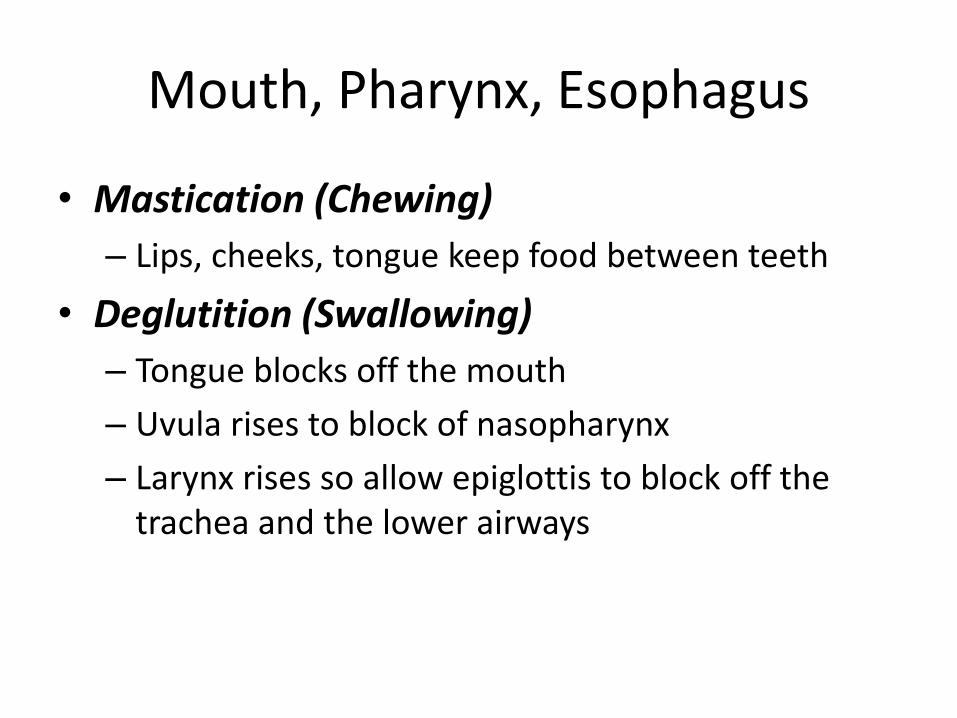
Mouth, Pharynx, Esophagus
• Mastication (Chewing)
– Lips, cheeks, tongue keep food between teeth
• Deglutition (Swallowing)
– Tongue blocks off the mouth
– Uvula rises to block of nasopharynx
– Larynx rises so allow epiglottis to block off the trachea and the lower airways
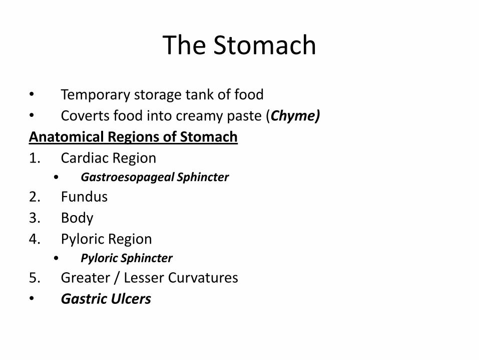
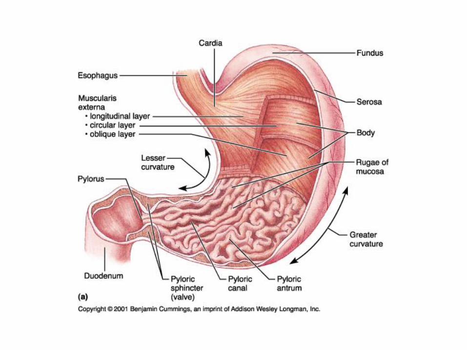
The Stomach
• Temporary storage tank of food
• Coverts food into creamy paste (Chyme)
Anatomical Regions of Stomach
1. Cardiac Region• Gastroesopageal Sphincter
2. Fundus
3. Body
4. Pyloric Region• Pyloric Sphincter
5. Greater / Lesser Curvatures
• Gastric Ulcers
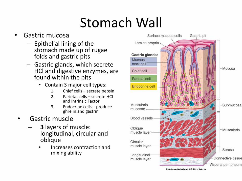
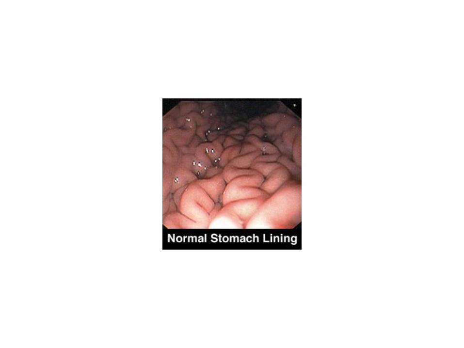
Stomach Wall• Gastric mucosa
– Epithelial lining of the stomach made up of rugaefolds and gastric pits
– Gastric glands, which secrete HCl and digestive enzymes, are found within the pits• Contain 3 major cell types:
1. Chief cells – secrete pepsin2. Parietal cells – secrete HCl
and Intrinsic Factor3. Endocrine cells – produce
ghrelin and gastrin
• Gastric muscle– 3 layers of muscle:
longitudinal, circular and oblique• Increases contraction and
mixing ability
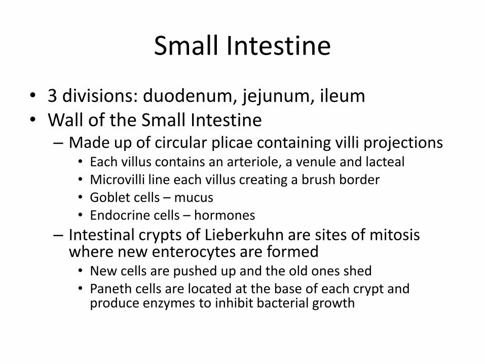
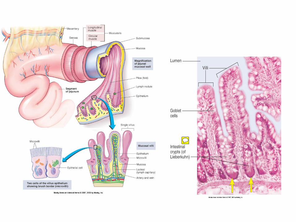
Small Intestine
• 3 divisions: duodenum, jejunum, ileum• Wall of the Small Intestine
– Made up of circular plicae containing villi projections• Each villus contains an arteriole, a venule and lacteal• Microvilli line each villus creating a brush border• Goblet cells – mucus• Endocrine cells – hormones
– Intestinal crypts of Lieberkuhn are sites of mitosis where new enterocytes are formed• New cells are pushed up and the old ones shed• Paneth cells are located at the base of each crypt and
produce enzymes to inhibit bacterial growth
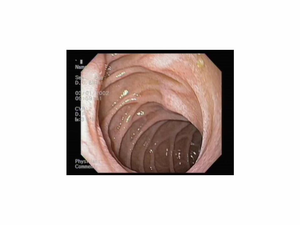
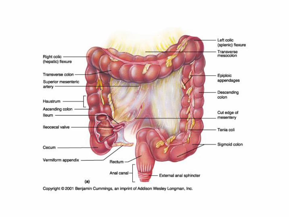
The Large Intestine
• Larger diameter than small intestine– Almost half as long as small intestine
• Absorbs water, eliminates waste (feces)Large Intestine Subdivisions1. Cecum2. Appendix3. Colon: ascending, transverse, descending, sigmoid4. Rectum5. Anal Canal
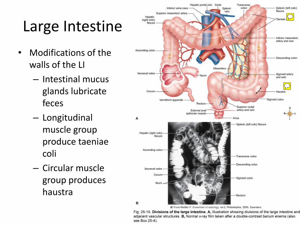
Large Intestine
• Modifications of the walls of the LI
– Intestinal mucus glands lubricate feces
– Longitudinal muscle group produce taeniaecoli
– Circular muscle group produces haustra
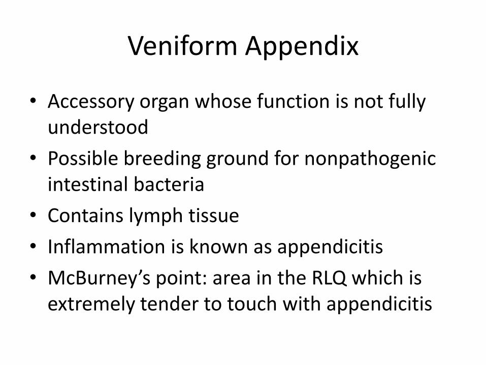
Veniform Appendix
• Accessory organ whose function is not fully understood
• Possible breeding ground for nonpathogenic intestinal bacteria
• Contains lymph tissue
• Inflammation is known as appendicitis
• McBurney’s point: area in the RLQ which is extremely tender to touch with appendicitis
32
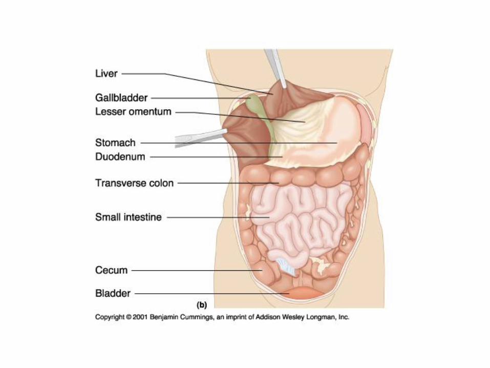
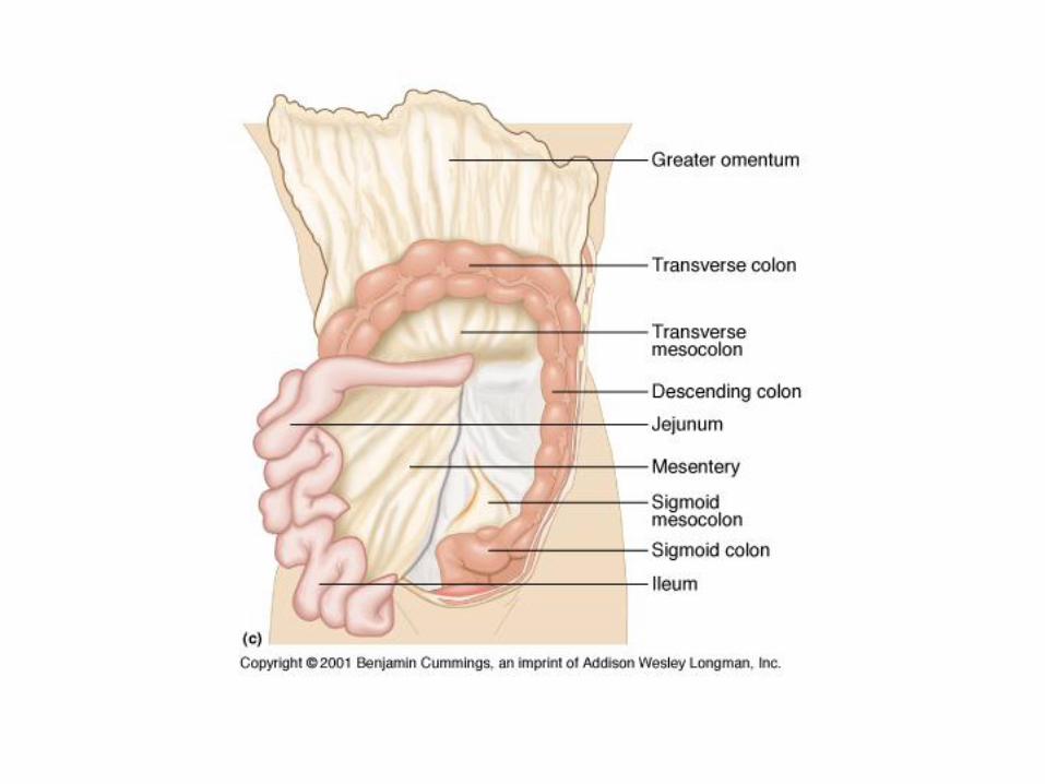
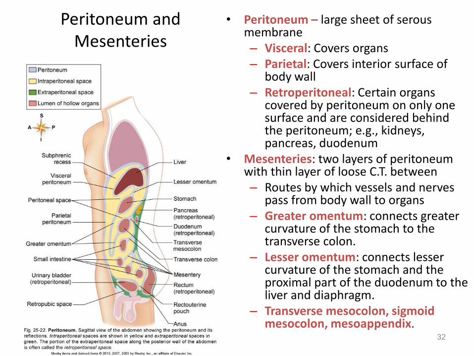
Peritoneum and Mesenteries
• Peritoneum – large sheet of serous membrane– Visceral: Covers organs– Parietal: Covers interior surface of
body wall– Retroperitoneal: Certain organs
covered by peritoneum on only one surface and are considered behind the peritoneum; e.g., kidneys, pancreas, duodenum
• Mesenteries: two layers of peritoneum with thin layer of loose C.T. between– Routes by which vessels and nerves
pass from body wall to organs– Greater omentum: connects greater
curvature of the stomach to the transverse colon.
– Lesser omentum: connects lesser curvature of the stomach and the proximal part of the duodenum to the liver and diaphragm.
– Transverse mesocolon, sigmoid mesocolon, mesoappendix.
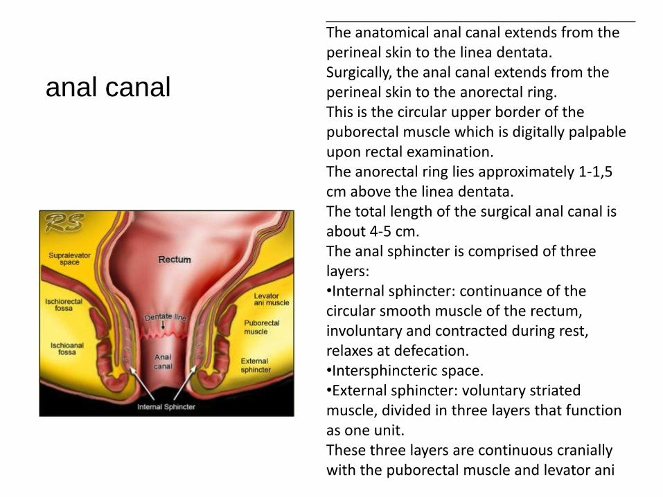
The anatomical anal canal extends from the perineal skin to the linea dentata.Surgically, the anal canal extends from the perineal skin to the anorectal ring.This is the circular upper border of the puborectal muscle which is digitally palpable upon rectal examination.The anorectal ring lies approximately 1-1,5 cm above the linea dentata.The total length of the surgical anal canal is about 4-5 cm.The anal sphincter is comprised of three layers:•Internal sphincter: continuance of the circular smooth muscle of the rectum, involuntary and contracted during rest, relaxes at defecation.•Intersphincteric space.•External sphincter: voluntary striated muscle, divided in three layers that function as one unit.These three layers are continuous cranially with the puborectal muscle and levator ani
anal canal