biology 2011 human anatomy-msk 2020 course …
TRANSCRIPT

1
BIOLOGY 2011
HUMAN ANATOMY-MSK “Where one has to work their phalanges to the periosteum!”
2020
COURSE SYLLABUS/LAB MANUAL

2
BIOLOGY 2011
HUMAN ANATOMY - MSK Instructor: Donna Newhouse, M.Sc., Ph.D(c) Office: NOSM Medical School Building (MSW 3009) Office Phone # (807) 766-7387 Phone: Cell (807) 474-9016 E-mail address: [email protected]
[email protected] Office Hours: By appointment GA’s & TA’s: Mark Schutte (GA) [email protected] James Fimognari (GA) [email protected] Andrea Zapcic (GA) [email protected] Tehya Smith (TA) [email protected] Required Texts: Principles of Human Anatomy (13e). Author: Tortora & Nielsen (2017) Atlas of Human Anatomy (7e.). Author: Netter (2017)
Mark Breakdown Lecture: Topics Course Weight Date/Time MT Integument, Bones 30% October 23rd, 2020
(6:00-7:30 pm) Final Bones, Muscles, Joints 30% TBA Lab: Lab Exam 1 Integument, Bones,
Joints 20% October 9th, 2020
(6:00-7:30 pm) Lab Exam 2 Muscles (and bones) 20% November 27th, 2020
(6:00-7:30 pm) Assignments: Assignment 1 Introduction Video NIL (required) September 27, 2020
(due 11:59 pm) Assignment 2 MT Reflection 3% Bonus November 8, 2020
(due 11:59 pm) **Dates are subject to change
3
Course Description: Anatomical systems/topics covered are the integumentary, muscular, and skeletal systems as well as arthrology. Lecture and Lab Examinations: There is one midterm (MT) and a final exam and two Lab exams. Both the MT and final exams will consist of a variety of questions types such as fill-in-the-blank (FITB), T/F, MCQ, matching, short answer, etc.). These exams will be out of 100-200 marks each. The length of exams will be dependent of the number of questions and as well as the type of questions. These exams will assess you on the information covered within the recorded lectures or any ASSIGNED readings from the textbook. Ninety (90) minutes and three (3) hours will be allotted for the exams, respectively. The exams may not take the full time, however, you should set that time aside in your schedules/calendars. The final exam will assess the information covered from the MT on. There are two lab exams and will include approximately 25-50 questions. Each question will consist of an image and there will be multiple structures that you will have to identify. You will be tested on bones, models, radiological, and histological materials. Given that the labs are “online”, images of the material from the lab will be provided (D2L) and the on-line lab exam will be based LARGELY BUT NOT SOLELY on those images. If you cannot write an exam on the scheduled day/time, then please contact me in advance via email ([email protected]) All exams are rounded up to the nearest whole number. Therefore, no additional marks are awarded individuals at the end of the course, except for the bonus assignment. Assignment 1: Introduction Video You will be required to upload an introduction video of yourself to the D2L site. This will allow me an opportunity to “meet” you virtually and get to know a little bit about you which is particularly important given that we, regrettably do not have any formal face to face interactions during this course. Please include anything you would like to share about yourself. You could include such infromation as your career aspirations, where you are from, whether you have siblings and/or pets, play sports, have any hobbies, or volunteer, summer empolyment, etc. Assignment 2: MT Reflection Some background reading on this is provided under “Reflective Learning” on the Biol 2011 D2L site. For this assignment, I would like you to critically reflect on how you did on the midterm exam. The objective of this assignment is that it may improve your ability to analyze how you learn (i.e. metacognitive abilities) which may lead to lessons learned on how best to prepare for subsequent exams.
4
As this is just a 3% bonus assignment, my expectations are for a one page, single spaced, typed submission. Remember, as noted in the posted article, that a good critical reflection includes "(1) linking past, present, and future experience; (2) integrating cognitive and emotional experience; (3) considering the experience from multiple perspectives; (4) reframing; (5) stating the lessons learned; and (6) planning for future learning or behaviour."
Course and University Policies The policies set out below are for the students' benefit. These policies are set forth to ensure that all students are treated fairly. Please read thoroughly. Behavioral standards:
Please refer to the Student Code of Conduct - Academic Integrity.
Netiquette:
Please communicate with me via your Lakehead e-mail account. It is appropriate
to address me as Donna or Professor Newhouse. Always use Biology 2011 FDE
2020 in the subject line of any email you send to me. I will respond to all e-
mails in a timely fashion (usually within 24 hours, with the exception of
weekends). If you would like to arrange to meet via a Zoom call for purposes of
office hours, I am happy to do so, but I would ask that students contact me in
advance, so that we can agree to meet at a mutually convenient date/time.
Academic Integrity Statement:
I understand and agree that:
(1) Unless otherwise allowed by the course instructor, I must complete the
assignments in this course without the assistance of anyone else.
(2) Unless otherwise allowed by the course instructor, I must not access any
sources or materials (in print, online, or in any other way) to complete any course
exam.
I further understand and agree that, if I violate either of these two rules, or if I
provide any false or misleading information about my completion of course
assignments or exams, I may be prosecuted under the Lakehead University
Student Code of Conduct – Academic Integrity, which requires students to act
ethically and with integrity in academic matters and to demonstrate behaviours
that support the University’s academic values.
Copyright:
All instructional, reference, and administrative materials prepared for this course are protected in their entirety by copyright. Students are expected to comply with

5
this copyright by only accessing and using the course materials for personal educational use related to the course, and that the materials cannot be shared in any way, without the written authorization of the course instructor. If this copyright is infringed in anyway, students may be prosecuted under the Lakehead University Student Code of Conduct – Academic Integrity, which requires students to act ethically and with integrity in academic matters and to demonstrate behaviours that support the University’s academic values. Copyright Compliance: I understand and agree that all instructional, reference, and administrative materials to which I am given access in this course (the "course materials"), whether they consist of text, still or kinetic images, or sound, whether they are in digital or hard copy formats, and in whatever media they are offered, are protected in their entirety by copyright, and that to comply with this copyright and the law
(1) I may access and download the course materials only for my own personal and non-commercial use for this course; and (2) I am not permitted to download, copy, or store (in any medium) any text, image, or sound component of the course materials for any other purpose whatsoever, or to forward or share, transmit, broadcast, show, post or play in public, adapt, or change in any way any text, image, or sound component of the course materials, except as expressly authorized, and only to the extent authorized, in writing, by the course instructor."
I further understand and agree that, if I infringe the copyright of the course materials in any way, I may be prosecuted under the Lakehead University Student Code of Conduct – Academic Integrity, which requires students to act ethically and with integrity in academic matters and to demonstrate behaviours that support the University’s academic values. Regulations It is the responsibility of each student registered at Lakehead University to be familiar with, and comply with all the terms, requirements, regulations, policies and conditions in the Lakehead University Academic Calendar. This includes, but is not limited to, Academic Program Requirements, Academic Schedule of Dates, University and Faculty/School Policies and Regulations and the Fees and Refund Policies and Schedules (Lakehead University Regulations webpage, 2020-21). Academic Integrity A breach of Academic Integrity is a serious offence. The principle of Academic Integrity, particularly of doing one’s own work, documenting properly (including use of quotation marks, appropriate paraphrasing and referencing/citation), collaborating appropriately, and avoiding misrepresentation, is a core principle in university study. Students should view the Student Code of Conduct - Academic Integrity for a full description of academic offences, procedures when Academic

6
Integrity breaches are suspected and sanctions for breaches of Academic Integrity. Supports for Students – there are many resources available to support students. These include but are not limited to:
Health and Wellness
Student Success Centre
Student Accessibility Centre
Library
Lakehead International
Indigenous Initiatives
Lakehead University is committed to achieving full accessibility for persons with disabilities. Part of this commitment includes arranging academic accommodations for students with disabilities and/or medical conditions to ensure they have an equitable opportunity to participate in all of their academic activities. If you are a student with a disability and think you may need accommodations, you are strongly encouraged to contact Student Accessibility Services (SAS) and register as early as possible. For more information, please contact Student Accessibility Services (SC0003, 343-8047 or [email protected])
LABORATORY SCHEDULE 2020 BLOCK 1: September 9 - October 4 Integument, Bones and Joints October 9 LAB EXAM #1 BLOCK 2: October 21 - November 22 Muscles (and some bones) November 27 LAB EXAM #2
7
LECTURE OUTLINE (Subject to Change)
I. Introduction A. Definition of Anatomy B. Anatomical Position, Planes, Movements C. Organization of the body (1) cells (2) tissues (3) organs (4) organ systems (5) membranes (serous, synovial, mucous, cutaneous) C. Anatomical Terminology II. Tissues and Tissue Types A. Tissue Types (1) epithelial (2) connective (3) muscular (4) nervous III. Integumentary System
A. Functions of the Skin B. Structure of the Skin C. Accessory Structures of the Skin D. Clinical considerations

8
IV. Skeletal System (Osseous connective tissue) A. Skeletal Development and Function (a) composition and structure of bones B. Review of Human Bones (1) axial division (a) skull (cranium, facial bones) (b) hyoid bone (c) trunk (vertebrae, ribs, sternum) (2) appendicular division (a) upper (pectoral) appendages (b) lower (pelvic) appendages C. Joints (1) synarthroses (2) amphiarthroses (3) diarthroses (4) types of movement across joints (5) types of joint injuries V. Muscular System A. Muscle Tissue (1) skeletal (striated) (2) smooth (unstriated) (3) cardiac (4) properties of muscle tissue (5) functions (6) structure of skeletal muscles B. Principle muscles of the body (1) Muscles of facial expression (2) Muscles of upper extremity (a) shoulder joint (b) muscles moving the shoulder (c) muscles moving the upper arm (d) muscles moving the lower arm (e) muscles which move the hand (f) muscles which move the fingers and thumb (forearm/hand) (3) Muscles of the lower extremity (a) hip joint (b) muscles which move the thigh (c) muscles which move the lower leg (d) muscles which move the foot and toes (4) Muscles of the abdominal wall (5) Muscles that move the head and spine (6) Muscles of the pelvic floor (7) Muscles which move the chest wall
9
LABORATORY OUTLINE SKELETAL SYSTEM AND JOINTS
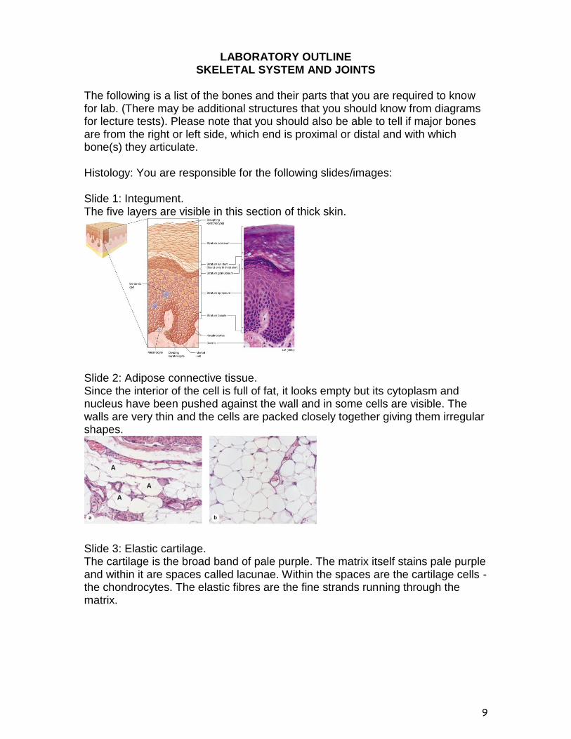
The following is a list of the bones and their parts that you are required to know for lab. (There may be additional structures that you should know from diagrams for lecture tests). Please note that you should also be able to tell if major bones are from the right or left side, which end is proximal or distal and with which bone(s) they articulate. Histology: You are responsible for the following slides/images: Slide 1: Integument. The five layers are visible in this section of thick skin.
Slide 2: Adipose connective tissue. Since the interior of the cell is full of fat, it looks empty but its cytoplasm and nucleus have been pushed against the wall and in some cells are visible. The walls are very thin and the cells are packed closely together giving them irregular shapes.
Slide 3: Elastic cartilage. The cartilage is the broad band of pale purple. The matrix itself stains pale purple and within it are spaces called lacunae. Within the spaces are the cartilage cells - the chondrocytes. The elastic fibres are the fine strands running through the matrix.

10
Slide 4: Hyaline cartilage. On this slide, the cartilage is the area of lacunae (stained purple). It has the chondrocytes just as elastic cartilage does but there are no elastic fibres.
Slide 5: Compact bone. The characteristic of this slide is the Haversian system that is found only in compact bone. Although they are not visible, the osteocytes are in the lacunae.
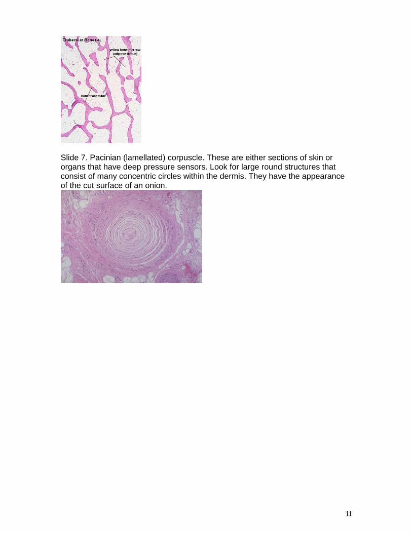
Slide 6: Cancellous (spongy) bone. There is no Haversian system (see slide 5), only irregular rods of bone (called trabeculae) are produced that form a network filled with marrow. The bone is stained bright red or pink, depending on the slide, and the cells that produce the bone (osteocytes) are visible in the lacunae.
11
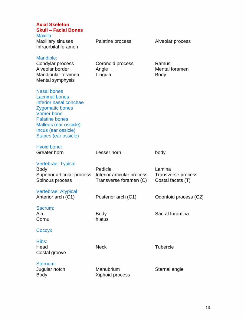
Slide 7. Pacinian (lamellated) corpuscle. These are either sections of skin or organs that have deep pressure sensors. Look for large round structures that consist of many concentric circles within the dermis. They have the appearance of the cut surface of an onion.

12
Integument Epidermiss Stratum basale Stratum spinosum Stratum granulosum Stratum lucidum Stratum corneum Dermis Stratum papillarosum Stratum reticularosum Pore Dermal papillae Arrector pili m. Sebaceous gland Sweat gland Sweat gland duct Hair root Cutaneous blood vessels Pacinian corpuscles Hair medulla Hair follicle receptor Hair follicle Hair cortex Hair shaft Hair papilla Huxley’s layer Henle’s layer Inner root sheath Outer root sheath Free nerve ending Ruffini corpuscle Krause’s end bulb Meissner’s corpuscle Hypodermis Adipose tissue Axial Skeleton Skull - Cranium Frontal bone: Frontal sinus Supraorbital ridge Supraorbital foramen Coronal suture Parietal bone: Squamousal suture Lambdoidal suture Sagittal suture Grooves of middle meningeal aa.
Temporal bone: Mastoid process Mandibular fossa Zygomatic process Stylomastoid foramen Styloid process Petrous portion Squmaous portion External acoustic meatus Internal acoustic meatus Occipital bone: Foramen magnum Occipital condyles Jugular foramen Hypoglossal foramen Groove of transverse
sinus Groove of sigmoid sinus
Sphenoid bone: Sphenoid sinus Foramen ovale Foramen rotundum Foramen spinosum Optic foramen Superior orbital fissure Inferior orbital fissure Sella turcica Ethmoid bone: Crista galli Cribriform foramina Cribriform plate Perpendicular plate Superior nasal concha Middle nasal concha
13
Axial Skeleton Skull – Facial Bones Maxilla: Maxillary sinuses Palatine process Alveolar process Infraorbital foramen Mandible: Condylar process Coronoid process Ramus Alveolar border Angle Mental foramen Mandibular foramen Lingula Body Mental symphysis Nasal bones Lacrimal bones Inferior nasal conchae Zygomatic bones Vomer bone Palatine bones Malleus (ear ossicle) Incus (ear ossicle) Stapes (ear ossicle) Hyoid bone: Greater horn Lesser horn body Vertebrae: Typical Body Pedicle Lamina Superior articular process Inferior articular process Transverse process Spinous process Transverse foramen (C) Costal facets (T) Vertebrae: Atypical Anterior arch (C1) Posterior arch (C1) Odontoid process (C2) Sacrum: Ala Body Sacral foramina Cornu hiatus Coccyx Ribs: Head Neck Tubercle Costal groove Sternum: Jugular notch Manubrium Sternal angle Body Xiphoid process

14
Appendicular Skeleton Pectoral Girdle Scapula: Vertebral (medial) border Axillary (lateral) border Superior angle Inferior angle Spine Acromion Glenoid fossa Coracoid process Supraspinous fossa Infraspinous fossa Subscapular fossa Clavicle: Sternal (medial) end Acromial (lateral) end Conoid tubercle Appendicular Skeleton Upper Extremity Humerus: Head Deltoid tuberosity Capitulum Coronoid fossa Olecranon fossa Trochlea Medial epicondyle Lateral epicondyle Supracondylar ridges Intertubercular groove Greater tubercle Lesser tubercle Ulna: Trochlear notch Olecranon process Coronoid process Radial notch Head Styloid process Radius: Head Radial tuberosity Neck Styloid process Carpal bones: Scaphoid Lunate Trapezium Capitate Triquetral (triquetrum) Pisiform Trapezoid Hamate (hook of hamate) Metacarpal bones: Metacarpal I Metacarpal II Metacarpal III Metacarpal IV Metacarpal V Phalanges: Proximal phalanx Middle phalanx Distal phalanx

15
Appendicular Skeleton Pelvic Girdle
Os Coxae: Pubis symphysis Acetabulum Obturator foramen Greater sciatic notch Lesser sciatic notch Ischial spine Anterior superior iliac spine Ischial tuberosity Pubis Anterior inferior iliac spine Ischium Ilium Posterior superior iliac spine Posterior inferior iliac spine Appendicular Skeleton Lower Extremity
Femur: Head Neck Greater trochanter Lesser trochanter Linea aspera Medial condyle Lateral condyle Intercondylar fossa Patella: Base Apex Tibia: Lateral condyle Medial condyle Medial malleolus Tibial tuberosity Nutrient foramen Intercondylar eminence Fibula: Head Neck Lateral malleolus Tarsal Bones: Talus Calcaneus Navicular Cuboid Medial (1st) cuneiform Intermediate (2nd) cuneiform Lateral (3rd) cuneiform Phalanges: Distal phalanx Middle phalanx Proximal phalanx Model of Bone: Periosteum Lamellae - interstitial Lamellae - concentric Osteocyte Canaliculi Osteon Central canal lacuna Perforating fibres Perforating canals

16
The following is a list of structures associated with the knee, shoulder, elbow, and hip joints which you are responsible to know: Appendicular Skeleton Joints
Knee: Anterior cruciate ligament Medial meniscus Lateral meniscus Posterior cruciate ligament Quadriceps tendon Patellar ligament Medial collateral ligament Lateral collateral ligament Posterior meniscofemoral ligament
Shoulder: Acromioclavicular ligament Coracoacromial ligament Coracohumeral ligament Superior trnasverse scapular ligament
Tendon - Long head of biceps brachii m.
Transverse humeral retinaculum
Coracoclavicular ligament Conoid ligament Trapezoid ligament Elbow: Medial collateral ligament Lateral collateral ligament Annular ligament Hip: Iliofemoral ligament Ischiofemoral ligament Pubofemoral ligament
17
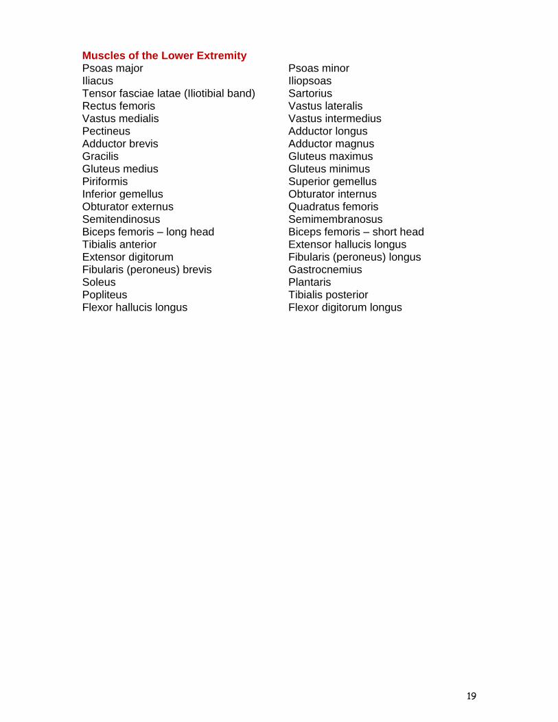
MUSCULAR SYSTEM This section lists the muscles you need to know for the lab portion of the course. In addition to being able to identify muscles, you are responsible for origin(s), insertion(s) and action(s) for the major muscle groups in the human. The only tendon you should know is the Achilles (calcaneal) tendon. Histology: You are responsible for the microscopic anatomy of the three types of muscles. The three types of muscle are available shown in images below Striated (skeletal) muscle. This is the major component of skeletal muscles, which pull on bones to cause body movements. Skeletal muscle fibres are long, large cylinders that contain many nuclei. Notice the obvious banding pattern and the fact that these large cells are multinucleated. Smooth muscle. It is so named because there are no visible striations in its fibres. These fibres are spindle-shaped and contain one centrally located nucleus. Smooth muscle primarily occurs in the walls of hollow organs. It generally acts to squeeze substances through these organs by alternately contracting and relaxing Cardiac muscle. Cardiac muscle is found in the walls of the heart. It contracts to propel blood through the blood vessels. Like skeletal muscle fibres, cardiac muscle fibres are striated. However, they differ in two ways: (1) cardiac fibres are generally uninucleated (one nucleus) and (2) cardiac cells branch and join at unique cellular junctions called intercalated discs.
You are not responsible for identifying muscle types in cross section (xs), only in longitudinal section (ls).

18
Muscles of the Head/Neck Region Head: Masseter Temporalis Buccinator Orbicularis oris Orbicularis oculi Frontalis Occipitalis Zygomaticus major Zygomaticus minor Neck: Platysma Sternocleidomastoid sternohyoid Sternothyroid Thyrohyoid stylohyoid Anterior scalene Middle scalene Posterior scalene Levator scapula Muscles of the Thorax/Abdomen/Back Thorax: Pectoralis minor Pectoralis major Serratus anterior External intercostal Internal intercostal Abdomen: Internal abdominal oblique Transversus abdominis Rectus abdominis External abdominal oblique Back: Latissimus dorsi Rhomboideus major Rhomboideus minor Erector spinae Trapezius Quadratus lumborum Muscles of the Upper Extremity Teres minor Teres major Supraspinatus Infraspinatus Subscapularis Deltoid Biceps brachii – long head Biceps brachii – short head Coracobrachialis Brachialis Pronator teres Flexor carpi radialis Palmaris longus Flexor carpi ulnaris Flexor digitorum superficialis Flexor digitorum profundus Flexor pollicis longus Pronaor quadratus Triceps brachii – long head Triceps brachii – lateral head Triceps brachii – medial head Brachioradialis Extensor carpi radialis longus Extensor carpi radialis brevis Extensor digitorum Extensor carpi ulnaris Abductor pollicis Extensor pollicis brevis Extensor pollicis longus Flexor pollicis brevis Abductor pollicis brevis Opponens pollicis Adductor pollicis Flexor digiti minimi Abductor digiti minimi Opponens digiti minimi Supinator lumbricals
19
Muscles of the Lower Extremity Psoas major Psoas minor Iliacus Iliopsoas Tensor fasciae latae (Iliotibial band) Sartorius Rectus femoris Vastus lateralis Vastus medialis Vastus intermedius Pectineus Adductor longus Adductor brevis Adductor magnus Gracilis Gluteus maximus Gluteus medius Gluteus minimus Piriformis Superior gemellus Inferior gemellus Obturator internus Obturator externus Quadratus femoris Semitendinosus Semimembranosus Biceps femoris – long head Biceps femoris – short head Tibialis anterior Extensor hallucis longus Extensor digitorum Fibularis (peroneus) longus Fibularis (peroneus) brevis Gastrocnemius Soleus Plantaris Popliteus Tibialis posterior Flexor hallucis longus Flexor digitorum longus