biologically relevant lighting: an industry perspective
TRANSCRIPT
fnins-15-637221 June 1, 2021 Time: 18:47 # 1
PERSPECTIVEpublished: 07 June 2021
doi: 10.3389/fnins.2021.637221
Edited by:Jamie Zeitzer,
Stanford University, United States
Reviewed by:Michael Herf,
f.lux Software LLC, United StatesDaniel S. Joyce,
University of Nevada, Reno,United States
*Correspondence:Robert Soler
Specialty section:This article was submitted to
Sleep and Circadian Rhythms,a section of the journal
Frontiers in Neuroscience
Received: 04 December 2020Accepted: 05 May 2021
Published: 07 June 2021
Citation:Soler R and Voss E (2021)
Biologically Relevant Lighting: AnIndustry Perspective.
Front. Neurosci. 15:637221.doi: 10.3389/fnins.2021.637221
Biologically Relevant Lighting: AnIndustry PerspectiveRobert Soler* and Erica Voss
BIOS Lighting, Carlsbad, CA, United States
Innovations in LED lighting technology have led to tremendous adoption rates andvastly improved the metrics by which they are traditionally evaluated–including colorquality, longevity, and energy efficiency to name a few. Additionally, scientific insighthas broadened with respect to the biological impact of light, specifically our circadianrhythm. Indoor electric lighting, despite its many attributes, fails to specifically addressthe biological responses to light. Traditional electric lighting environments are biologicallytoo dim during the day, too bright at night, and with many people spending much of theirlives in these environments, it can lead to circadian dysfunction. The lighting industry’sbiological solution has been to create bluer days and yellower nights, but the technologycreated to do so caters primarily to the cones. A better call to action is to providebiologically brighter days and biologically darker nights within the built environment.However, current lighting design practices have specified the comfort and utility ofelectric light. Brighter intensity during the day can often be uncomfortable or glary, andreduced light intensity at night may compromise visual comfort and safety, both of whichwill affect user compliance. No single lighting solution will effectively create biologicallybrighter days and biologically darker nights, but rather a variety of parameters need to beconsidered. This paper discusses the contributions of spectral power distribution, hueor color temperature, spatial distribution, as well as architectural geometry and surfacereflectivity, to achieve biologically relevant lighting.
Keywords: social jet lag, circadian, lighting, melanopsin, Opn4
INTRODUCTION
The discovery of a novel Intrinsically Photosenstive Retinal Ganglion Cell (ipRGC) in the eyehas changed our understanding of the role of light in our everyday lives (Provencio and Foster,1995; Provencio et al., 1998; Brainard et al., 2001; Thapan et al., 2001; Berson et al., 2002;Panda et al., 2002; Dacey et al., 2005; Güler et al., 2008; Hughes et al., 2016). These ipRGCscontain a photopigment melanopsin with an in-vitro spectral sensitivity around 480 nm. However,after lens transmission this spectral sensitivity is shifted to longer wavelengths between 487 and496 nm (Spitschan, 2019), and it is now understood that these ipRGCs are responsible for severalphysiological effects such as circadian synchronization, tracking seasonal changes, acute alertness,working memory improvements and mood improvements (Gaggioni et al., 2014; LeGates et al.,2014; Rodgers et al., 2016; Fernandez et al., 2018; Brown, 2020). The role of rods and coneson ipRGC response is still being debated. Some studies have shown no contribution of S-cone
Frontiers in Neuroscience | www.frontiersin.org 1 June 2021 | Volume 15 | Article 637221
fnins-15-637221 June 1, 2021 Time: 18:47 # 2
Soler and Voss Biologically Relevant Lighting: An Industry Perspective
(Spitschan et al., 2019) while others show an exposuretime dependent contribution of S-cone (Brown et al., 2021)when evaluating melatonin suppression. Further studies showa reduced circadian impact of blue colored environments(Mouland et al., 2019). Less information is available on rod andcone contributions for other ipRGC driven responses.
In order to apply a biologically relevant lighting solutionwithin the built environment, we must first understand thechallenge at hand. Today, we spend most of our time indoors,removed from daylight which contains an important naturaland robust daytime and nighttime signal (Knoop et al., 2020).Daylight has been replaced with electric lighting which hasbeen engineered and optimized for visual efficiency and canbe switched on or off at any time of the day. Moreover,today’s light level recommendations (while sufficient for visualtasks) are insufficient for proper circadian signaling. Wefind ourselves immersed in environments where the lightingis too biologically dim for our brains to receive a properdaytime signal and too biologically bright to provide a propernighttime signal. This lack of delineation between day andnight can lead to circadian drift and exacerbate social jet lag(Roenneberg and Merrow, 2016), which have been associatedwith a whole host of negative health outcomes: decreases inlearning and attention, increased risk of obesity, addiction,and cardiovascular disease (Roenneberg et al., 2012; Zarrinparet al., 2015; Roenneberg and Merrow, 2016; Sulli et al., 2018).Moreover, this appears to be a widespread phenomenon thataffects the majority of people. In fact, it has been shownthat 87% of non-shift workers have some form of circadiandysfunction and the associated health risks previously mentioned(Roenneberg and Merrow, 2016), illustrating that the currentlighting environment may be insufficient for proper circadianentrainment and amplitude. Therefore, biologically relevantlighting should provide biologically brighter days and biologicallydarker nights within the built environment. However, in practicethere are additional constraints. Energy consumption, visualcomfort (i.e., glare) associated with brighter days, and visualacuity and safety related to darker nights. It should be notedthat while everyone should benefit from brighter days anddarker nights, it may not be additive, there may be morebenefit from darker nights compared to brighter days (Skeldonet al., 2017), and vice versa, depending on existing conditionsand population type. We do know that large differencesexist between individuals for nighttime melatonin suppression(Phillips et al., 2019).
In order to create truly biologically relevant lighting, thefollowing factors must be considered in conjunction with oneanother: spectral composition, color, intensity, and distributionof the light, as well as the geometry and reflectivity of the builtenvironment. As long as we remain in the built environment,there is no single strategy that can provide the optimum circadianlighting environment. Instead, we must use a multi-facetedapproach to achieve biologically relevant lighting that is focusedon biologically brighter days and darker nights. Ultimately, weneed to understand how both circadian lighting factors andvisual lighting factors can be addressed to create spaces that areboth biologically relevant for day and night while still providing
comfortable and well-designed spaces (Berman et al., 1991; Reaand Ouellette, 1991).
CURRENT INDUSTRY STANDARDS,METRICS AND RECOMMENDATIONS
The WELL Building StandardTM uses a series of designcategories–Air, Water, Nourishment, Light, Movement, ThermalComfort, Sound, Materials, Mind, Community, and Innovation–to create a point-based framework that determines how muchwellness can be delivered to building occupants. A minimumof 40 points is required to achieve any type of WELLcertification, with nine total points available from the Lightconcept. Circadian lighting and daylight exposure are keyFeatures within the WELLTM “Light” concept. The CircadianLighting Design Feature uses vertical melanopic equivalentdaylight illuminance (EDI) as a criterion for minimum daytimecircadian stimulus. Vertical melanopic EDI is measured based onthe occupants’ primary location within a space. Circadian lightlevel measurements are meant to quantify the light reaching theoccupants’ eye and as such are taken 4′ above finished floor (or18′′ above the task plane) in the primary viewing direction ofthe occupant. The amount of vertical melanopic EDI required isdependent on how much daylight availability there is. It rangesfrom 109 vertical melanopic EDI (for one point) when adequatedaylight is present to 218 vertical melanopic EDI (for threepoints) when sufficient daylight is not present.
Melanopic EDI originated from The InternationalCommission on Illumination (CIE) issuance of the CIE S026/E:2018, and uses the same melanopic sensitivity function asthe Lucas toolbox, with peak sensitivity at 490 nm. Additionally,the CIE has proposed melanopic Daylight Efficiency Ratio(melanopic DER) as a metric for determining the biologicalpotential for a light spectrum to activate melanopsin relative tovisual illuminance (lumens/m2) (Lucas et al., 2014; InternationalCommission on Illumination, 2018).
THE ROLE OF LED TECHNOLOGY INBIOLOGICALLY RELEVANT LIGHTING
Light emitting diodes (LEDs) lighting has seen tremendousadoption rates due to its energy efficiency and longevity. Asof 2018 residential products saw LED penetration in 33% ofA-lamps and 45% of recessed downlights, while office lighting sawLED penetration in 20% of linear installations (Navigant, 2019,2020). How does LED lighting impact these newly discoveredphysiological responses to light? Some claim that LEDs containa large blue peak, sending too many daytime signals and thusdeeming them unsuitable for nighttime use (Tosini et al., 2016).Here I will demonstrate that the contrary is true and in fact, thereexists a fundamental dip in the melanopic region (from 470 to500 nm) due to the way LED white light spectrum is generated.This means that white LEDs actually perform poorly when itcomes to producing the necessary melanopic stimulation relativeto its perceived color temperature and visual brightness.
Frontiers in Neuroscience | www.frontiersin.org 2 June 2021 | Volume 15 | Article 637221

fnins-15-637221 June 1, 2021 Time: 18:47 # 3
Soler and Voss Biologically Relevant Lighting: An Industry Perspective
Color Matching FunctionsColor matching functions shown in Supplementary Figure 1are used to convert any spectral power distribution to a (x,y)point on the CIE 1931 color space diagram. These (x,y) colorpoints are how the lighting industry communicates color. Whenreferring to white light, these color coordinates are binned basedon the temperature of a black body radiator in Kelvin (referredto as K). The following equations are used to convert spectruminto Tristimulus X, Y, and Z, then to color coordinates (x,y).Tristimulus Y is also the luminous efficiency function, used tocalculate lumens.
X =
780nm∑λ=380nm
S (λ) x (λ) ∂λ
Y =
780nm∑λ=380nm
S (λ) y (λ) ∂λ
Z =
780nm∑λ=380nm
S (λ) z (λ) ∂λ
WhereS(λ) = Spectral power distribution of the light sourcex(λ) = spectral tristimulus value from Figure 1.y(λ) = spectral tristimulus value from Figure 1.z(λ) = spectral tristimulus value from Figure 1.
x =X
X + Y + Z
y =Y
X + Y + Z
Wherex = x-coordinate on CIE 1931 color space diagram.y = y-coordinate on CIE 1931 color space diagram.One way to think about this is as follows: Z stimulation
results in bluer color perception, Y stimulation results in greenercolor perception, and X stimulation results in redder colorperception. This blue perception from Z stimulation versusmelanopic stimulation is key to disentangling visual perceptionfrom physiological effect. Z has highest sensitivity from 430 to450 nm, whereas melanopic blue-green has highest sensitivityfrom 480 to 500 nm. Thus, a spectral power distributioncan be derived with heightened melanopic stimulation withoutappearing cold in color temperature. Likewise, a spectral powerdistribution can be created that has less melanopic stimulationwhile still appearing cold in color temperature.
Traditional LED TechnologyLight emitting diodes are semiconductors that utilize a materialcomposition to emit a specific wavelength of light. There aretwo classes of LEDs; InGaN (blue) and AlInGaP (red), whichare doped to produce a desired wavelength (Shaolin et al., 2015).However, a green gap exists in this doping process that makesgreen light very inefficient to produce from a discrete LED source
(Auf Der Maur et al., 2016). Thus, a common approach to LEDlight is to utilize a monochromatic blue LED with peak emissionbetween 445 and 455 nm combined with a combination of greenand red phosphors that are excited by the blue wavelengths ofthe LED (Mueller-Mach et al., 2005; Nishiura et al., 2011; Liuet al., 2015). This combination is what provides the LED itssignificant energy efficiency and allows for a much wider rangeof CCTs and spectral control than previous technologies. Anexample of this approach is shown in Supplementary Figure 2for a 6,500◦K LED spectra. What can be seen from this LEDspectra is a peak emission that coincides with the peak sensitivityfor Z from the color matching functions. By pinpointing theZ sensitivity, LEDs provide the most visually blue-looking lightsource with the least amount of energy appropriated in the blueregion of the spectrum, relative to other light sources. This is animportant tactic for energy efficiency, as the Luminous EfficiencyFunction (tristimulus y) is very low in the blue region. A commonmisconception about LED lighting is that they produce more bluelight than other sources, due to the narrow, high-amplitude peakof the blue wavelengths. However, this is not the case, LED lightis in fact engineered to produce the least amount of blue light perlumen for any given color temperature of light, in order to gainthe highest luminous efficacy.
Spectral Simulation MethodSpectral simulation was created using an excel based spectrumdatabase and calculator containing selectable Blue LED withscaler factor to increase or decrease LED simulated intensity. OneBlue LED was selected and combined with various amounts ofthree unique phosphors. A combination of three (3) color pointscan produce any of the colors in the space, however, a fourth colorpoint adds an additional degree of freedom that allows lumens tobe maintained throughout simulations. Each spectral simulationshown in this study are within 1% of lumen and color pointtargets. Lumen target was chosen arbitrarily at 717 lumens. Colorpoint targets were obtained as central points for each ANSI colorbin, obtained from LED datasheets from Lumileds (San Jose, CA,United States) and are as follows:
1,800 K: (x,y) = (0.549, 0.408)2,200 K: (x,y) = (0.502, 0.415)2,700 K: (x,y) = (0.458, 0.410)3,000 K: (x,y) = (0.434, 0.403)3,500 K: (x,y) = (0.407, 0.392)4,000 K: (x,y) = (0.380, 0.380)5,000 K: (x,y) = (0.345, 0.355)6,500 K: (x,y) = (0.312, 0.328)
Spectral Data450 nm royal blue LED data was obtained from Lumileds wastranslated to 410, 420, 430, 440, 460, 470, 480, 490, and 500 nmdata for this simulation. Phosphor data was obtained from MerckEMD (Darmstadt, Germany). Phosphors were selected to havelonger wavelength while having capability to meet the desiredcolor points within the CIE 1931 color space, which is usedto determine white color points. This was done to minimizeeffect of phosphor on melanopic content. Phosphors have peakwavelengths at 547 nm (YYG-547-210), 585 nm (OGA-585), and
Frontiers in Neuroscience | www.frontiersin.org 3 June 2021 | Volume 15 | Article 637221
fnins-15-637221 June 1, 2021 Time: 18:47 # 4
Soler and Voss Biologically Relevant Lighting: An Industry Perspective
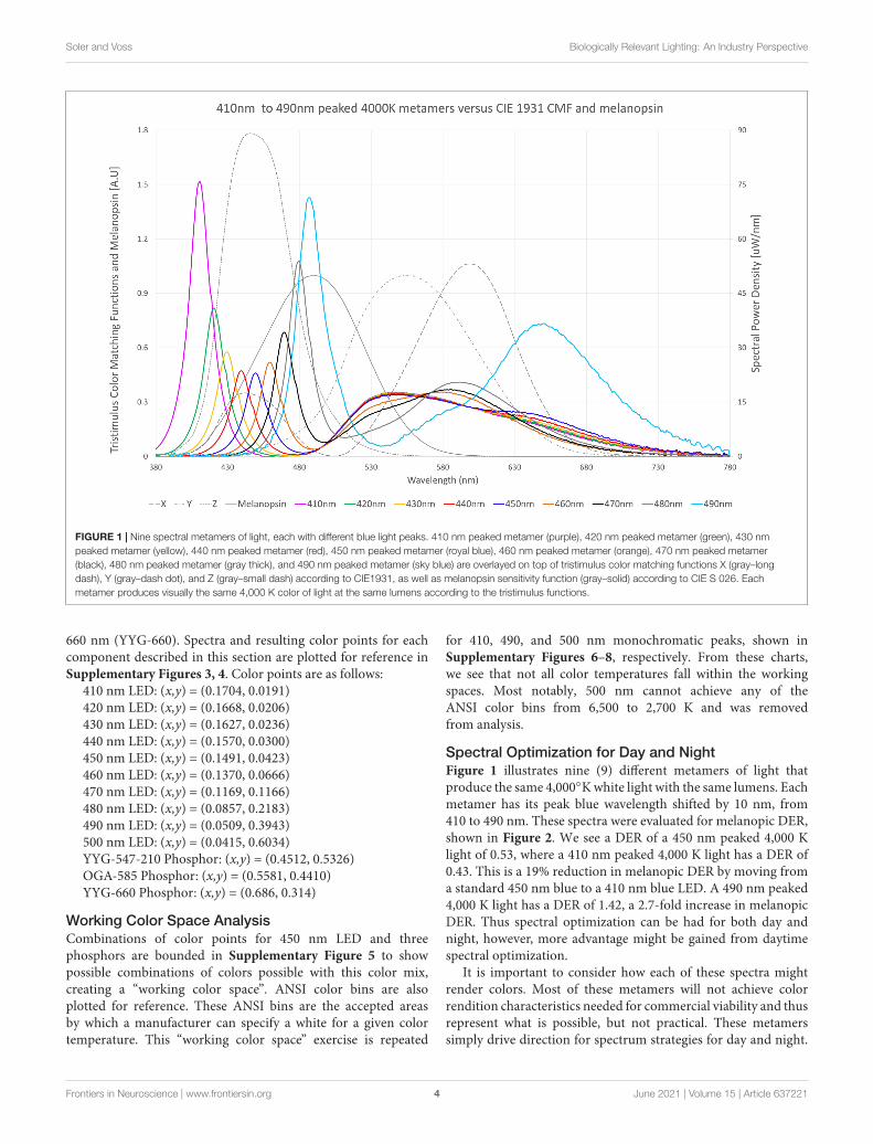
FIGURE 1 | Nine spectral metamers of light, each with different blue light peaks. 410 nm peaked metamer (purple), 420 nm peaked metamer (green), 430 nmpeaked metamer (yellow), 440 nm peaked metamer (red), 450 nm peaked metamer (royal blue), 460 nm peaked metamer (orange), 470 nm peaked metamer(black), 480 nm peaked metamer (gray thick), and 490 nm peaked metamer (sky blue) are overlayed on top of tristimulus color matching functions X (gray–longdash), Y (gray–dash dot), and Z (gray–small dash) according to CIE1931, as well as melanopsin sensitivity function (gray–solid) according to CIE S 026. Eachmetamer produces visually the same 4,000 K color of light at the same lumens according to the tristimulus functions.
660 nm (YYG-660). Spectra and resulting color points for eachcomponent described in this section are plotted for reference inSupplementary Figures 3, 4. Color points are as follows:
410 nm LED: (x,y) = (0.1704, 0.0191)420 nm LED: (x,y) = (0.1668, 0.0206)430 nm LED: (x,y) = (0.1627, 0.0236)440 nm LED: (x,y) = (0.1570, 0.0300)450 nm LED: (x,y) = (0.1491, 0.0423)460 nm LED: (x,y) = (0.1370, 0.0666)470 nm LED: (x,y) = (0.1169, 0.1166)480 nm LED: (x,y) = (0.0857, 0.2183)490 nm LED: (x,y) = (0.0509, 0.3943)500 nm LED: (x,y) = (0.0415, 0.6034)YYG-547-210 Phosphor: (x,y) = (0.4512, 0.5326)OGA-585 Phosphor: (x,y) = (0.5581, 0.4410)YYG-660 Phosphor: (x,y) = (0.686, 0.314)
Working Color Space AnalysisCombinations of color points for 450 nm LED and threephosphors are bounded in Supplementary Figure 5 to showpossible combinations of colors possible with this color mix,creating a “working color space”. ANSI color bins are alsoplotted for reference. These ANSI bins are the accepted areasby which a manufacturer can specify a white for a given colortemperature. This “working color space” exercise is repeated
for 410, 490, and 500 nm monochromatic peaks, shown inSupplementary Figures 6–8, respectively. From these charts,we see that not all color temperatures fall within the workingspaces. Most notably, 500 nm cannot achieve any of theANSI color bins from 6,500 to 2,700 K and was removedfrom analysis.
Spectral Optimization for Day and NightFigure 1 illustrates nine (9) different metamers of light thatproduce the same 4,000◦K white light with the same lumens. Eachmetamer has its peak blue wavelength shifted by 10 nm, from410 to 490 nm. These spectra were evaluated for melanopic DER,shown in Figure 2. We see a DER of a 450 nm peaked 4,000 Klight of 0.53, where a 410 nm peaked 4,000 K light has a DER of0.43. This is a 19% reduction in melanopic DER by moving froma standard 450 nm blue to a 410 nm blue LED. A 490 nm peaked4,000 K light has a DER of 1.42, a 2.7-fold increase in melanopicDER. Thus spectral optimization can be had for both day andnight, however, more advantage might be gained from daytimespectral optimization.
It is important to consider how each of these spectra mightrender colors. Most of these metamers will not achieve colorrendition characteristics needed for commercial viability and thusrepresent what is possible, but not practical. These metamerssimply drive direction for spectrum strategies for day and night.
Frontiers in Neuroscience | www.frontiersin.org 4 June 2021 | Volume 15 | Article 637221
fnins-15-637221 June 1, 2021 Time: 18:47 # 5
Soler and Voss Biologically Relevant Lighting: An Industry Perspective
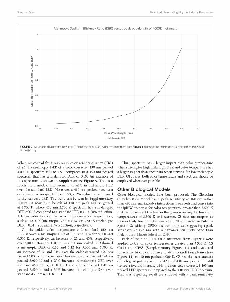
FIGURE 2 | Melanopic daylight efficiency ratio (DER) of the nine 4,000 K spectral metamers from Figure 1 organized by their peak blue emission on the X-axis(410–490 nm).
When we control for a minimum color rendering index (CRI)of 80, the melanopic DER of a color-corrected 490 nm peaked4,000 K spectrum falls to 0.83, compared to a 450 nm peakedspectrum that has a melanopic DER of 0.59. An example ofthis spectrum is shown in Supplementary Figure 9. This is amuch more modest improvement of 41% in melanopic DERover the standard LED. Moreover, a 410 nm peaked spectrumonly has a melanopic DER of 0.58, a 2% reduction comparedto the standard LED. The trend can be seen in SupplementaryFigure 10. Maximum benefit of 410 nm peak LED is gainedat 2,700 K, where 410 nm 2,700 K spectrum has a melanopicDER of 0.33 compared to a standard LED 0.41, a 20% reduction.A larger reducation can be had with warmer color temperatures,such as 1,800 K (melanopic DER = 0.18) or 2,200 K (melanopicDER = 0.31), a 56 and 25% reduction, respectively.
On the colder color temperature end, standard 450 nmLED showed a melanopic DER of 0.73 and 0.86 for 5,000 and6,500 K, respectively, an increase of 23 and 45%, respectively,over 4,000 K standard 450 nm LED. 490 nm peaked LED showeda melanopic DER of 0.93 and 1.12 for 5,000 and 6,500 K,an increase of 12 and 34% over the color-corrected 490 nmpeaked 4,000 K LED spectrum. However, color-corrected 490 nmpeaked 5,000 K had a 27% increase in melanopic DER overstandard 450 nm 5,000 K LED and color-corrected 490 nmpeaked 6,500 K had a 30% increase in melanopic DER overstandard 450 nm 6,500 K LED.
Thus, spectrum has a larger impact than color temperaturewhen striving for high melanopic DER and color temperature hasa larger impact than spectrum when striving for low melanopicDER. Of course, both color temperature and spectrum should beemployed whenever possible.
Other Biological ModelsOther biological models have been proposed. The CircadianStimulus (CS) Model has a peak sensitivity at 460 nm ratherthan 490 nm and includes interactions from rods and cones intothe ipRGC response for color temperatures greater than 3,500 Kthat results in a subtraction in the green wavelengths. For colortemperatures of 3,500 K and warmer, CS uses melanopsin asits sensitivity function (Figueiro et al., 2008). Circadian PotencySpectral Sensitivity (CPSS) has been proposed, suggesting a peaksensitivity at 477 nm with a narrower sensitivity band thanmelanopsin (Moore-Ede et al., 2020).
Each of the nine (9) 4,000 K metamers from Figure 1 wereapplied to CS for color temperatures greater than 3,500 K (CSCool) and CPSS (Supplementary Figure 11) and evaluatedfor relative biological potency relative to itself (SupplementaryFigure 12) at 410 nm peaked 4,000 K. CS has the least amountof biological potency with the 420 and 430 nm spectra, but stillwe see a fivefold increase with the non-color-corrected 490 nmpeaked LED spectrum compared to the 410 nm LED spectrum.This is a surprising result for a model with a peak sensitivity
Frontiers in Neuroscience | www.frontiersin.org 5 June 2021 | Volume 15 | Article 637221
fnins-15-637221 June 1, 2021 Time: 18:47 # 6
Soler and Voss Biologically Relevant Lighting: An Industry Perspective
at 460 nm. The reason is longer wavelength blues stimulate theY tristimulus color matching function. This has to be counter-balanced by less green phosphor, which is primarily in thesubtraction portion of the CS sensitivity curve.
The CPSS also has a minimum potency with the 420 nm LEDspectrum, but still sees a 4.2-fold increase in biological potencyfrom a non-color-corrected 490 nm peaked LED spectrumcompared to a non-color-corrected 410 nm LED spectrum. Againthis is a surprising effect from a curve with spectral sensitivitypeak at 477 nm. This is due to the shape of the 490 nmLED, which has tremendous overlap of the 480 nm LED and470 nm LED spectra.
In other words, the 490 nm peak is so large that it overshadowsshorter wavelength LEDs. This large peak is required because thetristimulus color matching functions are much less sensitive at490 nm compared to shorter wavelengths. In other words, thebiological potential of a 490 nm peaked spectrum found in thisanalysis may not be due to the peak sensitivity of melanopsinat 490 nm, but rather due to the insensitivity of the tristimulusfunctions at longer wavelengths, allowing for more total radiantenergy for the same visual lumens and perceived color.
OTHER KEY VARIABLES
While spectrum and color temperature of the light sources areof primary importance, there are secondary considerations thatpertain to the biological relevance of the delivery of light. Howthe light exits the luminaire and how light bounces off the wallscan contribute to biological relevance.
Geometry of the Built Environment onVertical IlluminanceCommercial buildings often have a grid ceiling, often referred toas a drop ceiling, that include an array of lighting and heating,ventilation, and air conditioning (HVAC) systems integratedinside. This approach in architecture is the most cost-effective forwiring lighting and routing air ducts. This approach points lightsdownward toward desks and floors to optimize luminance andcontrast of objects to be seen. This provides efficiency for visualapplications, but lacks direct illumination into the occupantseyes, making this approach inefficient for providing the verticalillumination necessary for circadian daytime exposure.
Current lighting standards and recommendations for lightingare solely placed on the visual criteria as it relates to completingtasks that occur on a horizontal plane (i.e., on a desk). Forexample, an office may require 300 lumens per square meter (lux)to fall on the desk but make no criteria for what falls on theeyes of the occupants. Key factors related to how much verticallight you can achieve from these common luminaires are roomgeometry and wall reflectance. The ratio of wall area to floor areais referred to as the room cavity ratio (RCR), as this quantifies theopportunity for light to bounce off a vertical surface and redirectits weight from the horizontal plane to the vertical plane.
RCR = 2.5 ×Total wall area above work plane
Floor area
Larger RCR values correspond to more wall area per floor area.For example, a private office may have an RCR of 5, while an openplan office has a RCR closer to 1.
Simulation of various RCRs at 50% wall reflectivity wasachieved using AGi32 lighting design software from LightingAnalysts (Littleton, CO, United States) with typical lightingfixtures. Space types for the simulations included the following:
Open Office (46′ × 86′ × 9′) with recessed light fixtures,RCR = 0.8
Open Office (46′ × 86′ × 9′) with suspended light fixtures 18′′below the ceiling, RCR = 1.1
Classroom (20′ × 30′ × 9′) using recessed fixtures, RCR = 2.1Classroom (20′ × 30′ × 9′) with suspended light fixtures 18′′
below the ceiling, RCR = 2.7Break Room (12′ × 20′ × 9′) with recessed light fixtures,
RCR = 3.3Break Room (12′ × 20′ × 9′) with suspended light fixtures 18′′
below the ceiling, RCR = 4.3Office/Conf. Room (12′ × 12′ × 9′) with recessed light
fixtures, RCR = 4.2Office/Conf. Room (12′ × 12′ × 9′) with suspended light
fixtures 18′′ below the ceiling, RCR = 5.4Each space type outlined above also used the following
luminaire types/manufacturers:Recessed Downlights / Ledra Alphabet NU3RD (Tustin, CA,
United States)Recessed Wall Washer / Ledra Alphabet NU3RW (Tustin, CA,
United States)Recessed 2 × 2 Troffer / Pinnacle Lucen (Denver, CO,
United States)Recessed 2 × 4 Troffer / Pinnacle Lucen (Denver, CO,
United States)Direct Linear Pendant / Pinnacle Edge3 (Denver, CO,
United States)Indirect Linear Pendant /Axis Beam4 (Lasalle, QC, Canada)Direct / Indirect Linear Pendant / Axis Surround Lite (Lasalle,
QC, Canada)Direct / Indirect Circular Area Pendant / Prudential P4000
(Los Angeles, CA, United States)An example of these AGi32 calculations is shown in
Supplementary Figures 13–15. Supplementary Figure 16illustrates the benefit in vertical to horizontal ratio usingdifferent fixture types versus RCR. In summary, an openplan office will provide 50 vertical lux at the occupant’seyes for every 100 lux on the desktop. Whereas a privateoffice will provide 80 vertical lux at the occupant’s eyesfor every 100 lux on the desk surface, a 60% boost invertical illumination.
These calculations were repeated for 70% wall reflectivityand 90% wall reflectivity. A boost in vertical to horizontal ratiowas not observed due to wall reflectivity, however, a boost intotal illuminance was observed. Supplementary Figures 17, 18illustrate these vertical illumination benefits of higher wallreflectivity as a function of RCR. These Figures illustrate thatvertical illumination benefits were minor for small RCRs, butmuch stronger for larger RCRs. In other words, increasing wallreflectivity in a large open plan office will yield a meager benefit,
Frontiers in Neuroscience | www.frontiersin.org 6 June 2021 | Volume 15 | Article 637221
fnins-15-637221 June 1, 2021 Time: 18:47 # 7
Soler and Voss Biologically Relevant Lighting: An Industry Perspective
but increasing wall reflectivity in a private office will have asignificant benefit to vertical illumination.
Luminaire Light DistributionLight distribution is another characteristic that plays a criticalrole in achieving vertical illuminance. Pendants are a type ofluminaire that are mounted to the ceiling and suspended in thespace. Some pendants are fully luminous, such that it distributeslight in all directions. We evaluated the Purelight RoundLuminous pendant from Selux (Highland, NY, United States) thatfocus the majority of their energy downward toward the taskplane. Our AGi32 analysis showed that these luminous surfacedpendants provided 22% more vertical illumination on average forthe same amount of light on the task plane.
Different lighting strategies can also be applied at night.Typical LED “Edison-type” light bulbs do not have the sameisotropic distribution as a standard incandescent light source.These light bulbs direct the majority of the light in the directionopposite of screw-in base. When applied into a standard tablelamp with the screw base downward, light is directed onto the80% reflective ceiling. Mirror light bulbs are fairly common anddesigned to reduce direct glare and can be used in place of thesestandard Edison LED bulbs to redirect the light from the 80%reflective ceiling toward a much lower reflectance floor. A studywas conducted in a 14′ × 10′ × 8′ (L ×W × H) room, where asingle table lamp was illuminated in a corner of the room usingboth a regular Edison-type light bulb and a mirror type lightbulb. Vertical illumination was measured facing the lamp from2′ to 10′ away using both a standard LED lamp and a mirrorlamp of the same lumen output. Measurements are provided insupplemental Supplementary Figure 19 which shows that thistechnique yielded a 35% reduction in vertical illumination, whilestill maintaining light availability.
ControlsControls play an integral role in biologically relevant lighting.Controls should be configured to create brighter days anddarker nights on a consistent and predictable cadence. Thiscan be set to be in phase with the solar cycle or can be setto a specific time, such as 6 a.m. to 8 p.m. The later maybepreferred at higher latitudes. At a bare minimum, this controlsystem would comprise of automatic dimmers that increase anddecrease intensity according to time. However, a biologicallyrelevant lighting system should consider intensity, spectrum,and distribution. Intensity and spectrum should be tied togetherto maximize day versus night delineation. Blending between abrighter intensity of blue enriched spectrum with peak emissionat 490 nm of the highest accepted color temperature and dimmerintensity of blue depleted spectrum with peak emission between410 and 450 nm of lowest acceptable color temperature. Furtherday/night delineation value can be obtained by including spatialdistribution. Nighttime scenes should include no indirect light(lights pointed at the ceiling) and should have direct light(lights pointed at the task) with blue depleted spectrum ofminimal intensity to achieve necessary tasks. Daytime scenesshould include direct and indirect light, both with blue enrichedspectrum with peak emission at 490 nm of highest accepted color
temperature and of highest accepted intensity both in terms ofcomfort and energy constraints.
PERSONAL CIRCADIAN LIGHTINGDEVICES
One final strategy to help provide biologically relevant lightingwithin a space is the use of personalized devices that can supplysupplemental vertical lighting. These personalized devices couldbe located relatively close to the occupant and primarily providevertical illumination, for example something akin to a light boxor a table lamp. These types of devices could conceivably add200 melanopic EDI or more at the eye of the occupant andcould be tailored to the needs of an individual. Ideally, this typeof intervention would be automated to dynamically transitionbetween day and night illumination throughout the day.
DAYLIGHTING
The data presented here provides quantified strategies forimplementing biologically brighter days and darker nights intothe built environment. While not quantified in this analysis,daylight exposure is best for providing circadian benefits(Knoop et al., 2020), but it should be noted that vertical lightexposure from windows drops off dramatically with distancefrom said window. Skylights and daylight harvesting strategiesare encouraged to bring that daylight deeper into the space tomaximize its benefits.
COMPOUNDING THE STRATEGIES FORBRIGHTER DAYS AND DARKER NIGHTS
Standalone strategies for brighter days and darker nights areoutlined in this article and while some individual interventionsmay not seem like they apply a space or application, thecombination of these strategies compound their benefits to createa biologically relevant lighting environment.
For example, a person spends their day in the office understandard 3,500 K LED lighting with 300 lux at the task planeand 150 lux at the eye. A standard 3,500 K LED spectrum hasa melanopic DER of about 0.51, thus the melanopic EDI at theeye is 76.5. The same person spends their evening in a homewith luminaires populated with 2,700 K LED light bulbs. Theseluminaires provide approximately 50 lux at the eye. 2,700 K LEDspectrum has a melanopic DER of about 0.4, thus a nighttimemelanopic EDI of 20. This is a typical example of how light isbiologically too dim for daytime use (76.5 melanopic EDI) andtoo bright for evening (20 melanopic EDI), with a day-to-nightratio of 3.8:1. Its not certain if more benefit would be gained frombrighter days or darker nights, it is good practice to improve bothand increase that day-to-night ratio.
At the office, the simple incorporation of a spectrallyoptimized daytime light source with a slightly cooler colortemperature of 4,000◦K will lead to a 58% boost in daytime
Frontiers in Neuroscience | www.frontiersin.org 7 June 2021 | Volume 15 | Article 637221
fnins-15-637221 June 1, 2021 Time: 18:47 # 8
Soler and Voss Biologically Relevant Lighting: An Industry Perspective
biological potency. This would increase 76.5 melanopic EDI fromthe example to 121 melanopic EDI. While at home, incorporatingspatially optimized light bulbs with a slightly warmer colortemperature of 2,200◦K lighting provides a 60% reduction innighttime biological potency. This would decrease 20 melanopicEDI from the example to eight melanopic EDI. Combiningthese two strategies would increase the day-to-night ratio in theexample from 3.8:1 to 15.1:1. This is a 392% increase in thedelineation of daytime versus nighttime.
The proposed goal is to combine as many of these individualstrategies and put them into practice (when possible) whilemaintaining excellent light quality, appropriate design aesthetic,and achieving user/occupant compliance. These strategies areitemized here:
Daytime strategy Standalone benefit
(1) Color-corrected spectrally optimized daytime spectrum ∼ (+41%)
(2) Colder color temperatures ∼ (+10%) per 500 K
(3) Spatially optimized luminaire ∼ (+22%)
(4) Private office ∼ (+60%)
(5) Private office with highly reflective walls ∼ (+150%)
(6) Personal circadian luminaire +200 melanopic EDI
Nighttime strategy Standalone benefit
(1) Warmer color temperatures ∼(−15%) per 500 K
(2) Spatially optimized luminaire ∼(−35%)
(3) Spectrally optimization Up to −20%
DATA AVAILABILITY STATEMENT
The raw data supporting the conclusions of this article will bemade available by the authors, without undue reservation.
AUTHOR CONTRIBUTIONS
All spectral analysis, industry perspective, data collection, andanalysis were done by RS. Lighting design simulation was doneby EV, an employee of BIOS lighting.
SUPPLEMENTARY MATERIAL
The Supplementary Material for this article can be found onlineat: https://www.frontiersin.org/articles/10.3389/fnins.2021.637221/full#supplementary-material
Supplementary Figure 1 | Tristimulus sensitivity according to CIE 1931 colormatching functions and melanopsin sensitivity according to CIE S 026.
Supplementary Figure 2 | Standard off-the-shelf 6,500 K LED with peak blueemission at 450 nm and broad phosphor excitation emission from 480 to 780 nm
compared to tristimulus sensitivity according to CIE 1931 and melanopsinsensitivity according to CIE S 026.
Supplementary Figure 3 | Spectral Power Distribution (SPD) for eachcomponent used in simulation. (A) SPD of 410 nm LED; (B) SPD of 420 nm LED;(C) SPD of 430 nm LED; (D) SPD of 440 nm LED; (E) SPD of 450 nm LED; (F)SPD of 460 nm LED; (G) SPD of 470 nm LED; (H) SPD of 480 nm LED; (I) SPD of490 nm LED; (J) SPD of 500 nm LED; (K) SPD of 547 nm peaked phosphorYYG-547-210; (L) SPD of 585 nm peaked phosphor OGA-585; (M) SPD of660 nm peaked phosphor YYG-660.
Supplementary Figure 4 | Color plots of each component used in simulation.Color points are labeled A through M, in reference to spectral power distributionsof said color plot from Supplementary Figure 3.
Supplementary Figure 5 | Working space of 450 nm LED (E), 547 nm peakedphosphor YYG-547-210 (K), 585 nm peaked phosphor OGA-585 (L), and 660 nmpeaked phosphor YYG-660 (M). Any color points within the triangle plotted herecan be created. ANSI color temperature bins for 6,500, 5,000, 4,000, 3,500,3,000, and 2,700 K white light are plotted for reference.
Supplementary Figure 6 | Working space of 410 nm LED (A), 547 nm peakedphosphor YYG-547-210 (K), 585 nm peaked phosphor OGA-585 (L), and 660 nmpeaked phosphor YYG-660 (M). Any color points within the triangle plotted herecan be created. ANSI color temperature bins for 6,500, 5,000, 4,000, 3,500,3,000, and 2,700 K white light are plotted for reference.
Supplementary Figure 7 | Working space of 490 nm LED (I), 547 nm peakedphosphor YYG-547-210 (K), 585 nm peaked phosphor OGA-585 (L), and 660 nmpeaked phosphor YYG-660 (M). Any color points within the triangle plotted herecan be created. ANSI color temperature bins for 6,500, 5,000, 4,000, 3,500,3,000, and 2,700 K white light are plotted for reference.
Supplementary Figure 8 | Working space of 500 nm LED (J), 547 nm peakedphosphor YYG-547-210 (K), 585 nm peaked phosphor OGA-585 (L), and 660 nmpeaked phosphor YYG-660 (M). Any color points within the triangle plotted herecan be created. ANSI color temperature bins for 6,500, 5,000, 4,000, 3,500,3,000, and 2,700 K white light are plotted for reference.
Supplementary Figure 9 | Blended spectrum that achieves a minimum of 80 CRIand maximizes energy efficiency and melanopic daylight efficiency ratio (DER).
Supplementary Figure 10 | Melanopic DER versus color temperature ofcolor-corrected optimized daytime spectrum (dashed line), color-correctedoptimized nighttime spectrum (dash-dot line), and standard LED (solid line).
Supplementary Figure 11 | Nine spectral metamers of light, each with differentblue light peaks. 410 nm peaked metamer (purple), 420 nm peaked metamer(green), 430 nm peaked metamer (yellow), 440 nm peaked metamer (red), 450 nmpeaked metamer (royal blue), 460 nm peaked metamer (orange), 470 nm peakedmetamer (black), 480 nm peaked metamer (dark gray), and 490 nm peakedmetamer (sky blue), from Figure 1, are overlayed on top of melanopsin sensitivityfunction (gray–solid) according to CIE S 026, CS cool sensitivity function(gray–small dash) and CPSS (gray–dash dot). Each metamer produces visually thesame 4,000 K color of light at the same lumens according to thetristimulus functions.
Supplementary Figure 12 | Relative weights of m-DER, CS, and CPSS of thenine spectral metamers from figure S10 (Y-axis) versus peak blue wavelength ofmetamer (X-axis). Weights are relative to themselves at the410 nm peaked metamer.
Supplementary Figure 13 | Elevation view of AGi32 lighting simulation softwareand lighting calculation points of an open office layout. Calculation points arehorizontal (with lines pointed up from points) and vertical (with lines pointed leftfrom points). Blue lines reference building structure. Black lines reference furniture.
Supplementary Figure 14 | Plan view of AGi32 lighting simulation software andvertical lighting calculation points of an open office layout. Calculation points arevertical with lines pointed in direction of view. Blue lines reference buildingstructure. Black lines reference furniture. Red lines reference light fixtures.
Supplementary Figure 15 | Rendering of isometric view using AGi32 lightingsimulation software.
Frontiers in Neuroscience | www.frontiersin.org 8 June 2021 | Volume 15 | Article 637221

fnins-15-637221 June 1, 2021 Time: 18:47 # 9
Soler and Voss Biologically Relevant Lighting: An Industry Perspective
Supplementary Figure 16 | Vertical to Horizontal light ratio versus room cavityratio with 90% reflective painted walls for of downlight luminaires (gray–solid),direct pendent luminaires (black–solid), wall washer luminaires (gray–dashed),indirect/direct pendent luminaires (black–dashed), indirect only pendent luminaires(gray–dash dot), and recessed general illumination luminaires (black–dash dot).
Supplementary Figure 17 | Boost in vertical melanopic EDI versus RCR ofspaces with 90% wall reflectivity compared to spaces with 50% wall reflectivity ofdownlight luminaires (gray–solid), direct pendent luminaires (black–solid), wallwasher luminaires (gray–dashed), indirect/direct pendent luminaires(black–dashed), indirect only pendent luminaires (gray–dash dot), and recessedgeneral illumination luminaires (black–dash dot).
Supplementary Figure 18 | Boost in vertical melanopic EDI versus RCR ofspaces with 70% wall reflectivity compared to spaces with 50% wall reflectivity ofdownlight luminaires (gray–solid), direct pendent luminaires (black–solid), wallwasher luminaires (gray–dashed), indirect/direct pendent luminaires(black–dashed), indirect only pendent luminaires (gray–dash dot), and recessedgeneral illumination luminaires (black–dash dot).
Supplementary Figure 19 | Measurements of vertical illumination (lux) at 40′ ′
height for a space with a single table lamp containing a standard LED A19 lightbulb “upward lamp” (black–solid) or a A19 light bulb with a mirror coating“downward lamp” (black–dashed) versus distance away from lamp.Measurements taken facing the lamp.
REFERENCESAuf Der Maur, M., Pecchia, A., Penazzi, G., Rodrigues, W., and Di Carlo, A. (2016).
Efficiency drop in green InGaN/GaN light emitting diodes: the role of randomalloy fluctuations. Phys. Rev. Lett. 116:027401. doi: 10.1103/PhysRevLett.116.027401
Berman, S. M., Jacobs, R. J., Bullimore, M. A., Bailey, L. L., Ghandi, N., andGreenhouse, D. S. (1991). An Objective Measure of Discomfort Glare. FirstInternational Symposium On Glare. New York, NY: Lighting Research Institute.
Berson, D. M., Dunn, F. A., and Takao, M. (2002). Phototransduction by retinalganglion cells that set the circadian clock. Science 295, 1070–1073. doi: 10.1126/science.1067262
Brainard, G. C., Hanifin, J. P., Greeson, J. M., Byrne, B., Glickman, G., Gerner, E.,et al. (2001). Action spectrum for melatonin regulation in humans: evidencefor a novel circadian photoreceptor. J. Neurosci. Off. J. Soc. Neurosci. 21,6405–6412.
Brown, T. M. (2020). Melanopic illuminance defines the magnitude of humancircadian light responses under a wide range of conditions. J. Pineal. Res.5:e12655. doi: 10.1111/jpi.12655
Brown, T. M., Thapan, K., Arendt, J., Revell, V. L., and Skene, D. J. (2021). S-conecontribution to the acute melatonin suppression response in humans. J. PinealRes. doi: 10.1111/jpi.12719 ∗vol page,
Dacey, D. M., Liao, H., Peterson, B. B., Robinson, F. R., Smith, V. C., Pokorny,J., et al. (2005). Melanopsin-expressing ganglion cells in primate retina signalcolour and irradiance and project to the LGN. Nature 433, 749–754. doi:10.1038/nature03344
Fernandez, D., Fogerson, P., Lazzerini, O., Thomsen, M., Layne, R., Severin, D.,et al. (2018). Light affects mood and learning through distinct retina-brainpathways. Cell 175, 71–84.e18. doi: 10.1016/j.cell.2018.08.004
Figueiro, M. G., Bierman, A., and Rea, M. S. (2008). Retinal mechanisms determinethe subadditive response to polychromatic light by the human circadian system.Neurosci. Lett. 438, 242–245. doi: 10.1016/j.neulet.2008.04.055
Gaggioni, G., Maquet, P., Schmidt, C., Dijk, D., and Vandewalle, G. (2014).Neuroimaging, cognitioon, light and circadian rhythms. Front. Syst. Neurosci.8:126. doi: 10.3389/fnsys.2014.00126
Güler, A. D., Ecker, J. L., Lall, G. S., Haq, S., Altimus, C. M., Liao, H. W.,et al. (2008). Melanopsin cells are the principal conduits for rod-coneinput to non-image-forming vision. Nature 453, 102–105. doi: 10.1038/nature06829
Hughes, S., Jagannath, A., Rodgers, J., and Hankins, M. W. (2016). Signallingby expressing photosensitive retinal ganglion cells. Nat. Publ. Group44, 1–8.
International Commission on Illumination (CIE) (2018). CIE System for Metrologyof Optical Radiation for ipRGC-Influenced Responses to Light. Vienna: CentralBureau CIE.
Knoop, M., Stefani, O., Bueno, B., Matusiak, B., Hobday, R., Wirz-Justice, A., et al.(2020). Daylight: What makes the difference? Ligh. Res. Technol. 52, 423–442.doi: 10.1177/1477153519869758
LeGates, T. A., Fernandez, D. C., and Hattar, S. (2014). Light as a central modulatorof circadian rhythms, sleep and affect. Nat. Rev. Neurosci. 15:443.
Liu, G. H., Zhou, Z. Z., Shi, Y., Liu, Q., Wan, J. Q., and Pan, Y. B. (2015). Ce:YAGtransparent ceramics for applications of high power LEDs: Thickness effects andhigh temperature performance. Mater. Lett. 139, 480–482. doi: 10.1016/j.matlet.2014.10.114
Lucas, R. J., Peirson, S. N., Berson, D. M., Brown, T. M., Cooper, H. M., Czeisler,C. A., et al. (2014). Measuring and using light in the melanopsin age. TrendsNeurosci. 37, 1–9. doi: 10.1016/j.tins.2013.10.004
Moore-Ede, M., Heitmann, A., and Guttkuhn, R. (2020). Circadian potencyspectrum with extended exposure to polychromatic white LED lightunder workplace conditions. J. Biol. Rhyt. 35, 405–415. doi: 10.1177/0748730420923164
Mouland, J. W., Martial, F., Watson, A., Lucas, R. J., and Brown, T. M. (2019).Cones support alignment to an inconsistent world by suppressing mousecircadian responses to the blue colors associated with twilight. Curr. Biol. CB29, 4260–4267.e4. doi: 10.1016/j.cub.2019.10.02
Mueller-Mach, R., Mueller, G., Krames, M. R., Höppe, H. A., Stadler, F., Schnick,W., et al. (2005). Highly efficient all-nitride phosphor-converted white lightemitting diode. Phys. Status Soli. 202, 1727–1732. doi: 10.1002/pssa.200590014
Navigant. (2019). Energy Savings Forecast of Solid-State Lighting in GeneralIllumination Applications. U.S. Department of Energy Report, DOE/EERE 2001.
Navigant. (2020). Adoption of Light-Emitting Diodes in Common LightingApplications. U.S. Department of Energy Report. 1–56. Available online at: https://www.energy.gov/sites/prod/files/2020/09/f78/ssl-led-adoption-aug2020.pdf(accessed January 20, 2021).
Nishiura, S., Tanabe, S., Fujioka, K., and Fujimoto, Y. (2011). Properties oftransparent Ce:YAG ceramic phosphors for white LED. Optical Mater. 33,688–691. doi: 10.1016/j.optmat.2010.06.005
Panda, S., Sato, T. K., Castrucci, A. M., Rollag, M. D., DeGrip, W. J., Hogenesch,J. B., et al. (2002). Melanopsin (Opn4) requirement for normal light-inducedcircadian phase shifting. Science 298, 2213–2216. doi: 10.1126/science.1076848
Phillips, A. J. K., Vidafar, P., Burns, A. C., McGlashan, E. M., Anderson, C.,Rajaratnam, S. M. W., et al. (2019). High sensitivity and interindividualvariability in the response of the human circadian system to eveninglight. Proc. Natl. Acad. Sci. 116, 12019–12024. doi: 10.1073/pnas.1901824116
Provencio, I., Cooper, H. M., and Foster, R. G. (1998). Retinal projectionsin mice with inherited retinal degeneration: implications for circadianphotoentrainment. J. Comp. Neurol. 395, 417–439. doi: 10.1002/(sici)1096-9861(19980615)395:4<417::aid-cne1<3.0.co;2-4
Provencio, I., and Foster, R. G. (1995). Circadian rhythms in mice can be regulatedby photoreceptors with cone-like characteristics. Brain Res. 694, 183–190. doi:10.1016/0006-8993(95)00694-l
Rea, M. S., and Ouellette, M. J. (1991). Visual performance using reaction times: abasis for application. Light. Res. Tech. 23, 135–144.
Rodgers, J., Hankins, M. W., Peirson, S. N., and Foster, R. G. (2016). Signallingby melanopsin (OPN4) expressing photosensitive retinal ganglion cells. Eye 44,1–8. doi: 10.1038/eye.2015.264
Roenneberg, T., Allebrandt, K. V., Merrow, M., and Vetter, C. (2012).Social jetlag and obesity. Curr. Biol. 22, 939–943. doi: 10.1016/j.cub.2012.03.038
Roenneberg, T., and Merrow, M. (2016). The circadian clock andhuman health. Curr. Biol. 26, R432–R443. doi: 10.1016/j.cub.2016.04.011
Shaolin, H., Sheng, L., Zhi, Z., Han, Y., Zhiyin, G., and Haisheng, F. (2015). A novelMOCVD reactor for growth of high-quality GaN-related LED layers. J. CrystalGrowth 415, 72–77. doi: 10.1016/j.jcrysgro.2014.12.038
Frontiers in Neuroscience | www.frontiersin.org 9 June 2021 | Volume 15 | Article 637221
fnins-15-637221 June 1, 2021 Time: 18:47 # 10
Soler and Voss Biologically Relevant Lighting: An Industry Perspective
Skeldon, A. C., Phillips, A. J. K., and Dijk, D. (2017). The effects of self-selectedlight- dark cycles and social constraints on human sleep and circadian timing: amodeling approach. Sci. Rep. 7:45158. doi: 10.1038/srep45158
Spitschan, M. (2019). Melanopsin contributions to non-visual and visualfunction. Curr. Opin. Behav. Sci. 30, 67–72. doi: 10.1016/j.cobeha.2019.06.004
Spitschan, M., Lazar, R., Yetik, E., and Cajochen, C. (2019). No evidence for anS cone contribution to acute neuroendocrine and alerting responses to light.Curr. Biol. CB 29, R1297–R1298. doi: 10.1016/j.cub.2019.11.031
Sulli, G., Manoogian, E., Taub, P., and Panda, S. (2018). Training the circadianclock, clocking the drugs,and drugging the clock to prevent, manage, and treatchronic diseases. Trends Pharmacol. Sci. 39, 812–827. doi: 10.1016/j.tips.2018.07.003
Thapan, K., Arendt, J., and Skene, D. J. (2001). An action spectrum for melatoninsuppression: evidence for a novel non-rod, non-cone photoreceptor system inhumans. J. Physiol. 535(Pt. 1), 261–267. doi: 10.1111/j.1469-7793.2001.t01-1-00261.x
Tosini, G., Ferguson, I., and Tsubota, K. (2016). Effects of bluelight ontheon thecircadian system and eye physiology. Mol. Vis. 22, 61–72.
Zarrinpar, A., Chaix, A., and Panda, S. (2015). Daily eatingpatterns and their impact on health and disease. TrendsEndocrinol. Metabol. 27, 69–83. doi: 10.1016/j.tem.2015.11.007
Conflict of Interest: RS is a founder of BIOS Lighting, a for-profit lightingmanufacturer. EV is currently employed by the company BIOS Lighting, a for-profit company.
Copyright © 2021 Soler and Voss. This is an open-access article distributed under theterms of the Creative Commons Attribution License (CC BY). The use, distributionor reproduction in other forums is permitted, provided the original author(s) andthe copyright owner(s) are credited and that the original publication in this journalis cited, in accordance with accepted academic practice. No use, distribution orreproduction is permitted which does not comply with these terms.
Frontiers in Neuroscience | www.frontiersin.org 10 June 2021 | Volume 15 | Article 637221