biological ligands for metal ions -...
TRANSCRIPT

– 3 –
Biological Ligands for Metal Ions
INTRODUCTION
In the previous chapter we have explained the basic notions involved in the coordination
chemistry of metal ions. We now consider the potential ligands that could be involved in
binding metals in metalloproteins. We can divide them into those which are naturally
occurring amino acids in the protein itself; amino acids that have been chemically modi-
fied in order to bind specific metal ions, such as Ca2�; low-molecular weight inorganic lig-
ands, such as carbonate, cyanide and carbon monoxide; metal-binding organic cofactors
that have been introduced into the protein (such as porphyrins, corrins and iron–sulfur
(Fe–S) clusters, the molybdenum cofactor, MoCo, the CuZ centre of nitrous oxide reduc-
tase and the FeMoCo and P-clusters of nitrogenase); and, finally, metal-binding molecules
excreted from the cell and then taken up as the metal chelate (such as siderophores).
As was pointed out in the previous chapter, biologically important metal ions and their
ligands can be classified according to the hard–soft theory of acids and bases (Table 2.1).
While there are exceptions, most metal ions bind to donor ligands as a function of prefer-
ences based on this concept, with hard acids (metal ions) binding preferentially to hard
bases (ligands) and soft acids to soft bases.
PROTEIN AMINO ACID RESIDUES (AND DERIVATIVES) AS LIGANDS
Of the 20 amino acids present in proteins, only a relatively small number are potential
metal ligands. The ligand groups, which are encountered most often, are the thiolate of
Cys, the imidazole of His, the carboxylates of Glu and Asp, and the phenolate of Tyr
(Figure 3.1). Less frequently we encounter the thioether group of Met, the amino group of
Lys and the guanidino group of Arg, and the amide groups of Asn and Gln. Metal ions can
also bind to peptide bonds, through the carbonyl or the deprotonated amide nitrogen, and
to the terminal amino and carboxyl groups of the protein.
Cysteine can bind to either one or two metal ions, and is frequently found as a ligand to
iron (in Fe–S clusters—see later) and to Cu� (for example in the copper chaperones, which
transfer copper to specific copper-binding proteins). Histidine can bind metal ions in two
27

positions, and has a strong preference for Cu2�. The carboxylate oxygens of aspartate (and
glutamate, which is not included in Figure 3.1) are preferential ligands for the alkali and alka-
line earth metals such as Ca2�. They can bind a single metal ion in either a mono- or bi-dentate
(chelating) mode, or bind two metal ions in a bi-dentate, bridging mode. Many proteins of
the blood-clotting cascade contain �-carboxyglutamic acid (Figure 3.1), which binds Ca2�
more strongly than glutamate itself. Vitamin K is a cofactor for the enzymatic conversion of
glutamic acid to �-carboxyglutamic acid in these vitamin K-dependent proteins after their
biosynthesis (an example of post-translational modification). Vitamin K antagonists (such as
Warfarin, originally developed as a rat poison) are used therapeutically as anti-coagulants.
28 Biological Inorganic Chemistry/R.R. Crichton
Figure 3.1 Principal protein amino acid side-chain metal–ion binding modes (the metal ion repre-sented as a dark filled circle) and (right) the structure of the Ca2�-binding �-carboxyglutamate foundin proteins of the blood-clotting cascade.

Fe3� also shows a strong affinity for the oxygen donor atoms of carboxylates as well as the
phenoxide of tyrosine. Like cysteine, the sulfur ligand of methionine is often found bound to
iron, for example in electron-transfer haemoproteins such as cytochrome c.
AN EXAMPLE OF A NON-PROTEIN LIGAND: CARBONATE
AND PHOSPHATE
In addition to the amino acid side chains mentioned above, a number of other low molec-
ular weight ligands are found in metalloproteins. These include cyanide and carbon monox-
ide, which we will describe later in this chapter. Here we consider carbonate and phosphate
anions in the context of the super family of iron-binding proteins, the transferrins.
Transferrins, which transport iron in serum and other extracellular fluids in mammals,
are part of a super family of proteins, all of which function by a ‘Venus fly trap’ mecha-
nism. They are made up of two homologous lobes, termed N- and C-lobes, each of which
binds a single atom of ferric iron together with a ‘synergistic’ carbonate anion. Each lobe
is composed of two domains indicated in Figure 3.2, which close together upon iron and
carbonate binding1. The ferric iron is bound in almost ideal octahedral geometry with four
Biological Ligands for Metal Ions 29
Figure 3.2 Ribbon diagram of the C-lobe of human transferrin with the two domains shown in differ-ent colours (cyan for C1 and green for C2). The inset shows the four protein ligand residues togetherwith the arginine residue which stabilizes binding of the synergistic carbonate ion (both in magenta).(Reprinted with permission from Mason et al., 2005. Copyright (2005) American Chemical Society.)
1 The Venus fly trap is a carnivorous swamp flower of the sundew family, native to the Carolinas; it has leaves
with two-hinged blades that snap shut to trap insects.

ligands supplied by the protein and the remaining two contributed by the carbonate anion.
The charge on the ferric ion is matched by the three anionic ligands contributed by the
protein itself (two tyrosines and one aspartate), while the charge on the carbonate is sta-
bilized by the positive charge on an arginine residue, which is highly conserved in each
lobe of all mammalian transferrins. In the open configuration, the carbonate can bind in
the bottom of the metal-binding pocket. Upon binding of the Fe3�, two of the four pro-
tein ligands are already in place, and closure of the domains brings the other two into
place. Iron release from transferrin is characterized by a large conformational change, in
which the two domains move away from each other and is thought to involve protonation
of the carbonate and movement of the arginine residue, destabilizing the coordination of
the iron atom.
In pathogenic bacteria, such as Haemophilus influenzae and various species of
Neisseria, Fe3� is transported by ferric-binding proteins, which have an overall structure
almost identical to that of one of the iron-binding lobes of transferrin. Again there are four
protein ligands to the metal, almost identical to transferrin (except that aspartate is
replaced by glutamate): However the carbonate is replaced by a phosphate, and only one
of the phosphate oxygens (alternatively oxygen atoms of the phosphate) binds the metal
with the sixth coordination position taken by a water molecule.
ENGINEERING METAL INSERTION INTO ORGANIC COFACTORS
As we will see in subsequent chapters, many metalloproteins have their metal centres
located in organic cofactors (Lippard and Berg, 1994), such as the tetrapyrrole por-
phyrins and corrins, or in metal clusters, such as the Fe–S clusters in Fe–S proteins or
the FeMo-cofactor of nitrogenase. Here we discuss briefly how metals are incorporated
into porphyrins and corrins to form haem and other metallated tetrapyrroles, how Fe–S
clusters are synthesized and how copper is inserted into superoxide dismutase.
CHELATASE: TERMINAL STEP IN TETRAPYRROLE METALLATION
The insertion of a divalent metal ion into a tetrapyrrole is the final step in the biosynthesis
of haem (Fe2�), chlorophyll (Mg2�), cobalamin (vitamin B12—Co2�) and the coenzyme
F430 (Ni2�) involved in methane production in methanogenic bacteria. These are all derived
from a common tetrapyrrole precursor, uroporphyrinogen III (Figure 3.3). The insertion of
each of these metal ions involves a group of enzymes called chelatases, of which the best
characterized is ferrochelatase, which inserts Fe2� into protoporphyrin IX in the terminal
step of the haem biosynthetic pathway. The different chelatases are thought to have simi-
lar mechanisms, which involve, as the first step, the distortion of the tetrapyrrole porphyrin
upon binding to the enzyme to give a saddled structure (Figure 3.4a) in which two oppo-
site pyrrole rings are slightly tilted upwards while the other two pyrrole rings are tilted
slightly downwards. In the figure, the two unprotonated nitrogen atoms of the pyrrole
rings point upwards, while the two protonated nitrogens point downwards with respect
to the porphyrin ring. Subsequent to the distortion of the porphyrin ring, the first
30 Biological Inorganic Chemistry/R.R. Crichton

metal–porphyrin bond is formed (Figure 3.4b), followed by other ligand exchange steps
leading to the formation of a complex in which the iron atom is sitting on top of the por-
phyrin, with two of its nitrogen atoms coordinated to the metal while the other two are
still protonated. This is followed by the sequential deprotonation of the two pyrrole nitro-
gen atoms coupled with formation of the metallated porphyrin. The saddling of the por-
phyrin is an out-of-plane deformation, which exposes both the protons and the lone pairs
of the nitrogen atoms of the porphyrin molecule in an appropriate arrangement for metal
insertion.
The structure of several ferrochelatases has been determined, and it is clear that the por-
phyrin rings B, C and D are held in a very tight grip by conserved amino acids, whereas
the A ring is distorted. Two metal–ion binding sites have been identified, one located at
the surface of the molecule, occupied by a fully hydrated Mg2� ion, and the other located
in the porphyrin-binding cleft, close to the distorted porphyrin ring A, with its nitrogen
pointing towards His183 and Glu264 (Figure 3.5). It has been proposed that the metal ion
on the outermost site, by ligand exchange with a series of acidic residues arranged along
Biological Ligands for Metal Ions 31
Figure 3.3 The tetrapyrrole biosynthetic pathways. Chelatases selectively insert Fe2� to form haem,Mg2� to form chlorophyll, Co2� to form cobalamin and in methanogenic bacteria Ni2� to formcoenzyme F430.
Figure 3.4 Mechanism of porphyrin metallation. (a) Out-of-plane saddle structure in which two pyr-role rings with unprotonated nitrogens (blue spheres) point upwards, while the other two, protonated(blue and white spheres) point downwards. (b) Steps in the mechanism for incorporation of the metalion (red) into the porphyrin (pyrrole rings in green), described in the text. (From Al-Karadaghi et al.,2006. Copyright 2006, with permission from Elsevier.)

the helical edge of a �-helix2, would be shuttled to the inner site, to be exchanged withthe pyrrole nitrogens, resulting in insertion of the metal ion into the porphyrin. The twosites, occupied respectively by a Zn2� ion and a fully hydrated Mg2� ion, are �7 Å apart.Two of the ligands to the Zn2� ion in the outer site, His183 and Glu264 are invariant inall ferrochelatases. The side chains of Glu272, Asp268 and Glu272 are aligned along the�-helix, in a line connecting the two metal sites. Only a �-helix can provide such an align-ment of side chains. This is reminiscent of several other metalloproteins, such as nitro-genase, in which residues in �-helices function to coordinate metal ions involved inenzymatic activity.
IRON–SULFUR CLUSTER CONTAINING PROTEINS
For the first billion years of evolution the environment was anaerobic; this meant that, sinceiron and sulfur were abundant, proteins containing Fe–S clusters were probably abundant,and therefore were among the first catalysts that Nature had available to it. They are distrib-uted in virtually all the living organisms, but their recognition as a distinct class of metallo-proteins only occurred after the discovery of their characteristic EPR spectra in the oxidizedstate in the 1960s. This second class of iron-containing proteins have iron atoms bound tosulfur, either bound to the polypeptide chain by the thiol groups of cysteine residues or elsewith both inorganic sulfide and cysteine thiols as ligands. The biochemical utility of theseFe–S clusters resides not only in their possibility to easily transfer electrons, but also in theirtendency to bind the electron-rich oxygen and nitrogen atoms of organic substrates.
Fe–S proteins contain four basic core structures, which have been characterized crystal-lographically both in model compounds (Rao and Holm, 2004) and in iron–sulfur proteins.These are (Figure 3.6), respectively, (A) rubredoxins found only in bacteria, in which the[Fe–S] cluster consists of a single Fe atom liganded to four Cys residues—the iron atom
32 Biological Inorganic Chemistry/R.R. Crichton
2 For more information concerning this unusual type of helix see Chapter 4.
Figure 3.5 Structure (a), porphyrin (b) and metal ion-binding sites (c) in Bacillus subtilis fer-rochelatase. In (c) the two metal ions are a Zn2� ion (grey) and a fully hydrated Mg2� ion. The sidechains of Glu272, Asp 268 and Glu264 are aligned along a �-helix (green). (From Al-Karadaghiet al., 2006. Copyright 2006, with permission from Elsevier.)

can be in the �2 or �3 valence; (B) rhombic two iron–two sulfide [Fe2–S2] clusters—
typical stable cluster oxidation states are �1 and �2 (the charges of the coordinating
cysteinate residues are not considered); (C) cuboidal three-iron–four sulfide [Fe3–S4]
clusters—stable oxidation states are 0 and �1; and (D) cubane four iron–four sulfide
[Fe4–S4] clusters—stable oxidation states are �1 and �2 for ferredoxin-type clusters and
�2 and �3 for ‘Hipip’3 clusters. Electrons can be delocalized, such that the valences of
individual iron atoms lie between ferrous and ferric forms. Low molecular weight proteins
containing the first and the last three types are referred to as rubredoxins (Rd) and ferre-
doxins (Fd), respectively. The protein ligands are frequently Cys residues, but a number of
others are found, notably His, which replace two of the thiol ligands in the high-potential
[Fe2–S2] Rieske proteins.
IRON–SULFUR CLUSTER FORMATION
Numerous Fe–S proteins are known in each of the three kingdoms of living organisms, i.e. in
Eubacteria, Archaebacteria and Eukaryotes, and their multiple functions in electron transport
and catalysis are reviewed in Chapter 13. In contrast to most other cofactors, they are essen-
tially of an inorganic nature consisting simply of iron cations (Fe2� or Fe3�) and inorganic
sulfide anions (S2�). Our understanding of the way in which these clusters are assembled
has evolved rapidly in the last few years (Lill and Muhlenhoff, 2005, 2006 ; Lill et al., 2006)
and we summarize our current understanding of the eukaryotic mitochondrial iron-cluster
assembly (ICA) machinery here. The mitochondrial ISC assembly system is strikingly sim-
ilar to that in bacteria, and it is now clear that mitochondria play a prime role in Fe–S pro-
tein biogenesis, since they are not only responsible for the maturation of Fe–S proteins inside
but also outside of the organelle. The current view of Fe–S protein biogenesis in eukaryotes
(Figure 3.7) involves the interplay of three complex multi-protein systems, referred to as ISC
assembly, ISC export and CIA (cytosolic iron–sulfur protein assembly machinery).
Biological Ligands for Metal Ions 33
Figure 3.6 Structures of common iron–sulfur centres (C - Cys).
3 Hipip: high potential iron–sulfur protein.

The general concept of ISC biogenesis involves the transient de novo synthesis of an
ISC, with the sulfur derived from cysteine being delivered as sulfide to a so-called scaf-
fold protein complex (Isu1/2). The ISC is then finally transferred to apoproteins with the
help of additional ISC proteins (Figure 3.8). Iron, as Fe2�, is imported in a membrane
potential-dependent process facilitated by the carrier proteins Mrs3 and Mrs4, together
34 Biological Inorganic Chemistry/R.R. Crichton
Figure 3.7 The three systems involved in the generation of Fe–S proteins in eukaryotes. (From Lillet al., 2006. Copyright 2006, with permission from Elsevier.)
Figure 3.8 Current working model for the mechanism of ISC assembly in mitochondria. (FromLill et al., 2006. Copyright 2006, with permission from Elsevier.)

with further unknown proteins (X). The sulfide required for ISC synthesis is supplied
by the pyridoxal phosphate-dependent cysteine desulfurase, Nfs1. In this reaction, the
sulfur atom of free cysteine is transferred to a conserved cysteine of Nfs1 to form a per-
sulfide as a reaction intermediate. The sulfur is then transferred directly to the scaffold
protein complex, a necessary process to avoid the potential unregulated release of toxic
sulfide. The reduction of sulfur to sulfide requires electrons derived from NADH via an
electron-transfer chain involving the ferredoxin reductase Arh1 and the ferredoxin
Yah1. The binding of iron to the scaffold protein complex Isu1/Isu2 seems to require
the protein Yfh1 (known in man as frataxin4) as an iron chaperone, which donates its
iron to Isu1.
Additional ISC proteins are required later in the process for the insertion of ISC into
mitochondrial Fe–S proteins. Isa1 and Isa2 are required specifically and in addition for the
maturation of aconitase-type Fe–S proteins.
COPPER INSERTION INTO SUPEROXIDE DISMUTASE
As we will discuss later, in Chapter 8, free copper levels are extremely low within cells
because the copper is bound to a family of metallochaperones, which are subsequently
involved in the incorporation of copper into copper-containing proteins. The mechanism
proposed for copper insertion into the Cu/Zn superoxide dismutase, SOD1, is presented in
Figure 3.9. The copper chaperone, CCS, acquires copper as Cu� from a copper transporter
and then docks with the reduced dithiol form of SOD1 (Steps I and II) to give a docked
Biological Ligands for Metal Ions 35
4 Frataxin is the protein involved in Friedreich’s ataxia, the most common ataxia found in man, associated with
massive iron accumulation in mitochondria.
Figure 3.9 Proposed mechanism of copper insertion into SOD1 by its metallochaperone CCS.(From Culotta et al., 2006. Copyright 2006, with permission from Elsevier.)
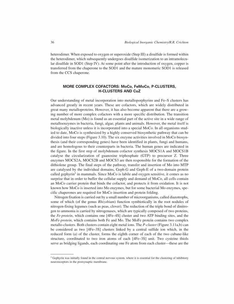
heterodimer. When exposed to oxygen or superoxide (Step III) a disulfide is formed within
the heterodimer, which subsequently undergoes disulfide isomerization to an intramolecu-
lar disulfide in SOD1 (Step IV). At some point after the introduction of oxygen, copper is
transferred from the chaperone to the SOD1 and the mature monomeric SOD1 is released
from the CCS chaperone.
MORE COMPLEX COFACTORS: MoCo, FeMoCo, P-CLUSTERS,
H-CLUSTERS AND CuZ
Our understanding of metal incorporation into metalloporphyrins and Fe–S clusters has
advanced greatly in recent years. These are cofactors, which are widely distributed in
great many metalloproteins. However, it has also become apparent that there are a grow-
ing number of more complex cofactors with a more specific distribution. The transition
metal molybdenum (Mo) is found as an essential part of the active site in a wide range of
metalloenzymes in bacteria, fungi, algae, plants and animals. However, the metal itself is
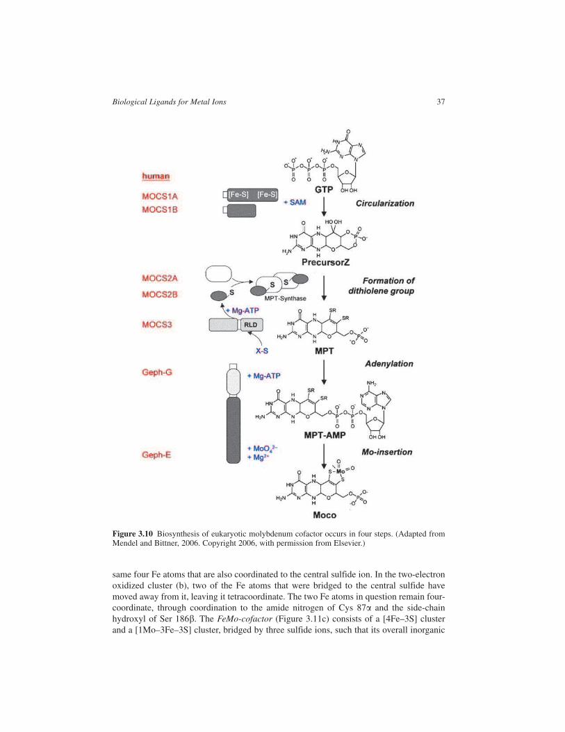
biologically inactive unless it is incorporated into a special MoCo. In all organisms stud-
ied to date, MoCo is synthesized by a highly conserved biosynthetic pathway that can be
divided into four steps (Figure 3.10). The six enzyme activities involved in MoCo biosyn-
thesis (and their corresponding genes) have been identified in plants, fungi and humans,
and are homologous to their counterparts in bacteria. The human genes are indicated in
the figure. In the first step of molybdenum cofactor synthesis MOCS1A and MOCS1B
catalyse the circularization of guanosine triphosphate (GTP) to precursor Z. Three
enzymes MOCS2A, MOCS2B and MOCS3 are then responsible for the formation of the
dithiolene group. The final steps of the pathway, transfer and insertion of Mo into MTP
are catalysed by the individual domains, Geph-G and Geph-E of a two-domain protein
called gephyrin5 in mammals. Since MoCo is labile and oxygen sensitive, it comes as no
surprise that in order to buffer the cellular supply and demand of MoCo, all cells contain
an MoCo carrier protein that binds the cofactor, and protects it from oxidation. It is not
known how MoCo is inserted into Mo enzymes, but for some bacterial Mo enzymes, spe-
cific chaperones are required for MoCo insertion and protein folding.
Nitrogen fixation is carried out by a small number of microorganisms, called diazotrophs,
some of which (of the genus Rhizobium) function symbiotically in the root nodules of
nitrogen-fixing legumes (such as peas, clover). The reduction of the triple bond of dinitro-
gen to ammonia is carried by nitrogenases, which are typically composed of two proteins,
the Fe-protein, which contains one [4Fe–4S] cluster and two ATP binding sites, and the
MoFe-protein, which contains both Fe and Mo. The MoFe protein contains two complex
metallo-clusters. Both clusters contain eight metal ions. The P-cluster (Figure 3.11a,b) can
be considered as two [4Fe–3S] clusters linked by a central sulfide ion which, in the
reduced form (a) of the cluster, forms the eighth corner of each of the two cubane-like
structure, coordinated to two iron atoms of each [4Fe–3S] unit. Two cysteine thiols
serve as bridging ligands, each coordinating one Fe atom from each cluster—these are the
36 Biological Inorganic Chemistry/R.R. Crichton
5 Gephyrin was initially found in the central nervous system, where it is essential for the clustering of inhibitory
neuroreceptors in the postsynaptic membrane.
same four Fe atoms that are also coordinated to the central sulfide ion. In the two-electron
oxidized cluster (b), two of the Fe atoms that were bridged to the central sulfide have
moved away from it, leaving it tetracoordinate. The two Fe atoms in question remain four-
coordinate, through coordination to the amide nitrogen of Cys 87� and the side-chain
hydroxyl of Ser 186�. The FeMo-cofactor (Figure 3.11c) consists of a [4Fe–3S] cluster
and a [1Mo–3Fe–3S] cluster, bridged by three sulfide ions, such that its overall inorganic
Biological Ligands for Metal Ions 37
Figure 3.10 Biosynthesis of eukaryotic molybdenum cofactor occurs in four steps. (Adapted fromMendel and Bittner, 2006. Copyright 2006, with permission from Elsevier.)

composition is [1Mo–7Fe–9S]. The cofactor is bound to the protein by one Cys and one
His residue at either end of the structure, and the Mo ion is coordinated approximately
octahedrally by three sulfide ions from the cofactor itself, the terminal imidazole nitrogen
of the histidine residue of the protein and two oxygens from a molecule of the unusual tri-
carboxylic acid homocitrate, which is an essential component of the cofactor. The com-
plexity of the enzyme systems required to synthesize both the P-cluster and the FeMoCo
cluster, together with the proteins required for their insertion into functionally active nitro-
genase, have combined to render the biotechnological dreams of cloning nitrogen fixation
into other crop plants an illusion.
Yet another organic cofactor of extraordinary complexity is represented by the H-cluster
of microbial hydrogenases. In this case, an unusual coordination of cyanide and carbon
monoxide ligands to a metal centre was found, notably by spectroscopic methods, since
the electron density of carbon, nitrogen and oxygen cannot easily be differentiated by
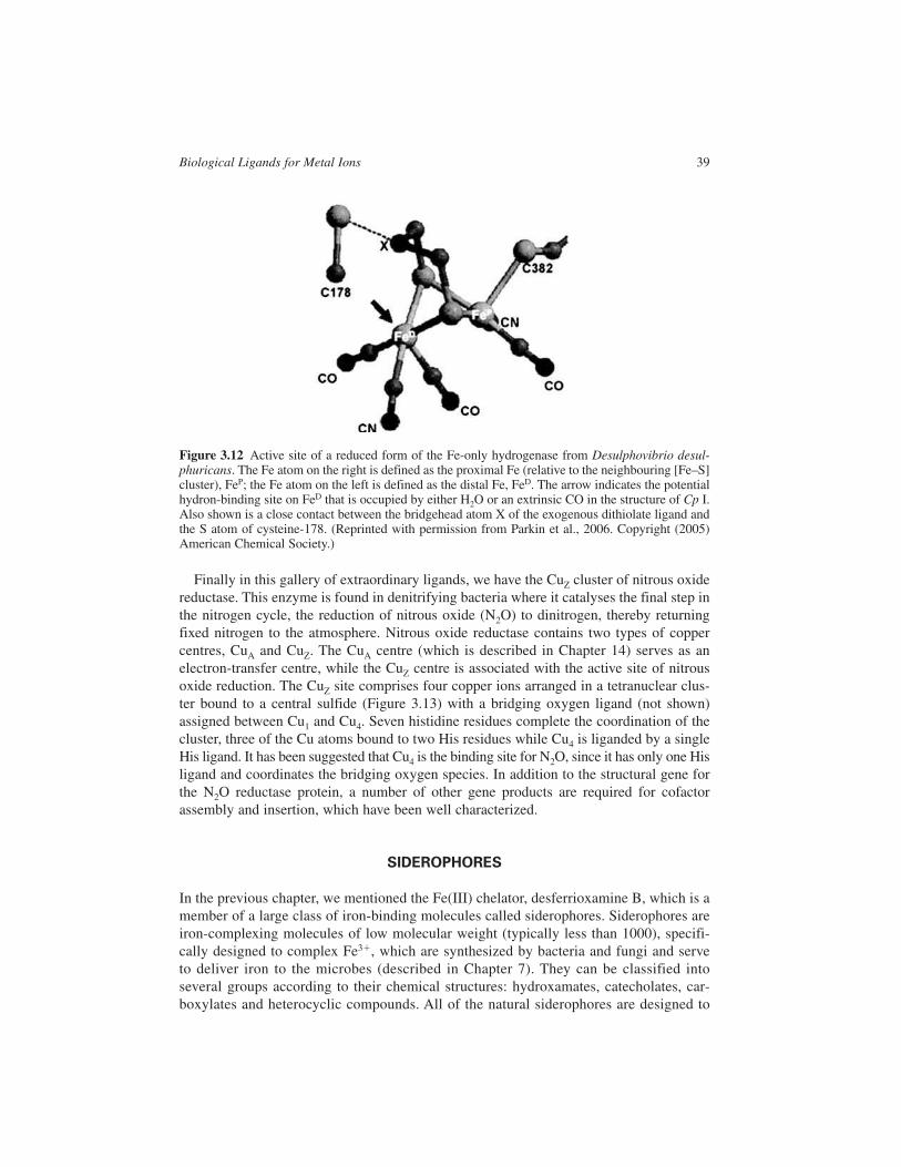
X-ray crystallography. This is illustrated in Figure 3.12 for the Fe-only hydrogenase from
Desulfovibrio desulfuricans. The two active site Fe atoms are each coordinated by one CO
and one CN ligand and are bridged by an unusual organic 1,3-dithiolate ligand. The Fe
atom designated FeP is bound to the protein by a cysteine residue, which is itself bridged
to a 4Fe–4S cluster. The other, FeD has a second CO ligand bound in the reduced form,
which changes to become a bridging ligand upon oxidation. The nature of the bridgehead
atom (C, N or O) in the 1,3-dithiolate ligand could not be determined unequivocally from
the X-ray data, and is therefore shown as X in the figure.
38 Biological Inorganic Chemistry/R.R. Crichton
Figure 3.11 (a) and (b) the P-cluster of nitrogenase in its reduced and oxidized state and (c) theFeMo-cofactor. The molecules are represented with C green, N blue, O red, S yellow, Fe orange andMo pink. (From Voet and Voet, 2004. Reproduced with permission from John Wiley & Sons., Inc.)
Finally in this gallery of extraordinary ligands, we have the CuZ cluster of nitrous oxide
reductase. This enzyme is found in denitrifying bacteria where it catalyses the final step in
the nitrogen cycle, the reduction of nitrous oxide (N2O) to dinitrogen, thereby returning
fixed nitrogen to the atmosphere. Nitrous oxide reductase contains two types of copper
centres, CuA and CuZ. The CuA centre (which is described in Chapter 14) serves as an
electron-transfer centre, while the CuZ centre is associated with the active site of nitrous
oxide reduction. The CuZ site comprises four copper ions arranged in a tetranuclear clus-
ter bound to a central sulfide (Figure 3.13) with a bridging oxygen ligand (not shown)
assigned between Cu1 and Cu4. Seven histidine residues complete the coordination of the
cluster, three of the Cu atoms bound to two His residues while Cu4 is liganded by a single
His ligand. It has been suggested that Cu4 is the binding site for N2O, since it has only one His
ligand and coordinates the bridging oxygen species. In addition to the structural gene for
the N2O reductase protein, a number of other gene products are required for cofactor
assembly and insertion, which have been well characterized.
SIDEROPHORES
In the previous chapter, we mentioned the Fe(III) chelator, desferrioxamine B, which is a
member of a large class of iron-binding molecules called siderophores. Siderophores are
iron-complexing molecules of low molecular weight (typically less than 1000), specifi-
cally designed to complex Fe3�, which are synthesized by bacteria and fungi and serve
to deliver iron to the microbes (described in Chapter 7). They can be classified into
several groups according to their chemical structures: hydroxamates, catecholates, car-
boxylates and heterocyclic compounds. All of the natural siderophores are designed to
Biological Ligands for Metal Ions 39
Figure 3.12 Active site of a reduced form of the Fe-only hydrogenase from Desulphovibrio desul-phuricans. The Fe atom on the right is defined as the proximal Fe (relative to the neighbouring [Fe–S]cluster), FeP; the Fe atom on the left is defined as the distal Fe, FeD. The arrow indicates the potentialhydron-binding site on FeD that is occupied by either H2O or an extrinsic CO in the structure of Cp I.Also shown is a close contact between the bridgehead atom X of the exogenous dithiolate ligand andthe S atom of cysteine-178. (Reprinted with permission from Parkin et al., 2006. Copyright (2005)American Chemical Society.)

selectively chelate Fe(III), which under aerobic conditions is the predominant and poten-
tially bioinaccessible form of iron in the environment. This means that they usually con-
tain hard O-donor atoms as ligands, and form thermodynamically extremely stable
complexes with Fe(III). Examples are given in Figure 3.14. Ferrichrome (pFe � 25.2)6,
first isolated from the smut mould Ustilago in 1952, the best characterized of the
hydroxamate siderophores, has a cyclic hexapeptide backbone to which are attached
three molecules of N-acyl-N-hydroxy-L-ornithine. Enterobactin (pFe � 35.5), the pro-
totype of the catecholate siderophores, is the principal siderophore produced by
Eschericia coli. It is a cyclic triester of dihydroxybenzoyl-serine. When enterobactin
binds iron, the six deprotonated hydroxyl groups of the dihydroxybenzoyl (or cate-
cholate) functions wrap around the metal ion at the centre of the molecule (Figure 3.15).
It is clear from this figure that recognition of the ferric enterobactin does not involve
recognition of the metal, and that the receptor will have no difficulty distinguishing
between the apo- and the ferri-enterobactin. Staphyloferrin A, the iron-transporting
siderophore of Staphylococci, contains a D-ornithine backbone to which two citric acid
residues are linked, which are clearly involved in Fe(III) binding. Yersiniabactin, an
example of a heterocyclic siderophore, is from the highly pathological Yersinia family.
In ferric–yersiniabactin the iron atom is coordinated by the three nitrogens and three
negatively charged oxygen atoms, arranged in a distorted octahedral arrangement.
40 Biological Inorganic Chemistry/R.R. Crichton
Figure 3.13 Structure of the CuZ centre in nitrous oxide reductase. The central sulfide interacts withall four copper atoms. (From Rees, 2002. Copyright 2002 Annual Reviews.)
6 pFe as defined in Chapter 2.

Biological Ligands for Metal Ions 41
Figure 3.14 Chemical structures of selected siderophores to demonstrate the four major structuralclasses.
Figure 3.15 Iron incorporation into apo-enterobactin.

The importance of iron for a bacteria-like E. coli can be illustrated by fact that 14 genes
alone are required for enterobactin-mediated iron uptake, including those for its synthesis,
export, transport of the ferric–enterobactin back into the cell and iron release (Figure 3.16).
In total, E. coli has at least 8 uptake systems for iron, encoded by some 50 genes.
REFERENCES
Al-Karadaghi, S., Franco, R., Hansson, M., Shelnutt, J.A., Isaya, G. and Ferreira, G.C. (2006)
Chelatases: distort to select? TIBS, 31, 135–142.
Culotta, V.C., Yang, M. and O’Halloran, T.V.O (2006) Activation of superoxide dismutases: putting
the metal to the pedal, Biochim. Biophys. Acta, 1763, 747–758.
Lill, R., Duftkiewitz, R., Elsässer, H.-P., Hausmann, A., Netz, D.J.A., Pierik, A.J., Stehling, O.,
Urzika, E. and Mühlenhoff, U. (2006) Mechanisms of iron–sulfur protein maturation in mito-
chondria, cytosol and nucleus of eukaryotes, Biochim. Biophys. Acta, 1763, 652–667.
Lill, R. and Mühlenhoff, U. (2005) Iron–sulfur protein biogenesis in eukaryotes, TIBS, 30, 133–141.
Lill, R. and Mühlenhoff, U. (2006) Iron–sulfur protein biogenesis in eukaryotes: components and
mechanisms, Annu. Rev. Cell Dev. Biol., 22, 457–486.
Lippard, S.J. and Berg, J.M. (1994) Principles of Bioinorganic Chemistry, University Science Books,
Mill Valley, CA, 411 pp.
Mason, A.B., Halbrooks, P.J., James, N.G., Connolly, S.A., Larouche, J.R., Smith, V.C.,
MacGillivray, R.T.A. and Chasteen, N.D. (2005) Mutational analysis of C-lobe ligands of human
serum Transferrin: insights into the mechanism of iron release, Biochemistry, 44, 8013–8021.
Mendel, R.R. and Bittner, F. (2006) Cell biology of molybdenum, Biochim. Biophys. Acta, 1763,
621–635.
Parkin, A., Cavazza, C., Fontecilla-Camps, J.C. and Armstrong, F.C. (2006) Electrochemical
investigations of the interconversions between catalytic and inhibited states of the [FeFe]-
hydrogenase from Desulphovibrio desulfuricans, JACS, 128, 16808–16815.
Rao, P.V. and Holm, R.H. (2004) Synthetic analogues of the active sites of iron–sulfur proteins,
Chem. Rev., 104, 527–559.
Rees, D.C. (2002) Great metalloclusters in enzymology, Annu. Rev. Biochem., 71, 221–246.
Voet, D. and Voet, J.G. (2004) Biochemistry 3rd edition, John Wiley & Sons, Chichester.
42 Biological Inorganic Chemistry/R.R. Crichton
Figure 3.16 The genes involved in enterobactin-mediated iron uptake in E. coli.