biological and chemical aspects of agrocin 434 as a ... · summary the involvement of factors other...
TRANSCRIPT

ñ{þ\
4*l-Ð ^
u..¿^
q,$tlllFUS
.di1 'l'. 5 -7 sEP ]t$fi
BIOLOGICAL AND CHEMICAL
AS A ST]PPLEMENTARY BIOCONTROL AGENT
F'OR CRO\ryN GALL
NORMA N. FAJARDO
M.Sc. (University of the Philippines at Los Baños)
A thesis submitted for the degree of Doctor of Philosophy in the Faculty of Agricultural
and Natural Resource Sciences at the University of Adelaide
Departments of Crop Protection and Plant Science
Waite Campus
The University of Adelaide
May 1995

i;,j .t I 3
TABLE OF CONTENTS
Summary
Declaration
Acknowledgments
Abbreviations
CHAPTER 1 General Introduction
Review of Literature
1.1 Introduction
1.2 The genus Agrobacterium
1.3 The role of plasmids in crown gall induction
1.4 The molecular mechanism of pathogenesis
1.4.1 Chemotaxis and attachment
1.4.2 T-DNA processing and transfer
1.4.3 T-DNA integration and expression
1.5 Biological control of crown gall
1.5.1 The K84 system
1.5.2 Other potential biological control agents
Aims and Scope of Study
CHAPTER 2 Screening Agrobacteria for Inhibition by Agrocin 434
Innoduction
Materials and Methods
Results and Discussion
2.1 ScreeningAgrobacterium strains for sensitivity to agrocin434
2.2 F;ffectof media on sensitivity to agrocin434
2.3 Effect of media and pH on agrocin bioassays
Conclusions
I
1V
V
vi
I
1
1
2
3
4
4
5
7
9
9
72
T4
15
15
t6
23
23
26
29
32

CHAPTER 3 Growth of Strain K1143 and Agrocn434 Production in a
Chemically Defined Medium
Introduction
Materials and Methods
Results and Discussion
3.1 Growth of strain Kl143 in MMG
3.2 Agrocin434 Production in MMG
Conclusions
CHAPTER 4 Restriction Endonuclease Mapping of Plasmid pAgK1318
Introduction
Materials and Methods
Results and Discussion
4.1 Isolation and purification of plasmid DNA
4.2 Genetic mapping
Conclusions
CHAPTER 5 Agrocin 434: Its Uronic Acid Component
Introduction
Materials and Methods
Results and Discussion
5.1 Isolation and purification of agrocin 434
5.2 Identification of the uronic acid component of agtocin 434
5.3 Determination of the nature of linkages
Conclusions
CHAPTER 6 General Discussion
33
33
34
36
36
37
38
39
39
40
45
45
46
54
55
55
56
59
59
60
62
64
65

References
Appendices
Appendix A Bacterial media and antibiotics
Appendix B Miscellaneous buffers and solutions
Appendix C Publications arising from this thesis
72
9l
9l
98
101

SUMMARY
The involvement of factors other than agrocin 84 in the biological control of
crown gall by Agrobacterium struns K84 and K1026 has been suggested in a number of
studies. This thesis examines the possible role in the biological control process of an
antibiotic compound, agrocin 434, produced by the biocontrol strains in addition to the
well-chara cterized adenine nucleotide agrocin 84.
A total of 66 Agrobacterium isolates from Australia, Europe and North
America were tested for sensitivity to agrocin 434 using standard agrocin bioassay
methods. All strains of biovar 2 tested were sensitive to agrocin 434 with the exception
of 6 strains which produced agrocin 434 themselves. These included K84 and two
agrocin 434-producing derivatives plus three isolates from New South Wales which were
subsequently shown to be pathogenic. In contrast, all strains of biovars 1, 3 and A. rubi
tested were resistant to agrocin 434. Selected isolates representing these three groups
were also tested for agrocin 434 production to check if the resistance could be due to the
production of this agrocin. None of those strains produced agrocin 434. lt appears that
sensitivity to agrocin 434 is a general characteristic of biovar 2 agtobacteria and may, at
least in part, be chromosomally encoded.
It was also shown that media had an influence on the expression of
sensitivity to agrocin 434. \Vhether this is due to inactivation of the agrocin, or to
changes in the test strains brought about by growing on a particula¡ medium remains to
be clarified.
The genes involved in the biosynthesis of agrocin 434 are encoded on a large
plasmid (300-400 kb) carried by K84 and some of its derivatives (Donner et aL,1993)'
A derivative of K84 (strain Kl143) which carries only this large plasmid, designated

ll
pAgK434, has been isolated (Donner et aI., 1993). A buffered glucose-glutamate
medium was found to support both the growth of Kl143 and agrocin 434 production.
Agrocin 434 was produced throughout the growth cycle even though its antibiotic
activity is characteristic of a secondary metabolite. Agrocin production did not appear to
be regulated by glucose.
Another derivative of K84, strain K1318, carries a deleted version of
pAgK434 (the designation pAgK1318 is recommended). This strain produces a
biologically inactive fragment of agrocin 434, designated nucleoside 4176, suggesting
that part of the agrocin 434 biosynthetic pathway is encoded on pAgK1318 and that
genes for this would most probably be located in the vicinity of the deletion. The
smaller size of pAgK1318 (30-90 kb) facilitates the localization of genes involved in
agrocin 434 synthesis as compared to the much larger pAgK434. On the assumption that
the relevant genes are clustered in a manner similar to the agrocin 84 synthesis genes,
mapping of pAgK1318 was undertaken. Conditions were optimized for the preparation
and purification of plasmid DNA from strain K1318. A protocol for the minipreparation
of plasmid DNA by the alkaline lysis method was scaled up and DNA was recovered
from low melting point agarose gels using the enzyme agarase. The procedure is simple,
fast and very reliable, yielding high quality DNA useful for most recombinant DNA
applications.
XbaI digests of plasmids pAgK13l8 and pAgK434 were compa¡ed and the
results supported the assumption that a single large deletion event had given rise to
plasmid pAgK1318 (McClure NC, unpublished). A novel 10.5 kb Xbalftagment of
pAgK1318 encompasses the site of deletion and could be expected to contain some of
the genes that encode the production of the biologically inactive agrocin 434 fuagment.
A physical map of the region inclusive of, and flanking, the novel 10.5 kb Xbalfragment
was constructed with respect to the restriction endonucleases BamHI, Hindlfl and SmaI

lll
using the techniques of molecular cloning, DNA-DNA hybridization, and multiple and
partial endonuclease digestions.
A provisional structure had been proposed for agrocin 434 as a disubstituted
cytidine nucleoside (Donner SC, unpublished). The substituent at,¡. ¡r{4-position of
cytidine was described as a p-l,4-hexuronosyl-glucopyranosyl moiety. The nature of the
uronic acid component and its linkage remained to be established. Strong acid
hydrolysis of agrocin 434 yielded glucuronic acid and glucose by electrophoretic
comparisons with authentic samples. Sodium borohydride reduction of the acid
hydrolysate yielded products whose electrophoretic properties in alkaline, as well as in
borate and zinc complexing buffers, were identical with the reduction products of
glucuronic acid and glucose. NMR studies have confirmed the nature of the linkages.
The available evidence is consistent with a B-1,4-glucuronopyranosyl-cr-1,N4-
glucopyranosyl substituent on the cytidine moiety of the nucleoside.

lv
DECLARATION
This work contains no material which has been accepted for the award of any other
degree or diploma in any university or other tertiary institution and, to the best of my
knowledge and belief, contains no material previously published or written by another
person, except where due reference has been made in the text.
I give consent to this copy of my thesis, when deposited in the University Library, being
available for loan and photocopying
Norma N. Fajardo
May 1995

v
ACKNOWLEDGMENTS
I am deeply grateful to my supervisors, Dr. Max E. Tate, Dr. Nicholas C'
McClure and Prof. otto schmidt, for their guidance, encouragement and support
throughout the course of this study. I also wish to thank Dr. Bruce G. Clare, my former
supervisor, for his encouragement and continued keen interest in my work in spite of his
retirement.
I am grateful to Dr. David Liljegren for his help with university matters.
I would also like to thank Dr. Peter J. Murphy and Scott Donner for helpful
discussions, Dr. Graham Jones for advice on cystallization and Dr. Kith Jayasena for
assistance during manuscript preparation'
I sincerely thank the former members of Bruce Clare's lab, Garry Rosewarne
and Natalie Dillon, for their help and friendship.
For assistance with laboratory and departmental matters, I am indebted to
Terry Feckner, Gary Taylor, Anke Johnsen, and Emma Cabot'
I wish to thank Dr. Bob Brownlee of La Trobe University for NMR analysis
of agrocin samples.
I thank the many friends I have made in the Departments of Crop Protection
and plant Science for their invaluable support and the warmth of their company.
Financial support from AusAid is gratefully acknowledged. I also thank the
government of the Republic of the Philippines and, in particular, the University of the
Philippines for giving me the opportunity to study in Australia'
Finally, I wish to express my deepest gratitude to my family for their love
and support. I especially thank my husband, Freddie, and our four children, Brian,
Marvin, yasmin and Alvin. Their love has inspired and sustained me through the most
difficutt times in my studY.

vl
ABBREVIATIONS
Amp
cm
DNase
DIG
FW
('Þ
TIVPE
IR
Km
kb
KV
I
I
mg
ml
fnm
mg
rtl
mM
min
M
nm
NMR
OD
RM
RNase
Ampicillin
centimeter
deoxyribonuclease
digoxigenin
formula weight
gram
high voltage paper electrophoresis
infrared
kanamycin
kilobase
kilovolt
lambda
liter
microgram
microliter
micrometer
milligram
milliliter
millimolar
minute
molar
nanometer
nuclear magnetic resonance
optical density
relative mobilitY
ribonuclease

vll
SDS
T-DNA
Ti
Tris
UV
w/v
sodium dodecyl sulfate
transferred-DNA
tumor-inducing
tris(hydroxymethyl) aminomethane
ultraviolet
weight per unit volume

å
i
It:
CHAPTER ONE
GENERAL INTRODUCTION
REVIEW OF LITERATURE
1.1 INTRODACTION
Crown gall is a plant cancer which causes significant international losses to
crops such as stone fruit, grapes, pome fruit, nuts, roses and other ornamentals (Schroth
and Moller, 1976; Moore and W'arren, 1979; Panagopoulos st al., 1979; Alconero,
1980; Kennedy and Alcorn, 1980; Ken, 1989). The disease is characterized by tumor
formation at infected wound sites, usually at the crown (hence the name crown gall) or
on the root system (Figure 1.1). It affects mainly dicotyledonous plants and a few
gymnosperms (Lippincott and Lippincott, 1975; De Cleene and De Ley, 1976).
Monocots appear to be naturally resistant but some have been shown to be susceptible
following artificial inoculation (Hooykaas-Van Slogteren et al., 1984; Graves and
Goldman, 1986; Grimsley et a1.,1987).
Since 1907 when Smith and Townsend first reported the bacterial origin of
this neoplastic disease of plants (Braun, 1982), a vast amount of research has been
undertaken worldwide in order to understand the molecular mechanism of crown gall
pathogenesis. The momentum for such research has been sustained by advances in
molecular biotogy and genetic techniques which have revolutionized the understanding
of the plant-bacterial interactions involved in crown gall induction. Key steps in the
process are now known although there are many facets of the pathogenesis which are
poorly understood.

Figure 1.1 Crown gall on almond induced by pathogenic strain K27. Artows indicate
galls. (Photo courtesy of Ali Reza Ahmadi)

Ð

,)
A biological control system (Kerr, 1980; Kerr, 1989; Jones and Kerr, 1989;
Fa¡rand, 1990; Clare, 1993) developed against the disease has found acceptance in many
countries a¡ound the world. This biological control system is based on the activities of a
non-pathog erllc Agrobacterium strain, K84. Evidence has gradually accrued (cooksey
andMoore,1982; Shim¿r a1.,1987; Macrae eta1.,1988; Lopezetal',1989; Vicedoet
a1.,1993; Farrand, 1990; Farrand and Wang, 1992) that the'full range of mechanisms
by which this extraordinarily successful agent inhibits pathogenesis still remains to be
clarified. Examination of the possible role of factors other than agrocin 84 in the
biocontrol system forms a central part of this thesis'
1.2 THE GENT]S AGROBACTBRIUM
Crown gal| is caused by pathogenic strains of. Agrobacterium, a soil
microorganism which belongs to the family Rhizobiaceae along with Rhizobium,
Brad.yrhizobium and Phyllobacterium (Jordan, 1984). Members of the genus are Grarn-
negative, single-celled, non-sporeforming rods motile by means of a few peritrichous
flagella (Ken and Brisbane, 1983). Although all strains grow between 20 and 28oC' the
optimum temperature for growth is 25-28oC.
The taxonomy of agrobacteria has been the subject of controversy and much
of it stems from the use of species epithets that are closely associated with unstable
pathogenic characteristics (Youn g et al., 1992). For example, the names A' tumefaciens
and A. rad,iobacter have been applied to strains which differ only in the presence or
absence, respectively, of a tumor-inducing (Ti) plasmid which can be either lost or
transferred between strains (Ken, t969; Van Lareb eke et a1., 1975). Another example is
the association of the names A. tumefaciens and A. rhizogenøs with tumor-inducing and
root-inducing capacity, respectively, again emphasizing transient pathogenic attributes'
Even without such a distinction, these two are considered separate species on the basis of
phenotypic characteristics and DNA relatedness (Holmes and Roberts, 1981; Kersters
and De Ley, 1984). If names are considered without regard to their descriptive

3
connotations, there are four valid species, namely , A. tumefaciens, A. rhiZogenes, A' vitis
andA. rubi (Ophel and Kerr, 1990; Young et al., L992)'
To avoid the confusion in the nomenclature of Agrobacterium, there has been
a strong tendency to use biovar designations which are based on characteristics that are
chromosomallydetermined(Kerr, 1992; Clare, 1995). Biovars l,2and3correspondto
A. tumefaciens, A. rhizogenes and A. vilis, respectively, while A' rubi has no biovar
designation (ophel and Kerr, 1990; Kerr,1992). Agrobacteria can thus be referred to as
biovar l-3 strains or A. rubiwithout regard to their pathogenic characteristics. Recently,
on the basis of 165 ribosomal RNA (rRNA) studies, Sawada et aI. (1993) proposed the
replacement of A. tumefaciens by A. radiobacter as the valid species epithet for biovar 1
strains.
In nature, agrobacteria have been found to have specific host associations
(Ophel and Kerr, 1937). White biovar 1 strains have been associated with a wide range
of dicotyledonous hosts, biovar 2 strains have mostly been found in stone fruit. Biovar 3
strains affect grapevines virtually exclusively while A. rubí have been isolated only from
Rubus spp. (ophel and Kerr, 1987; Ker'"., 1992). However, many agrobacteria have
been shown to have a wider host range in artificial inoculations (Ophel and Kerr, 1987)'
1.3 THE ROLE OF PIA.SMIDS IN CROWN GALL INDUCTION
Crown gall is a neoplastic disease that results from the stable integration of a
discrete segment of a large plasmid (the tumor-inducing or Ti-plasmid) into the nuclear
genome of the infected plant. It was several decades after the bacterial nature of the
disease was recognized before the Ti-plasmid was finally identified as the tumor-
inducing principle. A key discovery was made by Kerr (1969) who reported that
virulence could be transferred to non-pathogenic isolates in a developing crown gall
tumor suggesting the possibility that a transmissible extrachromosomal element
determined pathogenicity. zaenen et aI. (1974) later found that virulent strains of
agrobacteria carry one or more large plasmids and suggested that genetic information for

4
crorvn gall induction could reside on one or several of those plasmids' The role of the
Ti-plasmid in crown gall induction was firrnly established when van Larebeke et aI'
(1975) showed that the acquisition of the large plasmid from pathogenic strains was the
reason why non-pathogenic isolates became virulent in mixed infections in planta' It
was subsequently demonstrated that only a portion of the Ti-plasmid was transferred
from pathogenic agrobacteria to plant cells (Chiltoî et at', 1977) where it is stably
integrated into the host nucleus (Yadav et a1.,1980)'
I.4THEMùLE1UI.A,RME0HANISMoFPATHOGENESIS
Since the discovery of the relationship between Tiplasmid and pathogenicity
much work has gone into understanding the molecular mechanism that leads to the
tumorigenic phenotype. This mechanism is now known in some detail and is the subject
of several recent reviews (Winans, 1992; Zambryski,1992; Clare and McClure' 1995)'
Several discrete steps are involved in crown gall induction. The pathogenic
process is initiated by bacterial recognition and attachment to susceptible wounded plant
tissues. Then a sequence of events leads to the transfer of a specific segment of the Ti-
plasmid (the transfened DNA or T-DNA) to the plant cell nucleus where it becomes
covalently integrated. The genes on the T-DNA are expressed in the plant cells' As well
as expressing functions which result in the tumorigenic growth, T-DNA genes also
encode the synthesis of a novel group of compounds termed opines which not only
provide the pathogen with an additional source of nutrients but also ensure the
organism,s spread and survival by inducing conjugal transfer of Ti-plasmids (Binns and
Thomashow, 1988; Zarnbryski, 1988; Cla¡e, 1990)'
1.4.1 Chemotaxß and Attachtnent
The initial event in crown gall induction is a positive chemotactic response to
plant exudates, mainly phenolic compounds and certain sug¿us, released by wounded
plant tissue (Stachel et a1.,1985; Ashby et al., 1987; Ashby et a1.,1988;)' Two levels

5
of chemotaxis towards plant metabolites have been demonstrated, one chromosomally
encoded and the other Ti-plasmid dependent (Ashby et aL, 1987; Parke et aI'' 1987;
Ashby et a1.,1988; Shaw et al., 1988).
The next step in the process is attachment of the bacteria to the plant tissue, a
process which necessarily involves bacterial cell-plant cell recognition' Little is known
about plant components required for attachment but agrobacterial components have been
identified. Three chromosomal virulence loci, chvA and chvB (Douglas et al., 1985) and
pscA (Thomashow et aI., tg87) have been shown to encode products required for
bacterial attachment during the infection process. The chvB locus appears to be involved
in the synthesis of a p-l,2-glucan which has been implicated in plant cell binding
(Zorreguieta et aI., 1988), while chvA may encode a transport function for the
polysaccharide (cangelosi et at.,1989). The pscA locus, shown to be related to the exoc
locus of Rhizobium melilotl (Marks et aL,1987), encodes a gene product which may
have a role in the production of surface polysaccharides important for attachment to plant
cells. V/hether this role is a direct one or not remains to be clarified (Thomashow et al',
1987).
Two other genetic loci have been implicated in bacterial attachment. The cel
locus is important in cellulose biosynthesis (Robertson ef al', 1988) while the closely-
linked att Eenes encode cell surface polypeptides which may play a role in bacterial
binding to plant cells (Matthyse, 1987; Robertson et a1.,1988). Mutations in these loci
lead to either loss or attenuation of virulence.
1.4.2 T-DNA Processing and Transfer
Mutational and transcriptional studies have confirmed that genes clustered in
a 40-kb segment of the Ti-plasmid known as the vir region afe responsible for the
excision and transfer of T-DNA ftom Agrobacterium to the plant cell nucleus' The vir
region comprises about 25 genes arranged in either 6 or 7 operons (virA-virG) which are
co-regulated thus forming a regulon (Winans, 1992; Hooykaas and Schilperoort' 1992)'

6
The majority of the vir genes are not expressed at detectable levels until they are
induced, in contrast to the known chromosomal virulence genes which are constitutively
expressed. The phenolic compounds acetosyringone and cr-hydroxyacetosyringone were
the first compounds to be identified as inducers of the vir regulon (Stachel et aI',1985)'
In addition to these two, other compounds such as monosaccharides, their derivatives
and analogues have subsequently been implicated invir gene induction (Veluthatrtbi et
al., L989; Ankenbauer and Nester, 1990; Cangelosi et a1.,1990; Shimoda et al', 1990)'
The vir gene regulatory system operates through 2 monocistronic genes, virA
and virG,which resemble genes of other two-component prokaryotic regulatory systems
such as envZ-ompR, ntrB-ntC and dctB-dctD (Nixon et aI., 1986; Parkinson and
Kofoid, Igg2). The constitutively expressed vdrA probably acts as a chemoreceptor for
plant wound metabolites and transmits this signal to the bacteria by activation (probably
by phosphorylation) of the VirG protein. VirG then activates the transcription of the
other yir genes (Stachel andZarú)ryski, 1986; Jin et aI., L99Oa Jin et ø/', 1990b; Turk
et al.,lgg4). products of the virB, C,D and E loci have been ascribed functions relating
to the cleavage of T-DNA at the border sequences and its processing and transfer to the
host. The virll (also known as pin\ is generally not essential for virulence but may be
involved in the protection of the bacteria during the infection process (Zambryski ' 1992)'
Experiments have shown that the 25bp imperfect direct repeats (LB and RB)
flanking the T-DNA on the Ti-plasmid are important in the transfer process (Yadav ef
aI., 1982; Zambryski et aI., lg82). In a concerted action proteins encoded by virD
recognize and cleave the bottom strand of the 25 bp border repeat sequence (Yanofsky ef
at.,1986). The nicked borders are then used for the generation of T-strands, molecules
considered to be the T-DNA intermediates that are transported from the bacteria into the
plant (Stachel et aI.,1986). In later experiments, it was established that the right border
alone is essential for the transfer of genes (rWang et aI',1984)' In octopine-type Ti-
plasmids there is, in addition to the 25bp border sequences' a specific 24bp sequence
called ,,overdrive " at !3-14 bp from the right border sequence which enhances T-DNA
transfer (Peralta et a1.,1986; Van Haaren et a1.,1987). Evidence indicates that VirCl

7
can bind to the overdrive sequence and that this interaction enhances T-strand production
(Toro et a1.,19S9). There is no sequence that closely resembles the octopine "overdrive"
near nopaline borders (Wang et aI.' 1987).
As the first step in T-strand transport is traversing the bacterial membrane,
some vir functions must provide for alteration of the bacterial membrane to facilitate T-
strand passage. Since most of the 1l virB open reading frames have sequences
characteristic of genes encoding either transmembrane or membrane-associated proteins,
VirB proteins are thought to form a structure (pore or channel) through which the DNA
is delivered into the plant cell (Kuldau et a1.,1990; Shirasu et aI',1990)'
The VirE proteins are likely candidates as components of the T-complex that
is transported to the plant cell nucleus. This is consistent with their being the most
abundant products of. vir induction (Citovsky et a1.,1988; Zambryski, 1992) and the
finding that the VirE2 polypeptide binds single-stranded DNA (Christie et aI., 1988)'
Experimental evidence suggest that the transferred form of the T-DNA is single-stranded
(Stachel et a1.,1986; Winans, 1992; Zambryski,1992; Tinland et aI',1994)' However'
double-stranded molecules may also be produced (Dürrenberger et al., 1989) and could
possibly play a role in the natural transformation process (Tintand et a1.,1994).
1.4.3 T-DNA Integration and Expression
Integration of the T-DNA into the plant nuclear genome results in the
expression of oncogenicity (onc) genes which code for the production of phytohormones'
The genes iaaM and iaaH encode enzymes' a monooxygenase and a hydrolase, which
together catalyze the conversion of tryptophan via indoleacetamide (IAM) into indole
acetic acid (IAA), a compound with auxin activity.(Thomashow et aI', 1984;
Thomashow et a\.,1986; Schroder et aI.,l9S4). The ipt gene codes for an isopentenyl
transferase, an enzyme which catalyzes the conversion of AMP into isopentenyl-AMP, a
compound with cytokinin activity (Akiyoshi et a1.,1984; Barry et al',1984)' Elevated

8
levels of these phytohormones give rise to uncontrolled cell proliferation and tumor
formation (Van Onckelen et al., 1984).
As well as expressing the onc genes, the T-DNA also directs the synthesis
(Holsters et. a1.,1980) of one or more of a novel group of amino acid derivatives
collectively called opines (Tempé and Goldman, 1982). The opines induce conjugal
transfer of Ti-plasmids among agrobacteria and also encode gene products required for
opine catabolism (Dessaux et al., 1992; Dessaux et al., 1993). Even before the
discovery of Ti-plasmids and T-DNA it had already been hypothesized, based on early
studies on opines, that the transfer of a portion of the bacterial genome into the plant cell
could be responsible for crown gall. The hypothesis was confirmed with the advent of
recombinant DNA techniques which lent genetic support for the hypothesis (Dessaux ef
al., 1992). That opines play a fundamental role in Agrobacterium-plant interactions -
that of "chemical mediators of parasitism"- is embodied in what is generally known as
the Opine Concept (Tempé and Petit, 1933). In this concept the bacteria sequester the
plant metabolic machinery for the production of compounds that will ensure their
survival and spread. The mechanism by which they achieve this is by genetic
engineering. J. Schell and his associates termed the process genetic colonization (Tempé
and petit, 1933). While initially it was thought that only the inciting bacteria are capable
of utilizing opines, later investigations have shown that microorganisms such as fungi
and other types of bacteria including pseudomonads and coryneform bacteria are also
able to catabolize opines (Beauchamp et a1.,1990; Nautiyal and Dion, 1990)'
In contrast to the vast amount of information that has been generated on the
agrobacterial aspect of DNA transfer, Iittle is known about the process of DNA
integration in the host plant. Willmitzer et aI. (1980) localized the position of the T-
DNA in the nucleus of the transformed ptant cell. The expression of bacterial genes in a
system that is known to recognize a different set of expression signals remained a
mystery for some time. That the T-DNA has eukaryotic properties was first confirmed
by experiments wherein transcription of T-DNA was shown to be inhibited by c[-
amanitin, an inhibitor of RNA polymerase II, suggesting that expression of T-DNA

9
genes depends upon this RNA polymerase (rwillmitzer et al',19S1)' Subsequently' the
complete T-DNA sequence of the octopine Tiplasmid was reported (Barker et a1.,1983)
and made possible a detailed analysis of the 5' and 3' non-coding regions of the T-DNA'
Indeed, eukaryotic expression signals such as the TATA box for transcription initiation
and the AATAAA box for transcription termination and polyadenylation were identified'
However, introns, a feature of many eukaryotic genes' were not found.
The transfer of DNA from Agrobacterium to the plant cell has been likened
to the bacterial conjugation system. Several lines of evidence derived mainly from
studies of incompatibility groups IncPl and IncW plasmids support this analogy
(Zambryski, 1992; Kado, 1993). The most direct support, however, comes from
Buchanan-Wollaston et al.'s (1987) demonstration that the oriT of the conjugative
bacterial ptasmid pRSF1010 can direct DNA transfer to the plant in place of the T-DNA
borders. It has been pointed out, though, that while the early stages of T-DNA
processing and transport may resemble bacterial conjugation, the later steps, particularly
those occurring inside the plant cell, share common features with viral infection
(Zarrrbryski, 1992).
1.5 BIOLOGICAL CONTROL OF CROWN GALL
L5.1 The K84 SYstem
The biological control of certain crown gall pathogens has been practiced on
an international scale for the past twenty years and utilizes a naturally-occurring non-
pathogenic biovar 2 agrobacterial strain, K84. The procedure that Kerr and associates
developed involves dipping planting materials such as cuttings, seeds or seedlings in
suspensions of K84 cells immediately before planting (New and Kerr, L972; Htay and
Ken,1974; Kerr, 1980).
Strain K84 was isolated from soil around an infected peach tree near
Adelaide, South Australia early in the 1970's (New and Kerr, 1972). Since 1973,K84
has been sold commercially in Australia (Jones et a1.,1983) and successful biological

10
control of crown gall on several hosts has been reported from many countries a¡ound the
world (Schroth and Moller, L976; Matthee et al., 1977; Moore, 1977; Moore, 1979;
Moore and'Wa¡ren,1979; Panagopoulos et a1.,1979). It must be emphasized, however,
that K84 is only preventive, not curative.
The rema¡kable success of K84 as a biological control agent stimulated much
interest in elucidating the mechanism(s) by which the control process occurs. Kerr and
Htay (1974) demonstrated that control is achieved through the production of a
bacteriocin which they called bacteriocin 84 (later changed to agrocin 84). Bacteriocins
are antibiotic compounds produced by certain strains of bacteria, which are inhibitory to
other strains of the same or closely related species (Kerr, 1982). Prior to the discovery of
the nucleotide bacteriocins, most definitions have emphasized the proteinaceous nature
of these compounds. Engler et al. (1975) coined the name agrocin to refer to low
molecular weight antibiotic substances produced by certain strains of. Agrobacterium'
which a¡e inhibitory to other strains of agrobacteria. The ma¡ked specificity of agrocins
against closely related strains is a characteristic that they share with the other
bacteriocins.
The genes for the biosynthesis of agrocin 84 a¡e located on a 47 .7 kb plasmid
called pAgK84 (Ellis et aI.,1979; Slota and Farrand, 1982) on which regions coding for
plasmid transfer and immunity to inhibition by agrocin 84 have also been identified
(Fanand et a1.,1985; Ryder et a1.,1937). V/ang et al. (1994), through mutational and
complementation analysis, have demonstrated that the 21-kb region on pAgK84
encoding agrocin 84 synthesis is organized into at least 5 transcriptional groups which
they have named agnA through agnE. They have also shown that the agn geîes arc
expressed in planta giving further support to the hypothesis that, in the soil, agrocin 84
production by strain K84 is important in the control of crown gall caused by sensitive
stains
Most of the studies on the genetics of sensitivity to agrocin 84 have been
carried out on the nopaline Ti-plasmid pTic58. The acc genes on this plasmid encode
transport and catabolism of the opines agrocinopine A and B (Ellis and Murphy, 1981;

1l
Ryder et al., 1984; Hayman and Farrand, 1988; Hayman et al., 1993). Transport of
these opines involves a periplasmic permease which has also been shown to actively take
up agrocin 84 due to its N6-D-glucofuranosyloxyphosphoramidate substituent (Murphy
and Roberts, L979; Ellis and Murphy, 1981; Murphy et al., 1981; Hayman and
Fa¡rand, 1988; Hayman et aL, 1993). Strains wi¡h acc genes are therefore sensitive to
agrocin 84 - this is the basis for the extraordinary selectivity of agrocin 84 for pathogens
carrying a nopaline Ti-plasmid.
Agrocin 84 (Figure 1.2) is a disubstituted adenine nucleotide analogue
(Murphy et al., 1981). From structure-function studies of the molecule, it was
established that uptake of the agrocin was determined by the N6-D-
glucofuranosyloxyphosphoramidate substituent and that toxicity was conferred by the 5'-
phosphoramidate moiety (Tate et a1.,1979; Murphy et a1.,1981).
Little is known about the exact mechanism of action of agrocin 84 in
inhibiting cell growth but Kerr and Tate (1984) have pointed out the structural similarity
of the lower (or alpha) face of the P-D-3'-deoxyarabinofuranosyl group of the agrocin 84
molecule to the dideoxynucleotides used in DNA sequencing reactions for chain
termination. Murphy et aI. (1981) have established that the presence of a fraudulent
nucleotide moiety alone is not sufficient for growth inhibition and that the minimum
moiety of agrocin 84 required for its toxic action is the S'-phosphoryl substituted
nucleoside. In studies using radiolabelled precursors, measurable effects on DNA
synthesis have been observed (McCardell and Pootj es, I976i Das ¿f aI., 1978).
The continued success of K84 as a biological control agent was potentially
threatened by the fact that pAgK84 could transfer conjugatively to tumorigenic strains
which would then acquire immunity to agrocin 84 (Ellis and Kerr, 1979; Panagopoulos
et a1.,1979; Fa¡rand et a1.,1935). Recombinant DNA techniques were used to construct
a deletion mutant of K84 which is unable to transfer its plasmid to other agrobacteria
(Jones et aI.,l9S8). This genetically-engineered derivative, strain K1026, was shown to
be as effective as K84 in preventing crown gall on almond seedlings in open air pot trials
(Jones and Kerr, 1989). Strain KlO26 was subsequently registered as the pesticide

Figure 1.2 Structure of agrocin 84, a 3'-deoxyarabinofuranosyladenine
bisphosphoramidate.

cH2OH
OHHO
oo-ll
-oH
MN,H
/\o-
+N
N
H oil
N- oo
HOM+
OH
AGROCIN 84
cHs

72
product Nogall* and became the first genetically-engineered microbe to be released for
commercial use (Kerr, 1989).
Another possible th¡eat to the effectiveness of both K84 and K1026 is their
acquisition of a Ti-plasmid which would make them agrocin 84 immune pathogens. This
situation is considered highly unlikely because both biocontrol strains carry the nopaline
catabolic plasmid pAtKS4b which belongs to the same incompatibility group as the
nopaline Ti-plasmids of the tumorigenic strains (Clare et a1.,1990). Plasmids belonging
to the same incompatibility group cannot exist within a bacterial cell at the same time.
Agrocin production is conceivably not the sole means by which control of
crown gall is effected by the biocontrol strains. It has been suggested that they are also
able to colonize roots efficiently giving them competitive advantage over their
pathogenic counterparts (Ellis et aI., 1979; Cooksey and Moore, 1982; Shim et al"
L987; Macrae et a1.,1988; Lopez et a1.,1989; Vicedo et a1.,1993)' However, Farrand
and Wang (lgg2) pointed out that there is a need for a more critical assessment of the
role of root colonization as the parameters involved are not clearly understood. They
also argued that there is a lack of consensus even on the defînition of colonization.
1.5.2 Other Potential Biological Control Agents
Both the success of K84 to control certain crown gall pathogens and its
failure to control others have emphasized the importance of finding other possible
biological antagonists. In Stonier's (1960) original paper on agrocins, he found that two
strains, H100 and T37, were able to inhibit several other strains of agrobacteria and
attributed this to the production of an antibiotic substance. Kerr and Htay (1974), during
experiments conducted to demonstrate that K84 inhibits pathogenic strains through
production of agrocin 84, found that a pathogenic strain, K108, produced its own
agrocin. It was later shown that agrocin 108 is also a fraudulent nucleotide (Kerr and
Tate, 1984).

13
In South Africa, Hendson et al. (1983) isolated strain D286, a pathogen that
spontaneously lost its pathogenicity. The agrocin it produced was found to inhibit strains
carrying nopaline-, octopine- and agropine-type Ti-plasmids. The physical
cha¡acteristics of agrocin D286 were consistent with its being a nucleotide, like agrocin
84. In a comparative study of D286 and K84, YanZyl et at. (L986) found that K84 was
generally superior to D286 in preventing tumor formation. In addition, it was shown that
no biotype 3 strains were inhibited by the two strains in vitro. Another agrocin-
producing South African isolate, J73, inhibited grapevine strains. This find was
particularly encouraging because of the known resistance of. A. vitis pathogens to control
by K84. However, J73 is a pathogen and its full potential can only be exploited once it is
cured of its Ti-plasmid.
In China, non-pathogenic strains HLB2 (Chen and Xiang, 1986; Pu and
Goodman, 1993) isolated from a hop gall, and E26 (Liang et a1.,1990) have been found
to inhibit crown gall on grapevines.
The search for other antagonists has not been limited to agrobacteria.
Cooksey and Moore (1930) investigated both the in vitro and in vivo inhibition of
pathogenic agrobacteria using fungal and bacterial antagonists. In field tests, galling
caused by some of the tumorigenic strains tested was reduced by isolates of Penicillium,
Aspergillus, Bacillus and Pseudomona* However, none was found to be more effective
than K84.
So far, the full potential of these prospective biocontrol agents have not been
adequately investigated. It will probably take some time before one can be found that
will match the remarkable success of K84 in the field.

t4
AIMS AND SCOPE OF STUDY
Inhibition by K84 of. Agrobacterium strains resistant to agrocin 84 suggests
that other mechanisms are important in the overall effectiveness of K84 in controlling
crown gall. Indeed a second agrocin has been isolated which is produced by the
biocontrol strains K84 and KIO26 and some of their derivatives (Donner et aI., 1993)'
The agrocin was designated agrocin 434 after the strain from which it was f,trst isolated,
K434,a derivative of K84 which has lost the agrocin 84 plasmid, pAgK84'
preliminary studies in this institute (Donner SC, unpublished) have suggested
that this novel agrocin is inhibitory to biovar 2 agrobacteria. A more extensive survey
was conducted in this study involving strains belonging to biovars 1,2 and 3 and A. rubi
in order to establish the selectivity properties of agrocin 434. The effect of media on
sensitivity to this particular antibiotic was also investigated.
The largest of the K84 plasmids, previously known as the cryptic plasmid
(300-400 kb), has been shown to encode functions involved in the biosynthesis of
agrocin 434. The name pAgK434 was thus recoÍtmended. The biosynthetic genes need
to be localized on this large plasmid. To this end it was deemed that one workable
approach would be to use a derivative of K84, Kl318, which carries a deleted version of
pAgK434 and which produces a biologically inactive fragment of agrocin 434'
Restriction endonuclease mapping of a region of pAgKl318 (the deleted plasmid) likely
to contain some of the genes involved in agrocin 434 synthesis was undertaken'
Any investigation on mechanisms of control by antibiotic compounds must
necessarily involve structure-activity relationships, hence the importance of knowing the
chemical structure of compounds being studied. The structure of agrocin 434 as a
disubstituted cytidine nucleoside has been partially elucidated (Donner sc,
unpublished). A particular aim of the present study was to fînally establish the nature of
the Na aldobiouronic acid substituent of agrocin 434'

15
CHAPTER TWO
SCREENING AGROBACTERIA FOR INHIBITION
BY AGROCIN 434
INTRODUCTION
The biological control of crown gall has been practiced on an international
scale since 1973 (Ken, 1989) using a non-pathogenic strain of Agrobacterium, strain
K84. An improved derivative of K84, strain K1026, developed by recombinant DNA
techniques (Jones et a1.,1988) has been approved for commercial release in Australia
and is now being marketed as the pesticide product NogallrM (Kerr, 1989)'
Whilst the efficacy of K84 and K1026 as biocontrol agents has been attibuted
to the inhibitory effect of agrocin 84 (Ken and Htay, 1974; Kerr, 1980), it now seems
likely that other mechanisms play a role in the biocontrol process. In field studies
conducted by Lopez et aI. (1989), both K84 and a mutant strain unable to produce
agrocin 84 controlled strains resistant to agrocin 84 in culture' In greenhouse
experiments, Cooksey and Moore (1982) were also able to reduce infection on tomato
seedlings caused by an agrocin 84-resistant strain by using a mutant that no longer
produced agrocin 84. They suggested that physical blockage of the infection site, among
other factors, could be responsible for the results obtained. Farrand and Wang (1992)
have also suggested that efficient root-colonizing ability and the production of other
agrocins could be important in the overall effectiveness of the K84 system.
A second antibiotic compound, designated agrocin 434,has been identified
produced by the biocontrol strains K84 and KlO26 and a derivative strain, K434, which
lacks the ability to produce agrocin 84 (Donner et a1.,1993). In addition,Donnet et al'

16
(1993) have shown that essential steps in the biosynthesis of agrocin 434 ate encoded on
what was previously known as the cryptic plasmid of K84 for which they recommended
the designation pAgK434. Preliminary studies have shown that agrocin 434 is effective
against biovar 2 pathogens. In order to determine the full range of strains inhibited by
agrocin 434, strain s of Agrobacteriumbelonging to biovars I,2 and 3 and A' rubi were
tested to determine their sensitivity to this novel agrocin'
Some authors have also shown that the choice of media can influence the in
vitro inhibl,tion of test strains by agrocin producers. For example, Dhanvantari (1983)
has demonstrated the sensitivity to agrocin 84 of several strains of the normally resistant
biotype 3 agrobacteria using a modified Stonier's medium' In another study, YanZyI et
at. (1986) observed marked differences in the in vitro susceptibility of pathogenic strains
of. A. tumefaciens to the two agrocin-producing strains K84 and D286 on three different
media. This aspect has also been investigated and the importance of adequate pH control
via well buffered media has been confìrmed.
MATERIALS AND METHODS
Bacteríal Straíns and Media
Bacterial strains used in this study a¡e listed in Tables 2.1 and 2.2. Details of
media preparation are given in Appendix A.
Strains belonging to biovars 1,2 and 3 were maintained on yeast mannitol
(yM) medium (Ellis and Kerr, lgTg) while those belonging to A. rubi were maintained
on yeast extract agar (YEA) (Rodriguez and Tait, 1983) supplemented with biotin (l
pg/mt), calcium pantothenate (0.2 pg/ml) and nicotinic acid (O.2 ¡tglßú)' Cultures were
kept at 4oC. When a strain was required a fresh slope was inoculated and incubated at
28oC for 48 hours.

t7
Table 2.1 Bactenal strains tested for sensitivity to agrocin 434.
Straina Pathogenic Other designation(s)å/source/origin
Biovar IKIK24
K31
K57
K64
Kl20K136
K145
Kl87K198
K230
K247
K301
K355
K549
K749
K821
K930
Biovar 2
K27
K29
K46
K47
K71
K84
K107
Kl08Kl14Kl18KT23
K128
K239
no
yes
yes
no
yes
yes
yes
yes
yes
yes
yes
yes
yes
yes
no
no
yes
yes
yes
yes
yes
no
yes
no
yes
yes
yes
yes
no
no
yes
ACP-WUI1; AG Lockhead, Canada
Kerr, Australia
Kerr, Australia
Kerr, Australia
Kerr, Australia
Kerr, Australia
NCPPB-398
NCPPB-396
A6; G Morel, France
IVP Roberts, Australia
C58; R Hamilton, USA
T37 NP4; Kerr, Australia
Ach5; J Schell, Belgium
Kerr, Australia
Kerr, Australia
C5S (p-); A Kondorosi via J TemPé
Kerr, Australia
4358; E Nester, USA
Kerr, Australia
Kerr, Australia
ICPB-TRIOI; J DeLeY
ICPB-TRIO7; J DeLeY
Kerr, Australia
NCPPB-2407; Kerr, Australia
Kerr, Australia
Kerr, Australia
Kerr, Australia
Kerr, Australia
Kerr, Australia
Kerr, Australia
Ag43; Panagopoulos, Greece

18
Table 2.1 Continued.
Straina Pathogenic Other designation(s)b/source/ori gin
Biovar 2
K250
K352
K434
K440
K44lK483
K563
K566
K568
K596
K602*
K640
K744
K745
Kl46K970
Kl143
Biovar 3
K252
K30g*
K374
K375
K376
K377
K52lK995
K1070
Klo72K1270
? Ag40; Panagopoulos, Greece
Kerr, Australia
Kerr, Australia
California, USA
California, USA
A4; LMoore, USA
ICPB-TR7; France
ICPB-TRl0l; France
NCIB-8196; Lippincott, USA
ATCC-15834; J Ellis
ATCC-11325
Kerr, Australia
Kerr, Australia
Kerr, Australia
Kerr, Australia
K Ophel, Australia
D Jones, Australia
Ag57 ; Panagopoulos, Greece
Kerr, Australia
Kerr, Australia
Kerr, Australia
Kerr, Australia
Kerr, Australia
Kerr, Australia
K Ophel, Australia
Kerr, Australia
Kerr, Australia
Burr, USA
no
no
yes
yes
yes
yes
yes
yes
yes
yes
yes
yes
yes
yes
yes
no
yes
yes
yes
yes
yes
yes
yes
yes
yes
yes
no

19
Table 2.1 Continued.
Straina Pathogenic Other designation(s)å/source/origin
A. rubi
K864
K867
K868
K869
K872
K1046*
Kl047
yes
yes
yes
yes
yes
yes
yes
Kerr, Australia
Kerr, Australia
Kerr, Australia
Kerr, Australia
Kerr, Australia
ATCC-13335, TR3
NCPPB1854
a Catalogue number in A. Kerr's culture collection'å ¡,fCC, American Type Culture Collection; ICPB, International Collection of Phytopathogenic
Bacteria; NCIÉ, National Cãitection of Industrial Bacteria (Scotland); NCPPB, National Collection of
Plant Pathogenic B acteria,* ATCC-11325 (K602),ATCC-13335 (TR3, Kl04ó) and K309 are type cultures of A' rhizogenes,
A. rubi and^4. virrs, respectively (Ophel and Ken, 1990)'
Table2.2 Strain K84 and derivatives used in this study.
Strain Plasmids Descrþtion/origin
K84
K434
Kl143
KT347
K1351
K1352
K1353
Kl355
pAgK84*
pAtKS4b**
pAgK434$
pAtKS4b
pAgK434
pAgK434
pAtKS4b
pAgK84
pAgK84
pAtKS4b
pAgK84
pAgK434
Biological control strain; produces both agrocins
84 and434.
Produces agrocin434.
Produces agrocin 434; D Jones
NC McClure
AR Ahmadi
Produces agrocin 84; AR Ahmadi
Produces agrocin 84; AR Ahmadi
Produces both agrocins 84 and434; AR Ahmadi
* Agrocin 84 plasmid (Slota and Farrand, 1982).** Nopaline catabolic plasmid (Clxe et al.,l99O).$ Agrocin 434 plasmid (Donner et ø1.,1 e93).

20
Screeningfor Sensitivity to Agrocín 434
Agrobacteriøz strains belonging to biovars !,2 and 3 and A. rubi were tested
for sensitivity to agrocin 434 ol three defined media, namely: (1) Stonier's medium
(1956) modified by increasing the concentration of phosphates to enhance its buffering
capaciry; (2) Stonier's medium as modified by Dhanvantari (1933) and (3) mannitol-
glutamate (MG) medium (Keane et al.,lg7o). Descriptions of these media are given in
Appendix A. Stonier's medium modified in its phosphate content will be referred to as
modified Stonier's medium and Dhanvantari's modification as Dhanvantari's medium. In
some of the tests conducted, the pH indicator bromothymol blue (BDH Chemicals), with
a pH range of 6.0-7.6, was added to the media at a concentration of 3 mg/l in order to
observe qualitative changes in the pH of the medium'
The bioassay technique described by Kerr and Htay (1974) was followed.
Agrocin 434 producer strain, Kl143, was spot-inoculated on the medium being tested
and incubated at 28oC for 48 hours. The producer strain was killed by inverting the plate
for 10 min over a filter paper disc saturated with chloroform. The plate was aired for at
least 30 min in a laminar flow cabinet to remove residual chloroform. About 4 ml of
buffered agar (Appendix A) cooled to about 50oC was then seeded with 1 ml suspension
of the test Strain and spread over the plate to form a continuous lawn. Plates were
incubated at 28oC for 48 hours before being examined for zones of inhibition.
Tests were also conducted in which filter paper discs moistened with aqueous
solutions of agrocin 434 pafüally purified by charcoal adsorption and/or high voltage
paper electrophoresis (HVPE) as described below were used in place of agrocin 434
producer colonies.
Isolation and Puríficatíon of Agrocín 434
Bacterial cultures were grown for 3-4 days in 10 ml of MMG, a glucose-
glutamate liquid medium (Murphy and Roberts, 1979) modified to contain L'74 g
K2HpOa and 5.44 g KH2POaper liter. The cultures were incubated with shaking at

2l
2goc. Cells were removed by centrifugation and 1.5 ml aliquots of the supernatant were
transferred to fresh tubes. To each aliquot was added 100 pl of an aqueous suspension of
activated charcoal (15 mg/ml). The mixture was left to stand for 10 min after mixing and
then centrifuged for 30 sec to pellet the charcoal. The supernatant was removed by
aspiration. The sample was desalted by two successive washes with 1 ml volumes of
distilled water. To recover the agrocin, the charcoal was resuspended in 200 ¡tI70Vo
ethanol, centrifuged and the supernatant collected in a fresh tube. This elution step was
repeated twice and the eluants combined and dried under vacuum (SpeedVac SVC 100'
Selby Scientific Instruments). The charcoal 707o ethanol desorbates were each dissolved
in l0 pt of deionized water for use in bioassays or for further purification.
Agrocin 434 was purified by high voltage paper electrophoresis (HVPE)
using the apparatus of Tate (1968). Samplès as well as electrophoretic standards
(Appendix B and chapter 5) were applied at 5-10 pVcm to the center of a 15 cm x 57 cm
strip of chromatography paper (Whatman Chr 1). The paper was immersed in the
appropriate buffer before loading in the HVPE apparatus. separations were carried out
at 1.8-3 kV for 20-30 min depending on the buffer system used (see Chapter 5 for a more
detailed description). Electrophoretograms were dried using a cool air blower and
agrocin 434 was detected by absorption of UV light (254 nm Transilluminator UVP).
UV-absorbing bands corresponding to agrocin 434 were cut from the
electrophoretograms and placed in a 0.5 ml tube with a pin hole in the bottom. The
small tube was then placed in a 1.5 ml tube without a lid, the paper saturated with
deionized water and centrifuged. Three successive 50-pl washes resulted in the elution
of most of the agrocin. The aqueous solution of agrocin 434 was either concentrated or
dried under vacuum and stored at -20oC until needed'
Electrophoretically homogeneous agrocin 434 samples were checked for
biological activity using K27 as the indicator strain'

))
The procedure described was used not only to produce agrocin 434 from
strain K1143 for use in bioassays or chemical studies but also for testing other
Agrobacterium struns for agrocin 434 production'
Plasmíd Is olation and Electrophoresß
Agrobacterium plasmids were isolated by a miniprep procedure based on
Farrand et at. (1985). Bacteria were grown in 5 ml nutrient broth (NB) to late log phase'
cultures were incubated with shaking at 28oC. Cells were harvested from 1'5 ml
samples by centrifugation. The bacterial pellet was washed with I ml LTE buffer (10
mM Tris-cl, I mM Na2EDTA, pH 8.0), 100 pl 5M NaCl and l0 ¡tl lo%o Na Sarkosyl
and centrifuged. After removing the supernatant by aspiration, the pellet was
resuspended in 100p1 of ice-cold Solution I (50mM glucose, 25 mM Tris-Cl' 10 mM
Na2EDTA, pH 8.0) containing lysozyme (2 mg/ml) and was left on ice for 5 min'
Bacteria were lysed by adding 200 pl of Solution tr (0.2 M NaOH, 17o SDS) and the
contents of the tube mixed by gentle inversion. The lysate was left to stand for 15 min at
room temperature and then neutralized by adding 50 ¡rl 2 M Tris-Cl, pH 7'0' It was left
to stand at room temperature for another 30 min after which 50 pl 5 M NaCl was added'
A single phenol extraction was carried out by adding an equal volume of phenol
(saturated with a 3% aqueous solution of Nacl) and maintaining an emulsion for 5 min
by gentle inversion. After centrifugation for 10 min' the upper aqueous phase was
transferred to a fresh tube with a wide-tipped pipette. DNA was precipitated by adding
0.1 vol 3 M Na acetate, pH 5.2, and2vol 1007o ethanol. The mixture was left to stand
for 15 min at room temperature. DNA was recovered by centrifugation for 10 min also
at room temperature. The resulting pellet was washed once wi¡h7}%o ethanol and dried
under vacuum for 5-10 min. It was dissolved in 20 pl LTE buffer containing DNase-free
RNase (20 pg/ml) and stored at -20oC until needed'

23
Plasmids were separated in agarose gels using standard methods (Sambrook
et a1.,1989), stained with ethidium bromide (5 pg/ml), destained, examined by uv light
and photographed using a Polaroid MP4land camera'
RESULTS AND DISCUSSION
2.1 Screeníng Agrobacterium strains for sensitivíty to Agrocín 434
A total of 66 Agrobacterium struns (Table 2.1), mostly pathogens' were
tested for sensitivity to agrocin 434 on modified Stonier's medium. The results of the
screening are summarized in Table2.3.
Table 2.3 In vitro sensitivity of Agrobacterium strains to agrocin 434
Strain Total no. tested Sensitive Resistant
Biovar 1
Biovar 2
Biovar 3
A. rubi
6
0
2430
18
11
18
11
77
0
0
In the tests conducted, results obtained with the charcoal desorbates and
electrophoretically homogeneous agrocin 434 conelated with those using killed producer
colonies. Figure 2.1 shows an agrocin 434 bioassay plate'
All strains of biovars I and 3 and A. rubi testedwere resistant to agrocin 434'
In contrast, all biovar 2 strains tested were sensitive with the exception of 6 strains.

Figure 2.1 An agrocin 434 bioassay plate with biovar 2 pathogen K27 as indicator
strain. Paper sfrip contains electrophoretically pure agrocin 434.


24
Three of these are K84 and its derivatives K434 and Kl143, all agrocin 434 producers.
The other 3, strains I(144,745 and746, arc Ti-type pathogenic isolates from Australia
which were subsequently shown to be agrocin 434 producers themselves. Figute 2.2
shows that the products obtained after isolating agrocins from the pathogenic isolates
have the same UV absorption characteristics and electrophoretic mobilities as agrocin
434. Bioassays (Figure 2.3) also showed the same inhibitory characteristics as agrocin
434. In addition each strain was found to contain a plasmid of approximately the same
size as pAgK434 as well as a pTi-sized plasmid. As all 3 isolates came from the same
source, it is possible that they are identical isolates of one strain.
Sensitivity to agrocin 434 thus appears to be a general characteristic of biovar
2 agrobacteria. The implication is that sensitivity to agrocin 434 may, at least in part, be
chromosomally encoded given that the biovar 2 strains tested included nopaline,
octopine and agropine strains. In this regard, it may be similar to agrocin J73 (rWebster
et a1.,1986), an agrocin produced by a biovar 2 nopaline strain, J73, active against a
broad range of agrobacteria, including grapevine isolates. 'Webster et aI. (1986)
concluded that sensitivity to agrocin J73 was chromosomally encoded as the agrocin was
active even against strains cured of their Ti plasmids. In contrast, sensitivity to agrocin
84 is conferred on the pathogens by their possession of agrocinopine catabolic genes
borne on their Ti plasmids (Ellis and Murphy, l98l; Hayman and Farrand, 1988). A
broad-host- range agrocin produced by isolate D286 from South Africa, D286 was found
to be active against nopaline-, octopine- and agropine-type Ti plasmids (Hendson et al.,
1983).
It can be inferred from the resistance of the agrocin 434 producers to the
effect of the antibiotic that immunity functions are also encoded on pAgK434. This
would be similar to pAgK84 which encodes both agrocin 84 synthetic and immunity
functions (Ryder et øL,1937). When pAgK434 was transferred to strain K2'7, a biovar 2
pathogen sensitive to agrocin 434,the resulting strain was resistant to agrocin 434 and
produced agrocin 434 thereby suggesting that immunity is plasmid-encoded
(NC McClure,.unpublished). However, a plasmidless derivative of K84, strain K1347,

Figure 2.2 Isolated agrocins from pathogenic biovar 2 strains KIM (l), KI45 (2) and
K746 (3) have UV absorption characteristics and electrophoretic mobilities similar to
those of agrocin 434 (indicated by the arrow) produced by Kl143 (remaining lanes).
M indicates the UV-absorbing marker 2-deoxyadenosine.

0zl,

Figure 2.3 Inhibitory characteristics of pathogenic isolates Í{144,K745 and K746, all
agrocin 434 producers, tested against biovar 2 pathogenK2T '

K744
K745
K746

25
was subsequently shown to be resistant to agrocin 434 suggesting that immunity may
also reside on the chromosome.
In order to check if the resistance of non biovar 2 strains to agrocin 434 could
be due to the production of this agrocin, representative strains from biovars I and 3 and
A. rubi were tested for agrocin 434 production. None of the strains tested produced
agrocin 434 (Table 2.4). Thus, immunity of non biovar 2 strains to agrocin 434 appears
to be functionally different from that of the agrocin 434 producers.
Table 2.4 Production of agrocin 434by non biovar 2 strains
Strain Agrocin 434
production*
Biovar I
Biovar 3
A. rubi
K120, K136(NCPPB-398)
K230(C58), K30l(Ach5)
K252(Ag 57), K309, K374,K377
K964, K867, K868, K869, K872,
K1046(ATCC-13335)
x +, produces agrocin 434; -, does not produce agrocin434
Inhibition zones produced by agrocin 434 were usually smaller in diameter
and diffuse when compared to the large and sharp zones obtained with agrocin 84 (Ken
and Htay, 1974) especially when killed producer colonies were used in bioassays. When
filter paper discs wet with charcoal desorbates or electrophoretically homogeneous
agrocin 434 were used, the zones of inhibition \ryere more clearly discernible.

26
During the course of the screening experiments, it was noted that strain K27
gave consistent results in bioassays. Thereafter, this strain was used as the indicator
strain for all experiments in which the biological activity of agrocin 434had to be tested.
2.2 Bffect of Media on Sensítivity to Agrocin 434
To determine the effect of media on sensitivity to agrocin 434, a subset of
strains representing biovars 1,2 and 3 and A. rubi were tested on two other defined
media namely, Dhanvantari's medium (Dhanvantari, 1983) and MG (Keane et a1.,1970).
The results are given in Table 2.5 and include data obtained using modified Stonier's
medium for comparison.
In Table 2.5 the response indicated is to both the agrocin 434 producer,
Kll43, and to electrophoretically homogeneous agrocin 434. For any particular strain,
any difference in response to these two is duly noted. Table 2.5 shows no difference in
response to agrocin 434 on modified Stonier's and Dhanvantari's media with the
exception of the biovar,3 strain K252. This strain showed a zone of inhibition when
tested against an agrocin 434 producer colony in the bioassay and none against
electrophoretically homogeneous agrocin 434. This suggests that K252 is resistant to
agrocin 434 but is sensitive to some other inhibitor(s) secreted by the agtocín 434
producer strain. This will be discussed in Section2.3.
Modified Stonier's and Dhanvantari's media differ in the following: (1)
ammonium nitrate as nitrogen source in Stonier's was replaced by L-glutamic acid in
Dhanvantari's; (2) modified Stonier's is more heavily buffered, and (3) Dhanvantari's
was supplemented with calcium pantothenate and nicotinic acid. From Table 2.5 it is
apparent that these differences did not have any observable effect on the sensitivity of
the tested strains to agrocin 434 when tested on the two media.

27
Table 2.5 Influence of media on the in vitro sensitivity of. Agrobacterium strains to
agrocin434.
Strain Agrocin 434 responseo'å
Stonier's Dhanvantari's MG
Biovar 1
K1
K24
K57
Kt20K136
Kr98
K301
Biovar 2
K27
K47
K566
K602
K744
K745
Íç46Kl143
+
+
+
+
+
+
+
+
K252
K377
K52IK1070
K1270
+l-c
Biovar 3
A rubi
K864
K868
K869
K872
K1046
a +, sensitive to agrocin 434; -rresistant to agrocin 434 ,
b R"rponr" is to both the agrocin 434 producer, Kl 143, and electrophoretically homogeneous
agrocin 434, unless otherwise stated'c Sensitive to Kl143; resistant to electrophoretically homogeneous agrocin 434.

28
MG medium differs from modified Stonier's and Dhanvantari's media by the
use of mannitol as a carbon source. In addition, it lacks some of the inorganic salts
present in the two alternative test media (Appendix A). Atl the strains tested were found
to be resistant to agrocin 434 on MG.
If bioassays were conducted using only agrocin 434 producer colonies,
repression of agrocin production could be given as a possible reason for the observed
resistance on MG. This is unlikely in this particular case because the same resistance
was observed when strains were tested against electrophoretically homogeneous agrocin
434
The observed resistance to agrocin 434 on MG may be due to inactivation of
the agrocin in this medium for as yet unknown reasons. Alternatively, metabolic
pathways may be operating in MG medium which give rise to products or changes in cell
composition that could account for the resistance observed. Moore (1979) suggested that
changes in cell permeability such as those resulting from alteration of the outer
membrane protein could influence agrocin sensitivity. In screening experiments
conducted by Spiers (1930) in which he also investigated the influence of media on
sensitivity to bacteriocin 84, he observed that while the bacteriocin was produced in all
the media that he used, for certain media expression of bacteriocin sensitivity was
obtained only when agar plugs containing the bacteriocin were inserted into another
medium for the sensitivity tests. Again, the implication is that there are components in
certain media that inhibit the expression of sensitivity.
Whether the observed resistance on MG is due to inactivation of agtocin 434
or to changes in the test strains induced by the medium is not clear at this point and
warrants further investigation. Furthermore, the use of electrophoretically pure agrocin
434 has eliminated the possibility that the observed resistance on MG is due to
repression of agrocin production.

29
2.3 Effect of Meilia and pH on Agrocin Bioassays
Prior to testing the influence of media on sensitivity of agrobacteria to
agrocin 434, five biovar 3 strains were tested on Dhanvantari's medium for sensitivity to
agrocin 84 (Table 2.6). Biovar 3 strains have been shown to be resistant to agrocin 84 in
vitro andnot subject to biological control by K84 (Ken and Panagopoulos, 1977; Btrr
and Katz, 1983). However, Dhanvanta¡i (1933) found that several biovar 3 strains were
sensitive to agrocin 84 when tested on his own modification of Stonier's basal medium.
The results obtained from the tests, though conducted on only a few strains,
support Dhanvantari's findings. Further tests were carried out using strain K252 for the
following reasons: (1) it showed a complex inhibition response (Table 2.6); (2) it is an
octopine strain and therefore not subject to control by agrocin 84, and (3) it was inhibited
by Kl143,'an agrocin 434-producing strain, when tested on Dhanvatari's medium. In
order to find out what could be responsible for the reactions obtained withK252, it was
tested against derivatives of K84 which produce either agrocin 84, agrocin 434,both ot
neither agrocin, on both modified Stonier's and Dhanvantari's media. In addition, since a
major difference between the two media is the high buffering capacity of modified
Stonier's, a parallel experiment was run in which the two media were supplemented with
bromothymol blue, a pH indicator with a range of 6.0-7.6 (yellow-blue) in order to
observe qualitative changes in the pH of the media. The results of these experiments are
given in Table 2.7 andFigure 2.4.
During the experiment, it was noted that two days after growing the producer
strains on plates, a faint halo of blue color was visible around the colonies of K84 and all
of its derivatives grown on Dhanvantari's medium supplemented with bromothymol blue.
This meant that the area around each producer colony on these plates had become more
alkaline as this indicator turns blue when the pH is greater than 7.6. From these results,
the observed zones of inhibition on Dhanvantari's medium can be postulated as a pH
effect due to growth inhibition of K252 in the alkaline conditions around the producer
colonies on a poorly buffered medium. Agrocin 84 cannot be implicated because K252

Figure 2.4 Inhibition of biova¡ 3 strain K252 by K84 (produces agrocins 84 and 434)
and derivative strains Kl143 (produces agrocin 434 only), KI352 (produces agrocin 84
only) and K1347 (produces neither agrocin).

K 13.17Kr352
Kl143K84

30
is an octopine strain and therefore unable to take up the antibiotic. Neither can the effect
be due to agrocin 434 as K252 was found to be resistant when tested against
electrophoretically homogeneous agrocin 434 (Table 2.5). The zone of inhibition then
that was observed when K252 was first tested against Kl143 (Table 2.5) was also likely
to be due to the inability of the strain to grow in the alkaline environment surrounding
the producer colony. In addition, since K252 was also inhibited by strain KI347, a
plasmidless K84 derivative producing neither agrocin 84 nor agrocin 434, the effect of
pH provides the simplest explanation for the observed inhibition.
Table 2.6 In vitro sensitivity of selected Agrobacteriumbiovar 3 strains to agrocin 84
on Dhanvantari's medium.
Strain Opine(s)utilizedø Observations
K252
K377
K521
K1070
Kt270
octopine
nopaline
octopine
cucumopine
octopine
cucumopine
Complex response - concentric zones of growth
and inhibition.
Complex response similar to that observed with
K252.
Not sensitive to agrocin 84
Sensitive to agrocin 84.
Sensitive to agrocin 84
o Ginen where known.

31
Table2.7 In vitro inhibition of strain K252 by K84 and derivative strains.
Responseo'åStrain
Stonier's medium Dhanvantari's medium
K84
K434
K1143
K1347
Kl351
KT352
K1353
Kr355
CR
+
+
+
+
+
CR
+
a +, inhibited; -, not inhibited by K84 and derivative strains; CR, complex response (see Table 2'6)å Overall poorer growth on Dhanvantari's compared to that on Stonier's medium.
Other reports describing agrocin sensitivity obtained with certain media can
also be evaluated in terms of pH effects. For example, Cooksey and Moore (1980) have
observed in vitro inhibition by K84 of "insensitive" pathogens when grown on potato
dextrose agar (PDA) or on a minimal media (mannitol-glutamate) amended with glucose'
They have also noted that the effect of glucose was on the sensitivity of the pathogens
rather than on the production of agrocin 84. Bouzar and Jones (1992) found that biovar
2 strains produce more acid from glucose than biovar 1 and 3 agrobacteria and developed
a simple test for the presumptive identification of biovar 2 strains using this property. It
is possible that the inhibition of "insensitive" pathogens that Cooksey and Moore
observed was actually due to the inability of those strains to grow under the acidic
conditions that resulted from the growth of K84, a biovar 2 strain, on the media
containing glucose. Formica (1990) observed a similar acidification of AB medium
(McCardell and Pootj es, 1976), one that also contains glucose, when he used it to grow

32
strain K84. Finally, the inactivation of agrocin 84 at pH's below 4.0 and above 8'0
(Formica, 1990) should be considered when the involvement of this antibiotic compound
is being assessed. Pathways for the acid and alkaline degradation of agrocin 84 have
been elucidated (Tate et a1.,1979).
CONCLUSIONS
A total of 66 strains of Agrobacterium belonging to biovars 1,2 and 3 and A.
rubi were tested for sensitivity to agrocin 434, another agrocin produced by the
biocontrol strains K84 and KIO26 and some of their derivatives. Only the biovar 2
strains that do not produce the agrocin were found to be sensitive to agrocin 434. It
appears that sensitivity to this novel agrocin is a general characteristic of biovar 2 strains
and may, at least in part, be chromosomally encoded.
It was also shown that media had an effect on the expression of sensitivity to
agrocin 434. For the particular media tested, whether this is due to inactivation of the
agrocin, or to changes in the test strains brought about by growing on a particular
medium remains to be clarified. The use of electrophoretically pure agrocin 434 samples
has made possible the distinction between inhibition of production and decreased
sensitivity in the evaluation of bioassay results.
The study has also reinforced the importance of using sufficiently buffered
media so that pH changes are kept to a minimum when investigating aspects of inhibition
not related to pH effects.

33
CHAPTER THREE
GROWTH OF STRAIN K1143 AND AGROCIN 434 PRODUCTION
IN A CHEMICALLY DEFINED MEDIUM
INTRODUCTION
The biocontrol strain K84 carries three plasmids (Figure 3.1): (l) pAgK84, a
48 kb plasmid with genes that encode agrocin 84 synthesis and resistance (Farrand et aI.,
1985; Ryder et a1.,1987; 'Wang et aI.,1994); (2) pAtK84b, a 173 kb plasmid bearing
genes for the catabolism of nopaline (Sciaky et aI., t978; Clare et al., 1990); and (3) a
large cryptic plasmid (300-400 kb) of previously unknown function (Ellis and Kerr,
1979; Clare et a1.,1990). Donner et aI. (1993) have shown that the cryptic plasmid is
involved in the biosynthesis of agrocín 434. They have also isolated a strain, Kll43,
which carries only this large plasmid now designated pAgK434 (Figure 3.1). For studies
on the synthesis of agrocin 434, KI143 is a useful strain to use as it produces only
agrocin 434, even though the production of other, as yet unidentified agents cannot be
discounted.
The growth of Agrobacterium rhizogenes strain KII43 in a chemically
defined medium and production of agrocin 434 during the growth cycle was
investigated. The aim was to obtain information as to how factors such as pH and
glucose content of the medium influence the two processes. As the production of
agrocin 434 f.or both biological and chemical studies would have to be scaled up
eventually, this information should prove useful when this task is undertaken.

Figure 3.1 Biocontrol strain K84 carries 3 plasmids: (A) pAgK434, previously known
as the cryptic plasmid; (B) pAtKS4b, the nopaline catabolizing plasmid; and (C)
pAgK84, encoding agrocin 84 synthesis and immunity to agrocin 84. Derivative strains
K434 and K1143 also carry pAgK434 which encodes functions involved in the
biosynthesis of agrocin 434.
D refers to chromosomal DNA; 1, to uncut lambda phage DNA.

À K1143 K434 K84 À
A-> <- Il<-c
t)
-
r!r-, t-¡t ft
-
e
-.- --,
-
rI Il

34
MATERIALS AND METHODS
Bacterial Strains and Media
Agrocin 434-producing strain Kl143 and agrocin 434-sensitive strain K27
have been described in Chapter 2.
Details of bacterial media preparation are given in Appendix A. Bacterial
strains were maintained as described in Chapter 2. TY agar (Beringer, 1974) was the
preferred medium for the viable counts as colonies tend to aggregate on YM agar'
Buffered glucose-glutamate (MMG) was used as the growth and production medium.
Growth and Production Bxperiments
Inoculum was prepared by inoculating 40 ml of MMG with a loopful of
Kl143 from a Z-day old culture on YM slope and incubating for 30 hours at 28oC with
shaking. 5 ml of this starter culture was used to inoculate 25O ml of MMG in l-liter
culture bottles. The cultures were incubated with shaking at 28oC. These conditions
were employed for both the pilot experiment and the main growth and production
experiment.
A pilot experiment was conducted (1) to determine the frequency of
sampling for the main experiment, and (2) to get an approximate estimate of the dilutions
to be used for the viable count. During this pilot experiment, samples were collected at
8-hour intervals up to 96 hours. For each sample, optical density (OD) at 600 nm was
measured, glucose detected, and viable count made. Results were then used to plan the
main experiment.
During the main experiment, 3-ml samples were collected in 5-ml sterile
glass tubes at2- and4-hour intervals, depending on the growth rate, up to 56 hours. OD
measurements were made of each sample after which aliquots were taken for viable
counts and agrocin 434 isolation. The remainder of the 3-ml sample was used for
glucose and pH determinations.

35
Optical density measurements. OD was measured at 600 nm using a Turner
Model 330 spectrophotometer. The culture medium MMG was used as the blank.
Viable plate count. The drop count method of Miles and Misra (1938) was
used to determine bacterial cell concentrations. At each sampling a lO-fold dilution
series consisting of four dilutions (as determined from the pilot experiment) was
prepared using sterile buffered saline (Appendix B) as diluent. Three 2O-¡tL drops of
each dilution were spotted on TY agar. All four dilutions were plated on one petri dish.
The inoculated plates were incubated at 28oC for 24 or more hours until discrete colonies
could be counted.
Glucose detection. Glucose was detected by dipping the test area of a
glucose strip (Clinistix, Bayer Diagnostics) into the sample and comparing the resulting
color with a color chart 10 seconds after wetting. This test gives a semi-quantitative
estimation of glucose between the concentrations 14 mM and 28 mM.
pH determination. During the course of the growth experiment, as samples
had to be handled aseptically, pH measurements were made using indicator paper
(Merck) instead of pH electrodes. At the end of the experiment, however, the pH of the
culture was measured using a Metrohm 632pH meter.
Agrocin 434 production. The production of agrocin 434 was determined on
1.5-ml aliquots from samples collected at 4-hour intervals. The agrocin was isolated and
purified using the procedures described in Chapter 2. The UV absorbance of
electrophoretically pure samples was measured at274 nm using a Perkin Elmer Lambda
5 UV/VIS spectrophotometer. Biological activity of the agrocin 434 samples was
confirmed by the standard agrocin bioassay (Chapter 2) using Stonier's medium andK27
as indicator strain.

36
RESULTS AND DISCUSSION
3.1. Growth of Strain KII43 in MMG
Growth curves were derived for KI143 growing in MMG using both OD
measurements at 600 nm and viable counts (Figure 3.2). Both curves, one drawn on a
linear scale and the other on a log scale, show the expected bacterial growth pattern.
Bacterial populations reached maximum values of 10l0cells/ml. Viable counts,
however, should be treated with caution as they give only the number of cells that are
capable of giving rise to a colony on agar under conditions vastly different from those
found in the growth medium (Monod, 1949; Wang et al., 1979). For cultures that are
well dispersed OD measurement provides a convenient, inexpensive and adequate
estimate of bacterial density on a routine basis (Wang et a1.,1979).
As a culture grows, metabolic activity of the bacterial cells alters the
composition of the original medium. A change in pH is one of the factors that can bring
growrh to an end (Monod, t949). The buffering capacity of the medium and the type of
metabolites produced by the growing bacteria exert the greatest influence on pH during
growth. Bouzar and Jones (1992) have shown that biovar 2 strains of agrobacteria are
capable of producing large amounts of acid from glucose and have devised a simple test
to demonstrate this characteristic. Since KIl43 is a biovar 2 strain and glucose was a
component of the growth medium, apppreciable changes in pH could be expected if no
measures are taken to correct for this. The pH was measured during the course of the
experiment to determine its effect on the buffered medium and consequently on growth
and agrocin 434 production. The pH of the cultures decreased from 6.3 at the start of the
experiment to 5.8 when it was terminated. The phosphate buffer in the medium had been
effective in preventing large changes in pH during the growth of K1143. In an
analogous study involving K84, Formica (1990) found a better growth response on a

Figure 3.2 Growth curves derived for Kl143 growing in a buffered glucose-glutamate
(MMG) medium using OD measurements at 600 nm and viable counts
(logarithmic).

11
10
I
1
0
log cfu/ml 9-1 log Of
-2
370 10 20 30
Time (hour)
40 50 60
-41- log cfulmllog OD

37
modified Stonier's medium with phosphate buffer concentrations (50 mM and 100 mM)
that resisted changes in pH.
3.2. Agrocín 434 Production in MMG
Agrocin 434 was determined by isolating the compound and measuring its
concentration spectrophotometrically instead of the usual method of measuring zones of
inhibition produced on bioassay plates. The reason for this is that zones of inhibition
produced by agrocin 434 arc smaller and much fainter in comparison with the sharp
zones obtained with agrocin 84 thus making imprecise the exact correlation of inhibition
zone diameter with agrocin 434 concentration.
Figure 3.3 shows that agrocin434 was produced throughout the growth of
Kl143. In this respect it is similar to agrocin 84 whose production by strain K84 was
shown by Formica (1990) to coincide with the rapid phase of growth. Wang et al.
(1994) have also demonstrated the expression of all fíve agn (agrocin 84) genes at all
growth stages in both Stonier's minimal medium and AB minimal medium. By
definition, the antibiotic activity of both agrocins classifies them as secondary
metabolites (Vining, 1995; Wang et a1.,1979). However, their constitutive production
suggests otherwise.
Agrocin 434 production started well before glucose depletion was noted
(Figure 3.3) so clearly its production was not triggered by nutrient depletion, which
activates many secondary metabolic processes (Vining, 1995). The decline in agrocin
production after 40 hours could probably be attributed to the cultures being in the last
stages of the growth cycle. It appears that agrocin 434 production is not strongly
regulated by glucose. Ryder et al. (1987) have reported that glucose had no effect on
agrocin 84 production. Simitarly, Wang et al. (1994) have observed that the expression
of the agn genes do not appear to be influenced by carbon catabolite repression. In
contrast, the biosynthesis of the antibiotic oomycin A, responsible for biocontrol by

Figure 3.3 Agrocin 434 production by strain Kl143 in buffered glucose-glutamate
(MMG) medium.

glucose depleted
'---€- log cfu/ml+ agrocin conc'n
11
10
log cfu/ml 9
7
5
4
3
2
I
agrocin concrn(nmoUml sample)
I
00 30 40
Time (hours)
10 20 50 60

38
Pseudomonas fluorescens HY37a of Pythium ultimum-induced damping off disease, is
regulated by glucose (James and Gutterson, 1986: Gutterson et a1.,1988).
In his work on agrocin 84 production by strain K84, Formica (1990) has
investigated the effect of factors such as type of carbon source and supplementation with
opines, vitamins, cytokinins and acetosyringone on growth and agrocin 84 production.
Similarly, Wang et aL (1994) have determined the influence of carbon sources, pH and
the addition of opines and root exudates to the medium, on the expression of the agn
genes. Future investigations on the production of agrocin 434 may be conducted along
similar lines in order to gain a more detailed understanding of the factors that influence
the synthesis of this novel agrocin. Such studies would be aided by a detailed molecular
analysis of the agrocin 434 synthesis genes.
CONCLUSIONS
The glucose-glutamate medium MMG supports both the growth of strain
Kl143 and agroci¡434 production. No large changes in pH were observed in this
phosphate buffered medium. Agrocin 434 was produced throughout the growth cycle
even though its antibiotic activity is suggestive of a secondary metabolite. In addition, it
appears that, like that of agrocin 84, the production of agrocin 434 was not strongly
regulated by glucose concentration.

39
CHAPTER FOUR
RESTRICTION ENDONUCLEASE MAPPING
OF PLASMID pAgK1318
INTRODUCTION
The involvement of pAgK434 in the biosynthesis of agrocin 434 is the first
function assigned to what was previously known as the cryptic plasmid carried by the
biocontrol strain K84 and some of its derivatives (Donner et al., L993). The objective of
this study was to provide evidence to localize the relevant genes on this large plasmid
(300-400 kb). Many approaches can be taken to achieve this objective, e.g. the
production of transposon insertion mutants and molecular cloning of functional regions.
Genetic characterization can also be facilitated by the availability of a physical map of
the whole plasmid or at least relevant regions of it.
Mapping a plasmid of the size of pAgK434 can be laborious and difficult.
Fortunately, a derivative of K84, strain Kl318, carrying a deleted version of pAgK434
had been isolated (Jones and Rosewarne, unpublished). Strain Kl318 resulted from an
attempt to isolate Tn5 insertion mutants in the agrocin 434 biosynthetic pathway.
However, it has subsequently been shown that strain K1318 caries a Tn5 insertion in the
chromosome and that loss of ability to produce agrocin 434 is the result of a single large
deletion in plasmid pAgK434 (NC McClure, personal communication).
Strain K1318 produces a biologically inactive fragment of agrocin 434,
designated nucleoside 4176, which is missing a uronic acid substituent (Figure 4.1)' It
has been shown that functions involved in the synthesis of this inactive fragment of
agrocin 434 are encoded on the deleted plasmid (designated pAgKl318). If the genes

Figure 4.1 The biologically inactive fragment of agrocin 434 (Donner SC,
unpublished), designated nucleoside 4l76,lacks the glucuronic acid component attached
to glucose via a p-l,4linkage in agrocin 434.

cooH CH20H o
OHN
oHOCH N2
o
OH
oHO o
HOH H
N
Proposed AGROCIN 434
H
oH N
o
'\l coNH2
NUCLEOSIDE 4T76

40
responsible for the synthesis of agrocin 434 are clustered, as is the case for agrocin 84
(Farrand et a1.,1985; Ryder et aI., t987; Wang et aI., 1994), genes encoding those
functions would most probably be located in the vicinity of the deletion. The smaller
size of pAgKl3l8 as compared to pAgK434 has facilitated the production of a
restriction map for this deleted plasmid.
This chapter describes the mapping of a region of pAgKl318 which is likely
to contain some of the genes involved in the biosynthesis of agrocin 434 and which will
enable construction of a complete restriction map of pAgKl318. The map could prove
useful not only in localizing the genes that are involved in the biosynthesis of agrocin
434but also in studying other, as yet unidentified, functions encoded on pAgKl3lS and
pAgK434.
MATERIALS AND METHODS
Bacterial Straíns, Plasmíds and Media
Bacterial strains and plasmids used in this study are listed in Table 4.1.
Details of media and antibiotic preparation are described in Appendix A. Strain K1318
was maintained on YM supplemented with kanamycin, Km, at a concentration of
200pg/ml. For plasmid preparations, K1318 was grown in YEB (Van Larebeke et al.,
Ig77), also with Km, at 28oC. Strain JMl09 was grown at37oC in LB broth (Miller,
1972) and maintained on LB agar. Prior to transformation, JM109 was grown on M9
minimal medium (Sambrook et a1.,1989). Ampicillin, Amp, was added to give a final
concentration of 50 pg/ml where appropriate.
Isolation and Purìfication of Plasmid DNA
At the start of this study, a large scale alkali lysis method based on the
procedures of Casse et at. (1979) and Farrand et aL (1985) was used for the preparation

4l
of plasmid DNA from K1318. Cultures were grown in NB (Oxoid) medium with Km.
Plasmid DNA was then recovered from low melting point agarose gels (Boehringer-
Mannheim or SeaPlaque) by treatment with the enzyme agarase (Boehringer-Mannheim)
following the manufacturer's instructions. This large scale method proved to be
unsatisfactory for K1318, giving very variable and often low yields.
Table 4.1 Bacterial strains and plasmids used in experiments described in this chapter
Strain Plasmid Description
A. rhizogenes K1318
E. coli JM109
pAgKl318
pUC18
pcnu@-zzr(+)
Cured derivative of K434 (pAgK84-) with Tn5 insertion in
chromosome and deletion in plasmid pAgK434, (DA Jones
and G Rosewarne, unpublished).
Host strain carrying lacl9ÂM15 on an F' episome for
blue/white color screening, (Yanisch-Perron et aI., 1985),
gift from JP Rao, also supplied by Stratagene as competent
cells (Epicurian Coti@Jùt t09).
23 kb, Ampr, (Yanisch-Perron et a1.,1985), gift from NC
McClure.
3.0 kb, Ampr, supplied by Promega'
Modifications were then made to the miniprep procedure for Agrobacterium
plasmids described in Chapter 2 andthe resulting method was used for the remainder of

42
the study. In this method, bacterial cultures were grown f.or 24 hours in 10 ml YEB'
Cells were collected by centrifugation in 10ml tubes at 9000 rpm for 5 min using a
Sorvall centrifuge (Superspeed RC2-B). The bacterial pellet was washed with I ml LTE
buffer (10mM Tris-Cl, lmM Na2EDTA, pH 8.0), 100p1 5 M NaCl and 20pl 10% Na
Sarkosyl and then centrifuged. The supernatant was removed by aspiration and the pellet
was resuspended in 200p1 of ice-cold Solution I (50 mM glucose, 25 mM Tris-Cl, 10
mM Na2EDTA, pH 8.0) containing lysozyme (2mglrnl). The suspension was left on ice
for 5min. Bacteria were lysed by adding 400 ¡rl of Solution II (0.2 M NaOH, 17o SDS)
and incubated at 34oC f.or 20 min. The suspension was neutralized by adding 100 pl 2 M
Tris-Cl, pH 7.0, mixed and then left to stand at room temperature for 30 min. The lysate
was adjusted to about 37o NaCl (w/v) by adding 100prl5 M NaCl. An equal volume (800
pl) of phenol (saturated with a 3Vo aqueols solution of NaCl) was added. An emulsion
was maintained for 5 min by gentle inversion. Using a wide-tipped pipette, the upper
aqueous phase was collected in a fresh tube after centrifugation for l0 min. The DNA
was precipitated by adding an equal volume of isopropanol and recovered by
centrifuging for 15 min at room temperature. The pellet was washed once withT0%o
ethanol and dried under vacuum for 5-10 min. It was then dissolved in 20 pl LTE buffer
containing DNase-free RNase (20 ¡tgtml) and stored at -20oC until needed. In this
procedure all mixing were done by gentle inversion to avoid shearing the plasmid DNA.
Crude plasmid preparations were then run in low melting point agarose gels and plasmid
DNA was recovered by treatment with agarase.
To isolate recombinant plasmids from E. coli, the miniprep procedure of
Sambrook et al. (1989) was followed.
Re striction Endonucleas e Digestion
Restriction enzymes were obtained from Boehringer-Mannheim. Digestions
were carried out according to the manufacturer's instructions. Partial digests were
obtained by limiting the reaction time so that not all possible sites in the DNA are

43
cleaved by the particular enzyme. When double digests were required, the DNA was
incubated with the two enzymes of interest in the buffer most suitable for the double
digest following the manufacturer's recommendations. DNA fragments, especially large
ones (>9 kb), were recovered from low melting point agarose gels by agarase treatment..
In addition, small restriction fragments were purified using a Geneclean@ kit (Bio 101,
La Jolla, California). rù/hen DNA fragments required no separation, the mixture was
purified by phenoVchloroform (TE-saturated phenoVchloroform 1:1) extraction followed
by ethanol precipitation.
Electrophoresß of DNA
DNA molecules were separated in agarose gels using standard
electrophoretic methods (Sambrook et øL,1989). The following were used as molecular
size standards: (1) uncut À phage DNA (Sigma), (2) a mixture of fragments from the
cleavage of l, with HindÜ,(Promega), and (3) the 1 Kb DNA ladder (Gibco BRL)' After
electrophoresis, gels were stained with ethidium bromide (5pg/ml-), destained, examined
by UV light and photographed.
Molecular Cloníng
Ligations. DNA ligations were carried out using T4 DNA ligase
(Boehringer-Mannheim) in the supplied buffer. Ligation mixtures were incubated
overnight at 15oC.
Preparation of competent cells and transformation. E. coli JM109 was the
host strain used in transformation experiments. Competent cells were prepared
following the method described by Promega (Protocols and Applications Guide, 1991,
Promega Corp.). The transformation protocol described by Sambrook et aI. (1989) was
followed. When commercially prepared competent cells were used, transformations
were carried out according to the manufacturer's instructions. Transformation mixtures

4
were plated on H-agar (Appendix A) supplemented with AmP, X-Gal and IPTG.
Recombinant colonies were white.
Anal)¡sis of recombinant plasmids. Recombinant plasmids were analyzed for
the correct size of insert DNA by digestion with the appropriate restriction enzyme and
separation in agarose gels.
Southern Analysìs
Southern blotting. V/ith minor modifications, the capillary transfer method
(Southern, 1975) as described by Sambrook ¿f aI. (1989) was used to transfer DNA to
positively-charged nylon membranes (Boehringer-Mannheim). The specific procedure
involved transferring DNA from a single gel simultaneously to two nylon membranes.
DNA was fixed by baking at l2OoC for 30 min.
DNA labelling. DNA probes were prepared by random primed labelling with
digoxigenin-l1-dUTP (DIG-11-dUTP). The labelling reactions were performed using
the DIG DNA Labeling Kit (Boehringer-Mannheim).
Hybridization. Hybridizations were done according to the protocol described
in The DIG System l-lser's Guide for Filter Hybridization (Boehringer-Mannheim) with
some modifications. To serve as blocking agent, powdered skim milk was used to a final
concentration of 2Vo (wtv) in the hybridization buffer. Hybridization reactions in 507o
formamide were carried out overnight at 42oC in a shaking water bath (The Belly
DancerÆIybridization Water Bath, Stovall, Life Science Inc., Greensboro, NC, USA).
For washing at high stringency, membranes were washed twice with 2X SSC, 0.17o SDS
(w/v) for 5 min at room temperature then twice with 0.lX SSC, 0.17o SDS (w/v) for 15
min at 68oC. Washes were performed with gentle agitation.
chemiluminescent detection and autoradiograph]¡' DlGlabelled DNA was
detected by using the chemiluminescent substrate LumigenwPPD [4-methoxy-4-(3-
phosphate-phenyl)-spiro(1,2-dioxetane-3,2'-adamantane)disodium saltl (Boehringer-
Mannheim) following the manufacturer's instructions. The procedure involved immuno-

45
detection of hybridized probes with an alkaline phosphatase-conjugated Anti-
digoxigenin antibody.
Membranes were exposed to X-ray film (Kodak X-omatru) between
intensifying screens. Exposures were carried out at room temperature. Exposure times,
usually between 5 min to 2 hours, were adjusted to obtain the desired signal intensity.
RESULTS AND DISCUSSION
4,1 Isolatíon and Purification of Plasmìd DNA
In order to facilitate the mapping experiments, an efficient method was
needed to isolate and purify plasmid DNA from strain K1318. The large scale procedure
(Casse et aI., t979; Farrand et a1.,1985) used at the start of the study did not work well
for this particular strain. Yields were very low and varied greatly from batch to batch.
Some batches yielded no useful products. A method was then developed by scaling up
an alkaline lysis procedure based on Farrand et al. (1985) routinely used in our
laboratory for the minipreparation of Agrobacterium plasmids. The procedure worked
for pAgKl318 giving 4-5 times greater yields for the same volume of original cultures
compared to the large scale method used initially. In addition to being reliable, the
method is simple. All operations after centrifugation of the bacterial cells can be
performed at the bench.
Purified plasmid DNA was recovered from low melting point agarose gels by
digesting agarose with the enzyme agarase. Average recovery is about 7O-85Vo of good
quality plasmid DNA that is suitable for cloning and restriction endonuclease analysis.
The method was also chosen for its simplicity as compared to purification by isopycnic
ultracentrifugation in CsCl-ethidium bromide (EtBr) gradients which is time consuming
and requires more expensive instruments and chemicals. Furthennore, agarase is not a

46
health hazard like EtBr which, though only moderately toxic, is a powerful mutagen
(Sambrook et aI., 1989).
4.2 Genetíc Mapping
To select the enzymes with which to map pAgK1318, the plasmid was
digested with the restriction endonucleases BamHI, EcoRI, HindllI, PstI, SacI, Sall,
SmaI, Sphl andXbaI. XbaI gave the least number of identifiable fragments (7), followed
by SmaI (10), with the remaining enzymes generating more. XbaI was chosen to
commence the mapping of pAgKl318. The restriction pattern on cutting pAgKl318
with XboI is shown in Figure 4.2. The largest fragments (designated XbaI-AB for
convenience) were thought to be either identical or very similar in size as they were not
resolved even by extensive electrophoresis on long gels. These fragments were isolated
from low melting point agarose gels and further digested with the restriction
endonucleases BamHI, BcïI, Bg[n, EcoRI, Hindln,NsiI, Psfl, SacI, Sa[I and SmaI. SmaI
gave the least number of identifiable fragments (8) with the rest giving 12 or more. For
the initial genetic manipulations of XbaI-AB, Hindnl was chosen. As cloning was part
of the experiments planned, it would be easier to clone HindIII fragments that generate
cohesive ends than Smalfragments that carry blunt ends.
For the mapping experiments, the smaller XbaI fragments (< 10.5 kb) as well
as the fragments generated by HindIlI digestion of XbaI-AB were subcloned. From the
subcloning of the XbaI-ABHindIII fragments, four distinct clones were obtained in
which the insert DNA had a HindlII end and an XbaI end. This supported the
assumption thatXbal-AB consists of two fragments. DlG-labelled probes of. alltheXbal
fragments were prepared.
With some modifications, the technique of Danna (1980) was used in
ordering the XbaI fragments through partial digests. In Danna's procedtrre, individual
partial digestion products were purified from large-scale digests and then incubated with
excess enzyme to complete the digestion. Identification of the fragments after complete

Figure 4.2 XbaI restriction digestion pattern of plasmid pAgKl318 (lanes l, 2 and 3)
Fragments were separated on 03Vo agarcse gel. L,]u Hindfr; K, I Kb DNA ladder.

t-12 3K
< 4.3< 4.o
< 0.6
tr-
aa-
E-I'
-
-D
a;
I
I-at-
-a tEtt-
-t-¡t

47
digestion enabled one to determine which final products were contained in a given partial
digest. In this study, partial digests were analyzedby Southern hybridization using the
DlG-labelled probes. Figure 4.3, for example, shows hybridization of the partial digest
with the Xbal4.0 and 4.3 kb probes. Similar hybridizations were carried out with the
remainder of the probes. The composition of a specific partial digestion product was
determined by which probes hybridized to it. Using the results given in Table 4.2, the
Xbalfragments were ordered with the exception of the largest fragments which could not
be resolved (Figure 4.4), All the smaller fragments were found to be contiguous.
Table 4.2 Analysis of XbaI partial digestion products by hybridization with DIG-
labelled XåøI fragments.
Size of partial Probes hybridizing to product (kb) Total (kb)
digestion product (kb)
2I
18.9
17.5
14.5
13.5
12.8
8.4
1o.5,4.3,4.0,2.3,0.6
10.5,4.3,4.O
10.5,4.0, 2.3,0.6
10.5,4.0
10.5,2.3,O.6
1o.5,2.3
4.3,4.0
2.3,0.6
2t.7
18.8
17.4
14.5
13.4
12.8
8.3
2.9 2.9

Figure 4.3 Xbal partial digest (P) of pAgKl3l8 and autoradiograms of Southern blots
probed with the DlG-labelled 4.0 (2) and 4.3 (l) kb Xbal fragments. Similar
hybridizations of the partial digest were carried out using the DlG-labelled 10.5, 2.3 and
0.6 kb Xbalfragments as probes.
K, 1 Kb DNA ladder; L,L Hindß..

1 L PK 2
l 21
18.9
17.5
14.5
8'4 -+ (t
4'3 * lD'
b lot
üf <- 8.4
- <-- 4.O
blot. -get
r-
-
ü
OIü
; ;
O
r-(D{DT

Figure 4.4 Plasmid pAgKl3lS XbaI fragments ordered through Southern analysis of
partial digestion products.

I
4.3 kb 4.0 kb 10.5 kb 2.3 kb
0.6 kb
XbaI - ABXbaI - AB

48
When XbaI digests of pAgK434 and pAgK1318 were compared, the results
supported the assumption that a single large deletion event had given rise to plasmid
pAgKl318. A novel 10.5 kb XbaI fragment of pAgKl318 encompasses the site of
deletion (NC McClure, unpublished) and could be expected to contain at least some of
the genes involved in the production of the biologically inactive fragment of agrocin 434'
This assumption is dependent on the clustering of genes involved in agrocin 434
production, which will only be confirmed by further investigations. Fine mapping of the
region inclusive of, and flanking, the novel Xbalfragment was carried out with respect to
the restriction endonucleases BamHI, HindIJJ' and SmaL
Southern analysis of complete BamH\ HindIlI and SmaI digests of
pAgKl318 were performed using the DlG-labelled probes (Figure 4.5). The results of
these hybridizations (Table 4.3) were used to order the relevant fragments of each of the
three digests with respect to the Xbalfragments. The accuracy of the order obtained for
each restriction enzyme was checked by digesting each Xbalfragment with that enzyme
(Figure 4.6). Additional smaller fragments of the HindÍII digest within the novel 10.5
kb XbaI fragment that were missed during Southern analysis were revealed by the
HindlII digestion of the 10.5 kb fragment. Table 4.4 summarizes the results of these
multiple enzyme digestions. As a further check on the internal consistency of the map
obtained, a Southern blot of the complete BamHI, HindIII and SmaI digests of
pAgKl318 was probed with the DlG-labelledXbal-AB. Results are given in Table 4.5
and incorporates information from Southern analysis using the smaller XbøI fragments'
It shows thatXbal-AB hybridized only with those fragments that did not hybridize with
the alternative XbaI probes except in certain cases where the fragments involved are
found at the borders of the region being mapped.

Figure 4.5 Hybridization of complete BamHll (3), I/izdlTt (2) and Smal (1) digests of
pAgK1318 with the DlG-labelled 10.5 kb XbaI fragment. Similar hybridizations were
canied out using DlG-labelled probes made from the remaining Xbal fragments of
pAgKl318.
K, I Kb DNA ladder.

12 3 K 12 3
+25
-raI' <- 11.9
+ 8.5+ 7.O
<- 4.2
+ 3.4
+ 1.5
blot
ËL¡ &t¡lrI ¡..lÊfh¡.$. -{
gel
-tsa
-t
J'
É Y-------
rr*.i
+,¿
¿i.'.

Figure 4.6 Digestion of pAgKl3l8 Xbalfragments 0.6 kb (A), 2.3 kb (B), 4.0 kb (C)
and 4.3 kb (D) with the restriction endonucleases B¿mHI (3), HindIII (2) and Smal (l).
Fragments were separated on 2Vo agarcse gels. Similar hybridizations were carried out
with the 10.5 kb XbaIftagment.
K, 1 Kb DNA ladder; L,L Hindfi.

BA
Gel 1
Gel 2
K1231123K
K1231123KDc
102
I3
O¡
O --
a --(F
-

49
Table 4.3 Analysis of complete BamHI, HindIII and SmaI digests of pAgKl318 by
hybridiz ation with DlGlabell ed Xb aI fragments.
RestrictionFragment(s) hybridizing to DlGlabelled probes (kb)
enzyme 10.5 kb 4.3 kb 4.0 kb 2.3 kb 0.6 kb
BamHI 11.9
7.0
4.2*
4.4
9.1*
4.2
4.4*
11.9 tr.9
HindItr 7.0 4.7
3.4 2.8
1.5 2.3
4.7
0.6*
7.O
1.5
1.5
SmaI 25 6.1
9.5x
6.r
8.5*
25 25
8.5
* weakly hybridizing

50
Table 4.4 Products from the digestion of pAgK13l8 XbaI fragments with the
restriction endonucleases BørzHI, HindU.[ and SmaL
RestrictionProduct(s) from the digestion of Xbalfragments (kb)
enzyme 10.5 kb 4.3 kb 4.0 kb 2.3 kb 0.6 kb
BamHI 7.0
0.8
3.8 2.3 0.63.4
0.52.6 0.6
HindIII 5.0
1.5
0.2 (2)
2.3 2.O3.5 0.6
3.4 t.2
0.7
0.4 0.3
SmaI 7.2
3.3
2.7 2.3
1.3
3.4
0.9
0.6

Ii1l¡,iÏfr fl Aí,{lliiì l"' AAqiïY
Ti' ¡ il u r'iliËrä{rlTy fií: Åü[L¿ii ilË
Table 4.5 Summary of the Southern analysis of complete BamHI, HindIII
digests of pAgKl318.
Restriction enzyme Fragment size (kb) Probe(s) hybridizing to fragment (kb)
BamHI
14.5
11.9
9.t
7.5
7.0
6.7
6.4
6.0
4.4a
4.4b
3.8
2.7
2.1
1.3
1.2
XbaI-AB
10.5,2.3,0.6,XbaI-AB
4.3*,XbaI-AB
XbaI-AB
10.5
XbaI-AB
XbaI-AB
XbaI-AB
4.3,4.0*
XbaI-AB
10.5,4.0
Xbal-AB
Xbal-AB
XbaI-AB
XbaI-AB
XbaI-AB
4.2
HinðIJI
11.8
to.7
8.5a,b
7.8
7.0
4.8
Xbal-AB
XbaI-AB
XbaI-AB
XbaI-AB
1o.5,2.3
Xbal-AB

52
Table 4.5 Continued
Restriction enzyme Fragment size (kb) Probe(s) hybridizing to fragment (kb)
Hindnl
4.7
3.5
3.4a
3.4b
2.8
2.4
2.3a
2.3b
2.1
1.5a
1.5b
1.1
0.6
4.3,4.O
XbaI-AB
r0.5
XbaI-AB
4.3, XbaI-AB
XbaI-AB
4.3
Xbal-AB
XbaI-AB
10.5
2.3, 0.6, Xbal-AB
XbaI-AB
4.O*
Smal
25
13.0
9.5
8.8
8.5
6.r
5.4
1.6
0.9
2.3, 0.6, XbaI-þ¡B
XbaI-AB
4.3*,XbaI-AB
XbaI-AB
10.5,4.0x
4.3,4.0
XbaI-AB
XbaI-AB
(not in gel transferred)
*weakly hybridizing

53
The map of the region of pAgK1318 inclusive of, and flanking, the novel
10.5 kb XbaI ftagments with respect BamHI, HindIU, and SmaI is shown in Figure 4.7.
From the map, it is possible to predict that on cutting XbaI-AB with I/indIII a 2.1 kb
fragment should be contiguous to the 4.3kbXbalfragment and a 0.6 kb fragment should
be next to the 0.6 Xbal fragment. Earlier in the study, HindIII fragments of XbaI-AB
were subcloned and recombinants with insert DNAs of sizes 2.1 and 0.6 kb with XbaI
and Hindnl ends were obtained.
In the map proposed, fragments smaller than 0.2 kb may have been excluded.
Gels used for Southern analysis were run for extended periods to obtain better resolution
of the larger fragments. Consequently, fragments up to 0.5 kb could have "run off' the
gel. However, this should have been corrected for during multiple endonuclease
digestions of the XbaI fragments as separations were carried out using various
concentrations of agarose gel ranging ftom I-2Vo.
The map can be used initially with a view to localizing the genes that may be
involved in agrocin 434 synthesis. For example, reasonable-sized fragments in the
vicinity of the deletion can be labelled and used to probe the existing HindlII fragment
library of total K434 DNA to detect clones which will contain part or all of the region
involved in agrocin 434 synthesis. Alternatively, such fragments can be cloned into a
broad-host range vector and tested for production of nucleoside 4176, the biologically
inactive fragment of agrocin 434. In addition, such clones can be used for
complementation studies with strain Kl3l8 to obtain production of agrocin 434.
Construction of the complete map of pAgKl318 should be facilitated by the
availability of clones of the HindIII fragments of XbaI-AB. As mentioned earlier in the
discussion, four distinct clones were obtained in which the insert DNA had a Hindlllend
and an XbaI end. These DNAs will be very useful as probes. The SmaI fragments of
XbaI-AB can also be used as probes. Ordering of the fragments can then be carried out
in a manner analogous to what had been done to generate a partial map of pAgK1318,

Figure 4.7 Mrap of the region of pAgKl318 inclusive of, and flanking, the novel 10.5 kb
XbaI fragment with respect to the restriction endonucleases BamHI, Hindnl and SmaI.

0.6 kb
XbaI- ABXbaI XbaI - AB
Smal 9.5 kb
Hind trI
BamHI 9.1 kb
6.1 kb 8.5 kb 25 kb
lt.9 kb
2.8 kb 2.3 kb
4.3 kb
4.4 kb
4.7 kb
4.0 kb
4.2 kb
ËlllË 3.4 kb
7.0 kb
10.5 kb
7.0 kb
2.3 kb I
pJ¿
n

54
that is, mapping by a combination of multiple and partial endonuclease digestions and
Southern analysis.
It has been suggested that the stepwise biosynthesis of antibiotics would
require a dozen or more genes (Ken and Tate, 1984). Experimental findings appear to
bear this out. For example, it has been found that at least 15 kb of the Pseudomonas
fluorescens HV37a genome is involved in the biosynthesis of oomycin A, the antibiotic
implicated in the ability of this strain to prevent infection of cotton seedlings by Pythiunt
ultimum (Gutterson, 1990). It has also been established that the biosynthesis of agrocin
84 is encoded on a contiguous 21-kb segment of pAgK84 (Ryder et al., t987; Wang et
at., L994). Figure 4.8 shows that agrocin 434 is a disubstituted cytidine nucleoside. As
such it is reasonable to predict that agrocin 434 may require less genetic information for
its biosynthesis compared to the disubstituted adenine nucleotide agrocin 84 (Figure 4.8)
which requires additional pathways for the synthesis of the fraudulent "core" nucleoside.
CONCLUSIONS
Good yields of high quality DNA were obtained from a procedure developed
for the preparation and purification of plasmid DNA fromAgrobacterium strain Kl318,
a K84 derivative carrying genes that encode synthesis of a biologically inactive fragment
of agrocin 434. The procedure is simple, fast and very reliable.
A map of the region of pAgKl318 considered to contain some of the genes
involved in the biosynthesis of agrocin 434 has been constructed using the restriction
endonucle ases BamHI, HindlII, SmaI and XbaI. The map should prove useful in
localizing genes involved in agrocin 434 synthesis and in studying other functions
encoded on pAgK1318, and on the larger plasmid pAgK434.

Figure 4.8 The chemical structures of agrocin 84 and agrocin 434. Agrocin 84 is a
disubstituted adenine nucleotide while agrocin 434 is a disubstituted cytidine nucleoside.

OHHO
H-o Ho- N
M+N
\H o
N-Po=C/ o
o//o-P
N
N)
Ho+4oxJc
HsC CHs
M
AGROCIN 84
CH zoH ocooH oH o
OH OH
o
H
N
oN
'\l OH
coNH2
Proposed AGROCIN 434

55
CHAPTER FIVE
AGROCIN 434: ITS URONIC ACID COMPONENT
INTRODUCTION
The name agrocin (Engler et aI., 1975) applies to low molecular weight
antibiotic compounds produced by certain strains of Agrobacterium, which are inhibitory
to other agrobacteria. A compound with these characteristics was first reported by
Stonier (1960). Since then, a number of different agrocins have been reported (Ken and
Htay, 1974; Hendson eta1.,1983; 'Webster eta1.,1986; ChenandXiang, 1986). The
most well-characterized of these agrocins is the adenine nucleotide agrocin 84 (Ken and
Htay, L974) because it is a major component in the biological control of crown gall by
the non-pathogenic Agrobacterium sffain K84 and its genetically-engineered derivative
KIO26 (Jones and Kerr, 1989; Ryder and Jones, 1990).
It has been found that both biocontrol strains produce another antibiotic
called agrocin 434. This agrocin was first isolated from a K84 derivative strain, K434,
from which it gets its name (Donner et aI., 1993). A provisional structure has been
proposed for agrocin 434 as a disubstituted cytidine nucleoside (Donner and Tate,
unpublished). The substituent at the ¡4-position was described as a B-1,4-hexuronosyl-
glucopyranosyl moiety. The nature of the uronic acid component has not been
established.
The main aim of this study was to determine the nature of the uronic acid
component of the agrocin 434 molecule. Other chemical aspects relevant to the
elucidation of the complete chemical structure of the antibiotic have also been
investigated.

56
MATERIALS AND METHODS
Bacterial Strains and Media
Agrobacterium strainsK2T, Kl143 and Kl318 used in this study have been
described in Chapters 2 and 4. They were routinely maintained also as described in
those two chapters.
Buffered glucose-glutamate (MMG) was used as the agrocin 434 production
medium. Details of its preparation as well as of the other bacterial media employed in
this study are given in APPendix A.
Chemícals
All solvents and solid reagents used, supplied mostly by Sigma and BDH
Chemicals, were analytical grade or better.
Isolatíon and Purífication of Agrocin 434
Isolation. Agrocin 434 was produced in the liquid medium MMG from strain
Kl143. The procedure developed in Chapter 3 was used for the production of agrocin
434 in 250-ml cultures. Before the agrocin was isolated, the cultures were incubated
with shaking for 4-5 days at 28oC
The isolation protocol given in Chapter 2 was modified for the processing of
250-ml cultures. Using a GSA rotor, the cultures were centrifuged at 4oC (Sorvall
Superspeed RC2-B centrifuge) for 10 min at 9000 rpm. The supernatant was decanted
into clean centrifuge bottles. About O.25 g activated charcoal was added to each bottle.
The mixtures were left to adsorb for 30 min and then centrifuged for 10 min at 9000 rpm
to pellet the charcoal. The samples were desalted by two successive washes with 200 rnl
distilled water. Agrocin 434 was recovered by elution in 30 ml TOVo ethanol. The
elution step twas repeated twice.

57
The eluants were pooled and ethanol was removed using a rotary vacuum
evaporator (Büchi EL 131 Rotavapor) with the water bath (Büchi 46t'Water Bath)
maintained at 32oC. The residue was dissolved in a minimum amount of. TOVo ethanol
(about 20 ml), transferred to 2 ml tubes and centrifuged to remove the fine particles of
charcoal that usually contaminated the residue. Ethanol was removed by drying under
vacuum (SpeedVac SVC 100, Selby Scientific Instruments). The charcoal TOVo ethanol
desorbates were each dissolved in 20 pl of deionized water for further purification.
purification. Agrocin 434 was purified by high voltage paper electrophoresis
(HVPE) as described in Chapter 2. The buffer used for preparative HVPE was formic-
acetic buffer, pH 1.75 (Appendix B) and separations were cartied out at 3 kV for 15 min.
Agrocin samples for chemical studies were separated using Whatman 2O Cht
chromatography paper.
UV-absorbing bands were eluted from electrophoretograms by the procedure
described in Chapter 2. The eluants were pooled and filtered through a O.22 ¡tm
membrane filter (MiIex@-GS) and then lyophilized. Samples were checked for
biological activity by the bioassay procedure given in Chapter 2 using K27 as indicator
strain. The UV absorption spectra of the samples were obtained using a Perkin Elmer
Lambda 5 UV/Vis spectrophotometer.
Hydrolysis of Agrocin 434
Agrocin 434 samples were hydrolyzed with 1 M HCI for 3 hours at l10oC.
B or ohy drid e Re d.uc tio n
portions of the agrocin 434 acid hydrolysate were reduced with sodium
borohydride (20 mg/ml) for 30 min at room temperature'

58
Analysis of Hyilrolysis and Reduction Products
products from the hydrolysis and reduction of agrocin 434 were examined by
HVPE as described in Chapter 2. The buffer systems used were formic-acetic acid
buffer (pH 1.75), citrate buffer (pH 6.0), sodium tetraborate complexing buffer (pH 9.4)
and ammonium bicarbonate buffer (pH 9.2). These buffers are further described in
Appendix B. Separations carried out with formic-acetic buffer were usually run at 3 kV
for 15 min while those with the other three buffers were done at l'8 kV for 30 min.
Longer runs were sometimes employed when greater resolution of components was
desired
The electrophoretic method of Miyamoto and Nagase (1981) for the
separation of uronic acids from glycosaminoglycans was modified for this study.
Chromatography paper (V/hatman Chr 1) was used in place of Titan Itr cellulose acetate
plates so that samples could be resolved using HVPE (Tate, 1963). In addition, formic-
acetic acid buffer containing zinc acetate to a final concentration of 0.1 M was used in
order to maintain constant pH in preference to the unbuffered 0.1 M zinc acetate.
Two sets of standards (Appendix B) consisting of various dyes, pyrimidine
bases and nucleosides were run alongside samples.
Hydrolysis and reduction products were compared with authentic samples of
sugars, sugar alcohols and uronic acids which included arabinose, glucose, ribose,
xylose, glucitol, xylitol, galacturonic acid, glucuronic acid and glucuronolactone.
Staining
After examination with UV light, hydrolysis and reduction products were
identified by staining the electrophoretograms using the methods of Trevelyan (1950)
with some modifications.
The silver nitrate reagent (Appendix B) was used for the detection of vicinal
diols. The paper strip was dipped in the silver nitrate reagent and allowed to dry. It was
developed in a 2Vo ethanolic solution of NaOH (w/v), gently steamed and fixed with

59
thiosulfate reagent (107o sodium thiosulfate, 1.57o sodium metabisulfite). The paper was
then soaked for at least 30 min in water to remove excess reagent and dried. A positive
reaction was indicated by dark brown to black spots on a white background'
The presence of reducing sugars was detected by staining with tetrazolium
reagent (Appendix B). The electrophoretogram was dipped in this reagent and the
solvent allowed to dry. It was then developed in ethanolic NaOH (prepared as in silver
staining), dried and steamed. Excess tetrazolium reagent was removed by soaking in
water for at least 30 min. The paper strip was allowed to dry. The presence of a
reducing sugar was indicated by an intensely red spot due to the formazan compound
formed by the reduction of triphenyltetrazolium chloride in alkaline solution.
RESULTS AND DISCUSSION
5.1 Isolation and Purification of Agrocín 434
The procedure developed in this study for the large-scale production of
agrocin 434 andthe subsequent purification by HVPE gave an average yield of about 2
mg electrophoretically pure agrocin 434 per Iiter. Samples exhibited the characteristic
UV absorption curve associated with agrocin 434 (Figure 5.1). This compound shows
maximum absorption at274 nm in aqueous solutions (pH = 7) and a bathochromic shift
to 288 nm on being made 0.1 M in HCl. The IR spectrum was recorded and compared
with cytidine (Figure 5.2). The agrocin 434 spectrum is consistent with what would be
expected of a molecule having a cytidine core.
Attempts to crystallize agrocin 434 from aqueous solutions for a
confirmatory X-ray crystal structure determination, and as the hydrochloride from
methanolic solutions using diethyl ether as precipitant have not been successful to date.

Figure 5.1 The ultraviolet (UV) spectrum of electrophoretically homogeneous agrocrn
434 showing the cha¡acteristic absorption maximum at 274 nm in water (-) and the
bathochromic shift to 288 nm in 0.1 M HCI (---).

ooco¡to!,l¡
ËI . il.
t:T . 1:l
1:1 .Ë
1:l .l
[r . r¡r
1:1 . .{.
rrT . Z:
r:1 . il
Ë.1
Fl r'-I
:¡ .i L{t: .44
Wavelength, nm
288
^l\\\T4
\I
I
I
I
I
t
I
I
I
\\
IIll
IIII
III
\\\\\\
\\\\
\\
I
I

Figure 5.2 The infrared (IR) spectrum of electrophoretically homogeneous agrocin 434
compared with that of cytidine.

Cytldlne
d)o(o @NlO
rOtoo
o)@N
o
oÍ ç(f¡ rO
CO
È(ft N
o)
o,ll)ct)
Agrocln 4Í14
4000
Absorbance / Wavenumber (cm-l)
File#2: CYTIDINE
1000
Overlay Y-Zoom CURSOR
5/9/95 5:38 PM Res=None
3000 2500

60
5.2 Identífication of the llronic acid cornponent of Agrocin 434
The uronic acid component of the proposed aldobiouronic acid substituent at
the Na position of cytidine was identified by a combination of degradative reactions and
electrophoretic separations. Acid hydrolysis yielded components whose mobilities were
indistinguishable from those of authentic samples of glucuronic acid and glucose, the
other component of the proposed aldobiouronic acid moiety, when examined by HVPE
(Table 5.1; Figure 5.3). In addition sodium borohydride reduction of the acid
hydrolysate gave reduction products whose electrophoretic mobilities were
indistinguishable from the corresponding reduction products of glucuronic acid and
glucose (Table 5.1).
A third component of the agrocin 434 hydrolysate was detected with relative
mobility values of 0.64 in borate buffer and 0.55 in ammonium bicarbonate buffer.
These mobilities are what would be expected of the aldobiouronic acid moiety.
That the uronic acid component is glucuronic acid and not galacturonic acid
was shown unequivocally in borate buffer and formic-acetic acid buffer containing 0.1 M
zinc acetate as the two hexuronic acids were very well resolved in these complexing
buffer systems (Table 5.1) in contrast to the poor separation in the non-complexing
ammonium bicarbonate buffer.
The reduction products of the two uronic acids exhibited markedly different
mobilities in formic-acetic acid buffer containing zinc acetate cation complexing buffer
but not in the borate or bicarbonate buffer systems (Table 5.1).

Figure 5.3 Electrophoretogram showing comparison of agrocin 434 hydrolysis
products (H) with authentic samples of glucuronic acid (GluUA), galacturonic acid
(GalUA) and glucose (Glu). Fructose (Fru) was the zero mobility marker (RM=O). High
voltage paper electrophoresis (Tate, 1968) was performed for 45 min at 1800 V.

r,-,-
(- )
+)(

61
Table 5.1 Relative electrophoretic mobilities of agrocin 434 degradation products using
a zero mobility marker (RMoc = 0'0) and a standard dyestuff' Orange G
(RMoc = 1.0).
RMoco StainbCompound
1 2 3 A B
Agrocin 434 hydrolysate 1.16 0'50 0.82
0.96 0 0.55
0.64
Glucuronic acid
Galacturonic acid
Glucose
Ribose
Reduction oroducts
Agrocin 434 hydrolysate
(+ BFI¿-)
Glucuronic acid (+ BH4-)
1.16 0.48 0.82
1.02 0.06
1.01 -0.06 0.81
0.71 -0.r7 0
1.01 -0.06 0.84
-o.17
Galacturonic acid (+ BH+-)
Glucose (+ BH¿-)
t.oz -0.24 0.78
o.72 -0.06 0
0
0
0.78
+
+
+
+
+
+
+
+
+
+
+
+
+
+
0.96 0
0.73
+
+
+
+
+
+
a Buffer systems used: (l) 0.05 M sodium tetraborate, pH9.4; (2) 035 M formic acid/1.03 Macetic acidi/0.1 M zinc acetate; (3) O.zM ammonium bicafbonate/O.1 M ammonia,pIJ9.2'
å Stuins, used: (A) Silver nitrate reagent for vicinal diols; (B) Tetrazolium reagent for reducing
sugars,

62
In the absence of authentic samples, the distinction of glucuronic acid from
other uronic acids was inferred from studies using the cationic complexing zinc acetate
system which was the basis for the modified zinc-containing buffer, used in the current
study, which enhanced the separation of glucuronic and galacturonic acids. Following
on from the important work of St. Cyr (1970) who introduced the use of the zinc acetate
complexing system (Table 5.2), Miyamoto and Nagase (1981) found that excellent
electrophoretic separations of glucuronic acid from all other uronic acids could be
achieved on Titan III cellulose plates using 0.1 M zinc acetate. In this system, they have
demonstrated that glucuronic formed the weakest complex with the zinc cation and
migrated furthest towards the anode (the positively-charged electrode). Galacturonic
acid was found to have about the same mobility as iduronic acid indicating that the two
formed the strongest complexes with zinc (Table 5.2).
In order to compa.re the efficiency of separation between glucuronic acid and
galacturonic acid in buffered and unbuffered zinc acetate solutions, the two uronic acids
were separated by HVPE (Tate, 1963). It was found that separation was better using
buffered zinc acetate (Table 5.2). In addition, the large increases in current that can be
expected from using unbuffered zinc acetate due to deposition of zinc hydroxide on the
cathode were minimizedby the use of the buffered system.
5.3 Determination of the Nature of Linkages
NMR data (Donner SC, unpublished) suggests the involvement of a B-1,4
linkage between the uronic acid and glucose in the agrocin 434 molecule. In an attempt
to independently confirm this, agrocin 434 was treated with B-glucuronidase (P-D-
glucuronide glucuronosohydrolase, EC 3.2.1.31) which catalyzes the hydrolysis of B-
glucuronides (Levvy and Conchie, 1966; Wakabayashi, 1970; Fishman, t974). The
attempt was unsuccessful. An extensive search of literature revealed that B-
glucuronidase will only remove glucuronic acid residues when they are located in non-

63
reducing terminal positions of tetrasaccharides or larger fragments (Rodén, 1980).
Neither the agrocin 434 molecule nor its disaccharide aldobiouronic fragment were
substrates for this enzyme.
Table 5.2 Comparison of electrophoretic mobilities of hexuronic acids in 0.1 M zinc
acetate (pH 6.2) and buffered 0.1 M zinc acetate (pH 3.0).
RM4
Hexuronic Acid 0.1M Zinc Acetate Buffered 0.1M
P IIc Irlild Zinc l¡cetatee
Glucuronic acid 1.00 1.00 r.00 1.00
Mannuronic acid o.75
Guluronic acid 0.44
Iduronic acid 0.32 o.29
Galacturonic acid 0.30 0.42 o.l2
a Mobility relative to glucuronic acid; RMglucuronic acid = 1.00.
á F.orn St. Cyr (1970); Shandon electrophoresis apparatus, ó00 V, 90 min.c Calculated from Miyamoto and Nagase (1981) with correction for electroendoosmosis based on
St. Cyr's (1970) mobilities; LKB Multiphor apparatus, 500 V, 30 min, 4oC.d Tut"', (1968) apparatus, 1800 V, 45 min.
Agrocin 434 samples have been subsequently analyzed by high field NMR
spectrometry in the laboratory of Dr. Robert Brownlee at La Trobe University, Victoria.

&
Although minor impurities in the sample precluded the confirmation of all t'eatures of the
proposed structure, the nature of the pyranosyl linkage between glucuronic acid and
glucose was confirmed by Dr. Brownlee (personal communication) to be p-I,4. The
aldobiouronic acid substituent is therefore cellobiouronic acid, present as an c¡-linked
cellobiouronic moiety at position Na of the cytidine nucleoside as shown in Figure 5.4.
It is noteworthy that this same aldobiouronic unit was first reported as the
repeating unit responsible for the rough exocellular polysaccharides of pneumococcus.
The remarkable specificity exhibited by pneumococcus has been attributed to the
chemical composition of the capsular polysaccharides (Goebel, 1935; Reeves and
Goebel, l94l; Heidelberger and Rebers, 1958).
CONCLUSIONS
The current investigation has established that agrocin 434 contains a B-1,4-
glucuronopyranosyl-cr,-1,N4-glucopyranosyl substituent at the N4 position of cytidine in
the nucleoside as shown (Figure 5.4).
High voltage paper electrophoresis was found to be effective in the
separation of uronic acids. The method is simple, rapid, inexpensive, and required no
prior derivatization of the uronic acids. The modified zinc acetate buffer system, in
particular, gave good resolution of the uronic acids and their reduction products using
HVPE.

Figure 5.4 The proposed structure of agrocin 434.

cooH H
oHOC H2
o oHO
HOHH
N
HOOH
N
(N
o
o H'\l co
Proposed AGROCIN 434
NHz

65
CHAPTER SIX
GENERAL DISCUSSION
There is much current interest in the use of biological agents to control the
spread and severity of economically important crop diseases' The use of chemicals'
which is the nofin at the present, is expensive. In addition, there has been a growing
awareness of the deleterious effects of chemicals on the environment and the
development of pathogen resistance to chemical agents. The result has been an interest
in finding microbial antagonists which may be used as alternatives to, or in combination
with, agricultural chemicals.
The biological control of crown gall using strain K84 has been successfully
practiced in many parts of the world since its introduction more than twenty years ago'
A transfer-deficient derivative, K1026, was subsequently developed to safeguard the
procedure from possible breakdown. K1026, as the pesticide product Nogallru, became
the first ever genetically-engineered microorganism to be released for commercial
application.
In both K84 and Kl}26,the production of an antibiotic compound known as
agrocin 84, is thought to play a major role in the control of certain crown gall pathogens'
Strong support for this hypothesis comes from the work of Wang et aI' (1994) in which
they have demonstrated the expression in planta of genes encoding the biosynthesis of
agrocin g4 (agn)genes. That other factors also contribute to the control process has been
suggested by studies in which strain K84 was shown to be active against agrocin 84-
resistant pathogens (cooksey and Moore , 1982 Lopez et a1.,1989)'
A second antibiotic compound, designated agrocin 434, has been isolated
from the biocontrol strains (Donner et aI', t9g3). This agrocin is also produced by
derivative strains such as K434 and Kl l43 both of which do not produce agrocin 84. In

66
this study, it was shown that agroci n 434 inhibits, in vitro , a wide range of biovar 2
agrobacteria that included both agrocin 84-sensitive and -resistant strains. While agrocin
434 may be less inhibitory in plate bioassays as compared to agrocin 84, it nevertheless
extends the potential range of pathogens controlled by K84 to agrocin 84-resistant biovar
2 pathogens.
An implication of the sensitivity of biovar 2 strains to agrocin 434 is that this
property may, at least in part, be chromosomally encoded. In contrast, sensitivity to
agrocin 84 is conferred on the pathogen by their possession of agrocinopine catabolic
genes borne on their Ti-plasmids (Ellis and Murphy, 1981; Hayman and Farrand' 1988)'
Genes involved in the biosynthesis of agrocin 434 have been located on what
was previously known as the cryptic plasmid (300-400 kb) and the designation pAgK434
has been recoÍtmended in recognition of this function (Donner et al',1993)' It can be
inferred from the insensitivity of the agrocin 434 producer strains to inhibition by the
agrocin that some immunity functions a¡e encoded on the agrocinogenic plasmid' In this
regard, pAgK434 is simila¡ to pAgK84 which encodes both agrocin 84 synthetic and
immunity functions (Ellis er aI., 1979; Farrand et a1.,1985; Ryder et aI', 1987; Wang et
a1.,1994). The involvement of genes on pAgK434 in resistance to agrocin 434 has been
demonstrated by transferring the plasmid to an agrocin 434-sensitive strain (NC
McClure, unpublished). The resulting strain was resistant to agrocin 434 and had
acquired the ability to produce it as well. However, the isolation of a plasmid-free
derivative of K84, K1347 (McClure et al., Igg4), which is resistant to agrocin 434,
suggests that some immunity functions may also reside on the chromosome'
Mapping pAgK434 was considered as one of the strategies that would
facilitate genetic studies on agrocin 434 as a supplementaly agent for the biological
control of crown gall. However, mapping a plasmid the size of pAgK434' which is
about 300-400 kb, could be both laborious and difficult. Fortunately, a strain carrying a
deleted version of pAgK434, has been isolated (Jones and Rosewarne, unpublished)'
This strain, called K1318, produces a biologically inactive fragment of agrocin 434'
designated nucleoside 4176 (Figure 4.1). The resident plasmid, pAgKl3l8' is much

67
smaller in size (80-90 kb) and the result of a single large deletion in pAgK434 (NC
McClure, personal communication). It has been shown to encode functions involved in
the synthesis of the inactive fragment of agrocin 434. On the assumption that genes
involved in the synthesis of agrocin 434 arc clustered in a manner analogous to the
agrocin 84 synthesis genes (Ryder et aL, 1987; Wang et al., t994), mapping of
pAgKl318 was undertaken. As the likelihood of locating some of the genes is greatest
in the vicinity of the deletion, fine mapping of the region inclusive of, and flanking' a
novel 10.5 kb Xbalfragment was carried out. The result is a physical map of a2l'7 kb
segment of pAgKl318 with respect to the restriction endonucleases BamHI' HindIlI,
SmaIandXbal.
The map should facilitate subsequent mutational and functional studies
relevant to agrocin 434. For example, TnS mutagenesis can be used to target genes
involved in the biosynthesis of nucleoside 4176. Loss of this function can be readily
monitored by electrophoretic (HVPE) and spectrophotometric (UV absorption)
techniques. Reasonable-sized fragments can be Cloned into broad-host-range vectors to
test for nucleoside 4176 production. Alternatively, DNA fragments within the mapped
region can be labelled and used to probe the existin g Hindlll fragment library of total
K434 DNA to isolate clones which will contain part or all of the agrocin 434 synthesis
genes. As strain K1318 is resistant to agrocin 434, the map should also prove useful in
localizing genes that encode immunity to agrocin 434. Finally, construction of the
complete map of pAgK1318, and eventually of pAgK434, should be facilitated by this
partial physical map and some of the clones generated in the process of constructing it'
The effect of media on the expression of sensitivity to agrocin 84 has been
well-documented (cooksey and Moore, 1980; Spiers, 1980; Dhanvantari, 1983; van
Zyl et aI., 1986). The same was found to be true for the expression of sensitivity to
agrocin 434. Whether this is due to non-production/inactivation of agrocin 434 on the
particular medium, or to changes in the test strains as a result of growth on the medium
remains to be cla¡ified. This part of the study has reiterated the importance of instituting
proper controls in the conduct of comparative studies in different laboratories'

68
Specifically, it emphasizes the importance of using a sufficiently buffered medium in in
vitro assays when investigating aspects of inhibition not related to pH effects' Because
pH changes exert a profound influence on many metabolic processes, adequate pH
control is important for the correct interpretation of experimental data'
The inhibitory effect of agrocin 434 has been demonstrated not only in plate
bioassays but also in leaf disc bioassays and stem inoculations with a variety of tomato
and tobacco cultivars (McClure et aI.,lgg4). This study involved testing the biovar 2
pathogen K27 against a range of K84 derivative strains producing either agrocin 84,
agrocin 434 as well as a strain (KI347) producing neither agrocin. Interestingly, there
was no significant difference in callus prevention between Kl143, an agrocin 434
producer, and K84 which produces both agrocins. Thus, a role for agrocin 434 in the
overall biocontrol process is indicated. In these plant bioassays a clear reduction of gall
formation by the pathogen was obtained with K1347. These results point to the
involvement of other as yet unidentified factors in the demonstrable biological control by
strains K84 and K1026.
For studies on agrocin 434, strain Kl143 was employed as it has been shown
to produce only agroc in 434. Growth of this strain and production of agrocin 434 in a
buffered glucose-glutamate medium (MMG) was investigated' Agrocin 434 was
produced throughout the growth cycle. In this respect it is similar to agrocin 84 whose
production was shown to coincide with the rapid phase of growth (Formica, 1990)'
Consistent with Formica's observation, Wang et aI. (1994) have demonstrated that all
agn Iocí were expressed at all growth stages. Thus, while the antibiotic activity of
agrocin 84 and 434 classifies them as secondary metabolites (Vining, 1995; Wang et aI"
lgTg) their constitutive production suggests otherwise'
It was also observed that agrocin 434 production was not a result of carbon
catabolite repression as production started well before glucose depletion in the medium'
In contrast, the biosynthesis of the antibiotic oomycin Aby Pseudomonas fluorescens
HY37a has been found to be regulated by glucose (James and Gutterson' 1986;
Gutterson et a1.,1988). It should be rewarding to extend the study of agrocin 434

69
production to include other carbon sources because of the influence of these compounds
on the type and levels of antibiotics produced. Combined with the knowledge of the
kind and relative amounts of other chemicals that may be present in the rhizosphere, the
information could prove useful in predicting whether the antibiotic will be produced dn
sirø (Dowling and O'Gara, 1994).
The current investigation has established that agrocin 434 contains a p-l,4-
glucuronopyranosyl-cr-l,N4-glucopyranosyl substituent at the ¡4 position of cytidine in
the nucleoside. Interestingly, agrocin 434 shares common features in its chemical
structure with agrocin 84 (Figure 4.8). Both molecules have modified 3' nucleosides as
core components and both have carbohydrate substituents which are essential for
selective antibiotic activity (Murphy et al., 1981). Removal of the glucuronic acid
moiety from agrocin 434 yields a biologically inactive molecule (nucleoside 4176)
produced by a mutant strain Kl121 (Donner SC, unpublished) and its derivative strain
K1318. Whether the glucuronic acid substituent is important for selective uptake or for
toxicity remains to be elucidated. It seems likely that the role of glucuronic acid
substituent has to do with selective uptake if a parallel can be drawn with agrocin 84. In
this molecule, the glucose moiety is recognized by the permease whose proper substrate
is agrocinopine A (Murphy, 1982; Ken and Tate, 1984)'
The nucleoside component of agrocin 434 could be responsible for the
toxicity of the molecule because it may interfere in any of a number of steps involved in
nucleic acid synthesis. But it may not be as simple as this. As Murphy et aI. (1981)
have demonstrated for agrocin 84, the fraudulent nucleoside moiety is not sufficient for
inhibiting bacterial growth. Rather, it is the 5'-phosphoryl substituted nucleoside which
is required for toxicity. The exact nature of the agrocin 434 moiety which is toxic to
sensitive strains will have to be determined in similar structure-activity studies.
Another substituted cytidine nucleoside is agrocin 108 (Kerr and Tate, 1984)'
It was reported as being produced by a pathogenic strain , Kl08 (Kerr and Htay' 1974)'
No further details have been reported other than that it is a 3'-O-p-D-xylopyranosyl
cytidine with a 5'-phosphodiester substituent. Hendson et al. (L983) have also reported

70
another agrocin, D286, which is active against agrocin 84-resistant strains just like
agrocin 434. Agrocin D286 exhibits the characteristic adenine-N6-phosphoramidate UV
absorption maximum at.264 nm, with a shoulder at27O nm (Robefts et al" 1977;
Hendson et a1.,1983). There are no further reports on structure-activity correlations
regarding agrocin D286 but Kerr and Tate (1984) suggested that its wider host range
may be related to the nature of the substituents to the adenine core'
It has been shown that genetic determinants for the production of agrocin 84
a¡e located on a contiguous 21-kb span of pAgK84. Agrocin 84 has been shown to be a
disubstituted adenine nucleotide while the proposed structure of agrocin 434 is that of a
disubstituted cytidine nucleotide. Because the fraudulent "core" nucleoside of agrocin
g4 probably requires more steps for its biosynthesis, it is reasonable to predict that the
synthesis of agrocin 434 willinvolve a DNA encoding region of a lesser magnitude than
21 kb. Another piece of information that may be useful in genetic studies is that strain
K1318 encodes a fragment of agrocin 434lacking the uronic acid component' The
implication is that genes involved in the transfer of the glucuronic acid moiety to the
nucleoside core \ryere deleted when the parent strain of K1318 was TnS mutagenized'
These genes possibly encode functions relevant to the enzyme B-glucuronopyranosyl
transferase
While the production of agrocin 434 in siru has not been demonstrated, this
does not argue against a role for agrocin 434 in controlling agrocin 84-resistant strains.
A number of studies have also suggested that efficient root colonization may be a factor
in the control process. Farrand and \Mang (lgg2), however, raised the need for defining
the parameters involved in root colonization in order to critically assess its role in
biocontrol. Vicedo et al. (1993) and Wang et al. (1994) have discussed the role of
factors in addition to agrocin 84, including additional agrocins, in the biological control
of agrocin 84-resistant pathogens. Dowling and o'Gara (1994), in their review of the
metabolite s of. Pseudotnonas involved in plant disease control, reported that there is a
growing body of evidence supporting the theory that metabolite production gives the
producing strain a selective advantage in the very competitive environment of the plant

7t
rhizosphere. The success of strain K84 as a biological control agent can thus be better
appreciated in terms of how it has adapted to its particular niche in the rhizosphere'
In summary, it has been shown that agrocin 434, produced by the biocontrol
strains K84 and K1026 in addition to agrocin 84, is inhibitory to biovar 2 agrobacteria'
A partial physical map of the region considered to contain some of the genes involved in
the synthesis of agrocin 434 has been constructed' A buffered glucose-glutamate
medium (MMG) has been shown to support the growth of K1!43, a K84 derivative
strain, and the production of agrocin 434by this strain. Agrocin 434 production appears
not to be regulated by glucose. Finally, it has been established that agrocin 434 contains
a p-1,4-glucuronopyranosyl-cr-1,N4-glucopyranosyl substituent at the Na position of
cytidine in the nucleoside.
The exact mode of action of agrocin 434 in inhibiting crown gall caused by
biovar 2 pathogens will need to be addressed in future investigations. In addition , the
genetic basis of agrocin 434 biosynthesis and immunity to its effect also need to be
elucidated. Synthesis of the compound on the basis of its proposed structure and
demonstration of biological activity of the synthesized product will constitute a final
proof of structure. Colonization and competition need to be studied more intensively in
order to define their exact roles in biological control by K84' When a thorough
understanding of the factors that have contributed to the remarkable success of this
microbial agent is achieved, the development of other biological control agents to equal
its success may be more attainable.

72
REFERENCBS
Akiyoshi DE, Klee H, Amasino RM, Nester EW and Gordon MP. 1984. T-DNA of
Agrobacterium tumefaciens encodes an enzyme of cytokinin biosynthesis'
Proceedings of the National Academy of Sciences USA 81: 5994-5998'
Alconero R. 1980. Crown gall of peaches from Maryland, South Carolina' and
Tennessee and problems with biological control. Plant Disease 64: 835-838.
Ankenbauer RG and Nester EW. 1990. Sugar-mediated induction of Agrobacterium
tumefaciens virulence genes: structural specificity and activities of
monosaccharides. Journal of B acteriology 17 2z 6442-6446.
Ashby AM, 'Watson MD, Loake GJ and Shaw cH. 1988. Ti plasmid-specified
chemotaxi s of Agrobacterium tumefaciens C58C1 toward vir-inducing phenolic
compounds and soluble factors from monocotyledonous and dicotyledonous plants.
Journal of Bacteriology 170: 4181-4187.
Ashby AM, 'Watson MD and Shaw CH. 1987. A Ti-plasmid determined function is
responsible for chemotaxis of Agrobacteriumtumefaciens towards the plant wound
product acetosyringone. FEMS Microbiology Letters 4lz 189-192.
Barker RF, Idler KB, Thompson DV and Kemp JD. 1983. Nucleotide sequence of the
T-DNA region ftom Agrobacterium tumefaciens octopine Ti plasmid pTil5955.
Plant Molecular Biology 2z 335-350.
Barry GF, Rogers SG, Fraley RT and Brand L. 1984. Identification of a cloned
cytokinin biosynthetic gene. Proceedings of the National Academy of Sciences
USA 81: 4776-4780.
Beauchamp CJ, Chilton WS, Dion P and Antoun H. 1990. Fungal catabolism of crown
gall opines. Applied and Environmental Microbiology 56: 150-155.
Beringer JE. 1974. R factor transfer in Rhizobium leguminosarum' The Journal of
General MicrobiologY 84: 188-198.

73
Binns AN and rhomashow MF. 19gg. cell biology of Agrobacterium infection and
transformation of plants. Annual Review of Microbiology 422 575-606'
Bouzar H and Jones JB. 1gg2. Distinction of biovar 2 strains of. Agrobacterium from
other chromosomal groups by differential acid production' Letters in Applied
MicrobiologY 15: 83-85.
Braun AC. Ig82. A history of the crown gall probleÛìs, PP' 155-210 in: Molecular
Biology of Plant Tumors. G Kahl and JS Schell, eds' Academic Press' New York'
Buchanan-Wollaston V, Passiatore JE and Cannon F. L987 ' The mob and oriT
mobilization functions of a bacterial ptasmid promote its transfer to plants' Nature
3282172-t75.
Bun TJ and Katz BH. 1983. Isolation of Agrobacterium tumefaciens biovar 3 from
grapevine galls and sap and vineyard soil. Phytopathology 732 163-165'
Cangelosi GA, Ankenbauer RG and Nester EV/. 1990. Sugars induce the
Agrobacterium vitulence genes through a periplasmic binding protein and a
transmembrane signal protein. Proceedings of the National Academy of Sciences
USA 87: 6708-6712.
Cangelosi GA, Martinetti G, Leigh JA, Lee CC, Theines C and Nester EW' 1989' Role
of Agrobacterium tumefaciens chvA protein in export of beta-!,2-glucan. Journal
of BacteriologY 171: 1609-1615.
Casse F, Boucher C, Julliot JS, Michel M and Dénarié J' t979' Identification and
characterization of large plasmids in Rhizobium meliloti using agarose gel
electrophoresis. The Journal of General Microbiology 113: 229-242'
Chen X and Xiang W. 1986. A strain of Agrobacterium radiobacter inhibits growth and
gall formation by biotype 3 strains of. Agrobacterium tumefaciens from grapevine'
Acta Microbiologica Sinica 262 L93-199'

74
chilton M-D, Drummond MH, Merlo DJ, Sciaky D, Montoya AL, Gordon MP and
Nester EW. 1977. Stable incorporation of plasmid DNA into higher plant cells:
the molecular basis of crown gall tumorigenesis. Cell 11: 263-271'
christie PJ, Ward JE, Winans sc and Nester EW. 1988. The Agrobacterium
tumefaciens virE2 gene product is a single-stranded-DNA-binding protein that
associates with T-strands. Journal of Bacteriology ]70z 2659-2667 '
citovsky v, De Vos G andzanbryski P. 1988. Single-stranded DNA binding protein
encoded by the virE locus of Agrobacterium tumefaciens' science 240: 501-504'
clare BG. 1990. Agrobacterium in plant disease, biological disease control and plant
genetic engineering. Science Progress, Oxford 742I-I3'
Clare BG. lgg3. Agrobacterium: biological plant disease control, pp' 129-146 rn:
Advanced Engineered Pesticides. L Kim, ed. Marcel Dekker, Inc', New York'
clare BG. 1995. Agrocins, pp. 619-632 in: Genetics and Biochemistry of Antibiotic
Production. LC Vining and c Stuttard, eds. Butterworth-Heinemann, Boston'
Clare BG and McClure NC. 1995. Agrobacterium. pp.22L-236 in: Pathogenesis and
Host Specificity in Plant Diseases, Vol. I: Prokaryotes. us Singh, RP Singh and
K Kohmoto, eds. Elsevier Science Ltd, Oxford'
Clare BG, Kerr A and Jones DA. 1990. Characteristics of the nopaline catabolic plasmid
in Agrobacterium strains K84 and KLO26 used for biological control of crown gall
disease. Plasmid 232126-137 '
Cooksey DA and Moore LW. 1980 Biological control of crown gall with fungal and
bacterial antagonists. Phytopathology 70: 506-509'
cooksey DA and Moore LS/. 1982. Biological control of crown gall with an agrocln
mutant of. Agrobacterium radiobacter. Phytopathology 722 919-921.
Danna KJ. 1980. Determination of fragment order through partial digests and multiple
enzyme digests. Methods in Enzymology 65: 449-467 '

75
Das PK, Basu M and chatterjee GC. 1978. Studies on the mode of action of agrocin 84
The Journal of Antibiotics 3l': 490-492.
De Cleene M and De Ley J. 1976. The host range of crown gall. The Botanical Review
422 389-466
Dessaux Y, Petit A and Tempé J. 1992. Opines in Agrobacterium biology, pp' 109-136
in: Molecular Signals in Plant-Microbe Communications' DPS Verma, ed' CRC
Press, Inc.
Dessaux Y, Petit A and Tempé J. tgg3. chemistry and biochemistry of opines,
chemical mediators of parasitism. Phytochemistry 34: 31-38.
Dhanvantari BN. 1983. Etiology of grape crown gall in ontario. canadian Journal of
Botany 61: 2641-2646.
Donner SC, Jones DA, McClure NC, Rosewarne GM, Tate ME, Kerr A, Fajardo NN and
Clare BG. lgg3. Agrocin 434, a new plasmid encoded agrocin from the
biocontrol Agrobacterium strains K84 and K1026, which inhibits biovar 2
agrobacteria. Physiological and Molecular Plant Pathology 422 185-194'
Douglas CJ, Staneloni RI, Rubin RA and Nester EW. 1985. Identification and genetic
analysis of an Agrobacterium tumefaciens chromosomal virulence region. Journal
of Bacteriology 161: 850-860.
Dowling DN and o'Gara F. lgg4. Metabolites of Pseudomona.s involved in the
biocontrol of plant disease. Trends in Biotechnology l2z 133-741.
Dürrenberger F, Crameri A, Hohn B, Koukolftová-Nicola Z' 1989' Covalently bound
virD2 protein of Agrobacterium tumefaciens protects the T-DNA from
exonucleolytic degradation. Proceedings of the National Academy of Sciences
USA 86: 9154-9158.

76
Ellis JG and Kerr A. Ig7g. Transfer of agrocin 84 production from strain 84 to
pathogenic recipients: a comment on the previous paper. pp. 579-583 in: Soil-
Borne Plant Pathogens. B Schippers and W Gams, eds. Academic Press, London'
Ellis JG and Murphy PJ. 1981. Four new opines from crown gall tumours - their
detection and properties. Molecular and General Genetics 181: 36-43'
Ellis JG, Kerr A, van Montagu M and Schell J' 1979. Agrobacterium" genetic studies
on agrocin 84 production and the biological control of crown gall' Physiological
Plant PathologY 15: 3Il-3L9.
Engler G, Holsters M, Van Montagu M and schell J. 1975. Agrocin 84 sensitivity: a
plasmid determined property in Agrobacterium tumefaciens' Molecular and
General Genetics 138: 345-349.
Farrand sK. 1990. Agrobacterium radiobacter strain K84: a model biocontrol system,
pp. 679-691 in: New Directions in Biological Control: Alternatives for
Suppressing Agricultural Pests and Diseases. RR Baker and PE Dunn, eds' V/iley-
Liss, New York.
Farrand SK and Wang C. lgg2. Do we really understand crown gall control by
Agrobacterium radiobacter strain K84?, pp.287-293 in Biological Control of
Plant Diseases. EC Tjamos, GC Papavizas and RJ Cook, eds' Plenum Press' New
York.
Farrand SK, Slota JE, Shim J-s and Kerr A. 1985. Tn5 insertions in the agrocin 84
plasmid: the conjugal nature of pAgK84 and the locations of determinants for
transfer and agrocin 84 production. Plasmid 13: 106-117.
Fishman wH. 1974. p-Glucuronidase. pp. 929-943 in Methods of Enzymatic
Analysis, Vol. 2, 2nd edition. HU Bergmeyer, ed. Academic Press, New York'
Formica JV. 1990. Conditions for the production of agrocin 84 by Agrobacterium
radiobacter K84. Journal of Industrial Microbiology 5z 33-44'

77
Goebel WF. 1935. Chemo-immunological studies on the soluble specific substance of
pneumococcus. II. The chemical basis for the immunological relationship
between the capsular polysaccharides of Types Itr and VIII pneumococcus' The
Journal of Biological Chemistry 110: 391-398'
Graves ACF and Goldman SL. 1986. The transformation of Zea mays seedlings with
Agrobacterium tumefaciens. Plant Molecular Biology 7z 43-50.
Grimsley N, Hohn T, Davies JW and Hohn B' 1987. Agrobacterium mediated delivery
of infectious maize streak virus into maize plants. Nature 3252 t77-I79'
Gutterson N. 1990. Microbial fungicides: recent approaches to elucidating mechanisms'
Critical Reviews in Biotechnology 10: 69-91'
Gutterson N, Ziegle JS, Warren GJ and Layton TJ. 1988' Genetic determinants for
catabolite induction of antibiotic biosynthesis in Pseudornonas fluorescens HV37a'
Journal of Bacteriology 170: 380-385.
Hayman GT and Farrand sK. 1988. Characterization and mapping of the agrocinoprne-
agrocin 84 locus on the nopaline Ti plasmid pTiC58. Journal of Bacteriology 170:
1759-1767.
Hayman GT, von Bodman SB, Kim H, Jiang P and Fa¡rand sK. 1993. Genetic analysis
of the agrocinopine catabolic region of Agrobacterium tumefaciens Ti plasmid
pTiC58, which encodes genes required for opine and agrocin 84 transport' Journal
of BacteriologY 175: 5575-5584.
Heidelberger M and Rebers PA. 1958. Cross reactions of polyglucose in
antipneumococcal sera. vI. Precipitation of Type vItr and Type III antisera bv Þ-
glucans. Journal of the American Chemical Society 80: 116-118'
Hendson M, Askjaer L, Thomson JA and van Montagu M' 1983' Broad-host-range
agrocin of Agrobacterium tumefaciens. Applied and Environmental Microbiology
452 1526-t533.

78
Holmes B and Roberts P. 1981. The classification, identification and nomenclature of
agrobacteria. The Journal of Applied Bacteriology 502 443-467 .
Holsters M, Silva B, Van Vliet F, Genetello C, De Block M, Dhaese P, Depicker A'Inzé
D, Engler G, villaroel R, Van Montagu M and Schell J. 1980' The functional
organization of the nopaline A' tumefacdens plasmid pTiC58' Ptasmid 3" 212-230'
Hooykaas PJJ and Schilperoort RA. lgg2. Agrobacterium and plant genetic
engineering. Plant Molecular Biology 19: 15-38'
Hooykaas-Van Slogteren GMS, Hooykaas PJJ and Schilperoort RA' 1984' Expression
of Ti plasmid in monocotyledonous plants infected with Agrobacterium
tumefaciens. Nature 31-1,z 7 63-7 64'
Htay K and Kerr A. 1974. Biological control of crown gall: seed and root inoculation'
The Journal of Applied Bacteriology 372 525-530'
James Jr DV/ and Gutterson NI. 1986. Multiple antibiotics produced by Pseudomonas
fluorescens HV37a and their differential regulation by glucose' Applied and
Environmental Microbiology 52: 1 183-1 189'
Jin S, Prusti RK, Roitsch T, Ankenbauer RG and Nester EW. 1990a' Phosphorylation
of the virG prote in of Agrobacterium tumefaciens by the autophosphorylated virA
protein: essential role in biological activity of virG. Journal of Bacteriology 172:
4945-4950.
Jin S, Roitsch T, Ankenbauer RG, Gordon MP and Nester EW' 1990b' The virA
protein of Agrobacterium tumefaciens is autophosphorylated and is essential for vir
gene regulation. Journal of Bacteriology l72z 525-53O'
Jones DA and Kerr A. 1989. Agrobacterium radiobacter strain K1026, a genetically
engineered derivative of strain K84, for biological control of crown gall' Plant
Disease 73: 15-18.

79
Jones DA, Ryder MH, Clare BG, Farrand SK and Kerr A. 1988' Construction of a Tra-
deletionmutantofpAgK84tosafeguardthebiologicalcontrolofcrowngall.Molecular and General Genetics 2122207-2I4'
Jordan DC. 1984. Family III. Rhizobiaceae conn 1938, pp' 234-256 in: Bergey's
Manual of systematic Bacteriology, vol. 1. NR Krieg and JG Holt, eds' williams
and Wilkins, Baltimore.
Kado cI. 1993. Agrobacterium-mediated transfer and stable incorporation of foreign
genesinplants,pp.243.254in:BacterialConjugation.DBClewell,ed.Plenum
Press, New York.
Keane PJ, Kerr A and New PB. 1970. Crown gall of stone fruit' tr' Identification and
nomenclatu re of. Agrobacteriumisolates. Australian Journal of Biological sciences
23: 585-595.
Kennedy BW and Alcorn SM. 1gg0. Estimates of u.S. crop losses due to procaryote
plant pathogens. Plant Disease 642 674-676'
Kerr A. 1969. Transfer of virulence between isolates of Agrobacterium' Nature 223:
tt75-7176
Kerr A. 1980. Biological control of crown gall through production of agrocin 84' Plant
Disease 642 25-30.
Ken A. 1982. Biological control of soil-borne microbial pathogens and nematodes' pp'
429-463 in: Advances in Agricultural Microbiology. NS Subba Rao' ed'
Butterworth Scientific, London'
KenA'1989'Commercialreleaseofageneticallyengineeredbacteriumforthecontrol
of crown gall. Agricultural Science 2z 4l-44'
Kerr A. tggz. The genus Agrobacterium, pp. 2214-2235 in: The Prokaryotes' 2nd
edition. A Balows, HG Touper, M Dworkin, w Harder and KH Schleifer' eds'
Springer-Verlag, New York'

80
Kerr A and Brisbane PG. 1983. Agrobacterium, pp. 27 -43 in: Plant Bacterial Diseases
A Diagnostic Guide. PC Fahy and GJ Persley, eds. Academic Press, Australia'
Kerr A and Htay K. 1974. Biological control of crown gall through bacteriocin
production. Physiological Plant Pathology 4z 37-44'
Kerr A and Panagopoulos CG. 1977. Biotypes of Agrobacterium radiobacter vat'
tumefaciens and their biological control. Phytopathology 90: 172-L79'
Kerr A and Tate ME. 1984. Agrocins and the biological control of crown gall'
Microbiological Sciences 1: 1-4.
Kersters K and De Ley J. 1984. Genus III. Agrobacterium, pp' 244-254 in: Bergey's
Manual of Systematic Bacteriology, vol' 1. NR Krieg and JG Holt, eds' 'lñ/illiams
and rWilkins, Baltimore.
Kuldau GA, De Vos G, Owen J, McCaffrey G and Zambryski P. 1990. The virB operon
of Agrobacterium tumefaciens pTiC58 encodes l1 open reading frames'
Molecular and General Genetics 22lz 256-266'
Levvy GA and conchie J. 1966. B-Glucuronidase and the hydrolysis of glucuronides'
pp. 301-364 in: Glucuronic Acid, Free and Combined. GJ Dutton, ed' Academic
Press, New York.
Liang YJ, Di YB, Zhoa JY and Ma DQ. 1990. Strain 826 of Agrobacterium
radiobacter efficiently inhibits crown gall formation of grapevines' chinese
Science Bulletin 35: 1055-1056.
Lippincott JA and Lippincott BB. Ig75. The genus Agrobacterium and plant
tumorigenesis. Annual Review of Microbiology 292 377-405.
López MM, Gorris MT, Salcedo CI, Montojo AM and Miró M' 1989' Evidence of
biological control of Agrobacterium tumefaciens strains sensitive and resistant to
agrocin g4 by different Agrobacterium radiobacter strains on stone fruit trees'
Applied and Environmental Microbiology 55: 74I-746'

81
McCardell BA and pootjes CF. 1976. Chemical nature of agrocin 84 and its effect on a
virulent strain of. Agrobacterium tumefaciens' Antimicrobial Agents and
ChemotheraPY 10: 498-502.
McClure NC, Ahmadi AR and Clare BG. 1994. The role of agrocin 434 produced by
Agrobacterium strain K84 and derivatives in the biological control of
Agrobacteriumbiovat 2 pathogens. pp. 125-127 in: Improving Plant Productivity
with Rhizosphere Bacteria. MH Ryder, PM Stephens and GD Bowen, eds'
CSIRO Division of Soils, Adelaide.
Macrae S, Thomson JA and Van Staden J. 1988. Colonization of tomato plants by two
agrocin-producing strains of Agrobacterium tumefaciens. Applied and
Environmental Microbiology 542 3133-3137 '
Marks JR, Lynch TJ, Karlinsey JE and Thomashow MF. 1987. Agrobacterium
tumefaciens virulence locus pscA is related to the Rhizobium meliloti ¿xoC locus'
Journal of Bacteriology 169: 5835-5837.
Matthee FN, Thomas AC and Du Plessis HJ. t977. Biological control of crown gall'
The Deciduous Fruit Grower 27t 3O3-3O7 .
Matthysse AG. 1987. Characterization of nonattaching mutants of. Agrobacterium
tumefaciens. Journal of Bacteriology 169: 313-323'
Miles pA and Misra SS. 1938. The estimarion of the bactericidal power of blood.
Journal of Hygiene 38t 732-740.
Miller JH. 1972. Experiments in molecular genetics. Cold Spring Harbor Laboratory'
Cold Spring Harbor, New York.
Miyamoto I and Nagase S. 1981. A simple and rapid electrophoretic method for the
separation of glucuronic acid and iduronic acid. Analytical Biochemistry 115:
308-310.
Monod J lg4g. The growth of bacterial cultures. Annual Review of Microbiology 3:
37t-394.

82
Moore L'W. Ig77. prevention of crown gall on Prunus roots by bacterial antagonists
PhytopathologY 67 z I39-lM'
Moore LW. 1979. Practical use and success of Agrobacterium radiobacter strain 84 for
crown gall control. pp. 553-568 in: Soil-Borne Plant Pathogens' B Schippers and
W Gams, eds. Academic Press, London'
Moore LW and Warren G. 1979. Agrobacterium radiobacter strain 84 and biological
control of crown gall. Annual Review of Phytopathology L7z 163-179 '
Murphy PJ. Ig82. Agrocin 84 selectivity and toxicity \n Agrobacterium. PhD thesis,
University of Adelaide, Adelaide, South Australia'
Murphy PJ and Roberts wP. lg7g. Abasis for agrocin 84 sensitivity in Agrobacterium
radiobacter. The Journal of General Microbiolo gy l]{z 207 -213 '
Murphy PJ, Tate ME and Kerr A. 1981. Substituents at N6 and C-5' control selective
uptake and toxicity of the adenine-nucleotide bacteriocin, agrocin 84' in
agrobacteria. European Journal of Biochemistry 115: 539-543.
Nautiyal cS and Dion P. 1990. Characterization of the opine-utilizing microflora
associated with samples of soil and plants. Applied and Environmental
MicrobiologY 56: 257 6-2579.
New PB and Kerr A. 1972. Biological control of crown gall: field measurements and
glasshouse experiments. The Journal of Applied Bacteriology 352 279-287 '
Nixon BT, Ronson cw and Ausubel FM. 1986. Two-component regulatory systems
responsive to environmental stimuli share strongly conserved domains with the
nitrogen assimilation regulatory genes ntrB and nttC. Ptoceedings of the National
Academy of Sciences USA 83: 7850-7854'
ophel K and Kerr A. 1987. Ecology of Agrobacterium'. plasmids and biovars, pp' 3-5
in: Molecula¡ Genetics of Plant-Microbe Interactions. DPS Verma and N Brisson'
eds. Martinus Nijhoff Publishers, Dordrecht, The Netherlands'

83
Ophel K and Kerr A. 1990. Agrobacterium vifis sp' nov' for strains of Agrobacteriunt
biovar 3 from grapevines. International Journal of Systematic Bacteriology 40:
236-24r.
Panagopoulos CG, Psallidas PG and Alivizatos AS. 1979' Evidence of a breakdown in
the effectiveness of biological control of crown gall, pp' 569-578 in: Soil-Borne
Plant Pathogens. B Schippers and w Gams, eds. Academic Press, London'
parke D, ornston NL and Nester EW. 1987. Chemotaxis to plant phenolic inducers of
virulence genes is constitutively expressed in the absence of the Ti plasmid in
Agrobacterium tumefaciens. Joumal of Bacteriology 169: 5336-5338'
Parkinson JS and Kofoid EC. Igg2. Communication modules in bacterial signaling
proteins. Annual Review of Genetics 26271-II2'
Peralta EG, Hellmiss, R and Ream W. 1986' Overdrive, a T-DNA transmission
enhancer on the Agrobacterium tumefaciens tumour-inducing plasmid' The
EMBO Journal 5z ll37-t142.
pu X-A and Goodman RN. 1993. Tumor formationby Agrobøcterium tumefaciens is
suppressed by Agrobacterium radiobacter HLB-Z on grape plants' American
Journal of Enology and Viticulture 44: 249-254'
Reeves RE and Goebel wF. 1941. Chemoimmunological studies on the soluble specific
substance of pneumococcus. V. The structure of the Type trI polysaccharide' The
Journal of Biological Chemistry 139:511-519'
Roberts wP, Tate ME and Ken A. 1977. Agrocin 84 is a 6-N-phosphoramidate of an
adenine nucleotide analogue. Nature 2652 379-381'
Robertson JL, Holliday T and Matthysse AG. 1988. Mapping of Agrobacterium
tumefaciens chromosomal genes affecting cellulose synthesis and bacterial
attachment. Joumal of Bacteriology 170: 1408-1411'

84
Rodén L. 1980. Structure and metabolism of connective tissue proteoglycans' pp'267-
371 in: The Biochemistry of Glycoproteins and Proteoglycans' WJ Lennarz' ed'
Plenum Press, New York.
Rodriguez RL and Tait RC. 1983. Recombinant DNA techniques: an introduction'
Addison-V/esleY, Reading, Mass'
Ryder MH and Jones DA. 1990. Biological control of crown gall' pp' 45-63 in:
Biological control of Soil-Borne Plant Pathogens. D Hornby, ed' cAB
International, Wallingford, UK.
Ryder MH, Slota JE, Scarim A and Farrand SK. 1987' Genetic analysis of agrocin 84
production and immunity in Agrobacterium spp. Journal of Bacteriology 169:
4184-4189.
Ryder MH, Tate ME and Jones GP. 1984. Agrocinopine A, a tumor-inducing plasmid-
coded enzyme product, is a phosphodiester of sucrose and L-arabinose' The
Journal of Biological Chemistry 2592 9704-9710'
St. Cyr. 1970. Electrophoresis of hexuronic acids. Journal of chromatography 472 284-
287.
Sambrook J, Fritsch EF and Maniatis T. 1989. Molecular Cloning' A Laboratory
Manual, 2nd edition. Cold Spring Harbor Laboratory Press, Cold Spring Harbor'
New York.
Sawada H, Ieki H, Oyaizu H and Matsumoto S. Lgg3. Proposal for rejection of
Agrobacterium tumefaciens andrevised descriptions for the genus Agrobacterium
and for Agrobacterium radiobacter and Agrobacterium rhizogenes' International
Journal of Systematic Bacteriology 43: 694-702'
Schroder G, 'Waffenschmidt S, Weiler EV/ and Schroder J' 1984' The T-region of Ti
plasmids codes for an enzyme synthesizing indole-3-acetic acid. European Journal
of Biochemistry 138: 388-392.

85
schroth MN and Motler wJ. 1976. Crown gall controlled in the field with a
nonpathogenic bacterium. Plant Disease Reporter 60'. 27 5 -27 8.
Sciaky D, Montoya AL and Chilton M-D. 1978. Fingerprints of Agrobacterium Ti
plasmids. Plasmid lz 238-253.
Shaw CH, Ashby AM, Loake GJ and Watson MD. 1988. One small step: the missing
link in crown gall. Oxford Surveys of Plant Molecular and Cell Biology 5z 177-
183.
Shim J-S, Farrand SK and Ken A. 1987. Biological control of crown gall: construction
and testing of new biocontrol agents. Phytopathology 772 463-466'
shimoda N, Toyoda-Yamamoto A, Nagamine J, usami S, Katayama M, Sakagami Y
and Machida M. 1gg0. control of expression of Agrobacterium vir genes by
synergistic actions of phenolic signal molecules and monosaccharides. Proceedings
oftheNationalAcademyofsciencesUsAsT:6684-6688.
Shirasu K, Morel P and Kado CI. 1990. Characterization of the virB operon of an
Agrobacterium tumefaciens Tiplasmid: nucleotide sequence and protein analysis'
Molecular Microbiolo gy 4z 1153-1 163'
Slota JE and Farrand SK. 1982. Genetic isolation and physical characterization of
pAgK84, the plasmid responsible for agrocin 84 production' Plasmid 8: 175-186'
Southern E. 1975. Detection of specific sequences among DNA fragments separated by
gel electrophoresis. Journal of Molecular Biology 98: 503-517'
spiers AG. 1980. Biological control of Agrobacterium species in vitro. N'Z' Journal of
Agricultural Research 232 133-737 '
stachel sE and Zambryski pc. 19g6. virA andvirG control the plant-induced activation
of the T-DNA transfer process of A. tumefaciens. Cell 46: 325-333'

86
Stachel SE, Messens E, Van Montagu M and Zambryski P' 1985. Identification of the
signal molecules produced by wounded plant cells which activate the T-DNA
transfer process in Agrobacterium tumeþciens. Nature 3l8z 624-629'
Stachel SE, Timmerman B andZambryski P. 1986. Generation of single-stranded T-
- DNA molecules during the initiat stages of T-DNA transfer from Agrobacterium
tumeþciens to plant cells. Nature 3222 706-7L2'
Stonier T. 1956. Labeling crown gall bacteria with P32 for radioautography. Joumal of
Bacteriology 7 2z 259 -268
Stonier T. 1960. Agrobacterium tumefaci¿ns conn. II' Production of an antibiotic
substance. Journal of Bacteriology 79: 889-898'
Tate ME. 1968. Separation of myoinositol pentaphosphates by moving paper
electrophoresis (MPE). Analytical Biochemistry 232 l4I-149.
Tate ME, Murphy PJ, Roberts WP and Kerr A. lg7g. Adenine N6-substituent of
agrocin 84 determines its bacteriocin-like specificity. Nature 280: 697-699.
Tempé J and Goldmann A. Ig82. Occurrence and biosynthesis of opines, pp' 427-449
in: Molecular Biology of Plant Tumors. G Kahl and JS Schell, eds' Academic
Press, New York.
Tempé J and Petit A. 1983. La piste des opines, pp. 14-32 in Molecular Genetics of
the Bacteria-Plant Interaction. A Rihler, ed' Springer-Verlag, Berlin'
Thomashow LS, Reeves S and Thomashow MF. 1984. Crown gall oncogenesis:
evidence that a T-DNA gene from the Agrobacterium Ti plasmid pTiA6 encodes
an enzyme that catalyzes synthesis of indoleacetic acid' Proceedings of the
National Academy of Sciences USA 81: 5071-5075'
Thomashow MF, Hugly S, Buchholz v/G and Thomashow LS. 1986. Molecular basis
for the auxin-independent phenotype of crown gall tumor tissues' Science 231:
616-618.

87
Thomashow MF, Karlinsey JE, Marks JR and Hurlbert RE. 1987. Identification of a
new virulence locus in Agrobacterium tumefaciens that affects polysaccharide
composition and plant cell attachment. Journal of Bacteriology 169: 3209-3216'
Tinland B, Hohn B and Puchta H. t994. Agrobacterium tumefaciens transfers single-
stranded tranferred DNA (T-DNA) into the plant cell nucleus. Proceedings of the
National Academy of Sciences USA 91: 8000-8004'
Toro N, Datta A, Carmi OA, Young C, Prusti RK and Nester EW' 1989' The
Agrobacterium tumefaciens vircl gene product binds to overdrive, a T-DNA
transfer enhancer. Journal of Bacteriology 171: 6845-6849.
Trevelyan'WE, Procter DP and Harrison JS. 1950. Detection of sugars on paper
chromatograms' Nature t66z 444-445'
Turk SCHJ, van Lange RP, Regensburg-TuTnk TJG and Hooykaas PJJ' 1994'
Localization of the VirA domain involved in acetosyringone-mediated vir gene
induction inAgrobacterium tumeþciens. Plant Molecula¡ Biology 252 899-9O7 '
van Haaren MJJ, Sedee NJA, Schilperoort RA and Hooykaas PJJ. 1987. Overdrive is a
T-region enhancer which stimulates T-strand production in Agrobacterium
tumefaciens. Nucleic Acids Research 15: 8983-8997'
Van Larebeke N, Genetello C, Hernalsteens JP, De Picker A,Zaenen I, Messens E' van
Montagu M and Schell J. 1977. Ttansfer of Ti plasmids between Agrobacterium
strains by mobilization with the conjugative plasmid RP4. Molecula¡ and General
Genetics 1.52z ll9-124.
Van Larebeke N, Genetello C, Schell J, Schilperoort RA, Hermans AK, Hernalsteens JP
and van Montagu M. Ig75 Acquisition of tumour-inducing ability by non-
oncogenic agrobacteria as a result of plasmid transfer. Nature 2552 742-743'
Van Onckelen H, Rudelsheim P, Hermans R, Horemans S, Messens E, Hernalsteens J-P'
van Montagu M and Greef JD. 1984. Kinetics of endogenous cytokinin, IAA and
ABA levels in relation to the growth and morphology of tobacco crown gall tissue'
Plant Cell Physiology 252 IOIT -1025.

88
YanZylFGH, Strijdom BW and Staphorst JL. 1986. Susceptibility of Agrobacterium
tumeþciens strains to two agrocin-producing Agrobacterium sffains' Applied and
Environmental Microbiology 521 234-238'
Veluthambi K, Krishnan M, Gould JH, Smith RH and Gelvin SB' 1989' Opines
stimulate induction of the vir genes of. Agrobacterium tumefaciens Ti-plasmid'
Journal of Bacteriology 171: 3696-3703.
Vicedo B, Peñalver R, Asins MJ and López MM. 1993' Biological control of
Agrobacterium tumefaciens, colonization, and pAgK84 transfer with
Agrobacterium radiobacter K84 and the Tra- mutant strain K1026' Applied and
Environmental Microbiology 59: 309-3 15'
Vining LC. 1995. Genetic and environmental control of antibiotic production, pp. 1-7
in: Genetics and Biochemistry of Antibiotic Production. LC Vining and c
S tuttard, eds. Butterworth-Heinemann' B oston'
Wakabayashi M. 1970. p-glucuronidases in metabolic hydrolysis' pp' 519-602 in:
Metabolic conjugation and Metabolic Hydrolysis, vol. II. V/H Fishman, ed'
Academic Press, New York.
Wang C-L, Farrand SK and Hwang I. t994. Organization and expression of the genes
on pAgK84 that encode production of agrocin 84. Molecular Plant-Microbe
Interaction s 7 z 47 2-481.
wang DIC, Cooney cL, Demain AL, Dunnill P, Humphrey AE and Lilly MD' 1979'
Fermentation and Enzyme Technology. John Wiley and Sons, New York'
V/ang K, Genetello C, Van Montagu M and Zambryski P. L987 ' Sequence context of
the T-DNA border repeat element determines its relative activity during T-DNA
transfer to plant cells. Molecular and General Genetics 2l0z 338-346'
wang K, Herrera-Estrella L, van Montagu M and Zambryski P. t984. Right 25 bp
terminus sequence of the nopaline T-DNA is essential for and determines direction
of DNA transfer fromAgrobacteriumto the plant genome' Cell 382 455-462'

89
Webster J, Dos Santos M and Thomson JA. 1986. Agrocin-producing Agrobacterium
tumefaciens strain active against grapevine isolates. Applied and Environmental
Microbiolo gY 522 2I7 -219.
v/illmitzer L, De Beuckeleer M, Lemmers M, Van Montagu M and schell J' 1980'
DNA from Ti plasmid present in nucleus and absent from plastids of crown gall
plant cells. Nature 2872 359-361.
Willmitzer L, Schmalenbach V/ and Schell J. 1981. Transcription of T-DNA in
octopine and nopaline crown gall tumours is inhibited by low concentrations of c¡-
amanitin. Nucleic Acids Research 9z 4801-4812'
winans S. Igg2. Two-way chemical signaling in Agrobacterium-plant interactions
Microbiological Reviews 56: l2-3I'
Yadav NS, Postle K, Saiki RK, Thomashow MF and Chilton M-D' 1980' T-DNA of a
crown gall teratoma is covalently joined to host plant. Nature 287" 458-461'
Yadav NS, Vanderleyden J, Bennett DR, Barnes WM and Chilton M-D' 1982' Short
direct repeats flank the T-DNA on a nopaline Ti-plasmid. Proceedings of the
National Academy of Sciences USA 79: 6322-6326
yanisch-perron C, Vieira J and Messing J. 1985. Improved M13 phage cloning vectors
and host strains: nucleotide sequence of the M13mp18 and pUCl9 vectors' Gene
33:103-119.
Yanofsky MF, Porter SG, Young C, Albright LM, Gordon MP and Nester EW' 1986'
The virD operon of Agrobacterium tumefaciens encodes a site-specific
endonuclease. Cell 472 471-477.
Young JM, Takikawa Y, Gardan L and Stead DE. 1992. Changing concept in the
taxonomy of plant pathogenic bacteria. Annual Review of Phytopathology 302 67-
105.

90
Zaenenl, Van Larebeke N, Teuchy H, Van Montagu M and Schell J' 1974' Supercoiled
circular DNA in crown-gall inducin g Agrobacterium struns. Journal of Molecular
Biology 862 IO9-I27.
Zarrrbryski p. 1988. Basic processes underlying Agrobacterium-mediated DNA transfer
to plant cells. Annual Review of Genetics 222 l-30'
Zambryski pC. Igg2. Chronicles from the Agrobacterium-plant cell DNA transfer
story. Annual Review of Plant Physiology and Plant Molecular Biology 432 465-
490.
Zambryski P, Depicker A, Krüger K and Goodman H. 1982. Tumor induction by
Agrobacterium tumefaciens: analysis of the boundaries of T-DNA' Journal of
Molecular and Applied Genetics lz 36t-37O.
Zoneguieta A, Geremia RA, Cavaignac S, Cangelosi GA, Nester EW and Ugalde RA'
1988. Identificationof theproduct of anAgrobacteriumtumefacienschromosomal
virulence gene. Molecular Plant-Microbe Interactions 1: l2I-I27.

91
H-Agar
H base olate. per liter
tryPtone
NaCl
Bacto agar
H too asar. per liter
tryptone
NaCl
Bacto agar
APPENDICES
APPENDIX A Bacterial Media and Antibiotics
1og
8g
1)oL-ë
log
8g
7g
10
\û
5s
Prepare base plates (approx. 20 ml per plate) with appropriate antibiotic.
Overlay each plate with approximately 4 ml of top agar containing antibiotic, X-gal (5-
bromo-4-chloro-3-indolyl-p-D-galactoside) and IPTG (isopropylthio-B-D-galactoside).
Per 40 ml top agar, add 160 pl X-gal (50 mg/ml; Promega) and 200 pI IPTG
(100 mM; Sigma).
Luria Bertani (LB) broth' per liter
Tryptone
Yeast extract
NaCl
Adjust to pH 7.2withNaOH.
For the preparation of plates, add 15 g agü per liter.

92
M-9 minímal medíum, Per liter
Na2HPO4
KH2POa
NH4CI
NaCl
agarose
MG medíurn, perliter
Mannitol
L-glutamic acid
KzHPO+
MgSO¿ '7H20
NaCl
Biotin
Adjust to pH 7.0.
6g
3g
1g
0.5 g
15g
Add water to approximately 1 liter. Autoclave. Cool to 50oC. Add:
l MMgSO4 2ml
I M CaCt2 0.1 ml
l Mthiamine-HCl lml
2OVo (wtv) glucose 10 ml
log
2g
0.5 g
o.2 g
0.2 g
200 pg

MMG medium, per liter
1g
0.5 g
lg
1.74 g
5.44 e
93
When
Sodium glutamate
Tri-sodium citrate
(NH¿)zSO¿
K2HPOa
KH2POa
Dissolve the solids in about 954 ml distilled water and autoclave
cool, add the following filter-sterilized solutions:
0.57o Fe(NO3)3 lml
O.02Vo biotin lÍtt
IVo thiamine 1 rnl
Doy's trace elements * 1 Inl
10% MgSO 2m1
107o glucose 50 ml
Final pH is about 6.3.
t Doy's trace elements
MnSO4 .HzO 1 g
H3BO3 300 mg
ZnSO+ .7HzO 300 mg
Na2MoO4 '2H2O 25 mg
CuSO¿ 25 mg
CoCl2'6HzO 25 mg
Dissolve solids and dilute to 100 ml with distilled water. Filter sterilize

94
Natríent broth, Per liter
Nutrient broth (Oxoid)
Modift,ed Stoníer's medium, per liter
13g
Tri-potassium citrate
Sodium glutamate
CaSO¿
MgSO+ '7H2O
NaCl
NH¿NOg
NaIIzPO+
K2HPOa
Fe-EDTA *
MnCl2 x
ZnCl2*
Biotin
Agar
pH 7.0
1og
)o
0.1 g
0.2 e
0.2 e
2.7 e
0.3 g
0.88 g
2.5 mI
0.1 mg
0.5 mg
200 pg
15g
x Stock solutions:
Fe-EDTA -278 mg FeSOa +372mg Na2EDTA per 100 ml
MnCl2 - 0.01 g Per 100 ml
ZnCl2- 0.05 g Per 100 ml

95
Dhanvantari's modification of Stoníer's medìum, per liter
Tri-potassium citrate
Sodium glutamate
L-glutamic acid
CaSOa
MgSOa '7H2O
NaCl
Fe(NO¡)¡
MnClz
ZnCI2
NatlzPO¿
K2HPOa
Biotin
Calcium pantothenate
Nicotinic acid
Aga¡
1og
2g
2g
0.1 g
0.2 e
0.2 g
5mg
0.1 mg
0.5 mg
1.5 mg
4.4 mg
200 pg
10 mg
10 mg
15g
5g
3g
1.3 g
15g
TY medium, per liter
Tryptone
Yeast extract
CaCI2'6HzO
Agar

96
YM medium, perliter
YEA medíuraq per liter
Mannitol
Yeast extract
KzHPO¿
MgSOa .7H2O
NaCI
FeCl3
CaCl2
Agar
Adjust to pH 7.0.
Nutrient broth
Yeast extract
Sucrose
MgSOa .7H2O
Agar
log
1g
0.5 g
0.2 e
o.2 g
0.01 g
0.2 e
15g
13.3 g
lg\û
0.24 g
15g
For A. rubi the following growth factors are added per 100 ml YEA medium:
Biotin 100 Pg
Calcium pantothenate 20 ¡tg
Nicotinic acid 2O ltg
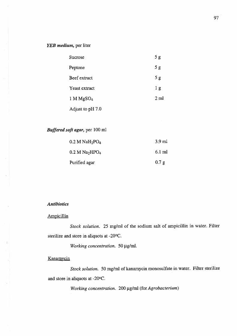
97
YEB median, per liter
Sucrose
Peptone
Beef extract
Yeast extract
I M MgSOa
Adjust to pH 7.0
Buffered soft agar, per 100 ml
O.2MNaH2POa
0.2 M NazHPO¿
Purified agar
5g
5g
5o
lg
2ml
3.9 ml
6.1mI
0.7 s
Antíbiotics
Amoicillin
Stock solution. 25 mglnl of the sodium salt of ampicillin in water. Filter
sterilize and store in aliquots at -20oC.
Working concentration. 50 pg/rtt.
Kanamvcin
Stock solution. 50 mg/ml of kanamycin monosulfate in water. Filter sterilize
and store in aliquots at -20oC.
Working concentra.tion. 2OO pglml (for Agrobacterium)

98
APPENDIX B Miscellaneous Buffers and Solutions
Physiological Saline (Buffered Saline), per 100 ml
NaCl
0.2 M NaH2POa
0.2 M Na2HPO4
pH7.3
Støndard Markers
Set I, per ml
2-deoxyadenosine
Orange G
Xylene cyanol
Fructose
Nitrobenzene sulfonate
Sodium azide (preservative)
Set 2, per ml
Cytosine
Cytidine
Uracil
Uridine
0.85 g
3.9 ml
6.1mI
5mg
1mg
lmg
10 mg
4mg
1mg
2mg
2mg
2mg
2mg
2mgNitrobenzene sulfonate

99
HVPE Buffers
Ammoníam Bícarbonate Baffer (0.2 M NH¿HCO¡/O.I M NH:; pH 9.2)
Dissolve 15.8 g NHaHCO3 (FW=79) and approximately 7 ml of 28-3OVo
(w/w) NH3 per liter solution.
Borate Buffer (0.05 M; pH 9.a)
Dissolve 19.07 g Na2BaOT . l0 H2O (FW=381.38) per liter solution.
Cítrate Buffer (0.05 M; pH 6.0)
Dissolve 10.5 g citric acid (FW=210.14) and 5.7 g NaOH (FW=40) per liter
solution.
Formic-Acetíc Acid Buffer (0.75 M HCOOH1.03 M CH:COOH; pH 1.75)
Dissolve 28.4 mI 98Vo HCOOH and 59.2 ml glacial CH3COOH per liter
solution.
Formic-Acetic Acid Buffer contaìning 0.1 M Zinc Acetate (pH 3)
Dissolve 2t.95 g (CH3COO)2Zn.2H2O (FW=219.49) and dilute to I liter
with formic-acetic acid buffer.
Staíns
Silver Nitrate Reagent, per liter
Dissolve 2 g AgNO3 fW=169.87) in 20 rnl water. Dilute to I liter with
acetone.

100
Tetrazolium Reagent, per 100 ml
Dissolve 0.2 gtetrazolium (2,3,5-triphenyl tetrazolium chloride; FW=334.8)
in about l0 ml methanol. Dilute to 100 ml with chloroform.

101
APPENDIX C Publications Arising from this Thesis
I)onner SC, Jones DA, McClure NC, Rosewarne GM, Tate ME, Kerr A' Fajardo
NN and Clare BG. 1993. Agrocin 434, a new plasmid encoded agrocin
from the biocontrol Agrobacterium strains K84 and KIO26, which inhibits
biovar 2 agrobacteria. Physiological and Molecular Plant Pathology 42¿ I85-
r94.
Fajardo NN, Tate ME and Clare BG. 1994. Agrocin 434: an additional biological
control component for crown gall, pp. 128-130 in: Improving Plant
Productivity with Rhizosphere Bacteria. MH Ryder, PM Stephens and GD
Bowen, eds. CSIRO Division of Soils, Adelaide.

APPENDD( I)
ADDITIONAL DISCUSSION OF RECENT RELEVANT WORI(
A recent publication by Penalver et al., (L994) described production of a new antibiotic-like
substance, active in vitro against A.tumefaci¿ns. This zubstance, designated ALS 84 was
different from agrocin 84 and produced on mannitol-glutamate medium. Sensitivity was not
determined by presence/absence of a Ti plasmid and ALS 84 had a bacteriostatic effect against
a range of other bacterial genera. In this respect this proposed substance is also distinct from
agrocin 434 which active exclusively against A.rhizogenes (biovar 2) pathogens. The effects of
ALS 34 have only been observ ed in vitro and the chemical nature of this substance has not yet
been elucidated. However it is important to consider the activity of this alternative product
when interpreting the inhibition res,ults observed during this snrdy. In this study it has been
suggested tlat the inhibition observed with certain biovar 3 strains was the result of a pH
effect. There is also the possibility that additional inhibitory products zuch as ALS 84 may be
responsible for this effect. Altematively pH may affect the inhibitory activity of antibiotic-like
products. These possibilities must be investigated further to explain the in vitro effects
observed. In addition, the role these factors play in tle biocontrol process must be examined
with relevant in planta studies whioh are currently being oonducted.
The description of agrocin 84 as an antibiotic or bacteríocin requires clarification. As agrocin
84 does not kill the producer cell it cannot be classed as a bacteriocin, therefbre it is more
correct to class agrocin 84 as an antibiotic even though it appears to beproduced throughout
the growth cycle, unlike nxmy antibiotics which are secondary metabolites.
Vicedo et" al., (1995) have also recentþ described studies into the transfer of plasmids pTi,
pAtKS4b and pAgK84 between A.tumefaciens and strain K84 in aplanttumour. Plasmid
pAgKS4b belongs to the Inc Rhl inoompatibility group and is thought ot act as a barrier to
transfer of Ti plasmids ofthe same Inc goup. However the work of Vicedo et al., (1995) has
shown that strain K84 can act as a recipient for Inc Rhl Ti plasmids and that in some cases
pAgKS4b is replaced whereas in other cases recombination has occured between the Ti
plasmid and pAgKS4b

As the largest fragments of pAgKl3l8 are indistinguisable in size it is possible to show the
plasmid map obtained as a circular map, however the XbaI fragments A and B may be
differentiated on size basis after further investigation.
Reference to the paper by Wang et al., (1994) also requires additional discusion. This paper
showed that there were five agr tanscnptional groups or operons, although several of these
may be polycistronic so the exact number of genes is presentþ unlnoram. Although this paper
showed that agrocin 84 genes are expressed in planta it did not provide proof for a role for
agrocin 84 in the biological control process. The nature of the roles of the various agrocins in
the biological process is being investigated in an anotler study (Ahmadi et al., 1995).
Investigation into the role of carbon substrates in expression of agn genes showed that
expression levels were observed in minimal media, regardless ofthe carbon souÍce. In addition
ex¡rression of each øgz locus was not affected by the presence of other agn genes or the
nopaline catabolic plasmid pAgK84b, opines or root exudates in the culture media. All five
agnlociwere expressed throughout the gowth cycle.
Additional References
Ahmadi, A.R, McClure, N.C. and Clare, B.G. (1995). Factors involved in the bioogical
control of crowngall disease by derivatives of Agrobacterium rhizogenes K84. Presented at
"Agrobacterium and beyond" Intemational conference, Gif sur Yvette, France, September
1995.
Penalver, R, Vicedo, 8., Salcedo, C.L and Lopez, M.M. (1994). Agrobacterium radiobacter
strains K84, K1026 and K84 Agr- produce an antibiotic-like substance, active in vitro agains
A.tumefaciens andphytopathogentc Erwinia and.Pseudomonos qpp. Biocontrol Science and
Technolnqv 4 259-267.
Vicedo, B.,Lopez, M.J., Llop, P. and lope4 M.M. (1995). Transfer ofpTi, pAtKS4b and
pAgKSa phsmids betwee,n Agrobacterium tumefaciers and strain K84 ofl. radiobacter m a
tumour. Molecular oharacterisation of transferred plasmids. Presented at"Agrobacterium and
beyond" Intemational conference, Gif sur Yvette, France, September 1995.

Fajardo, N. N., Tate, M. E. & Clare, B. G. (1994). Agrocin 434: an additional
biological control component for crown gall. In M.H. Ryder, P.M. Stephens
and G.D. Bowen. (Eds.), Improving plant productivity with rhizosphere
bacteria: proceedings of the Third International Workshop on Plant Growth-
Promoting Rhizobacteria, Adelaide, South Australia, March 7-11 1994. (pp.
128-130). S. Aust., CSIRO Division of Soils.
NOTE:
This publication is included in the print copy
of the thesis held in the University of Adelaide Library.

Phl,tsiological and MolecuLar Plant Pathologt (1993) 42, 185-194 185
Agrocin 434, a new plasmid encoded agroc¡n from thebiocontr ol Agrobacterium strains K84 and K1026, whichinhibits biovar 2 agrobacteria
S. C. Dol¡N¡n*, D. A. Jownst, N. C. McClunof , G. M. RosnwenNtt, M. E. Tero*,A. Knnnt, N. N. Fe¡enoot and B. G. Cr,anetf*Plant Science Deþartment and lCrop Protection Deþarlment, Uniuersit2 of Adelaide, Waite Agricultural Research
Institute, Glen Osmond, SA 5064, AustraLia
(Aueþted for þub lication Februar2 1993)
Agrocin 434 is a previously unrecorded inhibitor produced by Agrobarterium strains KB4 and
Ki026, which also secrete agrocin 84. These strains are used for the biologicai control ofcrowngall disease of plants and agrocin 434 may contribute to the biocontrol processes. When strain K84was cured ol the plasmids which encode agrocin 84 production (pAgKBa) and nopalinecataboLism (pAtK84b), the derivative strains retained both agrocin 434 production and the
remaining 300 400 kb plasmid. This plasmid was mobiìized into a pìasmidless recipient and the
resulting transconjugants also produced agrocin434. Processes essential for biosynthesis ofagrocin434 are thus encoded on the large plasmid, for which the designation pAgK434 is recommended.Bioassays of the inhibitory effects of agrocin 434 were done with 68 Agrobacterium isolates fromAustralia, Europe and North America. Atl the biovar 2 (A. rhizogenes) isolates tested were
inhibited, with the exception of the strains which produced agrocin 434 themselves. None of the
biovar I (A. tumefaciens), biovar 3 (A. uitis), or A. rubi strains tested was sensitive to agrocin 434.
Preliminary chemical analyses indicated that the compound is a di-substituted cytidine nucleoside.
INTRODUCTION
Pathogenic agrobacteria cause crown gall and hairy root diseases of a wide range ofhost plants and are responsible for significant economic losses [4, 201. Etrectle controlof crown gall in a number of crops has been achieved in many countries through the
use of the non-pathogenic biological control straín Agrobacterium KB4, and its derivativeK1026 ll5, 16, 1Bl. Despite their wide-spread success, the biocontrol strains are noteffective against all crown gall pathogens and they do not control all hairy rootinfections [9]. We are therefore attempting to develop strains with wider ranges ofeffectiveness. To this end we are investigating factors involved in the biocontrol
Processes.Each of the biocontrol strains KB4 and K1026 produces agrocin 84, which is
specifically inhibitory to certain pathogenic agrobacteria in culture 116, 36) However,whilst it is accepted that agrocin 84 plays an essential part in controlling sensitive
fTo whom correspondence should be addressed.Abbreviations used in text: Amp, ampicillin; Inc, incompatibility; Km, kanamycin; Nm, neomycin;
Mob, mobilization; Ril, rifampicin; Str, streptomycin; Tc, tetracycline; Tn, transposon.
0885 5765/93/030185+ l0 $08.00/0 @ 1993 Academic Press Limited

1 86 S. C. Donner et a/.
pathogens, it appears that in þlanta biocontrol also involves other factors. For instance,field studies by López et aL. l24l have shown that both strain KB4 and a mutant unableto produce agrocin 84 are able to reduce infection by some pathogenic agrobacteriawhich are insensitive to agrocin 84 in culture. These effects may in part be due to thesuperior root colonizing ability of the biocontrol strains 131), to the influence of otheragrocins, or to as yet unidentified agents.
We have investigated the biological and chemical properties of additional inhibitorysecretions from the biocontrol strains that may possibly contribute to the overall effect.Agrocin 434 is one such compound. It was de tected initially as a product of strain KB4and is also produced by strain K434, a derivative of K84 lacking the ability tosynthesize agrocin 84. The new agrocin was consequently named after strain K434.Although strain K434 is unable to produce aglociir 84 iL rrever[hcless does haveinhibitory activity in uitro agarnst agrobacteria, including isolates insensitive to agrocin84. Strain 434 has lost pAgKB4, the 48 kb plasmid that encodes agrocin 84 synthesis,but retains the two other plasmids of its parent strain K84: pAtKB4b, of 173 kb andbearing nopaline catabolic genes [.], 16] and a 300-400 kb cryptic plasmid [J, 16]. WePresent evidence that: (a) sensitivity to agrocin 434ts acharacteristic ofbiovar 2 strainsof Agrobacterium and; (b) that genes lor the biosynthesis of this agrocin are encoded onthe so-called cryptic plasmid. \{¡e proposc that this plasmid be designated pAgK434 toindicate it-s agrocinogenic attribute:it is referred to as such throughout the paper.
MATERIALS AND METHODS
Bacterial strains
Tlre biocontrol strain Agrobacterium K84, its agrocinogenic derivatives , the Agrobacteriumancl .8. coli strains used to produce these derivatives, and their relevant plasmids arelistecl in Table l.
The 68 Agrobacterium strains from Australia. Europe and North America, whichwere used in the survey, and their respective sensitivities to agrocin 434 are given inTable 2.
Bioassalts for agrocin 434
We usecl stanclard agrocin bioassay methocls 121), tn which zones of inhibition werecletected in lawns of sensitive strains overlaid on killed proclucer colonies. Whereappropriate, the proclucel colonies rvere leplaced by filter paper discs moistened withsolutions of crude or partially pulified agrocin recovered from 1'5-3.0 ml of liquidculture medium.
Agrocirt 134 in lirluid nudia; d¿lection and isolal¿o¡¿
Agrobacterium straius were growrl for 2 3 days at 25 oC in a shake culture in a glucose-glutamate, liquicl mediun l25l modified to contain 1.74 g KTHPO. and 5.44 gKH,PO4 per litre. Cells rvere removecl by centrifugation and I ml samples ofsupernatant werc mixed with 100 ¡rl portions of an aqueous suspension of activatedcharcoai (15 mgml 1). The charcoal was washed three times by suspension andcentrifugation in successirrc I ml volumes of water. After final centr-ifugation from\\.ater the charcoal lvas suspcndcd in 200 ¡rl of 70 J,o e thanol, centrifuged and the

Agrocin 434, a new plasmid encoded agrocin from Agrobacterium
T¡,eLn IBacterial strains and þlasmids us¿d in agrocin 434 þreþaration and detection
Strain Biovar* Plasmids Description
187
AgrobaúerittmK21
K84
K43+
K1t43
K7+9
Kl317
Escherichia coLi
HBIOl
pTiK27pAtK27pAgKB4fpAtKB4bfpAgKa34$pAtKB4bfpAgK434$pAgK43a$
1 pAgK434$
pJB3JI
2
q
Indicator strain sensitive to both agrocins 84
and 434.Biological control strain, produces both
agrocins 84 and 434.
Soontaneous mutant of K84 lacking pAgK84'p.odu.", agrocin 434 but not agrocin 84'
K434 .rt.d of pAtK84b, produces agrocrn
434 only; this studY.C58 pTilpAt-Rif Str', supplied by A Kon-
dorosi.K749 containing pAgK434 mobilized from
Kl143; this studY.
Plasmid is a Km" derivative of pR6B 45,
Tc"Tra+ [2].Helper plasmid in triparentaì matings [12]pfur-i¿ fl^ Inc olpTiC5B, Amp'Km'Nm''
1121.piuråi¿ has Tn::Mob transposon, Km' [32]'
2
2
HBlO1HBlO1
pRK2013pUCD1000
pSUPs011s17 I
*For purposes ofcÌarity, biovar designations are used rather than specific epithets'
tAgrocin 84 Plasmid [33]'{Nãpaline catabolic plasmid [5,-16]'
{egå.i" 434 plasmid 15, ß e' this studvl'
Supernatantcollected.Thisprocedurewasrepeatedtwice-moreandthecombined600 pl eluants were
",rupo.uàd to dryness under vacuum' These crrrde extracts were
each dissolved in 10 pl åf de-ionized water for use in bioassays or in isolating agrocin
+34' extracts exhibiting inhibitory activity in
(HVPE,), using the aPParatus of Tate
paper strips which were immersed in
S, O'OS M citrate, PH 5-6, or sodium
tetraborate, pH 9.4, and electrophoresis was ca: ied out at 2-3 kV for 20-30 min. Prior
to electropho..rir, ,ur,'fl.s of a solution containing the mobility. reference compounds
)noslne 17,29f were applied at the line of origin
dried and examined using transmitted UV
ï ï: ÏÏ J.3tr ï',î: i:'H'H J'.iiJ î:å::systems.
For partial purification of agrocin 434, crude extracts were electrophoresed in
formic/acetic acid. Discs containing the appropriate uV-absorbent spots were cut
from dried electrophoresis paper strips and .tri.a in five successive 50 ¡rl washes of

188S. C. Donner e¿ a/
Te¡r,c 2In vitro sensitiuitl o;fAgrobacterium strains to agrocin 434
S train Pathogenicity*Agrocin 434t
response
Biovar I ]46,4358, Ach5g,86, CsB, K3lK64, Kt20, Kt98, K3s5, Ks49NCPPB 396, NCPPB3gB, T37K484, K578, KBI7, KB2O KB22CsB (pTi ), CsB (p )
Biovar 2/'943,K27, K46, K7t, K10BK1r4, K118, K440, K9i0K744-K74644, ATCC I1325X*, TR7TRIOI, TRI05K84,K434.K1143K47,K97, K103, K104K123, Kt29, K130, K250K251
Biovar 34s57, K309+ *, K37 4, K37 5-KZ7 7Ks2t, K99s, Kt070, Kt072A. rubi
K!04. K867, K868, K869, K872K I 047, ATCCI 3335(TR g) u-
TiTiTiRio
TiTiTiRiRioooo
'_fi
Ti
TiTi
++
+
1+++
*O, non-pathogen; Ri, hairy, root-inducing pathoge'; .Ii, tumour_inducing pathogen.
f .*, inhibited by agrocin 434 in uitro; _, iot irf,iiit.a by agrocin 434.f isolates ol A. rubi are risted under this ta-, but irolu¡r, on Al n*efaciens, A. rhi4.qenes, andA. uilis, are listed under their jor, .irr.. there appears to be more generalagreement as to their meanings
,"åtiïïî;:"r.rlined were test >iìity to synthesise agrocin 434 using bioassays
**ATCCll325,ATCCl-3335(TRg) andK30garerypecultures of A.rhi4genes,A.rubi,and A. uitis, respectively [261.
de-ionized \'!'ater' The solutions were either concentrated or dried under vacuum forstorag-e' These partially purified preparations were used for bioassays and preliminarychemical studies.
Agrocin 434: þretiminarlt chem.ical studiesMeasurements were made of UV absolbance spectra of partially purified preparationsdissolved in water, in r N HCI, and after hydrorysis in 3 , ø.-i. acid at I l0 "c for 3 h.
Plasmid isolation and electroþhoresiss.mall scale plasmid preparations were done as described by Farrand et ar. lr0l, forAgrobacterium, and by sambrook et at. 1301, ror Escherichia ,ori. Either standard l30l orEckhardt [B] methods for agarose gel eleltroptroresis were used.

Agrocin 434, a new plasmid encoded agrocin from Agrobacterium 189
Curing strain K434 0f þAtKB4b to produce strain Kll43Strain K434 has the two plasmids pAtKB4b and pAgK434 (Table l). It was cured ofthe former to produce a derivative having the single remaining plasmid pAgK434.Cosmid pUCDl000 carries the incompatibility function of pTiC5B l12l and hence is
incompatible with pAtKB4b in strain K434 [5]. Cosmid pUCDl000 was transferredfrom -8. coli}JBl}l toK434 in a triparental mating with helper plasmid pRK2013 [11].Krn' Agrobacterium colonies were then subcultured in a succession of liquid media, free
of antibiotic. Km" isolates were obtained which contained pAgK434, but which had
lost both pAtKS4b and the unstable pUCDl000, as determined by agarose gel
electrophoresis. One of the resultant products was strain Kll43 (Table 1).
Mobili<ation of pAgKa3a from K1143 to K749'îní-mob was transferred from E. coli Sl7 -l (pSUPSOl I ) to Kl 143 in a patch-matingon TY agar at 28'C for 24h. Kll43 Km" colonies were selected on Bergersen's
medium [1] with 200 pg kanamycin per ml. These were patch-mated with HBl0l(pJB3JI) on TY agar: Km"Tc"K1l43 colonies were selected on Bergersen's mediumwith kanamycin (200 pg mt 1) and tetracycline (10 pg ml-t), and patch-mated on TYagar with the plasmidless biovar I strain K749. Transconjugant K749 colonies were
selected on biovar I selective medium 119, 201 with kanamycin (200 pg ml-r), and
screened for plasmids in Eckhardt gels. Colonies with the single plasmid pAgK434 were
tested for the complete range of biovar 1 characteristics [26] . Authentic biovar I strains
were then tested for agrocin 434 synthetic capacity in bioassays using charcoal
absorbed fractions of culture media, as described above, with K27 as the indicatorstrain. Inhibitory extracts were further purified by HVPE. UV absorbent spots fromthese extracts with the same electrophoretic mobility as agrocin 434 from strain Kl 143
were eluted from electrophoresis papers with de-ionized water and their UV absorbancespectra determined to ensure identity with agrocin 434. One of the biovar Itransconjugant products was strain Kl3l7 (Table 1).
R ESU LTS
Biouars and sensitiuit2 to agrocin 434
All biovar 2 strains tested were inhibited by agrocin 434,îrorn strain Kl143, with the
exception of KB4 and its derivatives, and the three Ti-type pathogenic isolates (K7++-K7+6) which proved to be agrocin 434 producers (Table 2).
In contrast, all biovar I and 3 and A. rubi strains tested were insensitive to agrocin434 (Table 2). Four representative strains, from each of the biovar 1,3 and A. rubi
groups (Table 2), were tested for agrocin 434 production in liquid media and in uitro
inhibitory capacrty. None of the l2 strains was an agrocin 434 producer.
Association betueen pAgK434 and agrocin 434 þroductionFigure I (a) shows the plasmids of the parent biocontrol strain K84 and those of its twoderivatives K434 and Kl143, all of which have pAgK434. Figure 1(b) shows the
presence of pAgK434 in both the donor biovar 2 strain Kll43 and Kl3l7, one of the
biovar 1 transconj ugant products of a K I 143 x K7 49 mating, and its absence from theplasmidless, biovar 1, recipient parent strain K749.

'- Þò* ffi
(al
-
190 S. C. Donner e¿ a/,
2 3 4
AB-c-D-
A
FIc. 1. Plasmids of Agrobacteriur¿ strains scparated by electrophorcsis in 0.70/o agarose gel. (a)Lanc l,,1DNA,48kb; lane 2, KB4; lane 3, K434; lane 4, Kl143. (b) Lane 1,K749;lane2,K1317; lanc 3, Kll43, ßand A, pAgK434; band B, pAtKB4b; band C, pAgKB4; band D,chromosomal DNA.
Agrocin 434 was produced by strains K84,K+34, Kll43, and Kl3l7 but not byK749. Figure 2 shorvs the results of bioassays in rvhich preparations from Kll43 andKi3l7 producecl zones of inhibition whereas K749 preparations clicl not. Figure 3
shows corresponding HVPE results demonstrating agrocin 434 presence in Kl143 andKl3l7 secretions and its absence from those olK749. The same association betweeninhibitory action, agrocin 434 synthesis, and the presence of pAgK434 was recordedfor a further 12 biovar I transconjugants produced by additional, independentKl l43 xK7+9 matings.
Cultures of each of the pathogenic biovar 2 isolates K74+-K1+6, which wereinsensitive to agrocin 434, produced a compound having the same inhibitory,electrophoretic and UV absorbance propertics as agrocin 434 (data not shown) . Inaddition, they each contained a plasmid ol approximately the same size as pAgK434as welì as a pTi-sized plasrnid (data not shown) . The three isolates came from the oneorchard and '"vere indistinguishable in all of thc cultural, biochernical, and plasmiclcharactcristics exarnined: they thus appear to be iclentical isolates of the one strailt.
Bioassal,, conditions
Colonies of strain KB4, which secrete both agrocin 84 and agrocin 434, exhibited thecharactelistically transparent zones of inhibition associated with agrocin 84 U5, 2ll.Incontrast, colonies secreting agrocin 434 alone procluced opaque zones ofinhibition thatwere smaller in diameter, much fainter, but still discernible. When discs impregnateciwith solutions of crude or partially purified agrocin 434 were used in bioassays, thezones of inhibition were more clearly visible (Fig. 2).
Agrodn 434: ch¿mical data
The UV absorbance spectra in water and I N HCt (Fig. 4) wele consistent with a aN
substituted cytidine nucleoside . Formic acid hydrolysis yielded cytidine. Electrophoresisin borate buffer showed no increase in relative mobility over that in ammonium
11
nrIIi|
(b)

Agrocin 434, a new plasmid encoded agrocin from Agrobacterium
Frc. 2. Bioassay ofcharcoal adsorbed fractions from liquicl media olerltrrrcs ofthc donor biovar
2 straiu Kl I43, u,hich contains pAgK434, the plasmidless biovar I recipient strain K749, and the
resultant biovar I tr'ansconjugant Kl3l7, wliich also contains pAgK434. Preparations lromK1143 and K1317 both produccd zones of inhibition but the K749 preparation was notinhibitory. K27 was used as the indicator strain
on
Frc. 3. High voltage paper electrophorcsis of charcoal adsorbed fractions from ìiquid culturemedia. Electrophoresis was in 0'05 u citrate, pH 5 0, at 2 kV for 30 min: photographed using
transmirted UV light. Lane 1, plasmidless strain K749;lanes 2 and 3, strains K1143 and K1317respectivelyJ each of which has the single plasmid pAgK434. AG indicates the positions of darkspots olUV-absorbent agrocin 434, which are prcsent in the K1143 and K1317 lanes bui absent
h'om the K749 lane. Ori, origin for samples and mobility relereuce compounds;2-d,2-dcoxyadenosine (UV absorbent) and xc, xylene cyanol (not visiblc in UV), indicate the positions
to which these mobility reference cornpounds rnigratcd. Wlritc spuLs are unidcntified, UV-flnorescent compounds
191
AG
K1317 Kl143

192 S. C. Donner e¿ a/
c)oÉcþI¡
0.2
0.1
0.0300
lVavelength (nm)360
Frc. 4. UV spectra of partially purified agrocin 434 in HrO (-) and I N HCI (------)absorbance maxima are 276 nm and 288 nm respectively.
carbonate, at the same pH. This establishes substitution at either position 2' or 3' of theribose moeity. Hence agrocin 434 appears to be a disubstituted cytidine nucleoside.
DISCUSSION
Sensitivity to agrocin 434 was exhibited by all biovar 2 strains that were tested (Table2), with the exceptions of wild-type or constructed agrocin 434 producer strains. Theimplication is that sensitivity to agrocin 434 is a general characteristic of biovar 2
strains and is presumably encoded chromosomally-at least in part. In contrast it has
been shown that agrocin 84 sensitivity is exhibited only by pathogenic agrobacteriawith acc genes borne on their Ti plasmids 17, l3l. Another possible inference to be
clrawn from the insensitivity to agrocin 434 shown by agrocin 434 producer strains is
that immunity to agrocin 434 is encoded on pAgK434. In this regard there may be aresemblance to pAgK84, which encodes both agrocin 84 immunity and synthesis [28].
None of the biovar I and 3 and A. rubi strains tested (Table 2) were inhibited byagrocin +3+. A representative selection of these strains (Table 2) was shown not toproduce agrocin 434. Hence their insensitivity to agrocin 434 appears to differfunctionally from the immunity exhibited by agrocin 434 producers.
The broader effective inhibitory range of agrocin 434 ín culture, u,hen comparedwith agrocin 84, suggests that agrocin 434 rnay possibly be one of the factorscontributing to in þlanta control by strain K84, and derivatives, of some Agrobacterium
pathogens which are insensitive to agrocin 8+ 16, 24).Our results indicate that essential steps in biosynthesis of agrocin 434 are encoded by
pAgK434, the largest of the reported strain KB4 plasmids. This conclusion is supportedby the evidence that all KB4 derivatives which contain this plasmid are agrocin 434producers and that one of the agrocinogenic derivatives,KII43, has this plasmid only(Table l). In addition, agrocin 434 synthesis was also exhibited by transconjugantbiovar I strains, such as K1317 (Figs.2 & 3), to which pAgK434was transferred byconjugation [Fig. I (b)]. Each of the pathogenic isolates (K744-K746) also has a
220

Agrocin 434, a new plasmid encoded agrocin from Agrobacter¡um 193
plasmid resembling pAgK434 in size, and each produces an inhibitor similar, oridentical, to agrocin 434.
This is the first published record of a functional capacity for a plasmid of 300-400 kb,cryptic group found relatively commonly amongst agrobacteria. However anassociation has been reported previously be tween catabolism of deoxyfructosyl aminoacids and the presence of the cryptic plasmid in a strain derived by curing strain C5B
of its Ti plasmid [27]. These plasmids represent a substantial portion of the totalgenome of their host cells, and the identities of other functions encoded on themremains an intriguing question.
Our preliminary results on the chemistry of agrocin 434 indicate that it is a di-substituted cytidine nucleoside (Fig. a) . Since its appears to have a 4N substitution itdiffers from agrocin 108 [23] and is also chemically distinct from the adenine-derivedagrocins, 84 and D286 114,23]. Agrocins have also been reported for strains }{I00 1341,HLB-2 l3),J73 [37], NCPPB 396 U4l, andT37 134),but since these compounds havenot been characterízed chemically it is not possible to make comparisons with agrocin43+.
This work was supported by grants from the Australian Research Grants Committee,the Australian Research Council and the Waite Institute Research Committee, and byscholarships from the Commonwealth Post-Graduate Research Scheme (S.C.D.) andthe Australian International Development Assistance Bureau (N.N.F.). We thankNatalie Dillon for skilled technical assistance.
R EFE R EN CES
l. Bergersen FJ. 1961. The growth of Rhizobium in synthetic media. Australian Journal of Biological Scienrcs
14: 349-360.2 Brewin NJ, Beringer JE, Johnston AWB. 1980. Plasmid-mediated transfer of host-range speciflcity
between two strains of Rhizobium leguminosarum Journal of General Microbiolog 120.413 420.3. Chen XY, Xiang WN. 1986. A strain of Agrobacterium radiobacter inhibits growth and gall formation by
biovar III strains of l. htmefaciens lrom grapevine. Acta Microbiologica Sinica 26: 193 199.4. Clare BG. 1990. Agrobacterium in plant disease, biological disease control and plant genetic engineering.
Science Progress, Oxford 74: 1-13.5. Clare BG, Kerr Ä,,Jones DÄ. 1990. Characteristics olthe nopaline catabolic plasmid in Agrobacteritun
strains KB4 and K1026 used for biological control of crown gall disease. PLasmid23:126 137.6 Cooksey DA, Moore LW, 1982. Biological control ol crown gall with an agrocin mutant of
Agrobat terium radiobac ler. Pþttoþatholog 12 : 919 921.7. Ellis JG, Murphy PJ. l98 1 . Four new opines from crown gall tumours-their de tection and propcrtie s.
Molecular and General Genetics l8l: 36 43.8. Eckhardt T. l978 A rapid method for the identification ofplasmid deoxyribonucleic acid in bacteria.
Plasmid l: 5B|5BB.9. Farrand SK. 1990. Agrobacterium radiobacter stain KB4: a model biocontrol system. In Baker RR, Dunn
PE. eds New Dir¿ctions in Biological Control: Alternaliuesfor Suþpressing AgriculturaL Pesls and Diseases,NcwYork: lViley-Liss, 679 691.
10. Farrand SK, Slota JE, Shirn J-S, Kerr A. 1985. Tn5 insertions in the agrocin 84 plasmid: Theconjugal nature of pAgK84 and the locations of determinants for transfer and agrocin 84production. Plasmid 13: 106 1 I 7.
l1 Gallie DR, HagiyaM, Kado CI. l9B5 Analysis ol Agrobaclerium tumefaciens plasmid pTiCsB replicationregion with a novel high-copy-number derivative. Journal of Bacteriologlt 16l: 103't 1041
12. Gallie DR, Novak S, Kado CI. 1985. Novel high- and low-copy stable cosmids lor use in Agrobacteriumand Rhi<obiutn. Plasmid 14: 171 175.
13. Hayrnan GT, Farrand SK. l9BB. Charactcrization and mapping of thc agrocinopinc-agrocin 84 locuson the nopaline Ti plasmid pTiC58. Journal of Bacteriologt l7O: 1759 1767.
t4 ÌvIPP 42

194 S. C. Donner e¿ a/.
14. Hendson M, A,skjaer L, Thomson J.4,, van Montagu M. 1983. Broad-host-range agrocin ofAgrobactetinn lunefatietts. Applied and Enuirontne¡tlal fuIinobiolog; 45: 1526 I533.
15. Jones DA, Kerr A. l9B9 AgrobacLeriunradiobacter strain K1026, a genetically cngineered derivative ofstrain K84, for biological control of clorvn gall. Plant Disease 73: 15 lB
16. Jones DA, Ryder MH, Clare BG, Farrand SK, Kerr A, 1988. Construction of a Tra deletion mutantof pAgKB4 to safeguard the bioìogical control of cros'n gall. Molecular and General Genelics 212:207 2t4.
l7 Kao JC, Perry KL, Kado CL l9B2. Indoleacetic acid complcmcntation and its relation to host rangespecifyirìg genes on the 'fi plasmid ol Agrobacletiun tutnefaciens Moleculat and Ge¡teral Genetics lB8:+25 +33.
I B. Kerr A. I 980. Biological coltrol of croln gall through production of agrocin 84. Plant Disease 64: 25 30l9 Kerr A. 1992 Thc Gerus AgrobarleritLtt. In Balorvs A, Toùpcr HG, Dworkin M, Harder W, Schlcifer
KH, eds. The Prokaryotes Second edn. New York: Spr'inger-Verlag,221+ 2235.20 Kerr Ä., Brisbane PG, 1983. Agrobacterinn Il Fah1, PC. Persley Gl, eds Planl BacteriaL Diseases. A
Diagnostic G¿il¿. New York: Acadernic Prcss, 27-43.2l Kerr A, Htay K. 197+. Biological control of crown gall thlough bacteriocin procluction. Phlsiological
Planl Pathology 4:37 4422. Kerr ,4., Panagopoulos CG. 1977. Biotypes of Agobacterium radiobacter var lwne;fodens and their
biological cortrol. PhltoþalhoLogy g0: 172 1?9.23 Kerr,4,TateME. l9B4.AgrocinsandthebiologicaÌcorrtrol ofcrorvn gzll À4inobi.ological ,Sdencesl:l-4.24. López MM, Gorris MT, Salcedo CI, Montojo AM, Miró M. 1989. Evidencc of biological control
of Agobacteriun lanefaciens strains sensitive and resistant to agrocin 84 by differelt Agrobactetiumradiobacter strains orì stone fruit trees. Applied and Enuironnenlal Microbíolop 55: 741-746.
25 Murphy PJ, Roberts WP. 1979. A l¡asis for agrocin 84 scnsitivity in Agrobaclerium radiobacter. JournaLoJ'GeneraL A4icrobioLog't, ll4: 207 213.
26. Ophel K, Kerr A. 1990. Agrobacteritnn allzi sp. nov. for strains of Agrobacterian biovar 3 from grapevines.Internalional Journal of Slstetnatic Bacteriologlt 40: 236 241.
27 RyderMH. l9S4. OpinesinCrounGalLandHairyRootDis¿ases. Ph.D.Thesis,UniversityofAdelaide,South Australia.
28 Ryder MH, Slota JE, Scarim A, Fanand SK, l9B7 Gcnetic analysis olagrocirì 84 production ancìirrnrurrity in .4grobacletiutn spp Joutnal of BacterìoLog'169: 4l84 4l89
29 Ryder MH, Tate ME, Jones GP, 1984. Agrocinopine A, a tumor-inducirg plasmid-coded enzyrneprocluct, is a phosphodiester of sucrose and L-arabinose. The Journal of Biological Chenústr1 259:9704 97t0
30 Sambrook J, Fritesch EF, Maniatis T. l9B9 Moletular Cloning. A Labomtory Mazttøl, Second edn,Coìd Spring Harbor, Cold Spring Harbor' Laboratory Press: USA.
31. ShimJ-S, Farrand SK, Kerr Á,. 1987. Biological control olclown gall : Construction and testing ofnerv biocontrol ageÌts. PlUttoþathologlt J7 ' +63-+66.
32. Simon, R. 1984. Hiøh fi'equency mobilisation of Gram-negative bacterial replicons by the ín uilroconstructed Tn5-mob transposon. MolecuLa¡ and General Genetics 196: 413 420.
33 SlotaJE, Farrand SK, 1982. Gcnctic isolation and physical char'acterization of pAgKB4, the plasmidresponsible lol agrocin 84 procìuction PLasnirl 8: I 75 l86
3.1 Stonier T. 1960. Agrobacteritnn tunefaciens Conn II. Produc¡ion of an antibiotic substancc Journal ofBatteriolo¿9' 79: BB9 B98
35 Tate ME. 1968 Separation olml,oinositol pentaphosphates b1, moving paper electrophoresis (À4PE)Anaþ,¡i¡¿¡ Biochenistrlt 23 l4l 149.
36 Tate ME, Murphy PJ, Roberts WP, Kerr 4.. 1979 Acleninc N6-sul¡stituent of agrocin 84 determinesits l¡acteriocin-like s¡recificitl' .Nahu e 280 : 697-699.
37 Webster J, Dos Santos M, Thomson JÄ. 1986. Agrocin-produci;tg tlgrobacterium tumefaciens strainactive agaiìrst grapevine isolates. Applied and EttuitonttenlaL fuIicrobiologlt 52:217-219.