biologia - studia.ubbcluj.ro · biologia 1 studia ubb editorial office: ... further subjected to...
TRANSCRIPT
BIOLOGIA1/2018

STUDIA UNIVERSITATIS BABEŞ-BOLYAI
BIOLOGIA
1 / 2018 January – June

EDITORIAL BOARD STUDIA UNIVERSITATIS BABEŞ-BOLYAI BIOLOGIA
EDITOR-IN-CHIEF:
Professor Octavian Popescu, Member of the Romanian Academy, Babeş-Bolyai University, Cluj-Napoca.
BOARD OF SUBJECT EDITORS:
Associate Professor Ioan Coroiu, Babeş-Bolyai University, Cluj-Napoca; Professor Nicolae Dragoş, Babeş-Bolyai University, Cluj-Napoca;
Professor László Gallé, Member of the Hungarian Academy, University of Szeged, Hungary;
Professor Michael Moustakas, Aristotle University, Thessaloniki, Greece; Professor Aharon Oren, Alexander Silberman Institute of Life Sciences, Jerusalem, Israel;
Professor Leontin Ştefan Péterfi, Associate Member of the Romanian Academy, Babeş-Bolyai University, Cluj-Napoca;
Professor László Rakosy, Babeş-Bolyai University, Cluj-Napoca; Senior Researcher Anca Sima, Associate Member of the Romanian Academy,
Institute of Citology and Cellular Pathology, Bucharest; Professor Helga Stan-Lötter, University of Salzburg, Salzburg, Austria;
Professor Corneliu Tarba, Babeş-Bolyai University, Cluj-Napoca.
LIST OF ASSOCIATE REVIEWERS:
Senior Researcher Adela Halmagyi, Institute of Biological Research, Cluj-Napoca; Professor László Rakosy, Babeş-Bolyai University, Cluj-Napoca;
Associate Professor Ioan Coroiu, Babeş-Bolyai University, Cluj-Napoca; Lecturer Rahela Carpa, Babeș-Bolyai University, Cluj-Napoca;
Lecturer Alin David, Babeș-Bolyai University, Cluj-Napoca; Lecturer Anca Farkaș, Babeș-Bolyai University, Cluj-Napoca;
Teaching Assistant Cristina Mircea, Babeș-Bolyai University, Cluj-Napoca; Biologist Sára Ferenți, University of Oradea, Faculty of Sciences, Oradea.
EXECUTIVE EDITORS:
Lecturer Karina Paula Battes, Babeş-Bolyai University, Cluj-Napoca; Lecturer Mirela Cîmpean, Babeş-Bolyai University, Cluj-Napoca.
Contact: [email protected]

YEAR Volume 63 (LXIII) 2018 MONTH JUNE ISSUE 1
PUBLISHED ONLINE: 2018-06-22
PUBLISHED PRINT: 2018-06-29 ISSUE DOI:10.24193/subbbiol.2018.1
STUDIA UNIVERSITATIS BABEŞ-BOLYAI
BIOLOGIA
1
STUDIA UBB EDITORIAL OFFICE: B.P. Hasdeu no. 51, 400371 Cluj-Napoca, Romania, Phone + 40 264 405352, www.studia.ubbcluj.ro
SUMAR – CONTENTS – SOMMAIRE – INHALT
REGULAR ARTICLES
V. BULIMAGA, M. PISOVA, L. ZOSIM, A. TROFIM, Procedures of partial purification for phycobiliproteins from cyanobacteria isolated from soils of Republic of Moldova .................................................................................... 5
N. TOMESCU, L. A. TEODOR, Terrestrial isopods (Isopoda, Crustacea) from the “Danube Delta” Biosphere Reserve .......................................................... 15
A. MESTECĂNEANU, R. GAVA, Ornithological observations on the Goleşti Basin (Argeş River, Romania) between February 2013 and January 2014 ..... 25

E. E. IMARHIAGBE, B. IKHAJIAGBE, Antibiotic susceptibility of bacterial isolates and water quality index of water sourced from closed ground water and open hand dug well in Koko Community, Delta State, Nigeria ..... 47
A. -L. BUTIUC-KEUL, L. JARDA, I. GOIA, I. HOLOBIUC, A. FARKAS, V. CRISTEA, Preliminary data regarding genetic diversity of several endangered and endemic Dianthus species from Romania generated by RAPD markers ................................................................................................ 59
N. TOMESCU, L. A. TEODOR, Protracheoniscus vasileradui – n. sp. (Crustacea, Isopoda, Crinochaeta) in the Romanian fauna ............................. 73
A. DAVID, A. N. STERMIN, E. SEVIANU, Clutch size and egg repeatability in three elusive bird species: Little Bittern (Ixobrychus minutus), Little Crake (Zapornia parva) and Water Rail (Rallus aquaticus) from north-west Romanian populations ............................................................................ 81
All authors are responsible for submitting manuscripts in comprehensible US or UK English and ensuring scientific accuracy.
Original pictures on front cover: Protracheoniscus vasileradui n. sp. (Crustacea, Isopoda, Crinochaeta) (male) © Nicolae Tomescu and Lucian Alexandru Teodor

STUDIA UNIVERSITATIS BABEŞ-BOLYAI BIOLOGIA, LXIII, 1, 2018 (p. 5-14) doi:10.24193/subbbiol.2018.1.01
Procedures of partial purification for phycobiliproteins from
cyanobacteria isolated from soils of Republic of Moldova
Valentina Bulimaga1, Maria Pisova1, Liliana Zosim1 and Alina Trofim1,
SUMMARY. Investigation of the new cyanobacterial strains, for use as potential sources of bioactive substances, including phycobiliproteins, encounters some difficulties due to presence of toxins (microcystins) produced by some cyanobacterial strains. Cyanobacteria phycobiliproteins are natural pigments with high potential for application as colorants in food, cosmetics and pharmaceuticals. The objective of the study was the elaboration of a procedure for Anabaena propinqua Setchell. et Gardn. phycobiliproteins separation from microcystins and a procedure of partial purification of phycobiliproteins from cyanobacteria Anabenopsis sp. The antioxidant capacity of partial purified phycoerythrin from Anabenopsis sp. was established. Keywords: antioxidant capacity, cyanobacteria, microcystins, phycobiliproteins.
Introduction Cyanobacteria possess a wide spectrum of actual and potential
biotechnological applications in diverse fields, such as agriculture, aquaculture, bioremediation, bioenergy and biofuels, nutraceuticals and pharmaceuticals, food industry, cosmetics and biomedical research (Abed et al., 2009; Chu, 2012; Lau et al., 2015; Manirafasha et al., 2016).
Investigation of the new cyanobacterial strains for use as potential sources of bioactive substances encounters some difficulties with the presence of toxins, including microcystins, produced by some cyanobacteria. Most microcystins are hepatotoxins (liver toxins). Hepatotoxins are produced by species of the genera Microcystis, Anabaena, Nodularia, Oscillatoria, Cylindrospermum (Bulimaga et al., 1 SRL “Phycobiotechnology”, Moldova State University, Chișinău, Republic of Moldova, 65A. M.
Kogălniceanu Street, MD 2009. Corresponding author: Alina Trofim, Moldova State University, Chișinău, Republic of Moldova, 5A.
M. Kogălniceanu Street, MD 2009, E-mail: [email protected]

V. BULIMAGA, M. PISOVA, L. ZOSIM, A. TROFIM
6
2014). Moreover, toxins can be eliminated in the nutritive media or can be extracted together with bioactive substances. The methods used for removing of microcystins from drinking water are mainly based on application of activated carbon (Pyo and Moon, 2005; Yan et al., 2006; Drogui et al., 2012).
At the same time, publications regarding the removal of microcystins from phycobiliproteins extracts are in very small number (Ehmann and Guthrie, 2011, 2015).
The goal of the present research was to elaborate the procedures for removal of microcystins from Anabaena propinqua phycobiliprotein extracts and partial purification of phycoerythrin from cyanobacteria Anabaenopsis sp.
Materials and methods The strains of investigated cyanobacteria Anabaena propinqua Setchell. et
Gardn. and Anabaenopsis sp. (isolated by A. Trofim) were cultivated and offered by SRL “Algology”, State University of Moldova, under the leadership of the professor V. M. Şalaru. The strains were isolated from the soils of the Cogalnic River Valley meadow, Cimişlia, Republic of Moldova.
Separation of phycobiliproteins extracted from biomass of cyanobacteria Anabaena propinqua from toxins was performed by the chromatographic method. The Amberlite XAD-2 (Sigma-Aldrich) column (20 x 0.5 cm) was washed with 10 volumes of bidistilled water. The aqueous suspension of Anabaena propinqua biomass (20 mg/ml) was supposed to freeze-thawed repeated procedure and subsequent maceration of the frozen mixture, using pestle in a mortar for 1 min. The macerate was centrifuged at 6000 rpm, 10 minutes. Extract (42 ml) was placed in the Amberlite column. The unabsorbed fraction was eluted with bidistilled H2O and the toxin-free phycobilliproteins solution was obtained. Toxins (microcystins) were adsorbed on the Amberlite and could be eluted with alcoholic solutions. To elute the toxins adsorbed on Amberlite, the column was washed with H2O, 20% C2H5OH, then with 96% C2H5OH. The identification of the toxin fractions was performed at 240 nm. The peptidic nature of microcystins was established by reaction with 0.35% ninhydrin.
Partial purification of phycobiliproteins extracts from cyanobacteria Anabaenopsis sp. by (NH4)2SO4 precipitation. Fractionation of phycobiliproteins was carried out by (NH4)2SO4 precipitation (Pandey et al., 2011; Chakdar and Pabbi, 2012) with our modification. To 70 ml 9.92 g (NH4)2SO4 were added to 25% saturation and after storage 1 hour at 4oC the suspension was centrifuged at 10000 rpm,10 minutes. A pink precipitate containing phycoerythrin was obtained. The precipitate was dissolved in 15 ml of water and after centrifugation the supernatant - (phycoerythrin 1) was collected and the insoluble residue (pink violet precipitate) poorly soluble in water has been removed. Then (NH4)2SO4 was added to the supernatant to

PROCEDURES OF PARTIAL PURIFICATION FOR PHYCOBILIPROTEINS FROM CYANOBACTERIA
7
60% saturation and, after 1 hour of storage at 4°C, the sample was centrifuged at 10000 rpm, 10 min. The precipitate was dissolved in H2O and the insoluble residue was removed by centrifugation. The pink violet supernatant was collected (phycoerithrin 3), and the pink residue was dissolved in 1.5 ml H2O and centrifuged. In the obtained supernatant phycoerythrin 2 was isolated. The preparations of phycoerythrin were further subjected to dialysis for 24 hours against 100 times volume of MilliQ water containing 3 mM sodium azide.
Antioxidant activity assessment of phycoerythrin preparations by ABTS
radical cation scavenging assay (Re et al., 1999). Antioxidant activity of phycoerythrin 1 isolated by 25% (NH4)2SO4 fractionation, as well as the fractions obtained by 25-60% (NH4)2SO4 precipitation - phycoerythrin 2 and 3 was determined by the reaction with the cation ABTS+ (2,2-azinobis-3-ethylbenzothiazoline-6-sulfonic acid). ABTS+ was generated by oxidation of ABTS with potassium persulfate. 7 mM ABTS solution and potassium persulfate (2.45 mM) were dissolved in deionized water. The reaction mixture was preserved at the room temperature for 12-16 hours in the dark before using. ABTS+ from the stock solution was diluted with ethanol to absorbance at 734 nm of 0.700 ± 0.020. Then 1 ml of diluted ABTS+ solution was mixed with 0.1 or 0.3 ml of the test sample (1.0 mg/ml) and after 6 min the absorbance was measured at 734 nm.
The % inhibition was calculated according to the equation:
%Inhibition
x 100 %
where Abst0 min is the extinction value of the ABTS + and Abst6min solution is the extinction value of the ABTS + solution after 6 min of incubation with the samples. All determinations were performed in 3 replicates.
Results and discussion Removing of microcystins from Anabaena propinqua phycobiliproteins
extract. In the present study the Anabaena propinqua phycobiliproteins extract has been analyzed. Freezing and thawing method and subsequent maceration of the frozen mixture, using pestle in a mortar, were selected as efficient way to obtain aqueous extract of phycobiliproteins from Anabaena propinqua. The extraction of phycobiliproteins was accompanied by the presence of toxins (microcystins).
Separation of toxins (microcystins) from phycobiliproteins was performed by the chromatographic method on Amberlite XAD-2 (Fig.1). Toxins (microcystins) were adsorbed on the Amberlite and could be eluted with alcoholic solutions. To elute the toxins adsorbed on Amberlite, the column was washed with H2O, 20% C2H5OH, then with 96% C2H5OH. The identification of the toxin fractions was performed at 240 nm. The peptide nature of microcystins was established by reaction with 0.35% ninhydrin.

V. BULIMAGA, M. PISOVA, L. ZOSIM, A. TROFIM
8
Figure 1. Scheme of microcystins removing from Anabaena propinqua phycobiliproteins extract.
Partial purification of Anabaenopsis sp. phycobiliproteins by two steps
(NH4)2SO4 precipitation. As a result of the partial purification of phycobiliproteins aqueous extract by two steps fractionation with (NH4) 2SO4 (Table 1, Fig. 2) three preparations of phycoerythrin were obtained: phycoerythrin 1 from precipitate isolated after 25% (NH4)2SO4 fractionation, phycoerythrin 3 from precipitate obtained by 25-60% (NH4)2SO4 fractionation and phycoerythrin 2 from residue resulted after solubilization of the precipitate obtained by 25-60% (NH4)2SO4 fractionation.
Table 1. Partial purification of phycobiliproteins extracts of cyanobacteria
Anabaenopsis sp by (NH4)2SO4 precipitation
Phycoerythrin fractions A565 A620 A650 A280 Phycoerythrin purity (A565/A280)
Phycobiliproteins extract 0.400 0.212 0.120 0.400 1.0
Phycoerythrin 1 0.881 0.184 0.122 0.569 1.54
Phycoerythrin 2 1.045 0.220 0.142 0.528 1.98
Phycoerythrin 3 1.210 0.826 0.097 0.481 2.5
CYANOBACTERIUM Anabaena propinqua filtration
Residual biomass
Cells wall destruction
Extraction, centrifugation
Liquid culture
Supernatant (phycobiliproteins, microcystins)
Separation by chromatography
Phycobiliproteins (Unabsorbed fraction)
Biomass
Microcystins

PROCEDURES OF PARTIAL PURIFICATION FOR PHYCOBILIPROTEINS FROM CYANOBACTERIA
9
Scheme of partial purification of Anabaenopsis sp. phycobiliproteins by two steps (NH4)2SO4 precipitation is presented in Fig. 2.
Figure 2. Scheme of partial purification of Anabaenopsis sp. phycobiliproteins by two steps (NH4)2SO4 precipitation
It has been established that the two consecutive steps of phycobiliproteins
purification by (NH4)2SO4 precipitation was efficient for partial purification of phycoerythrin from Anabaenopsis sp. aqueous extract (Table 1, Fig. 3a, b).
UV-VIS absorbance of phycoerythrin preparations obtained from Anabaenopsis sp. extract at the first and second step of fractionation by (NH4)2SO4
revealed that the maximum content of phycoerythrin with the highest purity (A565/A280=2.5) was detected in the phycoerythrin fraction obtained by 25-60% (NH4)2SO4 precipitation (Fig. 3b). The preparation contains phycocyanin besides phycoerythrin. The purity of phycoerythrin 1 is the lowest (A565/A280=1.54) in comparison with the other phycoerythrin preparations. A high absorbance at 280 nm was observed, that can be connected with presence of ballast proteins in this solution (Fig. 3a).
Phycobiliproteins extract
25% (NH4)2SO4 precipitation
Precipitate (phycoerythrin 1) Supernatant (phycoerythrin and phycocyanin et al.)
25-60%(NH4)2SO4 precipitation
Precipitate (phycoerythrin and phycocyanin)
Supernatant
Precipitate solubilization and
centrifugation Phycoerythrin 3 Precipitate residue (Phycoerythrin 2)

V. BULIMAGA, M. PISOVA, L. ZOSIM, A. TROFIM
10
a b
Figure 3. UV-VIS absorbance of phycoerythrin preparations obtained from Anabaenopsis sp extract at the 1-st and second step of purification by (NH4)2SO4: a) phycoerythrin 1 (PE1) and phycoerythrin 2(PE2); b) phycoerythrin 3 (PE3), containing both phycoerythrin and
phycocyanin (PC)
Antioxidant capacitaty of phycoerithrin preparations obtained from cyanobacteria Anabenopsis sp. assessed by ABTS+ method.
Consumption of natural antioxidants, such as phycobiliproteins, that can
possibly scavenge free radicals has often been referred as an effective therapeutic option to alleviate free radicals induced cellular damage. Oxidative stress plays a key role in onset and progression of pathophysiological manifestation of many diseases, including cancer. Intra-cellular oxidative stress takes place under conditions of production of excessive ROS that cannot be mitigated by antioxidant defense system.
Partial purified phycoerithrin preparations obtained from cyanobacteria Anabenopsis sp. were tested for determination of its antioxidant capacity (Table 2). The obtained results allow us to conclude that the phycoerythrin 3 fraction constituted from phycoerythrin and phycocyanin, obtained from cyanobacterium Anabaenopsis sp. after partial purification by 25-60% (NH4)2SO4 precipitation, possesses a maximum antioxidant capacity (100%). In case of phycoerythrin 1 and phycoerythrin 2 lower values of antioxidant capacity (24.9 and 27.48%, respectively) are recorded.

PROCEDURES OF PARTIAL PURIFICATION FOR PHYCOBILIPROTEINS FROM CYANOBACTERIA
11
Table 2. Antioxidant capacitaty of phycoerithrin preparations obtained from
cyanobacteria Anabenopsis sp. determined by ABTS+ method
Sample A734 A734(0) - 734(exp)
% inhibition
0.1 ml 0.3ml Phycoerythrin fraction after the 1-st step of fractionation (25% (NH4)2SO4) 1. ABTS 2. Phycoerythrin 1
0.699±0.04 0.641±0.03
0.058
8.30
24.90
Phycoerythrin fractions after the 2second step of fractionation (25-60%(NH4)2SO4 ) 3. Phycoerythrin 2 4. Phycoerythrin 3
0.635±0.03 0.464±0.02
0.064 0.235
9.16 33.62
27.48 100
From previous research it was established that cyanobacterium Anabaena
propinqua contains 7.22–8.87% of phycobiliproteins from biomass, at the cultivation on the Drew media supplemented with NH4NO3. So, the phycoerythrin content was prevailing (4.29 – 5.10% of biomass) in comparison with phycocyanin and allophycocyanin content. The content of phycocyanin and allophycocyanin varies between 0.53 to 2.3% and 0.67 to 2.09%, respectively (Bulimaga et al., 2014). The high content of phycobiliproteins (up to 8.87%) in cyanobacterium Anabaena propinqua biomass makes it a source of perspective for obtaining natural colorants.
The extraction of phycobiliproteins can be carried out using phosphate buffers or distilled water. However, the water extraction is more preferable having the advantage of phycobiliproteins obtaining with a higher yield compared to extraction with buffers (Khatoona et al., 2018). The method for rapid phycobiliproteins extraction from cyanobacteria Synechococcus CCMP 833 is also known (Viskari and Colyer, 2003). The disadvantage of this method is the necessity in dialysis of phycobiliproteins extract for removing of detergent CHAPS, used in high concentration (3%) for culture cells disruption.
The use of Anabaena propinqua as a source of phycobiliproteins is limited due to the presence of microcystins that are extracted together with phicobiliproteins. The research carried out in the present paper has shown that AmberliteXAD-2 (hydrophobic copolymer of styrene-divinylbenzene resin) can be used as efficient adsorbent for microcystins.
The toxin-free phycobilliproteins fraction was not adsorbded on Amberlite column and could be eluted by H2O. For the separation of microcystins from phycobiliproteins, the authors Ehmann and Guthrie have used other resins, such as
Amberlite™ XAD 16HP, Amberlite™ FPX66, Diaion™ PS-DVB or Sepabeads™ SP70 (Ehmann and Guthrie, 2011, 2015).

V. BULIMAGA, M. PISOVA, L. ZOSIM, A. TROFIM
12
As a result of purification of Anabaenopsis sp. phycobiliproteins by two steps fractionation with (NH4)2SO4, three fractions of phycoerythrin: PE-1, PE-2, and PE-3 were obtained. According to the purity values (A620/A280) of phycoerythrin fractions, the purest fraction is PE-3(2.5) followed by PE-2(1.98) and PE-1(1.54) (Table 1).
Although the PE-3 fraction had a higher purity and could be used as a food and cosmetic pigment, it also contains phycocyanin, besides phycoerythrin (Fig. 3b). Further purification of the PE-3 fraction could be performed by chromatographic methods for use in immunodiagnostics or drug preparations. The other two phycoerythrin fractions contained a higher amount of ballast protein (Fig. 3a).
The scheme proposed in this study can be used to fractionate and obtain partially purified phycoerythrin fraction not only at Anabaenopsis sp. and Anabaena propinqua, but also to other cyanobacteria.
The analysis of the antioxidant activity of the obtained phycoerythrin fractions revealed the maximum antioxidant capacity of phycoerythrin 3, containing some quantity of phycocyanin. This fact is probably due to their synergistic action. The results are in accordance with the research results of various authors who related the high antioxidant capacity of C-phycocyanin from cyanobacteria Spirulina platensis (Bulimaga et al., 2012) and Synechococcus sp. (Sonani et al., 2017), as well as C-phycoerythrin from Phormidium sp. and Halomicronema sp. (Madamwar et al., 2015).
Conclusions Separation of Anabaena propinqua phycobiliproteins from toxins (microcystins)
by the chromatographic method on Amberlite was performed. The unabsorbed fraction was eluted with distilled H2O and the toxins-free phycobilliproteins solution was obtained. The procedure of phycobiliproteins isolation from microcystins was proposed. It has been established that the two consecutive steps of purification of phycoerythrin by (NH4)2SO4 precipitation were efficient for partial purification of phycobiliproteins from Anabaenopsis sp. aqueous extract. The maximum content of phycoerythrin with the highest purity (A565/A280=2.5) was detected in the phycoerythrin fraction obtained by 25-60% (NH4)2SO4 precipitation. The antioxidant capacity of phycoerythrin preparations has been established.

PROCEDURES OF PARTIAL PURIFICATION FOR PHYCOBILIPROTEINS FROM CYANOBACTERIA
13
REFERENCES
Abed, R. M., Dobretsov, S., Sudesh, K. (2009) Applications of cyanobacteria in biotechnology, J. Appl. Microbiol.106 (1):1-12
Bulimaga, V., Djur, S., Pisov, M., Rudi, L. Rudic, V. (2012) Antioxidant capacity of phycocyanin preparations obtained from Ge-enriched Spirulina biomass, Studia Universitatis Moldaviae, Ştiinţe reale şi ale naturii, 1 (51): 9-13 [in Romanian]
Bulimaga, V., Şalaru V. M., Zosim, L., Pisov, M., Trofim, A. (2014) Biochemical composition of the blue-green algae Anabaena propinqua at cultivation on the Drew nutritive media with and without nitrogen sources, 2nd International Conference on microbial technology. Octomber 9-10, Chisinau Moldova, pp.106
Bulimaga, V., Şalaru, V., Zosim, L., Pisov, M., Trofim, A. (2014) Blue-green algae (Cyanophyta) - sources of bioactive secondary metabolites, Studia Universitatis Moldaviae, Ştiinţe reale şi ale naturii, 1 (71): 96-107 [in Romanian]
Chakdar, H., Pabbi, S. (2012) Extraction and purification of phycoerythrin from Anabaena variabilis (CCC421), Phykos, 42 (1): 25 – 31
Chu, W. -L. (2012) Biotechnological applications of microalgae, Ie JSME., 6(Suppl1): S24-S37
Drogui, P., Daghrir, R., Simard, M.C., Sauvageau, C., Blais, J. F. (2012) Removal of microcystin-LR from spiked water using either activated carbon or anthracite as filter material, Environ Technol., 33(4-6):381-91
Ehmann, A., Guthrie, J. (2011) Methods for removal of microcystins and isolation of phycocyanin from Cyanobacteria. Patent. WO2011011174, Publication date 27.01.2011
Ehmann, A., Guthrie, J. (2015) Methods for removal of microcystins and isolation of phycocyanin from cyanobacteria, Patent US 9131724 B2, Publication date 15.09.2015
Khatoona , H., Leong, L. K. , Rahman N. A., Mian S., Begum, H., Banerjee S. Endut, A. (2018) Effects of different light source and media on growth and production of phycobiliprotein from freshwater cyanobacteria, Bioresource Technology, 249: 652–658
Lau, N. -S., Matsui, M., Abdullah, A. A. -A. (2015) Cyanobacteria: Photoautotrophic Microbial Factories for the Sustainable Synthesis of Industrial Products, BioMed Research International, Article ID 754934, http://dx.doi.org/10.1155/2015/754934
Madamwar, D., Patel, D. K., Desai, S. N., Upadhyay, K. K., Devkar, R. V. (2015) Apoptotic potential of C-phycoerythrin from Phormidium sp. A27DM and Halomicronema sp. A32DM on human lung carcinoma cells, EXCLI J., 14: 527–539
Manirafasha, E., Ndikubwimana T., Zeng , X., Lu, Y., Jing, K. (2016) Phycobiliprotein: Potential microalgae derived pharmaceutical and biological reagent, Biochemical Engineering Journal, 109: 282–296
Pandey, H. G., Bano, F., Fatma, T. (2011) Studies on Anabaena sp. NCCU-9 with special reference to phycocyanin, J. Algal Biomass Utilization, 2(1): 30–51
Pyo, D., Moon, D. (2005) Adsorption of microcystin LR by activated carbon fibers, Bulletin of the Korean Chemical Society, 26 (12):2089-2092

V. BULIMAGA, M. PISOVA, L. ZOSIM, A. TROFIM
14
Re, R., Pellegrini, N., Proteggente, A., Pannala, A., Yang, M., Rice-Evans, C. (1999) Antioxidant activity applying an improved ABTS radical cation decolorization assay, Free Radic Biol Med., 26 (9-10):1231-7
Sonani, R. R., Patel, S., Bhastana, B., Jakharia, K., Chaubey, M. G., Singh, N. K., Madamwar, D. (2017) Purification and antioxidant activity of phycocyanin from Synechococcus sp. R42DM isolated from industrially polluted site, Bioresource Technology, 245 (Pt A):325-331
Viskari, P. J., Colyer C. L. (2003) Rapid extraction of phycobiliproteins from cultured cyanobacteria samples, Anal Biochem. 319(2):263-71
Yan, H., Gong, A., He, H., Zhou, J., Wei, Y., Lv, L. (2006) Adsorption of microcystins by carbon nanotubes, Chemosphere, 62 (1):142-148

STUDIA UNIVERSITATIS BABEŞ-BOLYAI BIOLOGIA, LXIII, 1, 2018 (p. 15-23) doi:10.24193/subbbiol.2018.1.02
Terrestrial isopods (Isopoda, Crustacea) from the “Danube Delta”
Biosphere Reserve
Nicolae Tomescu 1, and Lucian Alexandru Teodor1
SUMMARY. The authors describe the fauna of terrestrial isopods and the habitats analysed in 13 sites located in the Biosphere Reserve “Danube Delta”: Letea Forest, Periprava Village, the Levees Maliuc, Caraorman, Dunavăţ, Sfântu Gheorghe, Sacalin Island, Popina Island, Enisala Fortress, Sălcioara (6 Martie) Forest, Doloşman Hill, Gura Portiţei and Lupilor Levee. In the investigated habitats 14 species of terrestrial isopods were identified: Hyloniscus riparius, Haplophtalmus orientalis, Cylisticus convexus, Porcellionides (= Metoponorthus) pruinosus, Orthometopon romanicus n. sp., Protracheoniscus politus, Porcellium collicola, Trachelipus arcuatus, Trachelipus nodulosus, Trachelipus rathkii, Trachelipus ratzeburgi, Trachelipus squamuliger, Armadillidium vulgare, Armadillidium jaqueti. Keywords: Danube Delta, terrestrial isopods. Introduction Research to investigate the flora and fauna of the “Danube Delta” Biosphere
Reserve was conducted in the period 1991-1994 under the supervision of the biologist Dr.Vasile Oţel from the Natural Sciences Museum “Danube Delta”, in Tulcea, Tulcea County. Researchers from Bucharest, Cluj, Iaşi and Constanţa participated in this study. Tomescu N. collected terrestrial isopod samples from 13 sites located in the “Danube Delta”Biosphere Reserve: Letea Forest, Periprava Village, the levees Maliuc, Caraorman, Dunavăţ, Sfântu Gheorghe, Sacalin Island, Popina Island, Enisala Fortress, Sălcioara (6 Martie) Forest, Doloşman Hill, Gura Portiţei and Lupilor Levee (Fig. 1).
1 Babeş-Bolyai University, Faculty of Biology and Geology, Department of Taxonomy and Ecology,
Cluj-Napoca, Romania. Corresponding author: Nicolae Tomescu, Babeş-Bolyai University of Cluj-Napoca, Department of
Taxonomy and Ecology, 5-7 Clinicilor Str., 400006, Cluj-Napoca, Romania. E-mail: [email protected]

N. TOMESCU, L. A. TEODOR
16
Figure 1. Map of the study sites investigated in the years 1991-1994, located in the
Biosphere Reserve “Danube Delta”: 1 – Letea Forest, 2 – Periprava Village, 3 –Maliuc Levee, 4 – Caraorman Levee, 5 – Dunavăţ Levee, 6 – Sântu Gheorghe Levee, 7 – Sacalin
Island, 8 – Popina Island, 9 – Enisala Fortress, 10 – Sălcioara (6 Martie) Forest, 11 – Doloşman Hill, 12 – Gura Portiţei, 13 – Lupilor Levee.
Fourteen isopod species were identified in the investigated sites, covering a
wide range of ecological conditions (habitat types and microhabitats). The species were identified using specific literature: Radu 1983, 1985, Schmalfuss 1993, 2003, Schmidt 1997, Tomescu 1992, Tomescu et al. 2015, Tomescu and Teodor 2016, Vandel 1962, Verhoeff 1907, Wächtler 1937.

TERRESTRIAL ISOPODS FROM THE DANUBE DELTA
17
Giurginca and Curcič (2003) and Tăbăcaru and Boghean (1989) published terrestrial isopod species lists from Dobrogea, where Popina Island and Enisala are the only locations from the “Danube Delta” Biosphere Reserve mentioned. Tomescu (1992) published terrestrial isopod species from the Caraorman Levee.
The investigated habitats are particularly rich in microhabitats, offering a large variety of environmental conditions for isopods with different ecological requirements (paludal, forest and grassland species).
Materials and methods Qualitative samples were collected from the 13 sites mentioned in the
introduction. Samples were collected directly with tweezers and by using a leaf litter sieve. Isopods were preserved in 70% alcohol. Identification of species followed morphological identification keys from the specialized literature.
The number of samples collected and number of habitats and microhabitats differ in the 13 investigated sites, so a quantitative analysis is impossible to perform. Estimates of the terrestrial isopod fauna were based only on qualitative information. The most frequently sampled habitats in 1991 and 1992 were those present on the Caraorman and Maliuc levees. The other sites were investigated in only one year and over short periods of time, of 1-2 days, following the program established by the project director, biologist dr. Vasile Oţel, who provided transportation of the researchers to the study sites in the Danube Delta.
Description of study site habitats Letea Forest is dominated by species of poplars, willows and shrubs,
growing mostly on dry soil, which is locally interrupted by patches of humid soil on uneven terrain. Thirteen samples from under litter and tree bark from fallen trees were collected in 1993.
Periprava Village is located in the vicinity of Letea forest and has
alternating patches of humid soil with rush and dry soils with rare willows and grasslands. The soil of the Danube bank is covered in plant detritus and wood deposits by the houses. Four samples were collected from this area in 1993.
Maliuc Levee. Seven samples were collected from the banks of the Sulina
channel, in 1991 and 1992, from habitats dominated by poplars ad willows. Isopods were sieved with a leaf litter sieve and collected under rocks and fallen trees.

N. TOMESCU, L. A. TEODOR
18
Caraorman Levee. Caraorman village is surrounded by forests dominated by poplar and willow, oak and ash, or poplar and ash, with forest glades, as well as by grasslands and marshes and rush- covered channel banks. This study site is characterised by a high diversity of habitats and microhabitats. Twenty-five samples were collected from this site in the years 1991-1992.
Dunavăţ Levee. Eight litter samples were collected in 1994 from the
following habitats: a willow forest with humid soil, under fallen tree trunks, on grasslands, under rocks and around an abandoned building.
Sfântu Gheorghe Levee. Nine samples were collected in 1994 from the
following habitats: a poplar forest, an alder forest at the outskirts of Sfântu Gheorghe locality, humid soil flood plains with sedges and rush and channel banks with grassy vegetation.
Sacalin Island. Three samples were collected in 1994 from very humid
sandy soil, dominated by rush and sedges and from the edges of rush patches, with less humid soil and with plant detritus.
Popina Island. Six samples were collected in 1992 from grasslands with
moderate soil humidity and from the lake shore. Enisala Fortress. One sample was collected in 1992, from a dry, rocky-soil
grassland located at the base of the fortress. Sălcioara (6 Martie) Forest is mostly composed of oak trees and glades and
a pine plantation with humid soil. Five samples were collected hhere in 1992. Doloşman Hill. Six samples were collected in 1992 from areas with grassy
vegetation, dry soil and lake shore with very humid soil. Gura Portiţei. Two soil samples were collected in 1994 from areas with
sandy, highly humid soil, covered with rush and a thick plant detritus layer, and from areas without rush and with moderate soil humidity.
Lupilor Levee. Three samples were collected in 1994 from areas with
sandy, highly humid soil, covered with rush and Sea Buckthorn, and from areas without rush, with dry soil and plant detritus.

TERRESTRIAL ISOPODS FROM THE DANUBE DELTA
19
Results and discussion
Terrestrial isopod species from Letea Forest
Five terrestrial isopod species were identified in the samples collected from Letea Forest: Hyloniscus riparius, Trachelipus arcuatus, Trachelipus rathkii Trachelipus ratzeburgi and Armadillidium vulgare (Table 1). The diversity of the investigated microhabitats is reflected by the diversity of the ecological preferences of the species: hygrophilous species (H. riparius), forest species (species from the genus Trachelipus) and grassland species (A. vulgare). H. riparius, T. rathkii and A. vulgare were present in large populations.
Terrestrial isopod species from Periprava Village
Five terrestrial isopod species were found in Periprava Village too: Hyloniscus riparius, Porcellionides (= Metoponorthus) pruinosus, Trachelipus rathkii, Armadillidium vulgare, Armadillidium jaqueti (Table 1).
Table 1. Terrestrial isopod species that were identified in the habitats
of the “Danube Delta”Biosphere Reserve
Species/ Sites 1 2 3 4 5 6 7 8 9 10 11 12 13 Hyloniscus riparius C. L. Koch, 1838 + + - + + + - - - - + - -
Haplophtalmus orientalis Radu Gh. V., Radu V. V., Cădariu M., 1955
- - - + - - - - - - + - -
Cylisticus convexus De Geer, 1778
- - - + - - - - - - - - -
Porcellionides (= Metoponorthus) pruinosus Brandt, 1833
- + + + - - - + + - + - -
Orthometopon romanicus Tomescu, Teodor, 2016
- - - - - - + - - - - + +
Protracheoniscus politus C. L. Koch, 1841 - - - - - - - - - + - - -
Porcellium collicola Verhoeff, 1907 - - - - - - - - - + - - -
Trachelipus arcuatus Budde-Lund, 1885
+ - + + - - - - - - - - -
Trachelipus nodulosus C. L. Koch, 1838
- - - - - - - + + - - - -
Trachelipus rathkii Brandt, 1833
+ + + + + + + - - - - + +
Trachelipus ratzeburg Brandt, 1833
+ - - - - - - - - - - - -

N. TOMESCU, L. A. TEODOR
20
Species/ Sites 1 2 3 4 5 6 7 8 9 10 11 12 13 Trachelipus squamuliger Verhoeff, 1907 - - - - - - - - - + - - -
Armadillidium vulgare Latreille, 1804
+ + - + + + - - + + + - +
Armadillidium jaqueti Dollfus, 1897 - + + - + - - + - - - - -
Total species 5 5 4 7 4 3 2 3 3 4 4 2 3
Stuied sites: 1 – Letea Forest, 2 – Periprava Village, 3 –Maliuc Levee, 4 – Caraorman Levee, 5 – Dunavăţ, 6 – Sântu Gheorghe Levee, 7 – Sacalin Island, 8 – Popina Island, 9 – Enisala Fortress, 10 – Sălcioara (6 Martie) Forest, 11 – Doloşman Hill, 12 – Gura Portiţei, 13 – Lupilor Levee.
Terrestrial isopod species from the Maliuc Levee
Four species of terrestrial isopods were found in the samples from Maliuc Levee: Porcellionides pruinosus, Trachelipus arcuatus, T. rathkii and Armadillidium jaqueti. T. arcuatus and A. jaqueti (Table 1) were found in relatively large populations.
Terrestrial isopod species from the Caraorman Levee
Seven terrestrial isopod species were identified in the habitats and microhabitats of the Caraorman Levee, all with different ecological requirements: Hyloniscus riparius, Haphlophtalmus orientalis, Clysticus convexus, Porcellionides pruinosus, Trachelipus arcuatus, T. rathkii and Armadillidium vulgare (Table 1). In the dry soil of the Caraorman forests we found numerous individuals of Armadillidium vulgare, usually a grassland species. Probably, the higher temperatures in the Delta forests favour the dispersal of A. vulgare in this habitat type. We found here large populations of the following species: Trachelipus arcuatus, T. rathkii and A. vulgare.
Terrestrial isopod species from the Dunavăţ Levee
Four terrestrial isopod species were found in the samples collected from the Dunavăţ Levee: Hyloniscus riparius, Trachelipus rathkii, Armadillidium vulgare and A. jaqueti (Table 1). The following species had large populations: Trachelipus rathkii and Armadillidium vulgare.
Terrestrial isopod species from the Sfântu Gheorghe Levee
Three terrestrial isopod species were identified in the samples collected from the Sfântu Gheorghe Levee: Hyloniscus riparius, Trachelipus rathkii and Armadillidium vulgare (Table 1). We found here large populations of the following species: Trachelipus rathkii and A. vulgare.

TERRESTRIAL ISOPODS FROM THE DANUBE DELTA
21
Terrestrial isopod species from the Sacalin Island
Two terrestrial isopod species were identified from the samples collected on Sacalin Island (Table 1): Orthometopon romanicus n. sp., collected only from very humid soil patches covered with rush and sedges, Trachelipus rathkii, collected from areas covered with plant detritus. O. romanicus had large populations, with hundreds of individuals per square meter.
Terrestrial isopod species from the Popina Island
Three terrestrial isopod species were identified from the samples collected on Popina Island: Porcellionides pruinosus, Trachelipus nodulosus and Armadillidium jaqueti (Table 1). P. pruinosus and T. nodulosus had large populations.
Terrestrial isopod species from the Enisala Fortress
Three terrestrial, grassland isopod species were found in the samples collected at Enisala Fortress: Porcellionides pruinosus, Trachelipus nodulosus and Armadillidium vulgare (Table 1). All three species had small populations.
Terrestrial isopod species from the Sălcioara (6 Martie) Forest
Four terrestrial isopod species were identified in the samples collected in the Sălcioara (6 Martie) Forest: Protracheoniscus politus, Porcellium collicola, Trachelipus squamuliger (newly mentioned species for Romania’s fauna by Tomescu et al. 2015) and Armadillidium vulgare (Table 1). P. collicola and T. squamuliger had large populations.
Terrestrial isopod species from the Doloşman Hill
Four terrestrial isopod species were identified in the samples collected from the Doloşman Hill: Hyloniscus riparius, Haplophtalmus orientalis, Porcellionides pruinosus and Armadillidium vulgare (Table 1).
Terrestrial isopod species from Gura Portiţei
Two terrestrial isopod species were identified in the samples collected from Gura Portiţei: Orthometopon romanicus and Trachelipus rathkii (Table 1). O. romanicus lives in microhabitats that are similar to those from Sacalin Island. Here too, its population counts hundreds of individuals per square meter.

N. TOMESCU, L. A. TEODOR
22
Terrestrial isopod species from Lupilor Levee
Three terrestrial isopod species were identified in the samples collected from Lupilor Levee: Orthometopon romanicus, in habitats similar to those on Sacalin Island and Gura Portiţei, Trachelipus rathkii and Armadillidium vulgare (Table 1).
The researches in continental Dobruja mentioned more terrestial isopod species. Tăbăcaru and Boghean (1989) mentioned 30 species, Giurginca and Ćurčić (2003) mentioned 41 species. Thus, terrestrial isopod communities from continental Dobruja are more ecological diverse then the Danube Delta ones (Table 1).
Conclusions Fourteen isopod species were identified in the 13 investigated study sites,
located in the Biosphere Reserve “Danube Delta”, of which one, Orthometopon romanicus, has been described as new for science by Tomescu and Teodor in 2016.
Species widely spread in the investigated sites were: Hyloniscus riparius, Porcellionides (= Metoponorthus) pruinosus, Trachelipus rathkii and Armadillidium vulgare.
Species with a limited spread in the investigated sites were: Haplophtalmus orientalis, Cylisticus convexus, Porcellium collicola, Trachelipus nodulosus, T. ratzeburgi, T. squamuliger and Orthometopon romanicus.
The number of terrestrial isopod species varies in the different investigated sites according to the number of habitat types and microhabitats, and with the ecological requirements of the species. A relatively large number of species were identified on the Caraorman Levee – seven species – Letea Forest and Periprava Village - each with five species – Maliuc and Dunavăţ levees, Sălcioara Forest and Doloşman Hill– each with four species.
The species: Hyloniscus riparius, Orthometopon romanicus, Porcellionides pruinosus, Trachelipus rathkii and Armadillidium vulgare had large populations in the habitats they inhabit.
REFERENCES
Giurginca, A., Ćurčić, B. S. (2003) A check-list of Oniscidea (Isopoda, Crustacea) from Dobruja (Romania), Arch. Biol. Sci., Belgrade, 55(1-2), 39-44
Radu, G. V. (1983) Crustacea, Isopoda, Oniscoidea, Oniscidae inferioare [in Romanian], Fauna R.S.R., IV(13), pp. 168

TERRESTRIAL ISOPODS FROM THE DANUBE DELTA
23
Radu, G. V. (1985) Crustacea, Isopoda, Oniscoidea, Crinocheta [in Romanian], Fauna R.S.R., IV(14), pp. 155
Schmalfuss, H. (1993) Die Land-Isopoden (Oniscidea) Griechenlands. 13. Beitrag: Gattung Orthometopon („Trachelipidae”) [in German], Stuttgarter Beitr. Naturk. Ser. A, 498, pp. 44
Schmalfuss, H. (2003) World catalog of terrestrial isopods (Isopoda: Oniscidea) [in German], Stuttgarter Beiträge zur Naturkunde, Serie A. 654, pp. 341
Schmidt, C. (1997) Revision of the European species of the genus Trachelipus Budde-Lund, 1908 (Crustacea: Isopoda: Oniscidea), Zoological Journal of Linnean Society, 121, 129-244
Tăbăcaru, I., Boghean, V. (1989) Dècouverte, en Dobrogea (Roumania), d‘une espèce troglobie du genre Trachelipus (Isopoda, Oniscoidea, Trachelipidae) [in French], Misc. Speol. Rom., 1, 53-75
Tomescu, N. (1992) Izopode terestre (Crustacea, Izopoda) din Delta Dunării [in Romanian], An. Şti. Inst. Delta Dunării, Tulcea, 89-90
Tomescu N., Teodor L. A., Ferenţi S., Covaciu-Marcov S. D. (2015) Trachelipus species (Crustacea, Isopoda, Oniscoidea) in Romanian fauna: morphology, ecology, and geographic distribution, North-Western Journal of Zoology, 11 (Supplement 1): 1-106
Tomescu N., Teodor L. A. (2016) Orthometopon romanicus n. sp. (Crustacea, Isopoda, Crinocheta: Agnaridae) in Romanian fauna, Studia UBB Biologia, 61(2): 63-68
Vandel, A. (1962) Isopodes terrestres (Deuxième Partie) [in French], Faune de France, 66, Ed. Paul Lechevalier, Paris, 582-592
Verhoeff, K. (1907) Über Isopoden. 10. Aufsatz: Zur Kenntnis der Porcellioniden (Körnerasseln) [in German], Sitzungsberichte der Gesellschaft Naturforschender Freunde zu Berlin, 8, 229-281
Wächtler, W. (1937) Ordung: Isopoda, Asseln [in German], Die Tierwelt Mitteleuropas II, Leipzig, 2, 288–293


STUDIA UNIVERSITATIS BABEŞ-BOLYAI BIOLOGIA, LXIII, 1, 2018 (p. 25-45) doi:10.24193/subbbiol.2018.1.03
Ornithological observations on the Goleşti Basin
(Argeş River, Romania) between February 2013 and January 2014
Adrian Mestecăneanu1, and Radu Gava2
SUMMARY. The results of the researches performed between February 2013 and January 2014 on the birds of the Goleşti reservoir from ROSPA0062 Lacurile de acumulare de pe Argeş are shown in this paper. The 91 observed species belong to 13 orders, Passeriformes being the richest (with 31 species). 53 species are totally or partially dependent on wetlands. The monthly variation of the number of species and individuals reflects both natural and artificial processes. A few species (Anas platyrhynchos, Aythya fuligula, Aythya ferina, Larus ridibundus) were noticeable because of their frequency and abundance and this is why they dictated the general dynamics of the local avifauna. As a result, the Anseriformes and Charadriiformes are the overdominant orders at general level. 21 species are included in the Annex I of the Birds Directive. The qualitative and quantitative alterations observed over time do not show necessarily the climatic changes, because the conditions of the basin did not remain constant. Keywords: anthropogenic pressure, avifauna, basin, Special Protected Area. Introduction The avifauna of the reservoirs from Romania has been the subject of many
works since the construction of the artificial lakes. In these papers the influence of the latter on the birds was constantly highlighted (Munteanu, 1978, Munteanu, 2000, Gache, 2002, Mitruly, 2002, Rang, 2002 etc.).
The first thematic study about the middle and upper hydrographic basin of the Argeş River was performed before the construction of the reservoirs (Mătieş, 1969). Their own avifauna was subsequently studied, when their major importance as places of stopover or wintering was revealed (Munteanu and Mătieş, 1983). Other data about them was published later, in a synthesis work on the aquatic birds from the winter quarters from Romania (Munteanu et al., 1989) and in 1997 it was suggested
1 Argeş County Museum, Armand Călinescu Str., No. 44, 110047, Piteşti, Argeş, Romania. 2 University of Piteşti, Târgu din Vale Str., No. 1, 110040, Piteşti, Argeş, Romania. Corresponding author: Adrian Mestecăneanu, Argeş County Museum, Armand Călinescu Str., No. 44,
110047, Piteşti, Argeş, Romania, E-mail: [email protected]

A. MESTECĂNEANU, R. GAVA
26
the possibility for some of these locations to become Important Bird Area (Gava, 1997). The research about the reservoirs’ ornithofauna between Vâlcele and Goleşti has been intensified after 2004 (Gava et al., 2004a,b, 2007, 2008, 2011, 2012, Mestecăneanu et al., 2004, 2006b, 2008, 2010, 2013, Conete et al., 2006, 2008, 2010, 2011, Mestecăneanu and Gava, 2013, 2015a,b, 2016a,b,c, 2017 etc.) and, in this context, some papers were dedicated to the birds from the Goleşti Basin (Mestecăneanu et al., 2005, 2006a, Conete et al., 2009, 2012). Also, a PhD Thesis referred to the birds from the area (Conete, 2011).
Materials and methods The Goleşti Basin is a component of the protected site ROSPA0062 Lacurile de
acumulare de pe Argeş (“The Basins from the Argeş River”), included in Natura 2000 Network. It is the southern element of this series of basins that begins at Zigoneni, i.e. upstream, and continues downstream with the reservoirs Vâlcele, Budeasa, Bascov and Piteşti (Fig. 1). Its features are: type – gravity dam/earth; kind of sealing – uphill embankment, concrete; height – 32 m; length – 7,866 m; volume – 78.5x10⁶ m3; area – 680 ha; length of the lake – 7 km; object – water supply, electricity, flood prevention, irrigation; area of catchment – 3100 km2; discharge flow – 3,760 m3/s; spill type – overflowing with the edges; gift in use - 1983 (cf. http://www.baraje.ro).
Figure 1. The map of the area with the place of the Goleşti Basin, marked with blue arrow.

ORNITHOLOGICAL OBSERVATIONS ON THE GOLEŞTI BASIN
27
The climate of the area is temperate-continental, with traits of plain. The average annual temperature of the air is 10ºC; in January, it is 2.7 ºC and in July it is nearly 21 ºC (Mâciu et al., 1982). The average annual temperature of the water is bigger with 1-3 ºC then the one of the air and it is ca. 9 ºC at Piteşti, 8 km far away from Goleşti. In the harsh winters, the ice bridge is formed in the first half of January and disappears in the last part of February (Barco and Nedelcu, 1974).
The vegetation is composed by species of the genera Carex, Juncus, Phragmites that, together with a few mature alders, grow toward the end of the lake where the right bank is covered with grass and other herbaceous species: Agrimonia eupatoria L., Carex sylvatica Huds., Urtica dioica L., Lysimachia nummularia L. and woody vegetation: Alnus glutinosa (L.), Prunus spinosa L., Crataegus monogyna Jacq., Rubus caesius L. etc. The floating vegetation emerges mainly in the upstream extremity: Myriophyllum verticilatum L., Sparganium erectum L., Mentha aquatica L., Polygonum mite L., Lemna minor L. etc. Toward the banks, Typha sp. can be found (Stancu, 2014).
Between the water surface and the forest of Quercus from the right bank, there is a bare band of gravel. Except the treed upstream end, the left bank and the dam have no vegetation because of the concrete bevel.
The area from vicinity is cultivated mainly with cereal crops, cabbage and grass. The fish fauna is diverse: Alburnus alburnus Linnaeus, 1758, Barbus barbus
(Linnaeus, 1758), Barbus meridionalis petenyi (Heckel, 1847), Carassius auratus gibelio (Bloch, 1782), Gobio gobio (Linnaeus, 1758), Squalius cephalus (Linnaeus, 1758), Rutilus rutilus (Linnaeus, 1758), Rhodeus amarus (Bloch, 1782), Pseudorasbora parva (Temminck & Schlegel, 1846), Cobitis taenia Linnaeus, 1758, Esox lucius Linnaeus, 1758, Perca fluviatilis Linnaeus, 1758, Lepomis gibbosus (Linnaeus, 1758), Sander lucioperca (Linnaeus, 1758) (Truţă and Dumitru, 2015). Many of the species represent the food source for the ichthyophagous birds.
There is a restricted road to the public circulation on the bevel of the dam. A motorway goes alongside the right end of the basin. Toward South (Recea and Cătanele), East (Udeni-Zăvoi) and West (Piteşti and Bradu) there are human settlements, situated under 500 m distance (Fig. 2).
The itinerary method combined to the one of fixed points of observations was used. Monthly, one field trip was performed, generally between the 10-th and 20-th day. Our main goal was to count the birds from the water surface and, in order to do that, we walked on the bevel – a place with good visibility. The amount of the birds found in big number was two times estimated, resulting mean values, and the strengths of the weakly represented species were precisely numbered. A particular care was to avoid as possible the double counting. The birds were visually and auditory identified. Two binoculars (10x50), a spotting scope (14-45x50) and a camera (42x optical zoom) were used.
The scientific norm and classification of the birds are compatible with the Hamlin Guide (Bruun et al., 1999).

A. MESTECĂNEANU, R. GAVA
28
Figure 2. Aerial view of the Goleşti Basin, by Google Earth. Results and discussion During the above mentioned interval of time, 91 species of birds were identified.
They represent less than a half of the 199 species recorded here during the preceding study (Conete et al., 2012) and this is mainly explained by the fact the period of monitoring was much shorter (a year versus eight years). So, if we eliminate the 86 subrecedent species from the Dzuba index of ecological significance point of view (that means rare and very rare species, represented by few individuals) previously recorded, we obtain a figure which is closer to the one registered now.
The species currently observed belong to 13 orders (Gaviiformes – 1 species, Podicipediformes – 4 species, Pelecaniformes – 3 species, Ciconiiformes – 7 species, Anseriformes – 13 species, Falconiformes – 6 species, Galliformes – 2 species, Gruiformes – 2 species, Charadriiformes – 20 species, Cuculiformes – 1 species, Apodiformes – 1 species, Coraciiformes – 1 species, Piciformes – 1 species and Passeriformes, the most numerous – 31 species), 4 (Columbiformes, Strigiformes, Caprimulgiformes and Piciformes) less than during 2003-2010.
53 species are totally or partially dependent on wetlands (Table 1). They belong to 9 orders: Gaviiformes – 1 species, Podicipediformes – 4 species, Pelecaniformes – 3 species, Ciconiiformes – 7 species, Anseriformes – 13 species, Falconiformes – 1 species, Gruiformes – 2 species, Charadriiformes, the best represented – 20 species, and Passeriformes – 2 species.
The number of species was the biggest in April (35) and August (40), corresponding to the migration periods (Table 2). A relatively big number was recorded in May – July (between 26 and 30 species), in the breeding period for the most of the species, while in the hiemal season, their number was, generally the

ORNITHOLOGICAL OBSERVATIONS ON THE GOLEŞTI BASIN
29
lowest (between 22 and 28). For the species dependent on wetlands, the principal maximum was in February (18 species) and the secondary one, in August (15 species); in April was registered the third peak (10 species). In the breeding period, the number of species was generally low (minimum 4 species, in May), while in the hiemal season the number of species increased from November (5 species) to February (18 species, as previously shown).
The 45,961 individuals counted along the year had a different dynamics (Table 2). The biggest strength was noticed in February (8,372 individuals) while lower peaks were recorded in August (7,505 individuals) and November (7,209 individuals). The maximum is over 45% of the one registered on all the basins from the Argeş River, upstream Piteşti, before 1980 (Munteanu and Mătieş, 1983), a number which is very close to that recorded currently only on five basins from the segment of the Argeş River between Vâlcele and Goleşti (Mestecăneanu and Gava, 2016a). The lowest values were between April and June (the minimum, in May – 184 individuals), but a small number was also registered in December (1,254 individuals). As regard the strength of the species dependent on wetlands, which totalised 44,078 individuals, the dynamics varied identically, but the values were lower than the firsts with 69 (in December) to 382 individuals (in the passage from September), that show that the input of the non-wetland species was small (it must be more significant, our attention being focused on the other group of species). The scarcity of individuals from April to June was also noticed on all the basins from the upper and middle course of the Argeş River until 1980, when 84 aquatic species were counted (Munteanu and Mătieş, 1983).
These reflect the migration periods, mainly for the birds dependent on wetlands, and, also, show that the Goleşti Basin is a very attractive place of wintering, when a moderate number of species provides a relative big number of individuals. As well as over 30 years ago (Munteanu and Mătieş, 1983), even now the basin is not good for breeding, because of the limited perimeter of aquatic and amphibious vegetation, but, in the future the situation is expected to become more favourable as a consequence of the developing of the natural silting and afforesting processes. Beside the intrinsic factors (the fluctuation of the water level, the variation of the food supply and shelters, in the cold time in relation with the gradual freezing or thawing of the water surface, that determine the migration etc.), the local dynamics of the avifauna can be negatively influenced by the anthropogenic elements (fishing, hunting, pasturage and other intrusions), as we discussed before (Mestecăneanu and Gava, 2015b, 2016a, c) but also positively, because large interventions on the surrounding dam lakes (the complete desiccations or the presence of many boats on the water, for instance) determine many birds to move, and thus to increase temporarily the number of individuals and species from here.
A correlation with the surface covered with ice cannot be made, because this was reduced (ca. 5%) and it was formed only in January. Instead, a simple correlation between the number of species dependent on wetland and the number of

A. MESTECĂNEANU, R. GAVA
30
fishermen can be obtained: it is -0.14 (negative and weak correlation) while the one between the number of individuals for the same species and the number of fishermen is -0.35 (negative and acceptable correlation, by Colton, 1974). That means that the fishermen affect to a certain extent the presence of the birds in the area, even if they were observed both on banks and on boats only in 75% of samples, with a maximum density (in April and October) of 0.04 persons/ha.
Table 1. The occurrence along the year and some ecological indexes.
No. Species
Jan
uar
y F
ebru
ary
Mar
ch
Apr
il
Ma y
Ju
ne
Jul y
A
ugu
st
Sept
emb
er
Oct
ober
N
ovem
ber
D
ecem
ber
Abs
olut
e ab
und
ance
Cla
ss o
f co
nst
ancy
Cla
ss o
f d
omin
ancy
Cla
ss o
f D
zub
a in
dex
I. Gaviiformes
1 Gavia arctica (Linnaeus, 1758)* + 1 C1 D1 W1
II. Podicipediformes
2 Podiceps cristatus (Linnaeus, 1758)* + + + + + + + + + + + + 760 C4 D2 W3
3 Podiceps grisegena Boddaert, 1783*
+ 2 C1 D1 W1
4 Podiceps nigricollis Brehm, 1831* + 2 C1 D1 W1
5 Tachybaptus ruficollis (Pallas, 1764)* + + 13 C1 D1 W1
III. Pelecaniformes
6 Phalacrocorax carbo (Linnaeus, 1758)* + + + + + + + + + + 301 C4 D1 W2
7 Phalacrocorax pygmeus (Pallas, 1773)* + + + + 52 C2 D1 W1
8 Pelecanus crispus Bruch, 1832* + + 13 C1 D1 W1
IV. Ciconiiformes
9 Ixobrychus minutus (Linnaeus, 1766)*
+ 1 C1 D1 W1
10 Egretta garzetta (Linnaeus, 1766)*
+ + + + + 90 C2 D1 W1
11 Egretta alba (Linnaeus, 1758)* + + + 4 C1 D1 W1
12 Ardeola ralloides (Scopoli, 1769)*
+ 1 C1 D1 W1
13 Ardea cinerea Linnaeus, 1758*
+ + + + + + + 79 C3 D1 W2

ORNITHOLOGICAL OBSERVATIONS ON THE GOLEŞTI BASIN
31
No. Species
Jan
uar
y F
ebru
ary
Mar
ch
Apr
il
Ma y
Ju
ne
Jul y
A
ugu
st
Sept
emb
er
Oct
ober
N
ovem
ber
D
ecem
ber
Abs
olut
e ab
und
ance
Cla
ss o
f co
nst
ancy
Cla
ss o
f d
omin
ancy
Cla
ss o
f D
zub
a in
dex
14 Nycticorax nycticorax (Linnaeus, 1758)*
+ + + 13 C1 D1 W1
15 Ciconia ciconia (Linnaeus, 1758)*
+ + 2 C1 D1 W1
V. Anseriformes
16 Cygnus olor (Gmelin, 1789)* + + + + + + + + + + + 173 C4 D1 W2
17 Cygnus cygnus (Linnaeus, 1758)* + 9 C1 D1 W1
18 Anser albifrons (Scopoli, 1769)* + + 660 C1 D2 W2
19 Anas platyrhynchos Linnaeus, 1758*
+ + + + + + + + + + + + 13812 C4 D5 W5
20 Anas penelope Linnaeus, 1758* + + + 50 C1 D1 W1
21 Anas querquedula Linnaeus, 1758*
+ + + 278 C1 D1 W2
22 Anas crecca Linnaeus, 1758* + + + + + + + + + 20023 C3 D3 W3
23 Anas clypeata Linnaeus, 1758* + + 19 C1 D1 W1
24 Tadorna tadorna (Linnaeus, 1758)*
+ + + 49 C1 D1 W1
25 Aythya fuligula (Linnaeus, 1758)* + + + + + + + + + 5209 C3 D5 W4
26 Aythya ferina (Linnaeus, 1758)* + + + + + + + + + + 10770 C4 D5 W5
27 Aythya nyroca Güldenstädt, 1770*
+ 2 C1 D1 W1
28 Bucephala clangula (Linnaeus, 1758)*
+ + + + 252 C2 D1 W2
VI. Falconiformes
29 Buteo buteo (Linnaeus, 1758) + + + + + 19 C2 D1 W1
30 Circus aeruginosus (Linnaeus, 1758)*
+ 3 C1 D1 W1
31 Falco subbuteo Linnaeus, 1758 + 1 C1 D1 W1
32 Falco vespertinus Linnaeus, 1766
+ 1 C1 D1 W1
33 Falco tinnunculus Linnaeus, 1758
+ + + + + + 11 C2 D1 W1
VII. Galliformes
34 Phasianus colchicus Linnaeus, 1758
+ + + 14 C1 D1 W1
35 Coturnix coturnix (Linnaeus, 1758)
+ 1 C1 D1 W1

A. MESTECĂNEANU, R. GAVA
32
No. Species
Jan
uar
y F
ebru
ary
Mar
ch
Apr
il
Ma y
Ju
ne
Jul y
A
ugu
st
Sept
emb
er
Oct
ober
N
ovem
ber
D
ecem
ber
Abs
olut
e ab
und
ance
Cla
ss o
f co
nst
ancy
Cla
ss o
f d
omin
ancy
Cla
ss o
f D
zub
a in
dex
VIII. Gruiformes
36 Gallinula chloropus (Linnaeus, 1758)*
+ 1 C1 D1 W1
37 Fulica atra Linnaeus, 1758* + + + + + + + + + + + 9831 C4 D3 W3
IX. Charadriiformes
38 Vanellus vanellus (Linnaeus, 1758)*
+ + + + 39 C2 D1 W1
39 Charadrius dubius Scopoli, 1786*
+ + 8 C1 D1 W1
40 Gallinago gallinago (Linnaeus, 1758)*
+ 1 C1 D1 W1
41 Limosa limosa (Linnaeus, 1758)*
+ 1 C1 D1 W1
42 Calidris alpina (Linnaeus, 1758)*
+ 1 C1 D1 W1
43 Calidris minuta (Leisler, 1812)* + 2 C1 D1 W1
44 Actitis hypoleucos (Linnaeus, 1758)*
+ + 25 C1 D1 W1
45 Tringa ochropus Linnaeus, 1758*
+ + + + 10 C2 D1 W1
46 Tringa glareola Linnaeus, 1758*
+ 22 C1 D1 W1
47 Tringa nebularia (Gunnerus, 1767) *
+ 4 C1 D1 W1
48 Philomachus pugnax (Linnaeus, 1758)*
+ 4 C1 D1 W1
49 Recurvirostra avosetta Linnaeus, 1758*
+ 7 C1 D1 W1
50 Himantopus himantopus (Linnaeus, 1758)*
+ 30 C1 D1 W1
51 Larus argentatus Pontoppidan, 1763*
+ + + + + + + + + + + + 2236 C4 D2 W3
52 Larus canus Linnaeus, 1758*
+ + + + 2025 C2 D2 W2
53 Larus ridibundus Linnaeus, 1766*
+ + + + + + + + + + + + 13163 C4 D5 W5
54 Larus minutus Pallas, 1776* + 4 C1 D1 W1
55 Chlidonias niger (Linnaeus, 1758)*
+ 9 C1 D1 W1

ORNITHOLOGICAL OBSERVATIONS ON THE GOLEŞTI BASIN
33
No. Species
Jan
uar
y F
ebru
ary
Mar
ch
Apr
il
Ma y
Ju
ne
Jul y
A
ugu
st
Sept
emb
er
Oct
ober
N
ovem
ber
D
ecem
ber
Abs
olut
e ab
und
ance
Cla
ss o
f co
nst
ancy
Cla
ss o
f d
omin
ancy
Cla
ss o
f D
zub
a in
dex
56 Chlidonias hybridus (Pallas, 1811)*
+ + 24 C1 D1 W1
57 Sterna hirundo Linnaeus, 1758*
+ + + + 30 C2 D1 W1
X. Cuculiformes
58 Cuculus canorus Linnaeus, 1758
+ 1 C1 D1 W1
XI. Apodiformes
59 Apus apus (Linnaeus, 1758) + 32 C1 D1 W1
XII. Coraciiformes
60 Upupa epops Linnaeus, 1758
+ + 5 C1 D1 W1
XIII. Passeriformes
61 Galerida cristata (Linnaeus, 1758)
+ + 6 C1 D1 W1
62 Alauda arvensis Linnaeus, 1758
+ + + + 10 C2 D1 W1
63 Riparia riparia (Linnaeus, 1758)
+ + + 251 C1 D1 W1
64 Hirundo rustica Linnaeus, 1758
+ + + + + + 204 C2 D1 W2
65 Delichon urbica (Linnaeus, 1758)
+ + 90 C1 D1 W1
66 Anthus trivialis (Linnaeus, 1758)
+ 9 C1 D1 W1
67 Anthus spinoletta (Linnaeus, 1758)
+ 40 C2 D1 W1
68 Motacilla flava Linnaeus, 1758
+ + + + + 32 C2 D1 W1
69 Motacilla alba Linnaeus, 1758
+ + + + + + 136 C2 D1 W1
70 Sturnus vulgaris Linnaeus, 1758
+ + + + + + + 371 C3 D1 W2
71 Pica pica (Linnaeus, 1758) + + + + + + + + + + + + 391 C4 D1 W2
72 Corvus monedula Linnaeus, 1758
+ + + + + + + + + + + 1260 C4 D1 W2
73 Corvus frugilegus Linnaeus, 1758
+ + + + + + + + + + + 1104 C4 D1 W2

A. MESTECĂNEANU, R. GAVA
34
No. Species
Jan
uar
y F
ebru
ary
Mar
ch
Apr
il
Ma y
Ju
ne
Jul y
A
ugu
st
Sept
emb
er
Oct
ober
N
ovem
ber
D
ecem
ber
Abs
olut
e ab
und
ance
Cla
ss o
f co
nst
ancy
Cla
ss o
f d
omin
ancy
Cla
ss o
f D
zub
a in
dex
74 Corvus corone cornix Linnaeus, 1758 + + + + + + + + 21 C3 D1 W1
75 Corvus corax Linnaeus, 1758 + + + + + + + 41 C3 D1 W1
76 Prunella modularis (Linnaeus, 1758) + 5 C1 D1 W1
77 Acrocephalus palustris Bechstein, 1798* + 30 C1 D1 W1
78 Sylvia communis Latham, 1787 + 4 C1 D1 W1
79 Phylloscopus collybita Vieillot, 1817 + 17 C1 D1 W1
80 Oenanthe oenanthe (Linnaeus, 1758) + 6 C1 D1 W1
81 Luscinia luscinia (Linnaeus, 1758) + 2 C1 D1 W1
82 Remiz pendulinus (Linnaeus, 1758)* + 8 C1 D1 W1
83 Passer domesticus (Linnaeus, 1758) + + + + 245 C2 D1 W1
84 Passer montanus (Linnaeus, 1758) + + 211 C1 D1 W1
85 Fringilla coelebs Linnaeus, 1758 + 47 C1 D1 W1
86 Carduelis chloris (Linnaeus, 1758) + + 35 C1 D1 W1
87 Carduelis spinus (Linnaeus, 1758) + 7 C1 D1 W1
88 Carduelis carduelis (Linnaeus, 1758) + + + + 65 C2 D1 W1
89 Carduelis cannabina (Linnaeus, 1758) + + + + 24 C2 D1 W1
90 Miliaria calandra (Linnaeus, 1758) + + + + + + + 34 C3 D1 W1
91 Emberiza citrinella Linnaeus, 1758 + + + 188 C1 D1 W1
Legend:* - birds that depend on wetlands; + - presence; C1 – accidental species, C2 – accessory species, C3 – constant species, C4 – euconstant species; D1, W1 – subrecedent species, D2, W2 – recedent species, D3, W3 – suddominant species, D4, W4 – dominant species, D5, W5 – eudominant species; AI, AII, AIII – annexes of the Birds Directive, Bern Convention and, respectively, Bonn Convention, A, B – parts of the annexes.

ORNITHOLOGICAL OBSERVATIONS ON THE GOLEŞTI BASIN
35
Table 2. The dynamics of the species and their strengths.
Month Jan. Feb. Mar. Apr. May Jun. Jul. Aug. Sep. Oct. Nov. Dec.
Number of species 24 28 27 35 28 30 26 42 24 26 25 22
Number of individuals 6,163 8,372 2,316 607 184 542 1,582 7505 3,278 6,949 7,209 1,254
Number of species* 8 18 7 10 4 6 6 15 6 6 5 6
Number of individuals* 6,036 8,300 2,189 523 49 354 1,477 7,228 2,896 6,767 7,074 1,185
Legend: * - birds that depend on wetlands.
Reffering to the ecological indexes, regarding the constancy, 57 species
(62.64%) were occasional (C1), 16 species (17.58%) were accessory (C2), 7 species (7.69%) were constant (C3) and 11 species (12.09%) were euconstant (C4), (Table 1, Fig. 3). The euconstant species were: Podiceps cristatus, Phalacrocorax carbo, Cygnus olor, Anas platyrhynchos, Aythya ferina, Fulica atra, Larus argentatus, Larus ridibundus, Pica pica, Corvus monedula, and Corvus frugilegus. Podiceps cristatus, Anas platyrhynchos, Larus argentatus, Larus ridibundus and Pica pica were observed every month, Cygnus olor, Fulica atra, Corvus monedula, and Corvus frugilegus were observed 11 times and Phalacrocorax carbo and Aythya ferina were observed 10 times (Table 1). Pica pica, Corvus monedula, and Corvus frugilegus (all residents, omnivorous, from Corvidae family) are the only non-wetland birds, while the others are birds adapted to the wetlands. Larus argentatus was represented by two subspecies: L. cachinnans and L. michahellis (different species, by other classifications), where the last, which breeds in Piteşti, was encountered all year round.
Figure 3. The species distribution according to the index of constancy (C1 – accidental species, C2 – accessory species, C3 – constant species, C4 – euconstant species).

A. MESTECĂNEANU, R. GAVA
36
By the dominance (Table 1, Fig. 4), the majority of the species (81) were subrecedent (89.09%). Only 4 (4.40%) were recedent, 2 (2.20%) were subdominant, and 4 (4.40%) were eudominant. It is surprising the absence of the dominant species. The distribution of the assembly suggests a large discrepancy between a big number of species represented by few individuals and a small number of species represented by many individuals, as we saw in other occasion, too (Mestecăneanu and Gava, 2016a). The group of the eudominant species is constituted by three species of Anseriformes (Anas platyrhynchos, Aythya fuligula, and Aythya ferina) and one species of Charadriiformes (Larus ridibundus). They are species with a vast range of food items (omnivorous, the first three, and zoofagous-polyfagous, the last one), and except Aythya fuligula, mainly winter visitor, the others are preponderantly partial migratory in our country (Bruun et al., 1999). Before 1980, Anas platyrhynchos was the most abundant species on the Argeş basins, too; it was followed by Anas crecca, Anas querquedula, Vanellus vanellus and then by Larus ridibundus (Munteanu and Mătieş, 1983).
Figure 4. The species distribution according to the index of dominancy (D1 – subrecedent species, D2 – recedent species, D3 – subdominant species,
D4 – dominant species, D5 – eudominant species).
By the Dzuba index of ecological significance, that takes into consideration both constancy and dominance, the most of the species (72) were subrecedent (89.09%); 11 (12.09%) were recedent, 4 (4.40%) were subdominant, 1 (1.10%, Aythya fuligula) was dominant and 3 (3.30%, Anas platyrhynchos, Aythya ferina, and Larus ridibundus) were eudominant (Table 1, Fig. 5).

ORNITHOLOGICAL OBSERVATIONS ON THE GOLEŞTI BASIN
37
Figure 5. The species distribution according to the index of Dzuba ecological significance (W1 – subrecedent species, W2 – recedent species, W3 – subdominant species,
W4 – dominant species, W5 – eudominant species). Anas platyrhynchos had the biggest strength in February (4,100 individuals)
and November (3,212 individuals), but big numbers were also registered in January (2,100) and October (2,000). The fewest individuals were observed in May (8 individuals) and generally between April and July (bellow 119). Even if it was a constant presence, the species is only probable breeding in the area. Aythya ferina had the biggest strengths in October (2,530 individuals) and February (2,350 individuals). The lowest numbers were registered from March to June, and in September. In May the figure was zero, the species being only possible breeding. Unexpectedly, the strength was null in December, too, when also the general number of all the observed species was low, by comparison to the one recorded in the other months of the hiemal season and on the other basins from the Argeş River at this time, fact that suggests a massive derange just before our visit. Larus ridibundus had the maximum strength in August (3,184 individuals) and in January (1,150 individuals). During the rest of the year it was lower, inclusively in December, usually one of the best represented months. Although it was registered in every field trip, it is only a possible breeder in the area (Fig. 6).
Regarding the density of these species (Table 3), we observe that Anas platyrhynchos attains 6.06 individuals/ha in February and 4.42 individuals/ha in November, Aythya ferina reaches 3.72 individuals/ha in October and 3.46 individuals/ha in February, and Larus ridibundus arrives to 4.68 individuals/ha in August and 1.69 individuals/ha, in January. All of them have 9.59 individuals/ha in February and 8.21 individuals/ha in August. In the same time, all the recorded species attain 12.31 individuals/ha in February and 11.04 individuals/ha in August, while the species dependent on wetlands have 12.21 individuals/ha in February, respectively 10.40 individuals/ha in August. Big values also

A. MESTECĂNEANU, R. GAVA
38
characterised October and November. Beside the very obvious migration of Larus ridibundus noticed in August, the increasing of the ratio stated now can be related to some species of Anseriformes and Charadriiformes: because they find shelter and food, the first ones gather on the basin for moulting from inadequate waters of the area and the second ones halt here in migration. Over 30 years ago, this density had maximum 15 individuals/ha (Munteanu and Mătieş, 1983), but the considered area is not clearly mentioned.
Figure 6. The monthly variation of the strength for the eudominant species from the Dzuba index point of view.
Table 3.
The monthly and yearly number of individuals/ha for some species.
Period
Jan
uar
y
Feb
ruar
y
Mar
ch
Ap
ril
May
Jun
e
July
Au
gust
Sept
embe
r
Oct
ober
Nov
emb
er
Dec
embe
r
Tot
al
Anas platyrhynchos
3.09 6.03 0.22 0.07 0.01 0.18 0.08 1.18 1.10 2.94 4.72 0.69 20.31
Aythya ferina 1.54 3.46 0.62 0.06 0.00 0.19 1.62 2.35 0.51 3.72 1.76 0.00 15.84
Larus ridibundus 1.69 0.10 0.11 0.04 0.00 0.01 0.06 4.68 0.62 0.08 1.10 0.02 8.52 Eudominant species
6.32 9.59 0.95 0.17 0.01 0.38 1.75 8.21 2.24 6.74 7.59 0.71 44.67
All species 9.06 12.31 3.41 0.89 0.27 0.80 2.33 11.04 4.82 10.22 10.60 1.84 67.59
Wetland species 8.88 12.21 3.22 0.77 0.07 0.52 2.17 10.63 4.26 9.95 10.40 1.74 64.82

ORNITHOLOGICAL OBSERVATIONS ON THE GOLEŞTI BASIN
39
By the index of relation (IR), as a result of the specific dominance, Anseriformes and Charadriiformes were the overdominant orders, the first with 72.02% and the second with 15.77%. There were not dominant orders, because the others are complementary (Fig. 7).
Figure 7. The participation of the orders to the formation of the avicoenose, by the index of
relation – IR (SA – the static axis, DA – the dominance axis).
The monthly dynamics (Fig. 8) shows Anseriformes permanently placed in the overdominant zone, except May, when it was complementary. Charadriiformes was overdominant in August, September and January, dominant in March, April, November and December, and complementary in the rest of the year. Together, the other orders were overdominant in May, especially due to the Passerifiormes, but also in March, April, June, July and September, on the background of the smaller strengths of Anseriformes and Charadriiformes.
Figure 8. The monthly dynamic of the orders, by the IR
(SA – the static axis, DA – the dominance axis).
SA DA
SA DA

A. MESTECĂNEANU, R. GAVA
40
Within the Anseriformes order (Fig. 9), Anas platyrhynchos, Aythya ferina and Aythya fuligula were the overdominant species, while the others are complementary, and within the Charadriiformes order (Fig. 10), Larus ridibundus and Larus argentatus were the overdominant species, Larus canus was the dominant species and the others were complementary.
The monthly dynamics of the species do not reveal continuous overdominant species (Fig. 11, Fig. 12). However, most of the time, Anas platyrhynchos and Aythya ferina, respectively Larus ridibundus and Larus argentatus, were in the overdominance zone. Aythya fuligula was overdominant in March, April and October, November, and Larus canus only in January. Notable is the group of the other species which was also overdominant in April, May, August, September and December (due to the presence of Anser albifrons), among the Anseriformes, and between April and July (due to Recurvirostra avosetta, Sterna hirundo, Vanellus vanellus, or Chlydonias hybridus), among the Charadriiformes.
Figure 9. The participation of the species to the Anseriformes coenose, by the IR (SA – the static axis, DA – the dominance axis).
Figure 10. The participation of the species to the formation of Charadriiformes coenose,
by the IR (SA – the static axis, DA – the dominance axis).
SA
DA

ORNITHOLOGICAL OBSERVATIONS ON THE GOLEŞTI BASIN
41
Figure 11. The monthly dynamic of the species inside the Anseriformes order, by the IR
(SA – the static axis, DA – the dominance axis).
Figure 12. The monthly dynamic of the species inside the Charadriiformes order, by the IR
(SA – the static axis, DA – the dominance axis). Regarding the protection, 21 species (23.07%: Gavia arctica, Phalacrocorax
pygmeus, Pelecanus crispus, Ixobrychus minutus, Egretta garzetta, Egretta alba, Ardeola ralloides, Nycticorax nycticorax, Ciconia ciconia, Cygnus cygnus, Aythya nyroca, Circus aeruginosus, Calidris alpina, Tringa glareola, Philomachus pugnax, Recurvirostra avosetta, Himantopus himantopus, Larus minutus, Chlidonias niger, Chlidonias hybridus, and Sterna hirundo) included in the Annex I by the Birds Directive – the Council Directive 2009/147/EC were observed in the perimeter of the basin. All of them are species dependent on wetlands and represent 39.62%.
SA DA
SA|
DA

A. MESTECĂNEANU, R. GAVA
42
Measures for the habitat protection to assure their survival and reproduction in their area of distribution must be applied (http://ec.europa.eu/environment/nature/legislation/). Among them, Phalacrocorax pygmeus, Egretta garzetta and Sterna hirundo are accessory. Phalacrocorax pygmeus was observed mainly in the hiemal season, and Egretta garzetta and Sterna hirundo were registered in the breeding period; they are possible, respectively probable breeding species on the basin. Regarding the dominance, all the species from the Annex are subrecedent. The most abundant were Egretta garzetta (90 individuals) and Phalacrocorax pygmeus (52 individuals).
Conclusions The avifauna observed between February 2013 and January 2014 on the
Goleşti Basin was diverse, with 91 species that belong to 13 orders. Among them, 53 are totally or partially dependent on wetlands.
The dynamics of the species and their strengths shows maximums that correspond to the migration and wintering times. Because of the unsatisfactory conditions, the breeding period was characterised by few species and individuals. The fluctuations reflect both the natural processes and the human pressure, a negative correlation between the fishing and the birds’ number being shown.
It seems to be an increase of the number of the individuals in relation to the strengths recorded in the zone of the basins over 30 years ago, but the fact is unclear because of the different method and area of data collecting. Some acts of euthrophication, the installation of vegetation and the climatic changes can be included here.
Several species (Anas platyrhynchos, Aythya fuligula, Aythya ferina, Larus ridibundus) were noticed due to their frequency and abundance. They trigger the dynamics of the local avifauna. As result, Anseriformes and Charadriiformes were the overdominant orders. The overdominant species were Anas platyrhynchos, Aythya ferina and Aythya fuligula inside the Anseriformes order, and Larus ridibundus and Larus argentatus inside the Charadriiformes order.
21 species are included in the Annex I by the Birds Directive; some of them are frequent species during winter (Phalacrocorax pygmeus), respectively summer (Egretta garzetta and Sterna hirundo). All of these and the big number of species and individuals justify the necessity of considering Goleşti Basin a protected area and the reason to diminish the negative anthropogenic impact as a priority in the future.

ORNITHOLOGICAL OBSERVATIONS ON THE GOLEŞTI BASIN
43
REFERENCES
Bruun, B., Delin, H., Svensson, L., Singer, A., Zetterström, D., Munteanu, D. (1999) Păsările din România şi Europa. Determinator ilustrat, Hamlyn Guide, Societatea Ornitologică Română, Octopus Publishing Group Ltd., pp. 320
Colton, T. (1974) Statistics in Medicine, Little Brown and Company, New York, pp. 224 Conete, D. (2011) Cercetări ecologice asupra avifaunei unor lacuri de baraj din zona
mijlocie a văii Argeşului, PhD thesis, Institutul de Biologie al Academiei Române Bucureşti, pp. 370
Conete, D., Gava, R., Mestecăneanu, A. (2008) Statutul de protecţie al păsărilor din zona lacurilor de acumulare de pe râul Argeş, Scripta Ornithologica Romaniae, Cluj-Napoca, 3: 68-75
Conete, D., Gava, R., Mestecăneanu, A. (2009) Ecological researches on the avifauna of the Goleşti basin in the hiemal and prevernal aspects (2008 - 2009), Annals, Food Science and Tehnology, Universitatea Valahia, Târgovişte, 10, 2: 530-536
Conete, D., Gava, R., Mestecăneanu, A. (2010) Observations regarding the occurrence of the species Phalacrocorax pygmeus Pallas, 1773 and Aythya nyroca Linnaeus, 1758 on the basins from the Argeş River (Romania), Studii şi Comunicări, Ştiinţele Naturii, Muzeul Judeţean Argeş, Piteşti, 18: 105-112
Conete, D., Mestecăneanu, A., Gava, R. (2006) Speciile de păsări din situl AIA „Lacurile de acumulare de pe Argeş” protejate pe plan naţional şi european, Argesis, Studii şi Comunicări, Ştiinţele Naturii, Muzeul Judeţean Argeş, Piteşti, 14: 103-115
Conete, D., Mestecăneanu, A., Gava, R. (2011) The breeding bird species from the middle hydrographichal basin of the Arges River (Romania), Research People and Actual Tasks on Multidisciplinary Sciences, Lozenec, Bulgaria, 3: 29-34
Conete, D., Mestecăneanu, A., Gava, R. (2012) Ornithological researches on the Goleşti Dam Lake (Argeş County, Romania) during 2003 – 2010, Analele Universităţii din Oradea, Fascicula Biologie, University of Oradea Publishing House, 19 (1): 84-92
Gache, C. (2002) Dinamica avifaunei în bazinul râului Prut, Publicaţiile Societăţii Ornitologice Române, Cluj-Napoca, pp. 210
Gava, R. (1997) Acumulările hidroenergetice de pe râul Argeş, posibile Arii de Importanţă Avifaunistică, Lucrările simpozionului Arii de Importanţă Avifaunistică din România, Publicaţiile S. O. R., Cluj-Napoca, 3: 39–42
Gava, R., Mestecăneanu, A., Conete, D. (2004a) The reservoirs of the Argeş River valley – important bird areas, Limnological Reports, Internat. Assoc. Danube. Res., Novi Sad, Sebia and Muntenegro, 35: 619-631
Gava, R., Mestecăneanu, A., Conete, D. (2007) The Avifauna of the Middle Basin of Argeş River Artificial Lakes, Analele Ştiinţifice ale Universităţii „Al. I. Cuza” Iaşi, s. Biologie animală, 53: 187-195
Gava, R., Mestecăneanu, A., Conete, D. (2008) Avifauna din zona lacurilor de acumulare din bazinul mijlociu şi superior al Argeşului şi statutul ei de protecţie, Lucrările Conferinţei Naţionale de Ecologie „Protecţia şi Restaurarea Bio şi Ecodiversităţii”, Societatea Română de Ecologie, Ars Docendi, Rezumat extins, pp. 11-13

A. MESTECĂNEANU, R. GAVA
44
Gava, R., Mestecăneanu, A., Conete, D. (2011). Species of birds rarely observed in the Important Bird Area „The Dam lakes of the Argeş River” during of the International waterbird Count (1999 – 2012), Argesis. Studii şi Comunicări, Ştiinţele Naturii, Muzeul Judeţean Argeş, Piteşti, 19: 79-86
Gava, R., Mestecăneanu, A., Conete, D. (2012) The mid-winter count of the waterfowls in 2012 on the basins from the Argeş River (IBA – “Lacurile de Acumulare de pe Argeş”). Scientific Papers. Current Trends in Natural Sciences, University of Piteşti, Faculty of Sciences, 1 (2): 6-11
Gava, R., Mestecăneanu, A., Conete, D., Mestecăneanu, F. (2004b) Recensământul păsărilor de baltă din ianuarie de pe lacurile din bazinul mijlociu al râului Argeş, în perioada 2000 – 2004, Argessis, Studii şi Comunicări, Ştiinţele Naturii, Muzeul Judeţean Argeş, Piteşti, 12: 125-132
Mătieş, M. (1969) Cercetări avifenologice de-a lungul bazinului mijlociu şi superior al Argeşului între 1 ianuarie – 31 mai 1968, Studii şi Comunicări, Muzeul Judeţean Argeş, 2: 73–90
Mestecăneanu, A., Gava, R. (2013) The avifauna from Vâlcele, Budeasa, Bascov, Piteşti and Goleşti basins observed in the prevernal season in 2013, Argesis, Studii şi Comunicări, Ştiinţele Naturii, Muzeul Judeţean Argeş, Piteşti, 21: 71-86
Mestecăneanu, A., Gava, R. (2015a) The avifauna from Vâlcele, Budeasa, Bascov, Piteşti, and Goleşti dam reservoirs observed in the autumnal season (2013, Drobeta, Seria Ştiinţele Naturii, Muzeul Regiunii Porţilor de Fier, Drobeta Turnu Severin, 25: 103-116
Mestecăneanu, A., Gava, R. (2015b) The avifauna from Vâlcele, Budeasa, Bascov, Piteşti, and Goleşti dam reservoirs observed in the hiemal season (2013 and 2014), Oltenia, Studii şi comunicări, Ştiinţele Naturii, Muzeul Olteniei Craiova, 31 (1): 154-165
Mestecăneanu, A., Gava, R. (2016a) A year of ornithological observations on the Vâlcele, Budeasa, Bascov, Piteşti, and Goleşti dam reservoirs from ROSPA0062 Lacurile de acumulare de pe Argeş. Oltenia, Studii şi comunicări, Ştiinţele Naturii, Muzeul Olteniei Craiova, 32 (1): 97-109
Mestecăneanu, A., Gava, R. (2016b) The census of the water birds from the dam basins from the Argeş River, between Vâlcele and Goleşti (January, 2015), Argesis, Studii şi Comunicări, Ştiinţele Naturii, Muzeul Judeţean Argeş, Piteşti, 24: 81-90
Mestecăneanu, A., Gava, R. (2016c) The influence of the habitats and anthropogenic pressure on birds, observed during February 2013 – January 2014 on the dam reservoirs from the Argeş River between Vâlcele and Goleşti, Scientific Papers, Current Trends in Natural Sciences, University of Piteşti, Faculty of Sciences, 5 (9): 18-27
Mestecăneanu, A., Gava, R. (2017) The Anseriformes from the basins of the Argeş River between Vâlcele and Golești (Argeș County, Romania) in February 2013 – January 2014, Scientific Papers, Current Trends in Natural Sciences, University of Piteşti, Faculty of Sciences, 6 (11): 135-147
Mestecăneanu, A., Conete, D., Gava, R. (2004) Contribuţii la cunoaşterea păsărilor clocitoare din bazinul mijlociu al râului Argeş, Scripta Ornithologica Romaniae, Cluj Napoca, 1: 17-20
Mestecăneanu, A., Conete, D., Gava, R. (2005) Observations of Monitoring Type about the Water Birds from the Goleşti Accumulation Lake – Argeş River, Drobeta, Muzeul Regiuni Porţilor de Fier, Seria Ştiinţele Naturii, Editura Universitaria Craiova, 15: 114-121

ORNITHOLOGICAL OBSERVATIONS ON THE GOLEŞTI BASIN
45
Mestecăneanu, A., Conete, D., Gava, R. (2006a) The avifauna of Basin Goleşti, Acta Universitatis Cibiniensis, „Lucian Blaga” University of Sibiu, 1, 1 (6): 117-126
Mestecăneanu, A., Conete, D., Gava, R. (2008) Avifauna lacurilor de acumulare de pe râul Argeş dintre Vâlcele şi Goleşti şi statutul ei de protecţie în baza Directivei Păsări, ECOS – Revistă de ecologie, educatie ecologica si ocrotire a naturii, Piteşti, 20: 53-61
Mestecăneanu, A., Conete, D., Gava, R. (2010) Ecological research-studies regarding the avifauna during the hiemal period from the basins area of the Argeş River between 2000 and 2010, Annals, Food Science and Tehnology, Universitatea Valahia, Târgovişte, 11, 2: 127-135
Mestecăneanu, A., Conete, D., Gava, R. (2013) The midwinter waterbird census from the basins Vâlcele, Budeasa, Bascov, Piteşti and Goleşti from the Argeş River (January 2013), Scientific Papers, Current Trends in Natural Sciences. University of Piteşti, Faculty of Sciences, 2 (3): 51-58
Mestecăneanu, A., Conete, D., Roşescu, M., Gava, R. (2006b) Observaţii avifaunistice pe valea râului Argeş, ECOS – Revistă de ecologie, educatie ecologica si ocrotire a naturii, Piteşti, 18: 68-75
Mitruly, A. (2002) Avifauna bazinelor acvatice antropice din Podişul Târnavelor, Publicaţiile Societăţii Ornitologice Române, Editura Risoprint, Cluj-Napoca, pp. 172
Munteanu, D. (1978) Avifauna ecosistemelor antropogene şi influenţa presiunii antropice asupra populaţiilor de păsări, Ecosistemele artificiale şi însemnătatea lor pentru omenire, Academia Republicii Socialiste România, Filiala Cluj-Napoca, Subcomisia Om şi Biosferă, Cluj-Napoca, 1: 264-277
Munteanu, D. (2000) Avifauna bazinului montan al Bistriţei Moldoveneşti, Aditura Alma Mater, Cluj-Napoca, pp. 250
Munteanu, D., Mătieş, M. (1983) Modificări induse de lacurile de acumulare în structura şi dinamica avifaunei, Analele Banatului, Ştiinţele Naturii, Muzeul Banatului, Timişoara, 1: 217–225
Munteanu, D., Toniuc, N., Weber, P., Szabó, J., Marinov, M. (1989) Evaluarea efectivelor păsărilor acvatice în cartierele lor de iernare din România (1988, 1989), Ocrotirea naturii şi mediului înconjurător, Bucureşti, 33 (2): 105–112
Rang, C. (2002) Studiul dinamicii unor comunităţi de păsri din bazinul mijlociu al râului Siret, inluzând zonele lacurilor de acumulare, Publicaţiile Societăţii Ornitologice Române, Cluj-Napoca, pp. 250
Truţă, A.-M., Dumitru D.-R. (2015) Research on Argeş River fish fauna in Budeasa-Goleşti Area. Scientific Papers, Current Trends in Natural Sciences, Universitatea din Piteşti, 4 (8): 95-105

STUDIA UNIVERSITATIS BABEŞ-BOLYAI BIOLOGIA, LXIII, 1, 2018 (p. 47-57) doi:10.24193/subbbiol.2018.1.04
Antibiotic susceptibility of bacterial isolates and water quality index of
water sourced from closed ground water and open hand dug well in Koko Community, Delta State, Nigeria
Emmanuel Esosa Imarhiagbe1 and Beckley Ikhajiagbe2,
SUMMARY. Water samples were collected from a semi urban community in Nigeria with the aim of investigating the water quality index and antibiotic profile of bacterial isolates of closed ground water and open hand dug wells. Physicochemical and microbiological analyses were carried out using standard analytical methods. pH of groundwater and hand dug well ranged from 4.16 to 5.74 and 4.83 to 5.22 respectively. The total suspended solid of water samples for hand dug well ranged from 1.2-9.2mg/l. Also iron concentrations for groundwater and hand dug well water samples ranged from 0.15-0.54 mg/l and 0.62-1.12 mg/l respectively. Microbial analysis of the water samples revealed the presence of bacteria such as S. aureus, Klebsiella sp., E. coli, B. subtilis, Pseudomonas, Aeromonas hydrophila and Enterobacter aerogenes and fungi such as Aspergillus niger, Penicillium notatum, Aspergillus flavus, Rhizopus stolonifer and Saccharomyces cerevisiae. The total heterotrophic bacterial count of water samples for groundwater and hand dug well samples indicated that values ranged from 2.9 - 4.4 ×10³ cfu/ml and 5.4 – 8.6 ×10³ cfu/ml respectively. Total coliform of water samples for groundwater and hand dug well samples indicated that values ranged from 5-8 MPN/100ml and 10-20 MPN/100ml respectively. E. coli count of water samples for groundwater and hand dug well samples indicated that values ranged from 0.0 MPN/100ml and 4-8 MPN/100ml respectively while total fungal count of groundwater and hand dug well samples indicated that values ranged from 0.0 - 6.0×10² cfu/ml and 3.5×10²-17.0×10³ cfu/ml respectively. Variable antibiotic susceptibility patterns were observed in antibiotic inhibitory zone (mm) among the tested bacterial isolates. Evaluation of Water Quality Index indicated values of 34.4 for groundwater source indicating good water quality and 67.31 for open hand dug well
1 Department of Environmental Management and Toxicology, Faculty of Life Sciences, University of
Benin, Benin City, Nigeria. 2 Environmental Biotechnology and Sustainability Research Group, Department of Plant Biology and
Biotechnology, Faculty of Life Sciences, University of Benin, Nigeria. Corresponding author: Beckley Ikhajiagbe, Environmental Biotechnology and Sustainability
Research Group, Department of Plant Biology and Biotechnology, University of Benin, Nigeria. E-mail: [email protected]

E.E. IMARHIAGBE, B. IKHAJIAGBE
48
indicating water was of poor quality. Findings from this study revealed that groundwater sources had better and acceptable quality compared to those of open hand dug wells, hence it is recommended that critical measures be put in place to ensure the safety of both sources of water in Koko community.
Keywords: antibiotic susceptibility, ground water, hand dug wells, microbio-logical analysis, water quality
Introduction Research findings have revealed that a considerable percentage of all
diseases which cause mortality in the developing countries are directly and indirectly related to poor drinking water quality (Jeffre, 2008), and over 20,000 children die per day (approximately six million annually) due to water borne diseases resulting from availability of safe drinking water (TWAS, 2002). The geological constraints limit accessibility of many human communities to water that is adequate in terms of quantity, quality and sustainability (Tchobanoglous et al., 2003) and lack of adequate supply of potable water is a critical challenge in Nigeria and other developing countries.
In these parts of the world, the usual sources of drinking water are streams, rivers, wells and boreholes which are mostly untreated and associated with various health risks (Agbarie and Obi, 2009). Paucity of infrastructure for effective treatment and distribution of water accounts for the incidence of high morbidity and mortality rate associated with water borne diseases in developing countries (Muhammed et al., 2007). The quality of water influences the health status of any population, hence, analysis of water for physical, biological and chemical properties including trace element contents are very important for public health studies (Orewole et al., 2007).
A reliable supply of clean wholesome water is highly essential in a bid to promote healthy living amongst the inhabitants of any defined geological region (Ndinwa et al., 2012). In advance industrialized world, delivery of safe drinking water and sanitation technologies are, however, not affordable (Ashaye et al., 2001; Adekunle et al., 2004). Consequently, given the renewed global commitment towards the millennium development goals (MDG) marked for 2015, the importance and contribution of locally sourced, low cost alternative drinking water schemes to sustainable access in rural, sub-urban and urban settings of developing countries cannot be over emphasized (UNDESA, 2004).
The objective of this study was to evaluate the antibiotic susceptibility profile of bacterial isolates and water quality index of closed groundwater and open hand dug wells water sourced from Koko community located in Delta State, Nigeria.
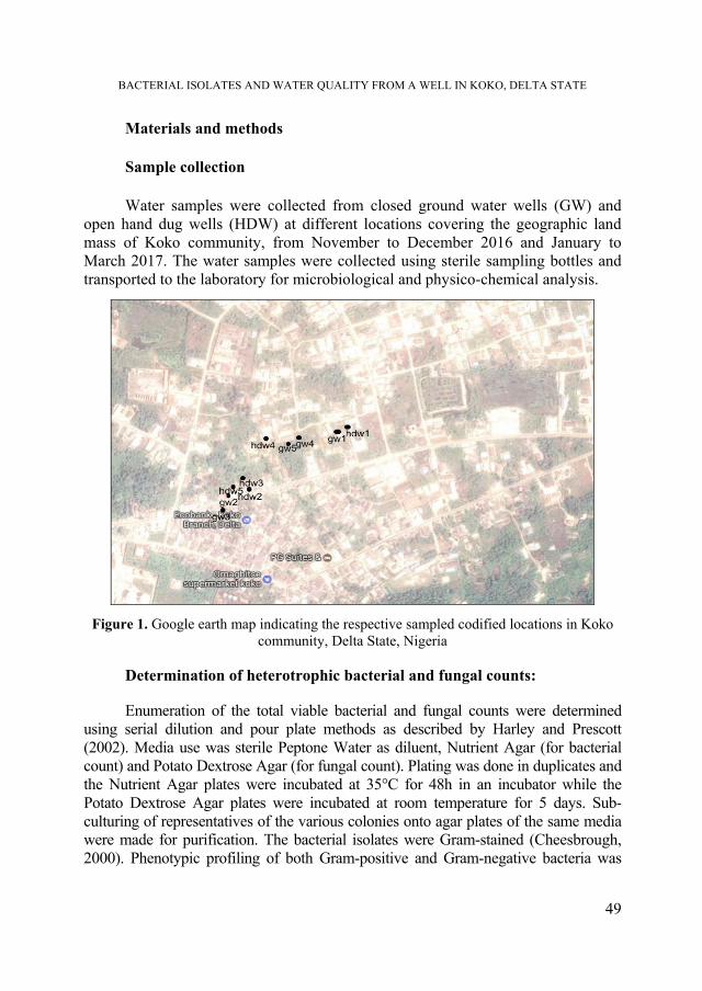
BACTERIAL ISOLATES AND WATER QUALITY FROM A WELL IN KOKO, DELTA STATE
49
Materials and methods
Sample collection
Water samples were collected from closed ground water wells (GW) and open hand dug wells (HDW) at different locations covering the geographic land mass of Koko community, from November to December 2016 and January to March 2017. The water samples were collected using sterile sampling bottles and transported to the laboratory for microbiological and physico-chemical analysis.
Figure 1. Google earth map indicating the respective sampled codified locations in Koko community, Delta State, Nigeria
Determination of heterotrophic bacterial and fungal counts: Enumeration of the total viable bacterial and fungal counts were determined
using serial dilution and pour plate methods as described by Harley and Prescott (2002). Media use was sterile Peptone Water as diluent, Nutrient Agar (for bacterial count) and Potato Dextrose Agar (for fungal count). Plating was done in duplicates and the Nutrient Agar plates were incubated at 35°C for 48h in an incubator while the Potato Dextrose Agar plates were incubated at room temperature for 5 days. Sub-culturing of representatives of the various colonies onto agar plates of the same media were made for purification. The bacterial isolates were Gram-stained (Cheesbrough, 2000). Phenotypic profiling of both Gram-positive and Gram-negative bacteria was

E.E. IMARHIAGBE, B. IKHAJIAGBE
undertaken using API 50CHB and API 20E strips (BioMerieux, Marsielle, France). Additional tests of spore stain and oxidase were also performed. Observable colonial characteristics of the fungal isolates which were noted, microscopic observation of portions of their mycelia and spores using wet mount technique were used in identifying the fungal isolates. Observed fungal spores and mycelial fragment were compared to illustrations contained in Barnett and Hunter (1972).
Determination of coliform and E. coli counts:
These tests were carried out according to methods stated by Cheesebrough (2006) and were conducted in three stages: presumptive stage, confirmatory stage and completed stage.
Determination of antibiotic susceptibility profile of the bacterial isolates:
The antibiogram of the isolates was determined using the disc diffusion method (Harley and Prescott, 2002). The test bacterial isolates were inoculated unto Muller-Hinton Agar and followed by application of the discs (Oxoid) impregnated with different antibiotics. Antibiotic disc contained the following antibiotics: Ciprofloxacin (CPX, 10 µg), Chloramphenicol (CH, 30 µg), Sparfloxacin (SP, 10 µg), Amoxacillin (AM, 30 µg), Augmentin (AU, 30 µg), Gentamicin (CN, 30 µg), Pefloxacin (PEF, 10 µg), Streptomycin (S, 30 µg), Erythromycin (E, 10 µg), Ampiclox (APX, 30 µg), Zinnacef (Z, 20 µg), Ofloxacin (OFX, 5 µg) and Recephin (R, 25 µg). After 24 hours of incubation at 30 oC, the diameter of the zone of inhibition of each antibiotic disc was measured using a ruler and recorded.
Physicochemical analyses:
pH, electrical conductivity, turbidity, total suspended solids (TSS), total dissolved solids (TDS), dissolved oxygen (DO), biochemical oxygen demand (BOD5) were analyzed according to standard analytical procedure (APHA, 1993, Ademoroti, 1996).
Evaluation of heavy metals:
The levels of iron (Fe), manganese (Mn), zinc (Zn), copper (Cu), chromium (Cr) and lead (Pb) were analysed using the atomic absorption spectrophotometer (AAS) (Buck Scientific, Model 210 VGP).
Evaluation of the water quality index of the sampled waters:
The WQI scores were generated using the Weighted Arithmetic Index method as described by Chatterjee and Raziuddin (2002). WQi = {[(Vactual – Videal) / (Vstandard – Videal)] x 100}
50

BACTERIAL ISOLATES AND WATER QUALITY FROM A WELL IN KOKO, DELTA STATE
51
Table 1.
Water Quality Index (WQI) rankings
Range Ranking/Status
0-25 Excellent
26-50 Good
51-75 Poor
76-100 Very poor
Above 100 Unsuitable for drinking
Source: Chatterjee and Raziuddin (2002)
Statistical analysis The non-parametric analogue of the unpaired student T test was utilized to
determine if the differences between the respective microbial and physicochemical data recorded for the ground water and hand dug well water samples was statistically significant (p<0.05).
The results of mean heterotrophic bacterial and fungal counts were in the order 102 to103 cfu/ml respectively. Generally, water samples from open hand dug wells had higher counts (5.4 x 10³ to 8.6 x 10³ cfu/ml) than ground water (table 2). Total microbial counts are important parameters which for indicative of the hygienic and portability properties of drinking water.
Table 2. The heterotrophic microbial and coliform counts of the water samples
Sampling month Water type Mean HBC
(×103cfu/ml) Mean HFC
(×102cfu/ml) TCC
(MPN/100ml) E. coli count
(MPN/100ml)
November 2016 GW 4.4 0.0 5 0.0l
HDW 6.3 5.5 10 5
December 2016 GW 3.6 0.0 5 0.0
HDW 5.4 3.5 10 4
January 2017 GW 4.2 4.0 7 0.0
HDW 6.5 7.5 10 5
February 2017 GW 2.9 4.0 5 0.0
HDW 8.0 9.0 10 4
March 2017
GW 3.7 6.0 8 0.0
HDW 8.6 17.0 20 8
p-values - 0.022 0.000 0.015 0.000
KEY: GW = Closed groundwater sample, HDW = hand dug well, HBC = Heterotrophic bacterial count, HFC = Heterotrophic fungal count, TCC = Total coliform count

E.E. IMARHIAGBE, B. IKHAJIAGBE
52
The results showed that the greatest frequency was observed for Bacillus spp. (30.1 %) and the least predominant among the bacterial isolates was Escherichia coli (6.0 %). Regarding the fungal isolates Aspergillus flavus was the most predominant (26.5 %) and Rhizopus stolonifer (11.8 %) was the least predominant (table 3). Bacterial isolates identified in the water samples include: Micrococcus sp., Klebsiella sp., Escherichia coli, Bacillus sp., Pseudomonas aeruginosa and Aeromonas hydrophila. Fungal isolates identified in the water samples collected include Aspergillus niger, Penicillium notatum, Aspergillus flavus, Rhizopus stolonifer and Saccharomyces cerevisiae. These microbial organisms are of public health importance. The high microbial load and variety of microorganisms detected and isolated from these water samples may be attributed to the poor sanitary and water handling practices.
Table 3.
Percentage frequency of occurrence of bacterial and fungal isolates from the respective water samples
Bacterial isolate No. of
bacterial isolates
% frequency of occurrence
Fungal isolate No. of fungal isolates
% frequency of occurrence
Micrococcus spp. 18 21.7 Aspergillus niger 8 23.5 Klebsiella spp. 7 8.4 Penicillium notatum 8 23.5 Escherichia coli 5 6.0 Aspergillus flavus 9 26.5 Bacillus spp. 25 30.1 Rhizopus stolonifera 4 11.8 Pseudomonas aeruginosa
8 9.6 Saccharomyces cerevisiae
5 14.7
Aeromonas hydrophila 20 24.1 n =83 100 n =34 100
Variable antibiotic susceptibility patterns were observed in the antibiotic
inhibitory zones (mm) among the tested bacterial isolates (Table 4). Findings showed that seven (7) antibiotics did not exhibit inhibition zones against tested Gram negative bacterial isolates (Klebsiella sp., Aeromonas hydrophila, E.coli and Pseudomonas aeruginosa) where as five (5) antibiotics showed no inhibition against Gram positive bacterial isolates (Micrococcus and Bacillus). The widest zone of inhibition was observed for ofloxacin (30.0 mm) against Klebsiella sp., which indicates susceptibility of the bacterial isolate. This observation could be ascribed to the fact that many bacteria isolated from the surface water possess an important ecological quality, namely that of resistance plasmids, which can be picked up in the course of selective processes. Researches had reported the increased incidences of bacterial resistance to antibiotics which contribute to the prevailing trend of antibiotic abuse and misuse by a larger human population most especially in the developing world (Omogbai and Ikenebomeh, 2013, Imarhiagbe et al., 2016). Spanggard et al. (1993) had earlier observed the possibility of members of bacterial groups residing in a common environment to express a similar antibiotics pattern if they share in a common pool of R-factor plasmids.

BACTERIAL ISOLATES AND WATER QUALITY FROM A WELL IN KOKO, DELTA STATE
53
Table 4.
Zone(s) of inhibition (mm) representing antibiotic sensitivity/resistance patterns elicited by the bacterial isolates against the test drugs
Gram +ve
PEF (10 µg)
CN (30 µg)
APX (30 µg)
Z (20 µg)
AM (30 µg)
R (25 µg)
CPX (10 µg)
S (30 µg)
SXT (30 µg)
E (10 µg)
(1) 0.0 13.0 0.0 17.0 0.0 20.0 24.0 0.0 0.0 11.0
(2) 19.0 25.0 0.0 20.0 12.0 28.0 30.0 14.0 0.0 18.0
Gram -ve
SXT(30 µg)
CH (30 µg)
SP (10 µg)
CPX (10 µg)
AM (30 µg)
AU (30 µg)
CN (30 µg)
PEF (10 µg)
OFX (5 µg)
S (30 µg)
(3) 0.0 08.0 0.0 18.0 0.0 14.0 20.0 0.0 27.0 15.0
(4) 0.0 0.0 0.0 15.0 0.0 0.0 16.0 0.0 20.0 0.0
(5) 0.0 0.0 0.0 20.0 0.0 0.0 18.0 0.0 25.0 0.0
(6) 0.0 13.0 15.0 22.0 0.0 12.0 20.0 10.0 30.0 10.0
KEY: (1) Micrococcus sp., (2) Bacillus sp., (3) Aeromonas hydrophila, (4) Pseudomonas aeruginosa, (5) E. col, (6) Klebsiella sp. Ciprofloxacin (CPX), Chloramphenicol (CH), Sparfloxacin (SP), Amoxacillin (AM), Augmentin (AU), Gentamicin (CN), Pefloxacin (PEF), Streptomycin (S), Erythromycin (E), Ampiclox (APX), Zinnacef (Z), Ofloxacin (OFX) and Recephin (R) S= Streptomycin.
Table 5 revealed the result of the physicochemical analysis of the water samples from closed groundwater and open hand dug well from Koko community. pH value is significant determinant for the suitability of water for several purposes. pH of the groundwater samples and hand dug well water samples ranged from 4.40-5.741 and 4.83-5.54 respectively. The findings show that water from this location is slightly acidic; this observation may be due to the geological conditions (King and Ekeh, 1990). Electrical conductivity for groundwater samples and hand dug well water samples ranged from 52.7-86.2 µS/cm and 145.3-330 µS/cm respectively. Measurement of turbidity has been described as a very important parameter when evaluating the quality of a drinking water. Findings from this study revealed that water sourced from closed ground water and open hand dug wells had a mean 0.0 and 2.82 NTU respectively. The highest values of TDS and TSS were observed in open hand dug wells (4.54 mg/L and 98.10 mg/L respectively). Dissolved oxygen for both groundwater samples and hand dug well water samples ranged from 5.5-7.1 mg/L and 4.5-6.6 mg/L respectively. BOD5 for groundwater samples and hand dug well water samples ranged from 0.4-1.1mg/L and 1.9-4.7mg/L. Also heavy metal analysis of water samples from closed groundwater and open hand dug wells is indicated in Table 5.

E.E. IMARHIAGBE, B. IKHAJIAGBE
54
Table 5.
Physicochemical analysis of water samples from closed Groundwater (GW) and open Hand Dug (HDW) wells from Koko community
Parameter
Water type
Month (F) (G)
(A) (B) (C) (D) (E) pH GW 5.19 5.74 4.54 4.40 4.16 4.81±0.65
6.5-8.5 HDW 5.54 4.83 5.12 4.92 5.22 5.13±0.28
EC (µS/cm) GW 60 57.8 63.8 52.7 86.2 64.1±12.99 NA
HDW 330 152.6 174.1 145.3 193.1 199.02±75.6 Turbidity (NTU)
GW 0.0 0.0 0.0 0.0 0.0 0.00±0.00 5.0
HDW 0.0 2.7 8.8 0.0 2.6 2.82±3.59 TSS (mg/l) GW 0.0 0.0 0.0 0.0 0.0 0.00±0.00
500 HDW 1.2 5.1 9.2 2.1 5.1 4.54±3.14
TDS (mg/l) GW 31 28.9 31.7 28.1 42.9 32.52±5.99 500
HDW 160 76.1 86.3 72.3 95.8 98.10±35.79 DO (mg/l) GW 5.9 6.3 5.5 6.8 7.1 6.32±0.65
NA HDW 6.1 4.5 5.2 5.9 6.6 5.66±0.82
BOD5 (mg/l) GW 0.4 0.0 0.0 1.5 1.1 0.60±0.67 NA
HDW 4.7 2.3 1.9 3.7 4.8 3.48±1.34 p-values - 0.000 0.000 0.000 0.000 0.000 0.000 -
(A) November 2016, (B) December, 2016, (C) January, 2017, (D) February 2017, (E) March 2017, (F) Mean ± S.D, (G) WHO limit, (NA) Not available
Table 6.
Water Quality Index of closed groundwater and open hand dug sources
Parameter
Closed ground water (GW) Open hand dug wells (HDW) Vi Si Wi qi Wiqi Vi Si Wi qi Wiqi
pH 4.81 6.5-8.5 0.22 0 0 5.13 6.5-8.5 0.22 0 0 EC (µS/cm) 64.1 250 0.37 25.6 9.51 199.02 250 0.37 79.61 29.54 TDS (mg/L) 32.5 500 0.004 6.50 0.02 98.10 500 0.004 19.62 0.07 TSS (mg/L) 0.00 500 0.003 0 0 4.54 500 0.004 0.91 0.003 DO (mg/L) 6.32 5 0.37 86.3 32.11 5.66 5 0.37 93.13 34.67 BOD5 (mg/L) 0.60 5 0.37 12.0 4.47 3.48 5 0.37 69.60 25.91
ΣWi= 1.34
ΣWiqi =
46.1
ΣWi= 1.34
ΣWiqi =
90.2 WQI (ΣWiqi / ΣWi) = 46.1/1.34 = 34.4 (ΣWiqi / ΣWi) = 90.2/1.34 = 67.31 Ranking Good Poor
Observed (Vi), Standard values (Si), Unit weights (qi), and quality rating (qi)
The observed water quality was supported by the findings from the
analytical physico-chemical parameters (Table 6). Water quality index (WQI) value obtained closed groundwater sources and open hand dug wells was 34.4 and 67.31 respectively. The derived water quality index of the respective water

BACTERIAL ISOLATES AND WATER QUALITY FROM A WELL IN KOKO, DELTA STATE
55
samples revealed that the overall water quality of the samples sourced from open hand dug wells was poor as compared with closed ground water. Based on the WQI values of the samples, it can be inferred that the water samples from open hand dug wells are unfit for direct consumption by the inhabitants of this community.
Table 7. Levels of heavy metals (mg/l) in water samples collected from closed groundwater and open hand dug wells sited in Koko town, Delta State
Parameter
Water type
Month (F)
(G) (A) (B) (C) (D) (E)
Fe GW 0.28 0.15 0.54 0.26 0.17 0.28±0.156 0.30 HDW 0.62 0.79 1.12 0.84 0.62 0.80±0.205
Mn GW 0.01 0.01 ND 0.01 ND 0.004±0.005 0.1 HDW 0.05 0.01 0.14 0.09 0.055 0.07±0.049
Zn GW 0.08 0.03 0.18 0.07 0.09 0.09±0.055 5.00 HDW 0.17 0.24 0.62 0.45 0.13 0.32±0.207
Cu GW ND 0.01 ND 0.004 ND 0.0028±0.004 1.0 HDW 0.02 0.02 0.04 0.061 0.027 0.03±0.017
Cr GW ND ND ND ND ND 0.00±0.00 0.050 HDW 0.003 ND 0.05 0.032 0.019 0.02±0.021
Pb GW ND ND ND ND ND 0.00±0.00 0.01 HDW ND ND 0.02 0.005 ND 0.005±0.009
p-values - 0.001 0.001 0.001 0.005 0.001 0.001
Key: (A) November 2016, (B) December, 2016, (C) January, 2017, (D) February 2017, (E) March 2017, (F) Mean ± S.D, (G) WHO limit, iron (Fe), manganese (Mn), zinc (Zn), copper (Cu),
chromium (Cr) and lead (Pb)
Generally, heavy metal levels were below recommended permissible levels
by World Health Organisation for drinking water. Iron concentration in both groundwater samples and hand dug well water samples ranged from 0.15-0.28mg/L and 0.62-1.12 mg/L (Table 7).
Conclusions The results obtained from this research indicate that the water samples collected
from this community had poor microbiological and overall Water Quality indices and therefore is unsuitable for direct human consumption. Several measures which include boiling, filtration and addition of flocculants such as aluminium salts would invariably enhance the potability of the water and reduce the risk of developing

E.E. IMARHIAGBE, B. IKHAJIAGBE
56
water borne gastroenteritis which could arise from the direct consumption of the contaminated water. The entitled authorities ought to ensure proper awareness for treatment of the water sources to safeguard the public health of the population.
REFERENCES Adekunle, L. V., Sridhar, M. K., Ajayi, A. A., Oluwale, P. A., Olawuyi, J. F. (2004) An
assessment of health and socio-economic implications of sachet in Ibadan, Nigeria: A Public Health Challenge. African Journal of Biomedical Research 7(1): 5–8
Ademoroti, C. M. A. (1996) Standard Methods for Water and Effluent Analysis. Foludex Press. Ltd. Ibadan, 182 pp.
Agbaire, P. O., Obi, C. G. (2009) Seasonal variations of some physico-chemical properties of River Ethiope Water in Abraka, Nigeria. Journal of Applied Science and Environmental Management 13(1): 55-57
American Public Health Authority (APHA) (1993) Standard Methods for Examination of Water and Wastewater.18th Edn.APHA.Washington D.C. 1421 pp.
Ashaye, O. A., Couple, A. A., Afolabi, O. O., Fasoyiro, S. B. (2001) Physicochemical properties of pure water samples in South Western, Nigeria. Journal of Food Technology 6(4): 119–120
Barnett, H. L., Hunter, B. B. (1972) Illustrated Genera of Imperfect Fungi. 3rd edn. Burgess Publishing Co. New York, 225 pp.
Chatterjee, C., Raziuddin, M. (2002) Determination of water quality index of a degraded river in Asanol industrial area, Raniganj, Burdwan, West Bengal. Nature, Environment and Pollution Technology 1(2): 181-189
Cheesebrough, M. (2006) District Laboratory Practice in Tropical Countries. Cambridge University Press. London, 434 pp.
Harley, J.P. and Prescott, L.M. (2002). Laboratory Exercises in Microbiology. 5th Edn. Mac Graw Hill, New York, 449 pp.
Imarhiagbe, E. E, Obayagbona, O. N, Osarenotor, O., Eghomwanre, A. F. (2016) Antibiotic sensitivity pattern of bacterial isolates and physico-chemical composition of maize flour sold in major markets in Benin City, Midwestern Nigeria, Studia UBB Biologia, 61(2): 5-12
Jeffre, H. (2008) Water Problems, Solutions and Conservation in the Developing World http:⁄⁄factsanddetails.com
King, R. P., Ekeh, I. B. (1990) The status and seasonality in the physico-chemical hydrology of a Nigerian head water stream. Acta Hydrobiol., 32, 313-328
Muhammad, B. G., Ismail, B. S., Ekhwan, T., Sujaul, I. M., Tan, C. C. (2007) A physicochemical assessment of the Behar River, Pahang, Malaysia. Global Journal of Environmental Research 1: 7-11

BACTERIAL ISOLATES AND WATER QUALITY FROM A WELL IN KOKO, DELTA STATE
57
Nwodo, C. S., Obinna, C. N., Adetayo, Y., Oluwadamisi, V. N. (2011) Assessment of water quality in Canaanland, Ota, Southwest Nigeria. Agriculture and Biology Journal 2(4): 577-583
Omogbai, B. A., Ikenebomeh, M. (2013) Microbiological characteristics and phytochemical screening of some herbal teas in Nigeria, Euro. Sci. J. 18, 149-160
Orewole, M. O., Mkainde, O. W., Adekalu, K., Shittu K. A. (2007) Chemical examination of piped water supply of Ile-Ife in South West Nigeria. Iran Journal of Environment Health Science 4(1): 51-56
Spanggard, B., Jorgenses, F. G., Huss, H. H. (1993) Antibiotic resistance in bacteria isolated from three freshwater farms and an unpolluted stream in Denmark, Aquaculture 195-207
Tchobanoglous, G., Burton, F. L., Stensel, H. D. (2003) Wastewater Engineering (Treatment Disposal Reuse) / Metcalf & Eddy, Inc. (4th Edition ed.). McGraw-Hill Book Company, 88pp.
Third World Academy of Sciences (TWAS) (2002) Safe Drinking Water: the need, the problem, solutions and action plan, Trieste, Italy. http:⁄⁄ www.g77.org⁄sshlest⁄TWAS
UN Department of Economic and Social Affairs (UNDESA) (2004) Urban agglomerations. Population Division of the Department of Economic and Social Affairs, United Nations, 78pp.
WHO (2006) Guidelines for drinking Water Quality: Recommendations. 3rd Edition, WHO. Geneva, 66pp.


STUDIA UNIVERSITATIS BABEŞ-BOLYAI BIOLOGIA, LXIII, 1, 2018 (p. 59-72) doi:10.24193/subbbiol.2018.1.05
Preliminary data regarding genetic diversity of several endangered and endemic Dianthus species from Romania generated by RAPD markers
Anca-Livia Butiuc-Keul1,2,, Liliana Jarda3, Irina Goia2,4, Irina Holobiuc5, Anca Farkas1,2 and Victoria Cristea3,2
SUMMARY. Conservation of endangered and endemic species of Dianhtus from Romania, requires the investigation of genetic polymorphism in the populations. Preliminary data were obtained by molecular characterization using RAPD markers. DNA amplification with the 9 RAPD primers of the individuals belonging to different populations of D. callizonus, D. giganteus ssp. banaticus, D. glacialis ssp. gelidus, D. henteri, D. nardiformis, D. pratensis ssp. racovitzae, D. spiculifolius and D. tenuifolius revealed low level of polymorphism within and between populations. Several polymorphic RAPD markers were identified being useful for investigation of genetic diversity. Out the 9 primers studied by us, only the primer OPB-07 ensured amplification in all species and primers OPA-13, OPE-04 and 1225 showed positive results in most of the species. The primers 4A-26 and 4A-27 ensured amplification only in D. spiculifolius and the primers 4A-23 and OPM-18 gave no results in none of the species. Keywords: Dianthus, endemic/endangered species, genetic polymorphism, RAPD markers.
Introduction Nowadays, in response to an alarming increase of species number with
different degrees of endangerment, the conservation of biodiversity has a great 1 Babeş-Bolyai University, Faculty of Biology and Geology, Department of Molecular Biology and
Biotechnology, 400084 Cluj-Napoca, Romania. 2 Babeş-Bolyai University, Center of Systems Biology, Biodiversity and Bioresources, Laboratory of
Plant Biology, Genetics, Microbiology and Biotechnology, 400006 Cluj-Napoca, Romania. 3 Babeş-Bolyai University, Alexandru Borza Botanical Garden, 400015 Cluj-Napoca, Romania. 4 Babeş-Bolyai University, Faculty of Biology and Geology, Department of Taxonomy and Ecology,
400015 Cluj-Napoca, Romania. 5 Institute of Biology, Romanian Academy, 060031 Bucharest, Romania. Corresponding author: Anca-Livia Butiuc-Keul, Babeş-Bolyai University, Faculty of Biology and
Geology, Department of Molecular Biology and Biotechnology, 400084 Cluj-Napoca, Romania, E-mail: [email protected]

A. L. BUTIUC-KEUL, L. JARDA, I. GOIA, I. HOLOBIUC, A. FARKAS, V. CRISTEA
60
importance. The concept of sustainable development implies the conservation of biodiversity both in situ and ex situ. Ex situ conservation is a viable alternative for highly endangered taxa in their natural environment, or may complete the in situ conservation methods (Marriott and Sarasan, 2010; Johnson et al., 2012; Butiuc-Keul, 2006; 2014; Cristea et al., 2014) that has consequences not only in science, but also in economy and culture. In order to develop a proper conservation management it is very important to know the genetic variability of in situ populations. In the case of in vitro conservation, it is also necessary to evaluate the somaclonal variability (i.e., the variability induced by the in vitro conditions) and to avoid the genetic uniformity. According to the International Union for Conservation of Nature (IUCN) there are approximately 33400 plant species threatened with extinction (Primack et al., 2008). Moreover, in 2014, there were 18291 plant species on IUCN red list (http://discover.iucnredlist.org/search?key=plants).
Genetic structure of plant populations, the level of genetic polymorphism within and among populations could offer valuable information in order to develop proper strategies for their conservation. The aim of conservation programs is to preserve a high number of populations, as well as their genetic structure and variability (Halmagyi and Butiuc-Keul, 2007; Butiuc-Keul, 2014). A characteristic of endangered and/or endemic plants is the maintenance of low levels of genetic variation in populations. Limited genetic diversity has been reported for many endangered and endemic plant species. The genetic structure and variability of endemic and rare plant populations were intensively studied by DNA markers (Luan et al., 2006; Breinholt el al., 2007; Cristea et al., 2014; Jarda et al., 2014).
Dianthus is one of the most diverse Mediterranean genera, more than 300
species were identified in Eurasia and Africa and over 100 species were found in Europe, more than 70 being endemic (Valente et al., 2010). In Romania, 58 Dianthus taxa have been recorded (Ciocârlan, 2009), of which 8 are endemic.
Our previous studies regarding the conservation of endemic and endangered species ensured valuable data for ex situ conservation strategies of several plant species of Dianthus as D. giganteus ssp. banaticus (Jarda et al., 2014), D. henteri (Cristea et al., 2010), D. spiculifolius (Butiuc-Keul et al., 2001; 2016; Cristea et al., 2006; 2009; 2013; 2014). Concerning the Dianthus genus our major interest was focused on the evaluation of genetic structure of populations and genetic variability within and between populations of several endemic and endangered species from Romania. Molecular markers as RAPD are valuable tools for such investigations because they are cost efficient, requires lower amounts of DNA and there is no necessary information about marker sequence. Thus, in this study we report the preliminary data regarding the usefulness of RAPD markers for the assessment of genetic polymorphism in the population of several Dianthus species from Romania.

GENETIC DIVERSITY OF SEVERAL DIANTHUS SPECIES FROM ROMANIA
61
Materials and methods
Plant material
The plant material was collected from different populations of the studied Dianthus taxa from Romania. Five individuals were collected from each location. Thus, D. callizonus was collected from 2 locations from Piatra Craiului Mountains: P1-Spîrlea refuge and Zăplaz (45°31'42.604"N, 25°12'00.174"E) and Diana refuge and Padina Popii, Piatra Craiului Mountains (45°33'29.184"N, 25°14'29.754"E); D. giganteus ssp. banaticus was collected from 3 populations: P1-Eșelnița (44°39'27.99"N, 22°17'47.42"E), P2-Domogled Mt. (44º52’36.20”N, 22º26’13.00”E) and P3-Țesna Gorge (44º58’03.30”N, 22º30’34.10”E); D. glacialis ssp. gelidus from 3 populations from Bucegi Mountains: P1-Omu Peak (45°22'51.004"N, 25°30'40.004"E), P2-Bâlea Lake, Făgăraş Mountains (45°31'40.004"N, 24°44'26.004"E), P3-Obârşia, Bucegi Mountains (45°22'50.004"N, 25°30'39.004"E); D. henteri from 3 populations from Vâlcea county: P1-Cornet (45°23'19.82"N, 24°18'28.54"E), P2-Călineşti Valey (45°22'19.34"N, 24°17'23.23"E), P3-Jiului Gorge (45°16'46.004"N, 23°23'19.004"E); D. nardiformis from 3 populations from Tulcea county: P1-Allah Bair (44°29'01.42"N, 28°13'24.88"E), P2-Consul Hill (45°10'55.19"N; 28°16'15.77"E), P3-Măcin (45°14'22.71"N, 28°35'01.01"E); D. pratensis ssp. racovitzae from 2 populations: P1-Dorobanț village, Iassy County (47°14'33"N, 27°34'55"E), P2-Spătaru, Buzău County (45°4'14.56"N, 26°47'1.64"E); D. spiculifolius from 2 populations: P1-ROSCI0027 Natura 2000 site, Hăşmaş Mountains (46°44'274"N, 25°47'584"E), P2-ROSCI0002 Natura 2000 site, Apuseni Natural Park, Vlădeasa Massif (N 46°35'454"N, 22°48'384"E); D. tenuifolius from 2 populations from Suceava county: P1-Stânişoarei Mountains (47°21'50.87"N, 25°36'10.88"E), P2-Bistriţei Mountains (47°23'30.00"N, 25°30'16.65"E).
RAPD analysis
DNA was isolated using the CTAB method (Doyle and Doyle, 1987), RAPD markers were obtained by PCR amplification, performed in 25 μL of mixture containing 2 mM MgCl2, 1 μM of each primer, 200 μM of each dNTP, 1.0 U of Taq (Fermentas) in reaction buffer (10mM TrisHCl pH 8.8, 50 mM KC1, 1.5 mM MgC12) and 25 ng of genomic DNA. Amplification programme: 1. T=94C, 4 min; 2. T=94C, 45 s; 3. primer alignment at 36 C, 45 s; 4. elongation T=72C, 50 s; steps 2-4, 35X. 9 primers have been tested as it can be seen in Table 1. Amplicons were separated on 1.5% agarose gel, stained with 0.5 μg mL-1 ethidium bromide. At least 2 independent PCR amplifications were performed for each primer.
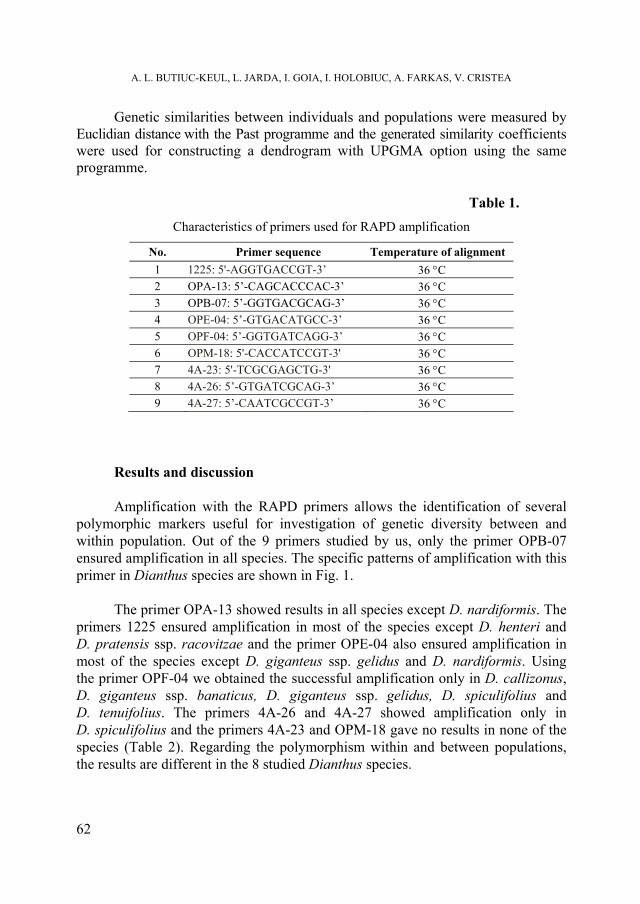
A. L. BUTIUC-KEUL, L. JARDA, I. GOIA, I. HOLOBIUC, A. FARKAS, V. CRISTEA
62
Genetic similarities between individuals and populations were measured by Euclidian distance with the Past programme and the generated similarity coefficients were used for constructing a dendrogram with UPGMA option using the same programme.
Table 1.
Characteristics of primers used for RAPD amplification
No. Primer sequence Temperature of alignment
1 1225: 5'-AGGTGACCGT-3’ 36 C 2 OPA-13: 5’-CAGCACCCAC-3’ 36 C 3 OPB-07: 5’-GGTGACGCAG-3’ 36 C 4 OPE-04: 5’-GTGACATGCC-3’ 36 C 5 OPF-04: 5’-GGTGATCAGG-3’ 36 C 6 OPM-18: 5'-CACCATCCGT-3' 36 C 7 4A-23: 5'-TCGCGAGCTG-3' 36 C 8 4A-26: 5’-GTGATCGCAG-3’ 36 C 9 4A-27: 5’-CAATCGCCGT-3’ 36 C
Results and discussion Amplification with the RAPD primers allows the identification of several
polymorphic markers useful for investigation of genetic diversity between and within population. Out of the 9 primers studied by us, only the primer OPB-07 ensured amplification in all species. The specific patterns of amplification with this primer in Dianthus species are shown in Fig. 1.
The primer OPA-13 showed results in all species except D. nardiformis. The
primers 1225 ensured amplification in most of the species except D. henteri and D. pratensis ssp. racovitzae and the primer OPE-04 also ensured amplification in most of the species except D. giganteus ssp. gelidus and D. nardiformis. Using the primer OPF-04 we obtained the successful amplification only in D. callizonus, D. giganteus ssp. banaticus, D. giganteus ssp. gelidus, D. spiculifolius and D. tenuifolius. The primers 4A-26 and 4A-27 showed amplification only in D. spiculifolius and the primers 4A-23 and OPM-18 gave no results in none of the species (Table 2). Regarding the polymorphism within and between populations, the results are different in the 8 studied Dianthus species.

GENETIC DIVERSITY OF SEVERAL DIANTHUS SPECIES FROM ROMANIA
63
a b c d
e f g h
Figure 1. RAPD patterns of different Dianthus species with OPB-07 primer (a- D.callizonus: 1-molecular marker, 2-Dc 1.1, 3-Dc 1.2, 4-Dc 1.3, 5-Dc1.4, 6-Dc 1.5, 7-Dc 1.6, 8-Dc 1.7, 9-Dc 1.8; b-Dianthus giganteus ssp. banaticus: 1-molecular marker, 2-Dgb 1.1, 3-Dgb 1.2, 4-Dgb 1.3, 5-Dgb 2.1, 6-Dgb 2.2, 7-Dgb 2.3, 8-Dgb 3.1, 9-Dgb 3.2, 10-Dgb 3.3; c- Dianthus giganteus ssp. gelidus: 1-molecular marker, 2-Dgg 1.1, 3-Dgg 1.2, 4-Dgg 1.3, 5-Dgg 1.4, 6-Dgg 1.5, 7-Dgg 2.1, 8-Dgg 2.2, 9-Dgg 2.3, 10-Dgg 2.4, 11-Dgg 2.5, 12-Dgg 3.1, 13-Dgg 3.2, 14-Dgg 3.3, 15-Dgg 3.4, 16-Dgg 3.5; d- Dianthus henteri: 1-molecular marker, 2-Dh 1.1, 3-Dh 1.2, 4-Dh 1.3, 5-Dh 1.4, 6-Dh 1.5, 7-Dh 2.1, 8-Dh 2.2, 9-Dh 2.3, 10-Dh 2.4, 11-Dh 2.5, 12-Dh 3.1, 13-Dh 3.2, 14-Dh 3.3, 15-Dh 3.4, 16-Dh 3.5; e- Dianthus nardiformis: 1-molecular marker, 2-Dn 1.1, 3-Dn 1.2, 4-Dn 1.3, 5-Dn 1.4, 6-Dn 1.6, 7-Dn 2.1, 8-Dn 2.2, 9-Dn 2.3, 10-Dn 3.1, 11-Dn 3.2, 12-Dn 3.3; f-Dianthus pratensis ssp. racovitzae: 1-molecular marker, 2-Dpr 1.1, 3-Dpr 1.2, 4-Dpr 1.3, 5-Dpr 1.4, 6-Dpr 1.6, 7-Dpr 2.1; g-Dianthus spiculifolius: 1-molecular marker, 2-6-negative controls, 7-Ds 1.1, 8-Ds 1.2, 9-Ds 1.3, 10-Ds 1.4, 11-Ds 1.5, 12-Ds 2.1, 13-Ds 2.2, 14-Ds 2.3, 15-Ds 2.4, 16-Ds 2.5; h- Dianthus tenuifolius: 1-molecular marker, 2-Dt 1.1, 3-Dt 1.2, 4-Dt 1.3, 5-Dt 2.1, 6-Dt 2.2, 7-Dt 2.3; number significance: first number-population, second number-individual). Separation on 1.5% agarose gel stained with 0.5 μg/mL ethidium bromide.
Genetic variability in population of D. callizonus was assessed by RAPD
markers generated with different primers as 1225, OPE-04, OPF-04, OPB-07 and OPA-13 (Table 2). All primers generated different polymorphic patterns but the highest polymorphism was observed with the primers OPE-04 and OPB-07. The dendrogram constructed based on Euclidian distance between individuals showed that they are clusterd in two groups of similarity (Fig. 2).

A. L. BUTIUC-KEUL, L. JARDA, I. GOIA, I. HOLOBIUC, A. FARKAS, V. CRISTEA
64
Table 2. Genetic polymorphism of Dianthus species based on RAPD markers
Species Population RAPD fragments 1225 OPA-13 OPB-07 OPE-04 OPF-04 4A-26 4A-27
D.callizonus P1 5 12 7 5 3 - - D. giganteus ssp.
banaticus P1 3 5 3 2 5 - - P2 2 7 3 2 3 - - P3 3 4 3 2 3 - -
D. giganteus ssp. gelidus
P1 4 3 3 - 4 - - P2 2 3 4 - 6 - - P3 4 3 4 - 5 - -
D. henteri P1 - 3 2 5 - - - P2 - 4 2 4 - - - P3 - 4 4 4 - - -
D. nardiformis P1 1 - 1 - - - - P2 2 - 1 - - - - P3 3 - 1 - - - -
D. pratensis ssp. racovitzae
P1 - 1 4 3 - - - P2 - 1 4 1 - - -
D. spiculifolius P1 5 6 5 5 4 2 4 P2 2 4 2 5 3 2 3
D. tenuifolius P1 2 3 1 2 2 - - P2 2 3 2 3 5 - -
Tree Diagram for 8 Variables
Single LinkageEuclidean distances
1.8 2.0 2.2 2.4 2.6 2.8 3.0 3.2
Linkage Distance
1.5
1.6
1.4
1.2
1.8
1.7
1.3
1.1
Figure 2. Dendrogram illustrating similarities between the individuals of
Dianthus callizonus based on RAPD markers.

GENETIC DIVERSITY OF SEVERAL DIANTHUS SPECIES FROM ROMANIA
65
In Dianthus giganteus ssp. banaticus, RAPD amplification was successfully obtained only with the primers 1225, OPE-04, OPF-04, OPB-07 and OPA-13 (Table 2). The primer 1225 showed 3 bands in the populations P1 and P3, and only 2 bands in the population P2. There is one specific band which is present only in population P3-Cheile Ţesnei, Mehedinţi. The populations from Eşelniţa and Domogled are extremely similar, the individuals showing the same RAPD pattern with this primer. Similar RAPD patterns were also obtained in all populations with the primer OPE-04. The amplification with the primer OPF-04 showed differences between individuals and populations, thus this primers is appropiate for genetic polymorphism assessment. RAPD markers generated with the primer OPB-07 are present in all individuals from P2 population and in some individuals from P1 and P3 populations, these patterns being low polymorphic. High genetic polymorphism was showed by OPA-13 primer, while higher polymorphism being identified in the populations P1 and P2 than in the population P3. Based on RAPD markers a dendrogram of similarity between individuals was generated (Fig. 3). Thus, the individuals of Dianthus giganteus ssp. banaticus are clustering in 3 groups. All groups are including individuals from all populations. The populations P1 and P3 are more similar than the population P2. In conclusion the primers OPF-04 and OPA-13 showed high genetic polymorphism between and within populations. The primer OPB-07 showed only genetic polymorphism within populations.
Tree Diagram for 9 Variables
Single LinkageEuclidean distances
1.6 1.7 1.8 1.9 2.0 2.1 2.2 2.3
Linkage Distance
2.2
2.1
3.3
1.2
3.2
3.1
2.3
1.3
1.1
Figure 3. Dendrogram illustrating similarities between the individuals of Dianthus giganteus ssp. banaticus based on RAPD markers.

A. L. BUTIUC-KEUL, L. JARDA, I. GOIA, I. HOLOBIUC, A. FARKAS, V. CRISTEA
66
RAPD amplification of Dinathus giganteus ssp gelidus was successfully obtained with primers 1225, OPF-04, OPB-07 and OPA-13 (Table 2). Amplification with 1225 primer generated 7 different bands showing high level of genetic polymorphism between and within populations. The OPF-04 primer generated high number of bands with polymorphic distribution between populations. The OPB-07 primer generated 5 different bands that showed genetic polymorphism between populations. The primer OPA-13 showed the same pattern of the individuals from P3 population and low genetic polymorphism between individuals from P1 and P2 populations. The dendrogram of similarity showed that the individuals from P3 population and several individuals from P1 popultion are clustered together, while the individuals from P2 population are clustered together in a distinct group. Several individuals from P1 population are clustered in other group at higher genetic distance (Fig. 4).
RAPD amplification in D. henteri was successfully obtained only with the following primers OPE-04, OPB-07 and OPA-13 (Table 2). DNA aplification with the primer OPE-04 generated 5 bands that are present in several individuals, thus this primer showed low genetic polymorphism between and within populations. Similar results were also been obtained with the primer OPB-07, 4 different bands being obtained that are present in some of the individuals. High genetic polymorphism was observed by DNA amplification with the primer OPA-13, 4 fragments were observed but their distribution is very different in the individuals belonging to this plant species. Similarity dendrogram constructed based on RAPD markers showed that the individuals from all populations are very similar. The individuals are clustered in 3 groups, the first and the second contain most of the individuals, while the third group contains only few individuals (Fig. 5).
Tree Diagram for 15 VariablesSingle Linkage
Euclidean distances
1.6 1.8 2.0 2.2 2.4 2.6 2.8 3.0
Linkage Distance
1.5
1.4
2.1
3.2
2.3
2.5
2.4
2.2
3.1
1.2
3.4
3.5
3.3
1.3
1.1
Figure 4. Dendrogram illustrating similarities between the individuals of
Dianthus giganteus ssp. gelidus based on RAPD markers.

GENETIC DIVERSITY OF SEVERAL DIANTHUS SPECIES FROM ROMANIA
67
Tree Diagram for 15 VariablesSingle Linkage
Euclidean distances
0.0 0.5 1.0 1.5 2.0
Linkage Distance
3.3
3.2
2.2
1.2
1.4
3.5
3.1
2.5
2.1
2.4
1.5
2.3
3.4
1.3
1.1
Figure 5. Dendrogram illustrating similarities between the individuals
of Dianthus henteri based on RAPD markers. Regarding the genetic variability in D. nardiformis, amplification with RAPD
markers was very difficult to achieve, only 2 primers as 1225 şi OPB-07 allowed amplification with scorable bands (Table 2). By amplification with the primer 1225, only 2 bands were obtained, the first is present only in the individuals from P2 and P3 populations and the second one is present in all individuals independent on the population. Thus, this primer detected low genetic polymorphism between populations, but not within population. By amplification with OPB-07 primers, the RAPD patterns of all individuals are the same, except several individuals from the population P1. The genetic polymorphism detected only with the primer 1225, did not ensure the different clustering of individuals.
In D. pratensis ssp. racovitzae RAPD amplification was also very difficult, successfully amplification was achieved only with 3 primers as OPE-04, OPB-07 and OPA-13 (Table 2). The amplification with OPE-04 primer generated 3 different fragments, each of them identified in one individual. Amplification with the primer OPB-07 generated 5 fragments that show several differences between individuals. The primer OPA-13 generated only 1 fragment that is present in all individuals. Despite of other species this primer showed no polymorphism in this species. Unfortunately the number of individuals collected form each population is very small and no pertinent conclusion could be showed by these results. The individuals of this species are clustered in 2 groups, in wich are mostly the individuals from population P1. The individual from population P2 belongs to the first cluster (Fig. 6).

A. L. BUTIUC-KEUL, L. JARDA, I. GOIA, I. HOLOBIUC, A. FARKAS, V. CRISTEA
Tree Diagram for 6 VariablesSingle Linkage
Euclidean distances
0.9 1.0 1.1 1.2 1.3 1.4 1.5 1.6 1.7 1.8
Linkage Distance
1.6
1.4
1.3
2.1
1.2
1.1
Figure 6. Dendrogram illustrating similarities between the individuals of Dianthus pratensis ssp. racovitzae based on RAPD markers.
In D. spiculifolius the RAPD amplification was achieved with the most of the primers, as 1225, OPF-04, OPE-04, OPB-07, OPA-13, 4A26 and 4A27 (Table 2). By amplification with 1225 primer we obtained 5 fragments that showed high genetic polymorphism of the individuals from both populations. Similar results were obtained with the primer OPF-04, 5 polymorphic fragments being identified. With the primer OPE-04, also 5 fragments were obtained, this primer showed genetic polymorphism within populations. By amplification with the primers 4A26 and 4A27 low genetic polymorphis was detected, thus the primer 4A26 generated 2 fragments, and the primer 4A27 generated 3 fragments. These primers showed genetic polymorphism between populations. The primer OPB-07 generated 6 fragment that showed genetic polymorphism between and within populations. The highest polymorphism was detected with the primer OPA13, 7 fragments being identified that showed genetic polymorphism within populations. Dendrogram of similarity shows that most of the individuals are clustered togheter in one group, and other group is formed only by the individuals 1.1, 2.1 and 2.3 (Fig. 7).
Amplification with RAPD primers of Dianthus tenuifolius was successfully obtained with the primers 1225, OPE-04, OPF-04, OPB-07 and OPA-13 (Table 2). The primer 1225 generated 2 fragment, the first fragment is present in all individuals and the second is present only in the individuals from Rodnei Mt. By amplification with OPE-04 primer, 4 fragments were obtained having a polymorphic distribution in the individuals belonging to different populations. The genetic polymorphism of the population P2 is very low. The amplification with the primer OPF-04 generated 5 fragments. The amplification with the primer OPB-07 showed only 2 fragments, the first being present in all individuals and the last one only in the individual 2.3.
68

GENETIC DIVERSITY OF SEVERAL DIANTHUS SPECIES FROM ROMANIA
69
Tree Diagram for 9 VariablesSingle Linkage
Euclidean distances
1.5 2.0 2.5 3.0 3.5 4.0
Linkage Distance
1.5
2.4
2.2
1.3
1.4
1.2
2.3
2.1
1.1
Figure 7. Dendrogram illustrating similarities between the individuals of
Dianthus spiculifolius based on RAPD markers. The primer OPA-13 generated 4 fragments with polymorphic distribution between individuals. The dendrogram of similarities showed 2 groups, one of them containing the individuals 1.3 and 2.1 and the secong group containing the individuals 1.1. and 2.2. The other individuals are more different from those included in these clusters (Fig. 8).
Tree Diagram for 6 VariablesSingle Linkage
Euclidean distances
1.9 2.0 2.1 2.2 2.3 2.4 2.5 2.6 2.7
Linkage Distance
2.3
1.2
2.2
2.1
1.3
1.1
Figure 8. Dendrogram illustrating similarities between the individuals of
Dianthus tenuifolius based on RAPD markers.

A. L. BUTIUC-KEUL, L. JARDA, I. GOIA, I. HOLOBIUC, A. FARKAS, V. CRISTEA
70
Despite of many morphological (Farsi et al. 2013) and cytological (Jafari and Behroozian, 2010) studies on different species of Dianthus, there are only limited information on the genetic diversity in the wild populations of these species. There are only few studies regarding the genetic diversity of D. caryophyllus using RAPD markers (Su Yeons, 2002) or D. polylepis and D. crinitus (Behroozian et al., 2013). Recent works revealed low genetic variability of 3 endemic and endangered species of Dianthus from Romania as D. spiculifolius Schur; D. giganteus d'Urv. subsp. banaticus (Heuff) and D. callizonus (Cristea et al. 2014; Jarda et al. 2014; Gabel et al. 2016).
Our preliminary data obtained with RAPD markers showed low level of polymorphism in the populations of the 8 Dianthus species. RAPD markers usually revealed high polymorphism even in the situation when other situs specific markers as SSR showed low polymorphism (Safari et al., 2013; Jarda et al., 2014) but was not the same in our study. Our results are in concordance with other data showing low diversity and polymorphism of Dianthus species from Iberian Peninsula (Balao et al., 2010) and Iran (Farsi et al., 2013) and to other data revealing low genetic polymorphism of plants having small populations isolated in fragmented mountain habitats (Duminil et al., 2007). Mountain and alpine plants have to cope with harsh environmental conditions and usually the low genetic diversity is compensated by clonal propagation, which produces rapid, but spatially limited spread of genotypes (Young et al., 2002; Gabel et al., 2016). This is also the situation of Dianthus species grown in severe environmental conditions (Stöcklin, 1992).
Conclusions Genetic diversity of Dianthus species revealed by RAPD markers is generaly
low. Out of 9 primers used in this study, only 6 generated reproducible and polymorphic patterns, useful for preliminary analysis of genetic polymorphism within and between populations.
Acknowledgements. This work was supported by the projects 31-008/2007 and
71/2014, Partnerships in priority areas - PN II, financed by Romanian Ministry of National Education (CNMP and UEFISCDI). Many thanks to Dr. Mihai Puşcaş Dr. Adrian Oprea, Dr. Gheorghe Coldea, Dr. Tudor Ursu and Dr. Liviu Filipaș for collecting the plant material.
REFERENCES Balao, F., Valente, L. M., Vargas, P., Herrera, J., Talavera, S. (2010) Radiative evolution of
polyploid races of the Iberian carnation Dianthus broteri (Caryophyllaceae), New Phytol. 187:542-551

GENETIC DIVERSITY OF SEVERAL DIANTHUS SPECIES FROM ROMANIA
71
Behroozian, M., Jafari, A., Farsi, M. (2013) RAPD analysis of genetic variation within and among natural population of two species of Dianthus L. (Caryophyllaceae) in NE Iran, Iran. J. Bot. 19 (2):194-201
Breinholt, J. W., Buren, V. R., Kopp, O. R., Stephen, C. L. (2007) Population genetic structure of an endangered Utah endemic, Astragalus ampullarioides (Fabaceae), Am. J. Bot. 96: 661-667
Butiuc-Keul, A. L, Şuteu, A., Munteanu-Deliu, C., Deliu, C. (2001) Study on the in vitro preservation of Dianthus spiculifolius, Schur., Contrib. Bot. 36: 137-147
Butiuc-Keul, A. L (2006) Markeri moleculari utilizaţi în genetică şi biotehnologia vegetală [in Romanian], Mega, Cluj-Napoca, pp. 17-57
Butiuc-Keul, A. L (2014) Biotehnologie generală [in Romanian], Presa Univeristară Clujeană, Cluj-Napoca, pp. 253-267
Butiuc-Keul, A. L., Farkas, A., Cristea, V. (2016) Genetic stability assessment of in vitro plants by molecular markers, Studia UBB Biologia LX1(1):107-114
Ciocârlan, V. (2009) Pictured Flora of Romania [in Romanian], Ceres, Bucureşti. Cristea, V., Puşcaş, M., Miclăuş, M., Deliu, C. (2006) Conservative micropropagation of
some endemic or rare species from the Dianthus genus, Acta Hort. 725: 357-364 Cristea, V., Deliu, C., Oltean, B., Butiuc-Keul, A. L., Brummer, A., Albu, C., Radu, G.L.
(2009), Soiless culture for pharmaceutical use and biodiversity conservation, Acta Hort. 843:157-164
Cristea, V., Palada, M., Jarda, L., Butiuc-Keul, A. L. (2013) Ex situ in vitro conservation of Dianthus spiculifolius, endangered and endemic plant species, Studia UBB Biologia, 58(1):57-69
Cristea, V., Brummer, A. T., Jarda, L., Miclăuş, M. (2010) In vitro culture initiation and phytohormonal influence on Dianthus henteri-a Romanian endemic species, Rom Biotechnol. Lett. 25:25-33
Cristea, V., Crăciunaş, C., Marcu, D., Palada, M., Butiuc-Keul, A. L. (2014) Genetic stability during in vitro propagation of Dianthus spiculifolius Schur, Prop. Ornam. Plants 14:26-31
Doyle, J., Doyle, J. L. (1987) A rapid DNA isolation procedure for small quantities of fresh leaf tissue, Phytochem. Bull. 19:11-15
Duminil, J., Fineschi, S., Hampe, A., Jordano, P., Salvini, D., Vendramin, G. G., Petit, R. J. (2007) Can population genetic structure be predicted from life-history traits. Am. Naturalist 169:662-672
Farsi, M., Behroozian, M., Vaezi, J., Joharchi, M.R., Memariani, F. (2013) The evolution of Dianthus polylepis complex (Caryophyllaceae) inferred from morphological and nuclear DNA sequence data: one or two species? Plant Syst. Evol. 299:1419-1431
Gabel, A. R., Sattler, J., Reisch, C. (2016) Genetic variation and performance of the alpine plant species Dianthus callizonus differ in two elevational zones of the Carpathians, Alp. Bot. doi:10.1007/s00035-016-0177-3.
Halmagyi, A., Butiuc-Keul, A. L. (2007) Conservarea resurselor genetice vegetale [in Romanian], Todesco, Cluj-Napoca, pp. 122-129
Jarda, L., Butiuc-Keul., A. L., Hőhn, M., Pedryć, A., Cristea, V. (2014) Ex situ conservation of Dianthus giganteus d’Urv. subsp. banaticus (Heuff.) Tutin by in vitro culture and assessment of somaclonal variability by molecular markers, Turk. J. Biol. 38:21-30

A. L. BUTIUC-KEUL, L. JARDA, I. GOIA, I. HOLOBIUC, A. FARKAS, V. CRISTEA
72
Johnson, T., Cruse-Sanders, J. M., Pullman, G. S. (2012) Micropropagation and seed cryopreservation of the critically endangered species Tennessee yellow-eye grass, Xyris tennesseensis Kral, In Vitro Cell Dev. Biol. Plant 48:369-376
Luan, S. S., Chiang, T. S., Gong, X. (2006) High genetic diversity vs. low genetic differentiation in Nouelia insignis (Asteraceae), a narrowly distributed and endemic species in China, revealed by ISSR fingerprinting, Ann. Bot. 98:583-589
Marriott, M., Sarasan, V. (2010) Novel micropropagation and weaning methods for the integrated conservation of a critically endangered tree species, Medusagyne oppositifolia, In Vitro Cell Dev. Biol. Plant 46:516-523
Primack, R. B., Pătroescu, M., Rozylowicz, L., Iojă, C. (2008) Fundamentele conservării diversităţii biologice, AGIR, Bucureşti, pp. 343-375
Safari, S., Mehrabi, A. A., Safari, Z. (2013) Efficiency of RAPD and ISSR markers in assessment of genetic diversity in Brassica napus genotypes, Int. J. Agri. Crop Sci. 5:273-279
Stöcklin, J. (1992) Differences in the location of subcotyledonary buds among Epilobium angustifolium L, E. dodonaei Vill and E. fleischeri Hochst (Onagraceae) and effects on architecture and population structure, Bot. J. Linn. Soc. 108:35-47
Su Yeons, K. (2002) Genetic relationship among Korean Dianthus species based on morphological characteristics and RAPD analysis, http://www.niha.go.kr/report_board/ report_board_view.asp?size_tag=&sno=1726&gubun=&p_type=11
Valente, L. M., Savolainen, V., Vargas, P. (2010) Unparalleled rates of species diversification in Europe, Proc. Royal Soc London B: Biological Sciences 277: 1489-1496
Young, A. G., Hill, J. H., Murray, B. G., Peakall, R. (2002) Breeding system, genetic diversity and clonal structure in the sub-alpine for Rutidosis leiolepis F. Muell. (Asteraceae), Biol. Conservation 106:71-78

STUDIA UNIVERSITATIS BABEŞ-BOLYAI BIOLOGIA, LXIII, 1, 2018 (p. 73-80) doi:10.24193/subbbiol.2018.1.06
Protracheoniscus vasileradui – n. sp. (Crustacea, Isopoda, Crinochaeta)
in the Romanian fauna
Nicolae Tomescu1, and Lucian Alexandru Teodor1
SUMMARY. We describe the terrestrial isopod species Protracheoniscus vasileradui n. sp. collected from the deciduous forest in the Liuborajdea Valley, Iron Gates (Porţile de Fier) area. We named it Protracheoniscus vasileradui n.sp., in the memory of Prof. V. Gh. Radu, who was a renowned specialist in terrestrial isopods. Keywords: isopod, specific characters, vasileradui Introduction Between 1967 and 1968 V. Gh. Radu and N. Tomescu conducted studies in
the habitats of the planned reservoir and neighbouring areas of the Iron Gates (Porţile de Fier). Over 500 terrestrial isopod individuals were collected in this period (Radu and Tomescu, 1975, p. 45-46). In these samples four terrestrial isopod species were found to be new for science. Three of these were described by Radu and published in Fauna of the SRR, Crustacea, Isopoda, vol. IV, fascicle 13, 1983. One of the species mentioned in the Fauna of Porţile de Fier (1975), belonging to the genus Protracheoniscus, could not be described at the time by Radu, due to health issues. We recently found 3 ♂ and 8 ♀ Protracheoniscus in the samples collected in that period and we named them Protracheoniscus vasileradui n.sp., in the memory of Prof. V. Gh. Radu, who was a renowned specialist in terrestrial isopods.
1 Babeş-Bolyai University, Faculty of Biology and Geology, Department of Taxonomy and Ecology,
Cluj-Napoca, Romania. Corresponding author: Nicolae Tomescu, Babeş-Bolyai University of Cluj-Napoca, Department of
Taxonomy and Ecology, 5-7 Clinicilor Str., 400006, Cluj-Napoca, Romania. E-mail: [email protected]

N. TOMESCU, L. A. TEODOR
74
Materials and methods The specimens of Protracheoniscus vasileradui were collected with the
help of a leaf litter sieve, from leaf litter collected in the deciduous forests of the Liuborajdea Valley. The study sites in the Liuborajdea Valley are located between the localities Moldova Veche and Orşova, between the kilometre stones 83 and 82.
Three males and 8 females of this species were collected. Two males have been dissected and microscopic slides of the taxonomically relevant organs were prepared with Euparal and Canada balsam. The extremity of the endopodites of the first male pleopods have been photographed from the slides in ventral position and laterally in alcohol, before dissection. The males and two females have been photographed dorsally, in alcohol, in order to have a whole-body image and the dorsal colouration. Two females were included in Canada Balsam, the six other females and a male are kept in 70% alcohol. These, the whole biological material and the microscopic slides from N. Tomescu’s terrestrial isopod collection are going to be donated to the Zoology Museum, Faculty of Biology and Geology of the Babeş-Bolyai University.
In describing the species Protracheoniscus vasileradui we will compare some characteristics with those of the species Protracheoniscus politus, a common, widespread species from the Romanian fauna. N. Tomescu (1972) described the ontogenetic postembryonic development of P. politus, and Tomescu et al. (2016) described the change in the morphology of the endopodites of the first male pleopods in relationship with post-reproductive moulting.
Results
Species description:
Size: males: 5 x 2 – 5.2 x 2 mm, females: 5.2 x 2 – 6 x 2.5 mm (Fig. 1 a, b).
Colour: dark-brown with yellow-orange spots.
Somatic features: cephalon: cephalic lobes poorly developed, large yellowish-orange spots on the cephalon (Fig. 1 c). Pleotelson shorter comparatively to that of the species P. politus (Fig. 1 d).

PROTRACHEONISCUS VASILERADUI – N. SP. IN THE ROMANIAN FAUNA
75
Appendages
Antennae. The second-last segment of the antenna is shorter than the last (Fig. 2 a).
Pereiopods. The seventh pereiopod pair of the male has a straight ventral side of the ischiopodite (Fig. 2 b). Males of the species P. politus have a slight curvature of the ventral side of the ischiopodite (Fig. 2 c).
Pleopods. The first pair of the male pleopod exopodites are approximatively triangular in shape, with the external side slightly concave (Fig. 3 a) and differing from the shape of the first pair of the male pleopod exopodites in P. politus (Fig. 3 e).
Endopodites of the male P. vasileradui have a wide basal half, with the external sides slightly curved (Fig. 3 b). The extremities of the endopodites are pointed (Fig. 3 c), with tips that are ventrally curved (Fig. 3 d). In males of the species P. politus, the first pair of the pleopod endopodites have a chitinous lobe at their extremities (Fig. 3 f, f’), which is shed with the exuvia during post-reproductive moulting and is regrown during the next year (Tomescu, 1972; Tomescu et al., 2016).
Derivatio nominis: we named the species Protracheoniscus vasileradui n.sp., in the memory of Prof. V. Gh. Radu, who was a renowned specialist in terrestrial isopods.

N. TOMESCU, L. A. TEODOR
76
Figure 1. Protracheoniscus vasileradui nov. spec. Holotyp, male and female dorsal view:
a. ♂ 5 x 2 mm, b. ♀ 5.5 x 2.2 mm, c. cephalic lobes, d. pleotelson.

PROTRACHEONISCUS VASILERADUI – N. SP. IN THE ROMANIAN FAUNA
77
Figure 2. Protracheoniscus vasileradui nov. spec. Holotyp, ♂ 5 x 2 mm, a. antenna, b. pereiopods 7. Protracheoniscus politus, ♂ 6.5 x 2.8, c. pereiopods 7.

N. TOMESCU, L. A. TEODOR
78
Figure 3. Protracheoniscus vasileradui nov. spec. Holotyp, ♂ 5 x 2 mm, a. exopod pleopods 1, b. endopod pleopods 1, c. apex of the endopod pleopods 1 – ventral view, d. apex of the endopod pleopods 1 – prophile view. Protracheoniscus politus, ♂ 6.5 x 2.8, e. exopod of pleopods 1, f. endopod of pleopods 1, f`. apex of the endopod pleopods 1.

PROTRACHEONISCUS VASILERADUI – N. SP. IN THE ROMANIAN FAUNA
79
Discussion The species Protracheoniscus vasileradui n. sp. has special morphological
characteristics in comparison to Protracheoniscus politus, a wide-spread species in Romania. The habitat, where the specimens of P. vasileradui were collected from, deciduous forests of the Liuborajdea Valley, in the Iron Gates (Porţile de Fier) area, seems to be a geomorphologically isolated area, that favoured, in time, the isolation of P. vasileradui populations. Research conducted in 1967 and 1968 aimed at recording plant and animal species in the area where a reservoir was planned to be formed, by building the Iron Gates dam on the Danube. The research was extended into the habitats of the valley slopes that were not covered by the water of the reservoir. The forest in the Liuborajdea Valley, where we collected the P. vasileradui specimens, is located on such a slope, and we believe these populations have survived until the present day. It is possible that the species is present also on areas in the vicinity of the Liuborajdea Valley, with similar ecological conditions.
Conclusions The specific morphological differences between Protracheoniscus vasileradui n.
sp. and Protracheoniscus politus C. L. Koch 1841 lie in the shape of the male’s first pair of exopodites and endopodites, and that of the male’s ischiopodites of the seventh pair of pereiopods. These are essential morphological characteristics in the separation of species belonging to the genus Protracheoniscus.
REFERENCES
Radu, G. V., Tomescu, N. (1975) Fauna de la Porţile de Fier, Isopoda [in Romanian], Ed. Acad. R. S. R. [in Romanian]
Radu, G. V. (1983) Crustacea, Isopoda, Oniscoidea, Oniscidae inferioare [in Romanian], Fauna R.S.R., IV(13), pp. 168
Radu, G. V. (1985) Crustacea, Isopoda, Oniscoidea, Crinocheta [in Romanian], Fauna R.S.R., IV(14), pp. 155

N. TOMESCU, L. A. TEODOR
80
Tomescu, N. (1972) Reproduction and ontogenetic development of Protracheoniscus politus, Rev. Roum. Biol., Zool., 17 (1), 31-39
Tomescu, N., Teodor, L. A., Ferenţi, S., Covaciu-Marcov, S. D. (2016) Two Protracheoniscus Species (Crustacea, Isopoda, Oniscidea) in Romanian Fauna: Morphology, Ecology and Distribution, Studia UBB Biologia, 61(1): 147-166

STUDIA UNIVERSITATIS BABEŞ-BOLYAI BIOLOGIA, LXIII, 1, 2018 (p. 81-88) doi:10.24193/subbbiol.2018.1.07
Clutch size and egg repeatability in three elusive bird species: Little
Bittern (Ixobrychus minutus), Little Crake (Zapornia parva) and Water Rail (Rallus aquaticus) from north-west Romanian populations
Alin David1, Alexandru Nicolae Stermin1, and Eliana Sevianu2
SUMMARY. Data were collected from Little Bittern, Water Rail and Little Crake nests located in North-Western Romania, between April and July of 2002 - 2006 and 2010 - 2012. The repeatability was calculated as intra-class correlation of length, breadth and egg volume coefficients. The total number of active nests considered for the present study was as follows: 43 for Little Bittern, 25 for Water Rail and 9 for Little Crake. The clutch size in Little Bittern ranged from 4 to 7 eggs, in Water Rail from 4 to 10 and in Little Crake from 4 to 8 eggs. The repeatability values of egg parameters varied between 0.844 to 0.860 in Little Bittern, 0.262 - 0.374 in Water Rail and 0.310 - 0.574 in Little Crake. Short-term environmental impact was strong in case of Water Rail and Little Cracke, while the genetic component had little influence. For these two species, the low repeatability could be explained by larger numbers of eggs in a clutch, reflecting in turn a longer laying period. Keywords: breadth, egg volume, laying period, length. Introduction Clutch size and egg characteristics vary among and within bird species (Lack,
1947; Klomp 1970; Figuerola and Green, 2005). These variations are important in revealing the individual’s fitness (Rockwell et al., 1987, Brisk and Sealy, 1989; Horak et al., 1997) and aspects about species evolution (Martin, 1995; Figuerola and Green, 2005). 1 Babeş-Bolyai University, Department of Taxonomy and Ecology, Cluj-Napoca, Romania. 2 Babeș-Bolyai University, Department of Environmental Science, Cluj-Napoca, Romania. Corresponding author: Alexandru Nicolae Stermin, Department of Taxonomy and Ecology,
Babeş-Bolyai University, Cluj-Napoca, Romania, E-mail: [email protected]
A. DAVID, A. N. STERMIN, E. SEVIANU
82
In bird species, clutch size may depend on many factors, such as the effective coverage capacity of the eggs during incubation (Rice and Kenyon, 1962) or the pressure from predators during nesting (in the sense that, the more eggs are being laid, the more exposed to predators they are) (Slagsvold, 1982; Arnold et al., 1987). The chances in offspring survival of large clutches usually decrease, in the sense that as the number of the offspring is larger, the possibility of being detected by predators increases (Cooch, 1961; Hilden, 1964; Lessells, 1986). Other factors influencing the clutch size are: food resources available during egg laying (Lark, 1974) and temperature during egg production (Great Tits, Parus major roosting in cooled nestboxes laid eggs 14% smaller than those roosting in heated nestboxes (Nager and Noordwijk, 1992)). In addition, significant effects on clutch size and egg characteristics are reported from parameters like: female age and experience (a statistically significant increase in egg size with age or experience was found in almost half (17/37) of the studies (Christians, 2002)); female mass and size; and other aspects of the phenotype (Christians, 2002).
The egg size and their intraclutch and interclutches variations are important in determining the offspring body size and weight, their survival probability and therefore the reproductive success (Schifferli, 1973; Williams, 1994; Surmacki, 2003). Variations in egg size are the consequences of genetic and environmental action (Surmacki, 2003), repeatability dimensions characterizing environmental conditions and food resources during laying period (Stermin et al., 2008).
By calculating the repeatability of egg size we can split the phenotypic variation in egg size into within-individual and among-individual components. Variation in egg size among individuals is caused by a combination of genetic and environmental differences, while the variation in egg size within an individual during several years is caused by temporary environmental differences between reproductive attempts (Falconer, 1989; Flint et al., 2001).
In this context, analyzing the repeatability in egg size can reveal information about the ecology and biology of bird species. These data bring valuable information about several bird species which are poorly studied, due to their elusiveness and rather inaccessible habitats. Some of the less studied bird species in Europe are Little Bittern Ixobrychus minutus, Little Crake Zapornia parva and Water Rail Rallus aquaticus (Marion et al., 2000; Stermin et al., 2011; Stermin et al., 2013).
Those three species coexist in dense wetland habitats and during their evolution each has adapted to occupy specific microhabitats inside the wetlands (Taylor, 1998; David, 2008; Stermin et al., 2012). Little Bittern and Little Crake are protected species and, along with the Water Rail, are now being affected by habitat loss and fragmentation (Marion et al., 2000; Brambilla et al., 2012). New information about their biology and ecology is important not only in solving the mystery of their elusive life, but also in designing and implementing conservation programs.

EGGS REPEATABILITY IN WETLAND BIRDS
83
Thus, the main goals of our study are: (i) to describe the variation of clutch size and egg parameters in Little Bittern, Water Rail and Little Crake populations from the Fizeş Basin (N-W Romania) and (ii) to calculate the repeatability and compare the results to other studies on other bird species by analyzing them in relation to the effect of the environment (sensus Bańbura and Zieliński, 1998).
Materials and methods The study area is located in the central part of the Transylvanian Plain, in
Romania (24°10’ E; 46°50’N). The wetlands surveyed here represent 1/3 from all Transylvanian wetlands. Most wetlands from the Fizeş Basin are man-made and used as fishing ponds; only two of them are natural, and were recently designated “Natura 2000” site (Special Protected Area) (David, 2008). The wetland areas range from 19 to 253 ha, being partially or totally covered by marsh vegetation.
Data were collected between April and July of 2002 - 2006 and 2010 - 2012. During this time, the areas suitable for Little Bittern, Little Crake and Water Rail nesting were checked at intervals of 2 to 5 days. Transect method was used in nest searching (Bibby et al., 2000). The Little Bittern nests were located in Lakes Știucii, Borzaş, Năsal, Ţaga Mare, Sucutard I, Roşieni, Tău Popii, Cătina and Legii Pond; the Little Crake nests were found in Lakes Sucutard I and Cătina, while the Water Rail nests were situated on Sic Reedbeds and Lake Știucii.
The length (L) and breadth (B) of eggs were measured to the nearest 0.1 mm with digital caliper. The volume was calculated using the following formula: V = k × L × B2 / 1000 (where k = 0.51) (Hoyt, 1979). The egg weight was measured in the field using a digital scale.
One-way analysis of variance (ANOVA) was carried out in order to obtain variance components. The repeatability was calculated as intra-class correlation coefficients (Lessells and Boag, 1987; Falconer and Mackay, 1995; Sokal and Rohlf, 1995), standard errors for repeatability values were calculated as described in Becker (1992).
Results and discussion A total of 223 eggs from 43 active Little Bittern nests, 209 eggs from 25
Water Rail nests and 56 eggs from 9 Little Crake nests were measured. The clutch size in Little Bittern ranged from 4 to 7 eggs, in Water Rail from 4 to 10 and in Little Crake from 4 to 8 (Table 1).

A. DAVID, A. N. STERMIN, E. SEVIANU
84
Table 1.
Clutch size characteristics in Little Bittern, Water Rail and Little Crake nests located in the Fizeş Basin (SD - standard deviation)
Little Bittern Water Rail Little Crake
Total number of clutches 43 25 9
Minimum number of eggs/clutch 4 4 4
Maximum number of eggs/clutch 7 10 8
Mean of eggs/clutch ± SD 5.32 ± 0.74 8.36 ± 1.55 6.22 ± 1.39
Egg measurements for Little Bittern, Water Rail and Little Crake are presented in table 2, while repeatability values are depicted in table 3.
Table 2.
Egg measurements from Little Bittern, Water Rail and Little Crake clutches located in the Fizeş Basin (SD - stardard deviation)
Little Bittern
(n = 223) Water Rail
(n= 209) Little Crake
(n=39)
Length (mm)
Minimum 30.80 26.59 28.87
Maximum 37.80 39.59 34.09
Mean ± SD 34.92 ± 1.45 35.92 ± 1.64 31.01 ± 1.38
Breadth (mm)
Minimum 22.10 23.88 20.58
Maximum 29.10 36.93 22.81
Mean ± SD 25.55 ± 0.92 25.96 ±1.24 21.76 ± 0.57
Volume (cm3)
Minimum 8.81 8.63 6.33
Maximum 16.15 24.14 8.42
Mean ± SD 11.66 ± 1.14 12.38 ± 1.42 7.49 ± 0.58
Weight (g)
Minimum - 9.9 6.60
Maximum - 15.10 8.90
Mean ± SD - 12.43 ± 0.95 7.71 ± 0.61

EGGS REPEATABILITY IN WETLAND BIRDS
85
Table 3.
Repeatability (R) of egg length, breadth and volume index in Little Bittern, Water Rail and Little Crake clutches located in the Fizeş Bazin (SE - standard
error; Df - degrees of freedom; F - variance ratio; P - significance level)
R SE df F P
Little Bittern
Length 0.844 3.055 42 23.374 < 0.001
Breadth 0.860 1.639 42 6.363 < 0.001
Volume 0.851 2.225 42 10.932 < 0.001
Water Rail
Length 0.374 9.326 23 8.537 < 0.001
Breadth 0.355 7.270 23 9.863 < 0.001
Volume 0.262 15.824 23 28.212 < 0.001
Little Crake
Length 0.574 10.771 8 16.092 <0.001
Breadth 0.517 3.288 8 7.294 <0.001
Volume 0.310 3.917 8 24.83 <0.001
Significant statistical differences between species were found regarding egg
measurements: length (F(2.468) = 196.60, df = 2, P < 0.001), breadth (F(2.468) = 262.95, df = 2, P < 0.001) and volume (F(2.468) = 252.80, df = 2, P < 0.001). Larger eggs were recorded in Water Rail, while Little Bittern and Little Crake had smaller eggs.
Previous studies reported that the Water Rail clutch size ranged from 5 to 16 eggs, with an average of 6 - 11 eggs (Flegg and Glue, 1973; Taylor, 1998). In the present study, the smallest clutch contained 4 eggs and the largest one 10 eggs, most of the clutches having 8 - 9 eggs. Moreover, the average egg size corresponded to those described by other authors, who observed egg sizes of 32 - 40 × 24.1 – 27.2 mm (Cramp and Simmons, 1980; Taylor, 1998). From this point of view, no differences were depicted between European populations and ones from the Fizeș Basin. A similar situation was recorded for Little Crake: its clutch size was reported to range from 4 to 11 eggs (Cramp and Simmons, 1980; Taylor, 1998), with an average size of 7 - 9 eggs. In this study, the 4 egg clutch was a replacement of the first clutch, destroyed by flooding. The average egg size was quite similar to those described in other studies, ranging from 28.5 - 32 × 12.2 - 22.2 mm (Dombrowski, 1912) or from 27.5 - 33.5 × 19 - 23 mm, with a weight of 6.3 - 8.7 g (Taylor, 1998).
In previous studies on Little Bittern, the clutch size ranged from 4 to 7 eggs (Samraoui et al., 2012); 1 to 6 eggs (Pardo-Cervera et al., 2010), 4 to 9 eggs (Cramp and Simmons, 1977), or 3 to 7 eggs (Martínez-Abraín, 1994). This clutch

A. DAVID, A. N. STERMIN, E. SEVIANU
86
size variation could be a consequence of various habitat quality, since Little Bittern was described as an area-sensitive species (Pezzo and Benocci, 2001; Benassi et al., 2009). The variation of egg sizes in the present study was comparable to previous literature, where the means for egg length were 35.1 ± 1.3 mm; for egg breadth 25.7 ± 0.7 mm, and for egg volume 11.8 ± 0.9 cm3 (Samraoui et al., 2012), and the mean egg size 34.2 mm × 25.6 mm (range 30.9 - 37.3 × 23.6 - 26.9 mm) (Pardo-Cervera et al., 2010).
Regarding the repeatability, the lowest values were recorded for volume and weight. These low values are normal in volume analysis, since it is a parameter that increases approximately with the cube of the linear size. On the other hand, weight is a parameter proportional to volume and density, and is therefore less related to linear dimensions, thus having a different effect on the repeatability calculation.
Repeatability values in Little Crake (0.574 - 0.260) and Water Rail (0.374 - 0.262) were low compared to those in Little Bittern (0.860 - 0.844), and those calculated in other birds (Boag and van Noordwijk, 1987; Hendricks, 1991; Potti, 1993; Bańbura and Zieliński, 1998; Jerzak et al., 2000; Tryjanowski et al., 2001; Christians, 2002; Zduniak and Antczak, 2003), which are generally higher than 0.6 (Christians, 2002). However, lower repeatability values were reported in the literature, in Greater Scaup, Aythia marila and American Oystercatcher, Haematopus palliates (0.36 in both cases) (Nol et al., 1984; Flint and Gran, 1999). Higher repeatability values were found in Redshank Tringa tetanus (Thompson and Hale, 1991), Northern Pintail Anas acuta (Flint and Grand, 1996) and Canada Goose Branta canadensis (Leblanc, 1989): 0.87, 0.89 and 0.92, respectively.
These findings highlighted the fact that in Water Rail and Little Crake the environmental impact was strong on short term and the genetic component had very little influence.
The low repeatability in Water Rail and Little Crake could be an effect of the large clutch size, which means a long laying period, compared with Little Bittern (with smaller clutch size and a shorter laying period). During a longer laying period, birds may face environmental variations that affect accessibility to food and thus the amount of resources and nutrients invested into an egg. Previous studies infered that in some bird species habitat quality and food supply may affect the eggs (Christians, 2002). On the other hand, other results show that food availability may have no effect on egg size (Arnold et al., 1991; Bolton et al. 1993; Jager et al., 2000).
A higher repeatability in Little Bittern could be caused by smaller clutch size, that implied a short laying period of time, and a lower probability of environmental changes.
The wetland habitats have rapidly changing parameters in terms of the water level, vegetation density and availability of food resources (Dyrcz and Zdunek, 1996). In wetlands, adaptability is a condition of survival, in contrast to birds living

EGGS REPEATABILITY IN WETLAND BIRDS
87
in urban environments, which have a higher rate of repeatability, living under more constant environmental conditions and where food resources are constant over longer periods (Surmacky et al., 2003).
REFERENCES
Arnold, T. W., Alisauskasand, R. T., Ankney, C. D. (1991) Egg composition of American coots in relation to habitat, year, laying date, clutch size, and supplemental feeding. Auk 108: 532-547
Bańbura, J., Zieliński, P. (1998) An analysis of egg-size repeatability in Barn Swallows Hirundo rustica. Ardeola 45: 183-192
Benassi, G., Battisti, C., Luiselli, L., Boitani, L. (2009) Areasensitivity of three reed bed bird species breeding in Mediterranean marshland fragments, Wetlands Ecol Manage 17: 555-564
Bibby, C. J., Burgess, N. D., Mustoe, S. H. (2000) Bird census techniques, London: Academic Press
Boag, P. T., van Noordwijk, A. J. (1987) Quantitative genetics. In: Buckley, P. A., Cooke, F. (ed.) Avian genetics, Academic Press, London, pp. 45-78
Bolton, M., Monaghan, P., Houston, D. C. (1993) Proximate determination of clutch size in lesser black-backed gulls: the roles of food supply and body condition, Canadian Journal of Zoology 71: 273-279
Christians, J. K. (2002) Avian egg size: variation within species and inflexibility within individuals, Biological Reviews 77: 1-26
Ciach, M. (2011) Habitat-related differences in egg size in the Spur-Winged Lapwing Vanellus spinosus. Ardeola 58: 335-341
David, A. (2008) Ecologia populaţiilor de păsări din Câmpia Fizeşului. Presa Universitară Clujeană, Cluj-Napoca [in Romanian]
Dombrowski, R. R. (1912) Ornis Romaniae: die Vogelwelt Rumänien's systematisch und biologisch-geographisch beschrieben, Staatsdruckerei, Bukarest [in German]
Dyrcz, A., Zdunek, W. (1996) Potential food resources and nestling food in the Great Reed Warbler Acrocephalus arundinaceus and Reed Warbler Acrocephalus scirpaceus at Milicz fish-ponds, Ptaki Śląska 11: 123-132
Falcone, D. S. (1989) Introduction to Quantitative Genetics, John Wiley and Sons, New York Figuerola, J., Green, A. (2005) A comparative study of egg mass and clutch size in the
Anseriformes. J Ornithol. DOI 10.1007/s10336-005-0017-5 Fint, P. L., Grand, J. B. (1996) Variation in egg size of the Northern Pintail, Condor 98:
162-165 Flegg, J. J. M., Glue, D. E. (1973) A Water Rail study, Bird Study 20: 69-80 Flint, P. L., Grand, J. B. (1999) Patterns of variation in size and composition of Greater
Scaup eggs: are they related? Wilson Bulletin 111: 465-471 Flint, P. L., Rockwell, R. F., Sedinger, J. S. (2001) Estimating repeatability of egg size, Auk
118: 500-503

A. DAVID, A. N. STERMIN, E. SEVIANU
88
Hendricks, P. (1991) Repeatability of size and shape of American Pipit eggs, Can. J. Zool. 69: 2624-2628
Hoyt, D. E. (1979) Practical methods for estimating volume and fresh weight of birds eggs, Auk 96: 73-77
Jager, T. D., Hulscher, J. B., Kersten, M. (2000) Egg size, egg composition and reproductive success in the oystercatcher Haematopus ostralegus, Ibis 142: 603-613
Jerzak, L., Bocheński, M., Kuczyński, L., Tryjanowski, P. (2000) Repeatability of size and shape of eggs in the urban Magpie Pica pica (Passeriformes: Corvidae) population, Acta zool Cracov 43: 165-169
Leblanc, Y. (1989) Variation in size and eggs of captive and wild Canada Geese, Ornis Scandinavica 20: 93-98
Marion, L., Ulenaers, P., Vanvessem, J. (2000) Herons in Europe, In: Kushalan, J. A., Hafner, H. (eds). Heron Conservation. Acad. Press, London
Martinez-Abrain, A. (1994) Notesonthebiology of Little Bittern Ixobrychus m. minutus during the breeding season in eastern Spain, Ardeola 41: 169-171
Nager, R. G., Noordwijk, A. J. V. (1992) Energetic limitation in the egg-laying period of Great Tits, Proceedings of the Royal Society of London Series B 249: 259-263
Nol, E., Baker, A. J., Cadman, M. D. (1984) Clutch initiation dates, clutch size, and egg size of the American Oystercatcher in Virginia, Auk 101: 855-867
Pardo-Cervera, F., Sørensen, I. H., Jensen, C., Ruiz, X., Sanchez Alonso, C. (2010) Breeding biology of the Little Bittern Ixobrychus minutus in the Ebro delta (NE Spain), Ardeola 57: 407-416
Pezzo, F., Benocci, A. (2001) Spatial behavior of the Little Bittern (Ixobrychus minutus): implications for conservation, Avocetta 25: 78
Potti, J. (1993) Environmental, ontogenetic, and genetic variation in egg size of Pied Flycatchers, Can. J. Zool. 71: 1534-1542
Samraoui, F., Nedjah, R., Boucheker, A., Alfarhan, A. H., Samraoui, B. (2012) Breeding ecology of the Little Bittern Ixobrychus minutus in northeast Algeria, Bird Study, iFirst,1-8, DOI:10.1080/00063657.2012.733335
Stermin, A. N., Pripon, L. R., David, A., Coroiu, I. (2011) Wetlands management for Little Crake (Porzana parva) conservation in a “Natura 2000” site, International Conference for Enviromental Sciences and Development, IPCBEE 4: 91-94
Stermin, A. N., David, A., Sevianu, E. (2013) An Evaluation of Acoustic Monitoring Methods for a Water Rail (Rallus aquaticus) Population in a Large Reed Bed, Waterbirds 36: 463-469
Surmacki, A., Stępiewski, J., Zduniak, P. (2003) Repeatability of egg dimensions within the clutches of Bearded Tit Panurus biarmicus, Acta Ornithologica 38: 123-127
Thompson, P. S., Hale, W. G. (1991) Age-related reproductive variation in the Redshank Tringa totanus, Ornis Scandinavica 22: 353-359
Tryjanowski, P., Kuczyński, L., Antczak, M., Skoracki, M., Hromada, M. (2001) Within–clutch repeatability of egg dimensions in the jackdaw Corvus monedula: a study based on a museum collection, Biologia Bratislava 56: 211-215
Zduniak, P., Antczak, M. (2003) Repeatability and within-clutch variation in egg dimensions in a Hooded Crow Corvus corone cornix population, Biol. Lett. 40: 37-42