biodegradation of griseofulvin by isolated from expired ... · disposal of unused and expired drugs...
TRANSCRIPT
Issues in Biological Sciences and Pharmaceutical Research Vol.2 (3), pp. 019-034,March 2014 Available online at http://www.journalissues.org/IBSPR/ © 2014 Journal Issues ISSN 2350-1588
Original Research Paper
Biodegradation of Griseofulvin by Bacillus subtilis isolated from expired pharmaceuticals raw materials
Accepted 27 February, 2014
*Maher Yahia Ahmed and Reda Ahmed Abd El-
Mageed Bayoum
Department of Botany and Microbiology, Faculty of Science
(Boys), Al-Azhar Univerisity, Cairo, Egypt.
*Corresponding Author's
Email: [email protected] Tel: +966-541387407
The enormous amount of the pollutants being dumped constantly from the pharmaceutical industries caused environment contamination. This pollutant can be removed by various technologies are more expensive; do not completely destroy rather transforming to desirable compounds. Biodegradation is used to eliminate the above contaminant completely from the environment with very low operation cost. Eleven bacterial isolates from six expired pharmaceutical raw materials on Tryptic Soy Agar (TSA) and Nutrient Agar (NA) medium from total different seventy-eight samples of expired pharmaceutical raw materials which collected from the storage room of Memphis Company for pharmaceutical and chemical industry, located in Cairo, Egypt. All bacterial isolates were identified by morphological, physiological and biochemical characteristics as according to Bergey’s Manual of Determinative Bacteriology. Studying the optimum conditions of Griseofulvin biodegrading enzyme was produced by Bacillus subtilis. The physical and chemical properties of biodegraded Griseofulvin were studied by using TLC, UV, IR, and HPLC analysis techniques. The enzyme responsible for the biodegradation process was precipitated by using saturated ammonium sulfate, and then concentrated using dialysis, purified using DEAE-Cellulose and sephadex G-200 column chromatography. The polyacrylamide gel electrophoresis was used for determining the Griseofulvin degrading enzyme purity, together with retention factor (Rf) and molecular masses (Mr). The biodegradation product of Griseofulvin is identified by using Gas Chromatography-Mass Spectrometry. Key words: Griseofulvin, biodegradation, expired pharmaceutical raw materials, Bacillus subtilis
INTRODUCTION The pharmaceutical industry products are present in everyday life. They help to pursue the modern way of living, and they contribute to our health and living level. For a long time the production of chemicals and pharmaceuticals as well as their usage and application caused heavy pollution of the environment and serious health effects (Sheldon, 2007). They may also enter the environment through the disposal of unused and expired drugs from manufacturing processes (Stackelberg et al., 2004). Pharmaceuticals include more than 4000 active ingredients with different physico-chemical and biological properties and distinct
modes of biochemical action (Beausse, 2004). In addition, the main sources for the occurrence of pharmaceuticals in the environment are the discharge of waste effluents from manufacturing processes, sewage treatment plants, the inappropriate disposal of unused or expired drugs and accidental spills during production or distribution (Dı´ az-Cruz and Barcelo´, 2004). Biodegradability of pharmaceuticals in aquatic environments has not been extensively studied (Ku¨mmerer et al., 2000). One way to minimize environmental release of pharmaceuticals could be the return of unused medicines to the pharmacy for
Issues Biol. Sci. Pharm. Res. 020
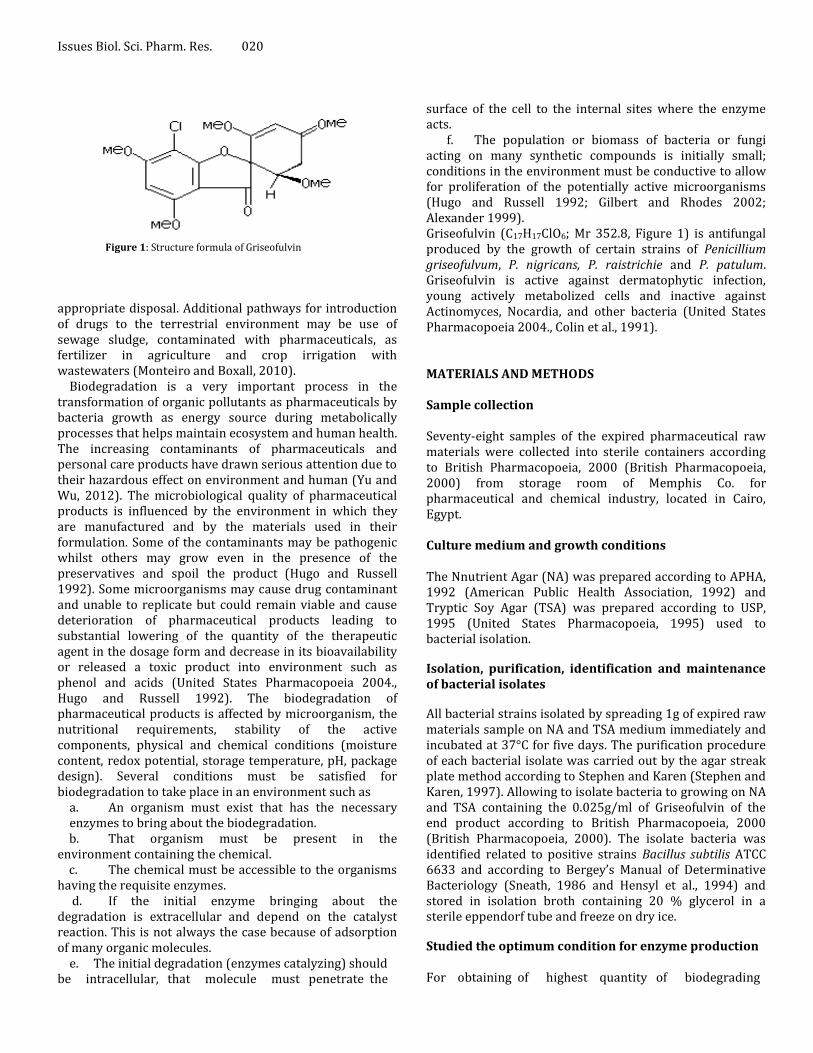
Figure 1: Structure formula of Griseofulvin
appropriate disposal. Additional pathways for introduction of drugs to the terrestrial environment may be use of sewage sludge, contaminated with pharmaceuticals, as fertilizer in agriculture and crop irrigation with wastewaters (Monteiro and Boxall, 2010).
Biodegradation is a very important process in the transformation of organic pollutants as pharmaceuticals by bacteria growth as energy source during metabolically processes that helps maintain ecosystem and human health. The increasing contaminants of pharmaceuticals and personal care products have drawn serious attention due to their hazardous effect on environment and human (Yu and Wu, 2012). The microbiological quality of pharmaceutical products is influenced by the environment in which they are manufactured and by the materials used in their formulation. Some of the contaminants may be pathogenic whilst others may grow even in the presence of the preservatives and spoil the product (Hugo and Russell 1992). Some microorganisms may cause drug contaminant and unable to replicate but could remain viable and cause deterioration of pharmaceutical products leading to substantial lowering of the quantity of the therapeutic agent in the dosage form and decrease in its bioavailability or released a toxic product into environment such as phenol and acids (United States Pharmacopoeia 2004., Hugo and Russell 1992). The biodegradation of pharmaceutical products is affected by microorganism, the nutritional requirements, stability of the active components, physical and chemical conditions (moisture content, redox potential, storage temperature, pH, package design). Several conditions must be satisfied for biodegradation to take place in an environment such as
a. An organism must exist that has the necessary enzymes to bring about the biodegradation.
b. That organism must be present in the environment containing the chemical. c. The chemical must be accessible to the organisms having the requisite enzymes. d. If the initial enzyme bringing about the degradation is extracellular and depend on the catalyst reaction. This is not always the case because of adsorption of many organic molecules.
e. The initial degradation (enzymes catalyzing) should be intracellular, that molecule must penetrate the
surface of the cell to the internal sites where the enzyme acts. f. The population or biomass of bacteria or fungi acting on many synthetic compounds is initially small; conditions in the environment must be conductive to allow for proliferation of the potentially active microorganisms (Hugo and Russell 1992; Gilbert and Rhodes 2002; Alexander 1999). Griseofulvin (C17H17ClO6; Mr 352.8, Figure 1) is antifungal produced by the growth of certain strains of Penicillium griseofulvum, P. nigricans, P. raistrichie and P. patulum. Griseofulvin is active against dermatophytic infection, young actively metabolized cells and inactive against Actinomyces, Nocardia, and other bacteria (United States Pharmacopoeia 2004., Colin et al., 1991). MATERIALS AND METHODS Sample collection Seventy-eight samples of the expired pharmaceutical raw materials were collected into sterile containers according to British Pharmacopoeia, 2000 (British Pharmacopoeia, 2000) from storage room of Memphis Co. for pharmaceutical and chemical industry, located in Cairo, Egypt. Culture medium and growth conditions The Nnutrient Agar (NA) was prepared according to APHA, 1992 (American Public Health Association, 1992) and Tryptic Soy Agar (TSA) was prepared according to USP, 1995 (United States Pharmacopoeia, 1995) used to bacterial isolation.
Isolation, purification, identification and maintenance of bacterial isolates
All bacterial strains isolated by spreading 1g of expired raw materials sample on NA and TSA medium immediately and incubated at 37°C for five days. The purification procedure of each bacterial isolate was carried out by the agar streak plate method according to Stephen and Karen (Stephen and Karen, 1997). Allowing to isolate bacteria to growing on NA and TSA containing the 0.025g/ml of Griseofulvin of the end product according to British Pharmacopoeia, 2000 (British Pharmacopoeia, 2000). The isolate bacteria was identified related to positive strains Bacillus subtilis ATCC 6633 and according to Bergey’s Manual of Determinative Bacteriology (Sneath, 1986 and Hensyl et al., 1994) and stored in isolation broth containing 20 % glycerol in a sterile eppendorf tube and freeze on dry ice.
Studied the optimum condition for enzyme production For obtaining of highest quantity of biodegrading
Maher and Reda 021
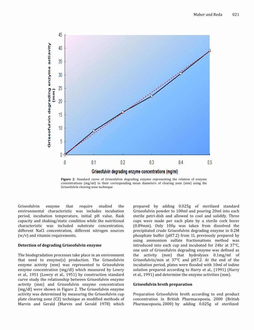
Figure 2: Standard curve of Griseofulvin degrading enzyme representing the relation of enzyme concentrations (mg/ml) to their corresponding mean diameters of clearing zone (mm) using the Griseofulvin clearing zone technique
Griseofulvin enzyme that require studied the environmental characteristic was includes incubation period, incubation temperature, initial pH value, flask capacity and shaking/static condition while the nutritional characteristic was included substrate concentration, different NaCl concentration, different nitrogen sources (w/v) and vitamin requirements. Detection of degrading Griseofulvin enzyme The biodegradation processes take place in an environment that need to enzyme(s) production. The Griseofulvin enzyme activity (mm) was represented to Griseofulvin enzyme concentration (mg/dl) which measured by Lowry et al., 1951 (Lowry et al., 1951) by construction standard curve study the relationship between Griseofulvin enzyme activity (mm) and Griseofulvin enzyme concentration (mg/dl) were shown in Figure 2. The Griseofulvin enzyme activity was determined by measuring the Griseofulvin cup plate clearing zone (CZ) technique as modified methods of Marvin and Gerald (Marvin and Gerald 1978) which
prepared by adding 0.025g of sterilized standard Griseofulvin powder to 100ml and pouring 20ml into each sterile petri-dish and allowed to cool and solidify. Three cups were made per each plate by a sterile cork borer (0.89mm). Only 100µ was taken from dissolved the precipitated crude Griseofulvin degrading enzyme in 0.2M phosphate buffer (pH7.2) from 1L previously prepared by using ammonium sulfate fractionations method was introduced into each cup and incubated for 24hr at 37°C, one unit of Griseofulvin degrading enzyme was defined as the activity (mm) that hydrolyzes 0.1mg/ml of Griseofulvin/min at 37°C and pH7.2. At the end of the incubation period, plates were flooded with 10ml of iodine solution prepared according to Harry et al., (1991) (Harry et al., 1991) and determine the enzyme activities (mm). Griseofulvin broth preparation Preparation Griseofulvin broth according to end product concentration in British Pharmacopoeia, 2000 (British Pharmacopoeia, 2000) by adding 0.025g of sterilized
Issues Biol. Sci. Pharm. Res. 022 standard Griseofulvin powder to 100ml of sterile phosphate buffer (pH7.2), inoculated with fresh, purified bacterial isolate. The flask was incubated at 37°C for 14 days. After incubation period the biodegraded raw material was collected by centrifugation at 4500 rpm for 15 minutes and examined by using the spectroscopical analysis. Griseofulvin identification method Physical test a- Dissolved 0.75g of from Standard Griseofulvin (St) or biodegraded Griseofulvin (T) in dimethylformamide and diluted to 10ml with the same solvent. The solution was cleared and less intensely colored than reference solution.
b- Suspended 0.25g from Standard Griseofulvin (St) or biodegraded Griseofulvin (T) in 20ml of methanol and 0.1ml of phenolphthalein solution was added. Not more than 1.0ml of 0.02M sodium hydroxide was required to change the color of the indicator. Standard Griseofulvin (St) concentration was 100%, the physical test done according to British Pharmacopoeia, 2000 (British Pharmacopoeia, 2000). Chemical tests Chemical reaction Dissolved 5 mg of T in 1ml of sulfuric acid and about 5mg of potassium dichromate powder was added according to British Pharmacopoeia, 2002 (British Pharmacopoeia, 2002). A wine-red color appeared means Griseofulvin was present.
UV spectroscopy detection
The maximum UV absorption of each T and St were measured at 291 nm according to British Pharmacopoeia, 2002 (British Pharmacopoeia, 2002) by Ultraviolet analyzed spectroscopically (160A-Shimadzu). The content of Griseofulvin was calculated according to equation. UV reading = UV biodegraded Griseofulvin reading (T) UV Standard Griseofulvin reading (St)
Thin Layer Chromatography (TLC) detection
Thin layer chromatography (TLC) analysis of Griseofulvin was carried out according to USP (1995) (United States Pharmacopoeia, 1995). Griseofulvin separated spot was detected and visible at wavelength 254 nm or 365 nm. To make sure of purification on TLC plate using the visual estimation by CS 9000 –Shimadzu and measured the retention factor (Rf) exactly at Y position for Griseofulvin standard and test.
IR detection Infrared (IR) spectrophotometers analysis of Griseofulvin was carried out according to British Pharmacopoeia, 2000
(British Pharmacopoeia, 2000) by using a Maston – Satelaite 113 spectrometer. IR spectrophotometer is used for recording spectra in the region 1800-4000cm-1. IR spectrophotometers use polychromatic radiation and calculated the spectrum in the frequency domain from the original data by fourier transformation. HPLC detection High Performance Liquid Chromatography (HPLC) was based on mechanisms of absorption, partition and ionexchange or size exclusion. HPLC analysis of Griseofulvin was carried out according to USP, 1995 (United States Pharmacopoeia, 1995). Extraction and purification of Griseofulvin biodegradation degrading enzyme Preparation Griseofulvin broth by adding 0.025g of sterilized standard Griseofulvin powder to 100ml of phosphate buffer (pH7.2), inoculated by Bacillus subtilis and incubated for five days. Filtration the broth through glass G-4 Metrical Membrane, filter paper and again through (0.45µ) filters. Collection the supernatant by centrifugation at 6000 rpm for 15 minutes at 20-25°C and precipitated by using different saturations (10-100%) of ammonium sulfate fractionations method according to Deutscher (Deutscher, 1990). The precipitated enzyme was collected and concentrated by using dialysis and then purified by using DEAE-Cellulose column chromatography according to Dale and Smith (Dale and Smith 1971) and sephadex G-200 column chromatography according to Andrews (Andrews 1969). The purity of an enzyme extraction and the molecular weight can be determined by SDS-polyacrylamide gel electrophoresis system according to the method modified by Hames and Rickwood (Hames and Rickwood, 1981). Identification of the Griseofulvin biodegradation products The biodegradation products of Griseofulvin was analyzed by using Gas Chromatograph mass spectrometer SHIMADZU GC/MS-QP 5050A at the Regional Center for Mycology and Biotechnology, Al-Azhar University Cairo, Egypt by using CLASS 5000 software. Griseofulvin was separated with a Helium flow on DB1, 25m; 0.53 mm ID; 5.0 μm film (J&W Scientific) column. The column oven temperature was increased from 40°C (0.5 min) -160°C (1min) at 10°C/min, 220°C (1min) at 5°C/min and 280°C (2min) at 4°C/min. The ion trap was set to 300°C, and the
Maher and Reda 023
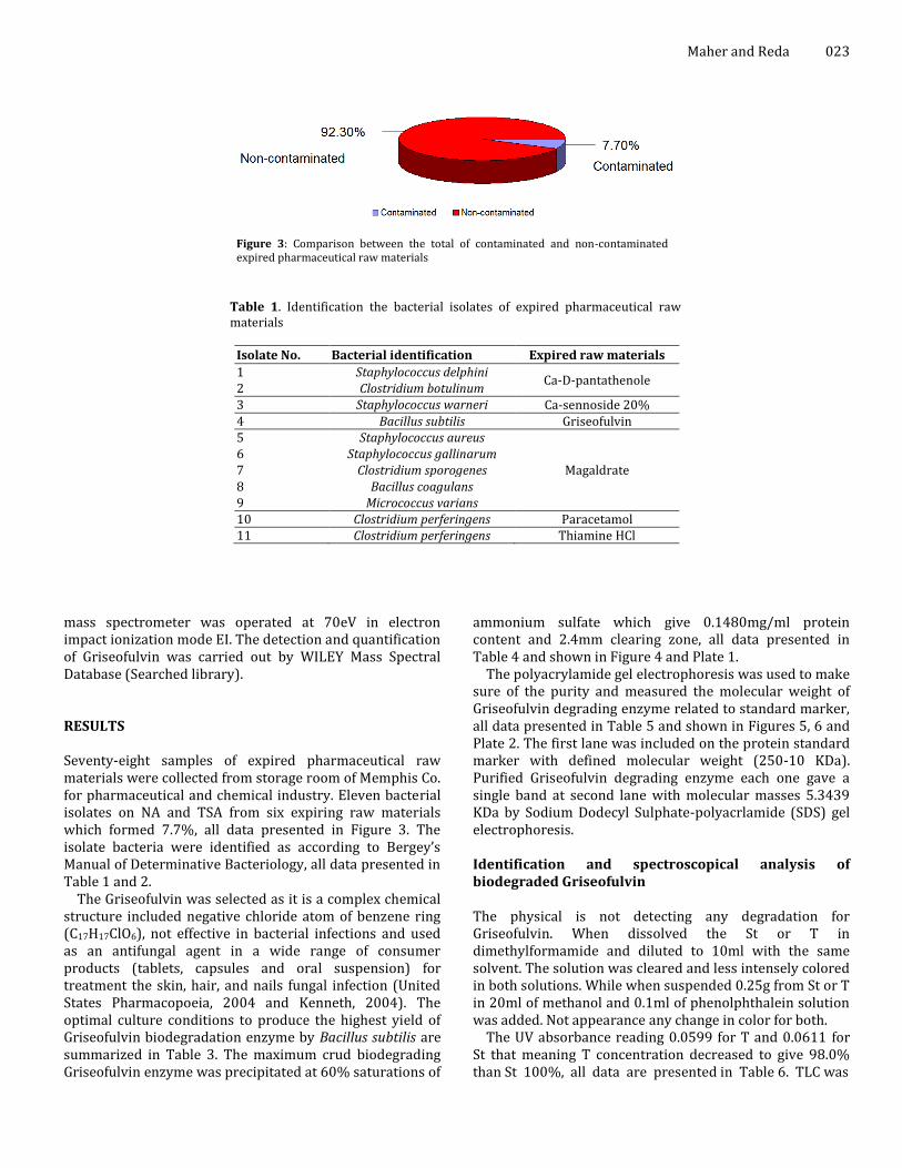
Figure 3: Comparison between the total of contaminated and non-contaminated expired pharmaceutical raw materials
Table 1. Identification the bacterial isolates of expired pharmaceutical raw materials
Isolate No. Bacterial identification Expired raw materials
1 Staphylococcus delphini Ca-D-pantathenole
2 Clostridium botulinum 3 Staphylococcus warneri Ca-sennoside 20% 4 Bacillus subtilis Griseofulvin 5 Staphylococcus aureus
Magaldrate 6 Staphylococcus gallinarum 7 Clostridium sporogenes 8 Bacillus coagulans 9 Micrococcus varians 10 Clostridium perferingens Paracetamol 11 Clostridium perferingens Thiamine HCl
mass spectrometer was operated at 70eV in electron impact ionization mode EI. The detection and quantification of Griseofulvin was carried out by WILEY Mass Spectral Database (Searched library). RESULTS Seventy-eight samples of expired pharmaceutical raw materials were collected from storage room of Memphis Co. for pharmaceutical and chemical industry. Eleven bacterial isolates on NA and TSA from six expiring raw materials which formed 7.7%, all data presented in Figure 3. The isolate bacteria were identified as according to Bergey’s Manual of Determinative Bacteriology, all data presented in Table 1 and 2.
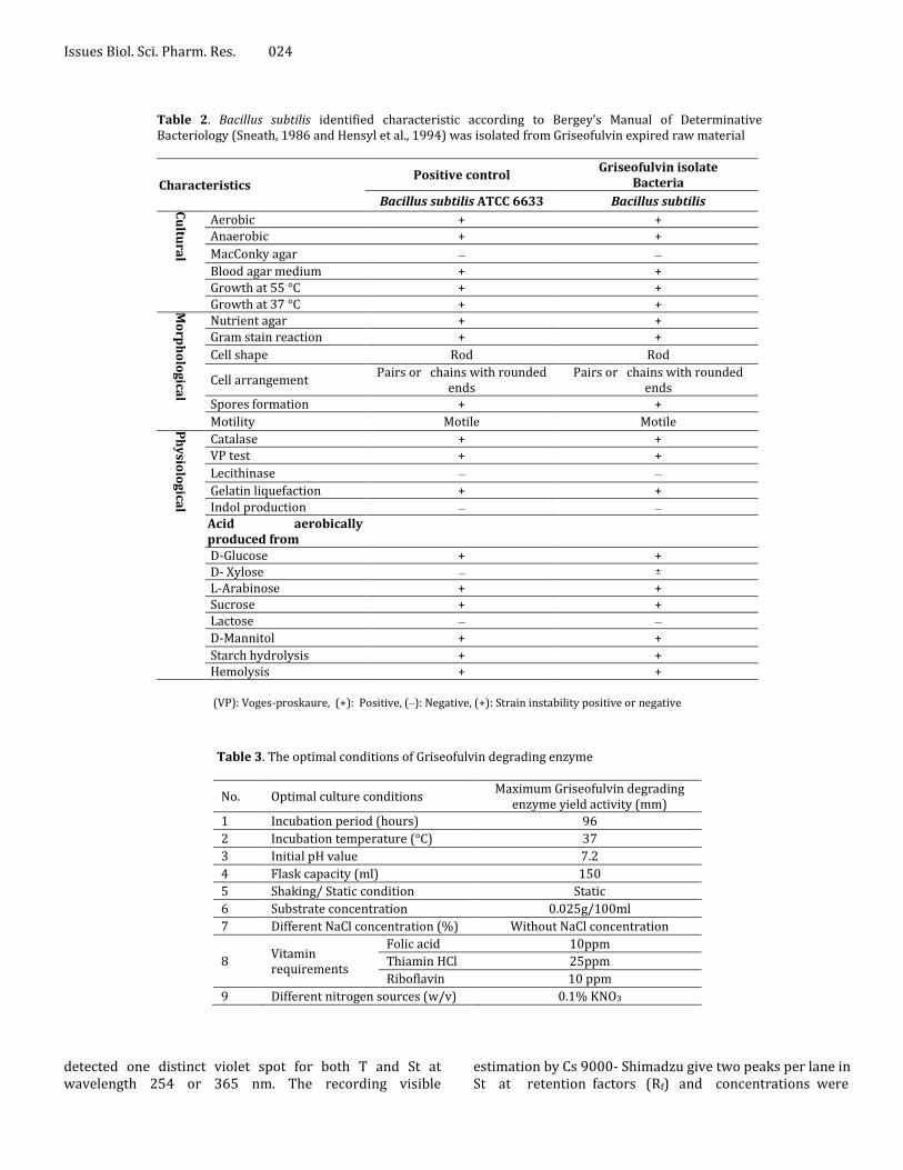
The Griseofulvin was selected as it is a complex chemical structure included negative chloride atom of benzene ring (C17H17ClO6), not effective in bacterial infections and used as an antifungal agent in a wide range of consumer products (tablets, capsules and oral suspension) for treatment the skin, hair, and nails fungal infection (United States Pharmacopoeia, 2004 and Kenneth, 2004). The optimal culture conditions to produce the highest yield of Griseofulvin biodegradation enzyme by Bacillus subtilis are summarized in Table 3. The maximum crud biodegrading Griseofulvin enzyme was precipitated at 60% saturations of
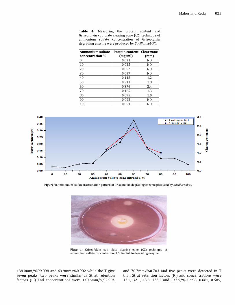
ammonium sulfate which give 0.1480mg/ml protein content and 2.4mm clearing zone, all data presented in Table 4 and shown in Figure 4 and Plate 1.
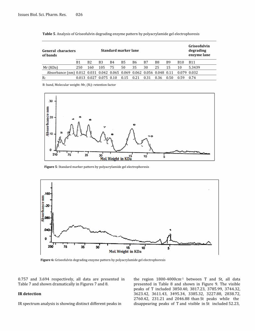
The polyacrylamide gel electrophoresis was used to make sure of the purity and measured the molecular weight of Griseofulvin degrading enzyme related to standard marker, all data presented in Table 5 and shown in Figures 5, 6 and Plate 2. The first lane was included on the protein standard marker with defined molecular weight (250-10 KDa). Purified Griseofulvin degrading enzyme each one gave a single band at second lane with molecular masses 5.3439 KDa by Sodium Dodecyl Sulphate-polyacrlamide (SDS) gel electrophoresis. Identification and spectroscopical analysis of biodegraded Griseofulvin The physical is not detecting any degradation for Griseofulvin. When dissolved the St or T in dimethylformamide and diluted to 10ml with the same solvent. The solution was cleared and less intensely colored in both solutions. While when suspended 0.25g from St or T in 20ml of methanol and 0.1ml of phenolphthalein solution was added. Not appearance any change in color for both.
The UV absorbance reading 0.0599 for T and 0.0611 for St that meaning T concentration decreased to give 98.0% than St 100%, all data are presented in Table 6. TLC was
Issues Biol. Sci. Pharm. Res. 024
Table 2. Bacillus subtilis identified characteristic according to Bergey’s Manual of Determinative Bacteriology (Sneath, 1986 and Hensyl et al., 1994) was isolated from Griseofulvin expired raw material
Characteristics Positive control
Griseofulvin isolate Bacteria
Bacillus subtilis ATCC 6633 Bacillus subtilis Cu
ltura
l
Aerobic + + Anaerobic + +
MacConky agar __ __
Blood agar medium + + Growth at 55 °C + + Growth at 37 °C + + M
orp
ho
log
ical
Nutrient agar + + Gram stain reaction + +
Cell shape Rod Rod
Cell arrangement Pairs or chains with rounded
ends Pairs or chains with rounded
ends Spores formation + +
Motility Motile Motile Ph
ysio
log
ical
Catalase + + VP test + +
Lecithinase __ __
Gelatin liquefaction + + Indol production __ __
Acid aerobically produced from
D-Glucose + + D- Xylose __ + L-Arabinose + + Sucrose + + Lactose __ __
D-Mannitol + +
Starch hydrolysis + + Hemolysis + +
(VP): Voges-proskaure, (+): Positive, (__): Negative, (+): Strain instability positive or negative
Table 3. The optimal conditions of Griseofulvin degrading enzyme
No. Optimal culture conditions Maximum Griseofulvin degrading
enzyme yield activity (mm) 1 Incubation period (hours) 96
2 Incubation temperature (°C) 37
3 Initial pH value 7.2
4 Flask capacity (ml) 150
5 Shaking/ Static condition Static
6 Substrate concentration 0.025g/100ml
7 Different NaCl concentration (%) Without NaCl concentration
8 Vitamin requirements
Folic acid 10ppm
Thiamin HCl 25ppm
Riboflavin 10 ppm
9 Different nitrogen sources (w/v) 0.1% KNO3
detected one distinct violet spot for both T and St at wavelength 254 or 365 nm. The recording visible
estimation by Cs 9000- Shimadzu give two peaks per lane in St at retention factors (Rf) and concentrations were
Maher and Reda 025
Table 4: Measuring the protein content and Griseofulvin cup plate clearing zone (CZ) technique of ammonium sulfate concentration of Griseofulvin degrading enzyme were produced by Bacillus subtilis.
Ammonium sulfate concentration %
Protein content (mg/ml)
Clear zone (mm)
0 0.031 ND
10 0.025 ND
20 0.052 ND
30 0.057 ND
40 0.148 1.2
50 0.213 1.8
60 0.376 2.4
70 0.165 1.3
80 0.095 1.0
90 0.092 ND
100 0.051 ND
Figure 4: Ammonium sulfate fractionation pattern of Griseofulvin degrading enzyme produced by Bacillus subtili
Plate 1: Griseofulvin cup plate clearing zone (CZ) technique of ammonium sulfate concentration of Griseofulvin degrading enzyme
138.0mm/%99.098 and 63.9mm/%0.902 while the T give seven peaks, two peaks were similar as St at retention factors (Rf) and concentrations were 140.6mm/%92.994
and 70.7mm/%0.703 and five peaks were detected in T than St at retention factors (Rf) and concentrations were 13.5, 32.1, 43.3, 123.2 and 133.5/% 0.598, 0.665, 0.585,
Issues Biol. Sci. Pharm. Res. 026
Table 5. Analysis of Griseofulvin degrading enzyme pattern by polyacrylamide gel electrophoresis
General characters of bands
Standard marker lane
Griseofulvin degrading enzyme lane
B1 B2 B3 B4 B5 B6 B7 B8 B9 B10 B11
Mr (KDa) 250 160 105 75 50 35 30 25 15 10 5.3439
Absorbance (nm) 0.012 0.031 0.042 0.065 0.069 0.062 0.056 0.048 0.11 0.079 0.032
Rf 0.013 0.027 0.075 0.10 0.15 0.21 0.31 0.36 0.50 0.59 0.74
B: band, Molecular weight: Mr, (Rf): retention factor
Figure 5: Standard marker pattern by polyacrylamide gel electrophoresis
Figure 6: Griseofulvin degrading enzyme pattern by polyacrylamide gel electrophoresis
0.757 and 3.694 respectively, all data are presented in Table 7 and shown dramatically in Figures 7 and 8. IR detection IR spectrum analysis is showing distinct different peaks in
the region 1800-4000cm-1 between T and St, all data presented in Table 8 and shown in Figure 9. The visible peaks of T included 3850.40, 3817.23, 3785.99, 3744.32, 3623.42, 3611.43, 3495.34, 3385.32, 3227.88, 2838.72, 2760.42, 231.21 and 2046.88 than St peaks while the disappearing peaks of T and visible in St included 52.23,

Maher and Reda 027
Plate 2: Standard marker and purified Griseofulvin degrading enzyme produced by Bacillus subtilis were patterned by polyacrylamide gel electrophoresis. A, standard marker lane and B, Griseofulvin degrading enzyme lane
Table 6. UV absorption spectra of standard and biodegraded Griseofulvin
UV test Test Standard
Wave length (nm) 291 291 Absorption (ג) 0.0611 0.0599
Concentration (%) 98.0 100
3450.23, 3397.20 and 2871.10.
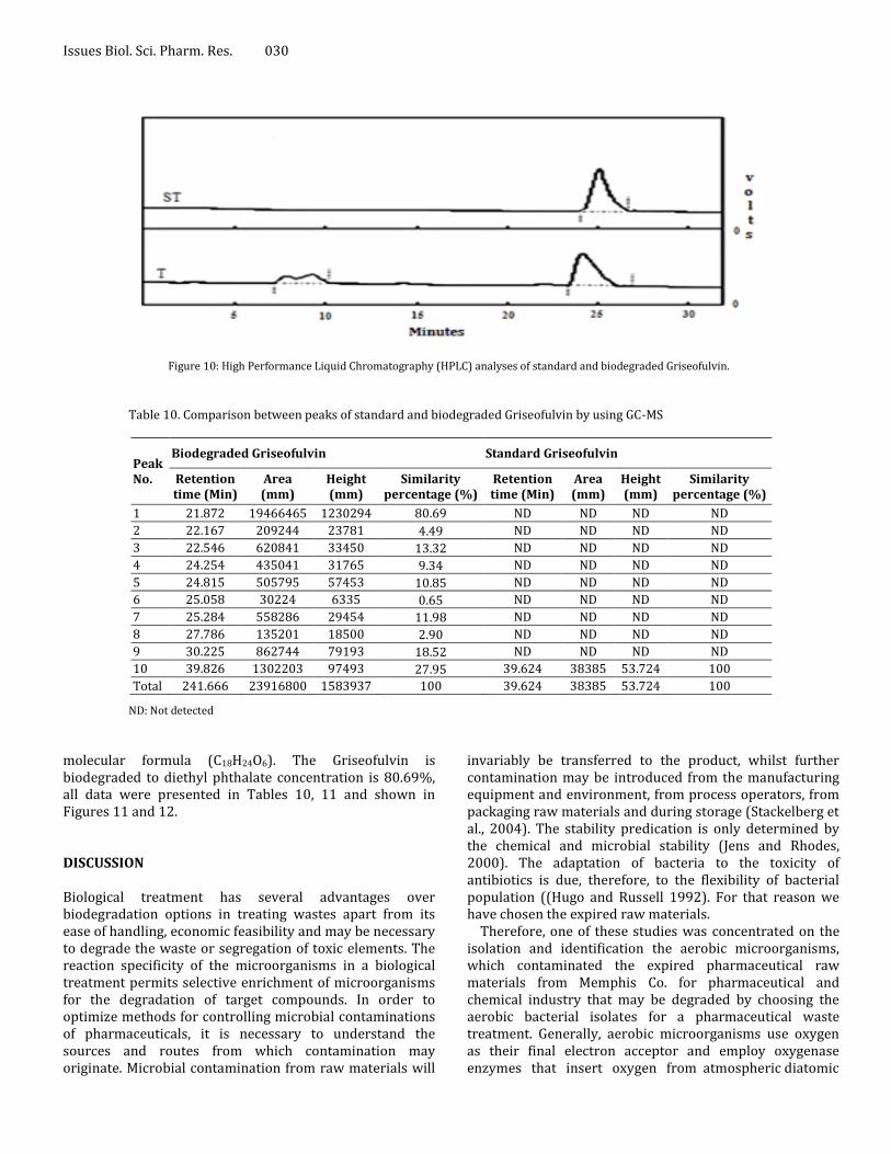
HPLC detection HPLC showing one peak was detected the Griseofulvin at area 7291458 and retention time 25.99 min in T while the St presented at area 7849993 and retention time 26.91 min. Two peaks were presented in T than St at area 928022 and 1703402 and retention time 8.97 and 10.58 min respectively. The T concentration was 0.208 mg/ml equal to %92.85 less than St 0.224 mg/ml equal to %100 that consider a good evidence for biodegradation of Griseofulvin occurring, all data presented in Table 9 and shown in Figure 10. Identification of the biodegraded Griseofulvin products The standard Griseofulvin showing one compound which appearing at retention time 39.624 min with peak area
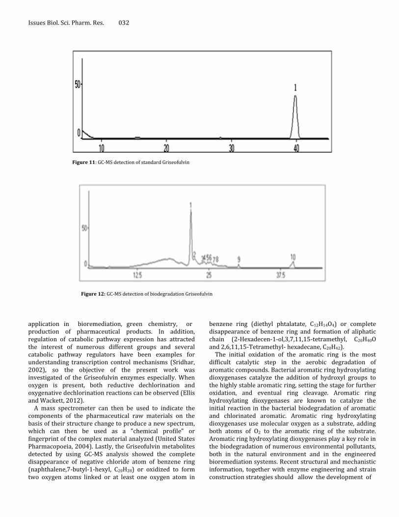
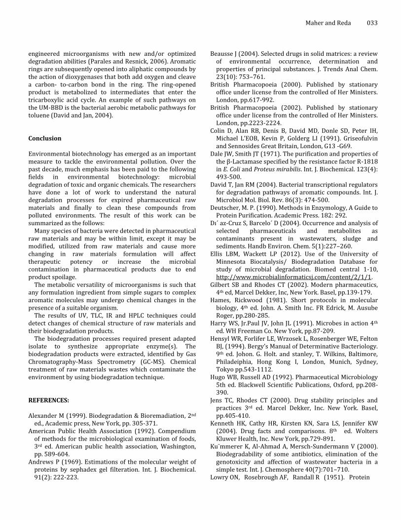
38385 and peak height 53.724, while the biodegradation Griseofulvin showing ten compounds, the similar compound appearing at retention time 39.826, with peak area 1302203 and peak height 97493. In T and St the compound was identified as Griseofulvin, molecular weight 352 and molecular formula (C17H17ClO6) while T Griseofulvin concentration is 5.40%. Nine metabolites produced from GC-Mass analysis for biodegraded Griseofulvin, which appearing as nine peaks at retention times 21.872, 22.317, 22.546, 24.254, 24.815, 25.058, 25.284, 27.786 and 30.225 min respectively, with peaks area 19466465, 209244, 620841, 435041, 505795, 30224, 558286, 135201 and 862744 respectively, while the peaks height 1230294, 23781, 33450, 31765, 57453, 6335, 29454, 18500 and 79193 respectively. The T compounds were identified as diethyl phthalate. It has a molecular weight 222 and molecular formula (C12H14O4). 2-Hexadecen-1-ol,3,7,11,15-tetramethyl has a molecular weight 296 and molecular formula (C20H40O). 2,6,11,15-Tetramethylhexadecane has a molecular weight 282 and

Issues Biol. Sci. Pharm. Res. 028
Table 7. Thin Layer Chromatography (TLC) recording visible estimation by Cs 9000- Shimadzu of standard and biodegraded Griseofulvin
Peak No.
Biodegraded Griseofulvin Standard Griseofulvin
Y position (Rf)
Area (mm)
Concentration (%)
Y position (Rf)
Area (mm)
Concentration (%)
1 63.9 1439.633 0.902 13.5 1057.375 0.598
2 138.0 158162.0 99.098 32.1 1175.102 0.665
3 ND ND ND 43.3 1035.300 0.585
4 ND ND ND 70.7 1243.086 0.703
5 ND ND ND 123.2 1338.324 0.757
6 ND ND ND 133.5 6527.805 3.694
7 ND ND ND 140.6 164308.2 92.994
Total area 159601.600 176685.200
Y position or (Rf): retention factor, ND: Not detected (relative to the dye front) and conventionally denoted as R f.
Figure 7: Thin Layer Chromatography (TLC) recording visible estimation by Cs 9000- Shimadzu of standard Griseofulvin
Figure 8: Thin Layer Chromatography (TLC) recording visible estimation by Cs 9000- Shimadzu of biodegraded Griseofulv molecular formula (C20H42). CIS-Spiro-Isohumulone I has a molecular weight 362, and molecular formula (C21H30O5). Isopentyl bezoate has a molecular weigh 192 and molecular
formula (C12H16O2). Naphthalene,7-butyl-1-hexyl has a molecular weight 268, and molecular formula (C20H28). Phenol,4-(1-Methyl-1-phenylethyl) has a molecular weight

Maher and Reda 029
Table 8. IR spectrum analysis of standard and biodegraded Griseofulvin
Biodegraded Griseofulvin Standard Griseofulvin
Peak No.
Wavenumbers (cm-1)
Peak No.
Wavenumbers (cm-1)
Peak No.
Wavenumbers (cm-1)
Peak No.
Wavenumbers (cm-1)
1 3850.40 14 ND 1 ND 14 3397.20 2 3817.23 15 3341.21 2 ND 15 3330.65 3 3785.99 16 3227.88 3 ND 16 ND 4 3744.32 17 3173.50 4 ND 17 3166.87 5 3726.62 18 2989.91 5 3708.24 18 2992.99 6 ND 19 2938.61 6 3652.23 19 2938.61 7 3656.01 20 2877.63 7 3654.21 20 2878.83 8 3603.82 21 2838.72 8 3611.40 21 ND 9 3623.42 22 ND 9 ND 22 2871.10 10 3611.43 23 2760.42 10 ND 23 ND 11 ND 24 2552.92 11 3450.23 24 2550.23 12 3495.34 25 2321.21 12 ND 25 ND 13 3385.32 26 2046.88 13 ND 26 ND
ND: Not detected.
Figure 9: IR spectrum analysis by Maston – Satelaite 113 spectrometer of standard and biodegraded Griseofulvin
Table 9. High Performance Liquid Chromatography (HPLC) analyses of standard and biodegraded Griseofulvin
Raw materials
Peak No. Width (mm)
Height (mm)
Area concentration (%) Area (mm) Retention time
(min)
Biodegraded Griseofulvin
1 1.23 19745 0 928022 8.97
2 2.4 26107 0 1703402 10.58
3 3.44 93873 0.208 7291458 25.99
Standard Griseofulvin
1 ND ND ND ND ND
2 ND ND ND ND ND
3 3.15 125118 0.224 7849993 26.91
ND: Not detected
212, and molecular formula (C15H16O). Benzenepropanol, acetate has a molecular weight 178, and molecular formula
(C11H14O2) and the final compound is 1,2-Benzenedicarboxylic. It has a molecular weight 336, and
Issues Biol. Sci. Pharm. Res. 030
Figure 10: High Performance Liquid Chromatography (HPLC) analyses of standard and biodegraded Griseofulvin.
Table 10. Comparison between peaks of standard and biodegraded Griseofulvin by using GC-MS
Peak No.
Biodegraded Griseofulvin Standard Griseofulvin
Retention time (Min)
Area (mm)
Height (mm)
Similarity percentage (%)
Retention time (Min)
Area (mm)
Height (mm)
Similarity percentage (%)
1 21.872 19466465 1230294 80.69 ND ND ND ND
2 22.167 209244 23781 4.49 ND ND ND ND
3 22.546 620841 33450 13.32 ND ND ND ND
4 24.254 435041 31765 9.34 ND ND ND ND
5 24.815 505795 57453 10.85 ND ND ND ND
6 25.058 30224 6335 0.65 ND ND ND ND
7 25.284 558286 29454 11.98 ND ND ND ND
8 27.786 135201 18500 2.90 ND ND ND ND
9 30.225 862744 79193 18.52 ND ND ND ND
10 39.826 1302203 97493 27.95 39.624 38385 53.724 100
Total 241.666 23916800 1583937 100 39.624 38385 53.724 100
ND: Not detected
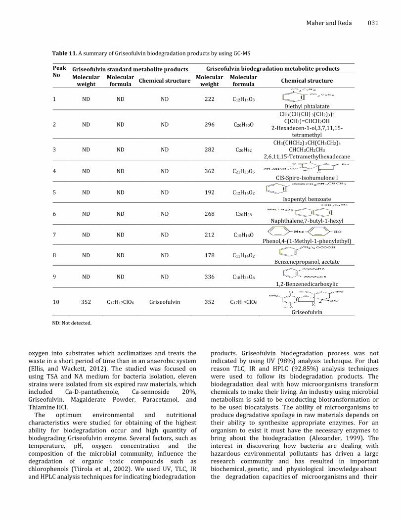
molecular formula (C18H24O6). The Griseofulvin is biodegraded to diethyl phthalate concentration is 80.69%, all data were presented in Tables 10, 11 and shown in Figures 11 and 12. DISCUSSION Biological treatment has several advantages over biodegradation options in treating wastes apart from its ease of handling, economic feasibility and may be necessary to degrade the waste or segregation of toxic elements. The reaction specificity of the microorganisms in a biological treatment permits selective enrichment of microorganisms for the degradation of target compounds. In order to optimize methods for controlling microbial contaminations of pharmaceuticals, it is necessary to understand the sources and routes from which contamination may originate. Microbial contamination from raw materials will
invariably be transferred to the product, whilst further contamination may be introduced from the manufacturing equipment and environment, from process operators, from packaging raw materials and during storage (Stackelberg et al., 2004). The stability predication is only determined by the chemical and microbial stability (Jens and Rhodes, 2000). The adaptation of bacteria to the toxicity of antibiotics is due, therefore, to the flexibility of bacterial population ((Hugo and Russell 1992). For that reason we have chosen the expired raw materials.
Therefore, one of these studies was concentrated on the isolation and identification the aerobic microorganisms, which contaminated the expired pharmaceutical raw materials from Memphis Co. for pharmaceutical and chemical industry that may be degraded by choosing the aerobic bacterial isolates for a pharmaceutical waste treatment. Generally, aerobic microorganisms use oxygen as their final electron acceptor and employ oxygenase enzymes that insert oxygen from atmospheric diatomic
Maher and Reda 031
Table 11. A summary of Griseofulvin biodegradation products by using GC-MS
Peak No
Griseofulvin standard metabolite products Griseofulvin biodegradation metabolite products
Molecular weight
Molecular formula
Chemical structure Molecular
weight Molecular
formula Chemical structure
1 ND ND ND 222 C12H14O3 Diethyl phtalatate
2 ND ND ND 296 C20H40O
CH3(CH(CH) 3(CH2)3)3 C(CH3)=CHCH2OH
2-Hexadecen-1-ol,3,7,11,15-tetramethyl
3 ND ND ND 282 C20H42 CH3(CHCH2) 3CH(CH3CH2)4
CHCH3CH2CH3 2,6,11,15-Tetramethylhexadecane
4 ND ND ND 362 C21H30O5 CIS-Spiro-Isohumulone I
5 ND ND ND 192 C12H16O2 Isopentyl benzoate
6 ND ND ND 268 C20H28 Naphthalene,7-butyl-1-hexyl
7 ND ND ND 212 C15H16O Phenol,4-(1-Methyl-1-phenylethyl)
8 ND ND ND 178 C11H14O2 Benzenepropanol, acetate
9 ND ND ND 336 C18H24O6 1,2-Benzenedicarboxylic
10 352 C17H17ClO6 Griseofulvin 352 C17H17ClO6
Griseofulvin
ND: Not detected.
oxygen into substrates which acclimatizes and treats the waste in a short period of time than in an anaerobic system (Ellis, and Wackett, 2012). The studied was focused on using TSA and NA medium for bacteria isolation, eleven strains were isolated from six expired raw materials, which included Ca-D-pantathenole, Ca-sennoside 20%, Griseofulvin, Magalderate Powder, Paracetamol, and Thiamine HCl.
The optimum environmental and nutritional characteristics were studied for obtaining of the highest ability for biodegradation occur and high quantity of biodegrading Griseofulvin enzyme. Several factors, such as temperature, pH, oxygen concentration and the composition of the microbial community, influence the degradation of organic toxic compounds such as chlorophenols (Tiirola et al., 2002). We used UV, TLC, IR and HPLC analysis techniques for indicating biodegradation
products. Griseofulvin biodegradation process was not indicated by using UV (98%) analysis technique. For that reason TLC, IR and HPLC (92.85%) analysis techniques were used to follow its biodegradation products. The biodegradation deal with how microorganisms transform chemicals to make their living. An industry using microbial metabolism is said to be conducting biotransformation or to be used biocatalysts. The ability of microorganisms to produce degradative spoilage in raw materials depends on their ability to synthesize appropriate enzymes. For an organism to exist it must have the necessary enzymes to bring about the biodegradation (Alexander, 1999). The interest in discovering how bacteria are dealing with hazardous environmental pollutants has driven a large research community and has resulted in important biochemical, genetic, and physiological knowledge about the degradation capacities of microorganisms and their
Issues Biol. Sci. Pharm. Res. 032
Figure 11: GC-MS detection of standard Griseofulvin
Figure 12: GC-MS detection of biodegradation Griseofulvin
application in bioremediation, green chemistry, or production of pharmaceutical products. In addition, regulation of catabolic pathway expression has attracted the interest of numerous different groups and several catabolic pathway regulators have been examples for understanding transcription control mechanisms (Sridhar, 2002), so the objective of the present work was investigated of the Griseofulvin enzymes especially. When oxygen is present, both reductive dechlorination and oxygenative dechlorination reactions can be observed (Ellis and Wackett, 2012).
A mass spectrometer can then be used to indicate the components of the pharmaceutical raw materials on the basis of their structure change to produce a new spectrum, which can then be used as a "chemical profile" or fingerprint of the complex material analyzed (United States Pharmacopoeia, 2004). Lastly, the Griseofulvin metabolites detected by using GC-MS analysis showed the complete disappearance of negative chloride atom of benzene ring (naphthalene,7-butyl-1-hexyl, C20H28) or oxidized to form two oxygen atoms linked or at least one oxygen atom in
benzene ring (diethyl phtalatate, C12H14O4) or complete disappearance of benzene ring and formation of aliphatic chain (2-Hexadecen-1-ol,3,7,11,15-tetramethyl, C20H40O and 2,6,11,15-Tetramethyl- hexadecane, C20H42).
The initial oxidation of the aromatic ring is the most difficult catalytic step in the aerobic degradation of aromatic compounds. Bacterial aromatic ring hydroxylating dioxygenases catalyze the addition of hydroxyl groups to the highly stable aromatic ring, setting the stage for further oxidation, and eventual ring cleavage. Aromatic ring hydroxylating dioxygenases are known to catalyze the initial reaction in the bacterial biodegradation of aromatic and chlorinated aromatic. Aromatic ring hydroxylating dioxygenases use molecular oxygen as a substrate, adding both atoms of O2 to the aromatic ring of the substrate. Aromatic ring hydroxylating dioxygenases play a key role in the biodegradation of numerous environmental pollutants, both in the natural environment and in the engineered bioremediation systems. Recent structural and mechanistic information, together with enzyme engineering and strain construction strategies should allow the development of
engineered microorganisms with new and/or optimized degradation abilities (Parales and Resnick, 2006). Aromatic rings are subsequently opened into aliphatic compounds by the action of dioxygenases that both add oxygen and cleave a carbon- to-carbon bond in the ring. The ring-opened product is metabolized to intermediates that enter the tricarboxylic acid cycle. An example of such pathways on the UM-BBD is the bacterial aerobic metabolic pathways for toluene (David and Jan, 2004). Conclusion Environmental biotechnology has emerged as an important measure to tackle the environmental pollution. Over the past decade, much emphasis has been paid to the following fields in environmental biotechnology: microbial degradation of toxic and organic chemicals. The researchers have done a lot of work to understand the natural degradation processes for expired pharmaceutical raw materials and finally to clean these compounds from polluted environments. The result of this work can be summarized as the follows:
Many species of bacteria were detected in pharmaceutical raw materials and may be within limit, except it may be modified, utilized from raw materials and cause more changing in raw materials formulation will affect therapeutic potency or increase the microbial contamination in pharmaceutical products due to end product spoilage.
The metabolic versatility of microorganisms is such that any formulation ingredient from simple sugars to complex aromatic molecules may undergo chemical changes in the presence of a suitable organism.
The results of UV, TLC, IR and HPLC techniques could detect changes of chemical structure of raw materials and their biodegradation products.
The biodegradation processes required present adapted isolate to synthesize appropriate enzyme(s). The biodegradation products were extracted, identified by Gas Chromatography-Mass Spectrometry (GC-MS). Chemical treatment of raw materials wastes which contaminate the environment by using biodegradation technique. REFERENCES: Alexander M (1999). Biodegradation & Bioremadiation, 2nd
ed., Academic press, New York, pp. 305-371. American Public Health Association (1992). Compendium
of methods for the microbiological examination of foods, 3rd ed. American public health association, Washington, pp. 589-604.
Andrews P (1969). Estimations of the molecular weight of proteins by sephadex gel filteration. Int. J. Biochemical. 91(2): 222-223.
Maher and Reda 033 Beausse J (2004). Selected drugs in solid matrices: a review
of environmental occurrence, determination and properties of principal substances. J. Trends Anal Chem. 23(10): 753–761.
British Pharmacopoeia (2000). Published by stationary office under license from the controlled of Her Ministers. London, pp.617-992.
British Pharmacopoeia (2002). Published by stationary office under license from the controlled of Her Ministers. London, pp.2223-2224.
Colin D, Alan RB, Denis B, David MD, Donle SD, Peter IH, Michael L’EOB, Kevin P, Golderg LI (1991). Griseofulvin and Sennosides Great Britain, London, G13 -G69.
Dale JW, Smith JT (1971). The purification and properties of the β-Lactamase specified by the resistance factor R-1818 in E. Coli and Proteus mirabilis. Int. J. Biochemical. 123(4): 493-500.
David T, Jan RM (2004). Bacterial transcriptional regulators for degradation pathways of aromatic compounds. Int. J. Microbiol Mol. Biol. Rev. 86(3): 474-500.
Deutscher, M. P. (1990). Methods in Enzymology, A Guide to Protein Purification. Academic Press. 182: 292.
Dı´ az-Cruz S, Barcelo´ D (2004). Occurrence and analysis of selected pharmaceuticals and metabolites as contaminants present in wastewaters, sludge and sediments. Handb Environ. Chem. 5(1):227–260.
Ellis LBM, Wackett LP (2012). Use of the University of Minnesota Biocatalysis/ Biodegradation Database for study of microbial degradation. Biomed central 1-10, http://www.microbialinformaticsj.com/content/2/1/1.
Gilbert SB and Rhodes CT (2002). Modern pharmaceutics, 4th ed, Marcel Dekker, Inc, New York. Basel, pp.139-179.
Hames, Rickwood (1981). Short protocols in molecular biology, 4th ed. John. A. Smith Inc. FR Edrick, M. Ausube Roger, pp.280-285.
Harry WS, Jr.Paul JV, John JL (1991). Microbes in action 4th ed. WH Freeman Co. New York, pp.87-209.
Hensyl WR, Forlifer LE, Wrzosek L, Rosenberger WE, Felton BJ, (1994). Bergy’s Manual of Determinative Bacteriology. 9th ed. Johon. G. Holt. and stanley, T. Wilkins, Baltimore, Philadeiphia, Hong Kong I, London, Munich, Sydney, Tokyo pp.543-1112.
Hugo WB, Russell AD (1992). Pharmaceutical Microbiology 5th ed. Blackwell Scientific Publications, Oxford, pp.208-390.
Jens TC, Rhodes CT (2000). Drug stability principles and practices 3rd ed. Marcel Dekker, Inc. New York. Basel, pp.405-410.
Kenneth HK, Cathy HR, Kirsten KN, Sara LS, Jennifer KW (2004). Drug facts and comparisons. 8th ed. Wolters Kluwer Health, Inc. New York, pp.729-891.
Ku¨mmerer K, Al-Ahmad A, Mersch-Sundermann V (2000). Biodegradability of some antibiotics, elimination of the genotoxicity and affection of wastewater bacteria in a simple test. Int. J. Chemosphere 40(7):701–710.
Lowry ON, Rosebrough AF, Randall R (1951). Protein
Issues Biol. Sci. Pharm. Res. 034
measurement with folin phenol reagent. Int. J. Biol. Chem. 193(1): 265-275.
Marvin JW, Gerald HW (1978). Antibiotics Isolation separation and purification, Netherlands, J. Elsevira Scientific Publishing Co. 15: 37-43.
Monteiro SC, Boxall ABA (2010). Occurrence and fate of human pharmaceuticals in the environment, In Reviews of environmental contamination and toxicology, Whitacre, D.M., Ed.; Springer: Summerfield, NC, USA. pp.53-154.
Parales RE, Resnick SM (2006). Aromatic ring hydroxylating dioxygenases. edited by Juan-Luis Ramos and Roger C. Levesque C Springer. Printed in the Netherlands. 4: 287-340.
Sheldon RA (2007). The E factor: fifteen years on. J. Green Chem. 9:1273–1283.
Sneath PH (1986). Bergey’s Manual of systematic Bacteriology, Williams & Wilkins (Boltimore, London, Los -Angeles, Sydney (2):536-1112.
Sridhar S, Khan S, Akella VR, Anjaneyulu Y (2002). Aerobic stabilisation of pharmaceutical wastewaters using large scale extended aeration activated sludge process. Int. J. Environ. Eng. 12: 459–505.
Stackelberg PE, Furlong ET, Meyer MT, Zaugg SD, Henderson AK, Reissman DB (2004). Persistence of
pharmaceutical compounds and other organic wastewater contaminants in a conventional drinking-water-treatment plant. Int. J. Sci Total Environ. 329:99–113.
Stephen AN, Karen EM (1997). Microbiology laboratory manual principal and applications. Prentice Hall, Upper River, New Jersey, pp.107-110.
Tiirola A, Mannisto K, Puhakka A, Kulomma S (2002). Isolation and characterization of Novosphingobioum spp. strain MT1: a dominant polychlorophenol degrading strain in groundwater bioremediation system. Int. J. Appl. Environ. Microbiol. 68(1): 173-180.
United States Pharmacopoeia (1995). The United States Pharmacopoeia. Rockville, MD: Asian ed. The United States Pharmacopeial Convention, Inc. Canada, pp.656-788.
United States Pharmacopoeia (2004). The United States Pharmacopoeia. Rockville, MD: Asian ed. The United States Pharmacopeial Convention, Inc. Canada, pp.1245-1246.
Yu Y, Wu LS (2012). Analysis of endocrine disrupting compounds, pharmaceuticals and personal care products in sewage sludge by gas chromatography-mass spectrometry. J. Talanta. 30(89): 258-263.