biochemical characterization of pantoate kinase, a novel ... filebiochemical characterization of...
TRANSCRIPT
Biochemical Characterization of Pantoate Kinase, a Novel EnzymeNecessary for Coenzyme A Biosynthesis in the Archaea
Hiroya Tomita,a Yuusuke Yokooji,a Takuya Ishibashi,a Tadayuki Imanaka,b,c and Haruyuki Atomia,c
Department of Synthetic Chemistry and Biological Chemistry, Graduate School of Engineering, Kyoto University Katsura, Nishikyo-ku, Kyoto, Japana; Department ofBiotechnology, College of Life Sciences, Ritsumeikan University Noji-Higashi, Kusatsu, Shiga, Japanb; and JST, CREST, Sanbancho, Chiyoda-ku, Tokyo, Japanc
Although bacteria and eukaryotes share a pathway for coenzyme A (CoA) biosynthesis, we previously clarified that most archaeautilize a distinct pathway for the conversion of pantoate to 4=-phosphopantothenate. Whereas bacteria/eukaryotes use pantothe-nate synthetase and pantothenate kinase (PanK), the hyperthermophilic archaeon Thermococcus kodakarensis utilizes two novelenzymes: pantoate kinase (PoK) and phosphopantothenate synthetase (PPS). Here, we report a detailed biochemical examina-tion of PoK from T. kodakarensis. Kinetic analyses revealed that the PoK reaction displayed Michaelis-Menten kinetics towardATP, whereas substrate inhibition was observed with pantoate. PoK activity was not affected by the addition of CoA/acetyl-CoA.Interestingly, PoK displayed broad nucleotide specificity and utilized ATP, GTP, UTP, and CTP with comparable kcat/Km values.Sequence alignment of 27 PoK homologs revealed seven conserved residues with reactive side chains, and variant proteins wereconstructed for each residue. Activity was not detected when mutations were introduced to Ser104, Glu134, and Asp143, suggest-ing that these residues play vital roles in PoK catalysis. Kinetic analysis of the other variant proteins, with mutations S28A,H131A, R155A, and T186A, indicated that all four residues are involved in pantoate recognition and that Arg155 and Thr186play important roles in PoK catalysis. Gel filtration analyses of the variant proteins indicated that Thr186 is also involved indimer assembly. A sequence comparison between PoK and other members of the GHMP kinase family suggests that Ser104 andGlu134 are involved in binding with phosphate and Mg2�, respectively, while Asp143 is the base responsible for proton abstrac-tion from the pantoate hydroxy group.
Coenzyme A (CoA) is an important coenzyme that is found inall three domains of life. CoA forms high-energy thioester
bonds with various carboxylic acids and plays a central role incarbon and energy metabolism. Its derivatives, such as acetyl-CoA, succinyl-CoA, malonyl-CoA, and hydroxymethylglu-taryl-CoA, are key metabolites in a wide range of pathwayswhich include sugar breakdown and �-oxidation, as well as thebiosynthesis of fatty acids and isoprenoid compounds.
In bacteria and eukaryotes, the mechanism of CoA biosynthe-sis has been examined in detail, and it is now known that theyshare common reactions (14, 27). CoA is synthesized from pan-tothenate via five enzymatic reactions: pantothenate kinase(PanK), 4=-phosphopantothenoylcysteine synthetase (PPCS), 4=-phosphopantothenoylcysteine decarboxylase (PPCDC), 4=-phos-phopantetheine adenylyltransferase (PPAT), and dephospho-CoA kinase (DPCK). In addition, microorganisms and plants cansynthesize pantothenate from L-valine and �-alanine throughfour enzymatic reactions: aminotransferase (AT), ketopantoatehydroxymethyltransferase (KPHMT), ketopantoate reductase(KPR), and pantothenate synthetase (PS).
Although much knowledge has accumulated on CoA biosyn-thesis in bacteria and eukaryotes, the corresponding pathway inarchaea has not been well examined. A number of genes that dis-play similarity to bacterial/eukaryotic genes are present on thearchaeal genomes, including those encoding PPCS, PPCDC, andPPAT. Among these, the PPCS and PPCDC from Methanocaldo-coccus jannaschii (13) and the PPAT from Pyrococcus abyssi (1, 17)have been biochemically characterized. A striking observation wasthat almost all of the archaeal genomes did not harbor genes cor-responding to PS and PanK. Genes predicted to be involved inthese reactions have been studied in Methanosarcina mazei (7, 23).By taking a comparative genomics approach, we have previously
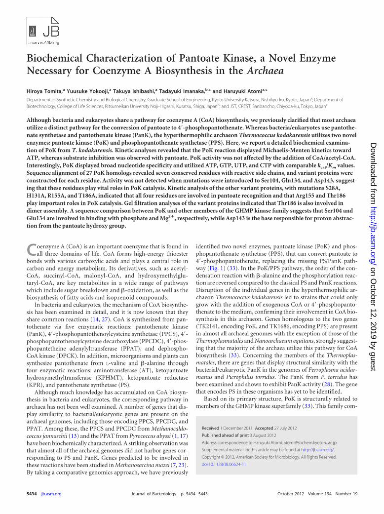
identified two novel enzymes, pantoate kinase (PoK) and phos-phopantothenate synthetase (PPS), that can convert pantoate to4=-phosphopantothenate, replacing the missing PS/PanK path-way (Fig. 1) (33). In the PoK/PPS pathway, the order of the con-densation reaction with �-alanine and the phosphorylation reac-tion are reversed compared to the classical PS and PanK reactions.Disruption of the individual genes in the hyperthermophilic ar-chaeon Thermococcus kodakarensis led to strains that could onlygrow with the addition of exogenous CoA or 4=-phosphopanto-thenate to the medium, confirming their involvement in CoA bio-synthesis in this archaeon. Genes homologous to the two genes(TK2141, encoding PoK, and TK1686, encoding PPS) are presentin almost all archaeal genomes with the exception of those of theThermoplasmatales and Nanoarchaeum equitans, strongly suggest-ing that the majority of the archaea utilize this pathway for CoAbiosynthesis (33). Concerning the members of the Thermoplas-matales, there are genes that display structural similarity with thebacterial/eukaryotic PanK in the genomes of Ferroplasma acidar-manus and Picrophilus torridus. The PanK from P. torridus hasbeen examined and shown to exhibit PanK activity (28). The genethat encodes PS in these organisms has yet to be identified.
Based on its primary structure, PoK is structurally related tomembers of the GHMP kinase superfamily (33). This family com-
Received 1 December 2011 Accepted 27 July 2012
Published ahead of print 3 August 2012
Address correspondence to Haruyuki Atomi, [email protected].
Supplemental material for this article may be found at http://jb.asm.org/.
Copyright © 2012, American Society for Microbiology. All Rights Reserved.
doi:10.1128/JB.06624-11
5434 jb.asm.org Journal of Bacteriology p. 5434–5443 October 2012 Volume 194 Number 19
on October 12, 2019 by guest
http://jb.asm.org/
Dow
nloaded from
prises a vast number of proteins that include the galactokinases,homoserine kinases, mevalonate kinases, and phosphomeval-onate kinases (3). Many members of the GHMP family are in-volved in intermediary metabolism and utilize ATP to phosphor-ylate their specific substrates. These kinases harbor a commonloop sequence rich in glycine/serine residues (4), which, in thecase of human mevalonate kinase (MvK), has been biochemicallyshown to be involved in binding with Mg2� and ATP. The specificresidue involved in this binding (Ser146) is highly conservedamong the members of the GHMP family (4). Glu193 from thisenzyme has also been shown to be involved in ATP binding (18).Furthermore, mutagenesis studies on Asp204 have indicated thatthis residue functions as the catalytic base to enhance the nucleo-philicity of the hydroxy group to attack the ATP �-phosphate(18). Both of these residues are conserved in a wide range ofGHMP family proteins. In the archaea, several proteins belongingto the GHMP kinase family have been biochemically and/or struc-turally examined, including the galactokinase (9, 30) from Pyro-coccus furiosus (Pf-GalK) and the mevalonate kinase (Mj-MvK)(32), homoserine kinase (Mj-HSK) (12), and shikimate kinase (5)from Methanocaldococcus jannaschii. Structural analyses of Pf-GalK, Mj-MvK, and Mj-HSK confirm the importance of the resi-dues described above. Ser107, Glu139, and Asp151 of Pf-GalK,Ser112, Glu144, and Asp155 of Mj-MvK, and Ser98, Glu130, andAsn141 of Mj-HSK correspond to Ser146, Glu193, and Asp204,respectively, of human MvK. In the case of Mj-HSK, no strongbase can be identified near the phosphoryl acceptor hydroxygroup, and Asn141 has been proposed to interact with the hydroxygroup of homoserine (12).
As the PoK/PPS pathway can be considered to be the predom-
inant route for CoA biosynthesis in the archaea, in this study, wehave performed the first detailed biochemical analysis of PoK.Here, we focused on the reaction kinetics, substrate specificity,and moreover, identification of the amino acid residues involvedin the catalysis of this novel enzyme, which can be considered anew archaeal member of the GHMP kinase superfamily.
MATERIALS AND METHODSStrains, media, and culture conditions. Cultivation of T. kodakarensisKOD1 (2, 16) and its derivative strains was performed under anaerobicconditions at 85°C in a nutrient-rich medium (ASW-YT) or a syntheticmedium (ASW-AA). ASW-YT medium consists of 0.8� artificial seawa-ter (ASW), 5.0 g liter�1 yeast extract, 5.0 g liter�1 tryptone, and 0.8 mgliter�1 resazurine. Prior to inoculation, 5.0 g liter�1 sodium pyruvate(ASW-YT-Pyr medium) or 2.0 g liter�1 elemental sulfur (ASW-YT-S0
medium) and Na2S were added to the medium until it became colorless.ASW-AA medium consisted of 0.8� ASW, a mixture of 20 amino acids,modified Wolfe’s trace minerals, a vitamin mixture, and 2.0 g liter�1
elemental sulfur (19, 25). In the case of plate culture used to isolate trans-formants, elemental sulfur and Na2S 9H2O were replaced with 2 ml of apolysulfide solution (10 g Na2S 9H2O and 3 g sulfur flowers in 15 ml H2O)per liter and 10 g liter�1 Gelrite was added to solidify the medium. Specificmodifications of the medium to isolate transformants are described in therespective sections below. Escherichia coli DH5� used for plasmid con-struction was cultivated at 37°C in Luria-Bertani (LB) medium containingampicillin (100 mg liter�1). Unless mentioned otherwise, all chemicalswere purchased from Wako Pure Chemicals (Osaka, Japan) or NacalaiTesque (Kyoto, Japan).
Production and purification of the wild-type pantoate kinase. TheTK2141 gene encoding pantoate kinase (PoK), with a His6 tag on its Nterminus, was overexpressed in T. kodakarensis. The TK2141 overexpres-sion strain (ETK2141) (33) was cultivated in ASW-YT-Pyr for 12 h at85°C. Cells were harvested, resuspended in 20 mM potassium phosphatebuffer (pH 7.4) containing 0.5 M KCl and 40 mM imidazole, and dis-rupted by sonication. After centrifugation (20,000 � g, 15 min), the su-pernatant was applied to His GraviTrap (GE Healthcare Biosciences, Pis-cataway, NJ), and the His-tagged proteins were eluted with 20 mMpotassium phosphate (pH 7.4), 0.5 M KCl, and 0.5 M imidazole. After thebuffer was exchanged for 50 mM Tris-HCl (pH 8.0) using a PD-10 col-umn (GE Healthcare Biosciences), the sample was applied to anion-ex-change chromatography (HiTrap Q HP; GE Healthcare Biosciences), andproteins were eluted with a linear gradient of NaCl (0 to 1.0 M) in 50 mMTris-HCl (pH 8.0) at a flow rate of 2.5 ml min�1. The protein concentra-tion was determined with the Bio-Rad protein assay system (Bio-Rad,Hercules, CA), using bovine serum albumin as a standard.
Examinations of pantoate kinase activity. PoK activity was measuredby quantifying the ADP generated from the PoK reaction with the pyru-vate kinase/lactate dehydrogenase (PK/LDH) reaction. The PoK reactionwas carried out either prior to or simultaneously with the PK/LDH reac-tion. In the former case, unless mentioned otherwise, the PoK reactionmixture contained 6 mM D-pantoate, 4 mM ATP (Oriental Yeast, Tokyo,Japan), 5.7 �g ml�1 recombinant PoK protein, 10 mM MgCl2, and 50 mMTris-HCl (pH 7.5). D-Pantoate was prepared by hydrolyzing D-pantolac-tone (Sigma-Aldrich, St. Louis, MO) in 0.4 M KOH for 1 h at 95°C. Afterthe reaction mixture was preincubated without ATP for 2 min at thedesired temperature, the reaction was initiated by the addition of ATP.The reaction was stopped by cooling the mixture on ice for 30 min, fol-lowed by the removal of PoK by ultrafiltration with an Amicon ultra0.5-ml 10K centrifugal filter (Millipore, Billerica, MA). An aliquot wasapplied to the PK/LDH reaction mixture, which contained 5 mM phos-phoenolpyruvate (PEP), 0.2 mM NADH (Oriental Yeast), 7.4 U ml�1/9.3U ml�1 PK/LDH enzymes from rabbit muscle (Sigma-Aldrich), 10 mMMgCl2, and 50 mM Tris-HCl (pH 7.5), and incubated at 42°C. The PK/LDH reaction mixture was preincubated for 2 min at 42°C, and the reac-tion was started by the addition of the aliquot of the PoK reaction. The
FIG 1 Two pathways from pantoate to 4=-phosphopantothenate. Bacteria andeukaryotes use the PS/PanK pathway (left), whereas most archaea utilize thePoK/PPS pathway (right).
Archaeal Pantoate Kinase
October 2012 Volume 194 Number 19 jb.asm.org 5435
on October 12, 2019 by guest
http://jb.asm.org/
Dow
nloaded from

decrease in absorption at 340 nm was measured using a spectrophoto-meter at 42°C. The decrease in NADH after the addition of an aliquot ofthe PoK reaction mixture without pantoate was subtracted from eachassay result. Standard activity measurements were performed by carryingout the PoK and PK/LDH reactions simultaneously at 42°C. The reactionmixture contained 6 mM pantoate, 4 mM ATP, 5 mM PEP, 0.2 mMNADH, 5.7 �g ml�1 of recombinant PoK, 14.8 U ml�1/18.6 U ml�1 ofPK/LDH enzymes, 10 mM MgCl2, and 50 mM Tris-HCl (pH 7.5). Themixture without ATP was preincubated for 2 min at 42°C, and ATP wasadded to start the reaction. The rate of the decrease in absorption at 340nm was measured consecutively. Kinetic parameters for pantoate andATP were determined using the standard method at 42°C with variousconcentrations of pantoate (with 4 mM ATP) and ATP (with 6 mM pan-toate). For analysis of the variant proteins, the reaction mixtures wereslightly modified and are described in the respective sections.
Thermostability and effects of pH and temperature. For examiningthermostability, purified PoK protein (0.57 mg ml�1) in 50 mM Tris-HCl(pH 8.0) was incubated for various periods of time at 60, 70, 80, or 90°C.After the protein solutions were cooled on ice for 30 min, PoK activity wasmeasured with the standard method. In order to examine the effects ofpH, the PoK reaction was first performed at various pHs, followed by thePK/LDH reaction. The PoK reaction mixtures contained one of the fol-lowing buffers at a final concentration of 50 mM: 2-morpholinoethane-sulfonic acid, monohydrate (MES) (pH 5.5 to 7.0), piperazine-1,4-bis(2-ethanesulfonic acid) (PIPES) (pH 6.5 to 7.5), HEPES (pH 7.0 to 8.0), Tris(Tris-HCl) (pH 7.5 to 8.0), N,N-bis(2-hydroxyethyl)glycine (bicine) (pH8.0 to 9.0), and 2-(N-cyclohexylamino)ethanesulfonic acid (CHES) (pH9.0 to 10). The pH values of all buffers except Tris-HCl were adjusted withNaOH. The reactions were performed at 75°C for 1, 3, and 5 min, and therate of ADP formation was used to calculate the activity. In order toexamine the effects of temperature, the PoK reaction was carried out atvarious temperatures for 1, 3, and 5 min, followed by the PK/LDH reac-tion at 42°C. The data obtained were used to make an Arrhenius plot. Therate constant k (s�1) was calculated for each temperature, based on theequation V � k[ES] Vmax � k[E]0, assuming that the observed velocitieswere almost equal to Vmax. Here, V (�mol s�1), [ES] (�mol), and [E]0
(�mol) represent the observed initial velocity, the concentration of en-zyme-substrate complexes, and the initial concentration of enzyme, re-spectively.
Effects of K�, CoA, acetyl-CoA, and L-cysteine. PoK activity in thepresence of various concentrations of K�, CoA, acetyl-CoA, or L-cysteinewas examined using the standard method at 42°C. We confirmed that theaddition of these compounds had no effect on the PK/LDH reaction.When we examined the effects of KCl, pantoate was prepared by hydro-lyzing pantolactone in 0.4 M NaOH instead of KOH.
Substrate specificity of pantoate kinase for phosphate donors. Inorder to examine the nucleotide specificity of PoK, various concentrationsof nucleoside triphosphate (NTP; ATP, GTP, UTP, or CTP) (Sigma-Al-drich) were added to the PoK reaction mixture and incubated at 42 or75°C. ATP, GTP, and UTP were trisodium salts, whereas CTP was a diso-dium salt. NaCl was added to adjust the Na� concentration, along with 5mM KCl. Although the reaction rates varied, we confirmed that eachnucleoside diphosphate formed in the PoK reaction could be accuratelyquantified with the PK/LDH reaction. The amounts of PK/LDH in thesecond reaction mixture were 7.4 U ml�1/9.3 U ml�1 for ADP, 22.2 Uml�1/27.9 U ml�1 for GDP and UDP, and 37.0 U ml�1/46.5 U ml�1 forCDP. When pyrophosphate or triphosphate was examined, the PoK reac-tion mixture with 4 mM pyrophosphate or triphosphate (Sigma-Aldrich)and 11.4 �g ml�1 PoK was coupled to the PPS reaction (33), and theformation of 4=-phosphopantothenate was examined with high-perfor-mance liquid chromatography. The PoK reaction was carried out at 75°Cfor 60 min. After cooling on ice for 30 min, enzyme was removed, and analiquot was applied to the PPS reaction mixture, which contained 300 �lml�1 of the PoK reaction mixture, 5 mM ATP, 5 mM �-alanine (Sigma),14.4 �g ml�1 recombinant PPS protein, 10 mM MgCl2, and 50 mM Tris-
HCl (pH 7.5). The PPS reaction was carried out at 75°C for 60 min. Aftercooling on ice for 30 min and removal of enzymes, an aliquot was appliedto a 250- by 4.60-mm Cosmosil 5C18-PAQ column (Nacalai Tesque).Compounds were separated with 20 mM NaH2PO4 (pH 3.0) at a flow rateof 1.0 ml min�1 and detected by the absorbance at 210 nm.
Expression and purification of the PoK variant proteins. The PoKoverexpression cassette (Pcsg::TK2141::TchiA) was amplified from the PoKoverexpression strain (33) with the primers 2141-EcoR/-NotF (see TableS1 in the supplemental material). After digestion of the amplified DNAfragment and pLC64 (24) with EcoRI and NotI, the cassette was insertedinto pLC64 in the same manner as the 3-hydroxy-3-methylglutaryl(HMG)-CoA reductase insertion reported by Santangelo et al. (24). Theplasmid constructed, pLC64/pok, was introduced into the T. kodakarensispok strain as follows. T. kodakarensis pok (pyrF trpE pok), whichshows tryptophan and uracil auxotrophy, was cultivated in ASW-YT-Pyrfor 12 h at 85°C. Cells were harvested and resuspended in 200 �l of 0.8�ASW, followed by incubation on ice for 30 min. After treatment with 3.0�g of pLC64 and further incubation on ice for 1 h, cells were cultivated inASW-AA medium without tryptophan (ASW-AA-W�) for 24 h at 85°C.Cells were harvested, diluted with 0.8� ASW, and spread onto solid ASW-AA-W�. After cultivation for 4 days at 85°C, transformants displayingtryptophan prototrophy were isolated and cultivated in ASW-AA-W�.The presence of the plasmids was confirmed by PCR using 8 primer sets(pLC64-F1/-R4, -F5/-R8, -F9/-R12, -F13/-R16, -F17/-R20, -F21/-R24,-F25/-R28, and -F29/-R2; see Table S1 in the supplemental material) thatanneal to pLC64. Inverse PCR was performed to construct the plasmidsfor overexpression of PoK variant proteins. Seven primer sets for intro-duction of point mutations (Ser28F/-R, Ser104F/-R, His131F/-R,Glu134F/-R, Asp143F/-R, Arg155F/-R, and Thr186F/-R; see Table S1)were used to amplify the entire pLC64/pok. After self-ligation and se-quence confirmation, plasmids were introduced into the T. kodakarensispok strain. After cultivation of these strains in ASW-AA-W� with ele-mental sulfur and 1 mM CoA for 24 h at 85°C, the strains were cultivatedin ASW-YT-Pyr with 0.5 mM CoA for 15 h at 85°C or 2 days at 70°C (forthe T186A variant). Cells were harvested by centrifugation, and the vari-ant proteins were purified by methods identical to that used for wild-typePoK. When we purified the T186A variant, the protein was further appliedto gel filtration chromatography using Superdex 200 (GE Healthcare Bio-sciences) at a flow rate of 1.0 ml min�1 after exchanging the buffer for 50mM Tris-HCl (pH 8.0) and 150 mM NaCl using an Amicon ultra 0.5-ml10K centrifugal filter. To examine the molecular mass of wild-type andvariant PoK proteins, gel filtration was carried out with a Superdex 200column with a mobile phase of 50 mM Tris-HCl (pH 7.5) containing 150mM NaCl at a flow rate of 0.8 ml min�1. The size markers were conalbu-min (75 kDa), albumin (67 kDa), ovalbumin (43 kDa), and carbonicanhydrase (29 kDa).
CD spectroscopy. Circular dichroism (CD) spectroscopy for the wild-type and all variant proteins was carried out at 42°C using a Jasco J-820spectropolarimeter. The far-UV spectra of the proteins were measuredfrom 200 to 260 nm in 50 mM Tris-HCl (pH 8.0). The instrument settingswere as follows: protein concentration, 0.2 mg liter�1; speed, 10 nm s�1;scans, average of 100; and path length, 1 mm.
RESULTSProduction and purification of recombinant pantoate kinase.Pantoate kinase (PoK, encoded by TK2141) was expressed as aninsoluble protein in E. coli (33), and therefore, it was expressed inits native host, T. kodakarensis ETK2141. As a result, recombinantPoK harboring a His6 tag sequence at the N terminus was purifiedto apparent homogeneity by nickel chelate affinity chromatogra-phy and anion-exchange chromatography. A single band corre-sponding to the molecular weight of PoK was observed after SDS-PAGE. The protein exhibited PoK activity and was used forfurther biochemical analyses.
Basic enzymatic properties of PoK. At 75°C, PoK showed high
Tomita et al.
5436 jb.asm.org Journal of Bacteriology
on October 12, 2019 by guest
http://jb.asm.org/
Dow
nloaded from

activity at neutral pH with a maximum at pH 7.5 in 50 mM PIPESor 50 mM Tris-HCl (data not shown). In terms of temperature,PoK showed the highest activity at 80°C (Fig. 2A). An Arrheniusplot of the data (Fig. 2B) displayed linearity between 55°C and80°C, indicating that the active site of PoK maintains its structurewithin this temperature range. The activation energy of the reac-tion was calculated to be 27.5 kJ mol�1. In terms of thermostabil-ity, PoK exhibited half-lives of 65 h and 11 h at 80 and 90°C,respectively. Decreases in activity could not be detected at 60 and70°C for at least 48 h. PoK activity was moderately stimulated inthe presence of potassium cations, with an optimal concentrationof 10 mM. The addition of CoA sodium salt, acetyl-CoA, andL-cysteine did not affect PoK activity at 42°C. We previously re-ported that a 1 to 2 mM concentration of CoA resulted in a slightdecrease (20 to 25%) in PoK activity (33), but we found here that
this decrease in activity was not due to CoA itself but to the lithiumcations in the CoA lithium salt reagent.
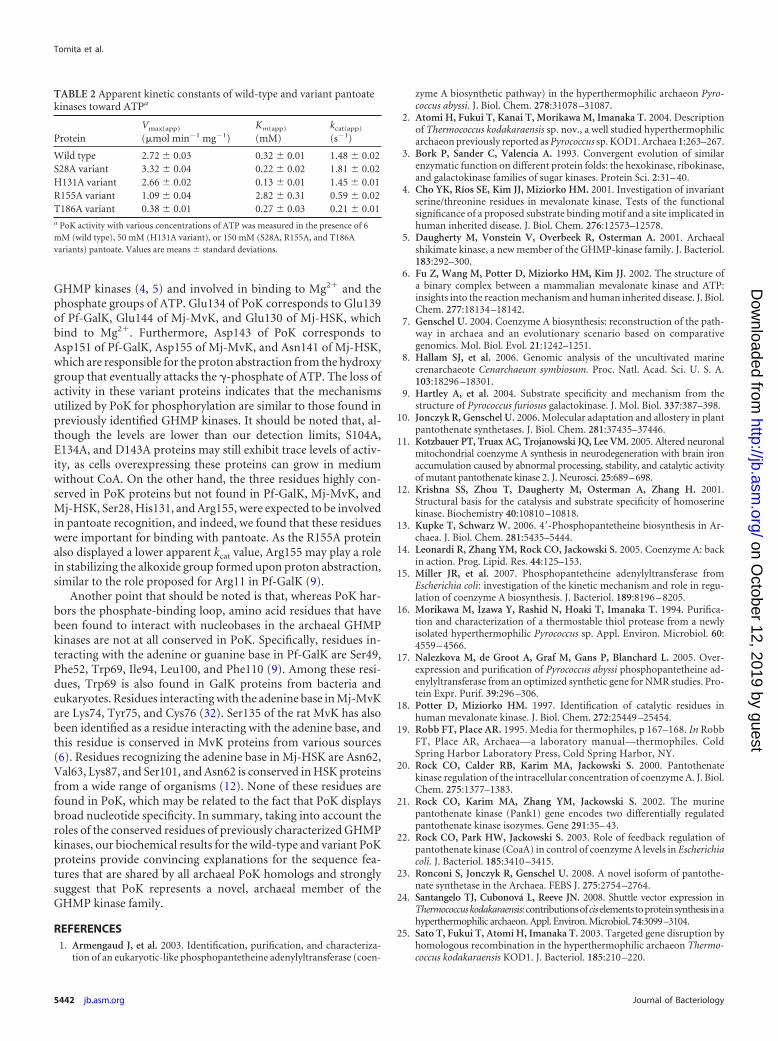
Kinetic examinations of the pantoate kinase reaction. Wecarried out kinetic studies on the pantoate kinase reaction. Activ-ity measurements were performed at 42°C in the presence of PK/LDH. The apparent kinetic constants for ATP were determined inthe presence of 6 mM pantoate. As shown in Fig. 3A, the reactionfollowed Michaelis-Menten kinetics. The apparent kinetic con-stants for pantoate were determined in the presence of 4 mM ATP.We previously concluded that the kinetics of PoK toward pantoatefollowed Michaelis-Menten kinetics (33), but examinations athigher substrate concentrations revealed that the initial velocitydecreases at concentrations higher than 6 mM pantoate (Fig. 3B).An equation taking into account substrate inhibition via bindingat a second inhibitory binding site for pantoate fit well to theobserved data. This model, toward the substrate homoserine, haspreviously been proposed for homoserine kinase from Escherichiacoli (26). The apparent kinetic constants toward ATP that wereobtained (apparent maximum initial velocity [Vmax(app)] �2.72 � 0.03 �mol min�1 mg�1 [mean � standard deviation],Km(app) � 0.32 � 0.01 mM, and kcat(app) � 1.48 � 0.02 s�1) werevery similar to those we reported previously (33). In the case of theother substrate, pantoate, the kinetic parameters obtained wereVmax(app) � 5.17 � 0.23 �mol min�1 mg�1, Ks(app) � 2.92 � 0.23mM, Ki(app) � 9.75 � 0.72 mM, and kcat(app) � 2.82 � 0.13 s�1.
Nucleotide specificity of PoK. In order to examine the nucle-otide specificity of PoK, the PoK reactions were performed in thepresence of 1, 3, or 5 mM ATP, GTP, UTP, or CTP at 75°C. To oursurprise, PoK displayed the highest activity with UTP, whereas theactivities with GTP and CTP were about half of that with ATP
FIG 2 The effects of temperature on PoK activity (A), and an Arrhenius plot ofthe data (B). The reactions were performed in the presence of 6 mM pantoateand 4 mM ATP. T, absolute temperature.
FIG 3 Kinetic analyses of wild-type PoK toward ATP (A), pantoate (B), and various nucleotides (C). (A) Initial velocity of PoK with various concentrations ofATP and 6 mM pantoate. (B) Initial velocity of PoK with various concentrations of pantoate and 4 mM ATP. (C) Initial velocity of PoK with variousconcentrations of ATP (open circles), UTP (closed circles), GTP (open squares), or CTP (closed squares) and 6 mM pantoate.
Archaeal Pantoate Kinase
October 2012 Volume 194 Number 19 jb.asm.org 5437
on October 12, 2019 by guest
http://jb.asm.org/
Dow
nloaded from

(data not shown). We performed kinetic examinations of the PoKreaction with each NTP at 42°C. As shown in Fig. 3C, all reactionsfollowed Michaelis-Menten kinetics, and the parameters obtainedare shown in Table 1. In this analysis, the PoK reaction was carriedout prior to the PK/LDH reaction. A slightly lower apparent Vmax
value toward ATP was obtained compared to that from the simul-taneous assay method, but the apparent Km value was similar. Theresults indicate that PoK displays broad substrate specificity to-ward NTPs with comparable kcat/Km values. In addition, we exam-ined whether PoK could utilize pyrophosphate or triphosphate bycoupling the reaction with the PPS reaction. While the formationof 4=-phosphopantothenate was clearly detected in the presence ofATP, we could not detect product formation with either pyro-phosphate or triphosphate in the PoK reaction, indicating thatPoK does not utilize pyrophosphate or triphosphate.
Conserved amino acid residues in archaeal PoK. PoK is anovel enzyme that does not display similarity to PanK, and thus,its structure-function relationship is of interest. In order to iden-tify the amino acid residues involved in catalysis and/or substraterecognition, we searched for highly conserved residues by aligningthe amino acid sequences of 27 PoK protein homologs from di-verse archaeal species. We found that the PoK homologs are pres-ent in the recently sequenced genomes of Thaumarchaeota Cenar-chaeum symbiosum (8) and Nitrosopumilus maritimus (31). Thealignment with nine representative sequences is shown in Fig. 4.As described above, PoK is structurally related to members of theGHMP kinase family, and thus, three representative sequenceswhose crystal structures have been determined, Pf-GalK, Mj-MvK, and Mj-HSK, are also aligned in Fig. 4. There are 25 residuesthat are highly conserved among the archaeal PoK sequences.Among these, seven residues (Ser28, Ser104, His131, Glu134,Asp143, Arg155, and Thr186) harbored reactive functionalgroups in their side chains. Interestingly, the position of the com-pletely conserved Ser104 from PoK corresponds to the Ser residuewhich plays an important role in Mg2� and ATP binding inGHMP kinases. The phosphate-binding loop which includes thisSer residue is also conserved in the PoK proteins. Likewise, thewell-conserved Glu134 and Asp143 of PoK align with the Gluresidue involved in Mg2� binding and the Asp residue that acts asthe catalytic base in GHMP proteins, respectively. Thr186 of PoKaligned well with the Thr residue of Mj-HSK, which was suggestedto be involved in phosphate binding. The other three residues,Ser28, His131, and Arg155, seem to be conserved only in PoK.
Wild-type and variant PoK proteins with single amino acidsubstitutions in the seven conserved residues were produced in T.kodakarensis. Genes were inserted into pLC64, a plasmid that dis-plays autonomous replication in both E. coli and T. kodakarensis(24). The plasmid was introduced into a pok strain in order to
avoid interaction of the variant protein with wild-type PoK en-coded in the native locus. We confirmed that the plasmids werestably maintained in T. kodakarensis under the culture conditionsapplied for gene expression. After cultivation, cells were disruptedand the proteins were purified by methods identical to those ap-plied for wild-type PoK, with the exception of the T186A protein.The expression levels of this protein, as well as those of the R155Avariant, to a lesser extent, were exceptionally low, and an addi-tional gel-filtration step was necessary to purify the protein. Cellswere inoculated into 5-fold-larger volumes of medium and grownat a suboptimal temperature of 70°C. The results of SDS-PAGEanalysis of the purified proteins are shown in Fig. 5A. In order tocompare the secondary structures of PoK variant proteins withthose of wild-type PoK, the CD spectrum of each protein wasmeasured. As shown in Fig. 5B, all variant proteins showed spectrathat were almost identical to that of the wild-type enzyme, sug-gesting that residue exchange did not disturb the overall mono-meric structure of the protein. In order to compare the quaternarystructures of the variant proteins with that of wild-type PoK, thepurified proteins were applied to gel filtration. Six of the sevenvariant proteins, with the exception of the T186A variant, elutedas a peak corresponding to a dimer. We observed multiple peaks inthe chromatogram of the T186A variant, suggesting that theThr186 residue is important in the dimer assembly of PoK. Theresults indicate that with the exception of the T186A protein, wecan interpret the changes in PoK activity observed in the variantproteins as direct consequences brought about by residue ex-change.
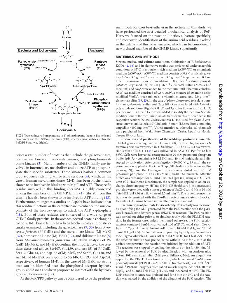
Activity measurements of the variant proteins. The activitiesof the wild-type and seven variant PoK proteins were examined inthe presence of 6 mM pantoate and 4 mM ATP at 42°C. As shownin Fig. 6A, decreases in activity were detected for all variant pro-teins, and activity could not be detected in five proteins. In orderto examine whether the loss of activity was due to a decrease in theaffinity toward substrate, we measured activity in the presence ofexcessively high substrate concentrations: 60 mM pantoate and 10mM ATP. As shown in Fig. 6B, high activities were detected for theS28A and H131A proteins, and we could also detect activity in theR155A and T186A proteins. On the other hand, we could notdetect activity in the S104A, E134A, and D143A proteins evenunder these conditions, indicating that these three residues arevital for PoK catalysis. Ser104, Glu134, and Asp143 are the threeresidues that were also conserved in all GHMP kinases.
The S28A, H131A, R155A, and T186A variant proteins dis-played sufficient levels of activity for kinetic examination (Fig. 7).The substrate concentrations applied for each variant protein areindicated in the legend of Fig. 7. When kinetic studies on the S28Avariant were performed, the kinetics of the S28A protein towardATP were similar to those of the wild-type protein (Fig. 7A),whereas the affinity for pantoate decreased (Fig. 7B). In addition,a sigmoidal curve was obtained and the substrate inhibition seenin the wild-type protein was not observed. The H131A proteinalso showed kinetics similar to those of the wild-type PoK in termsof ATP, and the affinity for pantoate displayed a significant de-crease, as in the case of the S28A protein (Fig. 7C and D). In thecase of the R155A protein, decreases in the affinities for both ATPand pantoate were observed (Fig. 7E and F). As for the T186Aprotein, which displayed a defect in dimer assembly, the apparentKm value for ATP was comparable with that of the wild-type pro-tein, whereas the affinity for pantoate decreased dramatically (Fig.
TABLE 1 Apparent kinetic constants of wild-type pantoate kinasetoward various NTPsa
SubstrateVmax(app)
(�mol min�1 mg�1)Km(app)
(mM)kcat(app)
(s�1)kcat(app)/Km(app)
(s�1 mM�1)
ATP 1.82 � 0.05 0.45 � 0.06 0.99 � 0.03 2.2GTP 0.58 � 0.02 0.43 � 0.07 0.32 � 0.01 0.74UTP 2.03 � 0.05 0.17 � 0.02 1.11 � 0.03 6.5CTP 0.87 � 0.02 0.34 � 0.04 0.47 � 0.01 1.4a PoK activity with various concentrations of NTP was measured in the presence of 6mM pantoate. Values are means � standard deviations or ratios.
Tomita et al.
5438 jb.asm.org Journal of Bacteriology
on October 12, 2019 by guest
http://jb.asm.org/
Dow
nloaded from

FIG 4 A sequence alignment of archaeal PoK proteins and galactokinase from Pyrococcus furiosus (Pf-GalK, AAL80569, PF0445) (sequence accession numbersare from NCBI; locus tags are also shown), mevalonate kinase from Methanocaldococcus jannaschii (Mj-MvK, AAB99088, MJ1087), and homoserine kinase fromM. jannaschii (Mj-HSK, AAB99107, MJ1104). The amino acid sequences of PoK homologs from 27 archaeal sequences were aligned with the ClustalW programprovided by the DNA Data Bank of Japan (DDBJ). Nine representative sequences from T. kodakarensis (Tko, BAD86330, TK2141), Halobacterium sp. NRC-1(Hba, AAG18848, VNG0251C), Archaeoglobus fulgidus (Afu, AAB89596, AF1646), M. jannaschii (Mja, AAB98974, MJ0969), Methanopyrus kandleri (Mka,AAM01947, MK0733), Pyrobaculum aerophilum (Pae, AAL64892, PAE3407), Aeropyrum pernix (Ape, BAA80948, APE1939), Sulfolobus solfataricus (Sso,AAK42544, SSO2397), and Cenarchaeum symbiosum (Csy, ABK78124, CENSYa1502), are shown. Highly conserved residues among the 27 PoK sequences, whichare present in at least 24 sequences, are indicated in red. Residues which were altered to alanine in this study are indicated with arrowheads along with their residuenumber in the T. kodakarensis (Tko) protein. Residues completely conserved in all 27 PoK sequences and Pf-GalK, Mj-MvK, and Mj-HsK are indicated withasterisks. Other PoK protein sequences used for the alignment but not shown here are from Haloarcula marismortui (AAV46584, rrnAC1672), Haloquadratumwalsbyi (CAJ51400, HQ1271A), Methanococcoides burtonii (ABE52109, Mbur1186), Methanococcus maripaludis (CAF29954, MMP0398), Methanosaeta thermo-phila (ABK15208, Mthe1433), Methanosarcina acetivorans (AAM04698, MA1279), Methanosarcina barkeri (AAZ72136, MbarA3255), Methanosarcina mazei(AAM31978, MM2282), Methanosphaera stadtmanae (ABC57568, Msp1187), Methanospirillum hungatei (ABD40583, Mhun0831), Methanothermobacter ther-mautotrophicus (AAB85083, MTH577), Natronomonas pharaonis (CAI48513, NP0844A), Nitrosopumilus maritimus (ABX13614, Nmar1718), Pyrococcus abyssi(CAB49305, PAB2407), P. furiosus (AAL81993, PF1869), Pyrococcus horikoshii (BAA30946, PH1827), Sulfolobus acidocaldarius (AAY80312, Saci0950), andSulfolobus tokodaii (BAB65525, ST0530).
Archaeal Pantoate Kinase
October 2012 Volume 194 Number 19 jb.asm.org 5439
on October 12, 2019 by guest
http://jb.asm.org/
Dow
nloaded from

7G and H). Furthermore, the initial velocity of the T186A proteinwas low even in the presence of high concentrations of substrate.The results indicate that Thr186 plays multiple roles involved inprotein assembly, pantoate binding, and PoK catalysis. Althoughwe could not propose equations that fit the [pantoate]-V plotsshown in Fig. 7, the [ATP]-V plots for all variant proteins followedMichaelis-Menten kinetics. The apparent kinetic constants ob-tained for the S28A, H131A, R155A, and T186A variants for ATP,along with those of the wild-type PoK, are shown in Table 2.
DISCUSSION
In this study, we carried out a detailed biochemical examination ofpantoate kinase (PoK), a novel enzyme involved in archaeal CoAbiosynthesis (33). The enzyme displayed several notable features,
including broad nucleotide specificity, substrate inhibition ob-served with pantoate, and a lack of inhibition in the presence ofCoA/acetyl-CoA.
We found that the apparent kcat/Km values of PoK for all ofthe nucleotides examined were comparable, indicating that any ofthe nucleotides could be used as a phosphate donor, dependingon the intracellular concentration. This was not due to the factthat our measurements were performed at relatively low temper-atures of 42°C, as we also observed the same broad nucleotidespecificity at 75°C (data not shown). In contrast, the PanK from E.coli displays a strict preference toward ATP. Interestingly, thePanK from P. torridus, which is an exceptional member of thearchaea that does not rely on the PoK/PPS system, also showsbroad nucleotide specificity (28). The lack of nucleotide specificitytoward ATP and the lack of inhibition by CoA may be related toone another, as the inhibition of PanK by CoA is due to the bind-ing of CoA competing with ATP binding through the ADP moi-eties found in both molecules (34).
Regardless of the different routes utilized to convert pantoateto 4=-phosphopantothenate, the biosynthesis of CoA can be re-garded as a costly process. In order to synthesize one molecule ofCoA, one molecule each of L-valine, �-alanine, and L-cysteine areconsumed, along with 5 molecules of ATP and an NADPH. There-fore, it can be expected that a regulatory mechanism exists toprevent the excess synthesis of CoA. In bacteria and eukaryotes,the regulatory mechanisms of CoA biosynthesis have been studiedin detail. In E. coli, the activities of PanK and PPAT are controlledby feedback inhibition by CoA and its thioesters (15, 22, 29). Mul-tiple PanK isozymes are found in mammalian cells, and severalhave been found to be inhibited by CoA, acetyl-CoA, malonyl-CoA, and/or palmitoyl-CoA (11, 20, 21). However, CoA andacetyl-CoA did not have any effect on PoK activity. Other enzymesin the upstream portion of the pathway, such as KPHMT, KPR, orthe other archaeon-specific enzyme PPS, may be the target of feed-back inhibition. In terms of competitive inhibition, PPS would bethe more likely candidate, as ATP (which contains an ADP moi-ety) is a substrate. We did observe substrate inhibition for PoK inthe case of pantoate. This substrate inhibition mechanism mayfunction in order to prevent excessive PoK reactions when pan-toate has accumulated in the cell. Similar features have been re-ported in PS from Arabidopsis thaliana and homoserine kinasefrom E. coli, which are inhibited by the substrates pantoate andhomoserine, respectively (10, 26). However, the moderate de-crease in activity at relatively high concentrations of pantoate doesnot strongly support a major regulatory role of this substrate in-
FIG 5 (A) SDS-PAGE analysis of the variant PoK proteins (3 �g each). Afterelectrophoresis, the gel was stained with Coomassie brilliant blue. (B) CDspectra in the far UV (200 to 260 nm) for variant PoK proteins. mdeg, millide-grees.
FIG 6 Activity measurements of variant PoK proteins in the presence of 6 mM pantoate and 4 mM ATP (A) or 60 mM pantoate and 10 mM ATP (B).
Tomita et al.
5440 jb.asm.org Journal of Bacteriology
on October 12, 2019 by guest
http://jb.asm.org/
Dow
nloaded from
hibition in CoA biosynthesis. Another alternative that should beexplored is regulation at the transcription/translation level.
We have identified seven amino acid residues that are highlyconserved in PoK homologs. Among these, our results indicatethat Ser104, Glu134, and Asp143 are vital for PoK activity. Se-
quence alignment indicates that the three residues correspond toresidues that play important roles in GHMP family enzymes.Ser104 of PoK corresponds to Ser107 of Pf-GalK, Ser112 of Mj-MvK, and Ser98 of Mj-HSK. All are included in a phosphate-binding loop [PX3GL(G/S)SSA] that is highly conserved in
FIG 7 Kinetic analyses of variant PoK enzymes. S28A protein toward ATP (A) and pantoate (B), H131A protein toward ATP (C) and pantoate (D), R155Aprotein toward ATP (E) and pantoate (F), and T186A protein toward ATP (G) and pantoate (H). Concentrations of pantoate were 50 mM in panel C and 150mM in panels A, E, and G. Concentrations of ATP were 4 mM in panels B, D, and H and 12 mM in panel F.
Archaeal Pantoate Kinase
October 2012 Volume 194 Number 19 jb.asm.org 5441
on October 12, 2019 by guest
http://jb.asm.org/
Dow
nloaded from
GHMP kinases (4, 5) and involved in binding to Mg2� and thephosphate groups of ATP. Glu134 of PoK corresponds to Glu139of Pf-GalK, Glu144 of Mj-MvK, and Glu130 of Mj-HSK, whichbind to Mg2�. Furthermore, Asp143 of PoK corresponds toAsp151 of Pf-GalK, Asp155 of Mj-MvK, and Asn141 of Mj-HSK,which are responsible for the proton abstraction from the hydroxygroup that eventually attacks the �-phosphate of ATP. The loss ofactivity in these variant proteins indicates that the mechanismsutilized by PoK for phosphorylation are similar to those found inpreviously identified GHMP kinases. It should be noted that, al-though the levels are lower than our detection limits, S104A,E134A, and D143A proteins may still exhibit trace levels of activ-ity, as cells overexpressing these proteins can grow in mediumwithout CoA. On the other hand, the three residues highly con-served in PoK proteins but not found in Pf-GalK, Mj-MvK, andMj-HSK, Ser28, His131, and Arg155, were expected to be involvedin pantoate recognition, and indeed, we found that these residueswere important for binding with pantoate. As the R155A proteinalso displayed a lower apparent kcat value, Arg155 may play a rolein stabilizing the alkoxide group formed upon proton abstraction,similar to the role proposed for Arg11 in Pf-GalK (9).
Another point that should be noted is that, whereas PoK har-bors the phosphate-binding loop, amino acid residues that havebeen found to interact with nucleobases in the archaeal GHMPkinases are not at all conserved in PoK. Specifically, residues in-teracting with the adenine or guanine base in Pf-GalK are Ser49,Phe52, Trp69, Ile94, Leu100, and Phe110 (9). Among these resi-dues, Trp69 is also found in GalK proteins from bacteria andeukaryotes. Residues interacting with the adenine base in Mj-MvKare Lys74, Tyr75, and Cys76 (32). Ser135 of the rat MvK has alsobeen identified as a residue interacting with the adenine base, andthis residue is conserved in MvK proteins from various sources(6). Residues recognizing the adenine base in Mj-HSK are Asn62,Val63, Lys87, and Ser101, and Asn62 is conserved in HSK proteinsfrom a wide range of organisms (12). None of these residues arefound in PoK, which may be related to the fact that PoK displaysbroad nucleotide specificity. In summary, taking into account theroles of the conserved residues of previously characterized GHMPkinases, our biochemical results for the wild-type and variant PoKproteins provide convincing explanations for the sequence fea-tures that are shared by all archaeal PoK homologs and stronglysuggest that PoK represents a novel, archaeal member of theGHMP kinase family.
REFERENCES1. Armengaud J, et al. 2003. Identification, purification, and characteriza-
tion of an eukaryotic-like phosphopantetheine adenylyltransferase (coen-
zyme A biosynthetic pathway) in the hyperthermophilic archaeon Pyro-coccus abyssi. J. Biol. Chem. 278:31078 –31087.
2. Atomi H, Fukui T, Kanai T, Morikawa M, Imanaka T. 2004. Descriptionof Thermococcus kodakaraensis sp. nov., a well studied hyperthermophilicarchaeon previously reported as Pyrococcus sp. KOD1. Archaea 1:263–267.
3. Bork P, Sander C, Valencia A. 1993. Convergent evolution of similarenzymatic function on different protein folds: the hexokinase, ribokinase,and galactokinase families of sugar kinases. Protein Sci. 2:31– 40.
4. Cho YK, Ríos SE, Kim JJ, Miziorko HM. 2001. Investigation of invariantserine/threonine residues in mevalonate kinase. Tests of the functionalsignificance of a proposed substrate binding motif and a site implicated inhuman inherited disease. J. Biol. Chem. 276:12573–12578.
5. Daugherty M, Vonstein V, Overbeek R, Osterman A. 2001. Archaealshikimate kinase, a new member of the GHMP-kinase family. J. Bacteriol.183:292–300.
6. Fu Z, Wang M, Potter D, Miziorko HM, Kim JJ. 2002. The structure ofa binary complex between a mammalian mevalonate kinase and ATP:insights into the reaction mechanism and human inherited disease. J. Biol.Chem. 277:18134 –18142.
7. Genschel U. 2004. Coenzyme A biosynthesis: reconstruction of the path-way in archaea and an evolutionary scenario based on comparativegenomics. Mol. Biol. Evol. 21:1242–1251.
8. Hallam SJ, et al. 2006. Genomic analysis of the uncultivated marinecrenarchaeote Cenarchaeum symbiosum. Proc. Natl. Acad. Sci. U. S. A.103:18296 –18301.
9. Hartley A, et al. 2004. Substrate specificity and mechanism from thestructure of Pyrococcus furiosus galactokinase. J. Mol. Biol. 337:387–398.
10. Jonczyk R, Genschel U. 2006. Molecular adaptation and allostery in plantpantothenate synthetases. J. Biol. Chem. 281:37435–37446.
11. Kotzbauer PT, Truax AC, Trojanowski JQ, Lee VM. 2005. Altered neuronalmitochondrial coenzyme A synthesis in neurodegeneration with brain ironaccumulation caused by abnormal processing, stability, and catalytic activityof mutant pantothenate kinase 2. J. Neurosci. 25:689–698.
12. Krishna SS, Zhou T, Daugherty M, Osterman A, Zhang H. 2001.Structural basis for the catalysis and substrate specificity of homoserinekinase. Biochemistry 40:10810 –10818.
13. Kupke T, Schwarz W. 2006. 4=-Phosphopantetheine biosynthesis in Ar-chaea. J. Biol. Chem. 281:5435–5444.
14. Leonardi R, Zhang YM, Rock CO, Jackowski S. 2005. Coenzyme A: backin action. Prog. Lipid. Res. 44:125–153.
15. Miller JR, et al. 2007. Phosphopantetheine adenylyltransferase fromEscherichia coli: investigation of the kinetic mechanism and role in regu-lation of coenzyme A biosynthesis. J. Bacteriol. 189:8196 – 8205.
16. Morikawa M, Izawa Y, Rashid N, Hoaki T, Imanaka T. 1994. Purifica-tion and characterization of a thermostable thiol protease from a newlyisolated hyperthermophilic Pyrococcus sp. Appl. Environ. Microbiol. 60:4559 – 4566.
17. Nalezkova M, de Groot A, Graf M, Gans P, Blanchard L. 2005. Over-expression and purification of Pyrococcus abyssi phosphopantetheine ad-enylyltransferase from an optimized synthetic gene for NMR studies. Pro-tein Expr. Purif. 39:296 –306.
18. Potter D, Miziorko HM. 1997. Identification of catalytic residues inhuman mevalonate kinase. J. Biol. Chem. 272:25449 –25454.
19. Robb FT, Place AR. 1995. Media for thermophiles, p 167–168. In RobbFT, Place AR, Archaea—a laboratory manual—thermophiles. ColdSpring Harbor Laboratory Press, Cold Spring Harbor, NY.
20. Rock CO, Calder RB, Karim MA, Jackowski S. 2000. Pantothenatekinase regulation of the intracellular concentration of coenzyme A. J. Biol.Chem. 275:1377–1383.
21. Rock CO, Karim MA, Zhang YM, Jackowski S. 2002. The murinepantothenate kinase (Pank1) gene encodes two differentially regulatedpantothenate kinase isozymes. Gene 291:35– 43.
22. Rock CO, Park HW, Jackowski S. 2003. Role of feedback regulation ofpantothenate kinase (CoaA) in control of coenzyme A levels in Escherichiacoli. J. Bacteriol. 185:3410 –3415.
23. Ronconi S, Jonczyk R, Genschel U. 2008. A novel isoform of pantothe-nate synthetase in the Archaea. FEBS J. 275:2754 –2764.
24. Santangelo TJ, Cubonová L, Reeve JN. 2008. Shuttle vector expression inThermococcuskodakaraensis: contributionsofciselementstoproteinsynthesis inahyperthermophilic archaeon. Appl. Environ. Microbiol. 74:3099–3104.
25. Sato T, Fukui T, Atomi H, Imanaka T. 2003. Targeted gene disruption byhomologous recombination in the hyperthermophilic archaeon Thermo-coccus kodakaraensis KOD1. J. Bacteriol. 185:210 –220.
TABLE 2 Apparent kinetic constants of wild-type and variant pantoatekinases toward ATPa
ProteinVmax(app)
(�mol min�1 mg�1)Km(app)
(mM)kcat(app)
(s�1)
Wild type 2.72 � 0.03 0.32 � 0.01 1.48 � 0.02S28A variant 3.32 � 0.04 0.22 � 0.02 1.81 � 0.02H131A variant 2.66 � 0.02 0.13 � 0.01 1.45 � 0.01R155A variant 1.09 � 0.04 2.82 � 0.31 0.59 � 0.02T186A variant 0.38 � 0.01 0.27 � 0.03 0.21 � 0.01a PoK activity with various concentrations of ATP was measured in the presence of 6mM (wild type), 50 mM (H131A variant), or 150 mM (S28A, R155A, and T186Avariants) pantoate. Values are means � standard deviations.
Tomita et al.
5442 jb.asm.org Journal of Bacteriology
on October 12, 2019 by guest
http://jb.asm.org/
Dow
nloaded from

26. Shames SL, Wedler FC. 1984. Homoserine kinase of Escherichia coli:kinetic mechanism and inhibition by L-aspartate semialdehyde. Arch.Biochem. Biophys. 235:359 –370.
27. Spry C, Kirk K, Saliba KJ. 2008. Coenzyme A biosynthesis: an antimi-crobial drug target. FEMS Microbiol. Rev. 32:56 –106.
28. Takagi M, et al. 2010. Pantothenate kinase from the thermoacidophilicarchaeon Picrophilus torridus. J. Bacteriol. 192:233–241.
29. Vallari DS, Jackowski S, Rock CO. 1987. Regulation of pantothenatekinase by coenzyme A and its thioesters. J. Biol. Chem. 262:2468 –2471.
30. Verhees CH, et al. 2002. Biochemical adaptations of two sugar kinasesfrom the hyperthermophilic archaeon Pyrococcus furiosus. Biochem. J.366:121–127.
31. Walker CB, et al. 2010. Nitrosopumilus maritimus genome reveals uniquemechanisms for nitrification and autotrophy in globally distributed ma-rine crenarchaea. Proc. Natl. Acad. Sci. U. S. A. 107:8818 – 8823.
32. Yang D, Shipman LW, Roessner CA, Scott AI, Sacchettini JC. 2002.Structure of the Methanococcus jannaschii mevalonate kinase, a member ofthe GHMP kinase superfamily. J. Biol. Chem. 277:9462–9467.
33. Yokooji Y, Tomita H, Atomi H, Imanaka T. 2009. Pantoate kinase andphosphopantothenate synthetase, two novel enzymes necessary for CoAbiosynthesis in the Archaea. J. Biol. Chem. 284:28137–28145.
34. Yun M, et al. 2000. Structural basis for the feedback regulation of Esche-richia coli pantothenate kinase by coenzyme A. J. Biol. Chem. 275:28093–28099.
Archaeal Pantoate Kinase
October 2012 Volume 194 Number 19 jb.asm.org 5443
on October 12, 2019 by guest
http://jb.asm.org/
Dow
nloaded from