biochemical and molecular characterization of 3 ... · carboxylase 2, beta) genes was performed by...
TRANSCRIPT

Biochemical and molecular characterization of 3-Methylcrotonylglycinuria inan Italian asymptomatic girl
Carla Cozzolino1§, Guglielmo RD Villani1,2,§ , Giulia Frisso1,2, Emanuela Scolamiero1, Lucia Albano1,Giovanna Gallo1, Roberta Romanelli1 and Margherita Ruoppolo1,2
1CEINGE Biotecnologie Avanzate, Naples, Italy.2Dipartimento di Medicina Molecolare e Biotecnologie Mediche, Università degli Studi di Napoli, “Federico
II”, Naples, Italy.
Abstract
3-Methylcrotonylglycinuria is an organic aciduria resulting from deficiency of 3-methylcrotonyl-CoA carboxylase(3-MCC), a biotin-dependent mitochondrial enzym carboxylating 3-methylcrotonyl-CoA to 3-methylglutaconyl-CoAduring leucine catabolism. Its deficiency, due to mutations on MCCC1 and MCCC2 genes, leads to accumulation of3-methylcrotonyl-CoA metabolites in blood and/or urine, primarily 3-hydroxyisovaleryl-carnitine (C5-OH) in plasmaand 3-methylcrotonyl-glycine (3-MCG) and 3-hydroxyisovaleric acid (3-HIVA) in the urine. The phenotype of 3-MCCdeficiency is highly variable, ranging from severe neurological abnormalities and death in infancy to asymptomaticadults. Here we report the biochemical and molecular characterization of an Italian asymptomatic girl, positive for thenewborn screening test. Molecular analysis showed two mutations in the MCCC2 gene, an already describedmissense mutation, c.691A > T (p.I231F), and a novel splicing mutation, c.1150-1G > A. We characterized the ex-pression profile of the splice mutation by functional studies.
Keywords: 3-Methylcrotonylglycinuria, MCCC2 mutations, 3-methylcrotonyl-CoA carboxylase deficiency, newborn screening,organic aciduria.
Received: April 05, 2017; Accepted: October 22, 2017.
Introduction
3-Methylcrotonylglycinuria (OMIM #210200,#210210) is an organic aciduria resulting from deficiencyof 3-methylcrotonyl-CoA carboxylase (3-MCC, EC6.4.1.4), a biotin-dependent mitochondrial enzyme in-volved in the fourth step of the catabolic pathway of theamino acid leucine (Sweetman and Williams, 2001) (Fig-ure 1). Its deficiency leads to accumulation in blood and/orurine of 3-methylcrotonyl-CoA metabolites, primarily3-hydroxyisovaleryl-carnitine (C5-OH) in plasma and 3-methylcrotonyl-glycine (3-MCG) and 3-hydroxyisovalericacid (3-HIVA) in urine (Arnold et al., 2012); patients oftenshow a secondary carnitine deficiency due to its conjuga-tion to 3-HIVA. The 3-methylcrotonylglycinuria is themost frequent organic aciduria detected by an increase ofC5-OH in tandem mass spectrometry-based newbornscreening programs (NBS) (Gallardo et al., 2001). Its prev-alence is highly variable ranging from 1:2,400 in the FaroeIslands (Thomsen et al., 2015) to 1:68,333 in the Chinese
population (Yang et al., 2015). Interestingly, the preva-lence could be lower in Brazil, and in fact, studies on fre-quencies of primary disorders of organic acid metabolismin high-risk Brazilian patients evidenced a frequency ofonly 0.92% of 3-Methyl-crotonylglycine-CoA carboxylasedeficiency (two patients among 218 with organic aciduria)(Wajner et al., 2002; Wajner et al., 2009).
3-MCC deficiency is inherited as an autosomal reces-sive trait due to mutations affecting one of the two genes,MCCC1 and MCCC2, encoding for two subunits, MCC�
and MCC�, respectively, that form the 3-MCC protein(Baumgartner et al., 2001). The � subunit includes the bio-tin carboxylase (BC) domain and the biotin carboxyl carrierprotein domain covalently bound with a biotin prostheticgroup, while the � subunit contains the carboxyltransferase(CT) domain (Grünert et al., 2012).
The clinical phenotype of 3-MCC deficiency ishighly variable, ranging from severe neurological abnor-malities and death in infancy (Baykal et al., 2005) toasymptomatic adults (Dantas et al., 2005; Forsyth et al.,2016). A severe presentation of 3-MCC deficit may includethe Reye-like illness, ketoacidosis, hypoglycemia, hyper-ammonemia, psychomotor retardation, seizures, symptomsof cardiorespiratory failure and coma. Mild phenotypes of-
Genetics and Molecular Biology, 41, 2, 379-385 (2018)Copyright © 2018, Sociedade Brasileira de Genética. Printed in BrazilDOI: http://dx.doi.org/10.1590/1678-4685-GMB-2017-0093
Send correspondence to Guglielmo RD Villan. Dipartimento diMedicina Molecolare e Biotecnologie Mediche, Università degliStudi di Napoli, “Federico II”, Via S. Pansini 5, 80131 Naples, Italy.E-mail: [email protected].§These authors equally contributed to the work
Research Article

ten include fatigue and weakness during catabolic episodesor mild developmental delay. Moreover, cardiomyopathy,brain atrophy and fatty infiltration of liver or muscle mayalso occur (Grünert et al., 2012). Nonetheless, most of thepatients diagnosed by NBS as affected by 3-MCC defi-ciency appear to be asymptomatic during the whole life(Forsyth et al., 2016).
In this report we present the biochemical and molecu-lar characterization of an Italian girl, positive for the new-born screening test. Molecular analysis showed two muta-tions in MCCC2 gene, a missense mutation and a splicingmutation. We characterized the expression profile of thenovel splice mutation by functional studies.
Materials and Methods
Biochemical analysis
The biochemical characterization of the patient wasobtained by analyzing amino acids and acylcarnitines indried blood spot (DBS) (at three days from birth) and in se-rum (at seven days from birth), organic acids in urine andbiotinidase enzyme activity on dried blood spot. For theseanalyses, labeled standards of amino acids and acylcar-nitines were purchased from Cambridge Isotope Labora-tories (Andover, MA, USA). External standard blood spotsfrom acylcarnitines and amino acids were from CDC (At-lanta, GA, USA).
Amino acids and acylcarnitines were analyzed asbutyl esters by using a triple quadrupole tandem mass spec-trometer API 4000 (AB/Sciex) connected to an Agilent1200 Series autosampler as described (Scolamiero et al.,2014; Scolamiero et al., 2015). Briefly, DBS from the new-born was punched into a 1.5 mL test tube and 200 �L ofmethanol containing labeled standards were added.
The standard concentrations were in the 500–2500�mol L-1 range for amino acids, and in the 7.6–152 �mol L-1
range for acylcarnitines. After 20 min of shaking at room
temperature, the samples were dried under a nitrogen flow.The extracted acylcarnitines and amino acids werederivatized to butyl esters with 80 �L of 3 N HCl in N-butanol at 65 °C for 25 min. After derivatization, the sam-ples were dried under nitrogen flow and resuspended in 300�L of acetonitrile/water (70:30) containing 0.1% formicacid. Forty microliters were injected in the flow injectionanalysis mode for the MS/MS analysis. For serum analysis,10 �L of the patient’s serum was spotted on a Schleicher&Schuell 903 grade filter paper (Whatman, Dassel, Ger-many) and dried overnight at room temperature. The nextday, the filter paper was punched into a 1.5 mL test tube andprocessed as described for DBS. Each sample was analyzedon an API 4000 triple quadrupole mass spectrometer (Ap-plied Biosystems-Sciex, Toronto, Canada) coupled withthe high performance liquid chromatograph Agilent 1100series (Agilent Technologies, Waldbronn, Germany), asdescribed (Scolamiero et al., 2015).
The extraction and quantification of the urinary or-ganic acids was performed as previously reported (Villaniet al., 2016). Urine samples were quantified for creatininein order to determine the amount of sample to be analyzed,equivalent to 0.5 �mol of creatinine, and NaOH 30% wasadded to the samples up to a final pH of 14.0. Subsequently,500 �L of hydroxylamine hydrochloride 2.5 g/L (in water)were added to each tube and samples were allowed to reactat 60 °C for 1 h. After acidification with H2SO4 2.5 N to a fi-nal pH of 1.0 internal standards were added: 10, 20 or 20 �lof a 100 �g/mL solution of dimethylmalonic acid, tropicacid and pentadecanoic acid (PDA), respectively. Three ex-tractions of the organic acids were performed by mixingvigorously each sample with 2 mL of ethyl acetate; the or-ganic phases were transferred to another tube, and approxi-mately 1 g of Na2SO4 was added. After 1 hour on bench,samples were centrifuged and the organic phase was trans-ferred to a new clean glass tube to be completely evapo-rated under a gentle nitrogen flow. Fifty microliters ofBSTFA was added to each dried sample and the deriva-tization reaction was performed at 60 °C for 30 min.Finally, 1 �L of the sample was used for GC/MS injection.Analyses were performed on an Agilent Technologies Mo-del 7890A gas chromatograph combined with a 5975Cmass spectrometer system and equipped with a split-modecapillary injection port held at 280 °C with a split ratio of10:1. The column (Agilent J&W GC column HP-5MS) wasdirectly interfaced to the ion source. The oven temperaturewas programmed from 70 °C to 280 °C at a rate of 10°C/min, and the helium flow program was 1 mL/min. Datawere acquired by repetitive scanning over a range of50–550 amu. The retention time and area of each peak wasautomatically determined and printed out by the MSD Pro-ductivity Chemstation software (Agilent Technologies).Quantitation of each analyte was performed referring to theinjected PDA quantity according to Tanaka et al. (1980a,b). The concentration of organic acids was normalized to
380 Cozzolino et al.
Figure 1 - The 3-MCC-catalyzed reaction and its position in the leucinecatabolic pathway. The dashed arrow indicates the metabolites that accu-mulate due to deficiency of 3-MCC.

the creatinine concentration of the urine sample and ex-pressed as mmol organic acid/mol creatinine.
Neonatal screening for biotinidase deficiency wasperformed by colorimetric semi-quantitative analysis(Heard et al., 1984).
Genetic analysis
Genomic DNA was extracted from peripheral bloodleukocytes of the patient and her parents using the “Nu-cleon” procedure (GE Healthcare, Little Chalfont, UK).Molecular analysis of the MCCC1 (methylcrotonoyl-CoAcarboxylase 1, alpha) and MCCC2 (methylcrotonoyl-CoAcarboxylase 2, beta) genes was performed by PCR, fol-lowed by direct sequencing of all exons and exon-intronboundaries, including 5’- and 3’-UTR regions (primers, se-quences and condition for PCR amplification are availableon request). PCR products were examined for sequencevariations using a Big Dye Primer Cycle Sequencing kitand an ABI 3730 DNA Analyzer (Applied Biosystems).Nucleotide positions were numbered on the basis of thecDNA sequence (GenBank, MCCC1: NM_020166.4 andMCCC2: NM_022132.4) according to the nomenclature ofden Dunnen and Antonarakis (2001).
Since the patient’s RNA was not available for study-ing the effect of the c.1150-1G > A novel variation on splic-ing pattern, we used an artificial minigene constructdesignated pMGene (Amato et al., 2012) kindly donated byF. Amato. A DNA fragment of approximately 1 kb, includ-ing the c.1150-1G > A variation in the MCCC2 gene, wasamplified from the genomic DNA of the patient and clonedinto the pMGene: the PCR product was obtained using theExpand High Fidelity PCR System (Roche Life Science,Penzberg, Germany), then digested by the KpnI restrictionenzyme and cloned into the KpnI-digested dephospho-rylated pMGene vector (primers, sequences and conditionsfor PCR amplification and cloning are available on re-quest). All clones were sequenced, and a wild-type and amutated form were retained for expression experiments.
tsA201 cells were grown in DMEM supplementedwith 10% fetal bovine serum and 1% penicillin/streptomy-cin in a humidified, 5% CO2 atmosphere at 37 °C. Theywere transfected with 2 �g of WT-pMGene or 2 �g ofMUT-pMGene plasmids, using FuGENE 6 (Roche), ac-cording to the manufacturer’s instructions. Forty-eighthours after transfection, cells were collected and total RNA
was extracted using TriPure Isolation Reagent (RocheApplied Science). Next, 1 �g of total RNA was reversetranscribed using SuperScript VILO cDNA synthesis kit(Life Technologies). Finally, 200 ng of cDNA were ampli-fied using the forward primer pMG-GFP-FW(5’-ACGACGGCAACTACAAGACC-3’) and reverseprimer pMG-bglob-ex3-REV (5’-CACACCAGCCACCACTTTC-3’) in a previously described protocol(Catanzano et al., 2010). The PCR products were subjectedto bidirectional cycle sequencing using a Big Dye PrimerCycle Sequencing kit and an ABI 3730 DNA Analyzer(Applied Biosystems).
To verify the potential role of the variants identifiedby genetic screening, we also checked their frequency inpopulation genomes databases, such as 1.000 Genomes(http://www.internationalgenome.org/) and ExAC(http://exac.broadinstitute.org/) consortia, which aggregateand harmonize exome sequencing data from a variety oflarge-scale sequencing projects. In silico bio-informaticevaluations were performed using Alamut Focus version0.9, a licensed software package available from InteractiveBiosoftware (www.interactive-biosoftware.com). Geno-mic sequences (WT and mutant) were processed by thispredictor software using five splicing (SpliceSiteFinder-like, MaxEntScan, Neural Network Splice, GeneSplicer,and Human SplicingFinder) and three missense predictionalgorithms (SIFT, Polyphen-2, MutationTaster). The ro-bustness of these bio-informatic tools is widely accepted(Frisso et al., 2016).
Results
The patient was a girl born to non-consanguineousparents after a full term normal delivery of dizygotic twins,weighing \ 2.57 kg. Physical examination was normal andshe was discharged from hospital 72 hours after birth. How-ever, while the amino acid profile was normal (data notshown), the acylcarnitine profile, obtained from driedblood spot during the newborn screening, presented a sig-nificant increase in 3-hydroxyisovaleryl-carnitine (C5-OH), along with a normal free carnitine (C0) concentration(Table 1). Similarly, a high concentration of C5-OH withnormal C0 concentration was reported as a consequence ofliver immaturity in a false-positive case, having a bloodC5-OH value normalized at five months of life (Rangel-Córdova et al., 2009). In addition, a recent study pointed
3-MCCD in an asymptomatic girl 381
Table 1 - Biomarkers found altered in the analyzed patient. These were quantified by LCMSMS analysis on dried blood spot and serum, by GCMS analy-sis in urine
Dried Blood Spot (DBS) (�mol/L) Serum (�moli/L) Urine (mmol/mol crea)
C5OH 4.09 (< 0.4) 4.58 (< 0.13) -
C0 15.29 (11-51) 8.7 (10-45) -
3-hydroxy-isovaleric acid - - 70 (0-1.3)
3-methylcrotonylglycine - - 441 (n.d.)
reference intervals are reported in brackets n.d.: not detectable.

out that the C5-OH level found in newborn screening is notsufficient for diagnostic or predictive purposes (Forsyth et
al., 2016). However, the detection of a C5-OH concentra-tion well above our cut-off led us to deepen the clinicalevaluation and to start prompt confirmatory testing in orderto investigate whether an inborn error of metabolism wasassociated to the biochemical alteration. The increase ofC5-OH acylcarnitine in blood/plasma may be ascribed todifferent pathologies, since several enzymes (3-methyl-crotonyl-CoA carboxylase, 3-methylglutaconic hydratase,3-hydroxy-3-methylglutaryl-CoA lyase, 2-methyl-3-hydroxybutyril-CoA dehydrogenase, beta-ketothiolase,holocarboxylase synthetase and biotinidase) may be defec-tive either in the leucine or the isoleucine catabolic pathway(Sweetman and Williams, 2001).
Although the child remained asymptomatic, the acyl-carnitine analysis performed on serum at 7 days from birthconfirmed the increase of C5-OH, but showed also a signif-icant decrease of C0 (Table 1). As stated above, elevationof C5-OH acylcarnitine levels in blood spot is not unique to3-MCC deficiency. Conditions such as 3-hydroxy-3-
methylglutaryl-coenzyme A lyase deficiency, �-ketothio-lase deficiency, multiple carboxylase deficiency resultingfrom holocarboxylase synthetase or biotinidase deficiency,2-methyl 3-hydroxybutyric acidemia, and 3-methylgluta-conic aciduria may occur with elevation of C5OH acyl-carnitine in asymptomatic infants. Therefore, the differen-tial diagnosis between different organic acidurias must beconsidered in the presence of isolated elevation of C5OHacylcarnitine levels. In our case, the analysis of the new-born urinary organic acids showed an abnormal profilecharacterized by an increase of 3-hydroxyisovaleric acidand a massive excretion of 3-methyl-crotonylglycine (Ta-ble 1, Figure 2). A biotinidase enzyme assay on dried bloodspot showed normal enzyme activity. A possible maternalorigin of the metabolic alteration, due to transplacentaltransfer of C5OH (Rangel-Córdova et al., 2009), was in-vestigated and excluded by the analysis of the mother’s se-rum acylcarnitine and urine organic acids (data not shown).These results were consistent with the diagnosis of 3-methyl-crotonyl-CoA carboxylase deficiency and justifiedthe molecular diagnosis.
382 Cozzolino et al.
Figure 2 - Patient’s urinary organic acids profile. (A) Partial organic acids profile from urine; arrows point to the main altered metabolites; (B-D) massspectra for these organic acids. 1TMS and 2TMS refer to the presence of 1 or 2 trimethylsylil ester groups, respectively, added during the derivatizationreaction.
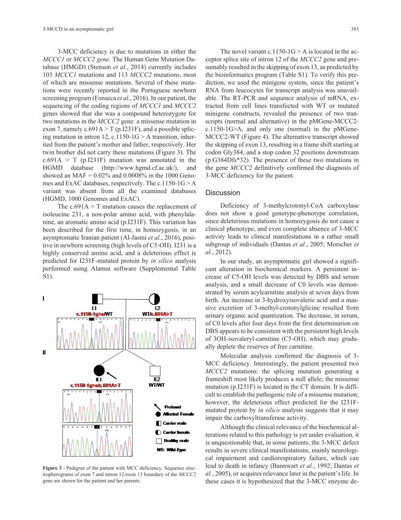
3-MCC deficiency is due to mutations in either theMCCC1 or MCCC2 gene. The Human Gene Mutation Da-tabase (HMGD) (Stenson et al., 2014) currently includes103 MCCC1 mutations and 113 MCCC2 mutations, mostof which are missense mutations. Several of these muta-tions were recently reported in the Portuguese newbornscreening program (Fonseca et al., 2016). In our patient, thesequencing of the coding regions of MCCC1 and MCCC2
genes showed that she was a compound heterozygote fortwo mutations in the MCCC2 gene: a missense mutation inexon 7, namely c.691A > T (p.I231F), and a possible splic-ing mutation in intron 12, c.1150-1G > A transition, inher-ited from the patient’s mother and father, respectively. Hertwin brother did not carry these mutations (Figure 3). Thec.691A > T (p.I231F) mutation was annotated in theHGMD database (http://www.hgmd.cf.ac.uk/), andshowed an MAF = 0.02% and 0.0008% in the 1000 Geno-mes and ExAC databases, respectively. The c.1150-1G > Avariant was absent from all the examined databases(HGMD, 1000 Genomes and ExAC).
The c.691A > T mutation causes the replacement ofisoleucine 231, a non-polar amino acid, with phenylala-nine, an aromatic amino acid (p.I231F). This variation hasbeen described for the first time, in homozygosis, in anasymptomatic Iranian patient (Al-Jasmi et al., 2016), posi-tive in newborn screening (high levels of C5-OH). I231 is ahighly conserved amino acid, and a deleterious effect ispredicted for I231F-mutated protein by in silico analysisperformed using Alamut software (Supplemental TableS1).
The novel variant c.1150-1G > A is located in the ac-ceptor splice site of intron 12 of the MCCC2 gene and pre-sumably resulted in the skipping of exon 13, as predicted bythe bioinformatics program (Table S1). To verify this pre-diction, we used the minigene system, since the patient’sRNA from leucocytes for transcript analysis was unavail-able. The RT-PCR and sequence analysis of mRNA, ex-tracted from cell lines transfected with WT or mutatedminigene constructs, revealed the presence of two tran-scripts (normal and alternative) in the pMGene-MCCC2-c.1150-1G>A, and only one (normal) in the pMGene-MCCC2-WT (Figure 4). The alternative transcript showedthe skipping of exon 13, resulting in a frame shift starting atcodon Gly384, and a stop codon 32 positions downstream(p.G384Dfs*32). The presence of these two mutations inthe gene MCCC2 definitively confirmed the diagnosis of3-MCC deficiency for the patient.
Discussion
Deficiency of 3-methylcrotonyl-CoA carboxylasedoes not show a good genotype-phenotype correlation,since deleterious mutations in homozygosis do not cause aclinical phenotype, and even complete absence of 3-MCCactivity leads to clinical manifestations in a rather smallsubgroup of individuals (Dantas et al., 2005; Morscher et
al., 2012).
In our study, an asymptomatic girl showed a signifi-cant alteration in biochemical markers. A persistent in-crease of C5-OH levels was detected by DBS and serumanalysis, and a small decrease of C0 levels was demon-strated by serum acylcarnitine analysis at seven days frombirth. An increase in 3-hydroxyisovaleric acid and a mas-sive excretion of 3-methyl-crotonylglicine resulted fromurinary organic acid quantization. The decrease, in serum,of C0 levels after four days from the first determination onDBS appears to be consistent with the persistent high levelsof 3OH-isovaleryl-carnitine (C5-OH), which may gradu-ally deplete the reserves of free carnitine.
Molecular analysis confirmed the diagnosis of 3-MCC deficiency. Interestingly, the patient presented twoMCCC2 mutations: the splicing mutation generating aframeshift most likely produces a null allele; the missensemutation (p.I231F) is located in the CT domain. It is diffi-cult to establish the pathogenic role of a missense mutation;however, the deleterious effect predicted for the I231F-mutated protein by in silico analysis suggests that it mayimpair the carboxyltransferase activity.
Although the clinical relevance of the biochemical al-terations related to this pathology is yet under evaluation, itis unquestionable that, in some patients, the 3-MCC defectresults in severe clinical manifestations, mainly neurologi-cal impairment and cardiorespiratory failure, which canlead to death in infancy (Bannwart et al., 1992; Dantas et
al., 2005), or acquires relevance later in the patient’s life. Inthese cases it is hypothesized that the 3-MCC enzyme de-
3-MCCD in an asymptomatic girl 383
Figure 3 - Pedigree of the patient with MCC deficiency. Sequence elec-tropherograms of exon 7 and intron 12/exon 13 boundary of the MCCC2
gene are shown for the patient and her parents.

fect cooperates with environmental factors or modifyinggenes to establish the pathological evolution of the disease(Baumgartner, 2005). Moreover, recent studies suggest thatalterations of the cellular redox homeostasis may poten-tially be involved in the pathophysiology of 3-MCC defi-ciency (Zanatta et al., 2013). Therefore, the accumulationof oxidative damage during the patient’s life could contrib-ute to explain neurological symptoms in late onset 3-MCCforms, with or without previous crises of metabolic decom-pensation.
With this in mind, aim of the present report is, first ofall, to emphasize the importance of differential diagnosisamong much more severe metabolic diseases, in asymp-tomatic infants with high levels of C5OH. Moreover, a con-stant monitoring is recommended for patients presentingwith 3-methylcrotonyl-CoA carboxylase deficiency, par-ticularly asymptomatic newborns. The possible absence ofsymptoms should not lead parents and paediatrics to under-estimate the pathology. It is also suggested, despite the ap-parent absence of clinical manifestations, to evaluate theopportunity of starting a preventive therapy in patientsidentified by abnormal biochemical findings, in order to re-duce the risk of neurological delay and cardiomyopathy.
References
Al-Jasmi FA, Al-Shamsi A, Hertecant JL, Al-Hamad SM andSouid AK (2016) Inborn errors of metabolism in the UnitedArab Emirates: Disorders detected by newborn screening(2011-2014). JIMD Rep 28:127-135.
Amato F, Bellia C, Cardillo G, Castaldo G, Ciaccio M, Elce A,Lembo F and Tomaiuolo R (2012) Extensive molecularanalysis of patients bearing CFTR-related disorders. J MolDiagn 14:81-9.
Arnold GL, Salazar D, Neidich JA, Suwannarat P, Graham BH,Lichter-Konecki U, Bosch AM, Cusmano-Ozog K, Enns G,Wright EL et al. (2012) Outcome of infants diagnosed with3-methyl-crotonyl-CoA-carboxylase deficiency by new-born screening. Mol Genet Metab 106:439–441.
Bannwart C, Wermuth B, Baumgartner R, Suormala T and Weis-mann UN (1992) Isolated biotin-resistant deficiency of 3-methylcrotonyl-CoA carboxylase presenting as a clinicallysevere form in a newborn with fatal outcome. J InheritMetab Dis 15:863-868.
Baumgartner MR, Almashanu S, Suormala T, Obie C, Cole RN,Packman S, Baumgartner ER and Valle D (2001) The mo-lecular basis of human 3-methylcrotonyl-CoA carboxylasedeficiency. J Clin Invest 107:495-504.
Baykal T, Gokcay GH, Ince Z, Dantas MF, Fowler B, Baum-gartner MR, Demir F, Can G and Demirkol M (2005) Con-sanguineous 3-methylcrotonyl-CoA carboxylase deficien-cy: Early-onset necrotizing encephalopathy with lethaloutcome. J Inherit Metab Dis 28:229-233.
384 Cozzolino et al.
Figure 4 - Minigene construct and RT-PCR results obtained in expression studies. (A) The minigene construct (pMGene) used in the study: a DNA frag-ment of approximately 1 kb was directly amplified from the genomic DNA of the patient and cloned into the pMGene as decribed in the Results section.All clones were sequenced, and a wild-type and a mutated clone were used for expression experiments. (B and C) The splicing pattern, evaluated byRT-PCR and sequence analysis of mRNA extracted from cell lines transfected with WT or mutated minigene constructs. Lane 1: molecular weightmarker, lane 2: RT-PCR of RNA obtained using the mutant clone; lane 3: RT-PCR of RNA obtained from the normal clone.

Catanzano F, Ombrone D, Di Stefano C, Rossi A, Nosari N,Scolamiero E, Tandurella I, Frisso G, Parenti G, RuoppoloM et al. (2010) The first case of mitochondrial acetoa-cetyl-CoA thiolase deficiency identified by expanded new-born metabolic screening in Italy: The importance of an inte-grated diagnostic approach. J Inherit Metab Dis 33:S91-S94.
Dantas MF, Suormala T, Randolph A, Coelho D, Fowler B, ValleD and Baumgartner MR (2005) 3-Methylcrotonyl-CoA car-boxylase deficiency: Mutation analysis in 28 probands, 9symptomatic and 19 detected by newborn screening. HumMutat 26:164.
den Dunnen JT and Antonarakis SE (2001) Nomenclature for thedescription of human sequence variations. Hum Genet109:121-4.
Fonseca H, Azevedo L, Serrano C, Sousa C, Marcão A andVilarinho L (2016) 3-Methylcrotonyl-CoA carboxylase de-ficiency: Mutational spectrum derived from comprehensivenewborn screening. Gene 594:203-210.
Forsyth R, Vockley CW, Edick MJ, Cameron CA, Hiner SJ, BerrySA, Vockley J, Arnold GL and Inborn Errors of MetabolismCollaborative (2016) Inborn Errors of Metabolism Collabo-rative, outcomes of cases with 3-methylcrotonyl-CoAcarboxylase (3-MCC) deficiency - report from the InbornErrors of Metabolism Information System. Mol GenetMetab 118:15-20.
Frisso G, Detta N, Coppola P, Mazzaccara C, Pricolo MR,D’Onofrio A, Limongelli G, Calabrò R, Salvatore F (2016)Functional studies and in silico analyses to evaluate non-coding variants in inherited cardiomyopathies. Int J Mol Sci17:E1883.
Gallardo ME, Desviat LR, Rodríguez JM, Esparza-Gordillo J,Pérez-Cerdá C, Pérez B, Rodríguez-Pombo P, Criado O,Sanz R, Morton DH et al. (2001) The molecular basis of3-methylcrotonylglycinuria, a disorder of leucine cata-bolsim. Am J Hum Genet 68:334-346.
Grünert SC, Stucki M, Morscher RJ, Suormala T, Bürer C, BurdaP, Christensen E, Ficicioglu C, Herwig J, Kölker S et al.
(2012) 3-methylcrotonyl-CoA carboxylase deficiency. Clin-ical, biochemical, enzymatic and molecular studies in 88 in-dividuals. Orphanet J Rare Dis 7:31.
Heard GS, Secor McVoy JR and Wolf B (1984) A screeningmethod for biotinidase deficiency in newborns. Clin Chem30:125-127.
Morscher RJ, Grünert SC, Bürer C, Burda P, Suormala T, FowlerB and Baumgartner MR (2012) A single mutation inMCCC1 or MCCC2 as a potential cause of positive screen-ing for 3-methylcrotonyl-CoA carboxylase deficiency. MolGenet Metab 105:602-606.
Rangel-Córdova EA, Martínez-de Villarreal LE and Torres-Sepulveda R (2009) 3-Methylcrotonyl-CoA carboxylase de-ficiency detected by tandem mass spectrometry in Mexicanpopulation. Medicina Universitaria 11:238-242.
Scolamiero E, Cozzolino C, Albano L, Ansalone A, Caterino M,Corbo G, di Girolamo MG, Di Stefano C, Durante A,Franzese G et al. (2015) Targeted metabolomics in the ex-panded newborn screening for inborn errors of metabolism.Mol Biosyst 11:1525-1535.
Scolamiero E, Villani GR, Ingenito L, Pecce R, Albano L, Cate-rino M, di Girolamo MG, Di Stefano C, Franzese I, Gallo Get al. (2014) B12 deficiency detected in expanded newbornscreening. Clin Biochem 47:312-317.
Stenson PD, Mort M, Ball EV, Shaw K, Phillips A and CooperDN (2014) The Human Gene Mutation Database: Building acomprehensive mutation repository for clinical and molecu-lar genetics, diagnostic testing and personalized genomicmedicine. Hum Genet 133:1-9.
Sweetman L and Williams JC (2001) Branched chain organicacidurias. In: Scriver CR, Beaudet AL, Sly WS and Valle D(eds) The Metabolic & Molecular Bases of Inherited Dis-ease. 8th edition. McGraw Hill, New York, pp 2125-2163.
Tanaka K, Hine DG, West-Dull A and Lynn TB (1980a) Gas-chromatographic method of analysis for urinary organic ac-ids. I. Retention indices of 155 metabolically importantcompounds. Clin Chem 26:1839-1846.
Tanaka K, West-Dull A, Hine DG, Lynn TB and Lowe T (1980b)Gaschromatographic method of analysis for urinary organicacids. II. Description of the procedure, and its application todiagnosis of patients with organic acidurias. Clin Chem26:1847-1853.
Thomsen JA, Lund AM, Olesen JH, Mohr M and Rasmussen J(2015) Is L-carnitine supplementation beneficial in 3-methylcrotonyl-CoA carboxylase deficiency? JIMD Rep21:79-88.
Villani GRD, Gallo G, Scolamiero E, Salvatore F and RuoppoloM (2016) “Classical organic acidurias”: Diagnosis andpathogenesis. Clin Exp Med 17:305-323.
Wajner M, Coelho DM, Ingrassia R, de Oliveira AB, BusanelloEN, Raymond K, Flores Pires R, de Souza CF, Giugliani Rand Vargas CR (2009) Selective screening for organic aci-demias by urine organic acid GC-MS analysis in Brazil: fif-teen-year experience. Clin Chim Acta 400:77-81 .
Wajner M, Raymond K, Barschak A, Luft AP, Ferreira G, Do-mingues G, Chiochetta M, Sirtori L, Goulart L, Pulrolnik Vet al. (2002) Detection of organic acidemias in Brazil. ArchMed Res 33:581-585.
Yang L, Yang J, Zhang T, Weng C, Hong F, Tong F, Yang R, YinX, Yu P, Huang X et al. (2015) Identification of eight novelmutations and transcript analysis of two splicing mutationsin Chinese newborns with MCC deficiency. Clin Genet88:484-488.
Zanatta Â, Moura AP, Tonin AM, Knebel LA, Grings M, LobatoVA, Ribeiro CA, Dutra-Filho CS, Leipnitz G and Wajner M(2013) Neurochemical evidence that the metabolites accu-mulating in 3-Methylcrotonyl-CoA carboxylase deficiencyinduce oxidative damage in cerebral cortex of young rats.Cell Mol Neurobiol 33:137-146.
Internet ResourcesBaumgartner M (2005) 3-Methylcrotonyl-CoA carboxylase defi-
ciency. Orphanet encyclopedia,http://www.orpha.net/data/patho/GB/uk-MCC.pdf.
Supplementary material
The following online material is available for this article:Table S1 - Summary of bioinformatics analysis using theAlamut software.
Associate Editor: Angela M. Vianna-Morgante
License information: This is an open-access article distributed under the terms of theCreative Commons Attribution License (type CC-BY), which permits unrestricted use,distribution and reproduction in any medium, provided the original article is properly cited.
3-MCCD in an asymptomatic girl 385