bioassay of a mixtur oef 3, 6, 7, 8 … · 1,2,3,6,7,8- and 1,2,3,7,8,9-hexachlorodibenzo-p-dioxin...
TRANSCRIPT

1980
National Cancer Institute CARCINOGENESIS Technical Report Series No. 202 NTP No. 80-13
BIOASSAY OF A MIXTURE OF
1, 2, 3, 6, 7, 8-HEXACHLORODIBENZO-p-DIOXIN
AND
1, 2, 3, 7, 8, 9-HEXACHLORODIBENZO-p-DIOXIN
(Dermal Study)
FOR POSSIBLE CARCINOGENICITY
CAS No. 57653-85-7 CAS No. 19408-74-3 NCI-CG-TR-202 NTP-80-13
U.S. DEPARTMENT OF HEALTH AND HUMAN SERVICES Public Health Service National Institutes of Health


BIOASSAY OF
1,2,3,6,7,8 and 1,2,3,7,8,9
HEXACHLORODIBENZO-p-DIOXINS
FOR POSSIBLE CARCINOGENICITY
CDermal Study)
Carcinogenesis Testing Program National Cancer Institute
National Institutes of Health Bethesda, Maryland 20205
and National Toxicology Program Research Triangle Park
Box 12233 North Carolina 27709
U.S. DEPARTMENT OF HEALTH AND HUMAN SERVICES Public Health Service
National Institutes of Health
NIH Publication No. 80-1758 August 1980

ii
BIOASSAY OF A MIXTURE OF
1,2,3,6,7,8- and 1,2,3,7,8,9HEXACHLORODIBENZO-p-DIOXINS FOR POSSIBLE CARCINOGENICITY
(Dermal Study)
Carcinogenesis Testing Program National Cancer Institute/National Toxicology Program
FOREWORD
This report presents the results of the bioassay of a mixture of 1,2,3,6,7,8- and 1,2,3,7,8,9-hexachlorodibenzo-p-dioxins conducted for the Carcinogenesis Testing Program, National Cancer Institute (NCI)/ National Toxicology Program (NTP). This is one of a series of experiments designed to determine whether selected chemicals have the capacity to produce cancer in animals. Negative results, in which the test animals do not have a greater incidence of cancer than control animals, do not necessarily mean that a test chemical is not a carcinogen, inasmuch as the experiments are conducted under a limited set of conditions. Positive results demonstrate that a test chemical is carcinogenic for animals under the conditions of the test and indicate that exposure to the chemical is a potential risk to man. The actual determination of the risk to man from chemicals found to be carcinogenic in animals requires a wider analysis.
CONTRIBUTORS
This bioassay was conducted at the Illinois Institute of Technology Research Institute (IITRI), Chicago, Illinois, initially under direct contract to NCI and later under a subcontract to Tracer Jitco, Inc., Rockville, Maryland, prime contractor for the NCI Carcinogenesis Testing Program.
The project director was Mr. A. Shefner (1); Dr. M. E. King (1) was the principal investigator for this study; and Dr. P. Holmes (1,2) assembled the data. Doses of the test chemical were selected by Dr. 0. G. Fitzhugh (3, 4). Mr. T. Kruckeberg (1) and Mr. K. Kaltenborn (1) were in charge of animal care. Histopathologic evaluations were performed by Dr. J. H. Rust (1). The pathology report and selected slides were evaluated by the NCI Pathology Working Group as described in Ward et al. (1978).
Animal pathology tables and survival tables were compiled at EG&G Mason Research Institute (5). Statistical analyses were performed by Dr. J. R. Joiner (4) and Ms. S. Vatsan (4) using methods selected for the bioassay program by Dr. J. J. Gart (6). Chemicals used in this bioassay were synthesized and analyzed under the direction of Dr. A. Gray (1), with the assistance of Mr. S. Cepa (1) and Mr. V. DaPinto (1). Further chemical analyses were conducted at Midwest Research Institute (7). The results of the chemical analytical work were reviewed by Dr. S. S. Olin (4).
iii
This report was prepared at Tracer Jitco (4) under the direction of Dr. L. A. Campbell, Acting Director of the Bioassay Program; Dr. S. S. Olin, Associate Director; Dr. R. L. Schueler, pathologist; Dr. D. J. Beach, reports manager; Dr. A. C. Jacobs, bioscience writer; and Dr. W. D. Theriault and Ms. M. W. Glasser, technical editors.
The following scientists at NCI (8) were responsible for evaluating the bioassay experiment, interpreting the results, and reporting the findings: Dr. Kenneth C. Chu, Jr., Dr. Michael P. Dieter, Dr. J. Fielding Douglas, Dr. Richard A. Griesemer, Dr. Charles K. Grieshaber, Dr. Thomas E. Hamm, Dr. William V. Hartwell, Dr. C. W. Jameson, Dr. Y. Jack Lee, Dr. Harry Mahar, Dr. James McCoy, Dr. Harry A. Milman, Dr. Thomas W. Orme, Dr. Marcelina B. Powers, Dr. Sherman F. Stinson, Dr. Jerrold M. Ward, and Dr. Carrie E. Whitmire.
(1) IIT Research Institute, 10 West 35th Street, Chicago, Illinois 60616. (2) Now with Stauffer Chemical Company, Richmond Research Center,
1200 South 47th Street, Richmond, California 94804. (3) Now at 4208 Dresden Street, Kensington, Maryland 20795. (4) Tracer Jitco, Inc., 1776 East Jefferson Street, Rockville, Maryland
20852. (5) EG&G Mason Research Institute, 1530 East Jefferson Street, Rockville,
Maryland 20852. (6) Mathematical Statistics and Applied Mathematics Section, Biometry
Branch, Field Studies and Statistics, Division of Cancer Cause and Prevention, National Cancer Institute, National Institutes of Health, Bethesda, Maryland 20205.
(7) Midwest Research Institute, 425 Volker Boulevard, Kansas City, Missouri 64110.
(8) Carcinogenesis Testing Program, National Cancer Institute, National Institutes of Health, Bethesda, Maryland 20205; and National Toxicology Program, Research Triangle Park, Box 12233, North Carolina 27709
iv
SUMMARY
A bioassay of a mixture of 1,2,3,6,7,8- and 1,2,3,7,8,9-hexachlorodibenzo-p-dioxins (HCDD) for possible carcinogenicity was conducted by dermal application of a suspension of this substance to Swiss-Webster mice.
HCDD (0.01 fig) suspended in 0.1 ml acetone was applied to the backs of 30 mice of each sex 3 days per week for 104 weeks. During the first 16 weeks, doses were 0.005/ng HCDD per application. An additional 30 mice of each sex were pretreated with one application of 50 g DMBA in 0.1 ml acetone 1 week before the initiation of the HCDD applications. As vehicle controls, 45 mice of each sex received 0.1 ml of acetone three times per week. Thirty animals of each sex served as untreated controls. Mean body weights of all test and vehicle control mice were comparable throughout the bioassay; mean body weights of untreated controls were higher than those of the test and vehicle-control groups.
In male mice, the incidence of alveolar/bronchiolar carcinomas in the group administered only HCDD was significantly higher (P=0.045) than that in the vehicle-control group; however, the incidence was not significantly higher when compared with untreated controls.
In male mice, the incidence of lymphomas or leukemias was significantly lower (P=0.011) in the group administered only HCDD when compared with the untreated controls.
In female mice, the incidences of fibrosarcomas of the skin were significantly higher (P=0.044) in animals administered HCDD (both with and without pretreatment with DMBA) than in the untreated-control group; however, when the incidences were compared with those of the vehicle controls (relative risk=3.037) the results were not significant.
Under the conditions of this bioassay, HCDD was not carcinogenic for male or female Swiss-Webster mice.
v

vi

TABLE OF CONTENTS
Page
I. Introduction 1
3II. Materials and Methods
A. Chemical 3
B. Dosage Preparation 3 4C. Animals 4 6
D. Animal Maintenance E. Subchronic Studies
6F. Chronic Studies G. Clinical Examinations and Pathology 9
H. Data Recording and Statistical Analyses 9
13 III. Results
A. Body Weights and Clinical Signs 13 B. Survival 13 C. Pathology 16 D. Statistical Analyses of Results 16
IV. Discussion 25
V. Conclusion 27
VI. Bibliography 29
APPENDIXES
Appendix A Summary of the Incidence of Neoplasms in Mice Administered HCDD by Dermal Application 31
Table Al Summary of the Incidence of Neoplasms in Male Mice Administered HCDD by Dermal Application 33
Table A2 Summary of the Incidence of Neoplasms in Female Mice Administered HCDD by Dermal Application 37
Appendix B Summary of the Incidence of Nonneoplastic Lesions 41 in Mice Administered HCDD by Dermal Application
Table Bl Summary of the Incidence of Nonneoplastic Lesions in Male Mice Administered HCDD by Dermal
43 Application
Table B2 Summary of the Incidence of Nonneoplastic Lesions in Female Mice Administered HCDD by Dermal Application 52
vii
Page
Appendix C Summary of the Incidence of Neoplasms in Mice Administered HCDD plus DMBA by Dermal Application 59
Table Cl Summary of the Incidence of Neoplasms in Male Mice Administered HCDD plus DMBA by Dermal Application 61
Table C2 Summary of the Incidence of Neoplasms in Female Mice Administered HCDD plus DMBA by Dermal Application 65
Appendix D Summary of the Incidence of Nonneoplastic Lesions in Mice Administered HCDD plus DMBA by Dermal Application 71
Table Dl Summary of the Incidence of Nonneoplastic Lesions in Male Mice Administered HCDD plus DMBA by Dermal Application 73
Table D2 Summary of the Incidence of Nonneoplastic Lesions in Female Mice Administered HCDD plus DMBA by Dermal Application 82
Appendix E Preparation of HCDD 91
Appendix F Analyses of HCDD 95
Appendix G Quarterly Analyses of HCDD Stock Solutions 105
TABLES
Table 1 HCDD Subchronic Dermal Studies in Mice 7
Table 2 HCDD Chronic Dermal Application Studies in Mice 8
Table 3 Analyses of the Incidence of Primary Tumors in Male Mice Administered HCDD or HCDD plus DMBA by Dermal Application 18
Table 4 Analyses of the Incidence of Primary Tumors in Female Mice Administered HCDD or HCDD plus DMBA by Dermal Application 21
viii

Page
FIGURES
Figure 1 Growth Curves for Mice Administered HCDD or 14 HCDD & DMBA by Dermal Application
Figure 2 Survival Curves for Mice Administered HCDD or 15 HCDD & DMBA by Dermal Application
ix

X
I. INTRODUCTION
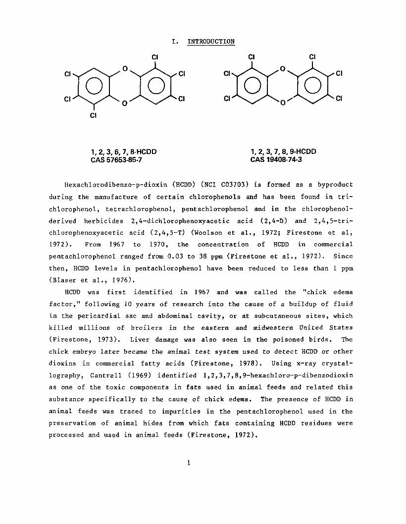
1,2,3, 6,7, 8-HCDD 1,2, 3,7, 8,9-HCDD CAS 57653-85-7 CAS 19408-74-3
Hexachlorodibenzo-p-dioxin (HCDD) (NCI C03703) is formed as a byproduct
during the manufacture of certain chlorophenols and has been found in tri
chlorophenol, tetrachlorophenol, pentachlorophenol and in the chlorophenol
derived herbicides 2,4-dichlorophenoxyacetic acid (2,4-D) and 2,4,5-tri
chlorophenoxyacetic acid (2,4,5-T) (Woolson et al., 1972; Firestone et al,
1972). From 1967 to 1970, the concentration of HCDD in commercial
pentachlorophenol ranged from 0.03 to 38 ppm (Firestone et al., 1972). Since
then, HCDD levels in pentachlorophenol have been reduced to less than 1 ppm
(Blaser et al., 1976).
HCDD was first identified in 1967 and was called the "chick edema
factor," following 10 years of research into the cause of a buildup of fluid
in the pericardial sac and abdominal cavity, or at subcutaneous sites, which
killed millions of broilers in the eastern and midwestern United States
(Firestone, 1973). Liver damage was also seen in the poisoned birds. The
chick embryo later became the animal test system used to detect HCDD or other
dioxins in commercial fatty acids (Firestone, 1978). Using x-ray crystal
lography, Cantrell (1969) identified 1,2,3,7,8,9-hexachloro-p-dibenzodioxin
as one of the toxic components in fats used in animal feeds and related this
substance specifically to the cause of chick edema. The presence of HCDD in
animal feeds was traced to impurities in the pentachlorophenol used in the
preservation of animal hides from which fats containing HCDD residues were
processed and used in animal feeds (Firestone, 1972).
1
Schwetz et al. (1973) found that a single oral dose of 100 j/g/kg HCDD was
lethal to male Sprague-Dawley rats. Severe weight losses and gross evidence
of liver damage were observed in pregnant Sprague-Dawley rats administered
oral doses of 100 /*g/kg day for 10 consecutive days. Doses of 10 or 100
(A g/kg/day of HCDD were fetotoxic, and a single dose of 100 ti g/kg was
teratogenic.
Although much has been published about the structurally related 2,3,7,
8-tetrachlorodibenzo-p-dioxin (TCDD), the literature on HCDD is very limited
and not all references specify which isomer was used. Biological effects of
HCDD appear to parallel, qualitatively, the biological effects of TCDD.
Toxicity appears to be partly correlated with the degree of chlorination at
the 2,3,7, or 8 position (McConnell and Moore, 1976). Although less potent
than TCDD, HCDD is active in the rabbit ear bioassay for acnegenic activity
and in the chick edema bioassay. Studies of aryl hydrocarbon hydroxylase
induction showed that TCDD was the most potent, followed by 1,2,3,4,7,8-HCDD,
1,2,3,7,8,9-HCDD, and 1,2,3,6,7,8-HCDD. 1,2,4,5,7,9-HCDD had no effect
(Bradlaw, 1975). 1,2,3,7,8,9-HCDD was 20% as effective as TCDD in inducing
aryl hydrocarbon hydroxylase (Poland et al., 1976). The isomers of HCDD used
in the present study (1,2,3,6,7,8 and 1,2,3,7,8,9) are both chlorinated at
the four lateral ring positions considered important for biological activity
(McConnell and Moore, 1976).
In the early 1970's, HCDD was one in a series of the chlorodibenzo-p
dioxins selected for testing by the Carcinogenesis Testing Program after
TCDD, a contaminant in 2,4,5-T, was found to be a potent teratogen (Courtney
et al., 1970; Sparschu et al., 1971). Results of preliminary toxicologic
analyses indicated that the dioxins were some of the most toxic substances
known. Long-term animal bioassays were initiated for all of the dioxins
that were released into the environment by the use of the herbicides and
microbicides they contaminated. A companion study of HCDD administered by
gavage (NCI, 1980) was conducted concurrently with this HCDD skin paint
study.
2
II. MATERIALS ANDMETHODS
A. Chemical
HCDD (Lot No. IIT 102) was synthesized at the Chemistry Division of IIT
Research Institute (IITRI), Chicago, Illinois (Appendix E). The white crys
talline solid was approximately 98.6% hexachlorodibenzo-p-dioxin, consisting
of a 1:2 mixture of the 1,2,3,6,7,8-(CAS 57653-85-7) and the 1,2,3,7,8,9-(CAS
19408-74-3) isomers (31% and 67% of the total HCDD, respectively).
After separation and purification, the isomers were identified by
comparing x-ray powder patterns with theoretical calculations and with the
reported x-ray data for the 1,2,3,7,8,9-isomer (Cantrell et al., 1969), as
well as by comparing melting point, gas-liquid chromatography, proton mag
netic resonance, and mass spectrometry (Gray et al., 1975). The mixture of
the two HCDD isomers used in the present study was similar to the HCDD
synthesized by an alternate route (Kende and DeCamp, 1975).
The following impurities were identified by vapor-phase chromatography
and mass spectrometry: bromopentachlorodibenzo-p-dioxin, less than 0.004%;
dichlorodibenzo-p-dioxin, 0.004%; trichlorodibenzo-p-dioxin, 0.004%;
tetrachlorodibenzo-p-dioxin, 0.07-0.09%; and pentachlorodibenzo-p-dioxin (at
least two isomers), 0.4%. No octachlorodibenzo-p-dioxin was found in HCDD
by either vapor-phase chromatography or mass spectrometry (Appendix F). The
chemical mixture will be referred to as HCDD.
The HCDD was stored in brown glass vials at room temperature in a dark
glove-box hood and was exposed to light only at 3-month intervals, when
samples were removed for preparation of stock suspensions in acetone.
The purity of the 7,12-dimethylbenz(a)anthracene (DMBA) purchased from
K & K Laboratories (Cleveland, Ohio) was not determined.
B. Dosage Preparation
Fresh stock suspensions of 2.5 n g/ml HCDD in acetone (Mallinkodt, Inc.,
St. Louis, Mo.) were prepared every 3 months. At the time of administration
of HCDD, the stock suspension was shaken well, and suitable aliquots were
3
added to additional acetone to give the desired concentrations of the test
chemical. DMBA was dissolved in acetone and applied in a volume of 100j/1.
The preparations of HCDD or DMBA in acetone were kept in brown glass bottles
with Teflon-lined caps. The bottles were sealed with tape, triple-bagged in
plastic, and stored at 4 C. The backs of all animals were clipped
weekly, and acetone and acetone suspension of HCDD and DMBA were applied to
the clipped areas of vehicle control and test groups of mice with automatic
pipettes equipped with disposable tips.
To determine the accuracy of the concentration of the HCDD in the stock
suspensions in acetone, samples were analyzed at IITRI when the stocks were
freshly prepared and at the end of the 3-month periods of use (Appendix G).
During the first 16 weeks of the chronic study, the concentration of HCDD in
the skin paint stock solution was 1.25 ft g/ml (or half the desired value).
Subsequently, the mean concentration of 16 samples containing a theoretical
level of 2.5 # g/ml was 2.83+0.77 11 g/ml. The coefficient of variation was
27%.
C. Animals
Male and female Swiss-Webster mice, obtained from Charles River Breeding
Laboratories, Inc., Wilmington, Massachusetts, were used in subchronic and
chronic studies. The animals used in the chronic studies were approximately
4 weeks old when received and were acclimated in the laboratory for 2 weeks
before the start of the bioassay. Those animals with no visible signs of
disease were then earmarked for individual identification and assigned to
dosed or control groups, using a table of random numbers. Because of animal
supply limitations, multiple shipments of mice received within a 2-week
period were used. The mice from each shipment were evenly distributed among
all test and control groups. All mice were approximately the same age when
placed on study.
D. Animal Maintenance
The mice were housed in temperature and humidity-controlled rooms. The
temperature was maintained at 20 to 22 C and the relative humidity was
4

40% to 50%. A system of exhaust ducts maintained a negative air pressure in
the animal rooms relative to the hallways and allowed 15 changes of room air
per hour. The exhaust system included a series of HEPA filters through which
all air from the animal rooms and hoods passed before being released into the
exterior atmosphere. Fluorescent lighting was provided 12 hours each day.
The mice were housed 10 per cage in clear 19" x 10-1/2" x 8" polystyrene
cages (Maryland Plastics, Federalsburg, Maryland). Each cage was fitted with
a special tight-fitting polystyrene lid adapted to hold two metal filter
housings and a water bottle. The filter housings contained FG 50 filters;
one was left open to the room atmosphere while the other was attached to a
hose that led to a pipe running the length of the shelf on the rack. Pipes
on each of the four shelves of the rack led to a large vertical pipe at the
end of the rack. The large pipe was connected by flexible hose to the HEPA
filter exhaust system. This arrangement provided a constant flow of air that
was filtered both as it entered and as it left the cages.
Because of the possible toxicity of the test chemical for laboratory
personnel, the cages and lids in the rooms housing the groups of animals
painted with the HCDD were used only once and were discarded every week. The
used cages and lids were triple-sealed in plastic bags and incinerated at
982 C, as was all waste material from the animal rooms and the hoods. The
glass water bottles and stainless steel sipper tubes from the used cages were
rinsed in the same rooms, using the organic solvent chlorothene N.U. (Central
Solvents, Chicago, Illinois) to dilute out any dioxin present, and were then
sanitized at 82 C in an automatic washer. The polystyrene cages in the
rooms housing the control groups of animals were recycled three times, and
the corresponding water bottles and sipper tubes were not rinsed in
chlorothene before washing. After 4 weeks of use, the cages housing the
control animals were also incinerated.
Disposable clothing was worn by all personnel and, after use, was
incinerated by the procedure used for the cages and other waste material.
The animals were provided with clean cages and fresh Absorb-Dri^iardwood
chip bedding (Lab Products, Inc., Garfield, N.J.) once per week. They were
fed Wayne® Lab Blox (Allied Mills, Inc., Chicago, Illinois) in pellet form
and were provided with fresh food when their cages were changed. Tap water
5
was made available ad libitum. Clean water bottles were provided once per
week, and the bottles were refilled once during the week.
E. Subchronic Studies
Subchronic dermal application studies were conducted to determine the
amount of test chemical to be used in the chronic studies. Groups of 10 mice
of each sex were administered HCDD by dermal application three times per week
for 13 weeks at doses ranging from 0.01 to 50 tt g per application. The
animals were observed daily for deaths. At the end of the study, necropsies
and histologic examinations of tissues were performed on several of the mice
in each dosed group. The rates of mortality and the incidences of histopath
ologic change for the different dosed groups are given in Table 1.
Applications of 50 or 25 jig HCDD caused 100% mortality in both male and
female mice. The 10 ng dose caused 80% mortality in female mice. The 25//g
application in male mice and the 10 n g application in female mice caused
depletion of cellular elements in lymphoid tissue. Moderate liver damage was
present even at the lowest doses (0.01/ig in males and 0.05 ;U.g in females).
The degree of liver damage was not strictly dose related in either sex. The
dose for the chronic study was chosen so that liver damage would be
minimized. The dose selected was 0.01 /j. g per application. This dose
corresponded to approximately 1.5//, g/kg/wk, based on an average mouse weight
of 20 g and on the use of three applications of HCDD per week.
F. Chronic Studies
The test groups, doses administered, and durations of the chronic dermal
studies are shown in Table 2. Thirty mice of either sex administered HCDD
alone, and an additional 30 mice of either sex given one application of 50/Ag
DMBA (at the same site as subsequent applications of HCDD) 1 week before the
initiation of the HCDD application, were housed in one room with untreated
control group No. 2. Vehicle controls were administered acetone alone and
were housed in a second room with untreated control group No 1. The vehicle-
control groups of each sex were shared with a dermal application study of
TCDD which was housed in a third room.
6
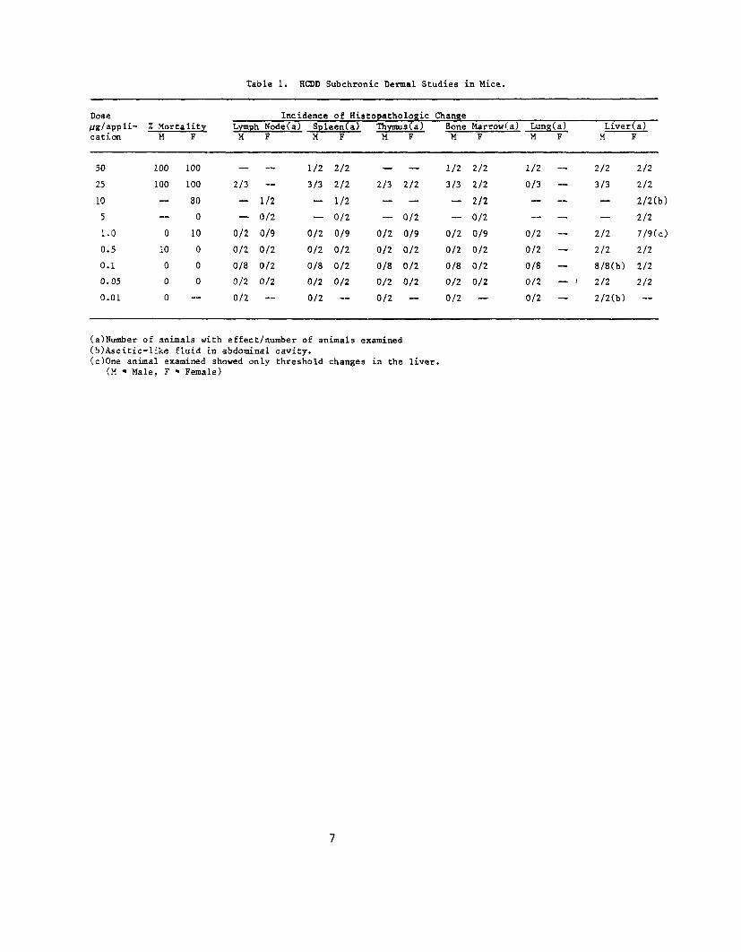
Table 1. HCDD Subchronic Dermal Studies in Mice.
Dose Incidence of Histopathologic Change ^/application
% Mortality M F
Lymph Node(a) Spleen(a) M F M F
Thymus(a) M F
Bone M
Marrow(a) Lung(a) F M F
Liver (a) M F
50
25
10
5
1.0
0.5
0.1
0.05
0.01
100
100
— — 0
10
0
0
0
100
100
80
0
10
0
0
0
—
— 2/3
—
— 0/2
0/2
0/8
0/2
0/2
—
— 1/2
0/2
0/9
0/2
0/2
0/2
—
1/2
3/3
—
— 0/2
0/2
0/8
0/2
0/2
2/2
2/2
1/2
0/2
0/9
0/2
0/2
0/2
—
— 2/3
—
— 0/2
0/2
0/8
0/2
0/2
— 2/2
— 0/2
0/9
0/2
0/2
0/2
—
1/2
3/3
—
— 0/2
0/2
0/8
0/2
0/2
2/2
2/2
2/2
0/2
0/9
0/2
0/2
0/2
—
1/2 —
0/3 _
— 0/2
0/2
0/8 —
0/2 — i
0/2
2/2
3/3
~
— 2/2
2/2
8/8(b)
2/2
2/2(b)
2/2
2/2
2/2(b)
2/2
7/9(c)
2/2
2/2
2/2
—
(a)Number of animals with effect/number of animals examined (b)Ascitic-like fluid in abdominal cavity. (c)One animal examined showed only threshold changes in the liver.
(M = Male, F = Female)
7

Table 2. HCDD Chronic Dermal Application Studies in Mice
Initial Time on Study Test No. of Dose (c) fJ.g/ Dosed Observed Group Animals(a,b) Room application (weeks) (weeks)
MALE
Untreated-Control Group No. 1 15 1C9 0 0 104
Untreated-Control Group No. 2 15 ISO 0 0 104
Vehicle-Control (d) 45 1C9 0 0 104
Dosed 30 1BO 0.01 104 0
Dosed plus DMBA 30 1BO 0.01(e) 101 0
FEMALE
Untreated-Control Group No. 1 15 1C9 0 0 104
Untreated-Control Group No. 2 15 1BO 0 0 104
Vehicle-Control (d) 45 1C9 0 0 104
Dosed 30 1BO 0.01 104 0
Dosed plus DMBA 30 1BO O.Ol(e) 104 0
(a) All animals were approximately 6 weeks of age when placed on study, regardless of shipment.
(b) Mice from multiple shipments covering a 2-week period were evenly distributed among all test and control groups.
(c) The HCDD was administered 3 days per week in acetone at a constant volume of 0.1 ml for each application. During the first 16 weeks, doses were 0.005/xg/application.
(d) Vehicle-controls received 0.1 ml acetone for each application. (e) Each animal was administered 50 u,g of dimethylbenzanthracene 1
week before to the initiation of dermal applications of HCDD.
8
G. Clinical Examinations and Pathology
Animals were observed twice daily for mortality. Body weights were
recorded every 2 weeks for the first 12 weeks and every month thereafter.
Moribund animals and those that survived to the termination of the study were
killed using sodium pentobarbital and necropsied.
Gross and microscopic examinations were performed on major tissues, major
organs, and all gross lesions from killed animals and from animals found
dead. Tissues were preserved in 10% neutral buffered formalin, embedded in
paraffin, sectioned, and stained with hematoxylin and eosin. The following
tissues and organs were taken at necropsy: skin, mandibular lymph node,
salivary gland, mammary gland, bone marrow, thymus, larynx, trachea, lungs
and bronchi, heart, thyroid, parathyroid, esophagus, stomach, duodenum,
colon, liver, gall bladder, pancreas, spleen, kidney, adrenal, gonads, nasal
cavity, brain, pituitary, spinal cord, skeletal muscle, sciatic nerve, and
all tissue masses.
Necropsies were also performed on all animals found dead, unless
precluded in whole or in part by autolysis or cannibalization. Thus, the
number of animals from which particular organs or tissues were examined
microscopically varies and does not necessarily represent the number of
animals that were placed on study in each group.
H. Data Recording and Statistical Analyses
Data on this experiment have been recorded in the Carcinogenesis Bioassay
Data System (Linhart et al., 1974). The data elements include descriptive
information on the chemicals, animals, experimental design, clinical obser
vations, survival, body weight, and individual pathologic results, as
recommended by the International Union Against Cancer (Berenblum, 1969).
Probabilities of survival were estimated by the product-limit procedure
of Kaplan and Meier (1958) and are presented in this report in the form of
graphs. Animals were statistically censored as of the time that they died
of other than natural causes or were found to be missing; animals dying from
natural causes were not statistically censored. Statistical analyses for a
possible compound-related effect on survival were performed using the method
9
of Cox (1972) to compare each dosed group with the control group. One-tailed
P values have been reported for all tests except for the departure from
linearity test, which is reported only when its two-tailed P value is less
than 0.05.
The incidence of neoplastic or nonneoplastic lesions has been given as
the ratio of the number of animals bearing such lesions at a specific
anatomic site (numerator) to the number of animals in which that site is
examined (denominator). In most instances, the denominators included only
those animals for which that site was examined histologically. However, when
macroscopic examination was required to detect lesions (e.g., skin or mammary
tumors) before histologic sampling, or when lesions could have appeared at
multiple sites (e.g., lymphomas), the denominators consist of the numbers of
animals necropsied.
The statistical analyses of tumor incidence are used to determine whether
animals receiving the test chemical developed a significantly higher propor
tion of tumors than did the control animals. As a part of these analyses,
the one-tailed Fisher exact test (Cox, 1970) was used to compare the tumor
incidence of a control group with that of a group of dosed animals. When
results from two dosed groups were compared simultaneously with those for a
single group, a correction to ensure an overall significance level of 0.05
may be made. The Bonferroni test for inequality (Miller, 1966) requires
that the P values for any comparison be less than or equal to 0.025. In
this case, HCDD and HCDD plus DMBA are compared with the vehicle control
group and, therefore, this correction is made where applicable.
Life table methods were used to analyze the incidence of tumors. Curves
of the proportions surviving without an observed tumor were computed as in
Saffiotti et al. (1972). The week during which an animal died naturally or
was killed was entered as the time point of tumor observation.
The approximate 95% confidence interval for the relative risk of each
dosed group, compared with its control, was calculated from the exact
interval on the odds ratio (Gart, 1971). The lower and upper limits of this
confidence interval have been included in the tables of statistical analyses.
The interpretation of the limits is that, in approximately 95% of a large
number of identical experiments, the true ratio of the risk in a dosed group
of animals to that in a control group would be within the interval calculated
10
from the experiment. When the lower limit of the confidence interval is
greater than one, it can be inferred that a statistically significant result
has occurred (P less than 0.025 one-tailed test when the control incidence
is not zero, P less than 0.050 when the control incidence is zero). When the
lower limit is less than unity but the upper limit is greater than unity, the
lower limit indicates the absence of a significant result while the upper
limit indicates that there is a theoretical possibility of the induction of
tumors by the test chemical, which could not be detected under the conditions
of this test.
11

12
III. RESULTS
A. Body Weights and Clinical Signs
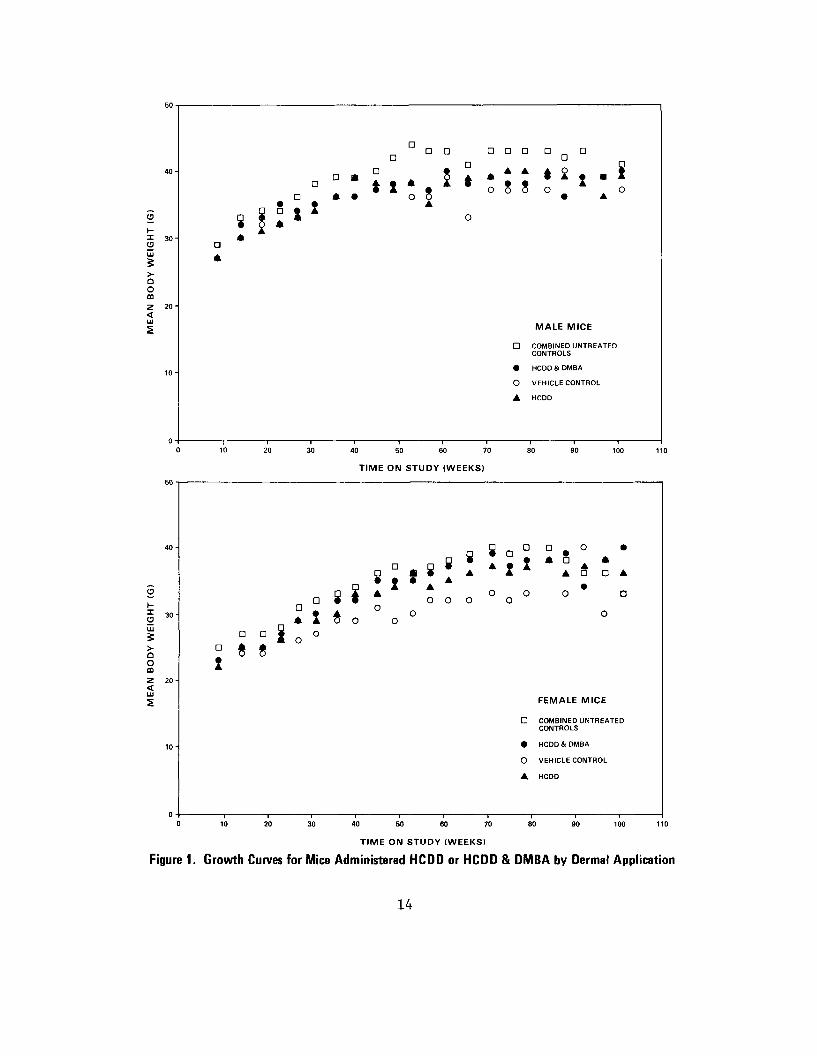
Throughout the bioassay, mean body weights of treated groups were
slightly greater than those of vehicle controls and slightly less than mean
body weights of untreated controls. Mean body weights were similar among the
groups of male or female mice administered HCDD, or HCDD plus DMBA; and mean
body weights of untreated controls were greater than those of the test and
vehicle-control groups (Figure 1). No other clinical signs were reported.
B. Survival
Estimates of the probabilities of survival for male and female mice
administered HCDD or HCDD plus DMBA by dermal application at the doses of
this bioassay, together with those of the controls, are shown by the Kaplan
and Meier curves in Figure 2. Five study groups were used for each sex: a
group administered HCDD alone, a group administered HCDD plus DMBA, a
vehicle-control group, and two untreated control groups. The two untreated-
control groups are pooled into one group. In male mice, the Cox test
comparing the survival of the pooled untreated-control group and the group
administered HCDD plus DMBA is significant (P=0.036) as a result of shortened
survival in the group with DMBA; however, the survival of each of the test
groups and the vehicle-control group are comparable. The results of the Cox
test in female mice are not significant, thus indicating comparable survival
among all groups.
In male mice, 23/30 (77%) of the pooled untreated-control group, 30/45
(67%) of the vehicle-control group, 25/30 (83%) of the group administered
HCDD alone, and 20/30 (67%) of the group administered HCDD plus DMBA lived
60 weeks or more. Nine of the 30 (30%) in the pooled untreated-control
group, 7/45 (16%) of the vehicle-control group, 2/30 (7%) in the group
administered HCDD alone, and 3/30 (10%) in the group administered HCDD plus
DMBA were alive at the end of study. In females, 28/30 (93%) of the combined
untreated-control group, 43/45 (96%) of the vehicle-control group, 24/30
13
TIME ON STUDY (WEEKS)
Figure 1. Growth Curves for Mice Administered HCDD or HCDD & DMBA by Dermal Application
14

TIME ON STUDY (WEEKS)
Figure 2. Survival Curves for Mice Administered HCDD or HCDD & OMBA by Dermal Application
15

(80%) of the group administered HCDD alone, and 26/30 (87%) of the group
administered HCDD plus DMBA lived beyond 60 weeks. There were 9/30 (30%) of
the pooled untreated-control group, 16/45 (36%) of the vehicle-control group,
8/30 (27%) of the group administered HCDD alone, and 7/30 (23%) of the group
administered HCDD plus DMBA alive at the end of the study at 102-106 weeks.
C. Pathology
Histopathologic findings on neoplasms in mice are summarized in
Appendixes A and C, Tables Al, A2, Cl, and C2; findings on nonneoplastic
lesions are summarized in Appendixes B and D, Tables Bl, B2, Dl, and D2.
Groups of animals receiving the test chemicals, the vehicle control, and
untreated controls are tabulated and summarized separately.
The types of tumors seen were typical for Swiss-Webster mice, except for
increased numbers of skin tumors which were not usually located on the back.
Most of the skin tumors were fibrosarcomas, with only an occasional fibroma
or epithelial tumor. There were two squamous cell carcinomas in male mice
given HCDD and DMBA. Although few of these skin tumors were seen, they
appeared in slightly higher incidences in dosed mice.
In conclusion, histopathologic examination provided no conclusive
evidence for the carcinogenicity of HCDD for the skin or internal organs of
female Swiss-Webster mice. Under the condition of this bioassay, however, a
slightly increased incidence of skin tumors may be associated with
administration of HCDD and HCDD plus DMBA.
D. Statistical Analyses of Results
Tables 3 and 4 contain the statistical analyses of the incidences of
those primary tumors that occurred in at least two animals of one group and
at an incidence of at least 5% in one or more than one group. The two
untreated-control groups are combined into one group in the analysis. Since
the doses in the two test groups did not differ in HCDD alone, no trend
analysis was made. The differences in tumor incidences between groups
receiving HCDD and those receiving HCDD plus DMBA were not statistically
significant.
16

In male mice, the incidence of animals with alveolar/bronchiolar
carcinomas in the lung is higher (P=0.045) in the group administered HCDD
than in the vehicle-control group. This value of 0.045 is above the level
of significance (0.025) used under the Bonferroni criterion to compare two
test groups with a common control group. No tests are significant when the
incidence of animals with this tumor in the group administered HCDD is
compared with that in the untreated-control group. The incidence of this
lesion in the untreated-control group is 4/28 (14%) as compared with 1/41
(2%) in the vehicle-control group. When the incidences of animals with
either adenomas or carcinomas of the lungs are used in the analysis, none of
the results are significant.
Statistical tests conducted upon the time to observation of the lung
tumors in the test groups of male rats compared with the matched controls
indicate no significant difference.
The two squamous cell carcinomas occurred in male mice administered
HCDD plus DMBA at weeks 60 and 94.
In female mice, the incidence of animals with fibrosarcomas of the skin
is significantly higher in each of the test groups (P=0.044) than in the
untreated control group; however, when the test groups are compared with the
vehicle-control group, the results are not significant.
In male mice, the incidence of animals with lymphomas or leukemias is
significantly lower (P=0.011) in the group administered HCDD than in the
untreated control group.
In each of the 95% confidence intervals for relative risk shown in the
tables, the value of one or less than one is included: this indicates the
absence of significant positive results. It should also be noted that each
of the intervals, except for the incidence of lymphomas in male mice, has an
upper limit greater than one indicating the theoretical possibility of tumor
induction by HCDD which could not be detected under the conditions of this
test.
17

Table 3. Analyses of the Incidence of Primary Tumors in Male Mice Administered HCDD or HCDD plus DMBA by Dermal Application (a)
Topograpy: Morphology
Integumentary System: Fibrosarcoma (b)
P Values (c,d)
Relative Risk (Combined Untreated Control) (e)
Lower Limit Upper Limit
Relative Risk (Vehicle Control) (e) Lower Limit Upper Limit
Weeks to First Observed Tumor
Integumentary System: Fibrosarcoma or Fibroma (b)
P Values (e,d)
Relative Risk (Combined Untreated Control) (e)
Lower Limit Upper Limit
Relative Risk (Vehicle Control) (e) Lower Limit Upper Limit
Weeks to First Observed Tumor
Integumentary System: Squamous-cell Carcinoma of the Skin (b)
P Values (c,d)
Relative Risk (Combined Untreated Control) (e)
Lower Limit Upper Limit
Relative Risk (Vehicle Control) (e) Lower Limit Upper Limit
Weeks to First Observed Tumor
Lung:Alveolar/Bronchiolar Adenoma (b)
P Value (c,d)
Relative Risk (Combined Untreated Control) (e)
Lower Limit Upper Limit
Relative Risk (Vehicle Control) (e)
Lower Limit Upper Limit
Weeks to First Observed Tumor
Combined UntreatedControl
3/29 (10)
55
3/29 (10)
55
0/29 (0)
_
2/28 (7)
59
Vehicle Control HCDD
3/42 (7) 6/30 (20)
N.S.
1.933 0.460 10.921
2.800 0.651 15.919
90 38
4/42 (10) 6/30 (20)
N.S.
1.933 0.460 10.921
2.100 0.544 9.195
46 38
0/42 (0) 0/30 (0)
N.S.
---
---
6/41 (15) 5/30 (17)
N.S.
2.333 0.421 22.894
1.139 0.301 4.023
75 76
HCDD plus DMBA
3/24 (13)
N.S.
1.208 0.176 8.191
1.750 0.250 11.932
60
3/24 (13)
N.S.
1.208 0.176 8.191
1.313 0.206 7.005
60
2/24 (8)
N.S.
Infinite 0.365 Infinite
Infinite 0.522 Infinite
60
3/24 (13)
N.S.
1.750 0.218 19.385
0.854 0.149 3.559
54

Table 3. Analyses of Che Incidence of Primary Tumors in Male Mice Administered HCDD or HCDD plus DMBA by Dermal Application (a)
(continued)
Combined Untreated Vehicle
Topography: Morphology Control Control
Lung: Alveolar /Bronchiolar Carcinoma (b) 4/28 (14) 1/41 (2)
P Values (c ,d)
Relative Risk (Combined Untreated Control) (e)
Lower Limit Upper Limit
Relative Risk (Vehicle Control) (e) Lower Limit Upper Limit
Weeks to First Observed Tumor 86 84
Lung: Alveolar/Bronchiolar AdenomaCarcinoma (b)
or 6/28 (21) 7/41 (17)
P Values (c ,d)
Relative Risk (Combined Untreated Control) (e)
Lower Limit Upper Limit
Relative Risk (Vehicle Control)Lower Limit Upper Limit
(e)
Weeks to First Observed Tumor 59 75
Hematopoietic System: All Lymphomas (b) 6/29 (21) 3/42 (7)
P Values ( c , d )
Relative Risk (Combined Untreated- Control) (e)
Lower Limit Upper Limit
Relative Risk (Vehicle Control) (e) Lower Limit Upper Limit
Weeks to First Observed Tumor 66 31
Hematopoietic System: Lymphoma or Leukemia (b) 6/29 (21) 4/42 (10)
P Values ( c , d )
Relative Risk (Combined Untreated Control) (e)
Lower Limit Upper Limit
Relative Risk (Vehicle Control) (e) Lower Limit Upper Limit
Weeks to First Observed Tumor 66 18
HCDD
5/30 (17)
P=0.045 (Vehicle Controls)
1.167 0.280 5.315
6.833 0.819
310.654
66
9/30 (30)
N.S.
1.400 0.515 4.155
1.757 0.655 4.854
66
0/30 (0)
P=0.011(N) (Untreated Controls)
0.000 0.000 0.590
0.000 0.000 2.286
0/30 (0)
P-O.Ol l (N) (Untreated Controls )
0.000 0.000 0.590
0.000 0.000 1.482
HCDD plus DMBA
3/24 (13)
N.S.
0.875 0.141 4.630
5.125 0.437
256.604
74
6/24 (25)
N.S.
1.167 0.358 3.752
1.464 0.454 4.397
54
2/24 (8)
N.S.
0.403 0.043 2.003
1.167 0.102 9.351
96
2/24 (8)
N.S.
0.403 0.043 2.003
0.875 0.083 5.550
96
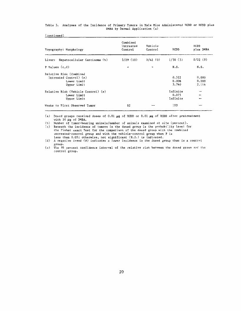
Table 3. Analyses of the Incidence of Primary Tumors in Male Mice Administered HCDD or HCDD plus DMBA by Dermal Application (a)
(continued)
Topography: Morphology
Combined UntreatedControl
Vehicle Control HCDD
HCDD plus DMBA
Liver: Hepatocellular Carcinoma (b) 3/29 (10) 0/42 (0) 1/30 (3) 0/22 (0)
P Values (c,d) N.S. N.S.
Relative Risk (Combined Untreated Control) (e) 0.322 0.000
Lower Limit 0.006 0.000 Upper Limit 3.740 2.114
Relative Risk (Vehicle Control) (e) Infinite —„Lower Limit 0.075
Upper Limit Infinite
_, Weeks to First Observed Tumor 82 — 103
(a) Dosed groups received doses of 0.01 fig of HCDD or 0.01 Us of HCDD after pretreatment with 50 H% of DMBA.
(b) Number of tumor-bearing animals/number of animals examined at site (percent). (c) Beneath the incidence of tumors in the do'sed group is the probability level for
the Fisher exact Test for the comparison of the dosed group with the combined untreated-control group and with the vehicle-control group when P is less than 0.05; otherwise, not significant (N.S.) is indicated.
(d) A negative trend (N) indicates a lower incidence in the dosed group than in a control group.
(e) The 95 percent confidence interval of the relative risk between the dosed group and tha control group.
20

Table 4. Analyses of the Incidence of Primary Tumors in Female Mice Administered HCDD or HCDD plus DMBA by Dermal Application (a)
Combined Untreated
Topography: Morphology Control
Integumentary System: Fibrosarcoma (b) 0/30 (0)
P Values (c,d)
Relative Risk (Combined Untreated Control) (e)
Lower Limit Upper Limit
Relative Risk (Vehicle Control) (e) Lower Limit Upper Limit
Weeks to First Observed Tumor —
Lung: Alveolar/Bronchiolar Adenoma (b) 2/30 (7)
P Values (c ,d)
Relative Risk (Combined Untreated Control) (e)
Lower Limit Upper Limit
Relative Risk (Vehicle Control) (e) Lower Limit Upper Limit
Weeks to First Observed Tumor 102
Lung: Alveolar/Bronchiolar Carcinoma (b) 2/30 (7)
P Values (c ,d)
Relative Risk (Combined Untreated Control) (e)
Lower Limit Upper Limit
Relative Risk (Vehicle Control) (e) Lower Limit Upper Limit
Weeks to First Observed Tumor 91
Lung: Alveolar/Bronchiolar Adenoma or Carcinoma (b) 4/30 (13)
P Value (c ,d)
Relative Risk (Combined Untreated Control) (e)
Lower Limit Upper Limit
Relative Risk (Vehicle Control) (c) Lower Limit Upper Limit
Weeks to First Observed Tumor 91
Vehicle Control
2/41 (5)
98
4/41 (10)
79
5/41 (12)
75
8/41 (20)
75
HCDD
4/27 (15)
P-0.044 (Untreated Controls)
Infinite 1.054
Infinite
3.037 0.467
31.347
57
4/27 (15)
N.S.
2.222 0.348
22.869
1.519 0.306 7.399
57
1/27 (4)
N.S.
0.556 0.010
10.031
0.304 0.007 2.493
89
4/27 (15)
N.S.
1.111 0.228 5.378
0.759 0.183 2.511
57
HCDD plus DMBA
4/27 (15)
P-0.044 (Untreated Controls)
Infinite 1.054
Infinite
3.037 0.467
31.347
77
1/25 (4)
N.S.
0.600 0.011
10.786
0.410 0.009 3.806
104
2/25 (8)
N.S.
1.200 0.093
15.432
0.656 0.066 3.628
100
3/25 (12)
N.S.
0.900 0.144 4.789
0.615 0.113 2.270
100
21
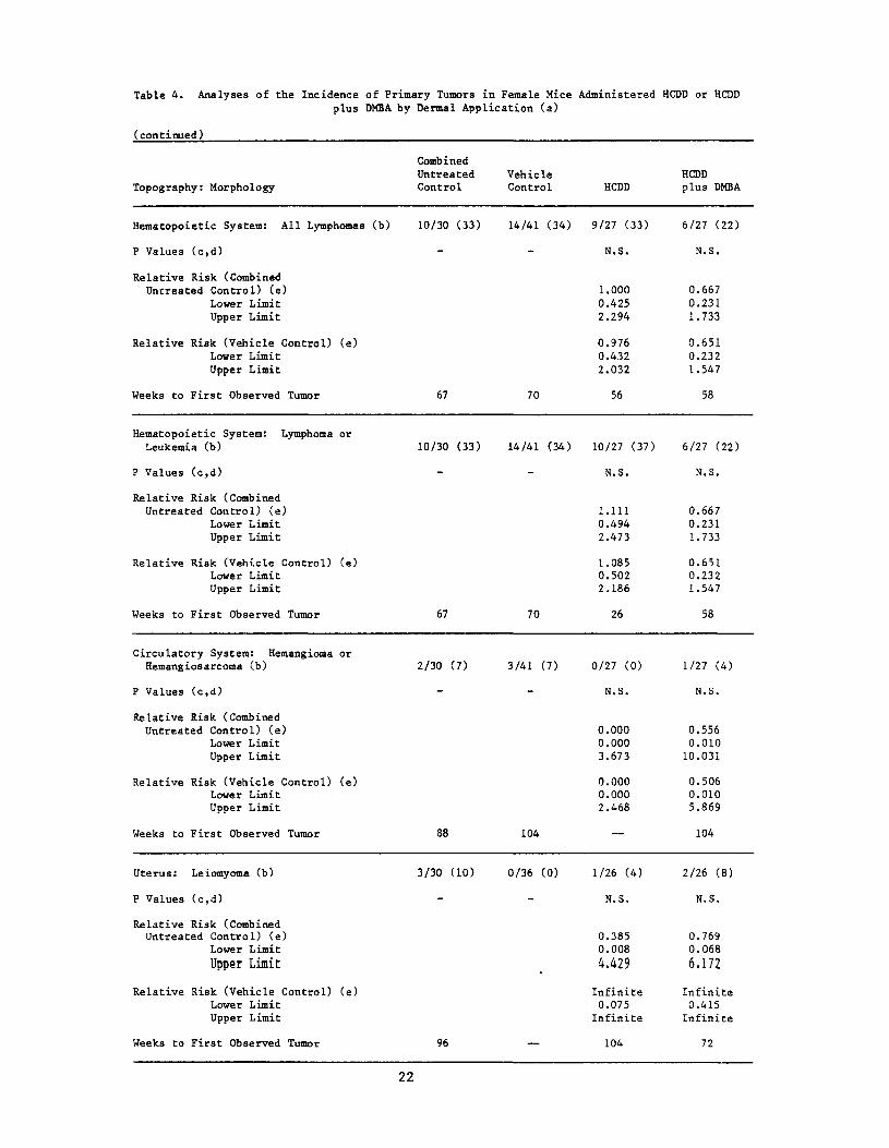
Table 4. Analyses of the Incidence of Primary Tumors in Female Mice Administered HCDD or HCDD plus DMBA by Dermal Application (a)
(continued)
Combined Untreated Vehicle
Topography: Morphology Control Control
Hematopoietic System: All Lymphomas (b) 10/30 (33) 14/41 (34)
P Values (c,d)
Relative Risk (Combined Uncreated Control) (e)
Lower Limit Upper Limit
Relative Risk (Vehicle Control) (e) Lower Limit Upper Limit
Weeks to First Observed Tumor 67 70
Hematopoietic System: Lymphoma or Leukemia (b) 10/30 (33) 14/41 (34)
P Values (c,d)
Relative Risk (Combined Untreated Control) (e)
Lower Limit Upper Limit
Relative Risk (Vehicle Control) (e) Lower Limit Upper Limit
Weeks to First Observed Tumor 67 70
Circulatory System: Hemangioma or Hemangiosarcoma (b) 2/30 (7) 3/41 (7)
P Values (c,d)
Relative Risk (Combined Untreated Control) (e)
Lower Limit Upper Limit
Relative Risk (Vehicle Control) (e) Lower Limit Upper Limit
Weeks to First Observed Tumor 88 104
Uterus: Leiomyoma (b) 3/30 (10) 0/36 (0)
P Values (c,d)
Relative Risk (Combined Untreated Control) (e)
Lower Limit
Upper Limit
Relative Risk (Vehicle Control) (e) Lower Limit Upper Limit
Weeks to First Observed Tumor 96 —
HCDD
9/27 (33)
N.S.
1.000 0.425 2.294
0.976 0.432 2.032
56
10/27 (37)
N.S.
1.111 0.494 2.473
1.085 0.502 2.186
26
0/27 (0)
N.S.
0.000 0.000 3.673
0.000 0.000 2.468
1/26 (4)
N.S.
0.385 0.008
4.429
Infinite 0.075 Infinite
104
HCDD plus DMBA
6/27 (22)
N.S.
0.667 0.231 1.733
0.651 0.232 1.547
58
6/27 (22)
N.S.
0.667 0.231 1.733
0.651 0.232 1.547
58
1/27 (4)
N.S.
0.556 0.010 10.031
0.506 0.010 5.869
104
2/26 (8)
N.S.
0.769 0.068
6.172
Infinite 0.415 Infinite
72
22
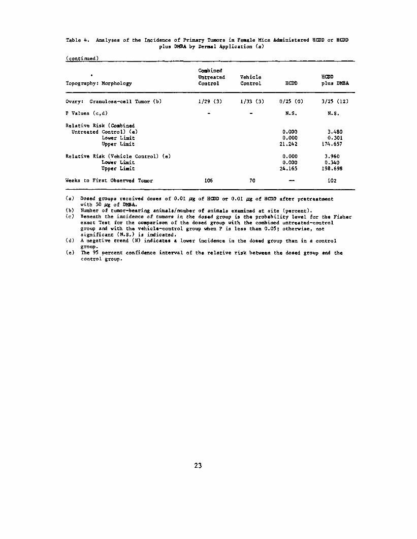
Table 4. Analyses of Che Incidence of Primary Tumors in Female Mice Administered HCDD or HCDD plus DHBA by Dermal Application (a)
(continued)
Combined * Untreated Vehicle HCDD
Topography: Morphology Control Control HCDD plus DMBA
Ovary: Granulosa-cell Tumor (b) 1/29 (3) 1/33 (3) 0/25 (0) 3/25 (12)
P Values (c,d) N.S. N.S.
Relative Risk (Combined Untreated Control) (e) 0.000 3.480
Lower Limit 0.000 0.301 Upper Limit 21.242 174.657
Relative Risk (Vehicle Control) (e) 0.000 3.960 Lower Limit 0.000 0.340 Upper Limit 24.165 198.698
ttoo FirsFirstt ObserveObservedd TumoTumorr 101066 7700 —WeekWeekss — 101022
(a) Dosed groups received doses of 0.01 Hg of HCDD or 0.01 fig of HCDD after pretreatment with 50 ug of DMBA.
(b) Number of tumor-bearing animals/number of animals examined at site (percent). (c) Beneath the incidence of tumors in the dosed group is the probability level for the Fisher
exact Test for the comparison of the dosed group with the combined untreated-control group and with the vehicle-control group when P is less than 0.05; otherwise, not significant (N.S.) is indicated.
(d ) A negative trend (N) indicates a lower incidence in the dosed group than in a control group.
(e) The 95 percent confidence interval of the relative risk between the dosed group and the control group.
23

24
IV. DISCUSSION
Throughout the bioassay the mean body weights of the groups of male or
female mice administered HCDD or HCDD plus DMBA were similar and were greater
than those of corresponding vehicle-control groups but less than those of
untreated control groups. The consequences of using only O.OOS/Lig rather than
0.01/ig HCDD per application during the first 16 weeks of the chronic study
were not considered to be significant.
After 80 weeks, survival was sufficient to detect late appearing tumors.
Alveolar/bronchiolar carcinomas occurred in the male mice administered
HCDD at an incidence that was significantly higher (P=0.045) than that in the
vehicle-control group, but the incidence was not significant when compared
with the untreated control group that had longer survival. Alveolar/
bronchiolar carcinomas did not occur in statistically significant incidences
in male mice pre-treated with DMBA before being administered HCDD, when
compared with either the vehicle or the untreated controls.
Compared with the untreated control group, the incidence of male mice
with lymphomas or leukemias was significantly lower (P=0.011) in the group
administered HCDD but not in the group administered with DMBA and HCDD.
Two squamous cell carcinomas in male mice administered HCDD plus DMBA
occurred in dosed skin near a hind leg or the tail. The possibility that
these areas may have been exposed accidentally during application of the test
chemical cannot be eliminated.
In female mice, fibrosarcomas in the skin occurred at incidences that
were significantly higher (P=0.044) in animals administered HCDD (both those
with and without pretreatment with DMBA) than in the untreated control
group. Although there were more female mice with fibrosarcomas in the two
groups administered HCDD when compared with the vehicle controls, the
increased incidences were not statistically significant. Thus, the
relationship between incidences of fibrosarcomas in female mice and HCDD
administration could not be clearly established.
In the present study, pretreatment with DMBA had no statistically
significant effect on skin tumor production in either male or female mice.
The significance of the two squamous cell carcinomas of the skin in male
25

mice administered both HCDD and DMBA cannot be determined in the absence of
a group exposed to DMBA alone. Male mice pretreated with DMBA, when compared
with male mice not pretreated, had a lower incidence of alveolar/bronchiolar
carcinomas and a higher incidence of lymphomas.
A bioassay of HCDD administered by gavage was run concurrently with the
present study (NCI, 1980). Under the conditions of that bioassay, HCDD was
carcinogenic for female Osborne-Mendel rats and for male and female B6C3F1
mice, inducing increased incidences of hepatocellular carcinomas, adenomas,
or neoplastic nodules. In the gavage studies, the B6C3F1 mice received 5,
2.5, or 1.25 /lg/kg/week (males) and 10, 5, or 2.5 /tg/kg/week (females).
These were high doses when compared with 1.5/ig/kg/week for male and female
Swiss-Webster mice in the present studies. The mice in the gavage studies
lived longer.
A dermal bioassay of TCDD in Swiss-Webster mice was conducted
concurrently in the room used for the present study. The protocols were
identical except that a lower dose was applied (NCI, 1980a). In the dermal
TCDD studies, there were statistically significant increased incidences of
fibrosarcomas in the integumentary system in female mice receiving 0.005 /xg
per application when compared with controls. This significant increase was
not found in the male mice receiving 0.001 /xg per application. In the
present dermal HCDD studies, both male and female mice received Q.Ql jj,g per
application. Thus, after dermal application under identical conditions, TCDD
was clearly carcinogenic for female mice, whereas HCDD (at a two-fold higher
dose) was not.
26

V. CONCLUSION
Under the conditions of this bioassay, HCDD was not considered
carcinogenic for male or female Swiss-Webster mice.
27

28
VI. BIBLIOGRAPHY
Armitage, P., Statistical Methods in Medical Research, John Wiley & Sons, Inc., New York, 1971, pp. 362-365.
Berenblum, I., ed., Carcinogenicity Testing! A Report of the Panel of Car cino genie ity of the Cancer Research Commission of UICC, Vol. 2^, International Union Against Cancer, Geneva, 1969.
Blaser, W. W., Bredeweg, R. A., Shadoff, L. A., and Stehl, R. H., Determination of chlorinated dibenzo-p-dioxins in pentachlorophenol by gas chromatography - mass spectrometry. Anal. Chem. 48(7);984-986, 1976.
Bradlaw, J. A., Garthoff, L. H., Graff , D. M., and Hurley, N. E., Detection of chlorinated dioxins: induction of aryl hydrocarbon hydroxylase activity in rat hepatoma cell culture. Toxicol. Appl. Pharmacol. 33_:166, 1975.
Cantrell, J. S., Webb, N. C., and Mabis, A. J., The identification and crystal structure of a hydropericardium-producing factor: 1,2,3,7,8,9hexachlorodibenzo-p-dioxin. Acta Cryst. B25; 150-151, 1969.
Courtney, K. D., Gaylor, D. W., Hogan, M. D., Falk, H. L., Bates, R. R., and Mitchell, I., Teratogenic evaluation of 2,4,5-T. Science 168:864-866, 1970.
Cox, D. R., Analysis of Binary Data, Methuen & Co., Ltd., London, 1970, pp. 48-52.
Cox, D. R., Regression models and life tables. _J. R. Statist. Soc. B34;187220, 1972.
Firestone, D., Etiology of chick edema disease. Environ. Health Perspect. 00:59-66, 1973.
Firestone, D., The 2,3,7,8-tetrachlorodibenzo-para-dioxin problem: a review. Ecol. Bull. (Stockholm) 2_7_:39-52, 1978.
Firestone, D., Ress, J., Brown, N. L., Barron, R. P., and Damico, J.N., Determination of polychlorodibenzo-p-dioxins and related compounds in commercial chlorophenols. J_. Assoc. Official Analyt. Chem. 55 :85-92,1972.
Gart, J. J., The comparison of proportions: a review of significance tests, confidence limits and adjustments for stratification. Rev. Int. Stat. Inst. 32:148-169, 1971.
Gray, A. P., Cepa, S. P., and Cantrell, J. S., Intervention of the Smiles rearrangement in synthesis of dibenzo-p-dioxins. 1,2,3,6,7,8- and 1,2,3,7,8, 9-hexachlorodibenzo-p-dioxin (HCDD). Tetrahedron Letters 33; pp. 2873-2876, 1975.
Kaplan, E. L. and Meier, P., Nonparametric estimation from incomplete observations. J[. Amer. Statist. Assoc. _53_:457-481, 1958.
29
Kende, A. S., and DeCamp, M. R., Smiles rearrangements in the synthesis of hexachlorodibenzo-p-dioxins. Tetrahedron Letters 33j 2877-2880, 1975.
Linhart, M. S., Cooper, J. A., Martin, R. L., Page, N. P., and Peters, J. A., Carcinogenesis bioassay data system. Comp. and Biomed. Res. 7^:230-248, 1974.
McConnell, E. E. and Moore, J. A., The comparative toxicity of chlorinated dibenzo-p-dioxin isomers in mice and guinea pigs. Toxicol. Appl. Pharmacol. 37:146, 1976.
Miller, R. G.,Co., New York,
Jr., Simultaneous 1966, pp. 6-10.
Statistical Inference, McGraw-Hill Book
NCI, National1,2,3,7,8?9
Cancer Institute, Bioassay Hexachlorodibenzo-p-dioxins
of_ a for
Mixture of Possible
1,2,3,6,7,8 and Carcinogenicity
(Gavage Study), DHHS Publication No. (NIH) 80-1754, Carcinogenesis Testing Program, National Cancer Institute, National Institutes of Health, Bethesda, Md., 1980.
NCI, National Cancer Institute, Bioassay £f 2,3,7,8-Tetrachlorodibenzo-pdioxin (Dermal Study), DHHS Publication No. (NIH) 80-1757, Carcinogenesis Testing Program, National Cancer Institute, National Institutes of Health, Bethesda, Md., 1980a.
Poland, A., Glover, E., and Kende, A., Stereospecific, high affinity binding of 2,3,7,8-Tetrachlorodibenzo-p-dioxin by hepatic cytosol. ^J. Biol. Chem. 25J.:4936-4946, 1976.
Saffiotti, U., Montesano, R., Sellakumar, A. R., Cefis, F., and Kaufman, D. G., Respiratory tract Carcinogenesis in hamsters induced by different numbers of administrations of benzo(a)pyrene and ferric oxide. Cancer Res. 32: 1073-1081, 1972.
Schwetz, B. A., Norris, J. M., Sparschu, G. L., Rowe, V. K., Gehring, P. J., Emerson, J. L., and Gerbig, C. G., Toxicology of chlorinated dibenzo-pdioxins. Environ. Health. Perspect. (5);87-99, 1973.
Sparschu, G. L., Dunn, F. L., and Rowe, V. K., Study of the teratogenicity of 2,3,7,8-tetrachlorodibenzo-p-dioxin in the rat. Food Cosmet. Toxicol. £:405412, 1971.
Ward, J. M., Goodman, D. G., Griesemer, R. A., Hardisty, J. F., Schueler, R. L., Squire, R. A., and Strandberg, J. D. , Quality assurance for pathology in rodent Carcinogenesis tests. ^J. Environ. Pathol. Toxicol. 2^:371-378, 1978.
Woolson, E. A., Thomas, R. F., and Ensor, P. D. J., Survey of polychlorodibenzo-p-dioxin content in selected pesticides. ^J. Agr. Food Chem. 20(2);351-354, 1972.
30

APPENDIX A
SUMMARY OF THE INCIDENCE OF NEOPLASMS IN MICE ADMINISTERED HCDD BY
DERMAL APPLICATION
31

32
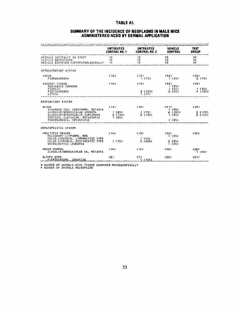
TABLE A1.
SUMMARY OF THE INCIDENCE OF NEOPLASMS IN MALE MICE ADMINISTERED HCDD BY DERMAL APPLICATION
ANIMALS INITIALLY IN STUDY ANIMALS NECROPSIED ANIMALS EXAMINED lilSTOPATHOLOGICALLY
INTEGUMENTARY SYSTEM
*SKIN FIBROSARCOMA
XSU5CUT TISSUE SEBACEOUS ADENOMA FIBROMA FIBnOSARCOMA LIPOMA
RESPIRATORY SYSTEM
SLUNG SQUAMOUS CELL CARCINOMA, METASTA ALVECLAR/BRONCIIIOLAR ADENOMA ALVEOLAFVBRCNCHIOLAR CARCINOMA CORTICAL CARCINOMA, METASTATIC FIBROSARCOMA, METASTATIC
HEMATOPOIETIC SYSTEM
^MULTIPLE ORGANS MALIGNANT LYMPHOMA, NOS HALIG.LYMPHOMA, LYMPHOCYTIC TYPE NALIG.LYKPHOfIA, HISTIOCYTIC TYPE GRANULOCYTIC LEUKEMIA
SBONE MARROW ALVEOLAR/BRONCHIOLAR CA, METASTA
ULYMPH NODE FIRROSARCOMA, INVASIVE
UNTREATED CONTROL NO. 1
15 14 14
( 14)
( 14)
( 13)
1 (85!) 2 (155!) 1 (8%)
( 14)
1 (75O
( 14)
(8)
UNTREATED CONTROL NO. 2
15 15 15
(15) 1 (7%)
( 15)
2 (135!) 1 (75O
(15)
1 (7%) 2 (135!)
( 15)
1 (75!) 3 (205!)
( 14)
(7) 1 (14X)
VEHICLE CONTROL
45 42 42
(42)1 (25()
(42) 1 (25O 1 (25:) 2 (550
(41) 1 (25O 6 (1550 1 ( 25! )
1 (25!)
(42) 1 (25O
2 (55!) 1 (25O
(40)
(25)
TEST GROUP
30 50 30
(30) 2 (75!)
(30)
1 (35!) 4 (1350
(30)
5 (1750 5 (175!)
(30)
(28)I (45!)
(21)
S NUMBER OF ANIMALS WITH TISSUE EXAMINED MICROSCOPICALLY X NUMBER OF ANIMALS NECROPSIED
33
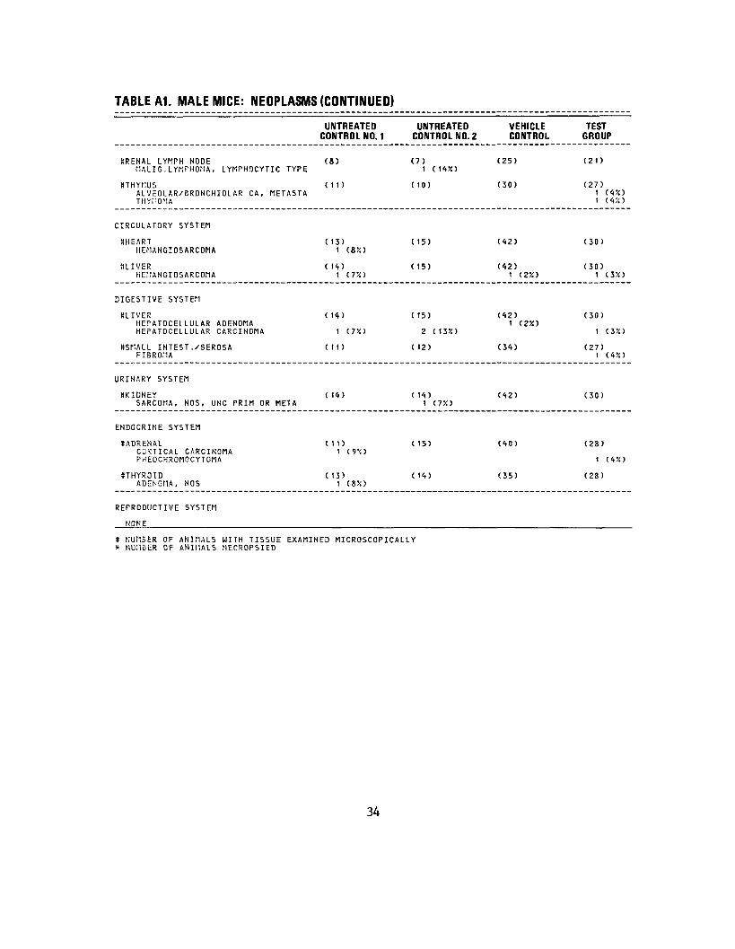
TABLE A1. MALE MICE: NEOPLASMS (CONTINUED)
UNTREATED CONTROL NO. 1
UNTREATEDCONTROL NO. 2
VEHICLE CONTROL
TEST GROUP
SRENAL LYMPH NODE tlALIG.LYMPHOMA, LYMPHOCYTIC TYPE
(8) (7)1 (145!) (25) (21)
STHYNUS ALVEOLAR/BRONCHIOLAR CA,THYI10NA
METASTA (It) (10) (30) (27)
11 (4!O (4X)
CIRCULATORY SYSTEM
8HEART HEMANGIOSARCOMA
( 13) 1 (8X)
(15) (42) (30)
SLIVER HEriANGIOSARCOMA
( 14) t <73O
(15) (42) 1 (2%)
(30)1 (3r<)
DIGESTIVE SYSTEM
SLIVER HEPATOCELLULAR ADENOMA HEPATOCELLULAR CARCINOMA
( 14)
1 (75O
(15)
2 (13X)
(42) 1 (2X)
(30)
1 (3X)
SSMALL INTEST./SEROSA FIBROMA
(11) (12) (34) (27)1 (45O
URINARY SYSTEM
tfKIDNEY SARCOMA, NOS, UNC PRIM OR META
( 14) (14)1 (7%) (42) (30)
ENDOCRINE SYSTEM
SADRENAL CCSTICAL CARCINOMA PHEOCHROMOCYTOMA
(11) 1 (9'<)
(15) (40) (28)
1 (4%)
STHYROID ( 13) (14)ADENOMA, NOS 1 (&'/.)
(35) (28)
REPRODUCTIVE SYSTEM
HONE
* NUMBER OF ANIMALS WITH TISSUE EXAMINED MICROSCOPICALLY * NUMBER OF ANIMALS NECROPSIED
34
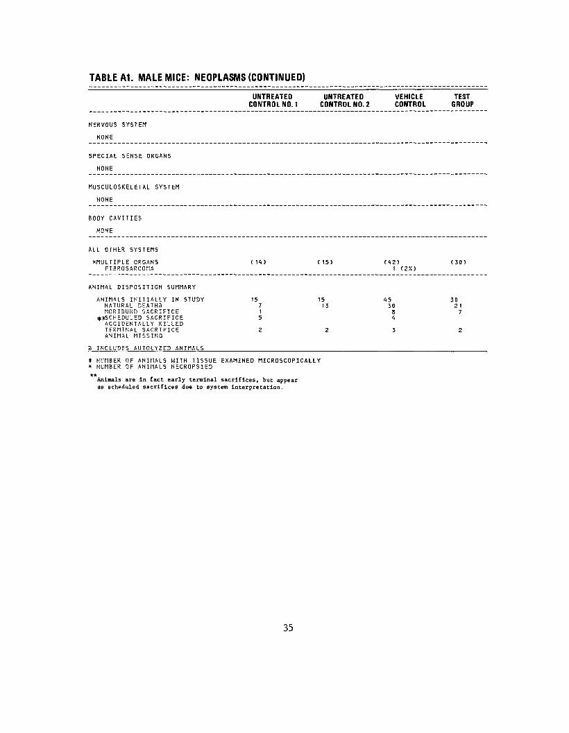
TABLE A1. MALE MICE: NEOPLASMS (CONTINUED)
UNTREATED UNTREATEDCONTROL NO. 1 CONTROL NO. 2
NERVOUS SYSTEM
NONE
SPECIAL SENSE ORGANS
NONE
MUSCULOSKELETAL SYSTEM
NONE
BODY CAVITIES
NONE
ALL OTHER SYSTEMS
^MULTIPLE ORGANS (14) (15)FIBROSARCOMA
ANIMAL DISPOSITION SUMMARY
ANIMALS INITIALLY IN STUDY 15 15NATURAL DEATH2 7 13MORIBUND SACRIFICE 1
*|cSCHEDULED SACRIFICE 5ACCIDENTALLY KILLED TERMINAL SACRIFICE 2 2 ANIMAL MISSING
a INCLUDES AUTOLYZED ANIMALS
* NUMBER OF ANIMALS WITH TISSUE EXAMINED MICROSCOPICALLY * NUMBER OF ANIMALS NECROPSIED ** Animals are in fact early terminal sacrifices, but appear as scheduled sacrifices due to system interpretation.
VEHICLE TEST CONTROL GROUP
(42) (30) 1 (2%)
45 30 30 21
8 7 4
3 2
35
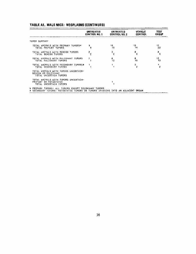
TABLE A1. MALE MICE: NEOPLASMS (CONTINUED)
UNTREATED UNTREATED VEHICLE TEST CONTROL NO. 1 CONTROL NO. 2 CONTROL GROUP
TUMOR SUMMARY
TOTAL ANIMALS WITH PRIMARY TUMORS'* 9 10 15 17 TOTAL PRIMARY TUMORS 9 15 19 22
TOTAL ANIMALS WITH BENIGN TUMORS 2 2 8 8 TOTAL BENIGN TUMORS 2 2 9 9
TOTAL ANIMALS WITH MALIGNANT TUMORS 7 8 8 12 TOTAL MALIGNANT TUMORS 7 12 10 13
TOTAL ANIMALS WITH SECONDARY TUMORS* t 1 2 1 TOTAL SECONDARY TUMORS 1 1 2 2
TOTAL ANIMALS WITH TUMORS UNCERTAINBENIGN OR MALIGNANT
TOTAL UNCERTAIN TUMORS
TOTAL ANIMALS WITH TUMORS UNCERTAINPRIMARY OR METASTATIC 1
TOTAL UNCERTAIN TUMORS 1
X PRIMARY TUMORS: ALL TUMORS EXCEPT SECONDARY TUMORS tt SECONDARY TUMORS: METASTATIC TUMORS OR TUMORS INVASIVE INTO AN ADJACENT ORGAN
36
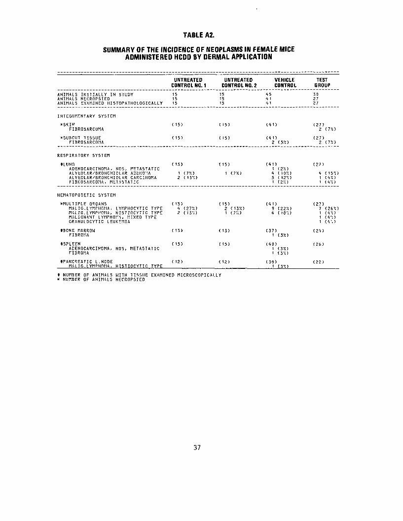
TABLE A2.
SUMMARY OF THE INCIDENCE OF NEOPLASMS IN FEMALE MICE ADMINISTERED HCDD BY DERMAL APPLICATION
UNTREATED UNTREATED VEHICLE CONTROL NO. 1 CONTROL NO. 2 CONTROL
ANIMALS INITIALLY IN STUDY 15 15 45 ANIMALS NECROPSIED 15 15 4 1 ANIMALS EXAMINED HISTOPATHOLOGICAL LY 15 15 41
INTEGUMENTARY SYSTEM
*SKIN ( 15) (15) (41) FIBROSARCOMA
*SUBCUT TISSUE ( 15) ( 15) (41) FIBROSARCOMA 2 (55!)
RESPIRATORY SYSTEM
SLUNG ( 15) ( 15) (41) ADENOCARCIHOMA, NOS, METASTATIC 1 (2X> ALVEOLAR/BRONCHIOLAR ADENOMA 1 (T/.t 1 (75!) 4 ( t 0 V. 1 ALVEOLAR/BRONCHIOLAR CARCINOMA 2 ( 13'0 5 ( 12'i) FIBROSARCOMA, METASTATIC 1 (2%)
HEMATOPOIETIC SYSTEM
^MULTIPLE ORGANS ( 15) ( 15) (41) MALIG.LYMPHCMA, LYMPHOCYTIC TYPE 4 (275!) 2 (13X) 9 (22X) MALIG.LYMPHOMA, HISTIOCYTIC TYPE 2 (13-'.) 1 (7!O 4 ( 10'!) MALIGNANT LYMPHOf:'\, MIXED TYPE GRANULOCYTIC LEUKEMIA
*BONE MARROW ( 13) ( 13) (37) FIBROMA 1 (3V.)
SSPLEEN ( 15) ( 15) (40) ADENOCARCINOMA, NOS, METASTATIC 1 (35!) FIBROMA 1 (3'0
((PANCREATIC L.NODE ( 12) ( 12) (30) MALIG.LYMPHPMA, HISTIOCYTIC TYPE 1 ( 3°< )
« NUMBER OF ANIMALS WITH TISSUE EXAMINED MICROSCOPICALLY * NUMBER OF ANIMALS NECROPSIED
TEST GROUP
30 27 27
(27) 2 (7V.)
(27) 2 (72)
(27)
4 (15'<) 1 (45!) 1 (45!)
(27) 7 (26'0 1 ( 4\) 1 (4%') 1 ( 4 '. )
(24)
(26)
(22)
37
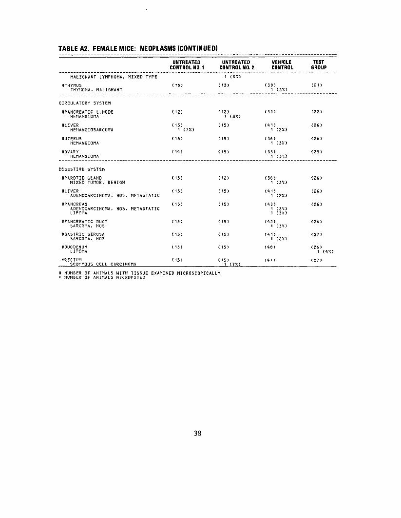
TABLE A2. FEMALE MICE: NEOPLASMS (CONTINUED)
MALIGNANT LYMPHOMA, MIXED TYPE
STHYMUS THYHOMA, MALIGNANT
CIRCULATORY SYSTEM
SPANCREATIC L . NODE HEMANGIOMA
OLIVER HEMANGIOSARCOMA
((UTERUS HEMANGIOMA
tOVARY HEMANGIOMA
DIGESTIVE SYSTEM
((PAROTID GLAND MIXED TUMOR, BENIGN
(tLIVERADENOCARCINOMA, NOS, METASTATIC
ttPANCREAS ADEHOCARCINOMA, NOS, METASTATIC LIPOI1A
((PANCREATIC DUCT SARCOMA, NOS
((GASTRIC SEROSA SARCOMA, NOS
((DUODENUMLIPOMA
*RECTUM S<!U."10US CELL CARCINOMA
UNTREATED CONTROL NO. 1
( 15)
(12)
(15) 1 (T/.t
( 15)
( 14)
( 15)
( 15)
( 15)
( 15)
( 15)
( 13)
( 15)
UNTREATED CONTROL NO. 2
1 (8::> ( 15)
( 12)i («*>
(15)
(15)
( 15)
( 12)
( 15)
( 15)
(15)
( 15)
( 15)
( 15) 1 (7X)
VEHICLE CONTROL
(39) 1 (3X>
(30)
(41) 1 (2X)
(36) 1 (3X)
(33) 1 (3'O
(36) 1 (.y-i)
(41) 1 (2X)
(10) 1 (3'0 1 (3%)
(40) 1 (3-0
(<t1) 1 (250
(40)
(41)
TEST GROUP
(21)
(22)
(26)
(26)
(25)
(26)
(26)
(26)
(26)
(27)
(26) 1 (4'<)
(27)
» NUMBER OF ANIMALS WITH TISSUE EXAMINED MICROSCOPICALLY * NUMBER OF ANIMALS NECROPSIED
38

TABLE A2. FEMALE MICE: NEOPLASMS (CONTINUED)
UNTREATED UNTREATEDCONTROL NO. 1 CONTROL NO.
VEHICLE2 CONTROL
TEST JBOUP
URINARY SYSTEM
^GENITOURINARY TRACT FIBROSARCOMA
(15) (15) (41)1 (25!) (27)
ENDOCRINE SYSTEM
fUDRENAL CORTICAL ADENOMA PHEOCH^OMOCYTOMA
(15)1 (7%) (14) (41) (26)
1 <«X>
REPRODUCTIVE SYSTEM
XVAGINA LEIOMYOMA
(15) (15) (<»1) (27)1 (<»?!)
SUTERUS ADEHOCARCINOMA, NOS LEIOMYOMA
(15)
2 ( IJX)
(15)
1 (7X)
(36)1 (3X> (26)
1 (45!)
(tCERVIX UTERI LEIOMYOMA
(15)1 (7X) (15) (36) (26)
(tOVARY LUTEOMA GRAHULOSA-CELL TUMOR LIPOMA
(1<t>1 (7X) (15)
1 (7X)
(33)
1 (3X) 1 (3X)
(25)
NERVOUS SYSTEM
NONE
SPECIAL SENSE ORGANS
NONE
MUSCULOSKELETAL SYSTEM
NONE
BODY CAVITIES
NONE
* NUMBER* NUMBER
OF A N I M A L S WITH TISSUE EXAMINED MICROSCOPICALLY OF A N I M A L S NECROPSIED
39
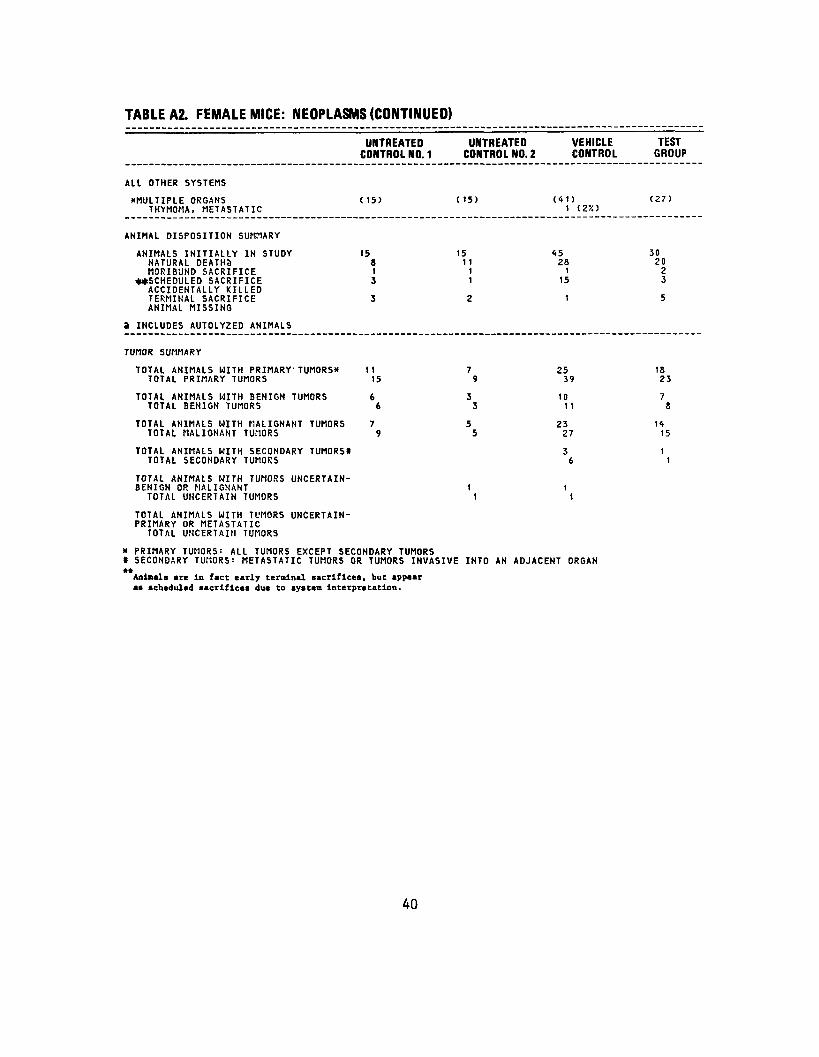
TABLE A2. FEMALE MICE: NEOPLASMS (CONTINUED)
UNTREATED UNTREATED VEHICLE TEST CONTROL NO. 1 CONTROL NO. 2 CONTROL GROUP
AIL OTHER SYSTEMS
XMULTIPLE ORGANS (15) (15) (41) (27) THYMOMA, METASTATIC 1 (2%)
ANIMAL DISPOSITION SUMMARY
ANIMALS INITIALLY IN STUDY 15 15 15 30 NATURAL DEATHS 8 11 28 20 MORIBUND SACRIFICE 1 1 1 2
**SCHEDULED SACRIFICE J 1 15 3 ACCIDENTALLY KILLED TERMINAL SACRIFICE 3 2 1 5 ANIMAL MISSING
3 INCLUDES AUTOLYZED ANIMALS
TUMOR SUMMARY
TOTAL ANIMALS WITH PRIMARY' TUMORS* 11 7 25 18 TOTAL PRIMARY TUMORS 15 9 39 23
TOTAL ANIMALS WITH BENIGN TUMORS 6 3 1 0 7 TOTAL BENIGN TUMORS 6 3 11 8
TOTAL ANIMALS WITH MALIGNANT TUMORS 7 5 23 14 TOTAL MALIGNANT TUMORS 9 5 27 15
TOTAL ANIMALS WITH SECONDARY TUMORS* 3 1 TOTAL SECONDARY TUMORS 6 1
TOTAL ANIMALS WITH TUMORS UNCERTAINBENIGN OR MALIGNANT 1 1
TOTAL UNCERTAIN TUMORS 1 1
TOTAL ANIMALS WITH TUMORS UNCERTAINPRIMARY OR METASTATIC
TOTAL UNCERTAIN TUMORS
* PRIMARY TUMORS: ALL TUMORS EXCEPT SECONDARY TUMORS I SECONDARY TUMORS: METASTATIC TUMORS OR TUMORS INVASIVE INTO AN ADJACENT ORGAN
40

APPENDIX B
SUMMARY OF THE INCIDENCE OF NONNEOPLASTIC LESIONS IN MICE ADMINISTERED HCDD BY
DERMAL APPLICATION
41

42

TABLE B1.
SUMMARY OF THE INCIDENCE OF NONNEOPLASTIC LESIONS IN MALE MICE ADMINISTERED HCDD BY DERMAL APPLICATION
ANIMALS INITIALLY IN STUDYANIMALS NECROPSIEDANIMALS EXAMINED HISTOPATHOLOGICALLY
INTEGUMENTARY SYSTEM
KSKINCYST, NOS EDEMA, NOS INFLAMMATION, NOS ULCER, NOS ULCER, ACUTE INFLAMMATION, ACUTE SUPPURATIVE ABSCESS, NOS INFLAMMATION ACUTE AND CHRONIC INFLAMMATION, ACUTE/CHRONIC INFLAMMATION, CHRONICULCER, CHRONIC INFLAMMATION PROLIFERATIVE NECROSIS, NOS MELANIN HYPERPLASIA, NOS HYPERPLASIA, PAPILLARY HYPERKERATOSISACANTHOSIS KERATIN-PEARL FORMATION
XSUBCUT TISSUEEPIDERMAL INCLUSION CYST ULCER, NOS INFLAMMATION, ACUTE SUPPURATIVE ABSCESS, NOS ULCER, CHRONIC GRANULATION, TISSUE NECROSIS, NOS
RESPIRATORY SYSTEM
SLUNG/BRONCHUSLYMPHOCYTIC INFLAMMATORY INFILTR
UNTREATED CONTROL NO. 1
15 14
14
C14)
1 (7X)
1 (7X)
( 14)
(13) 1 (8X)
UNTREATEDCONTROL NO. 2
151515
(15)
1 (7X)
1 (7X)
1 (75O
(15)
(15)
( NUMBER OF ANIMALS WITH TISSUE EXAMINED MICROSCOPICALLY X NUMBER OF ANIMALS NECROPSIED
43
VEHICLE CONTROL
45 42 42
(42)
1 (2%) 2 (5X> 3 (75O 1 (2X) 1 (2%)
1 (2X) 1 (2%)
1 (2%)
1 (2%) 1 (2?O 3 (75O 1 (2%)
(42)1 (2X) 1 (2SO 1 (2%)
1 (2X) 2 (5%)
(41)1 (2X)
TEST GROUP
30 30 30
(30) 1 (3X)
1 (35O 1 (3X)
1 (3%)
1 (3%)
1 (3%) 1 (3%) 1 (3%)
4 (13%)
1 (3X)
(30)
2 (7%) 1 (3*) 3 ( 105O 1 (3%) 1 (3X) 2 (7X)
(30)
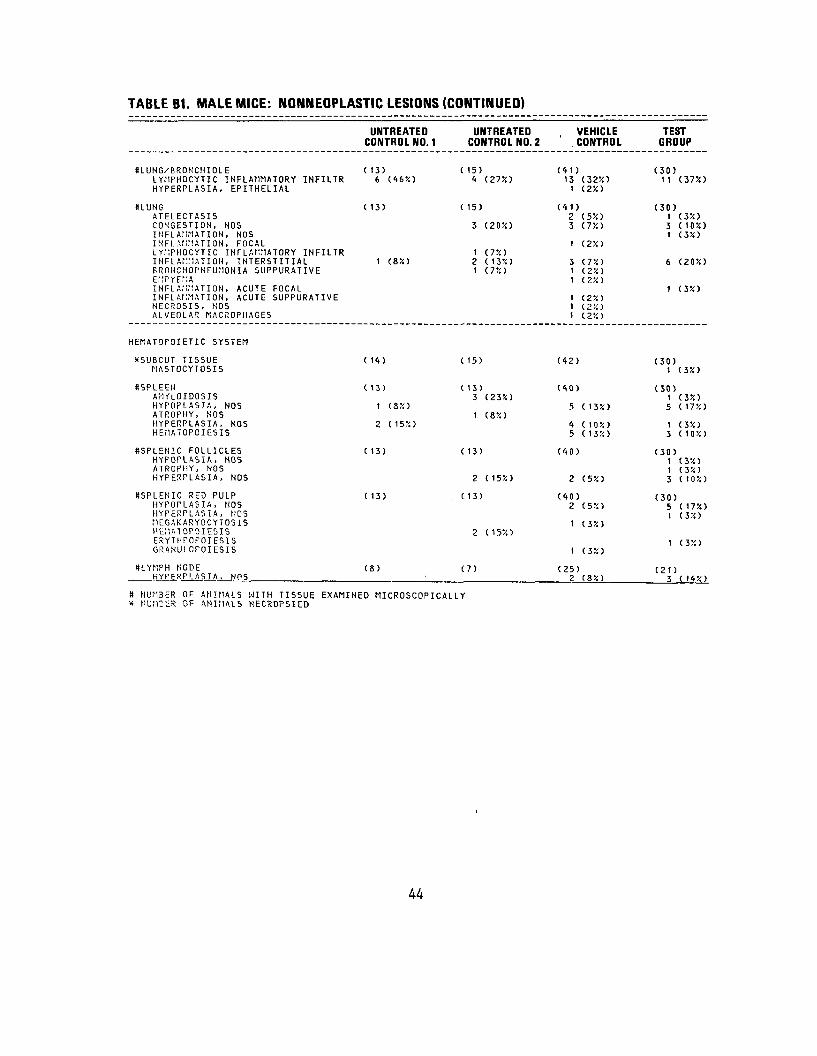
TABLE B1. MALE MICE: NONNEOPLASTIC LESIONS (CONTINUED)
HLUNG/BRONCHIOLELYHPHOCYTIC INFLAMMATORY INFILTRHYPERPLASIA, EPITHELIAL
KLUNGATELECTASI5 CONGESTION, NOS INFLAMMATION, NOS INFLAMMATION, FOCAL LYMPHOCYTIC INFLAMMATORY INFILTR INFLAMMATION, INTERSTITIALERONCHOPNEUMONIA SUPPURATIVE EMPYEMA INFLAMMATION, ACUTE FOCAL INFLAMMATION, ACUTE SUPPURATIVE NECROSIS, NOS ALVEOLAR P.ACROPHAGES
HEMATOPOIETIC SYSTEM
XSUBCUT TISSUEMASTOCYTOSIS
ItSPLEENAl-lfLOIDOSIS HYPOPLASIA, NOSATROPHY, NOS HYPERPLASIA, NOSHEMATOPOIESIS
SSPLENIC FOLLICLESHYFOPLASIA, NOS ATROPHY, NOS HYPERPLASIA, NOS
*SPLENIC RED PULPHYPOPLASIA, NOS HYPERPLASIA, I!OS MEGAKARYOCYTOSIS HEMA10POIESIS ERYTHPOPOIESIS GRANULOPOIESIS
ttLYMPH NODEHYPERPI.ASTA. NOS
UNTREATED CONTROL NO. 1
(13) 6 (46%)
(13)
1 (8%)
1 14)
(13)
1 (8%)
2 (15?.)
( 13)
( 13)
(8)
UNTREATED CONTROL NO. 2
( 15) 4 (27X)
( 15)
3 (20%)
1 (7X) 2 ( 13:<) 1 (7X)
( 15)
( 13) 3 (23X)
1 (8%)
( 13)
2 (15%)
t 13)
2 (15X)
(7)
VEHICLE CONTROL
(41) 13 (32X)
1 (2X)
(41) 2 (5X) 3 (75O
1 (2X)
3 (.T/.-) 1 <,2'/.1 1 (2%)
1 (2%) 1 (2X) 1 (2!O
(42)
(40)
5 ( 13%)
4 ( 1 0 '/. ) 5 ( 132)
(40)
2 (5%)
(40) 2 (5X)
1 (3%)
1 (3X)
(25) 2 <8X)
TEST GROUP
(30) 11 (37X)
(30) 1 (3X) 3 (105i) 1 (3X)
6 (20%)
1 (3%)
(30) 1 (3%)
(30) t (3%) 5 (17%)
1 (3%) 3 (10%)
(30) 1 (3%) 1 (3%) 3 ( 10%)
(30) 5 (17%) 1 (3%)
1 (3%)
(21 )3 ( 14%)
* NUMBER OF ANIMALS WITH TISSUE EXAMINED MICROSCOPICALLY x NUMBER OF ANIMALS NECROPSIED
44

TABLE B1. MALE MICE: NONNEOPLASTIC LESIONS (CONTINUED)
(CERVICAL LYMPH NODE ABSCESS, NOS HYPERPLASIA, NOS
(TRACHEAL LYMPH NODE HYPERPLASIA, tiOS
(PYLORIC LYMPH NODE HEMORRHAGE
(PANCREATIC L.NODE HYPERPLASIA, NOS
(LUMBAR LYMPH NODE HYPERPLASIA, LYMPHOID
(MESENTERIC L. NODE ABSCESS, NOS INFLAMMATION PROL IFERATIVE NECROSIS, CASEOUS
(RENAL LYMPH NODE HYPERPLASIA, NOS
•LUNG/BRONCHIOLE HYPERPLASIA, LYMPHOID
(PAROTID GLAND FIBROSING ADENOSIS
SLIVER HYPERPLASIA, NEUTROPHILIC HEMATOPOIESIS
(KIDNEY HYPERPLASIA, LYMPHOID
CIRCULATORY SYSTEM
(PANCREATIC L.NODE LYMPHANGIECTASIS
(MESENTERIC L. NODE LYMPHANPIECTASIS
UNTREATED CONTROL NO. 1
(8)
4 (50%)
(8) 1 (13X)
(8)
(8)
(8)
(8)
(8) 1 (13JO
(13)
( 12)
( 14)
( 14)
(8)
(8) 1 (13::)
UNTREATED CONTROL NO. 2
(7)
3 (*3ro
(7)
(7) 1 (145!)
(7)
(7)
(7)
(7)
( 15)
( 15)
( 15)
( 1<i)
(7)
(7)
VEHICLE CONTROL
(25)
10 (4 OX)
(25)
(25)
(25)
(25) 1 (4X)
(25) 1 (45!) 1 ( <t '/. ) 1 ( <t •/. )
(25)
(41) 1 (2X)
(36)
(42)
1 (2X)
(42) 1 (2'i)
(25) 1 (45!)
(25)
TEST GROUP
(21) 1 (53) 7 (33:;)
(21)
(21)
(21) 1 (55!)
(21)
(21)
(21) 1 (5X>
(30)i (3::)
(28) t CiX)
(30) 1 (3'<)
(30)
(21)
(21)
» NUMBER OF ANIMALS WITH TISSUE EXAMINED MICROSCOPICALLY X NUMBER OF ANIMALS NECROPSIED
45

TABLE B1. MALE MICE: NONNEOPLASTIC LESIONS (CONTINUED)
UNTREATED CONTROL NO. 1
UNTREATED CONTROL NO. 2
VEHICLE CONTROL
TEST GROUP
((HEART MINERALIZATION ABSCESS, NOS
( 13) 1 (.&•/.)
( 15) (42) (30)
1 (3X)
((HEART/ATRIUM THROMBOSIS, NOS
(13) ( 15) 2 C135O
(42) (30)
((MYOCARDIUM MINERALIZATION INFLAMMATION, CHRONIC INFLAMMATION, CHRONIC FOCAL INFLAMMATION PROLIFERATIVE NECROSIS, FOCAL
( 13) ( 15)1
2 1 1
m) ( 13X)C?:;)(?:;>
(42)
3 (7X>
(30)
1 1 (3!O (3'i)
((ENDOCARDIUM INFLAMMATION PROLIFERATIVE HYPERPLASIA, NOS
( 13) 1 (&•/.•> 1 (8rO
( 15) (42) (30)
XRENAL ARTERY ARTERIOSCLEROSIS, NOS
( 14) ( 15) (42) 1 (2X)
(30)
((URINARY BLADDER THROMBOSIS, NOS
( 13) ( 14) (39) (27) 1 (4X)
DIGESTIVE SYSTEM
((SALIVARY GLAND INFLAMMATION, FOCAL
C12) ( 15) 1 (7X)
(36) (28)
((PAROTID GLAND INFLAMMATION, NOS INFLAMMATION, FOCAL NECROSIS, FOCAL
C 12)
1 (85!)
( 15)1 (7X)
(36) 3 <.&•/.)
1 (3X)
(28)1 (4X)
((LIVER CONGESTION, NOS INFLAMMATION, FOCAL LYMPHOCYTIC INFLAMMATORY INFILTR INFLAM ;ATION, ACUTE FOCAL INFLAM 1ATION, ACUTE DIFFUSE INFLAfl 1ATION, ACUTE/CHRONIC CIRRHOSIS, NOS DEGENERATION, HYDROPIC
( 14) 1 (7X)
t <7X)
( 15)
2
1
( 13'/.)
<.!•/.->
(42)
3 (7%)
1 (2X1
1 (2-0
(30)
1 (3%)
(t NUMBER OF ANIMALS WITH TISSUE EXAMINED MICROSCOPICALLY * NUMBER OF ANIMALS NECROPSIED
46
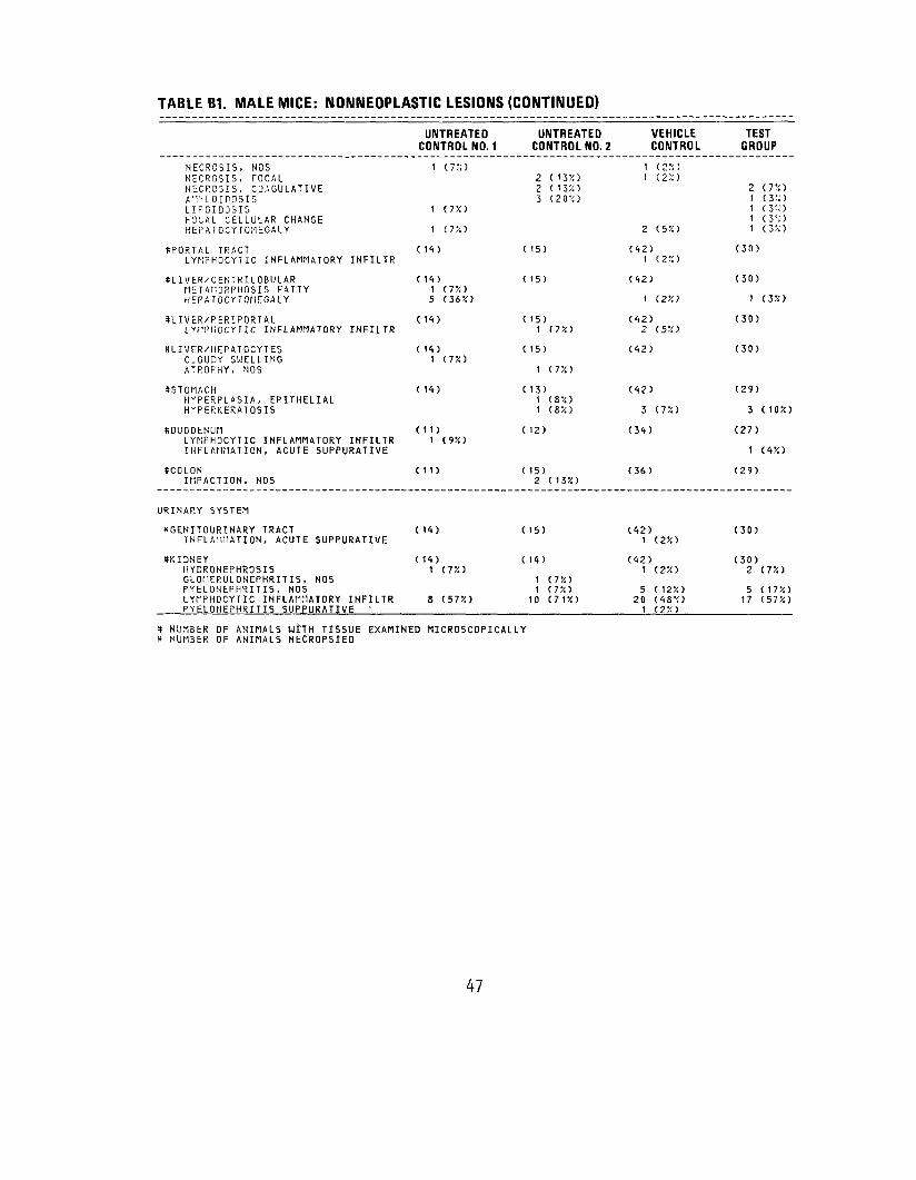
TABLE B1. MALE MICE: NONNEOPLASTIC LESIONS (CONTINUED)
UNTREATED UNTREATED VEHICLE TEST CONTROL NO. 1 CONTROL NO. 2 CONTROL GROUP
NECROSIS, NOS i (7%) 1 <2.v.) NECROSIS, rOCAL 2 (13%) 1 (2;:) NECRG3IS, COAGULATIVE 2 (135!) 2 (7%) AflUOIDOSIS 3 (20%) 1 (3%) LIFOIDOSIS 1 (7%) i (3;;) FOCAL C E L L U L A R CHANGE 1 (3%) HEPATOCYTOi-lEGALY 1 (7%) 2 (5%) 1 (3%)
ttPORTAL TRACT ( 14) ( 15) (42) (30) LYMPHOCYTIC INFLAMMATORY INFILTR I (2%)
KLIVER/CENTRILOBULAR ( 14) ( 15) (42) (30) METAMORPHOSIS FATTY 1 (7%) HEPATOCYTOriEGALY 5 (36%) 1 (2%) 1 (3%)
8LIVER/PERIPORTAL ( 14) ( 15) (42) (30) LYI1PHOCYTIC INFLAMMATORY INFILTR 1 (7%) 2 (5%)
SLIVER/HEPATOCYTES ( 14) ( 15) (42) (30) CLOUDY SWELLING 1 (75!) ATROPHY, NOS 1 (7%)
KSTOMACH ( 14) ( 13) (42) (29) HYPERPLASIA, EPITHELIAL 1 (8%) HYPERKERATOSIS 1 (8%) 3 (7%) 3 ( 10%)
8DUODENUM (11) ( 12) (34) (27) LYNPHOCYTIC INFLAMMATORY INFILTR 1 (9%) INFLAMMATION, ACUTE SUPPURATIVE 1 (4%)
SCOLON (11) ( 15) (36) (29) IMPACTION, NOS 2 ( 13%)
URINARY SYSTEM
((GENITOURINARY TRACT ( 14) ( 15) (42) (30) INFLAMMATION, ACUTE SUPPURATIVE t (2%)
((KIDNEY ( 14) ( 14) (42) (30) HYDRONEPHROSIS 1 (75!) 1 (2%) 2 (7%) GLOtlERULONEPHRITIS, NOS 1 (7%) PYELONEPHRITIS, NOS 1 (7%) 5 (12%) 5 ( 17%) LYMPHOCYTIC INFLAMMATORY INFILTR 8 (57%) 10 (71%) 20 (48%) 17 (57%) PYELONEPHRITIS SUPPURATIVE ' 1 (2%)
tt NUMBER OF ANIMALS wtTH TISSUE EXAMINED MICROSCOPICALLY * NUMBER OF ANIMALS NECROPSIED
47
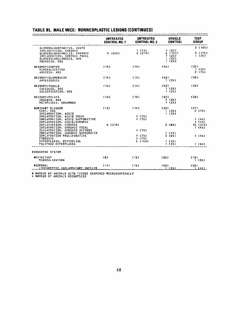
TABLE B1. MALE MICE: NONNEOPLASTIC LESIONS (CONTINUED)
UNTREATED CONTROL NO. 1
UNTREATED CONTROL NO. 2
VEHICLE CONTROL
TEST GROUP
GLOMERULONEPHRITIS, ACUTE INFLAMMATION, CHRONIC GLOilERULONEPHRITIS, CHRONICINFLAMMATION, CHRONIC FOCAL GLOMERULOSCLEROSIS, NOS NECROSIS, NOS
9 (64%) 1 (7%) 3 (21%)
1 (2%) 6 (14%) 1 (2%) 1 (2%) 1 (2%)
3 ( 10%)
5 (17%) 1 (3%)
KKIDNEY/CORTEXMINERALIZATION ABSCESS, NOS
(14) ( 14) (42) (30) 1 (3%) 2 (7%)
HKIDNEY/GLOMERULUSAMYLOIDOSIS
(14) ( 14) (42) 1 (2X)
(30)
SKIDNEY/TUBULECALCULUS, NOS CALCIFICATION, NOS
(14) ( 11) (42) 1 (25!) 1 (2%)
(30)
HKIDNEY/PELVISABSCESS, NOS METAPLASIA, SQUAMOUS
(14) (14) (42) 1 (2X) 1 (2%)
(30)
DURINARY BLADDERCAST, NOS INFLAMMATION, ACUTE INFLAMMATION, ACUTE FOCAL INFLAMMATION, ACUTE SUPPURATIVE INFLAMMATION, ACUTE/CHRONIC INFLAMMATION, CHRONICINFLAMMATION, CHRONIC FOCAL INFLAMMATION, CHRONIC DIFFUSE INFLAMMATION, CHRONIC SUPPURATIV INFLAMMATION PROLIFERATIVE FIBROSIS HYPERPLASIA, EPITHELIAL POLYPOID HYPERPLASIA
( 13)
4 (3U)
( 14)
t ay.) 1 (T/.)
1 (7%)
1 (7%) 1 (7%) 2 (14%)
(39) 1 (3%) 1 (35O
3 (85O
1 (3%) 3 (8%)
1 (3%) 1 (3X)
(27) 2
11
101
1
1
(7%)
(4%) (4%) (37%) (4%)
(4%)
(4%)
ENDOCRINE SYSTEM
•PITUITARYMINERALIZATION
(8) (12) (26) ( 19) 1 (5%)
•ADRENALLYMPHOCYTIC INFLAMMATORY INFILTR
(11) (15) (40) 1 (3X)
(28) 1 (4%)
* NUMBER OF ANIMALS WITH TISSUE EXAMINED MICROSCOPICALLY X NUMBER OF ANIMALS NECROPSIED
48
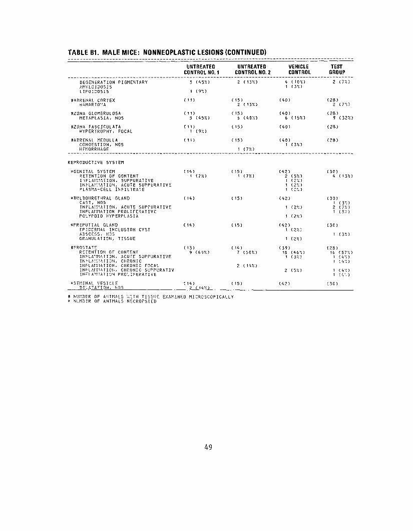
TABLE B1. MALE MICE: NONNEOPLASTIC LESIONS (CONTINUED)
UNTREATED UNTREATED CONTROL NO. 1 CONTROL NO. 2
DEGENERATION PIGMENTARY 5 (45%) 2 (13%) AMYLOIDOSIS LIPOIDOSIS 1 (9%)
((ADRENAL CORTEX (11) (15) HAMARTOMA 2 (13%)
ttZONA GLOMERULOSA (11) ( 15) METAPLASIA, NOS 5 (45%) 6 (40%)
SZONA FASCICULATA (11) ( 15) HYPERTROPHY, FOCAL 1 (9%)
SAPRENAL MEDULLA (11) ( 15) CONGESTION, NOS HEMORRHAGE 1 (7%)
REPRODUCTIVE SYSTEM
*GENITAL SYSTEM RETENTION OF CONTENT
( 14) t ( 7 •/. )
( 15) 1 (7%)
INFLAMMATION, SUPPURATIVE INFLAMMATION, ACUTE SUPPURATIVE PLASMA-CELL INFILTRATE
*BULBOURETHRAL GLAND CAST, NOS
( 14) ( 15)
INFLAMMATION, ACUTE SUPPURATIVE INFLAMMATION PROLIFERATIVE POLYPOID HYPERPLASIA
*PREPUTIAL GLAND (14) ( 15) EPIDERMAL INCLUSION CYST ABSCESS, NOS GRANULATION, TISSUE
((PROSTATE RETENTION OF CONTENT INFLAMMATION, ACUTE SUPPURATIVE
( 13) 9 (69!:)
( 14) 7 (50%)
INFLAMMATION, CHRONIC INFLAMMATION, CHRONIC FOCAL INFLAMMATION, CHRONIC SUPPURATIV
2 (14%)
INFLAMMATION PROLIFERATIVE
*SEMINAL VESICLE DILATATION, NOS
( 14) 2 (14%)
( 15)
* NUMBER OF ANIMALS WITH TISSUE EXAMINED MICROSCOPICALLY X NUMBER OF ANIMALS NECROPSIED
TEST GROUP
2 (7%)
(28) 2 (7%)
(28) 9 (32%)
(28)
(28)
(30) 4 (13%)
(30) 1 (3%) 2 (7%) 1 (3%)
(30)
1 (3%)
(23) 16 (57%) 1 ( 4 •'.) 1 (4%)
1 (4%) 1 <<•,•,)
(30)
1
(40)
(40) 6
(40)
(40) 1
(42) 2111
(42)
1
1
(42) 1
1
(39)
VEHICLE CONTROL
4 (10%) (3%)
(15%)
(3%)
(5%) (2%) ( 2 % ) (£%>
(2%)
(2%)
(2%)
(2%)
18 (46".) 1 (3%)
2 (5%)
(42)
49
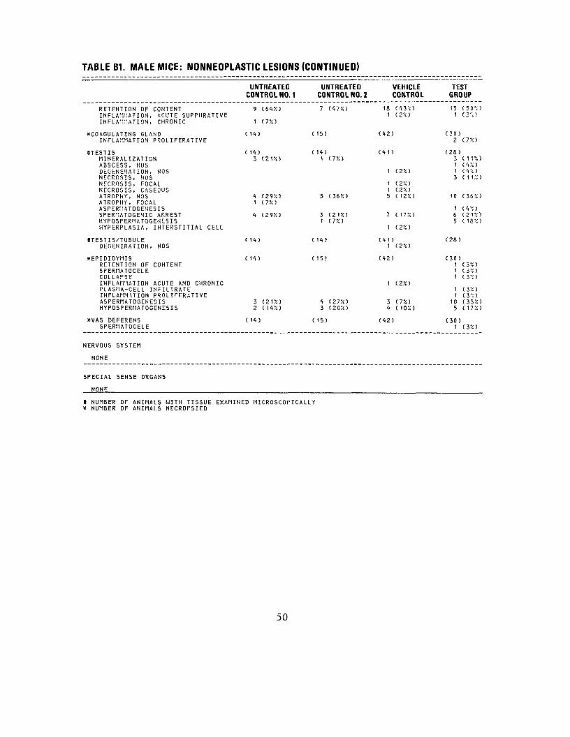
TABLE B1. MALE MICE: NONNEOPLASTIC LESIONS (CONTINUED)
UNTREATED CONTROL NO. 1
UNTREATEDCONTROL NO. 2
VEHICLE CONTROL
TEST GROUP
RETENTION OF CONTENTINFLAMMATION, ACUTE SUPPURATIVE INFLAMMATION, CHRONIC
9
1
(64%)
(7%)
7 (47%) 18 (43V.) 1 (2%)
15 1
(50%) (3%)
^COAGULATING GLANDINFLAMMATION PROL IFERATIVE
(14) (15) (42) (30) 2 (7%)
#TESTISMINERALIZATIONABSCESS, SOS DEGENERATION, NOS NECROSIS, NOS NECROSIS, FOCAL NECROSIS, CASEOUS ATROPHY, NOSATROPHY, FOCALASPERMATOGEUESIS SPERMATOGENIC ARRESTHYFOSPERMATOGENCSIS HYPERPLASIA, INTERSTITIAL CELL
(14) 3 (21%)
4 (29%) 1 (7%)
4 (29%)
(14)1 (7%)
5 (36%)
3(21%)1 (7%)
(41)
1 (2%)
1 (2%) 1 (2%) 5 ( 12%)
7 ( 17%)
1 (2%)
(28) 3 1 1 3
10
1 6 5
(11%) (4%) ( 4% ) (11%)
(36%)
( 4% ) (21%) ( 18%)
UTESTIS/TUBULEDEGENERATION, NOS
(14) (14) (41) 1 (2%)
(28)
XEPIDIDYMISRETENTION OF CONTENT SPERMATOCELE COLLAPSE INFLAMMATION ACUTE AND CHRONIC PLASMA-CELL INFILTRATE INFLAMMUION PROLI FERATI VE ASPERMATOGENESISHYPOSPERMATOGENESIS
(14)
3 (21%) 2 (14%)
(15)
4 (27%)3 (20%)
(42)
1 (2%)
3 (7%) 4 ( 10%)
(30)1 1 1
1 t
10 5
(3%) (3%) (3%)
(3%) (3%) (33%) (17%)
XVAS DEFERENSSPERMATOCELE
( 14) (15) (42) (30)1 C3%)
NERVOUS SYSTEM
NONE
SPECIAL SENSE ORGANS
NONE
* NUMBER OF ANIMALS WITH TISSUE EXAMINED MICROSCOPICALLY * NUMBER OF ANIMALS NECROPSIED
50

TABLE B1. MALE MICE: NONNEOPLASTIC LESIONS (CONTINUED)
UNTREATEDCONTROL NO. 1
UNTREATED CONTROL NO. 2
VEHICLE CONTROL
TEST GROUP
MUSCULOSKELETAL SYSTEM
NONE
BODY CAVITIES
^PERITONEUMINFLAMMATION PROLIFERATIVE
(14) (15) (42) 1 (2%)
(30)
ALL OTHER SYSTEMS
((MULTIPLE ORGANSLYMPHOCYTIC INFLAMMATORY INFILTR
CONNECTIVE TISSUE INFLAMMATION PROLIFERATIVEHYPERPLASIA, NOS
(14) 1 C7X)
(15) (42) (30) 1 (3X)
2 1
SPECIAL MORPHOLOGY SUMMARY
AUTO/NECROPSY/HISTO PERFAUTOLYSIS/NO NECROPSY
* NUMBER OF ANIMALS WITH TISSUE EXAMINED* NUMBER OF ANIMALS NECROPSIED
1
MICROSCOPICALLY
1 3
51
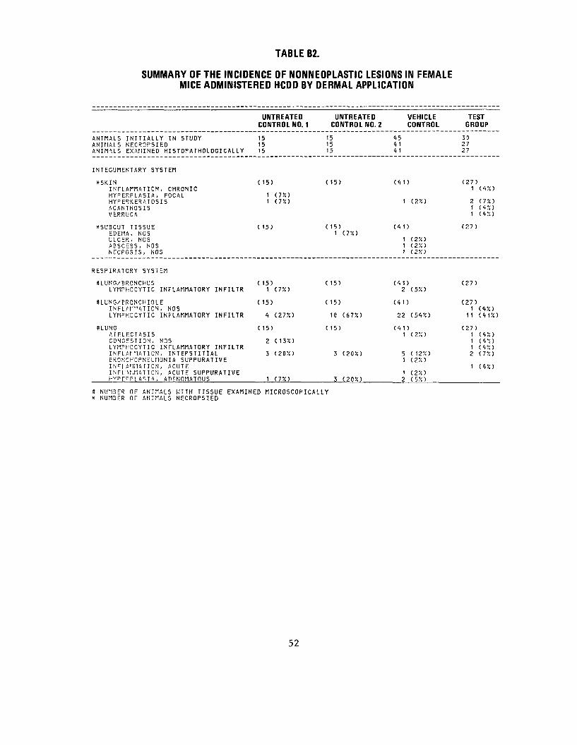
TABLE B2.
SUMMARY OF THE INCIDENCE OF NONNEOPLASTIC LESIONS IN FEMALE MICE ADMINISTERED HCDD BY DERMAL APPLICATION
UNTREATED CONTROL NO. 1 C
UNTREATEDONTROL NO. 2
VEHICLE CONTROL
ANIMALS INITIALLY IN STUDY ANIMALS NECROPSIED ANIMUS EXAMINED HISTOPATHOLOGI CAL L Y
15 15 15
151515
45 4 1 41
INTEGUMENTARY SYSTEM
XSKIN (15) (15) (41) INFLAMMATION, CHRONIC HYPERPLASIA, FOCAL 1 (7%) HYPERKERATOSIS 1 (7%) 1 (2X) ACANTHOSIS VERRUCA
*SUBCUT TISSUE t 15) (15) (41) EDEMA, NOS 1 (7%) ULCER, NOS 1 C2X) ABSCESS, NOS 1 (2X) NECPOSIS, NOS 1 (2X)
RESPIRATORY SYSTEM
SLUNG/BRONCHUS ( 15) (15) (41) LYMPHOCYTIC INFLAMMATORY INFILTR 1 (7%) 2 (5V.)
ULUNG/ERONCHIOLE ( 15) (15) (41) INFL,A1""ATION, NOS LYMPHOCYTIC INFLAMMATORY INFILTR 4 (27%) 10 (67%) 22 (54%)
SLUNG ( 15) (15) (41) ATELECTASIS 1 (2X) CONGESTION, NOS 2 (13X) LYMPHOCYTIC INFLAMMATORY INFILTR INFLAf NATION, INTERSTITIAL 3 (2050 3 (20X) 5 ( 12%) BRONCHOPNtUMONIA SUPPURATIVE 1 (2%) I N F L A M M A T I O N , ACUTE INFL U.F1ATION, ACUTE SUPPURATIVE 1 (2X) HVPEPPLASIA, ADENOMATOUS 1 (75O 3 (20%) 2 (5%)
* NU1BER OF ANIMALS WITH TISSUE EXAMINED MICROSCOPICALLY x NUMBER OF ANIMALS NECROPSIED
TEST GROUP
30 27 27
(27) 1 (4%)
2 (7%) 1 (4%) 1 (4%)
(27)
(27)
(27) 1 (4%)
11 (41%)
(27) 1 (4%) 1 (4%) 1 (4%) 2 (7%)
1 (4%)
52
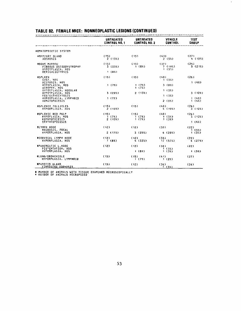
TABLE B2. FEMALE MICE: NONNEOPLASTIC LESIONS (CONTINUED)
VEHICLE CONTROL
(<ti) 2 (5%)
(37)7 (m) 1 (3'0
(40) 1 (3%)
3 (8%)
1 (3%)
1 (3%)
2 (5%)
(40) 4 (10%)
(40) 1 (3%) 1 (3%)
(30)
6 (20%)
(30) 17 (57%)
(30) 1 (3%) 1 (3%)
(41) 1 (2%)
(36) 1 (3%)
TEST GROUP
(27) 4 (15%)
(24) 5 (21%)
(26)
1 (4%)
3 ( 12%)
1 (4%) 1 (4%)
(26) 3 (12%)
(26) 3 (12%)
1 (4%)
(22) 1 (5%) 1 (5%)
(22) 6 (27%)
(22)
1 ( 5% )
(27)
(26)
HEMATOPOIETIC SYSTEM
*MAr;iARY GLAND ADEHOSIS
8BONE MARROl! FIBROUS OSTEODYSTROPHY HYPERPLASIA, NOS RETICULOCYTOSIS
OSPLEEN CYST, NOS NECROSIS, NOS HYPOPLASIA, NOS ATROPHY, NOS HYFEKFLASIA, NODULAR HYPERPLASIA, NOS MEG\KARYOCYTOSIS HYPERPLASIA, LYfiPHOID HEMATOPOIESIS
((SPLENIC FOLLICLES HYPERPLASIA, NOS
*SPLENIC RED PULP HYPOPLASIA, NOS HEMATOPOIESIS ERYTHROPOIESIS
((LYMPH NODE NECROSIS, FOCAL HYPERPLASIA, NOS
((CERVICAL LYMPH NODE HYPERPLASIA, NOS
((PANCREATIC L.HODE PIG'-'ENTATION, HOS HYPERPLASIA, NOS
((LUNG/BRONCHIOLE HYPERPLASIA, LYMPHOID
((PAROTID GLAND FIBROSING ADENOSIS
UNTREATED CONTROL NO. 1
( 15) 2 (13%)
( 13)3 (23%) 1 (8%)
( 15)
1 (7%)
3 (20%)
1 (7%)
(15) 2 (13%)
( 15) 1 (7%) 2 ( 13%)
( 12)
2 (17%)
( 12) 1 (8%)
( 12)
( 15)
( 15)
UNTREATED CONTROL NO. 2
(15)
( 13) 1 (8%)
C 15)
1 (7%) 1 (7%)
2 (13%)
(15)
(15) 1 (7%) 1 (7%)
( 12)
3 (25%)
( 12) 4 (33%)
( 12)
1 (8%)
( 15) 1 (7%)
( 12)
» NUMBER OF ANIMALS WITH TISSUE EXAMINED MICROSCOPICALLY * NUriBER OF ANIMALS NECROPSIED
53
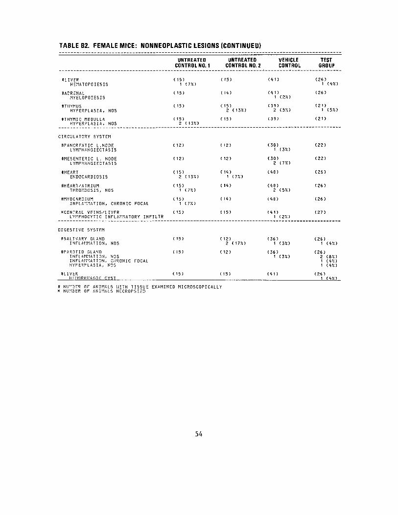
TABLE B2. FEMALE MICE: NONNEOPLASTIC LESIONS (CONTINUED)
SLIVER HEMATOPOIESIS
SADRENAL MYELOPOIESIS
8THYMUS HYPERPLASIA, NOS
tTHYMIC MEDULLA HYPERPLASIA, NOS
CIRCULATORY SYSTEM
•PANCREATIC L.NODE LYMPHANGIECTASIS
SMESENTERIC L. NODE LYMPHANGIECTASIS
SHEART EHDOCARDIOSIS
8HEART/ATRIUM THROMBOSIS, NOS
*MYOCARDIUM INFLAMMATION, CHRONIC FOCAL
XCENTRAL VEINS/LIVERLYMPHOCYTIC INFLAMMATORY INFILTR
DIGESTIVE SYSTEM
#SALIVARY GLAND INFLAMMATION, NOS
ttPAROTID GLAND INFLAMMATION, NOS INFLAMMATION, CHRONIC FOCAL HYPERPLASIA, NOS
SLIVER HEMORRHAGIC CYST
UNTREATED CONTROL NO. 1
( 15) 1 (75O
(15)
( 15)
( 15) 2 (13%)
( 12)
( 12)
( 15) 2 (13%)
( 15) 1 (7X)
( 15) 1 C7X)
( 15)
( 15)
C 15)
( 15)
UNTREATED CONTROL NO. 2
( 15)
C 14)
( 15) 2 (13X)
( 15)
( 12)
( 12)
( 14) 1 (7X)
( 1<t)
( 14)
( 15)
C 12) 2 (17%)
( 12)
C 15)
VEHICLE CONTROL
(41)
(41) 1 (2*)
(39) 2 (5X)
(39)
(30) 1 (3%)
(30) 2 (75O
(40)
(40) 2 (5%)
(40)
(41) 1 (2%)
(36) 1 (3X)
(36) 1 (3%)
(41)
TEST GROUP
(26) 1 (4%)
(26)
(21) 1 (55O
(21)
(22)
(22)
(26)
(26)
(26)
(27)
(26) 1 (4%)
(26) 2 (8X) 1 (4X) 1 (4X)
(26)1 (4X)
S NUMBER OF ANIMALS WITH TISSUE EXAMINED MICROSCOPICALLY x NUMBER OF ANIMALS NECROPSIED
54
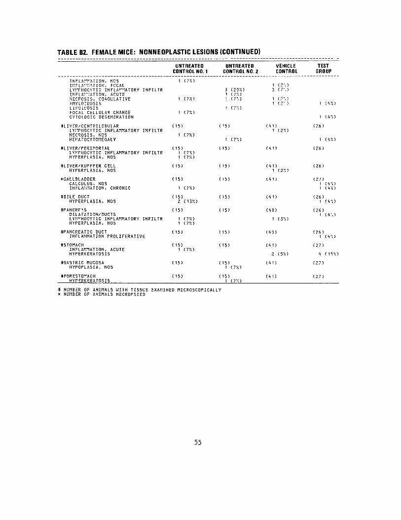
TABLE B2. FEMALE MICE: NONNEOPLASTIC LESIONS (CONTINUED)
INFLAMMATION, NOS iKFLArrmioN, FOCAL LYITHOCYTIC INFLAMMATORY INFILTR INFLAP1ATION, ACUTE NECPOSIS, COAGULATIVE AMYLOIDOSIS LIPOIDOSIS FOCAL CELLULAR CHANGE CYTOLOGIC DEGENERATION
SLIVER/CENTRILOBULAR LYMPHOCYTIC INFLAMMATORY INFILTR NECROSIS, NOS KEPATOCYTOriEGALY
HIVER/PERIPORTAL LYMFHOCYTIC INFLAMMATORY INFILTR HYPERPLA5IA, NOS
*LIVER/KUPFFER CELL HYPERPLASIA, NOS
^GALLBLADDER CALCULUS, NOS INFLAtirtATION, CHRONIC
SBILE DUCT HYPERPLASIA, NOS
SPANCRE1S DILATATION/DUCTS LYMRHOCYTIC INFLAMMATORY INFILTR HYPERPLASIA, NOS
((PANCREATIC DUCT INFLAMMATION PROL I FERATI VE
((STOMACH INFLAMMATION, ACUTE HYPERKERATOSIS
((GASTRIC MUCOSA HYPOPLASIA, NOS
SFORESTOMACH HYPERKERATOSIS
UNTREATED CONTROL NO. 1
1 (7%)
1 (T/.l
1 (T/.1
( 15)
1 (7?i)
( 15) 1 (7'<) 1 <.T/.1
( 15)
( 15)
1 (T/.)
( 15) 2 (135O
( 15)
1 (7X> 1 (7X)
( 15)
(15) 1 C7X)
( 15)
( 15)
UNTREATED CONTROL NO. 2
3 (20?O 1 (7%) 1 (T.I
1 (.T'.l
( 15)
1 ( 7 '<)
( 15)
C 15)
t 15)
C 15)
( 15)
( 15)
( 15)
( 15)1 (7X)
( 15) 1 (TO
VEHICLE CONTROL
1 (2%) 3 (T. )
1 C".) 1 (.2' )
(11) 1 (2X)
(41)
(<>1) 1 (2X)
(11)
(11)
(10)
1 (3'O
(10)
(11)
2 (5X)
(11)
(11)
TEST GROUP
1 (1%)
1 (1V.)
(26)
1 (1X)
(26)
(26)
(27) 1 (1'<> 1 (1X)
(26) 1 d'O
(26) 1 (1%)
(26) 1 (1V. )
(27)
1 ( 15'O
(27)
(27)
* NUMBER OF ANIMALS WITH TISSUE EXAMINED MICROSCOPICALLY X NUMBER OF ANIMALS NECROPSIED
55

TABLE B2. FEMALE MICE: NONNEOPLASTIC LESIONS (CONTINUED)
UNTREATED UNTREATED CONTROL NO. 1 CONTROL NO.
SDUODENUM ( 13) (15)NECROSIS, NOS 1 (78)
XRECTUM ( 15) (15) ULCER, ACUTE 1 (78) DESMOPLASIA 1 (78) NECROSIS, NOS 1 (78)
URINARY SYSTEM
8KIDNEY ( 15) (15) HYDRONEPHROSIS 1 (78) GLOMERULONEPHRITIS, NOS 1 (78) PYELONEPHRITIS, NOS 2 (138) LYMPHOCYTIC INFLAMMATORY INFILTR 5 (33%) 6 (408) PYELONEPHRITIS SUPPURATIVE 1 (78) GLOMERULONEPHRITIS, ACUTE INFLAMMATION, CHRONIC 2 M38) 1 (78) GLOMERULONEPHRITIS, CHRONIC 10 (678) 12 (808) INFLAMMATION, CHRONIC FOCAL GLOMERULOSCLEROSIS, NOS 2 ( 138) 2 (138) INFARCT, HEALED
8KIDNEY/GLOMFRUIUS ( 15) ( 15) AMYLOIDOSIS
SURINARY BLADDER ( 13) ( 13)LYMfHOCYTIC INFLAMMATORY INFILTR 1 (88) INFLAMMATION, ACUTE INFLAMMATION, ACUTE NECROTIZING 1 (88) INFLAMMATION, ACUTE/CHRONIC 2 (158) INFLAMMATION, CHRONIC 4 (318) 5 (388) INFLAMMATION, CHRONIC FOCAL INFLAMMATION PROL IFERATI VE 1 (88) ATROPHY, NOS METAPLASIA, SQUAMOUS
ENDOCRINE SYSTEM
•ADRENAL ( 15) ( 14) CONGESTION, NOS DEGENERATION PIGMENTARY 9 (608) & (578)
* NUMBER OF ANIMALS WITH TISSUE EXAMINED MICROSCOPICALLY X NUMBER OF ANIMALS NECROPSIED
VEHICLE 2 CONTROL
(40)
(41)
(41) 1 (28 )
1 (28) 26 (638)
1 (28) 1 (28)
16 (398) 1 (28) 2 (58) 1 (28)
(41)1 (28)
(30)
1 (38)
10 (338) 2 (78)
1 (38)
(41)2 (58) 22 (548)
TEST GROUP
(26)
(27)
(26)
1 (48)
14 (548)
1 (48)
7 (278)
3 (128)
(26)
( 19)
10 (538)
1 (58)
(26)1 (48)
16 (628)
56

TABLE B2. FEMALE MICE: NONNEOPLASTIC LESIONS (CONTINUED)
UNTREATED UNTREATED VEHICLE CONTROL NO. 1 CONTROL NO. 2 CONTROL
AMYLOID, NOS HYPERPLASIA, NOS
8ADRENAL CCRTEX C 15) ( 14) (11)HAMARTOMA 1 (7%) 1 (7%) 1 (2%) LYMPHOCYTIC INFLAMMATORY INFILTR 1 (750 1 (25!)
#ZONA GLOMERULOSA ( 15) ( 14) (41) METAPLASIA, HOS 6 (40%) 9 (64%) 20 (495!)
(•THYROID ( 15) ( 15) (39) INFLAMMATION, NOS 1 (75O HYPERPLASIA, FOL L I CUL AR-CEL L 1 (35!)
STHYROID FOLLICLE ( 15) ( 15) (39) GOITER COLLOID 1 (350
ttPANCREATIC ISLETS < 15) ( 15) (40) HYPERPLASIA, NOS 1 (7%)
REPRODUCTIVE SYSTEM
XMAMMARY GLAND ( 15) (15) (41) DILATATION/DUCTS 3 (20%) 1 (7%) 3 (75!)
XVAGINA ( 15) ( 15) (<i 1 ) INFLAMMATION, ACUTE/CHRONIC 1 (7%) INFLAMMATION PROL I PER ATI VE HYPERPLASIA, EPITHELIAL 1 (7%)
tfUTERUS C 15) ( 15) (36) MINERALIZATION 1 (35!) HEMORRHAGE 1 (35!) I N F L A M M A T I O N , ACUTE SUPPURATIVE ABSCESS, NOS 1 (3%) NECROSIS, NOS 1 (35!)
((CERVIX UTERI ( 15) ( 15) (36) INFLAMMATION, CHRONIC 1 ( 7 '/.t
SUTERUS/EHDOMETRIUM ( 15) ( 15) (36) HYPERPLASIA, NOS 1 C7X) 5 (335!) 8 (22%) HYPERPLASIA, CYSTIC 12 (805O 9 (605$) 22 (6150
ttOYARY ( 14) ( 15) (33) CYST, NOS 1 (7%) <t (12%)
8 NUMBER OF A N I M A L S U'lTH TISSUE EXAMINED MICROSCOPICALLY x NUMBER OF ANIMALS NECROPSIED
TEST GROUP
1 dX)1 (<t'/t)
(26)
(26) 12 (46%)
(20)
(20)
(26)
(27) 5 (19%)
(27)
2 (7%)
(26)
1 (4%)
(26)
(26) 2 (8%) 23 (88%)
(25)
57
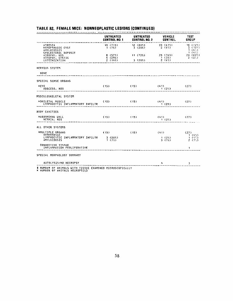
TABLE B2. FEMALE MICE: NONNEOPLASTIC LESIONS (CONTINUED)
UNTREATED CONTROL NO. 1
UNTREATEDCONTROL NO. 2
VEHICLE CONTROL
TEST GROUP
ATRESIAHEM3PRHAGIC CYSTAHYLOIDOSIS CHOLESTEROL DEPOSIT ATROPHY, NOSATROPHY, CYSTICLUTEINIZATION
10 (71%) 1 (17.)
8 (57'<) <t (29X)
2 C1<»:O
12 (80'O3 C20X)
11 (735O
3 (20X)
20 (6TO 2 (6".)
25 (76X) 1 (3X) 2 (6'O
18 (72V.) 3 ( 12'<> 1 (V. ) 1 Ci'O
20 ( 30 ''.I 2 (S%)
NERVOUS SYSTEM
NONE
SPECIAL SENSE ORGANS
*EYEABSCESS, NOS (15) (15) (41)
1 (2'O (27)
MUSCULOSKELETAL SYSTEM
"SKELETAL MUSCLELYMPHOCYTIC INFLAMMATORY INFILTR
( 15) (15) (<tt) 1 (2X)
(27)
BODY CAVITIES
"ABDOMINAL WALLHERNIA, NOS
( 15) (15) (<t1) 1 (2'O
(27)
ALL OTHER SYSTEMS
"MULTIPLE ORGANSHEMORRHAGE LYMPHOCYTIC INFLAMMATORY INFILTRAMYLOIDOSIS
( 15)
J (20X) 1 (7X)
(15) (<(1)
1 C2'<) 3 (7X>
(27) 1 (V!) 1 ( <i ". ) 2 (7%)
CONNECTIVE TISSUE INFLAMMATION PROL I FERATI VE 1
SPECIAL MORPHOLOGY SUMMARY
AUTOLYSIS/NO NECROPSY <t 3
* NUMBER OF ANIMALS WITH TISSUE* NUMBER OF ANIMALS NECROPSIfcD
EXAMINED MICROSCOPICALLY
58

APPENDIX C
SUMMARY OF THE INCIDENCE OF NEOPLASMS IN MICE ADMINISTERED HCDD PLUS DMBA
BY DERMAL APPLICATION
59

60
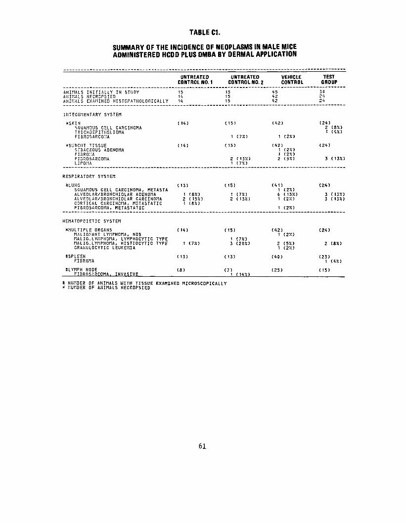
TABLE C1.
SUMMARY OF THE INCIDENCE OF NEOPLASMS IN MALE MICE ADMINISTERED HCDD PLUS DMBA BY DERMAL APPLICATION
ANIMALS INITIALLY IN STUDY ANIMALS NECR3PSIED ANIMALS EXAMINED HISTDPATHOLOGICALLY
INTEGUMENTARY SYSTEM
XSKIN SQUAMOUS CELL CARCINOMA TRICHOEPITIiELIOMA FIDROSARCOMA
xSUBCUT TISSUE SEBACEOUS ADENOMA FIBROKA FIBnOSARCOMA LIPOilA
RESPIRATORY SYSTEM
»LUSG SQUAMOUS CELL CARCINOMA, METASTA ALVEOLAR/BROHCHIOLAR ADENOMA ALVEOLAR/BRONCHIOLAR CARCINOMA CORTICAL CARCINOMA, METASTATIC FIBROSARCOMA, METASTATIC
HEMATOPOIETIC SYSTEM
XMULTIPLE ORGANS MALIGNANT LYflPHOMA, NOS MALIG.LYMPHOMA, LYMPHOCYTIC TYPE MALIG.LYMPHOMA, HISTIOCYTIC TYPE GRANULOCYTIC LEUKEMIA
8SPLEEN FIBROMA
SLYMPH NODE FIBROSARCOMA, INVASIVE
UNTREATED CONTROL NO. 1
15 11 14
( 14)
( 14)
( 13)
1 (8X) 2 (15X) 1 CSX)
(H)
1 C7X)
( 13)
(8)
UNTREATED CONTROL NO. 2
15 15 15
(15)
1 (75O
( 15)
2 cm> 1 (TO
(15)
1 (7X) 2 ( 13X)
(15)
1 (7X) 3 (20%)
( 13)
(7) 1 (HX)
VEHICLE CONTROL
45 42 42
(42)
1 (2X)
(42) 1 (2X) 1 (2X) 2 (5%)
(41) 1 (250 6 (15X) 1 (2%)
1 (25i)
(42) t (2X)
2 (5X) 1 (2X)
(40)
(25)
TEST GROUP
30 24 24
(24) 2 (8X) 1 (4X)
(24)
3 (135O
(24)
3 (13X) 3 (13-/0
(24)
2 (8X)
(23) 1 (4X)
(15)
# NUMBER OF ANIMALS UITH TISSUE EXAMINED MICROSCOPICALLY X NUMBER OF ANIMALS NECROPSIED
61
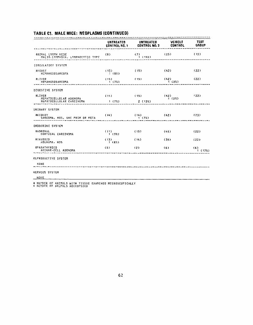
TABLE C1. MALE MICE: NEOPLASMS (CONTINUED)
UNTREATED UNTREATEDCONTROL NO. 1 CONTROL NO. 2
SRENAL LYMPH NODE (2) (7)MALIG.LYMPHONA, LYMPHOCYTIC TYPE 1 (14X)
CIRCULATORY SYSTEM
SHEART (13) OS)HCMAHGIOSARCOMA 1 (82)
SLIVER (14) (15)HEMANGIOSARCOMA 1 (7X)
DIGESTIVE SYSTEM
SLIVER (14) (15)HEPATOCELLULAR ADENOMA HEPATOCELLULAR CARCINOMA 1 (7X) 2 (13X)
URINARY SYSTEM
SKIDNEY (14) (14)SARCOMA, NOS, UNC PRIM OR META 1 (7%)
ENDOCRINE SYSTEM
SADRENAL (11) (15)CORTICAL CARCINOMA 1 (9X)
STHYROID (13) (14)ADENOMA, NOS 1 (8%)
SPARATHYROID (5) (2)ACINAR-CELL ADENOMA
REPRODUCTIVE SYSTEM
NONE
NERVOUS SYSTEM
NONE
# NUMBER OF ANIMALS WITH TISSUE EXAMINED MICROSCOPICALLY a NUMBER OF ANIMALS NECROPSIED
62
VEHICLE CONTROL
(25)
(42)
(42) 1 (2X)
(42)1 (2X)
(42)
(40)
(35)
(6)
TEST GROUP
( 15)
(22)
(22)
'22)
(23)
(22)
(22)
(6) 1 (17K)
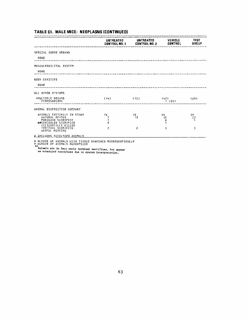
TABLE C1. MALE MICE: NEOPLASMS (CONTINUED)
UNTREATEDCONTROL NO. 1
UNTREATED CONTROL NO. 2
VEHICLE CONTROL
TEST GROUP
SPECIAL SENSE ORGANS
SOME
MUSCULOSKELETAL SYSTEM
NONE
BODY CAVITIES
NONE
'ALL OTHER SYSTEMS
*MULTIPLE ORGANS FIBROSARCOMA
(14) (15) (42)1 (2X) (24)
ANIMAL DISPOSITION SUMMARY
ANIMALS INITIALLY IN STUDY NATURAL DEATHS MORIBUND SACRIFICE
**SCHCDULED SACRIFICE ACCIDENTALLY KILLED TERMINAL SACRIFICE ANIMAL MISSING
15715
2
15 13
2
45 30
3 4
3
30 23
4
3
3 INCLUDES AUTOLYZED ANIMALS
* NUMBER OF ANIMALS WITH TISSUE EXAMINED MICROSCOPICALLY * NUMBER OF ANIMALS NECROPSIED ** Animals are in fact early terminal sacrifices, but appear
as scheduled sacrifices due to system interpretation.
63

TABLE C1. MALE MICE: NEOPLASMS (CONTINUED)
UNTREATED UNTREATED VEHICLE TEST CONTROL NO. 1 CONTROL NO. 2 CONTROL GROUP
TUMOR SUMMARY
TOTAL ANIMALS WITH PRIMARY TUMORS* 9 10 15 1 1 TOTAL PRIMARY TUMORS 9 15 19 16
TOTAL ANIMALS WITH BENIGN TUMORS 2 2 8 5 TOTAL BENIGN TUMORS 2 2 9 6
TOTAL ANIMAtS WITH MALIGNANT TUMORS 7 8 8 7 TOTAL MALIGNANT TUMORS 7 12 10 10
TOTAL ANIMALS WITH SECONDARY TUMORS* 1 1 2 TOTAL SECONDARY TUMORS 1 1 2
TOTAL ANIMALS WITH TUMORS UNCERTAINBENIGN OR MALIGNANT
TOTAL UNCERTAIN TUMORS
TOTAL ANIMALS WITH TUMORS UNCERTAINPRIMARY OR META3TATIC 1
TOTAL UNCERTAIN TUMORS 1
X PRIMARY TUMORS: ALL TUMORS EXCEPT SECONDARY TUMORS S SECONDARY TUMORS: METASTATIC TUMORS OR TUMORS INVASIVE INTO AN ADJACENT ORGAN
64

TABLE C2.
SUMMARY OF THE INCIDENCE OF NEOPLASMS IN FEMALE MICE ADMINISTERED HCDD PLUS DMBA BY DERMAL APPLICATION
VEHICLE CONTROL
45 <»1 41
(<tt)
(11)
2 (5X)
(41)1 (2X) 4 (10%) 5 (12%)
1 (2%)
(41) 9 (22%) 4 (10%)
(37)1 (3%)
(40)1 (3%) 1 (3%)
(30)1 (3%)
TEST GROUP
30 27 27
(27)1 (4%)
(27)1 (4%) 3 ( 1 1%)
(25)
1 (4%) 2 (8%) 1 (4%) 1 (4%)
(27) 2 (7%) 3 ( 1 1%)
(23)
(27)
( 16)I (6%)
ANIMALS INITIALLY IN STUDYANIMALS NECRGPSIEDANIMALS EXAMINED M ISTOPATHOLOGICAL LY
INTEGUMENTARY SYSTEM
XSKINSQUAMOUS CELL CARCINOMA
XSUBCUT TISSUEBASAL-CELL CARCINOMA FIBROSARCOMA
RESPIRATORY SYSTEM
SLUNGADENOCARCIHONA, NOS, METASTATIC ALVEOLAR/BKOHCHIOLAR ADLNOMAA L VEOLAR/BRONCHIOLAR CARCINOMAS'.RCO'-IA, NOS, METASTATIC FIBROSARCOr.A, METASTATIC
HEMATOPOIETIC SYSTEM
^MULTIPLE ORGANSHALIG. LYMPHOMA, LYMPHOCYTIC TYPEMALIC. LYMPHOMA, HISTIOCYTIC TYPE
SBQNE MARROWFIBROMA
SSPLEENADENOCARCINOMA, HOS, META5TATIC FIBROMA
tfPAHCREATIC L . NODEtIALIG. LYMPHOilA, HISTIOCYTIC TYPE
UNTREATED CONTROL NO. 1
15 15
15
(15)
( 15)
(15)
1 (7%) 2 (13%)
( 15) 4 (27%) 2 (13%)
(13)
(15)
( 12)
UNTREATEDCONTROL NO.2
151515
(15)
(15)
(15)
1 (T/.1
(15)2 (13%)1 (7%)
(13)
(15)
(12)
S NUMBER OF ANIMALS WITH TISSUE EXAMINED MICROSCOPICALLY X NUMBER OF ANIMALS NECROPSIED
65
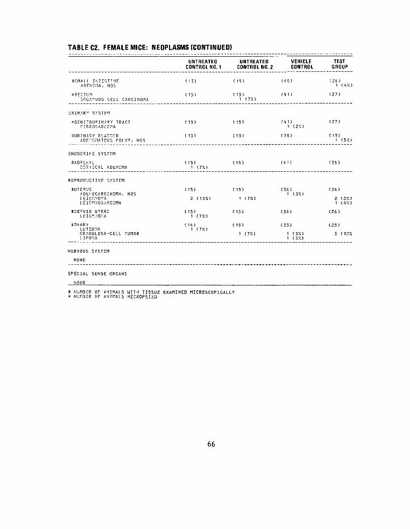
TABLE C2. FEMALE MICE: NEOPLASMS (CONTINUED)
VEHICLE CONTROL
(40)
(41)
(41) 1 (2JO
(30)
(41)
(36)1 (3JO
(36)
(33)
1 (3X> 1 (3X)
TEST GROUP
(26) 1 (4*)
(27)
(27)
( 19) 1 (57,1
(26)
(26)
2 (S'/.) 1 (4%)
(26)
(25)
3 (12X
SSMALL INTESTINE ADENOMA, NOS
XRECTUM SQUAMOUS CELL CARCINOMA
URINARY SYSTEM
^GENITOURINARY TRACT FIBROSARCOMA
SURINARY BLADDER ADEr'OMATOUS POLYP, NOS
ENDOCRINE SYSTEM
SADRENAL CORTICAL ADENOMA
REPRODUCTIVE SYSTEM
ttUTERUS ADENOCARCINOMA, NOS LEIOI1YOMA LEICMYOSARCOMA
SCEPVIX UTERI LEIOMCOMA
SOVARY LUTEOMA GRANULOSA-CELL TUMOR LIPONA
NERVOUS SYSTEM
NONE
SPECIAL SENSE ORGANS
NONE
UNTREATEDCONTROL NO. 1
C 13)
(15)
(15)
(13)
( 15)1 U'/.t
(15)
2 (13X)
(15)1 (7X)
(14)t (T/.1
UNTREATED CONTROL NO. 2
(15)
(15)1 (75C)
(15)
(13)
( 14)
(15)
1 (7%)
(15)
(15)
1 (7%)
S NUMBER OF ANIMALS WITH TISSUE EXAMINED MICROSCOPICALLY x NUMBER OF ANIMALS NECROPSIED
66
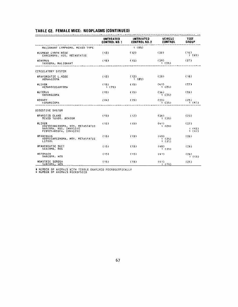
TABLE C2. FEMALE MICE: NEOPLASMS (CONTINUED)
UNTREATED CONTROL NO. 1
UNTREATED CONTROL NO. 2
VEHICLE CONTROL
TEST GROUP
MALIGNANT LYMPHOMA, MIXED TYPE 1 (8X)
KLUMBAR LYMPH NODE CARCINOMA, HOS, METASTATIC
( 12) ( 12) (30) ( 16) 1 (6X)
ftTHYMUS THYMOMA, MALIGNANT
(15) ( 15) (39) 1 (3X)
(27)
CIRCULATORY SYSTEM
SPANCREATIC L.NODE HEMANGIOMA
( 12) (12) 1 (8K)
(30) ( 16)
ftLIVER HEMANGIOSARCOMA
( 15) 1 (7%)
(15) (41) 1 <.2"'.1
(27)
KUTERUS HEMANGIOMA
(15) ( 15) (36) 1 (JX)
(26)
ttOVARY HtMANGICMA
( 14) ( 15) (33) 1 (3X)
(25) 1 (4 ':)
DIGESTIVE SYSTEM
tfPAROTID GLAND MIXED TUMOR, BENIGN
(15) (12) (36) 1 (3X)
(23)
SLIVER ADENOCARCINOMA, NOS, METASTATIC SARCOMA, NOS, INVASIVE FIBROSARCOMA, INVASIVE
(15) ( 15) (41) 1 (2V.)
(27)
11 (4X) (4 '.',•>
SPANCREAS ADENOCARCINOMA, NOS, METASTATIC LIPOMA
#PANCREATIC DUCT SARCOMA, NOS
•STOMACH SARCOMA, NOS
SGASTRIC SEROSA SARCOMA, HOS
( 15)
( 15)
( 15)
( 15)
( 15)
( 15)
( 15)
( 15)
# NUMBER OF ANIMALS WITH TISSUE EXAMINED MICROSCOPICALLY X NUMBER OF ANIMALS NcCROPSIED
(40) 1 ( 3 *-i )
1 (3%)
(40) 1 (3V.)
(41)
(41) 1 (.'X>
(26)
(26)
(26) 1 (4
(26)
•/.•>
67

TABLE C2. FEMALE MICE: NEOPLASMS (CONTINUED)
UNTREATEDCONTROL NO. 1
UNTREATED CONTROL NO. 2
VEHICLE CONTROL
TEST GROUP
MUSCULOSKELETAL SYSTEM
NONE
BODY CAVITIES
XABDOMINAL CAVITY LIPOMA
CIS) (15) (41) (27) 1 (45O
ALL OTHER SYSTEMS
^MULTIPLE ORGANS THYMOMA, METASTATIC FIBROSARCOMA
(15) (15) (41)1 (22) (27)
1 (4%)
ANIMAL DISPOSITION SUMMARY
ANIMALS INITIALLY IN STUDY NATURAL DEATHS MORIBUHD SACRIFICE
**SCHEDULED SACRIFICE ACCIDENTALLY KILLED TERMINAL SACRIFICE ANIMAL MISSING
1581 3
3
15 11
1 1
2
45 28
1 15
1
30 21
2 5
2
a INCLUDES AUTOLY7.ED ANIMALS
* NUMBER OF ANIMALS WITH TISSUE EXAMINED MICROSCOPICALLY * NUMBER OF ANIMALS NECROPSIED
**Animals are In fact early terminal sacrifices, but appear as scheduled sacrifices due to system interpretation.
68
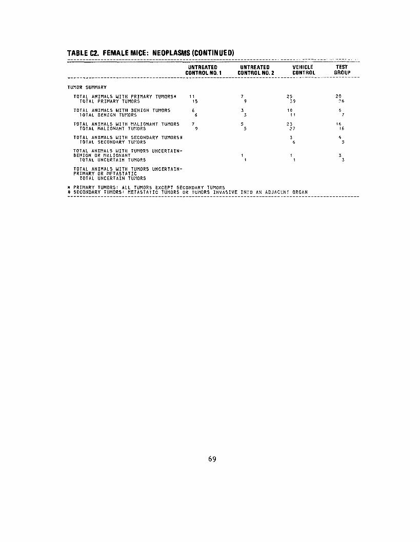
TABLE C2. FEMALE MICE: NEOPLASMS (CONTINUED)
UNTREATED UNTREATED VEHICLE TEST CONTROL NO. 1 CONTROL NO. 2 CONTROL GROUP
TUMOR SUMMARY
TOTAL ANIMALS WITH PRIMARY TUMORS* 11 7 25 20 TOTAL PRIMARY TUMORS 15 9 39 26
TOTAL ANIMALS UITH BENIGN TUMORS 6 3 10 6 TOTAL BENIGN TUMORS 6 3 11 7
TOTAL ANIMALS UITH MALIGNANT TUMORS 7 5 23 1ft TOTAL MALIGNANT TUMORS 9 5 27 16
TOTAL ANIMALS WITH SECONDARY TUMORS* 3 f> TOTAL SECONDARY TUMORS 6 5
TOTAL ANIMALS UITH TUMORS UNCERTAINBENIGN OR MALIGNANT 1 1 3
TOTAL UNCERTAIN TUMORS 1 1 3
TOTAL ANIMALS UITH TUMORS UNCERTAINPRIMARY OR METASTATIC
TOTAL UNCERTAIN TUMORS
X PRIMARY TUMORS: ALL TUMORS EXCEPT SECONDARY TUMORS # SECONDARY TUMORS: METASTATIC TUMORS OR TUMORS INVASIVE INTO AN ADJACENT ORGAN
69

70

APPENDIX D
SUMMARY OF THE INCIDENCE OF NONNEOPLASTIC LESIONS IN MICE ADMINISTERED HCDD PLUS DMBA
BY DERMAL APPLICATION
71

72

TABLE D1.
SUMMARY OF THE INCIDENCE OF NONNEOPLASTIC LESIONS IN MALE MICE ADMINISTERED HCDD PLUS DMBA BY DERMAL APPLICATION
UNTREATED CONTROL NO. 1 C
UNTREATEDONTROL NO. 2
VEHICLE CONTROL
ANIMALS INITIALLY IN STUDY 15 ANIMALS KECROPSIED 14 ANIMALS EXAMINED HISTOPATHOLOGTCALLY 14
151515
45 42 42
INTEGUMENTARY SYSTEM
XSKIN (14) (15) (42) EDEMA, NOS 1 (2%) INFLAMMATION, NOS 2 CSX) ULCER, NOS 1 C7%) 3 (7%) INFLAMMATION, NECROTIZING ULCFR, ACUTE 1 (2%) INFLAMMATION, ACUTE SNPPURATIVE 1 (2%) INFLAMMATION ACUTE AND CHRONIC 1 (2X) INFLAMMATION, ACUTE/CHROMIC 1 (2%) INFLAMMATION, CHRONIC 1 (72) ULCLR, CHRONIC 1 (7%) INFLAMMATION, CHRONIC NECROTIZIN INFLAMMATION PROL I FERAT I VE 1 (2X) NECROSIS, HOS EPIDERMODYSPLASIA VEREUCIFORMIS IIYPCRPLASIA, NOS 1 (2%) HYPIIRPLASIA, PAPILLARY 1 (2%) HYPCRKCRATOSIS 1 (7X) 3 (7X) ACAN1HOSIS 1 (7X> 1 (2%) VERRUCA
*SUECUT TISSUE C 14) (15) (42) EPIDERMAL INCLUSION CYST 1 (2X) INFLAMMATION, NOS ULCTR, NOS 1 (2X) INFLAMMATION, ACUTE SUPPURATIVE 1 (2-/.1 AESCESS, NOS GRANULATION, TISSUE 1 (2%) SCAR NECROSIS, NOS 2 (5X)
RESPIRATORY SYSTEM
*NASAL CAVITY (14) (15) (42) INFLAMMATION, ACUTE SUPPURATIVE
S NUMBER OF ANIMALS WITH TISSUE EXAMINED MICROSCOPICALLY * NUMBER OF ANIMALS NECROPSIED
TEST GROUP
30 24 24
(24)
8 (33%) 1 (4X)
1 (4%) 2 (8%)
1 (45O
1 (45O 1 (4X) 1 (4X)
2 (8%) 5 (21X) 1 (4X)
(24)
1 (4%)
1 (4X)
1 (4%) 2 (8X)
(24) 1 (4%)
73

TABLE 01. MALE MICE: NONNEOPLASTIC LESIONS (CONTINUED)
UNTREATED UNTREATED VEHICLE TEST CONTROL NO. 1 CONTROL NO. 2 CONTROL GROUP
SLUNG/BRONCHUS (13) ( 15) (41) (24)LYMPHCCYTIC INFLAMMATORY INFILTR 1 (8X) 1 (2X) 1 (4X)
SLUNG/BRONCHIOLE (13) ( 15) (41) (24) LYMFHOCYTIC INFLAMMATORY INFILTR 6 (46X) 4 (27X) 13 (32X) 5 (21X)
1HYPERPLASIA, EPITHELIAL (2::;
SLUNG (13) ( 15) (41) (24) ATELECTASIS *•) (5X) CONGESTION, NOS 3 (20X) 3 (7X> 6 (25X)INFLAMMATION, FOCAL 1 (2X) 1 ( 4 X )
( 0 '/. )LYI:?HCCYTIC INFLAMMATORY INFILTR 1 (7X) 2 INFLAMMATION, INTERSTITIAL 1 (SX) 2 (13X) 3 (7 TO 4 ( 17;:) BROJICHOPUEUMCNIA SUPPURATIVE 1 (7X) (2::)EMPYEMA 1 (2 7. ) INFLAMMATION, ACUTE SUPPURATIVE i (2;o NECROSIS. NOS 1 (2X> ALVEOLAR MACROPIIAGES 1 (27.1
HEMATOPOIETIC SYSTEM
XSKIH (14) ( 15) (42) (24) PARAXERATOSIS 1 (4%)
SBONE MARROW (14) ( 14) (40) (24)MYELOFI3ROSIS 1 (4X)
SSPLEEN (13) ( 13) (40) (23)AMYLOIDOSIS 3 (23X) ft ( 1 7 X ) HYPOPLASIA, NOS 1 (SX) 5 (13%) 2 (SX) ATROPHY, HOS 1 (SX) HYPERPLASIA, HCS 2 (15X) 4 ( !OX) MEGAKARYCCYTOSIS 1 (4X) HYPERPLASIA, GRANULOCYTIC 1 (4X) HYPERPLASIA, RETiCULUM CELL 1 ( <H :: ) HYPERPLASIA, LYMPKOIC 1 ( 4'i ) HEMATOPOIESIS 5 ( 13X) f (26X )
8SPLENIC FOLLICLES ( 13) ( 13) (40 (23) HYPOFLASIA, NOS 3 ( 13X) HYPERPLASIA, NOS 2 (15X) 2 (5X) 1 (4%)
r 1 1 )SSPLENIC RED PULP ( 13) ( 13) (40) \ C.*l 1
HYPOPLASIA, HOS 2 C V/:)
* NUMBER OF ANIMALS WITH TISSUE EXAMINED MICROSCOPICALLY X NUMBER OF ANIMALS NECaOi'SIilD
74

TABLE D1. MALE MICE: NONNEOPLASTIC LESIONS (CONTINUED)
MEGAKARYOCYTOSIS HCrU'TOPOIESIS ERYTKKOPOIESIS GRANULOPOIESIS
•LYMPH NODE NECROSIS, CASECUS HYPERPLA5IA, H3S HYPCRPLASIA, LY.'IPHOID
SCERVICAL LYMPH NODE MYPCRPLASIA, UOS
STRACHEAL LYMPH MODE HYPERPLASIA, NOS
SPYLORIC LYMPH NODE HLMORRHAGE
SLUMBAR LYMPH NODE HYPERPLASIA, LYMPHOID
SMESCNTE'JIC L. NODE ABSCESS, 1,05 INTLi-'MMATION PRO L ITERATIVE NLCRCSIS, CASEOUS
SRCHAL LYMPH NODE HYPERPLASIA, UOS
8BRACHIAL LYMPH NODE ABSCESS, NOS
SINGUINAL LYMPH NODE INFLAMMATION, CHRONIC
SLUNG/BRONCHIOLE HYPERPLASIA, LYMPHOID
SLIVER HCMATOPOIESIS
SKIDCEY HYPERPLASIA. LYMPIiOID
UNTREATED CONTROL NO. 1
(8)
C8> ft (50%)
(8) 1 (13X)
(3)
C8)
(8)
(8) 1 (13JO
(8)
(8)
C 13)
( 14)
( 14)
UNTREATED CONTROL NO. 2
2 ( 15X)
C7)
C7) 3 O3X)
(7)
(7) 1 C14JO
(7)
C7)
C7)
C7)
C7)
C 15)
( 15)
( 1<i)
VEHICLE CONTROL
1 (3X)
1 (3%)
C25)
2 (8X)
C25) 10 (40%)
(25)
C25)
(25) 1 (<*5O
(25) 1 C4JO 1 (4X) 1 ft?:)
(25)
(25)
(25)
(41) 1 (2X)
C',2)1 (2%)
(42)1 (?%)
TEST GROUP
1 (4X)
( 15) 1 (75O 1 (7X) 1 (7X)
( 15) 6 (40%)
( 15)
( 15)
( 15) 1 (7X)
( 15)
(15)
( 15) 1 (7X)
( 15) 1 (7X)
(24)
(22) 1 (5%)
(23)
» NUf.BER OF AHIflALS WITH TISSUE EXAMINED MICROSCOPICALLY * NUMBER OF ANIMALS HCCROP5IED
75

TABLE 01. MALE MICE: NONNEOPLASTIC LESIONS (CONTINUED)
UNTREATED CONTROL NO. 1
UNTREATED CONTROL NO. 2
VEHICLE CONTROL
TEST GROUP
CIRCULATORY SYSTEM
KPANCREATIC l.NCDE LYMPHANGIECTASIS
(8) C7) (25) 1 (4X)
( 15)
SMESENTERIC L. NDCE LYrPHANGIECTASIS
(8)i cm>
(7) (25) ( 15) 1 C75O
SHEART MINERALIZATION
( 13) 1 (8X)
C 15) (42) (22)
SHEART/ATRIUM THROMBOSIS, NOS
( 13) ( 15) 2 C 13X)
(42) (22) 1 (5%)
SMYOCARDILi:i MINERALIZATION INFLAMMATION, ACUTE SUPPURATIVE INFLAMMATION, CHRONIC FOCAL INFLAMMATION P"CLIFE"ATIVC NECROSIS, FCCAL
( 13) C 15) t C75O
2 C 13X) 1 ( 7 X ) 1 ( 7 V. )
(42)
3 (72)
(22)
2 (9;:) 1 (5X)
8EHDOCARDIUMINFLAMMATION PRO LI PER ATI VE HYPERPLASIA, NOS
( 13)1 (8X) 1 <8X>
( 15) (42) (22)
XRENAL ARTERY ARTERIOSCLEROSIS, NOS
C 1<t) ( 15) (4,2) I (22)
(24)
KHEPATIC SINUSOID RETENTION OF CONTENT
( 11) ( 15) (42) (22) 1 (52)
DIGESTIVE SYSTEM
SSALIVARY GLAND INFLAMMATION, FOCAL
( 12) ( 15) 1 (.T/.)
(36) (21)
SPAROTID GLAND INFLAMMATION, NOS INFLAMMATION, FOCAL NECROSIS, FOCAL
C 12)
1 (8X)
C 15) 1 (7X)
(3',) 3 (32)
1 (3%)
(21)
1 (52)
8LIVEP, CONGESTION, NOS
( 14) 1 <7X)
(15) (4?) (22)
* NUMBER CF ANIMALS WITH TISSUE EXAMINED MICROSCOPICALLY * NUMBER OF ANIMALS NECROPSIED
76
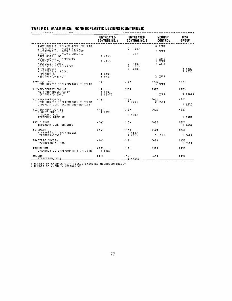
TABLE 01. MALE MICE: NONNEOPLASTIC LESIONS (CONTINUED)
LYMPHOCYTIC INFLAMMATORY INFILTR INrLAf.MATJON, ACUTE FOCAL INFLAMMATION, ACUTE DK-FUSE IHFLAK'IATION, ACUT E/CHRCN 1C CIRRHOSIS, f!OS DEGENERATION, HYDROPIC NECROSIS, liOS NECROSIS, FOCAL NECROSIS, COAGULATIVE AI1YLOIDOSIS AHYLOIDOSIS, FOCAL LIPOID03IS HEPATOCYTOMEGALY
SPORTAL TRACT LYMPHOCYTIC INFLAMMATORY INFILTR
SLIVER/CENTRILOBULAR METAMORPHOSIS FATTY HEPATOCYTOMEGALY
SLIVER/PERIPORTAL LYMPHOCYTIC INFLAMMATORY- INFILTR INFLAMMATION, ACUTE SUPPURATIVE
niVER/HEPATOCYTES CLOUDY SPELLING ATROPHY, NOS ATROPHY, DIFFUSE
SBILE DUCT INFLAMMATION, CHRONIC
((STOMACH HYPERPLASIA, EPITHELIAL IIYPERKERATOSIS
SGASTRIC MUCOSA HYPERPLASIA, NOS
8DUODENUM LYMPHOCYTIC INFLAMMATORY INFILTR
SCOLON IMPACTION, NOS
UNTREATED CONTROL NO. 1
1 (7JO
1 (7X)
1 C7X) 1 (.!'/.•>
C 1<t)
( 14) 1 (7%) 5 (36X)
t 14)
C 14) 1 (7%)
( 14)
( 14)
( 14)
(11) 1 (9X)
(11)
UNTREATED CONTROL NO. 2
2 ( 13JO
1 C750
2 (13%) 2 ( 135O 3 (20X)
( 15)
( 15)
( 15) 1 (7X)
( 15)
1 (7%)
(15)
( 13) 1 (8X) 1 (Z'/.l
(13)
(12)
( 15) 2 (13%)
VEHICLE CONTROL
3 (7%)
1 (2%)
1 (2X) 1 (2X) 1 (2X)
2 (5%)
(42) 1 (2%)
(42)
1 (2%)
(42) 2 (5%)
(42)
(42)
(42)
3 (7%)
(42)
(34)
(36)
TEST GROUP
1 (5X> 1 (5%)
(2?)
(22)
3 (14%)
(22)
1 (5%)
(22)
1 (5%)
(22) 1 (5%)
(23)
1 (4%)
(23) 1 (4%)
(19)
(19)
tt NUMBER OF ANIMALS WITH TISSUE EXAMINED MICROSCOPICALLY * NUMBER OF ANIMALS NECROPSIED
77
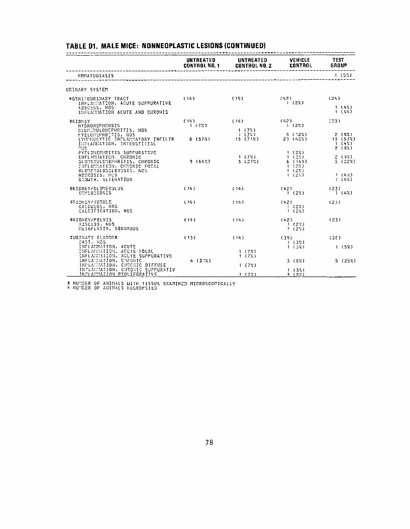
TABLE D1. MALE MICE: NONNEOPLASTIC LESIONS (CONTINUED) inULk u|. ivinkk mil***- ivuiviv^wr hnw iiw K.I.WIWIVW iLVWIV I llVWkV/
UNTREATED CONTROL NO. 1
UNTREATED CONTROL NO. 2
VEHICLE CONTROL
TEST GROUP
HENATODIASIS 1 (5X)
URINARY SYSTEM
*GENITC'JRINARY TRACTINFLAMMATION, ACUTE SUPPURATIVE ABSCESS, NOS INFLAMMATION ACUTE AND CHRONIC
(1*) ( 15) (42) 1 (2%)
(24)
1 (4JO 1 (4X)
SKIDNEYHYDRONEFHROSISGLOMCRULONCPHRITIS, NOS PYELONEPHRITIS, NOS LYMPIIOCYTIC INFLAMMATORY INFILTRINFLAMMATION, INTERSTIflAL PUS PYELONEPHRITIS SUPFURATIVE INFLAMMATION, CHRONIC GLOMERULONEPHRITIS, CHRONICINFLAMMATION, CHROMIC FOCAL GLCME"!!JLOSCLEROSIS, NOS NECROSIS, NOS GROWTH, ALTERATION
(14) 1 (7%)
& (57%)
9 (645!)
( 14)
1 (7X) 1 (7%)
10 (7U)
1 (7X) 3 (2U)
(42) 1 (2X)
5 (12%) 23 (4SX)
1 (254) 1 (2K) 6 ( 14X) 1 C2X) 1 (2%) 1 (27.)
(23)
2 (95!) 13 (575!)
1 (4%) 2 (9X)
2 (9%) 5 (225!)
1 (4%) 1 (4%)
8KIDNEY/GLOMERULUSAMYLOIDOSIS
(14) ( 14) (42) 1 (2X)
(23) 1 (45!)
SKIDHEY/TUBULECALCULUS, NOS CALCIFICATION, NCS
(14) ( K) (42) 1 (2%) 1 (2%)
(23)
SKIDNEY/PELVISASSCESS, NOS METAPLASIA, SQUAMOUS
(1<t) ( 14) (42) 1 (2*) 1 (2%)
(23)
SURINARY BLADDERCAST, NOS INFLAMMATION, ACUTE INFLAMMATION, ACUIE FOCAL INFLAflllATION, ACUTE SUPPURATIVE INFLAMMATION, CHRONICINFLAMMATION, CHRONIC DIFFUSE INFLAMMATION, CHRONIC 5UPPURATIV INFLAMMATION PROI. ITERATIVE
( 13)
4 (31%)
( 14)
1 C7JO 1 (77.)
1 (7%)
1 (7X>
(39) 1 (3%) 1 C3X)
3 (850
1 (35!) 3 (3%)
(20)
1 (55O
5 (255!)
8 NUM3ER OF ANIMALS UITI! TISSUE EXAMINED MICROSCOPICALLY X NUMBER OF ANIMALS NECROPSIED
78
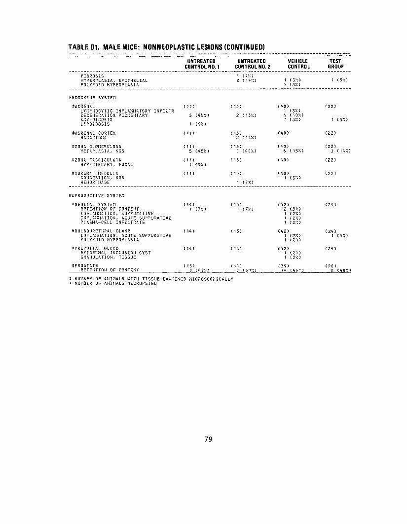
TABLE D1. MALE MICE: NONNEOPLASTIC LESIONS (CONTINUED)
UNTREATED UNTREATED VEHICLE TE 5T CONTROL NO. 1 CONTROL NO. 2 CONTROL GRC UP
FIEROSIS HYPERPLASIA, EPITHELIAL
1 ( 7 :; ) i C5»
POLYPOID HYPERPLASIA 1 (3X)
ENDOCRINE SYSTEM
SADRENAL LYMPHOCYTIC INFLAMMATORY INFILTR
(11) ( 15) (40) 1 (3X)
'22)
DEGENERATION PIGMENTARY AMYLOIDOSIS LIPOIDQSIS
5
1
(45X)
(9JO
2 (13X) 4 (10X) 1 (3X> 1 (5X)
#ADRENAL CORTEX HAMARTOMA
(11) ( 15) 2 (13/i)
(40) (22)
SZONA GLQMER'JLOSA METAPLASIA, NGS
(11) 5 (45!O
( 15) 6 (40V.)
(40) 6 ( 1 5X )
(22) 3 ( 14X)
*ZONA FA3CICULA1A HYPERTROPHY, FOCAL
(11)1 (9;o
( 15) (40) (22)
8ADRENAL MEDULLA CONGESTION, NOS HEMORRHAGE
(11) ( 15)
1 (7X)
(40) 1 (3X)
(22)
REPRODUCTIVE SYSTEM
XGENITAL SYSTEM RETENTION OF CONTENT
( 14) 1 (T/.t
( 15) 1 ( 7X)
(42) 2 (5X)
(24)
INFLAMMATION, SUPFURATIVE INFLAMMATION, ACUTE SU?PURATIVE PLASMA-CELL INFILTRATE
(2X)(2" ) (2::)
XBULBOURETHRAL GLAND INFLAMMATION, ACUTE SUPPURATIVE POLYPOID HYPERPLASIA
(14) ( 15) (42) (2X)(2-;)
(24)1 (4X)
XPREPUTIAL GLAND EPIDERMAL INCLUSION CYST GRANULATION, TISSUE
( 14) ( 15) (42) 1 (2X) 1 (2%)
(24)
SPROSTATE RETENTION OF CONTENT
( 13) 9 (69X)
(14) 7 ( 5 n •<)
(39) i S ( 4 A" )
(20) 8 (40X)
* NUMBER OF ANIMALS WITH TISSUE EXAMINED MICROSCOPICALLY * NUMBER OF ANIMALS NECROPSIED
79
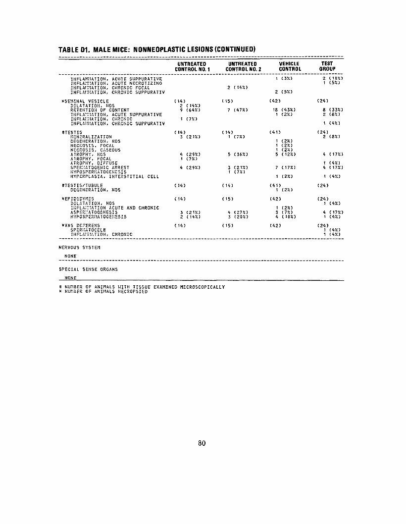
TABLE D1. MALE MICE: NONNEOPLASTIC LESIONS (CONTINUED)
INFLAMMATION, ACUTE SUPPURATIVE INFLAMMATION, ACUTE NECROTIZING INFLAMMATION, CHROMIC FOCAL INFLAMMATION, CHRONIC SUPPURATIV
XSEMIHAL VESICLE DILATATION, NOS RETENTION OF CONTENT INFLAMMATION, ACUTE SUPPURATIVE INFLAMMATION, CHRONIC INFLAMMATION, CHRONIC SUPPURATIV
KTESTIS MINERALIZATION DEGENERATION, NOS NECKOSIS, FOCAL NECPCSIS, CASEOUS ATROPHY, NCS ATROPHY, FOCAL ATROPHY, DIFFUSE SPERMATOGENIC ARREST KYPOSPERMATOGEKE3IS HYPERPLASIA, INTERSTITIAL CELL
8TESTIS/TU3ULE DEGENERATION, NOS
XEPIDIDYMIS DILATATION, NOS INFLAMMATION ACUTE AND CHRONIC ASPEKHATOGENESIS HYPOSPERMATOGEJ1ESIS
XVAS DEFERENS SPERMATOCELE INFLAMMATION, CHRONIC
NERVOUS SYSTEM
NONE
SPECIAL SENSE ORGANS
NONE
UNTREATED CONTROL NO. 1
( 14)2 (14%) 9 (64%)
1 (755)
C 14) 3 (2155)
4 (2955) 1 (7%)
4 (295!)
( 14)
(14)
3 (2155) 2 (1<iX)
( 14)
UNTREATED CONTROL NO. 2
2 <m)
( 15)
7 (4754)
( 14) 1 C7X)
5 C36X)
3 (21X) 1 (755)
( 14)
(15)
4 (275!) 3 (205!)
( 15)
VEHICLE CONTROL
1 (35!)
2 (555)
(42)
18 (4355) 1 (2X)
(41)
1 (25!) 1 (25!) 1 (2X.) 5 (125!)
7 (175!)
1 (255)
(41) 1 (2%)
(42)
1 (255) 3 (75!) 4 (1055)
(42)
TEST GROUP
2 (105!) 1 (55!)
(24)
8 (3355) 2 (85!)
1 (455)
(24) 2 (8%)
4 (1755)
1 (455) 4 (1755)
1 (4J5)
(24)
(24) 1 (455)
4 (1755) 1 (45!)
(24) 1 (455) 1 (455)
S NUMBER OF ANIMALS WITH TISSUE EXAMINED MICROSCOPICALLY * KUMDER OF ANIMALS NECROPSIED
80
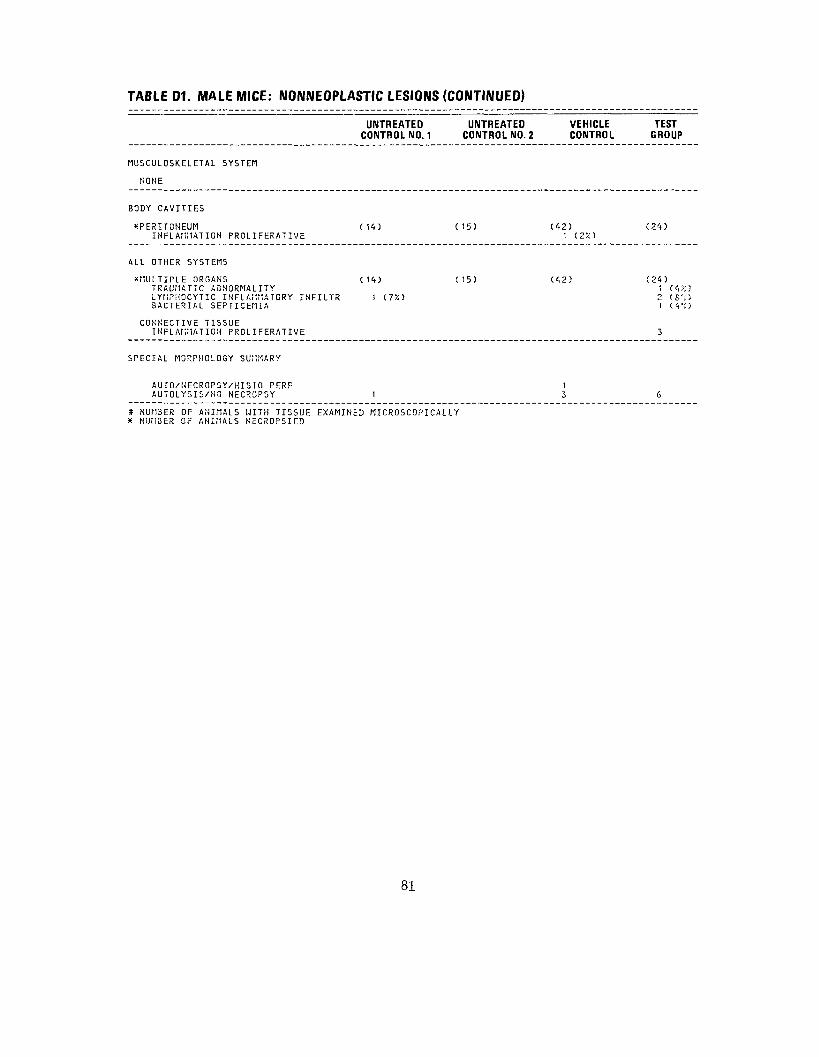
TABLE D1. MALE MICE: NONNEOPLASTIC LESIONS (CONTINUED)
UNTREATEDCONTROL NO. 1
UNTREATED CONTROL NO. 2
VEHICLE CONTROL
TEST GROUP
MUSCULOSKELETAL SYSTEM
NONE
BODY CAVITIES
XPERITONEUMINFLAMMATION PROLI FERATIVE
(14) (15) (<*2) 1 (2%)
(24)
ALL OTHER SYSTEMS
^MULTIPLE ORGANSTRAUMATIC ABNORMALITY LYiiPHOCYTIC INFLAMMATORY INFILTRBACTERIAL SEPTICEMIA
CONNECTIVE TISSUE INFLAMMATION PROL I FERAT I VE
(14)
1 (75O
(15) ( <, 2 ) (24) 1 (W>
2 (S'O 1 ( <i •: )
3
SPECIAL MORPHOLOGY SUMMARY
AUrO/HECROPSY/HISTO PERFAUTOLYSIS/NO NECROPSY 1
1 3 6
# NUMBER OF ANIMALS WITH TISSUE EXAMINED MICROSCOPICALLY X NUMBER OF ANIMALS NECROPSIED
81

TABLE D2.
SUMMARY OF THE INCIDENCE OF NONNEOPLASTIC LESIONS IN FEMALE MICE ADMINISTERED HCDD PLUS DMBA BY DERMAL APPLICATION
UNTREATED UNTREATED VEHICLE TEST CONTROL NO. 1 CONTROL NO. 2 CONTROL GROUP
ANIMALS INITIALLY IN STUDY 15 15 ',5 30 ANIMALS NECROPSIED 15 15 41 27 ANIMALS EXAMINED H ISTOPATHCLOGI CAL LY 15 15 4 1 ^7
INTEGUMENTARY SYSTEM
!<SKIN EDEMA, NOS NECROSIS, CASEOUS HYPLRPLASIA, FOCAL HYPER'CERATOSIS
( 15)
1 (7%) 1 (7%)
(15) (41)
1 (2%)
(27) 11 ( 4% ) ( 4 /;)
*SU3CUT TISSUE EDEMA, NOS ULCER, liOS ABSCESS, HOS IIHCROSIS, NOS
( 15) (15)1 (7%) (41)
1 (2%) 1 ( 2% )1 (2%)
(27)
2 (7%)
RESPIRATORY SYSTEM
SLUNG/BRONCHUS ( 15) (15)LYMPHOCYTIC INFLAMMATORY INFILTR t (7%)
(41)2 (5%)
(25) t (4%)
SLUNG/BRONCHIOLE (15) (15)LYMPHOCYTIC INFLAMMATORY INFILTR 4 (27%) 10 (67%)
(41) 22 (54%)
(25) 1 1 (44%)
tfLUNG (15) (15)ATELECTASIS CONGESTION, NOS 2 (13%) INFLAMMATION, INTERSTITIAL 3 (20%) 3 (20%)BRONCHOPNEUMONIA SUPPURATIVE INFLAMMATION, ACUTE SUPPURATIVE HYPERPLASIA, ADENOMATOUS 1 (7%) 3 (20%)
(41) 1 ( 2% )
5 (12%) 1 (2%) 1 ( 2 "/>)2 (5%)
(25) 2 (8%) 3 (12%) 6 ( 24% )
HEMATOPOIETIC SYSTEM
XMAKMARY GLAND (15) (15)ADENOSIS 2 (13%)
(41)2 (5%)
(27)
» NUMBER OF ANIMALS WITH TISSUE EXAMINED MICROSCOPICALLY * NUMBER OF ANIMALS NECROPSIED
82

TABLE D2. FEMALE MICE: NONNEOPLASTIC LESIONS (CONTINUED)
UNTREATED UNTREATED CONTROL NO. 1 CONTROL NO. 2
SBONE MARROW FIBROUS OSTEODYSTROPHY
( 13) 3 (23%)
( 13) 1 (8%)
HYPERPLASIA, NOS RETICULOCYTOSIS 1 (3%)
#5PLEEN CYST, NOS
(15) ( 15)
PIGMENTATION, NOS HEI105IDEROSIS HYPOPLASIA, NOS ATROPHY, NOS
1 (7%) 11 (75!) (7%)
HYPERPLASIA, NODULAR HYPERPLASIA, NOS 3 (20%) 2 (13%) MEGAKARYOCYTOSIS HYPERPLASIA, LYMPHOID 1 (7%) HEMATOPOIESIS
8SPLENIC FOLLICLES HYPOPLASIA, NOS
( 15) (15)
HYPERPLASIA, NOS 2 ( 13%)
SSPLENIC RED PULP HYPOPLASIA, NOS
( 15) 1 (7%)
( 15) 1 (7%)
HEMATOPCIESIS 2 (13%) 1 (7%)
8LYMPH NODE HYPERPLASIA, NOS
( 12) 2 (17%)
( 12) 3 (25%)
#CERVICAL LYMPHHYPERPLASIA,
NODE NOS
( 12) 1 (8%)
( 12) 4 (33%)
SPANCREATIC L.NODE PIGMENTATION, NQS
( 12) ( 12)
HYPERPLASIA, NOS 1 (8%)
8RENAL LYMPH NODE HYPERPLASIA, NOS
( 12) ( 12)
(tLUNG/BRONCHIOLE HYPERPLASIA, LYMPHOID
C 15) ( 15) 1 (7%)
*PAP.OTID GLAND FIBROSING ADENOSIS
(15) (12)
(tLIVER HEMATOPOIESIS
(15) ' (7%)
(15)
# NUMBER OF ANIMALS WITH TISSUE EXAMINED MICROSCOPICALLY * NUMBER OF ANIMALS NECROPSIED
VEHICLE CONTROL
(37) 7 (19%) 1 (3%)
(40) 1 (3%)
3 (8%)
1 (3%)
1 (3%)
2 (5%)
(40)
<t (10%)
(<tO) 1 (3%) 1 (3%)
(30) 6 (20%)
(30) 17 (57%)
(30) 1 (3%) 1 (3%)
(30)
(<t1> 1 (2%)
(36) 1 (3%)
(<t1)
TEST GROUP
(23) 7 (30%)
(27)
1 (<t%) 1 (<t%) 3 (11%)
3 (11%)
2 (7%) 1 (4%)
(27) 1 (4%)
(27)
1 (4%)
(16)
(16) 6 (38%)
( 16)
1 (6%)
( 16) 1 (6%)
(25)
(23)
(27)
83

TABLE D2. FEMALE MICE: NONNEOPLASTIC LESIONS (CONTINUED)
SADRENAL MYELOPOIESIS
8THYMUS ECTOPIA HYPERPLASIA, NOS
STHYMIC MEDULLA HYPERPLASIA, NOS
CIRCULATORY SYSTEM
SBONE MARROW PERIARTERITIS
SPANCREATIC L.NODE LYNPHANGIECTASIS
SMESENTERIC L. NODE LYMPHANGIECTASIS
ttHEART ENDOCARDIOSIS
SHEART/ATRIUM THROMBOSIS, NOS
ttMYOCARDIUM INFLAMMATION, CHRONIC FOCAL
XCENTRAL VEINS/LIVER LYMPHOCYTIC INFLAMMATORY INFILTR
DIGESTIVE SYSTEM
((SALIVARY GLAND INFLAMMATION, NOS
liPAROTID GLAND INFLAMMATION, NOS
SLIVER INFLAMMATION, NOS
UNTREATED CONTROL NO. 1
(15)
( 15)
(15) 2 (132)
( 13)
(12)
(12)
( 15) 2 ( 132)
( 15) 1 (72)
( 15) 1 (72)
( 15)
( 15)
( 15)
(15) 1 (72)
UNTREATEDCONTROL NO. 2
( 14)
(15)
2 (132)
(15)
(13)
(12)
(12)
(14)1 (72)
(14)
(14)
(15)
(12)2 (172)
(12)
(15)
# NUMBER OF ANIMALS WITH TISSUE EXAMINED MICROSCOPICALLY * NUMBER OF ANIMALS NECROPSIED
84
VEHICLE CONTROL
(41) 1 (22)
(39)
2 (52)
(39)
(37)
(30) 1 (32)
(3C) 2 (7%)
(40)
(40) 2 (52)
(40)
(41) 1 (22)
(36) 1 (32)
(36) 1 (32)
(41)
TEST GROUP
(26)
(27) 1 (42) 1 (42)
(27)
(23) 1 (42)
( 16)
( 16)
(27)
(27)
(27) 1 (42)
(27)
(23)
(23) 2 (92)
(27)

TABLE D2. FEMALE MICE: NONNEOPLASTIC LESIONS (CONTINUED)
UNTREATED CONTROL NO. 1
UNTREATED CONTROL NO. 2
VEHICLE CONTROL
TEST GROUP
INFLAMMATION, FOCAL LYMPHOCYTIC INFLAMMATORY INFILTR IHFLAf'iMATION, ACUTE INFLAMMATION, ACUTE DIFFUSE INFLAMMATION, CHRONIC NECROSIS, COAGULATIVE AflYLOIDOSIS LIPOIDOSIS FOCAL CELLULAR CHANGE
1
1
(7X)
(7%)
3 (20!!) 1 (7JO
1 (7%)
1 (7%)
1 (2X> 3 (7X)
1 (2X) 1 (2X)
111
(450 (4'X.) (4%)
SLIVER/CENTRILOBULAR LYI1PHOCYTIC INFLAMMATORYNECROSIS, NOS "ETAr'.OP.PHOSIS FATTY HEPATOCYTOtlEGALY
INFILTR ( 15)
1 (7%)
( 15)
1 (7%)
(41) 1 (2%)
(27)
1 (4>O
8LIVER/PERIPORTAL LYhPHOCYTIC INFLAMMATORYHYPERPLASIA, NOS
INFILTR ( 15)
1 (.T/.) \ (7X)
(15) (41) (27)
8LIVER/KUPFFER CELL HYPERPLASIA, NOS
( 15) ( 15) (41) 1 (2X)
(27)
XGALLDLADDERINFLAMMATION, CHRONIC
( 15) 1 (7X)
(15) (41) (27)
KBILE DUCT HYPERPLASIA, NOS
( 15) Z (1'3X)
( 15) (41) (27)
SPANCREAS LYMPKOCYTIC INFLAMMATORY INFILTR HYPERPLASIA, tiOS
(15) 1 (7X) 1 (T/.)
(15) (40) 1 (3X)
(26)
SSTOMACH IKFLArMATION, ACUTE HYPERKERATOSIS
( 15) 1 C75O
(15) (41)
2 (55O
(26)
1 (4X)
SGASTRIC MUCOSA HYPOPLASIA, NOS
( 15) (15) 1 (7!O
(41) (26)
SFORESTOMACH HYPERKERATOSIS
( 15) (15) 1 (75O
(41) (26)
ttSMALL INTESTINE PARASITISM
(13) (15) (40) (26) 1 (4%)
NUMBER OF ANIMALS WITH TISSUE EXAMINED MICROSCOPICALLY NUMBER OF ANIMALS NECROPSIED
85

TABLE D2. FEMALE MICE: NONNEOPLASTIC LESIONS (CONTINUED)
•DUODENUM NECROSIS, HOS
XRECTUM ULCER, ACUTE DESKOPLASIA NECROSIS, NOS
URINARY SYSTEM
8KIDHEY HYDRONEPHROSIS GLOtlERULONEPHRITIS, NOS PYELONEPHRITIS, NOS LYKPHOCYTIC INFLAMMATORY INFILTR PYELONEPHRITIS SUPPURATIVE GLOMERULONEPHRITIS, ACUTE INFLAMMATION, CK.10NIC GLOMERULONEPHRITIS, CHRONIC INFLAMMATION, CHROMIC FOCAL GLOMERULOSCLEROSIS, NOS INFAP.CT, HEALED
KKIDNEY/GLOMERULUS AMYLOID, NOS AMYLOIDOSIS
(KIDNEY/PELVIS DILATATION, NOS
•URINARY BLADDER LYMPHOCYTIC INFLAMMATORY INFILTR INFLAMMATION, ACUTE INFLAMMATION, ACUTE NECROTIZING INFLAMMATION, ACUTE/CHSOMC INFLAMMATION, CHRONIC INFLAMMATION, CHRONIC FOCAL INFLAMMATION PROLIFEF'.ATIVE ATROPHY, NOS METAPLASIA, SQUAMOUS
ENDOCRINE SYSTEM
•PITUITARY HYPERPLASIA, CHROMOPHOBE-CELL
UNTREATED CONTROL NO. 1
(13)
(15)
( 15) 1 (7%) 1 (7%)
5 (33%)
2 (13%) 10 (6/-0
2 (13%)
( 15)
( 15)
(13)
<t (31%)
(10)
UNTREATED CONTROL NO. 2
(15) 1 (7X)
(15) 1 (7X) 1 (7%) 1 (7X)
(15)
2 (13%) 6 (40%) i (/•:) 1 (T/.1
12 (80%)
2 (13%)
( IS)
(15)
( 13) 1 (3%)
1 (S>.) 2 (I5:i) 5 (3oX)
i (s;:)
(11)
VEHICLE CONTROL
CtO)
(^1)
(<(1) 1 (2%)
1 (2X) 26 (63%)
1 (2%) 1 (2%)
16 (39'i) 1 (2'<> 2 (5%) 1 (2%)
(<t1)
1 (2%)
(<t1>
(JO)
1 (3%)
10 (33X) 2 (7!'.)
1 (3X)
(35)
TEST GROUP
(26)
(27)
(27) 1 (4%)
16 (59%)
1 1 ( <- 1 % )
(27) 1 C5%)
(27) 1 (4%)
(19)
5 (26%)
1 (5%)
( 13) 1 (8X)
I NUMBER OF ANIMALS WITH TISSUE EXAMINED MICROSCOPICALLY * NUMBER OF ANIMALS NECROPSIED
86

TABLE D2. FEMALE MICE: NONNEOPLASTIC LESIONS (CONTINUED)
SADRENAL CONGESTION, NOS DEGENERATION PIGMENTARY
•ADRENAL CORTEX HAr'.RTOMA LYKPHOCYTIC INFLAMMATORY INFILTR
SZONA GLOMERULOSA METAPLASIA, HOS
8THYROID INFLAMMATION, NOS HYPERPLASIA, FOLLICULAR-CELL
STHYROID FOLLICLE GOITFR COLLOID
•PANCREATIC ISLETS HYPERPLASIA, NOS
REPRODUCTIVE SYSTEM
XMAIWRY GLAND DILATATION/DUCTS
*VAGINA INFLAMMATION, ACUTE/CHRONIC HYPERPLASIA, EPITHELIAL
8UTERUS MINERALIZATION HEMORRHAGE INFLAI'IIATION, ACUTE FOCAL ABSCESS, NOS INFLA! '1ATION, CHRONIC NECROSIS, NOS METAPLASIA, SQUAMOUS
•CERVIX UTERI INFLAMMATION, CHRONIC
#UTERUS/EHDO'1ETRIUM INFLAM 1ATIOH, CHRONIC SUPPURATIV
UNTREATED CONTROL NO. 1
C 15)
9 (60%)
(15) 1 (7%) 1 (72)
( 15) 6 (40%)
( 15)
C 15)
( 15)
( 15) 3 (20%)
( 15)
( 15)
( 15)
( 15)
UNTREATED CONTROL NO. 2
( 14)
3 (57%)
( 14) 1 (7%)
( 14) 9 (64%)
( 15) 1 (7V.)
( 15)
( 15) 1 (7%)
( 15) 1 (7%)
( 15) 1 (7%) 1 (7%)
( 15)
( 15) 1 (7%)
( 15)
VEHICLE CONTROL
(41) 2 (5%)
22 (54%)
(41) 1 (2%) 1 (2%)
(41) 20 (49%)
(39)
1 (3X)
(39) 1 (3%)
(40)
(41) 3 (7%)
(41)
(36) 1 (3%) 1 (3%)
1 (3X)
1 (3%)
(36)
(36)
TEST GROUP
(26)
16 (62%)
(26)
(26) 12 (46%)
(22)
(22)
(26) 1 (4%)
(27) 2 (7%)
(27)
(26)
1 (4%)
1 (4%)
1 (41/.)
(26)
(26)1 (4%)
# NUMBER OF ANIMALS WITH TISSUE EXAMINED MICROSCOPICALLY x NUMBER OF ANIMALS HECROPSIED
87
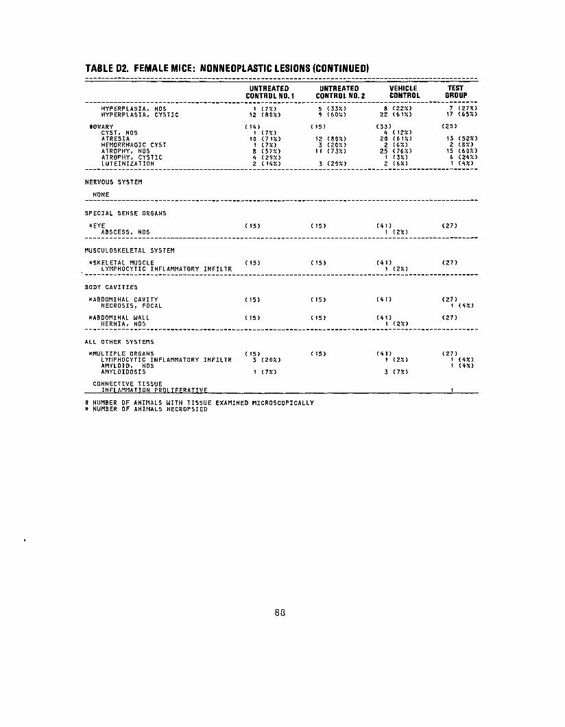
TABLE D2. FEMALE MICE: NONNEOPLASTIC LESIONS (CONTINUED)
HYPERPLASIA, NOS HYPERPLASIA, CYSTIC
*OVARY CYST, NOS ATRESIA HEMORRHAGIC CYST ATROPHY, NOS ATROPHY, CYSTIC LUTEINIZATION
NERVOUS SYSTEM
NONE
SPECIAL SENSE ORGANS
XEYE ABSCESS, NOS
MUSCULOSKELETAL SYSTEM
^SKELETAL MUSCLE LYMPHOCYTIC INFLAMMATORY INFILTR
BODY CAVITIES
XABDOMINAL CAVITY NECROSIS, FOCAL
XABDOMINAL WALL HERNIA, NOS
ALL OTHER SYSTEMS
XMULTIPLE ORGANS LYMPHOCYTIC INFLAMMATORY INFILTR AMYLOID, NOS AMYLOIDOSIS
CONNECTIVE TISSUE INFLAMMATION PROLIFERATIVE
* NUMBER OF ANIMALS WITH TISSUE EXAMINED* NUMBER OF ANIMALS NECROPSIED
UNTREATED CONTROL NO. 1
1 (7%) 12 (80%)
( 14) 1 (7%)
10 (71%) 1 (7%) 8 (57%) 4 (29%) 2 (14%)
(15)
( 15)
( 15)
( 15)
( 15) 3 (20%)
1 (7%)
MICROSCOPICALLY
UNTREATED CONTROL NO. 2
5 (33>!) 9 (60%)
( 15)
12 (80%) 3 (20%)
11 (73%)
3 (20%)
(15)
(15)
( 15)
( 15)
( 15)
VEHICLE CONTROL
8 (22%) 22 (61%)
(33) 4 (12%) 20 (61%) 2 (6%) 25 (76%) 1 (3%) 2 (6%)
(41) 1 (2%)
(41) 1 (2%)
(41)
(41) 1 (2%)
(41) 1 (2%)
3 (7%)
TEST GROUP
7 (27%) 17 (65X)
(25)
13 (52%) 2 (8%) 15 (60%) 6 (24%) 1 (4%)
(27)
(27)
(27) 1 (4%)
(27)
(27) 1 (4%) 1 (4%)
1
88
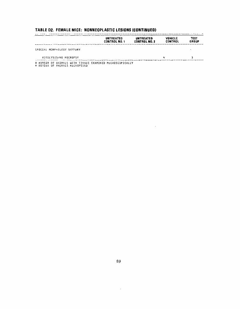
TABLE 02. FEMALE MICE: NONNEOPLASTIC LESIONS (CONTINUED)
UNTREATEDCONTROL NO. 1
UNTREATED CONTROL NO. 2
VEHICLE CONTROL
TEST GROUP
S P E C I A L M O R P H O L O G Y SUMMARY
A U T O L Y S I S / N O NECROPSY
* NUMBER OF ANIMALS WITH TISSUE EXAMINED MICROSCOPICALLY * NUMBER OF ANIMALS NECROPSIED
<t 3
89

90

APPENDIX E
PREPARATION OF HCDD
91

92

APPENDIX E
Preparation of HCDD
3,4,5-Trichlorophenol (Aldrich Chemical Co., Milwaukee, Wis.) was
brominated at 80 -90 C in glacial acetic acid and the product,
2-bromo-3,4,5-trichlorophenol, was recrystallized from methanol-water or
benzene-hexane. The potassium salt of 2-bromo-3,4,5-trichlorophenol was
prepared by treating the phenol with potassium hydride in benzene. The dry
potassium salt of 2-bromo-3,4,5-trichlorophenol was covered with a layer of
dry potassium carbonate and heated under sublimation conditions at
240 -270 C at 1 mm pressure for 20 hours. The crude product contained
hexachlorodibenzo-p-dioxins, bromohexachlorodibenzo-p-dioxins (from the
condensation of the 2,6-dibromo-3,4,5-trichlorophenol impurity in
2-bromo-3,4,5-trichlorophenol), and other minor impurities. The crude
product was debrominated by hydrogenolysis with lithium aluminum hydride at
0°C to yield HCDD (Gray et al., 1975).
93

94

APPENDIX F
ANALYSES OF HCDD
95

96

APPENDIX F
Analysis of HCDD
Midwest Research Institute
A. Vapor-Phase Chromatography
Lot No. IIT 102
a. System 1:
Instrument: Column: Detector: Oven Temperature I: Compound Concentration: Results:
b. System 2:
Instrument: Column: Detector: Oven Temperature I: Compound Concentration: Results:
Bendix 2500 3% OV-1, 1.8 m x 4 mm I.D. Electron capture, "3 1
160°C, isothermal
0.032 mg/ml in benzene One peak with a retention time identical to that of an authentic sample of tetrachlorodibenzo-p-dioxin. By comparison of the area of this peak with that of a weighed solution of tetrachlorodibenzo-p-dioxin, it was calculated that the tetrachloro compound was present at a concentration of 0.07%.
Varian 1400 3% OV-1, 1.8 m x 4 mm I.D. Electron capture, Sc- I
160°C, isothermal
Saturated (2 mg/ml in benzene) Major peak was not eluted in 45 minutes. Eight minor impurities detected.
97

Retention Time Retention (Relative to Percent
Peak
1 2 3 4 5 6 7 8
Time Tetrachloro(min) dibenzo-p-dioxin)
4.2 0.11 7.9 0.20 9.5 0.24
11.7 0.29 17.3 0.43 25.6 0.64 34.5 0.87 39.8 1.00
System 3:
Instrument: Column: Detector: Oven Temperature II: Compound Concentration: Results:
Possible by Identity Weight
Unknown _
Dichlorodibenzo-p-dioxin 0.004 Unknown Unknown Trichlorodibenzo-p-dioxin 0.004 Unknown Unknown Tetrachlorodibenzo-p-dioxin 0.01
Possible identities were assigned to peaks which had retention times identical to those for authentic samples of other chlorinated dibenzo-p-dioxins. Percentage compositions by weight were calculated by comparison of the areas of the impurity peaks in the weighed sample with the area of the tentatively identified chlorinated dibenzo-p-dioxin in a weighed solution of similar concentration. No authentic sample of monochlorodibenzo-p-dioxin was available. It is possible that the first peak is the monochloro compound. No percentage compositions were calculated for the unknown peaks because of the great variation in response of electron capture detectors to different compounds.
Varian 1400 3% OV-1, 1.8 m x 4 mm I.D. Electron capture, Sc I
225°C, isothermal
0.032 mg/ml in benzene Major peak and three impurities,
98

Retention Time
Peak (min)
1 4.0 2 7.8 3 8.6 4 15.7
Retention Time (Relative to Hexachlorodibenzo-p-dioxin) Identity
Area Relative to that of Hexachlorodibenzo-p-dioxin
0.250.500.541.00
d. System 4:
Instrument: Column: Detector: Oven Temperature III: Compound Concentration: Results:
Tetrachlorodibenzo-p-dioxin 0.08 Unknown Unknown Hexachlorodibenzo-p-dioxin 100.00
Nothing else eluted in 50 minutes.
The first peak was again quantitated against an authentic sample of tetrachlorodibenzo-p-dioxin. No authentic sample of pentachlorodibenzo-p-dioxin was available. The two peaks with retention times intermediate between tetra- and hexachlorodibenzo-p-dioxin could be pentachloro compounds, but there was no way to verify this. Octachlorodibenzo-p-dioxin was not detected; the detection limit in this sample is less than 0.004%.
Bendix 2500 3% Dexsil 400, 1.8 m x 2 mm I.D. Electron capture, 63jj
275°C, isothermal
0.032 mg/ml in benzene Major peak and two impurities with longer retention times.
Peak
Retention Time (min)
Retention Time (Relative to Hexachlorodibenzo-p-dioxin) Identity
1 2 3
2.1 3.5 4.8
1.00 1.70 2.30
Hexachlorodibenzo-p-dioxin Unknown Unknown
99
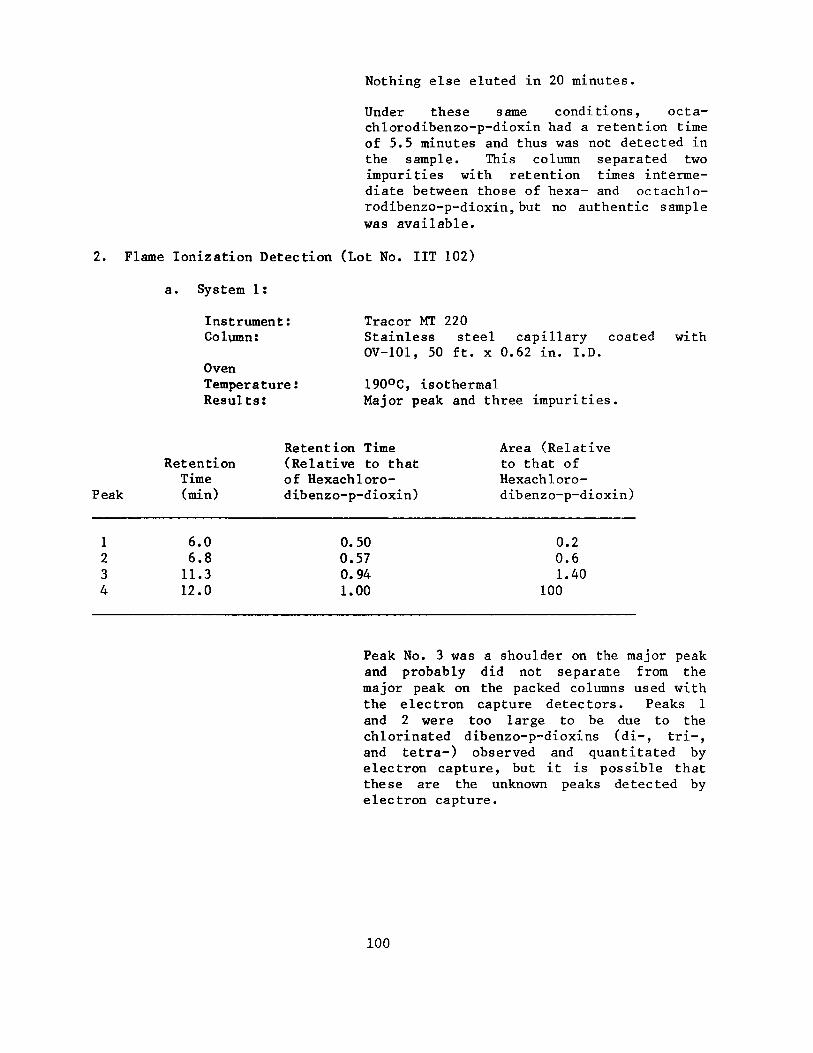
Nothing else eluted in 20 minutes.
Under these same conditions, octachlorodibenzo-p-dioxin had a retention time of 5.5 minutes and thus was not detected in the sample. This column separated two impurities with retention times intermediate between those of hexa- and octachlorodibenzo-p-dioxin, but no authentic sample was available.
2. Flame lonization Detection (Lot No. IIT 102)
a. System 1:
Instrument: Tracor MT 220 Column: Stainless steel capillary coated with
OV-101, 50 ft. x 0.62 in. I.D. Oven Temperature: 190°C, isothermal Results: Major peak and three impurities.
Retention Time Area (Relative Retention (Relative to that to that of
Time of Hexachloro- Hexachloro-Peak (min) dibenzo-p-dioxin) dibenzo-p-dioxin)
1 6.0 0.50 0.2 2 6.8 0.57 0.6 3 11.3 0.94 1.40 4 12.0 1.00 100
Peak No. 3 was a shoulder on the major peak and probably did not separate from the major peak on the packed columns used with the electron capture detectors. Peaks 1 and 2 were too large to be due to the chlorinated dibenzo-p-dioxins (di-, tri-, and tetra-) observed and quantitated by electron capture, but it is possible that these are the unknown peaks detected by electron capture.
100
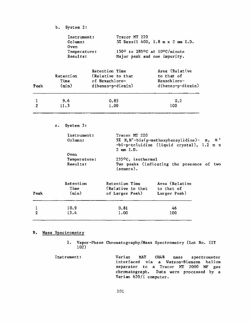
b. System 2:
Instrument: Tracor MT 220 Column: 3% Dexsil 400, 1.8 m x 2 mm I.D. Oven Temperature: 150° to 285°C at 10°C/minute Results: Major peak and one impurity.
Retention Time Area (Relative Retention (Relative to that to that of
Time of Hexachloro- Hexachloro-Peak (min) dibenzo-p-dioxin) dibenzo-p-dioxin)
1 9.6 0.85 0.2 2 11.3 1.00 100
c. System 3:
Instrument: Tracor MT 220 Column: 5% N,N'-bis(p-methoxybenzylidine) a, a'
-bi-p-toluidine (liquid crystal), 1.2 m x 2 mm I.D.
Oven Temperature! 235°C, isothermal Results: Two peaks (indicating the presence of two
isomers).
Retention Retention Time Area (Relative Time (Relative to that to that of
Peak (min) of Larger Peak) Larger Peak)
1 10.9 0.81 46 2 13.4 1.00 100
B. Mass Spectrometry
1. Vapor-Phase Chromatography/Mass Spectrometry (Lot No. IIT 102)
Instrument: Varian MAT CH4B mass spectrometer interfaced via a Watson-Biemann helium separator to a Tracor MT 2000 MF gas chromatograph. Data were processed by a Varian 620/i computer.
101

Column: Oven Temperature: Results:
Peak
Pentachlorodibenzo-pdioxin
Bromopentachlorodibenzo-p-dioxin
Peak Mass
Hexachlorodibenzo-p-dioxin
28(N2)262 264 325 327 329 356 388 390 391 392 394 396
Mass
354 356 358
434 436 438
3% OV-1, 1.8 m x 2 mm I.D.
210°C, isothermal Only one peak, that for the major component, was detected on the ion current monitor. Specific ion searches for other possible impurities indicated the presence of pentachlorodibenzo-p-dioxin and bromopentachlorodibenzo-p-dioxin; the searches gave no evidence for the presence of other chlorinated dibenzo-p-dioxins or tetrabromomonochloro- or bromohexachlorodibenzo-pdioxin.
Relative Intensities
74 100 85
138 100 51
Calculated Relative Intensities
61 100 66
98 100 53
Relative Intensity Intensities Calculated Relative to of Parent Relative Base Peak Ion Cluster Intensities
100 5 9
12 20 12
7 47 57 51 82 100 100 10 12 13 72 88 82 30 37 36 11 13 9
102
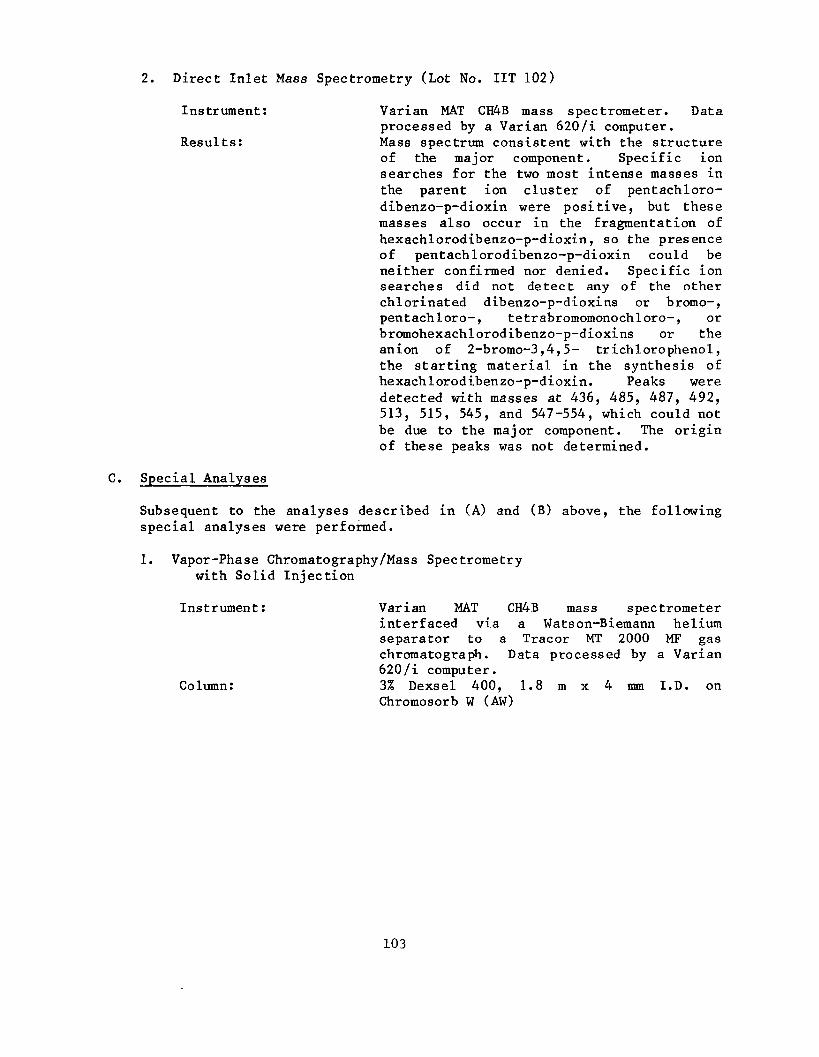
2. Direct Inlet Mass Spectrometry (Lot No. IIT 102)
Instrument: Varian MAT CH4B mass spectrometer. Data processed by a Varian 620/i computer.
Results: Mass spectrum consistent with the structure of the major component. Specific ion searches for the two most intense masses in the parent ion cluster of pentachlorodibenzo-p-dioxin were positive, but these masses also occur in the fragmentation of hexachlorodibenzo-p-dioxin, so the presence of pentachlorodibenzo-p-dioxin could be neither confirmed nor denied. Specific ion searches did not detect any of the other chlorinated dibenzo-p-dioxins or bromo-, pentachloro-, tetrabromomonochloro-, or bromohexachlorodibenzo-p-dioxins or the anion of 2-bromo-3,4,5- trich1orophenol, the starting material in the synthesis of hexachlorodibenzo-p-dioxin. Peaks were detected with masses at 436, 485, 487, 492, 513, 515, 545, and 547-554, which could not be due to the major component. The origin of these peaks was not determined.
C. Special Analyses
Subsequent to the analyses described in (A) and (B) above, the following special analyses were performed.
1. Vapor-Phase Chromatography/Mass Spectrometry with Solid Injection
Instrument: Varian MAT CH4B mass spectrometer interfaced via a Watson-Biemann helium separator to a Tracer MT 2000 MF gas chromatograph. Data processed by a Varian 620/i computer.
Column: 3% Dexsel 400, 1.8 m x 4 mm I.D. on Chromosorb W (AW)
103
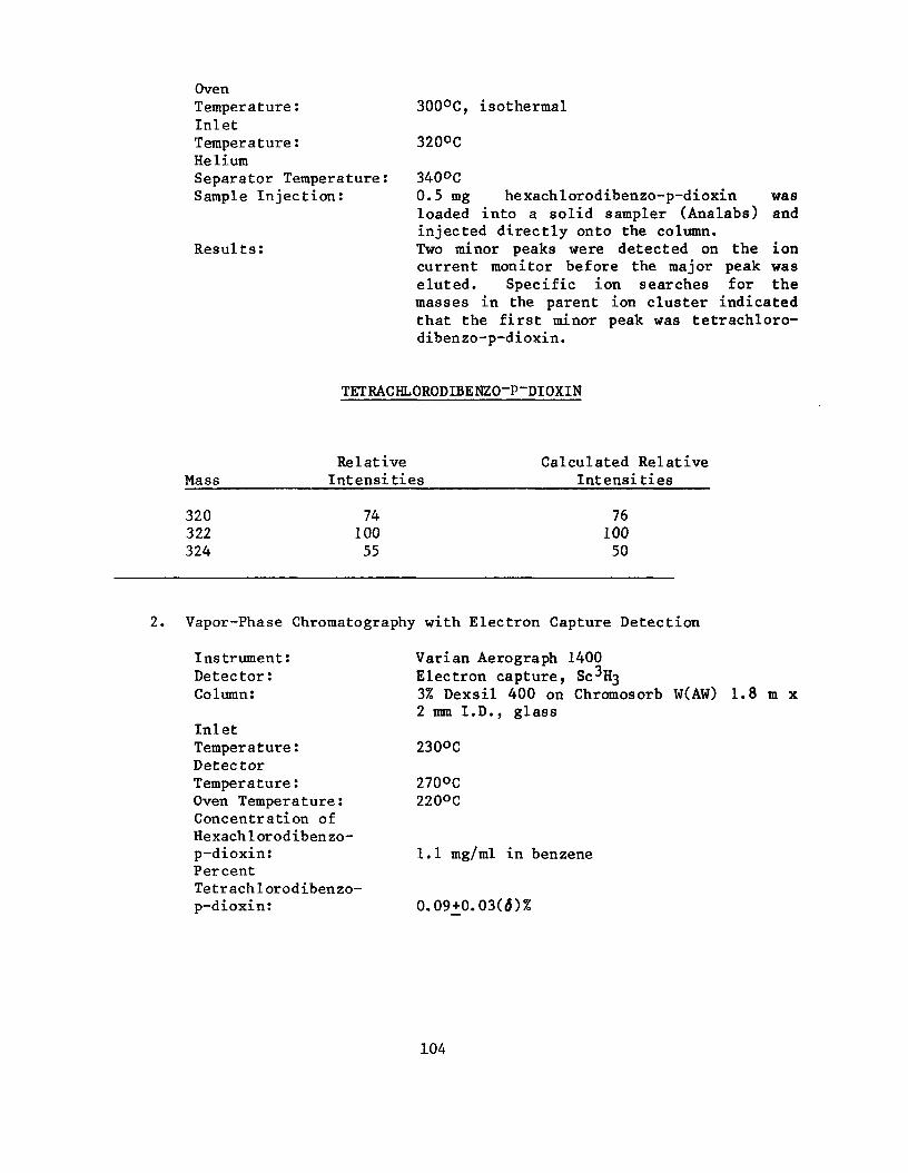
Oven Temperature: Inlet Temperature: Helium Separator Temperature: Sample Injection:
Results:
300°C, isothermal
320°C
340°C 0.5 mg hexachlorodibenzo-p-dioxin was loaded into a solid sampler (Analabs) and injected directly onto the column. Two minor peaks were detected on the ion current monitor before the major peak was eluted. Specific ion searches for the masses in the parent ion cluster indicated that the first minor peak was tetrachlorodibenzo-p-dioxin.
TETRACHLORODIBENZO-P-DIOXIN
Relative Calculated Relative Mass Intensities Intensities
320 74 76 322 100 100 324 55 50
2. Vapor-Phase Chromatography with Electron Capture Detection
Instrument: Detector: Column:
Inlet Temperature: Detector Temperature: Oven Temperature: Concentration of Hexachlorodibenzop-dioxin: Percent Tetrachlorodibenzop-dioxin:
Varian Aerograph 1400 Electron capture, 80- 3 3% Dexsil 400 on Chromosorb W(AW) 1.8 m x 2 mm I.D., glass
230°C
270°C 220°C
1.1 mg/ml in benzene
0.09+0.03(3)%
104

APPENDIX G
QUARTERLY ANALYSES OF HCDD STOCK SOLUTIONS
105

106

APPENDIX G
Quarterly Analyses of HCDD Stock Solutions
Stock solutions of HCDD in acetone were analyzed at the beginning and at
the end of each quarter by the IITRI Chemistry Division. The method of
analysis consisted of adding an internal standard (pentachlorodibenzo-p
dioxin, PCDD) to samples so that the internal standard concentration was
approximately the same as that of the sample being analyzed. The solution
containing sample and standard was then injected onto an electron capture/gas
chromatography system. The column was a 2 m x 1/8 in. Dexsil 300 with a
N_/CH, carrier - gas flow rate of 50 ml/min and an oven temperature of
275 C. Quantitation was achieved by manually measuring the area under the
resultant peaks with a planimeter and comparing with standard curves for the
internal standard and test compound. The standard curve was represented by
a third order polynomial equation fitting response to amounts.
The theoretical concentration for the stock solution was 2.5^<.g/ml. The
actual concentration as measured by the above method varied from 1.96 to
2.97 /ig/ml. The mean was 2.83Ju,g/ml and the coefficient of variation was
27%.
107

103
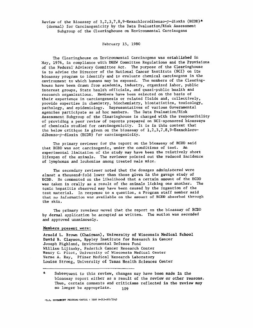
Review of the Bioassay of 1,2,3,7,8,9-Hexachlorodibenzo-p-dioxin (HCDD)* (dermal) for Carcinogenicity by the Data Evaluation/Risk Assessment
Subgroup of the Clearinghouse on Environmental Carcinogens
February 15, 1980
The Clearinghouse on Environmental Carcinogens was established in May, 1976, in compliance with DREW Committee Regulations and the Provisions of the Federal Advisory Committee Act. The purpose of the Clearinghouse is to advise the Director of the National Cancer Institute (NCI) on its bioassay program to identify and to evaluate chemical carcinogens in the environment to which humans.may be exposed. The members of the Clearinghouse have been drawn from academia, industry, organized labor, public interest groups, State health officials, and quasi-public health and research organizations. Members have been selected on the basis of their experience in carcinogenesis or related fields and, collectively, provide expertise in chemistry, biochemistry, biostatistics, toxicology, pathology, and epidemiology. Representatives of various Governmental agencies participate as ad hoc members. The Data Evaluation/Risk Assessment Subgroup of the Clearinghouse is charged with the responsibility of providing a peer review of reports prepared on NCI-sponsored bioassays of chemicals studied for carcinogenicity. It is in this context that the below critique is given on the bioassay of 1,2,3,7,8,9-Hexachlorodibenzo-p-dioxin (HCDD) for carcinogenicity.
The primary reviewer for the report on the bioassay of HCDD said that HCDD was not carcinogenic, under the conditions of test. An experimental limitation of the study may have been the relatively short lifespan of the animals. The reviewer pointed out the reduced incidence of lymphomas and leukemias among treated male mice.
The secondary reviewer noted that the dosages administered were almost a thousand-fold lower than those given in the gavage study of HCDD. He commented on the likelihood that a certain amount of the HCDD was taken in orally as a result of the animals licking one another. The toxic hepatitis observed may have been caused by the ingestion of the test material. In response to a question, a Program staff member said that no Information was available on the amount of HCDD absorbed through the skin.
The primary reveiwer moved that the report on the bioassay of HCDD by dermal application be accepted as written. The motion was seconded and approved unanimously.
Members present were:
Arnold L. Brown (Chairman), University of Wisconsin Medical School David B. Clayson, Eppley Institute for Research in Cancer Joseph Highland, Environmental Defense Fund William Lijinsky, Federick Cancer Research Center Henry C. Pitot, University of Wisconsin Medical Center Verne A. Ray, Pfizer Medical 'Research Laboratory Louise Strong, University of Texas Health Sciences Center
Subsequent to this review, changes may have been made in the bioassay report either as a result of the review or other reasons. Thus, certain comments and criticisms reflected in the review may no longer be appropriate.
•U.S. OOVEBHMEHT FEINTING OFFICE : 1980 0-311-201/3Ht5


NIH Publication No. 80-1758 August 1980