(bioarqueología) lewis (2006) the bioarchaeology of children
TRANSCRIPT


The Bioarchaeology of Children
This book is the first to be devoted entirely to the study of children’s
skeletons from archaeological and forensic contexts. It provides an exten-
sive review of the osteological methods and theoretical concepts of their
analysis. Non-adult skeletons provide a wealth of information on the
physical and social life of the child from their growth, diet and age at
death, to factors that expose them to trauma and disease at different
stages of their lives. This book covers non-adult skeletal preservation;
the assessment of age, sex and ancestry; growth and development; infant
and child mortality including infanticide; weaning ages and diseases of
dietary deficiency; skeletal pathology; personal identification; and expo-
sure to trauma from birth injuries, accidents and child abuse, providing new
insights for undergraduates and postgraduates in osteology, palaeopathol-
ogy and forensic anthropology.
M A R Y E. L EW I S is a lecturer at the University of Reading and has
taught palaeopathology and forensic anthropology to undergraduate and
postgraduate students for over 10 years. Mary is also an advisor to the
police and has served as a registered forensic anthropologist for the
Ministry of Defence.


Cambridge Studies in Biological and Evolutionary Anthropology
Series editors
H U M A N E C O L O G Y
C. G. Nicholas Mascie-Taylor, University of Cambridge
Michael A. Little, State University of New York, Binghamton
G E N E T I C S
Kenneth M. Weiss, Pennsylvania State University
H U M A N EV O L U T I O N
Robert A. Foley, University of Cambridge
Nina G. Jablonski, California Academy of Science
P R I M A T O L O G Y
Karen B. Strier, University of Wisconsin, Madison
Also available in the series
21 Bioarchaeology Clark S. Larsen 0 521 65834 9 (paperback)
22 Comparative Primate Socioecology P. C. Lee (ed.) 0 521 59336 0
23 Patterns of Human Growth, second edition Barry Bogin 0 521 56438 7 (paperback)
24 Migration and Colonisation in Human Microevolution Alan Fix 0 521 59206 2
25 Human Growth in the Past Robert D. Hoppa & Charles M. FitzGerald (eds.)
0 521 63153 X
26 Human Paleobiology Robert B. Eckhardt 0 521 45160 4
27 Mountain Gorillas Martha M. Robbins, Pascale Sicotte & Kelly J. Stewart (eds.)
0 521 76004 7
28 Evolution and Genetics of Latin American Populations Francisco M. Salzano &
Maria C. Bortolini 0 521 65275 8
29 Primates Face to Face Agustın Fuentes & Linda D. Wolfe (eds.)
0 521 79109 X
30 Human Biology of Pastoral Populations William R. Leonard & Michael H.
Crawford (eds.) 0 521 78016 0
31 Paleodemography Robert D. Hoppa & James W. Vaupel (eds.)
0 521 80063 3
32 Primate Dentition Daris R. Swindler 0 521 65289 8
33 The Primate Fossil Record Walter C. Hartwig (ed.) 0 521 66315 6
34 Gorilla Biology Andrea B. Taylor & Michele L. Goldsmith (eds.)
0 521 79281 9
35 Human Biologists in the Archives D. Ann Herring & Alan C. Swedlund (eds.)
0 521 80104 4

36 Human Senescence – Evolutionary and Biocultural Perspectives Douglas
E. Crews 0 521 57173 1
37 Patterns of Growth and Development in the Genus Homo. Jennifer L. Thompson,
Gail E. Krovitz & Andrew J. Nelson (eds.) 0 521 82272 6
38 Neandertals and Modern Humans – An Ecological and Evolutionary Perspective
Clive Finlayson 0 521 82087 1
39 Methods in Human Growth Research Roland C. Hauspie, Noel Cameron &
Luciano Molinari (eds.) 0 521 82050 2
40 Shaping Primate Evolution Fed Anapol, Rebella L. German & Nina G. Jablonski
(eds.) 0 521 81107 4
41 Macaque Societies – A Model for the Study of Social Organization Bernard
Thierry, Mewa Singh & Werner Kaumanns (eds.) 0 521 81847 8
42 Simulating Human Origins and Evolution Ken Wessen 0 521 84399 5
43 Bioarchaeology of Southeast Asia Marc Oxenham & Nancy Tayles (eds.)
0 521 82580 6
44 Seasonality in Primates Diane K. Brockman & Carel P. van Schaik 0 521 82069 3
45 Human Biology of Afro-Caribbean Populations Lorena Madrigal 0 521 81931 8
46 Primate and Human Evolution Susan Cachel 0 521 82942 9
47 The First Boat People Steve Webb 0 521 85656 6
48 Feeding Ecology in Apes and Other Primates Gottfried Hohmann, Martha
Robbins & Christophe Boesch (eds.) 0 521 85837 2
49 Measuring Stress in Humans: A Practical Guide for the Field Gillian Ice & Gary
James (eds.) 0 521 84479 7

The Bioarchaeology ofChildrenPerspectives from Biological and
Forensic Anthropology
Mary E. LewisUniversity of Reading

cambridge university pressCambridge, New York, Melbourne, Madrid, Cape Town, Singapore, São Paulo
Cambridge University PressThe Edinburgh Building, Cambridge cb2 2ru, UK
First published in print format
isbn-13 978-0-521-83602-9
isbn-13 978-0-511-25742-1
© Cambridge University Press 2007
2006
Information on this title: www.cambridge.org/9780521836029
This publication is in copyright. Subject to statutory exception and to the provision ofrelevant collective licensing agreements, no reproduction of any part may take placewithout the written permission of Cambridge University Press.
isbn-10 0-511-25742-2
isbn-10 0-521-83602-6
Cambridge University Press has no responsibility for the persistence or accuracy of urlsfor external or third-party internet websites referred to in this publication, and does notguarantee that any content on such websites is, or will remain, accurate or appropriate.
Published in the United States of America by Cambridge University Press, New York
www.cambridge.org
hardback
eBook (NetLibrary)
eBook (NetLibrary)
hardback

Contents
Acknowledgements page x
1 The bioarchaeology of children 1
1.1 Children in archaeology 1
1.2 A history of childhood 2
1.3 Children in biological anthropology 10
1.4 Children in forensic anthropology 13
1.5 Summary 19
2 Fragile bones and shallow graves 20
2.1 Introduction 20
2.2 Fragile bones? 23
2.3 Little people . . . little things . . . 26
2.4 The marginalised child? 30
2.5 Obstetric deaths 33
2.6 Summary 37
3 Age, sex and ancestry 38
3.1 Non-adult ageing 38
3.2 Sex determination 47
3.3 Ancestry 55
3.4 Summary 58
4 Growth and development 60
4.1 Introduction 60
4.2 Skeletal development and ossification 61
4.3 Prenatal and postnatal growth 62
4.4 Puberty and the growth spurt 64
4.5 Factors affecting growth 66
4.6 Growth studies: methods and concerns 68
4.7 Interpretations of past growth 74
4.8 Bone density 76
4.9 Estimations of stature 77
4.10 Summary 79
vii

viii Contents
5 Difficult births, precarious lives 81
5.1 Introduction 81
5.2 Infant mortality rates 81
5.3 Reconstructing child mortality 86
5.4 Infanticide 87
5.5 Summary 96
6 Little waifs: weaning and dietary stress 97
6.1 Introduction 97
6.2 Properties of human breastmilk 97
6.3 Weaning and infection 99
6.4 Ancient feeding practices 100
6.5 The osteological evidence 103
6.6 Weaning and bone chemistry analysis 115
6.7 Specific diseases of malnutrition 119
6.8 Summary 132
7 Non-adult skeletal pathology 133
7.1 Introduction 133
7.2 Non-specific infections 134
7.3 Endocranial lesions 141
7.4 Infantile cortical hyperostosis 143
7.5 Tuberculosis 146
7.6 Congenital syphilis 151
7.7 Skeletal pathology and personal identification 159
7.8 Summary 162
8 Trauma in the child 163
8.1 Introduction 163
8.2 Properties of paediatric trauma 163
8.3 Types of fracture 165
8.4 Birth trauma 168
8.5 Non-adult trauma in the archaeological
record 169
8.6 Physical child abuse 175
8.7 Clinical features of child abuse 176
8.8 Child abuse in the archaeological record 181
8.9 Summary 183
9 Future directions 184
9.1 Is absence of evidence evidence of absence? 185
9.2 Failure to adapt: children as non-survivors 186

Contents ix
9.3 Tom, Dick or Harriet? Sexing non-adults 187
9.4 The future 188
References 189
Index 248

Acknowledgements
Many friends and colleagues have provided advice and encouragement in the
preparation and writing of this book. In particular, Charlotte Roberts, Keith
Manchester and Jenny Wakely were instrumental in first introducing me to
human remains, and Charlotte Roberts has continued to provide invaluable
advice and support throughout my career. I am indebted to many who pro-
vided unpublished data, assistance, access to images and comments on drafts
of the text: Kristine Watts, Anthea Boylson, Louise Loe, Hella Eckardt, Louise
Humphrey, Roberta Gilchrist, Margaret Cox, Rebecca Gowland, Rebecca
Redfern, Gundula Muldner, Luis Rios, Bill White, Donald Ortner and Richard
Steckle. I also thank the team at Cambridge University Press, Tracey Sanderson,
Dominic Lewis, Emma Pearce and Anna Hodson.
I am thankful to all my friends and colleagues at the Department of
Archaeology in Reading for making it such a stimulating place to work and
who have allowed me the time and funds needed to finish this book. Finally, I
am grateful to my family for their continued support.
Figures 6.4, 8.4 and 8.5 are from the research slide collection of D. J. Ortner,
Department of Anthropology, Smithsonian Institution, Washington, DC, digi-
tised and made available through funds supporting National Science Foundation
grant SES-0138129 by R. H. Steckel, C. S. Larsen, P. W. Sculli and P. L. Walker
(2002), A History of Health in Europe from the Late Paleolithic Era to the
Present (Mimeo, Columbus, OH). These images are reprinted with their kind
permission. Figures 6.9–6.11, 7.1, 7.2 and 8.6 are of specimens from the Human
Remains Collection, Biological Anthropology Research Centre, Department of
Archaeological Sciences, University of Bradford. Many of these photographs
were taken by Jean Brown, to whom I am very grateful. Every effort has been
made to acknowledge copyright holders, but in a few cases this has not been
possible. Any omissions brought to my attention will be remedied in future
editions.
x

1 The bioarchaeology of children
1.1 Children in archaeology
This book reviews the current status of children’s skeletal remains in biological
and forensic anthropology. Child skeletons provide a wealth of information on
their physical and social life from their growth and development, diet and age
at death, to the social and economic factors that expose them to trauma and
disease at different stages of their brief lives. Cultural attitudes dictate where
and how infants and children are buried, when they assume their gender identity,
whether they are exposed to physical abuse, and at what age they are considered
adults. Similarly, children may enter the forensic record as the result of warfare,
neglect, abuse, murder, accident or suicide and the presence of young children
within a mass grave has powerful legal connotations. The death of a child under
suspicious circumstances is highly emotive and often creates intense media
coverage and public concern, making the recovery and identification of their
remains more pressing. In forensic anthropology, techniques used to provide a
biological and personal identification as well as the cause and manner of death
provide particular challenges.
The study of children and childhood in social archaeology emerged out of
gender theory in the 1990s, and has gradually increased in its sophistication,
moving children out of the realm of women’s work, to participating and active
agents in the past, with their own social identity, material culture and influence
on the physical environment around them. Children who were once invisible in
the archaeological record are slowly coming into view. The primary data for the
archaeology of childhood are the children themselves, and in order to progress
this new discipline, it is important to examine how bioarchaeologists derive the
data from which social interpretations are made, and the limitations that are
inherent in the methods and nature of immature skeletal material, including the
impact of the burial environment on their recovery.
Comparative studies of children from archaeological contexts have been com-
plicated by the eclectic use of terminology that both describes the skeleton as a
child and prescribes an age for the individual. For example, the use of the term
‘infant’ properly assigned to those under 1 year of age, has been used to describe
children aged up to 5 years, whereas ‘juvenile’ can be divided into ‘juvenile I’
1

2 The bioarchaeology of children
Table 1.1 Age terminology used in this volume
Term Period
Embryo First 8 weeks of intra-uterine life
Fetus From 8 weeks of intra-uterine life to birth
Stillbirth Infant born dead after 28 weeks gestation
Perinatal, perinate Around birth, from 24 weeks gestation to 7 postnatal days
Neonatal, neonate Birth to 27 postnatal days
Post-neonatal 28–346 postnatal days (1 year)
Infant Birth to 1 year
Non-adult ≤17 years
Child 1–14.6 years
Adolescent 14.6–17.0 years
Adult >17 years
or ‘juvenile II’ with a variety of ages assigned. One of the most popular terms
used by osteologists to describe children is ‘sub-adult’. This term is problem-
atic as it has been used to define a specific age category within the childhood
period. More fundamentally, sub-adult implies that the study of these remains
is somehow less important than that of the adults (i.e. sub = below). Through-
out this book children are described as ‘non-adults’ encompassing all children
recovered from the archaeological record up to the age of 17 years. Additional
terms divide this overarching category into critical physiological periods of the
child’s life (Table 1.1). These terms are used for ease of reference and provide
a biological basis for discussion; they are not intended to describe the complex
social experience of the youngest members of every society, past or present.
This book is divided into nine chapters, covering the development of child-
hood archaeology and the osteological study of non-adult remains; factors
affecting preservation; assessment of their age, sex and ancestry; growth and
development; infant and child mortality including infanticide; weaning ages
and diseases of dietary deficiency; skeletal pathology; and exposure to trauma
from birth injuries, accidents and child abuse. The final chapter considers some
future directions for the study of children in bioarchaeology. The following
sections explore the gradual development of childhood theory in archaeology
and the rise of research into non-adult skeletal remains in both biological and
forensic anthropology.
1.2 A history of childhood
Studies of the history of childhood began in 1960 when Philip Aries published
Centuries of Childhood: A Social History of Family Life. Aries argued that

A history of childhood 3
the ‘childhood’ we know today, which may perhaps be described as a period
of ‘cosseted dependency’ (Derevenski, 2000:4), did not exist until the early
modern period. Prior to this, parents were unsympathetic and detached from
their children, dressing them and expecting them to behave as miniature adults.
Such indifference was considered a coping mechanism to the constant threat
of infant mortality (Aries, 1960). In the past, we were led to believe, a child’s
upbringing was a combination of neglect and cruelty. Further debates in the
1970s developed the theme (De Mause, 1974; Shorter, 1976; Stone, 1977),
while later discourses began to challenge this traditional view (Attreed, 1983;
Hanawalt, 1986, 1993; Swanson, 1990; Shahar, 1992). Historians and social
archaeologists have now updated and revised our impressions of childhood. In
past societies, stages of life that correspond to childhood were recognised and
marked by social events or burial practices. Many parents loved their children,
sometimes to distraction. For example, Finucane (1997) concentrated on the
‘miracle’ texts of the medieval period which contained numerous tales of family
and village reactions to a child’s death or illness, with parents crippled by grief or
friends and relatives praying by a riverbank for the recovery of a drowned child.
Although important, these studies focussed on the attitude of adults towards
children, rather than viewing the past through a child’s eyes.
The study of children and childhood in archaeology emerged out of gender
theory in the 1990s (Derevenski, 1994, 1997; Moore and Scott, 1997). Previ-
ously, children had been considered ‘invisible’ in the archaeological record,
but a feminist reassessment of the past placed specific emphasis on gender and
age and with this, on the nature of childhood. Lillehammer (1989) was one
of the first to address the role of children in archaeology. She suggested that
through the use of burial, artefacts, ethnography and osteology we could gain
insight into the relationship the child had both with its physical environment
and the adult world. This was followed by an examination of documentary and
archaeological evidence for the child in the Anglo-Saxon and medieval peri-
ods (Coulon, 1994; Crawford, 1999; Orme, 2001), with Scott (1999) providing
a multicultural view on aspects of infancy and infanticide. Crawford (1991)
studied the Anglo-Saxon literature for clues as to when children were subject
to adult laws. Beausang (2000) expanded this theory of childhood to incorpo-
rate the concepts and practice of childbirth in the past, with the recognition of
birthing artefacts in the archaeological record. Although a promising start, these
studies have been criticised for maintaining the idea that children were passive
recipients in their communities, invariably linked to the activities of women
(Wilkie, 2000). Furthermore, the category of ‘child’ is often used in order to
investigate the construction of ‘adult’ (Derevenski, 2000). Neither approach
allows us to explore the role of the child as an independent agent in the past.
Wilkie (2000) went some way to redress this balance when she used evidence

4 The bioarchaeology of children
of the toy industry in the eighteenth and nineteenth centuries to illustrate how,
through their own material culture, children displayed their sense of identity
and defined their own distinctive social networks and liaisons.
1.2.1 Defining childhood
B O R E D O M !!! S H O O T I N G !!! S H E L L I N G !!! P E O P L E B E I N G
K I L L E D !!! D E S P A I R !!! H U N G E R !!! M I S E R Y !!! F E A R ! ! ! That’s my
life! The life of an innocent eleven-year-old schoolgirl!! . . . A child without
games, without friends, without sun, without birds, without nature, without
fruit, without chocolate or sweets . . . In short, a child without a childhood.
Extract from the diary of a child in the Sarajevo conflict, 1992; from Cunningham
(1995:1)
As this entry from the diary of a child in war-torn Sarajevo testifies, children
have an expectation of what childhood should be. No matter what period we are
examining, childhood is more than a biological age, but a series of social and
cultural events and experiences that make up a child’s life. Childhood can be
defined as a period of socialising and education, where children learn about their
society, gender roles and labour through play. The initial dependence on their
parents for nourishment and protection slowly diminishes as the child ages and
becomes an independent member of society. The time at which these transitions
take place varies from one culture to another, and has a bearing on the level of
interaction children have with their environment, their exposure to disease and
trauma, and their contribution to the economic status of their family and society.
The Western view of childhood, where children do not commit violence and
are asexual, has been challenged by studies of children that show them learning
to use weapons or being depicted in sexual poses (Derevenski, 2000; Meskell,
2000). What is clear is that we cannot simply transpose our view of childhood
directly onto the past.
Bogin (1997, 1998) takes an evolutionary approach to childhood theory.
Childhood is a period in the human life cycle not found in any other mammal,
and for Bogin this is defined as a period of time between the ages of 3 and 7
years, when ‘the youngster is weaned from nursing but still depends on older
people for feeding and protection’ (Bogin, 1997:64). The child is constrained
by its immature dentition, small digestive system and calorie-demanding brain,
which influence the type and amounts of food it can consume. ‘Juvenility’
occurs with the eruption of the permanent dentition, and when children are able
to procure and consume their own foods, as the brain and body growth diminish
to less than 50% of total energy needs, and they undergo a cognitive shift. This
period begins at the age of 7 and ends with the onset of puberty (c.10 years

A history of childhood 5
in girls, c.12 years in boys). Bogin (1998) asserts that in humans, childhood
performs several functions: an extended period for brain growth, time to acquire
technical skills, time for socialisation and an aid to adult reproduction. That is,
that the childhood period allows the mother to wean the child and produce other
offspring, by passing the energy expenditure of feeding and caring for the child
onto siblings and post-reproductive members of society, such as grandparents
(Key, 2000). This urge to care for the child is manipulated through the child’s
retention of its infantile appearance (large cranium, small face and body); that
is to say, children are ‘cute’. As the body and brain slow in their growth during
this period, they require less energy expenditure to feed but are protected during
times of hardship (Bogin, 1998). Many would object to this purely biological
view of childhood, as it ignores social theories of when a child becomes an
‘adult’ and a fully fledged member of a society, something that is culturally
defined. Hanawalt (2002) argues that in order for a child to survive, it must not
only be nursed, fed and kept warm (biological survival), but also be played with
and talked to (cultural survival).
1.2.2 Defining the child: biological versus cultural age
One of the resounding issues with the definition of a ‘child’ in archaeological
contexts is the use of physiological age to determine a social category (Gowland,
2001; Baxter, 2005). Physiological age is a biological reality, whereas ‘child’
is a culturally loaded term. The age at which an individual leaves the world of
dependency, learning and play, and takes on roles of work and social responsi-
bility is neither distinct nor universal. That there are three types of age category,
‘biological’, ‘chronological’ and ‘social’, is not denied, but in order to examine
the past life-course we need to have consistency in the raw data (the skeletal
remains), and use accurate osteological assessments of age and physiological
development as a marker from which to base our interpretations of the social
understanding of age in the past. Biological age is not irrelevant in the way
in which society treats a child. It affects children’s connection to their phys-
ical and social environment, from total dependency during infancy, to when
they begin to crawl, walk, talk and communicate with the adults and chil-
dren around them (Table 1.2). These abilities are physiologically determined
and they dictate how the child interacts. In particular, the misuse of the term
‘infant’ to refer to children between the ages of 1 and 3 years or 1 and 5 years
in studies that use skeletal evidence as their data misses this point. As an infant
(under 1 year), the child is particularly vulnerable to disease and death, and
its chances of survival significantly increase after the first year. Children who
die at around 2 years of age may be reflecting inadequate weaning methods or

6 The bioarchaeology of children
Table 1.2 Child development milestones from birth to 5 years
Birth to 8 months 8 months to 1.5 years 1.5 to 3 years 5 years
Lifts and holds up
head
Begins to crawl and
may stand aided by
furniture
Stands on one foot or
on tiptoe
Turns over unaided (7
months)
Can throw without
losing balance
Can run, skip, climb
and has a developed
sense of balance
Dresses and
undresses
Reaches towards
objects
Handles finger-foods
Uses spoons and cups
Imitates others
Becomes anxious
when separated from
loved ones
Understands people
and objects still exist
when they cannot be
seen
Smiling and gazing Shows affection by
kissing and hugging
Expresses pride,
pleasure,
embarrassment and
shame
Responds to name Listens to stories Tells stories
Explores environment Understands the future
and the past
Interacts with other
children
Social interaction
and role-playing
Gurgles and babbles to
communicate
Forms simple
sentences
Uses sentences to
communicate feelings
and needs
Asks questions
about the meaning
of words
Has no understanding
of ‘male’ and ‘female’
Understands ‘male’
and ‘female’ through
dress and over time,
but not changing
situations
Understands ‘male’
and ‘female’
through time and
situations: ‘gender
consistency’
Source: Collated from Berhrman et al. (1996) and Kohlberg (1966).
unsanitary conditions, and those that make it to 3 years are talking, playing and
actively mobile. By 5 years they are capable of contributing to the household
with minor chores. To categorise this most vital developmental period into one
age category, ‘infant’, will mask important physiological and, hence, social
advancements.
Derevenski (1997) refers to Kohlberg’s (1966) work on a child’s understand-
ing of gender roles. Before the age of 2, a child has no concept of male or
female but after 2 years of age, they begin to recognise males and females by

A history of childhood 7
visual prompts such as clothing. Between the ages of 3 and 4 years, a child’s
concept of gender becomes stable, and is understood through time. Hence, if
you are male when you are young, the child understands that you will be male
as an adult, but if a male begins to perform what the child perceives as female
roles, the male would become female. A stage of ‘gender consistency’ through
time and situation is not reached until the child is 5 (Table 1.2). Wiley and Pike
(1998) suggested the use of developmental stages rather than chronological
age to devise child mortality rates to take into account the activity of the child
(crawling, weaning, walking), which is often related to their cause of death
through exposure to disease and accidental injury. Although they propose this
method for use in modern communities where calendar age is rarely recorded,
the application of such developmental age categories into archaeological stud-
ies has the advantage of placing the child at the centre of the study by examining
the environment from their vantage point.
Although biological age categories provide data from which interpretations
are made, adult perceptions of the ability, maturity and responsibilities of chil-
dren at each age are culturally determined, and must be considered when trying
to ask questions about past child activity and health. In the later medieval
period, the ages of 8–12 years represented a time when children would begin
their apprenticeships (Cunningham, 1995), and children as young as 12 and
14 years could be married in ancient Egypt and Rome respectively, leaving
the realm of child for that of wife and mother. Childbirth is not a common
interpretation for the cause of death for older children within the burial record.
Today in the UK, children reach adulthood by degrees. At 16 they can legally
have sex, at 17 they can learn to drive, at 18 they can drink, get married and
vote, reflecting their status as full members of society. Crawford (1991) rightly
criticises archaeologists for their inconsistency in choosing the cut-off point
for children in archaeological reports, which vary from 15 years to 25 years in
some cases. These inconsistencies have a great impact on the way in which a
cemetery is interpreted. Moving an individual from one age category to another
can fundamentally change the profile of a cemetery when attempting to evaluate
the pattern of adult and non-adult burials, and to understand the significance of
their grave inclusions.
Attempts to define periods of transition in childhood have been carried out
by examining the burial of children and the engendered nature of their grave-
goods at certain ages. Gowland (2001, 2002) noted that at Romano-British
Lankhills in Hampshire, children were buried with gravegoods from the age
of 4 and the quantity of artefacts peaked between 8 and 12 years. Gowland
(2001) suggests that in these communities at least, age thresholds appear at
infancy (where perinates are interred outside of the cemetery area), at 4 years
and between 8 and 12 years where the quantity and wealth of gravegoods

8 The bioarchaeology of children
increases. Stoodley (2000) examined the presence of certain gravegoods within
burials from a large number of Anglo-Saxon graves in England. He noted that
‘masculine’ spears began to appear more frequently in male graves after the
age of 10–14 years, whereas ‘female’ beads and dress adornments appeared in
‘girl’s’ graves at between 10 and 12 years. This study suffers from a common
circular argument which stems from our inability to provide a biological sex for
non-adults, and a Westernised view of what is ‘masculine’ and what is ‘femi-
nine’. This circle was partially broken in Rega’s (1997) study of burials from
Bronze Age Mokrin in Yugoslavia, where children were sexed using canine
tooth-crown dimensions. Using these data, Rega revealed that all children were
provided with the same feminine engendered artefacts found in adult female
graves until around 17 years of age, when individuals sexed as male began
to be buried with artefacts associated with the male adult graves. Stoodley’s
(2000) age bracket in the Anglo-Saxon childhood life-course is supported by
Crawford’s (1991) analysis of contemporary records revealing that children as
young as 10 years could inherit property and be prosecuted under adult laws.
Kamp (2001) provides an excellent review of the development of childhood
studies and argues that the age categories employed by osteologists are often
selected and compared without reference to the society in which the children
lived. Biological or physical development and social markers of childhood are
not always related. This was demonstrated in Van Gennep’s (1960) The Rites
of Passage in which physical puberty did not always coincide with the rites of
passage that marked the adolescents’ entry into the adult world. Archaeological
evidence from the Anglo-Saxon period also attests to this, with male adult-type
gravegoods only appearing once an individual has reached 20–25 years (Stood-
ley, 2000), some 6 years after they would have reached puberty. While the
study of childhood has come some way in elucidating a particular section of
the human life-course, Gilchrist (2004) calls attention to the fact that other age
categories are still neglected, among them, what it was to be an adolescent in the
past.
1.2.3 Children in the archaeological record
Some artefacts have provided tangible links to children in the past. Footprints
(Roveland, 2000), death masks (Coulon, 1994), fingerprints on pots (Baart,
1990) and tooth marks in resin (Aveling, 1997) all prove that a child was there.
Wilkie’s (2000) discussion of toys that were designed, manufactured and sold
with children in mind forced historical archaeologists to acknowledge them as
actors in past society, but this concept has been slow to catch on in time periods
where the material evidence is not so rich. It may be that our association with
children and toys is based on Western ideals of what childhood should be, and

A history of childhood 9
Figure 1.1 Possible toys from the Ovcarovo ‘cult scene’. From Whittle (1996:94),
reproduced with kind permission from Cambridge University Press.
this has led some scholars to avoid toys as a route to the activities of children
(Derevenski, 1994). Nevertheless, humans learn through play, trial and error
and it is conceivable that small items or badly drawn or sculpted figures in
the archaeological record were used and created by children. Just as female
engendered space is now recognised in the past, it is time to start considering the
potential of identifying childhood spaces, where ‘women and children’ are no
longer seen as one entity and children are viewed as independent agents within
their own social space (Wilkie, 2000). Children have the imagination to make
toys out of sticks, stones and everyday household objects that will be invisible
in the archaeological record. In this way, children may influence the formation
processes of a site, perhaps by the movement of artefacts from their original site
of deposition (e.g. a midden), and the physical alteration of household objects.
A small pile of stones or an unusual collection of post-holes may indicate a child
was at play, and this possibility should be taken into account when interpreting
a site. Until recently, child activity in the archaeological record has been seen as
detracting from the real issues of adult behaviour (Bonnichsen, 1973; Hammond
and Hammond, 1981), rather than being viewed as informative of the child’s
interaction with its physical environment.
Possible toys have been recovered from various sites throughout Europe. Of
particular note are the small decorated clay figures, miniature furniture and tiny
bowls found at Ovcarovo, Bulgaria (Fig. 1.1), and the clay house and figurines
located in a house at Platia Magoula Zarkou, northern Greece, both finds dating
to the Neolithic (Whittle, 1996). Rossi (1993) identified two ivory dolls in the

10 The bioarchaeology of children
grave of a Roman child from Yverdon-les-Bains, Switzerland. Such items were
traditionally interpreted as ‘cult’ objects or foundation offerings, rather than
as a child’s playthings. On the other hand, the idea that all miniaturised items
represent toys is overly simplistic. Sillar (1994) noted that in the Andes, while
children will play with miniature pots, mimicking adult household practices
such as cooking and trade, such pots were also used by adults as donations at
shrines. In lithics analysis, small cores have been interpreted as being made by
children mimicking the adult knappers. Finlay (1997) suggests that inconsis-
tently made lithic artefacts may be the work of young apprentices, learning the
trade and that, as producers, children would make lithics in keeping with the
adult norms, rather than on a miniature scale. Bird and Bird (2000:462) argue
that differences between adult and child foraging patterns are not always about
the learning process, and that ‘children are not always practicing to be good
adults . . . but are predictably behaving in ways that efficiently solve immediate
fitness trade-offs’. If this pattern is predictable then we should be able to identify
it in the archaeological record. In particular, Bird and Bird (2000) examined
the different adult and child patterns of shellfishing in the Eastern Torres Strait
on the Great Barrier Reef. Due to their inexperience, children tended to collect
a wider variety of less valuable shellfish, which they proceeded to eat, leaving
them in small middens outside the settlement. Adults were able to exclusively
collect the most profitable and difficult-to-gather shellfish, avoiding the types
the children collected. In the archaeological record, two forms of shell midden
in different locations should be evident, with the more diverse and marginal
middens representing the foraging patterns of the children.
1.3 Children in biological anthropology
The study of children in biological anthropology has earlier beginnings than in
social archaeology, but they were no less focussed. Most studies were stimulated
by an interest in fertility levels, or the information that child survival could
provide on adult adaptation to their changing surroundings. These endeavours
were constantly being frustrated by the perceived notion that infant and child
remains could not survive the burial environment. It was only in the 1990s that
the study of non-adult skeletons began to concentrate on the information that
could be provided on the growth and health of the children themselves, providing
information on their activities and risk of infection or injury in contrasting
environments. Examination of the physical remains of children provides us
with the most direct and intimate evidence for them in the past. This section
outlines the development of the study of child skeletal remains in biological
anthropology and palaeopathology up until the present day.

Children in biological anthropology 11
Before the 1980s, studies of non-adult skeletal remains concentrated on
devising ageing and sexing methods based on medical studies (e.g. Schour and
Massler, 1941; Hunt and Gleiser, 1955). For example, Balthazard and Dervieux
(1921), Scammon and Calkins (1923) and later Olivier and Pineau (1960) pro-
vided data for fetal ageing using diaphyseal lengths, while Boucher (1955,
1957) assessed the use of the sciatic notch for sexing infant remains. In the
1960s studies on the physical growth of past populations began to emerge, and
would dominate research in non-adult remains for the next 40 years. The most
prolific researcher in this area at the time was Francis Johnston, who examined
the growth of children from Indian Knoll in Kentucky (Johnston and Snow,
1961; Johnston, 1962, 1968). Following Johnston’s example, by the late 1970s
the majority of studies that included child remains were focussed on diaphy-
seal length measurements to estimate growth attainment (Armelagos et al.,
1972; Y’Edynak, 1976; Merchant and Ubelaker, 1977). With the increasing
interest in palaeodemography, researchers began to assess the impact of under-
representation of child remains on life tables (Moore et al., 1975), but only a
few were interested in what these data could contribute to our understanding
of perinatal and child mortality (Brothwell, 1971; Henneberg, 1977; Mulinski,
1976). In palaeopathology, iron-deficiency anaemia as the underlying cause of
cranial porous lesions (porotic hyperostosis) was under increasing discussion,
with several studies examining its prevalence in non-adult crania (El-Najjar,
1977a; Lallo et al., 1977). However, the association of enamel hypoplasias and
Harris lines with childhood stress was indirectly determined using adult skeletal
and dental material (McHenry, 1968; McHenry and Schulz, 1976; Rose et al.,
1978). In 1978, Mensforth and colleagues heralded a way forward when they
examined the prevalence of anaemia and infection (i.e. porotic hyperostosis,
periostitis and endocranial lesions) in 452 infants and children from the Late
Woodland ossuary sample from the Libben site in Ottawa County, Ohio. For
the first time, the health of children in the past was the primary focus of study
(Mensforth et al., 1978). This research also demonstrated the importance of
healed and active lesions in determining the precise age at which children were
most at risk; the kind of detail not available when using adult evidence. In the
same year, Fazekas and Kosa (1978) published their detailed study of Hungar-
ian fetal skeletal remains, raising awareness of the number and morphology of
these tiny bones.
In 1980, Buikstra and Cook summed up child studies as being hindered
by poor preservation, lack of recovery and small sample sizes, despite, they
argued, many researchers becoming aware of their importance in determining
the overall success of a population (Buikstra and Cook, 1980). Instead, there
was a proliferation of papers on the lack of preservation of non-adult remains
compared to adults (Gordon and Buikstra, 1981; Von Endt and Ortner, 1984;

12 The bioarchaeology of children
Walker et al., 1988), an assumption that still prevails today. The prevalence of
stress indicators in children became more popular as researchers began to assess
the impact of agriculture, colonisation and urbanisation on child health (Blakey
and Armelagos, 1985; Storey, 1986, 1988). Jantz and Owsley (1984; Owsley and
Jantz, 1985) demonstrated changes in child health of the Arikara between AD
1600 and AD 1835 as a result of malnutrition and maternal stress brought about
by contact with European settlers. Goodman and Armelagos (1989) highlighted
the importance of children under 5 as the most sensitive members of society
to environmental and cultural insults, whose stress experience would impact
on the overall population’s ability to rally from disease in adulthood. Schultz
(1984, 1989) began to examine the health of non-adults from around the world,
employing histological analysis for evidence of scurvy and tuberculosis. In
Egypt, Brahin and Fleming (1982) reported on the health of child skeletal and
mummified remains, reporting the presence of tuberculosis, spina bifida and
osteogenesis imperfecta, while commenting on the lack of evidence for rickets.
Our inability to diagnose rickets in skeletal remains was about to come under
scrutiny (Stuart-Macadam, 1988).
By the 1990s, non-adults were becoming incorporated into biocultural stud-
ies of different populations (e.g. Stuart-Macadam, 1991; Grauer, 1993; Higgins,
1995; Ribot and Roberts, 1996). These studies were encouraged by the increase
in the non-adult material available. Children of known age and sex from
Christ Church Spitalfields and St Bride’s Church in London became accessible
(Molleson and Cox, 1993), and data began to be published on non-adults from
Wharram Percy (Mays, 1995) and St Thomas’ Church in Belleview, Ontario
(Saunders et al., 1993a; 1995). These samples encouraged a revival of meth-
ods to estimate the sex of non-adults (De Vito and Saunders, 1990; Mittler
and Sheridan, 1992; Schutkowski, 1993; Loth and Henneberg, 1996; Molleson
et al., 1998). Saunders (1992) carried out a review of non-adult growth studies,
outlining their advantages and limitations, particularly the issue of comparing
deceased children to living healthy modern populations (Saunders and Hoppa,
1993), while others began to highlight the potential and extent of pathological
evidence that could be derived from their study (Anderson and Carter, 1994,
1995; Lewis and Roberts, 1997). Nearly 10 years after Stuart-Macadam (1988)
had raised the issue, Ortner and colleagues began to address the diagnosis of
rickets and scurvy (Ortner and Ericksen, 1997; Ortner and Mays, 1998; Ortner
et al., 1999), while others identified sickle-cell anaemia, juvenile rheumatoid
arthritis (Still’s disease) and leprosy (Hershkovitz et al., 1997; Rothschild et al.,
1997; Lewis, 1998). By the end of the decade, dental microstructure was being
used to refine ageing techniques (Huda and Bowman, 1995) and stable iso-
tope analyses to address the age of weaning in contrasting past populations
throughout the world were beginning to dominate the literature (Katzenberg

Children in forensic anthropology 13
and Pfeiffer, 1995; Katzenberg et al., 1996; Schurr, 1997; Wright and Schwartz,
1997; Herring et al., 1998; Wright, 1998).
Today, studies of infant and child skeletal remains are receiving much more
attention. The publication of texts on non-adult osteology has increased the
number of researchers familiar with their identification and anatomy (Scheuer
and Black, 2000, 2004; Baker et al., 2005). Children are routinely included in
wide-ranging studies of health in the past (Steckel and Rose, 2002; Cook and
Powell, 2005), while the analysis of children themselves from sites all over
the world continues (Baker and Wright, 1999; Buckley, 2000; Lewis, 2002a;
Bennike et al., 2005; Blom et al., 2005). As we refine our ageing techniques
and statistical methods (Gowland and Chamberlain, 2002; Tocheri and Molto,
2002; Fitzgerald and Saunders, 2005) our understanding of the importance of
childhood diseases and their diagnosis is becoming more advanced and widely
publicised (Glencross and Stuart-Macadam, 2000; Ortner et al., 2001; Santos
and Roberts, 2001; Lewis, 2002b, 2004; Piontek and Kozlowski, 2002). New
understanding of trauma in the child has meant we can now reassess the evidence
for physical abuse and occupational injury, to gain a fuller understanding of the
child’s life experience in past society.
In biological anthropology, we still wrestle with the issue of children in the
archaeological sample representing the ‘non-survivors’ from any given popula-
tion. Their pattern of growth or frequency of lesions might not reflect that of the
children that went on to survive into adulthood (Wood et al., 1992; Saunders
and Hoppa, 1993). The early death of these individuals provides other chal-
lenges in the study of non-adult palaeopathology. Chronic diseases need time
to develop on the skeleton, but the children that enter the archaeological record
have usually died in the acute stages of disease before the skeleton has had time
to respond (Lewis, 2000). At the present time, studies that concentrate on non-
adult material are hindered by the inability to make reliable sex estimations, due
to absence of the secondary sexual characteristics evident on the adult skull and
pelvis. Although sexual dimorphism has been identified in utero, there is still
a disagreement about the validity of identifying morphological traits indicative
of sex in the non-adult skeleton (Saunders, 2000). However, the application
of ancient DNA analysis in determining the sex of non-adult skeletal material
holds promise for the future.
1.4 Children in forensic anthropology
Forensic anthropology is the application of biological anthropological tech-
niques to the study and identification of skeletal remains recovered from a crime
scene. Forensic anthropologists frequently work in conjunction with forensic

14 The bioarchaeology of children
pathologists and odontologists to suggest the age, sex, ancestry, stature (biolog-
ical identification) and unique features (personal identification) of the deceased
individual from the skeleton. Forensic anthropologists also contribute to the
understanding of skeletal trauma to aid in the determination of the cause and
manner of death. The data collected from the analysis constitute evidence to
be presented in a court of law. Children may enter the forensic record through
warfare (e.g. as child soldiers), abuse, murder, accident, suicide or neglect, but
the presence of young children within a mass grave has powerful legal connota-
tions and is highly emotive. The death of a child under suspicious circumstances
creates intense media coverage and public concern, making the recovery and
identification of the remains more pressing and objectivity more difficult to
maintain (Lewis and Rutty, 2003).
The biological and personal identification of children’s remains in forensic
anthropology is hindered by the paucity of techniques usually employed to pro-
vide such identification in adults (Kerley, 1976). Features that denote ancestry
and sex usually develop after puberty, when hormone levels increase and sex-
ual dimorphism becomes more apparent in the skull and pelvis. For example,
racial differences of the mid-facial projection and the appearance of the nasal
root develop during puberty, as do brow ridges used in sexing the skull. Estima-
tions of ancestry and sex are crucial to provide an accurate assessment of both
age and stature in skeletal remains, as they have an effect on the rate of growth
and development (see Chapter 3). An estimation of the minimum number of
individuals (MNI) is often easier to obtain in children, as sizes vary with age
and between individuals. However, in some cases children of similar age may
be recovered and size may not be a useful distinguishing feature, especially
where the epiphyses and developing dentition are concerned. Young children
seldom visit the dentist, or have major surgery, and their abuse or neglect can
hamper the one technique in which anthropologists are most confident when
examining child remains: age estimation.
Probably one of the greatest limiting factors in the development of stan-
dards for child identification stems from the lack of modern non-adult skeletal
collections. Parents rarely choose to donate their children’s bodies to medical
science, a situation not aided by events in England in 1999 (Burton and Wells,
2002). The case of the Royal Liverpool Children’s Hospital (Alder Hey) drew
out parental feelings towards the remains of their children when it was discov-
ered that pathologists had been ‘systematically stripping dead children of their
organs at autopsy and storing them, ostensibly for research purposes’ (Carvel,
2002:55). Large collections of modern infant and child skeletal remains of
known age, sex, ancestry and cause of death are rare, although some collections
of paediatric skulls exist (Shapiro and Richtsmeier, 1997).

Children in forensic anthropology 15
1.4.1 The child and the law
In 1998, the number of missing children in the UK was reported at 80 000 by
the International Centre for Missing and Exploited Children (ICMEC). Cases
of child murder in the USA have risen by 50% in the last 30 years and between
40 and 150 forensic cases involving children are handled annually (Morton and
Lord, 2002). In England and Wales, children less than 1 year of age are most at
risk of homicide (82 offences per million), compared to the overall risk of 15
per million in the total population (Intelligence and Security Council, 2000).
Humanitarian investigations, such as those carried out by the Argentine Forensic
Anthropology Team (EAAF) have recovered children’s remains from mass
graves in Guatemala and Argentina. An investigation of the Dos Rs massacre,
in El Peten (1982), revealed that 47.3% of the listed victims were children,
with the youngest victim only a few days old. During the excavation of a mass
grave in Kibuye in Rwanda 202 (44%) of the 460 bodies recovered were of
children (17% under 5 years; 14% 5–10 years; 13% 10–15 years) most showing
evidence for blunt force trauma. However, the figures are considered to be an
underestimate as decomposition and disarticulation of the remains meant that
many smaller bones could have been missed (Schmitt, 1998). Identification of
victims using DNA analysis is futile if no family members survive (EAAF,
1995). It is against the International Labour Organisation (ILO) Convention of
1982 for children under the age of 18 years to undergo compulsory recruitment
in state armies. Nevertheless, voluntary recruitment is legal for those of 15 years
and over under the Optional Protocol to the Convention on the Rights of the
Child on the Involvement of Children in Armed Conflict, which came into force
in 2002 (Harvey, 2003). Despite these rulings, in some conflicts child soldiers
are as young as 8 (Dufka, 1999). The presence of children younger than 15
years in a mass grave argues against claims that these graves represent soldiers
killed during legitimate conflict.
Another way in which children may enter the forensic record is as the result
of suicide. Suicides of children and adolescents are rare, but when they occur,
they generally involve ‘fail-safe’ methods such as hanging, running in front
of a train or jumping from a height (Schmidt et al., 2002). Motives include a
break-up of a relationship, conflicts at school or in the home, mental illness or
chronically disturbed family structures. Most at risk are usually males between
10 and 15 years, with cases rising into late adolescence and young adulthood. In
Turkey, Agritmis and colleagues (2004) reported 43 cases of suicides in which
72% comprised females, perhaps as a result of the social status of females and
their early marriage. Psychology studies have shown that children do not have
a distinct perception of death until the age of 7 or 8 years, and do not develop

16 The bioarchaeology of children
sophisticated reasoning until 12–14 years; hence their contemplation of suicide
is less likely (Schmidt et al., 2002).
1.4.2 Mute witness: cases of child identification
Thankfully, forensic anthropology cases involving infants and children are rare
in non-conflict areas. One of the earliest cases of child identification in England
is a historical one. In 1965, the skeleton of Lady Anne Mowbray, daughter
of the fifth Duke of Norfolk was disinterred during a cemetery clearance in
Westminster. Anne died in 1481 at the age of 8 years and 11 months, and the
remains were examined to see if her identity could be confirmed (Fig. 1.2).
Warwick (1986) established the age, and attempted stature and sex estimations
based on the femora. The dental remains were examined by Rushton (1965)
who identified agenesis of the second permanent molars in the mandible and
maxilla. Agenesis, but of different teeth, was also identified in the dentition of
12-year-old Edward V, Anne’s blood relative and his brother, Anne’s husband,
10-year-old Richard Duke of York. Molleson (1987) re-examined two sets of
skeletal remains discovered in 1674 in the Tower of London that were thought to
represent Edward and Richard, who, legend has it, were murdered in the Tower
in 1484. Age and sex estimates were established using Hunt and Gleiser’s
method (1955) of correlating the stage of dental eruption with the fusion of the
hand and wrist bones. The original examination of the Princes’ remains in 1933
by Tanner and Wright (Bramwell and Byard, 1989) asserted that the remains
were related not only due to dental agenesis, but because both of the skeletons
had wormian bones of the same shape and in the same place on their lambdoidal
sutures. Debates are still raging about the identity of these remains, and further
forensic investigations using chemical analysis may help to finally solve the
mystery once and for all.
Perhaps the first modern forensic case in the UK was published in 1958 when
Imrie and Wyburn were presented with the fragmentary remains of an unknown
child recovered from a hillside in England in 1955. With no previous cases to
guide them, Imrie and Wyburn (1958) reconstructed the growth cartilage for
the long bones from plasticine to estimate stature, and the triradiate cartilage
of the pelvis to estimate sex, and managed to identify a likely individual. In
1967, the Oklahoma City abductions in the USA presented anthropologists
with a particular problem. Although two girls aged 5 and 6 years had disap-
peared and were presumed dead, only one set of remains were recovered. The
bones, a dress and hair were examined by Clyde Snow (Snow and Luke, 1970)
who attempted to identify which child had been found and to rule out that
the remains belonged to an entirely different child. After the Branch Davidian

Children in forensic anthropology 17
Figure 1.2 Remains of Anne Mowbray in her coffin. From Clark and White
(1998:69), reproduced with kind permission from the Museum of London.
Compound incident near Waco in Texas in 1993, the bodies of 17 children
were recovered from the ruins. Their preliminary identification was based on
age assessments from the dentition and long bone measurements, in addition
to their association with the adult remains (Owsley et al., 1995). In cases of
such commingling of non-adult bones, an assessment of the MNI requires spe-
cialist knowledge of their skeletal elements (Scheuer, 2002), for example being
able to determine the side of a pars lateralis (part of the occipital) to determine
duplication.
The recovery of Stephan Jennings was the first case in the UK to involve
both an archaeologist and anthropologist in his recovery and identification. His
remains were identified on the basis of his leather sandals and estimated age of
around 3 years (Hunter, 1996). Careful examination by the anthropologist also
revealed that the rib fractures were peri-mortem rather than post-mortem, and
not caused when the stone wall that covered his body collapsed (C. Roberts,
pers. comm.). One of the most infamous child murder cases in the UK was the
Moors Murders. In 1965, the partially preserved bodies of Lesley Anne Downey
and John Kilbride were recovered from the Moors and identified mainly on the
basis of their clothing. The later recovery of Pauline Reade in 1986 followed
the advent of DNA analysis; however, this was unsuccessful and again, her
body was identified from her clothing. The problems of running a long-term

18 The bioarchaeology of children
investigation were illustrated when the dental records for Reade (and Keith
Bennett who is still missing) were destroyed in the 1960s (G. Knupfer, pers.
comm.). Since this time, positive identification of children has begun to rely
more on DNA analysis (Yamamoto et al., 1998). One of the youngest chil-
dren to be identified was a 13-month-old male victim of the Titanic disaster
in 1912, who was disinterred from the grave of the ‘Unknown Child’ in 2001
from the Fairview Lawn Cemetery in Halifax, Nova Scotia. Due to the acidic
nature of the soil, only a few fragments of bone and three teeth were recovered,
but mitochondrial DNA (mtDNA) was extracted from the teeth and the child
was identified as Eino Viljami Panula. His grave has since become a shrine
(Titley et al., 2004). More recently, Katzenberg and colleagues (2005) identi-
fied the body of an infant left behind after the clearance of a historical cemetery
through the combination of ageing through enamel microstructure, sex deter-
mination using DNA and maternal relatedness employing mtDNA from a living
relative.
For adult remains, forensic odontology plays a leading role in confirming an
identity. The situation is more problematic for child cases. Very few children
are taken to see a dentist before the age of 5 years (O’Brien, 1994) and few
have dental radiographs taken before the age of 10 for safety reasons. Dental
radiographs are then kept for 11 years for adults, and for children they are held
for 11 years or until they are 25 years old, whichever is longer, as regulated
by the Consumer Protection Act 1987. Where dental charts do exist, the lim-
ited amount of dentistry experienced by young children means that there is a
greater chance that their charts will be identical, with teeth at the same stage
of development. Such was found to be the case at Aberfan (Knott, 1967) when
in 1966, 116 children of the same age were killed when a waste tip slid down
the mountainside into the mining village, engulfing their school. An alterna-
tive method for identification is smile comparisons. Using this technique, a
photograph in life of the individual smiling is overlaid on a photograph of the
deceased’s front teeth. The morphology and orientation of the teeth from the
image and deceased can then be directly compared (Perzigian and Jolly, 1984).
With recent advances in technology, digital home videos can be used to pro-
vide comparisons for children where dental records do not exist (Marks et al.,
1997). Dental age estimated from a smiling photograph was instrumental in
securing the conviction of one of England’s most notorious serial killers. The
case of Fred and Rosemary West began with the discovery of three bodies in
the garden of 25 Cromwell Street, Gloucester in 1994 (Knight and Whittaker,
1997). Charmaine West, the daughter of Rosemary, was suspected to have been
killed at their previous home at 25 Midland Road. The positive identifica-
tion of one of the skeletons as Charmaine proved problematic when the same
teeth, on opposite sides of the jaw, provided the forensic odontologist with

Summary 19
two different ages, a year apart. When a photograph of the child was finally
acquired, both a positive identification and the relative time of her death could
be ascertained by comparing the extent of dental eruption in the skeleton to that
of the photograph, which also had the date it was printed on the back (Knight
and Whittaker, 1997). During the period in which the photograph was taken
and Charmaine was killed, Fred had been in prison. Rosemary was convicted
of murder.
1.5 Summary
The health and survival of the children from any given community is believed
to represent the most demographically variable and sensitive index of biocul-
tural change. Patterns of infant and child mortality have been shown to have
a profound effect on the crude death rates of a population and, when coupled
with evidence of childhood morbidity, have become accepted as a measure of
population fitness. Roth (1992) described childhood as the most sensitive por-
tion of the human life cycle, but it has taken us a long time to fully realise this
potential. Only in the last decade have studies into the growth, development and
health of non-adults been used to provide insights into the impact of the physical
and cultural environment on the children themselves. The osteological study of
non-adults occurred independently and at a much earlier period than theoretical
concepts of childhood in social archaeology, but both have now reached a level
of sophistication that should encourage communication and integration of the
disciplines. Only then can we develop a fuller understanding of what it was like
to be a child in the past.

2 Fragile bones and shallow graves
[t]he bone of infants being soft, they probably decay sooner, which might be
the cause so few were found here.
Thomas Jefferson (1788) commenting on remains from a Virginian
earthen mound (cited in Milner et al. (2000:473))
2.1 Introduction
One of the most commonly perceived limitations in the study of children from
archaeological contexts is their poor, or total lack of, preservation. The absence
of neonatal remains, or ‘infant-sized pits’ that no longer contain their ‘tiny
occupants’ (Watts, 1989:377), has led to the belief that children rarely survive
the burial environment, and even ‘dissolve’ in the ground. This issue has resulted
in the widespread neglect of studies into infant and child skeletal remains,
whose numbers are often believed to be too small for statistical analysis and
meaningful research. However, large numbers of non-adult remains have been
recovered from cemetery sites, and continue to be housed in museums and
universities ready for study (Table 2.1). In the UK, for example, the largest
include the multiperiod site of St Peter’s Church, Barton-on-Humber (n =c.1000), Romano-British Poundbury Camp (n = 395) and St Mary’s Spital
in London, where so far, 1740 non-adults have been identified (White, pers.
comm.). In North America, sites such as Indian Knoll have yielded 420 non-
adults (Johnston, 1962). Collections of known age and sex also exist, most
notably the 66 non-adults from Coimbra in Portugal with a known cause of death
(Santos and Roberts, 2001). The oldest non-adult remains include a 2-year-old
Neanderthal child dating from over 100 000 years ago (Akazawa et al., 1995).
In fact, at some sites non-adult remains can be better preserved than their adult
counterparts (Katzenberg, 2000). Buckberry (2000) reviewed the proportion
of non-adult remains recovered from Anglo-Saxon cemeteries and noted that
where adult bone preservation was good, so were the remains of the children.
In these cases, the percentage of non-adult remains recovered from the site
also increased. Just like adults, the histological structure of infant bones can be
excellent given the right burial circumstances (Colson et al., 1997), and it has
been possible to extract bone proteins (Schmidt-Schultz and Schultz, 2003),
DNA and stable isotope signatures from their skeletons.
20

Introduction 21
Table 2.1 Archaeological sites with large numbers of non-adult skeletons
Period Site
Number of
non-adults Reference
Palaeolithic Taforalt, Hungary 104 Acsadi and Nemeskeri (1970)
Neolithic Isbister, Scotland, UK 156 Chesterman (1983)
Neolithic Quaterness, Scotland, UK 72 Chesterman (1979)
Bronze Age Lerna, Greece 140 Angel (1971)
Iron Age Danebury, Hampshire, UK 50 Hooper (1991)
Pre-Dynastic Naga-ed-Der, Egypt 123 Lythgoe (1965)
Dynastic Gebelan and Asuit, Egypt 166 Masali and Chiarelli (1969)
4800–3700 BC Carrier Mills, Illinois, USA 62 Bassett (1982)
3000 BC Indian Knoll, Kentucky, USA 420 Johnston and Snow (1961)
150 BC–AD 750 Teotihuacan, Mexico 111 Storey (1986)
100 BC–AD 400 Gibson-Klunk, Illinois, USA 191 Buikstra (1976)
Roman Intercisa and Brigetio, Hungary 107 Acsadi and Nemeskeri (1970)
Romano-British Poundbury Camp, Dorset, UK 374 Farwell and Molleson (1993)
Romano-British Lankhills, Hampshire, UK 118 Clarke (1977)
Romano-British Winchester, Hampshire, UK 144 Roberts and Manchester
(2005)
Romano-British Cannington, Somerset, UK 148 Brothwell et al. (2000)
Anglo-Saxon Raunds Furnells,
Northamptonshire UK
208 Powell (1996)
Anglo-Saxon Edix Hill, Cambridgeshire UK 48 Duhig (1998)
Anglo-Saxon Empingham II, Rutland, UK 62 Timby (1996)
Anglo-Saxon Great Chesterford, Essex, UK 83 Waldron (1988)
Anglo-Saxon The Hirsel, Scotland, UK 153 Anderson (1989)
9th century AD Sopronkohida, Hungary 72 Acsadi and Nemeskeri (1970)
AD 970–1200 K2 and Mapungubwe, South
Africa
85 Henneberg and Steyn (1994)
AD 1050–1150 Meinarti, Sudan 101 Swedlund and Armelagos
(1969, 1976)
AD 1300–1425 Arroyo Hondo, Mexico 67 Palkovich (1980)
13th century AD Kane, Illinois, USA 60 Milner (1982)
Later Medieval St Helen-on-the-Walls, York, UK 200 Lewis (2002c)
Later Medieval Wharram Percy, Yorkshire, UK 303 Lewis (2002c)
Later Medieval Westerhus, Sweden 225 Brothwell (1986–7)
Later Medieval Jewbury, York, UK 150 Lilley et al. (1994)
Later Medieval Fishergate, York, UK 90 Stroud and Kemp (1993)
Later Medieval Royal Mint, London, UK 178 Waldron (1993)
Later Medieval Trowbridge Castle, Wiltshire, UK 84 Graham and Davies (1993)
Later Medieval St Nicholas Shambles, York, UK 51 White (1988)
Post-Medieval Christ Church Spitalfields,
London, UK
186 Lewis (2002c)
Post-Medieval Bathford, Somerset, UK 63 Start and Kirk (1998)
AD 1500–1900 Ensay, Scotland, UK 200 Miles (1989)
AD 1750–1785 Larson, South Dakota, USA 441 Owsley and Bass (1979)
Multiperiod St Mary Spital, London, UK c.1740 W. White (pers. comm.)
Multiperiod St Oswald’s Priory, Gloucester,
UK
128 Rogers (1999)
AD 1800–1832 Leavenworth, South Dakota,
USA
211 Bass et al. (1971)

22 Fragile bones and shallow graves
In 1973, Weiss estimated that 30–70% of a modern population would die
before reaching 15 years of age, with the highest mortality levels in individuals
between 1 and 5 years, and the lowest between 10 and 15 years of age (Weiss,
1973). Schofield and Wrigley (1979) reported that in modern Western popula-
tions the mortality rates of people under 10 years was only 2.4%, compared to
34% in pre-industrialised countries. It is now common to cite this modern pre-
industrial figure as the norm for child mortality in archaeological populations
from many periods all over the world. That 30% of the sample should con-
tain non-adults has become the gold standard by which under-representation is
measured. In many cemetery samples, the proportion of non-adults frequently
fails to reach this target. Modern pre-industrial societies are not directly anal-
ogous with those in the past, and they are not untouched by the pressures of
industrialisation that surrounds them. Factors that will affect infant and child
survival, such as their weaning diet, exposure to pollution and disease, living
conditions and economic status, are often dictated by the wealthier industrial
countries. Therefore, it is open to debate whether 30% child mortality is the
correct minimum we should expect from an archaeological sample where all
the children are included.
Some authors have argued that the usually low numbers of infant remains
in our archaeological populations may actually reflect true mortality rates
(Brothwell, 1986–7; Panhuysen, 1999), but cemetery samples revealing only 12
individuals dying under 1 year of age, over a 500-year period, suggest that sig-
nificant under-representation does exist (Lewis, 2002c). In the rare cases where
burial records from an excavated sample survive, the proportion of infants
reported to have died and those excavated are usually at odds. For example,
the low number of infants from the Voegtly cemetery in Pittsburgh, where 311
infants were reported to have died but only 200 children under 5 years were
recovered, was suggested by Jones and Ubelaker (2001) to demonstrated tapho-
nomic bias in preservation. Conversely, Herring and co-workers (1994) found
a greater number of infants in the skeletal sample from Belleview, Ontario than
were listed in the burial records, perhaps as the result of poor record-keeping
and the surreptitious burial of illegitimate and unbaptised babies. Nevertheless,
the proportion of non-adults recovered from a cemetery is an important tool
when trying to understand the nature of funerary treatment of the youngest
members of society. Sellevold (1997) suggests that in the medieval period, dif-
ferent types of church would attract a different congregation, and this may be
reflected in the age and sex distribution of those buried at the site. For example,
she states that 15% of those buried at Hamar Cathedral in Norway were infants
and children, compared to 22% in the small parish church of Sola. In the UK,
the early medieval pre-Christian cemeteries consistently show a paucity of
young children. The early Anglo-Saxon cemetery at Sewerby, North Yorkshire

Fragile bones? 23
produced only 5% of children under 5 years old, with West Heslerton, also
North Yorkshire providing only 6% (Lucy, 1994). In contrast, the earlier Roman
cemetery of Owlesbury, Hampshire had 35% of children under 5 years, the
later Anglo-Saxon cemetery at Norwich produced 45% child burials, and 48%
of the individuals recovered from later medieval Winchester were children
(Daniell, 1997:124). Lucy (1994) argues that this suggests that in the early
Anglo-Saxon period children were buried elsewhere, and that poor preserva-
tion cannot account for the lack of infant graves in these sites when compared
to earlier and later cemeteries. In such cases, the under-representation of non-
adults is usually explained in terms of taphonomic, methodological and cultural
factors.
2.2 Fragile bones?
Taphonomy is the scientific study of the processes of burial and decomposition
that influence the preservation of remains, from the cause and manner of death,
post-mortem anthropogenic alterations and animal scavenging, to the chemical
and physical breakdown of tissues (i.e. diagenesis). Much of the work carried out
into soft-tissue decomposition, and the influence of the burial environment on
the survival of bone elements, has been carried out in North America on modern
remains. These studies have shown that a complex interaction exists between
many internal and external factors relating to the body and burial environment.
These will affect the rate and nature of soft- and hard-tissue decay (e.g. age and
sex, presence of disease, open wounds, soil pH, temperature and burial depth).
As each case is unique, it is impossible to make broad assumptions about the
sequence of events during body decomposition. Taphonomic processes on non-
adult remains have not been fully documented in forensic archaeology due to
the rarity of cases. However, a child’s body can become skeletonised in just 6
days compared to several weeks for adult remains. Children’s bodies become
disarticulated more easily and are more susceptible to scavenging and dispersal
as their small size makes it easier for animals to move them around (Morton
and Lord, 2002). Their crania are also more fragile and prone to post-mortem
damage (Haglund, 1997). As for burned remains, Holck (2001) has recorded
that fetal remains can be completely destroyed in a house fire in under 20
minutes.
In forensic anthropology, it is often important to understand the rate of soft-
tissue decay in order to estimate time since death, and to piece together a series
of decomposition events that may have influenced the pattern and distribution of
scattered remains. The soft-tissue composition of children’s bodies means that
they will decompose at a different rate to adults. Their bodies cool more rapidly

24 Fragile bones and shallow graves
due to a greater surface-area-to-weight ratio (Smith, 1955:18), and infants who
die before their first feed will decompose at a slower rate than a fed infant,
due to the lack of organisms in the stomach, intestines and lungs (Bell et al.,
1996). Human infants are born with both brown and white adipose tissue,
which serves to insulate and store energy during growth (Kuzawa, 1998). This
tissue composition means children are prone to mummification rather than
saponification (adipocere formation). All these factors can have an affect on
the time since death that may be estimated for a child, especially if the rate of
decomposition is derived using animal analogues with lower levels of brown
adipose tissue.
An unusual feature in non-adult remains is the purple–blue coloration often
observed in the deciduous teeth and developing permanent crowns post-mortem.
Pigmented teeth are associated with certain pathological conditions (e.g. fluoro-
sis and congenital heart disease), and so-called ‘pink teeth’ in forensic contexts
are indicative of trauma and asphyxiation, as blood is forced into the pulp
cavities with the subsequent build-up of iron (Dye et al., 1995). Detailed exam-
ination of these pigmented teeth from burial environments has so far shown the
cause to be diagenetic. Archaeological ‘pink teeth’ are a consequence of fun-
gal infiltration, and the purple–blue coloured teeth the result of porous dental
enamel and the uptake of iron, manganese and organic matter solutions from
the surrounding soil (Dye et al., 1995; Mansilla et al., 2003).
Once the body has been reduced to hard tissues, the pace of deterioration
slows with bones often maintaining their external appearance for many hun-
dreds of years, until eventually bacterial and plant activity work to undermine
their chemical and physical structure. Living bone is made up of water, an inor-
ganic or mineral component in the form of hydroxyapatite (calcium, phosphate
and carbonate), and an organic matrix (principally collagen). The proportion of
these elements is governed by the age of the individual and type of bone (i.e.
compact or cancellous) being considered (Nielsen-Marsh et al., 2000). As with
soft-tissue decomposition, no single factor determines the level of bone preser-
vation, and a complex relationship exists between a number of overlapping
variables, both intrinsic and extrinsic. Intrinsic factors include the chemistry,
size, shape, density, porosity and age of the bone, whereas extrinsic factors
such as groundwater chemistry, clothing, soil type, temperature, oxygen levels,
flora and fauna all play a role in diagenesis. In general, however, the decay of
bone follows three phases: the chemical deterioration of the organic component,
the chemical deterioration of the mineral component, and the microbiological
attack of the composite form of the bone (Collins et al., 2002).
During the decay process, the organic matrix is subject to slow hydroly-
sis of proteins to peptides and eventually amino acids, while the inorganic
component undergoes spontaneous rearrangement of the crystalline matrix,

Fragile bones? 25
weakening the protein–mineral content and leaving the bone susceptible to
dissolution (Henderson, 1984). Microbial attack also has a profound influence
on both the macroscopic and microscopic preservation of bone, increasing its
porosity and hence its susceptibility to groundwater dissolution (Hedges, 2002).
Although studies focussing on the differential rate of adult and non-adult bone
decomposition are rare, Robinson and colleagues (2003) have shown that imma-
ture faunal remains are more porous than their adult counterparts, and that this
factor makes them more vulnerable to decay. The high organic and low inor-
ganic content of children’s bones (Guy et al., 1997), in addition to their small
size, certainly means they may be more susceptible to taphonomic processes.
Gordon and Buikstra (1981:589) suggest that in high alkaline or acidic soils
‘. . . all or most of the infants and children may be systematically eliminated
from the mortuary sample by preservational bias’. Walker and colleagues (1988)
demonstrated this problem when examining the burial records of adults and chil-
dren against skeletal remains recovered from Mission La Purisima, California.
The burial records indicated that 32% (150 out of n = 470) of the people
buried within the cemetery were under 18 years old, when only 6% (28) of the
skeletons represented individuals of this age. Poor preservation of the remains
as a whole was attributed to the acidic sandy soil in which they were buried,
which allowed water to permeate through the bone, with subsequent soaking
and drying disintegrating the fragile ribs and spine. The less dense cancellous
bone (e.g. ends of the long bones, spine), covered with only a thin layer of
cortical bone, has a larger surface area allowing for the more rapid decay of the
collagen and mineral matrix as it is exposed to the groundwater (Von Endt and
Ortner, 1984; Nawrocki, 1995). While cancellous bone decays more rapidly
than cortical bone, the infant skeleton actually contains less trabecular bone
than adults and older children, with the radius being the densest bone (Trotter,
1971). Bone mineral content also varies during childhood, with the lowest lev-
els in the post-neonatal period, returning to the denser neonatal levels at the
end of the second year. Thereafter, the mineral level steadily increases until the
adult level is reached.
Ingvarsson-Sundstrom (2003) when analysing her non-adult samples from
Greece, found that the small compact bones of the neural arches, phalanges,
zygomatics and temporal bones survived and were recovered well, but that only
28.6% of the long bones were preserved well enough to be measured for growth
and age. In the Inuit sites of Hudson Bay (AD 1200), Merbs (1997) noted small
empty graves and that cranial and leg bones were best represented among the
infants. In older children the scapulae, arms and pelvic bones survived best.
Adolescents and adults showed a better, similar pattern of preservation. There-
fore, it is expected that the recovery of non-adult remains will vary depending
on the age group and burial environment, with the epiphyses being particularly

26 Fragile bones and shallow graves
susceptible to groundwater penetration, and fetal remains surviving well in most
burial conditions.
2.3 Little people . . . little things . . .
The size and number of skeletal elements that make up the non-adult skeleton
varies with age, but ranges from 156 recognisable bone elements at birth (Table
2.2), to 332 at around 6 years as the epiphyses appear and fuse (Table 2.3), com-
pared to 206 bones in the adult skeleton. The number and complex morphology
of the non-adult skeleton can make identification challenging. While excellent
texts relating to non-adult skeletal anatomy exist (Fazekas and Kosa, 1978;
Scheuer and Black, 2000, 2004; Baker et al., 2005), the recovery of the small
bones, epiphyses and developing teeth can be hindered by lack of expertise of
the excavator, and will have a great impact on the information that can be gained
from the remains. Due to their porosity, immature remains are more susceptible
to colour change in the soil, making them harder to recognise, and the epiphyses
may mimic small stones and can easily be missed by the untrained eye. The
burial matrix, such as sandy and clay soil or gravel and stone inclusions, can
also severely hinder the identification and successful recovery of infant bones.
Rarely are diagrams outlining the bones of the non-adult skeleton provided on
a field recording form, and these at least would aid the excavator in ensuring
that all the bony elements are accounted for (Fig. 2.1).
Dental crowns develop in large alveolar crypts in the mandible and max-
illa. Once the gum (gingiva) has decomposed, this often results in the loss
of fragile and tiny developing crowns during excavation, processing and stor-
age. The pre-wrapping of all fetal and infant mandibles in tissue paper directly
after excavation is recommended to help preserve these tooth germs (Spence,
1986, cited in Saunders, 2000). Another common problem with tiny remains
is that they are often incorrectly identified as non-human, particularly as bird
or rodent bones. Several researchers have recounted tales of early excavations
where fetal remains were found with animal bone material that the excava-
tors failed to recognise (Robbins, 1977; Wicker, 1998; Ingvarsson-Sundstrom,
2003). The late discovery of the cremated remains of a 34–36-week fetus,
originally thought to be animal bone, among those of the ‘rich Athenian lady’
at Athens changed the interpretation of the burial tomb. The ritualised burial
and wealthy gravegoods were reconsidered as reflecting the fact that she died
young during pregnancy or childbirth, rather than purely being a statement of
her economic status (Liston and Papadopoulos, 2004). For the long bones at
least, the fusion of epiphyses, the density and lighter staining of the cortex
in animal remains should help distinguish them from human. It is important

Little people . . . little things . . . 27
Table 2.2 Number and type of bone elements expected in a neonate
Element Specific element Number Element
Specific
element Number
Frontal 2 Sacrum Centrum 5
Parietal 2 Lateral
elements
6
Occipital Pars basilaris 1 Neural arch 10
Pars lateralis 2 Manubrium 1
Pars squama 1 Sternum 4
Temporal Pars squama 2 Ribs 24
Petrosa 2 Clavicle 2
Tympanic ring 2 (attached) Scapula 2
Maxilla 2 Humerus 2
Zygomatic 2 Radius 2
Mandible 2 Ulna 2
Sphenoid Greater wings 2 Carpals Capitate,
hamate
4a
Body 1 Metacarpals 10
Nasal 2 Phalanges Proximal 10
Ethmoid 2 Intermediate 8
Concha 2 Distal 10
Vomer 1 Pelvis Ilium 2
Lacrimal 2 Pubis 2
Hyoid 3a Ischium 2
Atlas Neural arch 2 Femur 2
Axis Dens 1 Tibia 2
Centrum 1 Fibula 2
Neural arch 2 Tarsals Talus 2
C3–C7 Centrum 5 Calcaneus 2
Neural arch 10 Metatarsals 10
T1–T12 Centrum 12 Phalanges Proximal 10
Neural arch 24 Intermediate 8
L1–L5 Centrum 5 Distal 10
Neural arch 10
Total 263
aElements may not always appear during this period.
to remember that fetal and juvenile animal bones with unfused epiphyses are
also recovered, and a working knowledge of non-adult skeletal morphology is
crucial.
It is a general rule that the deeper the grave the better preserved the body as
it is protected from fluctuations in temperature, scavengers and removal from
the original site of deposition (Henderson, 1984). Scull (1997) noted that the
infant and child burials at Watchfield cemetery in Oxfordshire were in shallow

28 Fragile bones and shallow graves
Table 2.3 Number and type of bone elements expected in a 6-year-old child
Element Specific element Number Element Specific element Number
Frontal 1 Clavicle Shaft 2
Parietal 2 Scapula Body 2
Occipital Pars squama 1 Coracoid epiphysis 2
Pars basilaris 1a Humerus Shaft 2
Temporal 2 Proximal epiphysis 2
Maxilla 1 Radius Shaft 2
Zygomatic 2 Proximal epiphysis 2
Mandible 1 Ulna Shaft 2
Sphenoid 1 Distal epiphysis 2
Nasal 2 Carpals All but pisiform 14
Ethmoid 1 Metacarpals Shafts (epiphyses) 10 (10)
Concha 2 Phalanges Shafts (epiphyses) 28 (36)
Vomer 1 Pelvis Ilium, pubis, ischium 6
Lacrimal 2 Femur Shaft 2
Hyoid 1 Proximal epiphysis 2
Atlas Neural arch 1 Distal epiphysis 2
Anterior arch 1a Tibia Shaft 2
Axis 1 Proximal epiphysis 2
C3–C7 5 Distal epiphysis 2
T1–T12 12 Patella 2
L1–L5 5 Fibula Shaft 2
Sacrum 5 Proximal epiphysis 2
Coccyx 2 Distal epiphysis 2
Manubrium 1
Sternum 5 Tarsals 14
Xiphoid 1a Metatarsals Shafts (epiphyses) 10 (18)
Ribs 24 Phalanges Shafts (epiphyses) 28 (36)
Total 232
aElements may not always appear during this period.
graves. The infant burials that were recovered were found within or at the base
of ploughsoil, and therefore were more susceptible to ploughing or machine
stripping before excavation. Ingvarsson-Sundstrom (2003) reported that the
most frequent type of grave for children in the Lower Town of Asine was pit
graves, which were overlooked in favour of the more obvious built graves in
earlier excavations. Although depths of children’s graves from archaeological
investigations are rarely reported, Murail and Girard (2000) showed that chil-
dren under 15 years of age were buried at 1.40 m compared to 1.56 m for
adults and older children in the fifth-century rural cemetery of Chantambre,
France. Yet, in a previous study of two medieval cemeteries in Maastricht, The
Netherlands, Panhuysen (1999) found no difference in the depths of graves

Little people . . . little things . . . 29
fa
aa
ca ce
e
ad
ca a
eaca a
b
fe
c
b
fb a
cacae
eaaa
aae
a
ba
e ebae
e
a b
aae
a
a baa
a ae a
a aa
(cca)
adb e
ccaa ea
e ae eb ae
caa
ac
() ()
Figure 2.1 Diagrammatic inventory of non-adult bones.

30 Fragile bones and shallow graves
between adults and non-adults. Sellevold (1997) noted that a larger number of
dispersed bones from Sola and Simonsborg in medieval Norway were of non-
adults, and that their graves were more prone to disturbance perhaps due to their
shallow nature. Crawford (1991) has suggested that shallow burials of children
indicate the low regard in which they were held; however, she later reasoned
that it may be more due to practicalities and less of a need to bury at depth,
reflecting that it is harder to dig a deep grave of small dimensions (Crawford,
1993). In fact, Sellevold (1997) noted that grave length was not related to adult
or non-adult burials and that there was great variation in the size of non-adult
graves from the age of 1 year.
Once excavated, the importance of the analysis of non-adult remains is not
always recognised. Robbins (1977:15) reported that, along with fragmentary
remains, infant bones were ‘not saved for analysis’ from the Carlson Annis
shellmound in Kentucky, USA. In Scandinavia, before the 1980s, it was the
practice of museums to preserve only the intact skulls and, as children’s skulls
are made up of several fragile bones, they were generally disposed of (Sell-
evold, 1997). Buckberry (2005) reported that nearly half of all the infant
remains from St Peter’s Church, Barton-on-Humber, Lincolnshire were located
in the disarticulated material, as were the majority of infants from Fillingham,
Lincolnshire. These remains were probably originally in shallow graves and
disturbed by subsequent graves and ploughing. As many post-excavation exam-
inations do not include disarticulated or unstratified bones due to lack of time
and money, numerous child skeletons are probably hidden and will remain
unstudied in the stores of archaeological units and museums.
2.4 The marginalised child?
If any child be deadborn [she was to] see it buried in such a secret place as
neither hog nor dog nor any other beast may come onto it [nor] suffer any
such child to be cast into the jaques [latrines] or any other inconvenient place.
Oath of eighteenth-century midwives (Lane, 2001:121)
The earliest evidence for the special treatment of children in death comes from
the extraordinary burials from the Upper Palaeolithic. Formicola and Buzhiova
(2004) reported on the double burial of two children aged 9–10 and 12–13
years old from Sunghir in Russia. The remains were found head to head, with
ivory spears and rich gravegoods, and covered in red ochre. Palaeopathological
examination of the remains suggests that both were suffering from congenital
abnormalities producing bowing of the femora. This burial adds weight to the
theory that in the Upper Palaeolithic, visibly disabled individuals were afforded

The marginalised child? 31
special burial rites, first implied by the excavation of the triple burial of the same
date from Dolnı Vestonice (Trinkaus et al., 2001). Red ochre appears again in a
Neolithic collective burial from Ertebølle in Zealand, The Netherlands, where
several adult males and females were buried with infants and children. One male,
the last burial to be placed in the grave, was described as ‘cradling’ the body of
an infant in his arms. All the remains were sprinkled with red ochre ‘seemingly
most thickly around the children’ (Whittle, 1996:198). Boric (2002) discusses
the numerous red-ochre-covered infant burials from Vlasac and Lepenski Vir
in Neolithic Serbia, and in particular, the burial of a female with red ochre
covering her pelvis which contained a fetus. The significance of red ochre
is thought to symbolise blood, female menstruation, ancestral blood ties and
bloodshed. Boric (2002) also argues that the smothering of infant burials with
red ochre from Lepenski Vir represents protection. Perhaps not yet considered
members of that society, infants would not have undergone the protective rites
of passage afforded to the adults and older children. The discovery of a late Iron
Age burial of a 2-year-old from Bath in England provides more evidence that in
some periods at least, children were afforded exceptional burial rites, perhaps
due to illness. The child was found under a limestone slab, next to an adult male
with signs of tuberculosis. The child had a systemic inflammatory condition that
would have been visible during life. Both burials were inhumations in a cemetery
full of cremated individuals, and both were situated in an area considered to be
a sanctuary (Lewis and Gowland, 2005).
Examples of children’s remains located outside the confines of communal
burial grounds are a common finding throughout the world, and during all time
periods. Infant remains have been found under the floors of the world’s earli-
est agricultural settlements in Natufian sites in the Near East (12 500–10 200
BP) (Scott, 1999), and their remains have been found within domestic spaces
from the Neolithic to the Roman period. These burials have been variously
interpreted as the result of infanticide, as symbolising domestic or gendered
space (Scott, 1990), as due to a lack of social ranking, as a result of economic
necessity or as sacrificial burials (Watts, 1989; Wicker, 1998). These marginal
burials were not universal. For example, carefully wrapped fetal remains have
been recovered within the communal cemetery of the Roman Dakhleh Oasis in
Egypt (Tocheri et al., 2001). In the Mesolithic, infant burials have been located
within communal cemeteries, with children’s graves at Vedbaek, Denmark asso-
ciated with red ochre and animal bones, and one infant in particular being laid
under a swan’s wing, with a broken flint blade (Thorpe, 1996). The small
number of child burials in many of these periods suggests that the vast major-
ity may have been deposited elsewhere (Scott, 1999). The distinct absence of
children under 1 year of age at Mokrin in former Yugoslavia indicates that
they were purposefully excluded and not yet considered part of the community

32 Fragile bones and shallow graves
(Rega, 1997). Child cemeteries have been identified at Yasmina in Carthage
(present-day Tunisia) (Norman, 2002) and Lugano in Italy (Soren and Soren,
1999) and in the historic periods of Ireland, children were buried in separate
and marginal cemeteries or ‘cıllinı’ (Finlay, 2000). Evidence for one such burial
ground has also been reported in Wales (Manwaring, 2002). In some commu-
nities there is evidence that children underwent a different type of burial. For
instance, Chamberlain (1997) reported a higher number of child burials in caves
(39%) compared to long barrows (25%) in the British Neolithic, a pattern also
reflected in southern Europe. Cauwe (2001) found children’s remains clus-
tered in the northern sector of a Mesolithic tomb in Abri des Autours, southern
Belgium, an open area without natural protection, whereas the majority of the
adult burials were located in the southern sector in a more protected area. There
was also some evidence for the deliberate removal of their skulls.
In the later Anglo-Saxon and medieval periods, when children are included
in the communal cemeteries, they are often found to cluster, but these clusters
are not universally defined or easy to predict. For example, at St Andrews
Fishergate in York, 76% of the under-5s were found in the western third of
the cemetery (Stroud and Kemp, 1993), while excavations at Raunds Furnells
(Northamptonshire), Jarrow (Tyneside), Whithorn Priory (Galloway, Scotland)
and Winchester (Hampshire) have revealed groups of infants along the east
side of the church, where perhaps it was thought ‘holy’ water dripping off the
eaves would baptise them (Crawford, 1993). This practice seemed to die out
after 1066, and later child burials usually appeared in either the west or eastern
ends of the cemetery (Daniell, 1997). In St Olav’s monastery in Trondheim,
Norway infants had a burial place reserved for them close to the choir, which if
left unexcavated, would have meant the loss of 45% of the infant remains. At
Hamar Cathedral, Norway 57% of the remains recovered from the south-west
of the cemetery were children compared to only 8% in the west (Sellevold,
1997). Similarly, at Taunton (Somerset) in England, 85% of the non-adults
were located at the western end of the cemetery, while only three children were
excavated from the east (Rogers, 1984).
At Deir el Medina in Egypt, the cemetery seemed to have been divided
into age-determined sections, with the lowest part of the slope reserved for
infants and perinates, and even placentas in bloody cloths (Bruyere, 1937,
cited in Meskell, 1994). The infants were buried in containers associated with
the household including amphorae, fish baskets, boxes and chests, with some
small children and adolescents buried in roughly made coffins (Meskell, 1994).
Ethnographic sources indicate that prehistoric Iroquoians of Southern Ontario
buried infants along pathways believing their souls would influence the fertility
of passing women. Excavations of Iroquoian village sites have been interpreted
as reflecting this belief, with large numbers of infants located under the central

Obstetric deaths 33
corridor of longhouses, perhaps acting as a ‘path’ on which infertile women
walked (Saunders, 2000). If the whole area of the cemetery is not excavated, as is
often the case in urban developments, this could lead to an under-representation
of the youngest members of the society. At St Helen-on-the-Walls in York, UK
the recovery of only 12 infants was considered the result of infant burials being
clustered at the north side which remained unexcavated (Dawes and Magilton,
1980). However, this hypothesis later proved ill-founded, and the infants appear
to have been afforded another, unknown location for their burial (Grauer, 1991).
In the later medieval period, Protestant and Catholic dogma influenced the
inclusion of infants within cemeteries. Newborns were considered to be cor-
rupted by the original sin of their conception, and unbaptised or stillborn infants
were not permitted burial in consecrated ground (Orme, 2001:24). However,
in 1398 a royal licence was given to enclose the Hereford Cathedral cemetery
to prevent ‘the secret burials of unbaptised infants’ (Daniell, 1997:127). This
practice seems to be substantiated by the discovery of 24 infants shallowly
buried in a haphazard manner in an extended part of the cathedral cemetery
(Shoesmith, 1980). Daniell (1997:128) suggests this is the nearest equivalent
to a child cemetery that exists in medieval England.
In seventeenth-century England, there is documentary evidence for the dis-
posal of illegitimate children or infanticide victims in gardens, bogs, under the
floorboards or hidden in boxes, with some women entering the churchyard at
night to bury their babies (Gowing, 1997). We know little of the fate of the
hundreds of infants who died in ‘baby farms’ (where abandoned infants were
taken in by the parish), or whose bodies were discovered in the streets. Who
claimed their bodies from the Coroner? And if they were afforded burials,
where were they buried? In 1862, 13 unregistered infants were found within
St Peter’s Churchyard in London, where the sexton was running a lucrative
business charging a shilling for the clandestine burial of ‘stillborn’ infants
(Behlmer, 1979). Moreover, in the eighteenth and early nineteenth centuries,
the practice of using wet-nurses meant that children from towns and cities who
died whilst being nursed in the countryside were often buried there rather than
being returned home (Fildes, 1988a). This type of infant migration should be
taken into account when attempting to assess infant mortality levels in urban
and rural samples of this date.
2.5 Obstetric deaths
Infant remains may be better recovered when they are present in deeper, more
distinct adult graves, in particular, with females who died before or during
childbirth. These tiny bones may be overlooked in hurried excavation when

34 Fragile bones and shallow graves
sieving is not possible. Today, obstetric death rates for women between the
ages of 15 and 44 years in the UK are reported at 58.4 per 100 000, with
deaths during pregnancy low when compared to those of women 43 days post-
partum (Ronsmans et al., 2002). Obstetric deaths can result from puerperal
fever, dystocic presentation, obstructed labour due to a malformed pelvis, breach
birth or an oversized fetus (e.g. as the result of diabetes), haemorrhage or
premature detachment of the placenta. In the past, such hazards and infections
would have been similar. Although deaths during delivery or due to a lack of
prenatal care, instruments or appropriate and qualified assistance may have been
higher in the past. In 1775, the London Bills of Mortality reported 188 deaths
of women in ‘childbed’ (Roberts and Cox, 2003:316) but this would certainly
have been an underestimate, if only a small one, as deaths of women with a
child still inside the womb were omitted from the records (Eccles, 1977).
The presence of female burials with fetal remains in the archaeological record
can provide evidence for these hidden deaths. At least 24 cases have been
reported from excavations around Britain (Roberts and Cox, 2003), with fur-
ther isolated cases reported throughout the world. Arriaza and colleagues (1988)
reported 18 cases (14%) of childbirth related deaths in Chilean mummies dating
from 1300 BC to AD 1400, with one woman showing a full-term fetus in breach
position, and another described, dramatically, as clutching her umbilical cord
as she struggled to deliver a second child. When soft tissue is not preserved,
recognition of such deaths can only be made through meticulous excavation,
where an intimate link between the female and infant burial needs to be demon-
strated. The position of the child’s head or leg bones directly within the pelvic
cavity (i.e. overlying the sacrum between the ischium and pubic symphyses),
would be evidence of this link. Some of the earliest potential evidence for death
during childbirth was presented by Smith and Wood-Jones (1910), who reported
two cases of Egyptian females with deformed pelvises, perhaps as the result of
rickets, and fetal heads in the pelvic cavities.
The careful excavation of such neonatal remains is essential to establish
whether the child was placed within the grave after birth, was contained within
the pelvic cavity (in utero) or had been expelled during decomposition (Smith,
1955:25). For example, at Castledyke, Barton-on-Humber, a female carrying
a 37-week fetus appears to have been placed with her right hand over her
extended abdomen (Fig. 2.2), as hand and fetal bones were found to the left of
the lumbar spine, and the skull of the fetus rested on the left ilium in the birth
position (Boylston et al., 1998). Post-mortem disturbance of a double grave at
Whitehawk Neolithic camp in Brighton (Sussex, UK) made it unclear whether
the infant recovered was associated with the female (Curwen, 1934). Although
fragments of the child’s skull were found adhering to the inside of the female
pelvis, the age of the infant suggested the child had been placed in the grave

Obstetric deaths 35
Figure 2.2 Possible coffin birth. Note the position of the tibia under the pubic
symphyses. From Chadwick Hawkes and Wells (1975:49), reproduced with kind
permission from Barry Cunliffe.
after birth. Ancient DNA analysis could be used to assess the relationship of
the mother to the child. This distinction is essential as the placement of a child
within an adult grave (not necessarily that of a parent) as the result of coinciding
deaths was part of the Anglican tradition (Roberts and Cox, 2003:255). A child
may have been placed on top of the chest or stomach, resulting in bones coming
to rest within the pelvic cavity during decomposition. Two possible examples of
this practice come from St Helen-on-the-Walls in York, where a 9-month-old
child lies outside the abdominal cavity, on the left side of the pelvis, in one
case; and in another, a woman appears to have had a child laid to rest on her
stomach (Dawes and Magilton, 1980). Gilchrist and Slone (2005:71) report on
the guidelines of an Augustinian canon, John Mirk, dating to the late fifteenth
century, which states that: ‘A woman that dies in childing shall not be buried in
church, but in the churchyard, so that child should first be taken out of her and
buried outwith the churchyard.’ Evidence from monastic cemeteries indicates
that the notion of impurity of parous women led to some children being left
within the womb. Tiny infant burials placed within the mother’s grave, for
example in a small coffin as at Hull Augustinian Friary (Gilchrist and Sloane,

36 Fragile bones and shallow graves
Figure 2.3 Female with fetal remains on the left side. It is not clear if the child was
placed on top of the female or comes from within the womb. From Drinkhall and
Foreman (1998:366), reproduced with kind permission from The Continuum
International Publishing Group.
2005), may have been the result of obstetric hazards after all, but the babies
were removed from the womb post-mortem.
Møller-Christensen (1982) described a so-called ‘coffin birth’ in the
Aebelholt monastery in medieval Denmark. A coffin birth is supposed to occur
when the decomposition of the womb results in the post-mortem expulsion
of the unborn fetus, usually 48–72 hours after the death of the mother, but
this phenomenon has yet to be reported in the clinical or forensic literature.
This event was suggested at Kingsworthy Park (Hampshire, UK) (Chadwick
Hawkes and Wells, 1975), where the infant’s feet were still located under the
pubic symphyses (Fig. 2.3). Two Arikara females with fetuses apparently in
utero were recovered from the Larson site, South Dakota, USA (Owsley and
Bradtmiller, 1983), and an additional female was buried with ‘twins’ aged 24
weeks. Malgosa and colleagues (2004) presented a case from Bronze Age Spain
(1500–1000 BC) of a female with a child in an abnormal position during birth,
with the right arm of the child extended and located under the pubic symphyses,
perhaps as the result of a transverse lie birth presentation. The authors suggest

Summary 37
that death was due to haemorrhage and exhaustion on the part of the mother. At
St Nicholas Shambles in London, a fetus was found face down and ‘wedged’
into the pelvis (Wells, 1978), and at St Clements, Visby in Gotland, Sweden, the
fetus and young adult female both showed multiple exostoses that, it is argued,
obstructed the birth canal and led to their deaths (Sjøvold et al., 1974). Wells
(1975a) has stated that a deformed pelvis was the only direct evidence of an
obstetric death in the archaeological record.
The discovery of a female with fetal remains has important implications for
the analysis of trace elements, where pregnant and lactating women are reported
to have lower concentrations of zinc and calcium, but a higher concentration of
strontium (Blakely, 1989; King, 2001; Markland, 2003), although diagenetic
factors are a cause for concern. Studies of nitrogen levels in the hair of pregnant
females also suggests a decrease in δ15N values at birth compared to maternal
pre-pregnancy values (Fuller et al., 2004), which may be reflected in archaeo-
logical human bone collagen. Extrapolating such evidence could allow us to
analyse the number of lactating females within a cemetery, refine our estimate
of maternal mortality, and aid in the identification of pregnant and lactating
women within the forensic context.
2.6 Summary
Arguments that the remains of infants and children will not survive the burial
environment are unfounded, and in large cemetery excavations, non-adult
remains can be found in sufficient numbers to allow in-depth research into
their morbidity and mortality to be carried out. Evidence from the taphonomic
literature suggests that, while children’s bodies may decompose more quickly,
their bones have the potential to survive well in the same conditions that allow
for the good preservation of adult skeletal remains. The paucity of non-adults
in a cemetery sample can be the result of many additional factors, including
the skill of the excavator, curatorial bias and cultural exclusion. Funerary treat-
ment of non-adults in the past does not follow any specific pattern and varies in
extremes from the carefully placed ritualistic graves of the Upper Palaeolithic
and the child cemeteries of Hellenistic Greece and Rome, to their unceremoni-
ous burial in rubbish pits in post-medieval England. Patterning of graves within
communal cemeteries, where children are clustered in specific sections of the
graveyard for practical or ritual reasons, will also limit their recovery if the
entire cemetery is not excavated.

3 Age, sex and ancestry
3.1 Non-adult ageing
In bioarchaeology, the age-at-death of a child is used to make inferences about
mortality rates, growth and development, morbidity, weaning ages, congenital
and environmental conditions and infanticide. In forensic contexts, assigning
an age to a living child of unknown identity may be necessary when the child
is suspected of a crime; when penal codes differentiate law and punishment for
children of different ages (Schmeling et al., 2001; Foti et al., 2003); or if the child
is a refugee of uncertain age. For the deceased child, age estimation is considered
to be the most accurate biological identifier that a forensic anthropologist can
provide.
Age estimation of non-adults is based on a physiological assessment of den-
tal or skeletal maturation, and relies on the accurate conversion of biological
into chronological age. Error in the accuracy of this conversion can be intro-
duced by random individual variation, the effects of the environment, disease,
secular changes and genetics (Demirjian, 1990; Saunders et al., 1993a). Most
importantly, the age of development of the dentition and skeleton are known to
differ between the sexes, a biological assessment that has yet to be carried out
successfully in non-adults.
3.1.1 Dental development
Dental development (mineralisation and eruption) is less affected by environ-
mental influences than skeletal growth and maturation (Acheson, 1959), and
mineralisation of the dentition is the preferred method for producing an age
estimate for non-adults. The deciduous teeth begin to mineralise in the jaw
around 15 weeks gestation, beginning with the maxillary central incisors and
continuing until all of the deciduous teeth are fully erupted, when the child is
around 3 years of age. Development of the permanent dentition covers a period
from birth to around 14 years of age, with the eruption time of the third molar
considered the most variable, but roughly occurring around the age of 17 years
(Scheuer and Black, 2000). Differences between the sexes are evident, with
38

Non-adult ageing 39
females being on average 1–6 months ahead in their overall dental develop-
ment than males. This variability is also evident in individual teeth, with the
canines considered the most sexually dimorphic, and females being as much
as 11 months ahead of males in their development (Demirjian and Levesque,
1980).
There is a wealth of literature on the development and eruption of the decid-
uous and permanent dentition in children. Standards exist for various mod-
ern and ancient populations (Table 3.1) which employ both radiographic and
macroscopic techniques. Dental development follows a typical pattern and can
be divided into convenient stages, beginning with cusp mineralisation of the
deciduous maxillary incisors, and normally ending in root apex closure of the
third molar. The timing and sequence of this development differs between pop-
ulations (Lovey, 1983; Harris and McKee, 1990; Tomkins, 1996; Holman and
Jones, 1998; Willems et al., 2001) and the calcification of the third molar in
particular has been shown to be highly variable (Fanning and Moorrees, 1969;
Kullman, 1995; Bolanos et al., 2003). Such discrepancies can cause problems
when trying to assign a dental age to an individual with a different sequence
pattern to the standard being used, resulting in a mixture of younger and older
ages based on different teeth within one jaw (Owsley and Jantz, 1983). This
variation has also been noted in hominids from the Upper Pleistocene who,
when compared to modern humans, have advanced calcification of the sec-
ond and third molars, and delay of the canines (Tomkins, 1996). It has been
suggested that this variability has a genetic basis, and that the larger jaws of
the non-adult hominids allows for earlier eruption of the molars (Bermudez de
Castro and Rosas, 2001).
One of the most popular and easy to use macroscopic methods for dental age
estimation based on the extent of mineralisation was provided by Schour and
Massler (1941). Later Ubelaker (1989) modified this scheme to include larger
standard errors that took into account data from his American Indian (Arikara)
sample. Some authors have questioned the reliability of this method, as the orig-
inal data was, in part, taken from an earlier study by Logan and Kronfield (1933)
based on 20 children with debilitating illnesses and cleft palate, which may
have affected dental development (Smith, 1991). Hence, radiographic standards
derived by Moorrees and colleagues (1963a, b) for deciduous and permanent
dentition are preferred, as they are based on a larger sample of 246 (decidu-
ous) and 99 (permanent) teeth of White American males and females. Data for
the permanent teeth were refined and tabulated by Smith (1991) reducing the
inter- and intra-observer error inherent in the complex diagrams produced by
Moorrees and colleagues. The method relies on observation of the deciduous
canine, first and second deciduous molars and six of the permanent mandibular
teeth. Bolanos and colleagues (2000) warn that age estimations based on dental
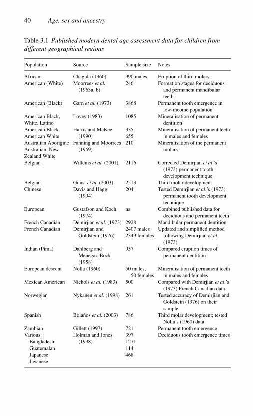
40 Age, sex and ancestry
Table 3.1 Published modern dental age assessment data for children from
different geographical regions
Population Source Sample size Notes
African Chagula (1960) 990 males Eruption of third molars
American (White) Moorrees et al.
(1963a, b)
246 Formation stages for deciduous
and permanent mandibular
teeth
American (Black) Garn et al. (1973) 3868 Permanent tooth emergence in
low-income population
American Black,
White, Latino
Lovey (1983) 1085 Mineralisation of permanent
dentition
American Black
American White
Harris and McKee
(1990)
335
655
Mineralisation of permanent teeth
in males and females
Australian Aborigine
Australian, New
Zealand White
Fanning and Moorrees
(1969)
210 Mineralisation of the permanent
molars
Belgian Willems et al. (2001) 2116 Corrected Demirjian et al.’s
(1973) permanent tooth
development technique
Belgian Gunst et al. (2003) 2513 Third molar development
Chinese Davis and Hagg
(1994)
204 Tested Demirjian et al.’s (1973)
permanent tooth development
technique
European Gustafson and Koch
(1974)
ns Combined published data for
deciduous and permanent teeth
French Canadian Demirjian et al. (1973) 2928 Mandibular permanent dentition
French Canadian Demirjian and
Goldstein (1976)
2407 males
2349 females
Updated and simplified method
following Demirjian et al.
(1973)
Indian (Pima) Dahlberg and
Menegaz-Bock
(1958)
957 Compared eruption times of
permanent dentition
European descent Nolla (1960) 50 males,
50 females
Mineralisation of permanent teeth
in males and females
Mexican American Nichols et al. (1983) 500 Compared with Demirjian et al.’s
(1973) French Canadian data
Norwegian Nykanen et al. (1998) 261 Tested accuracy of Demirjian and
Goldstein (1976) on their
sample
Spanish Bolanos et al. (2003) 786 Third molar development; tested
Nolla’s (1960) data
Zambian Gillett (1997) 721 Permanent tooth emergence
Various:
Bangladeshi
Guatemalan
Japanese
Javanese
Holman and Jones
(1998)
397
1271
114
468
Deciduous tooth emergence times

Non-adult ageing 41
calcification in modern children are most precise for those less than 10 years
of age, perhaps due to the greater number of stages to be scored and teeth that
can be observed. In addition, they suggest the most accurate ages can be gained
from the central maxillary incisors, the first mandibular molar and the canine
in boys, and the central maxillary incisors and the first and second mandibular
molars in girls.
Dental eruption begins as the cleft at the beginning of root formation miner-
alises and pushes the tooth up through the alveolar bone until it finally occludes
with the tooth above or below it (Gleiser and Hunt, 1955). Evidence of clini-
cal eruption, or the emergence of the tooth through the gum, is distinct from
the eruption through the alveolar bone seen in the hard tissues, which occurs
slightly earlier (Hillson, 1992), and this should be taken into account when
choosing ageing standards. Again, the timing and sequence of tooth emergence
has been shown to vary between populations and with disease (Dahlberg and
Menegaz-Bock, 1958; Niswander, 1965; Garn et al., 1973; Garn and Clark,
1976; Tomkins, 1996; Gillett, 1997), with caries, severe malnutrition or pre-
mature shedding of the deciduous teeth reported to delay the eruption of the
succeeding permanent dentition (Demirjian, 1990; Larsen, 1997). The third
molar, in particular, is highly variable in its time of eruption, and in some cases
may become impacted and not erupt at all. Tanguay and colleagues (1986) sug-
gested that standards of emergence for boys and girls would be more precise if
based on tooth length-for-age, rather than chronological age, as the length of the
tooth at time of emergence varies between the sexes. Liversidge and Molleson
(1999) produced such a method for measuring tooth length both macroscopi-
cally and from radiographs based on archaeological remains. When this stan-
dard was tested for accuracy on a modern sample, ages were underestimated
and accuracy decreased with age (Liversidge et al., 2003). Following these
studies, Muller-Bolla and colleagues (2003) introduced a simplified method of
dental age, using counts of erupted permanent teeth, based on 5848 modern (AD
2000–2001) White children from France. Caution is advised when choosing a
method of dental ageing for modern skeletonised remains, when developmental
and eruption timings were derived from archaeological samples of unknown
age, and vice versa.
For fetal remains, several dental development standards have been published
(Schour and Massler, 1941; Moorrees et al., 1963a; Kraus and Jordan, 1965;
Lunt and Law, 1974; Deutsch et al., 1985; and more recently Nystrom et al.,
2000; Nystrom and Ranta, 2003). The mineralisation of the deciduous dentition
is considered to be more robust to external influences such as socio-economic
conditions than the permanent teeth, although ancestral and sex differences still
exist (Liversidge et al., 2003). Dental emergence is not considered as reliable;
for example, Demirjian (1990) found that all the teeth, with the exception

42 Age, sex and ancestry
of the first deciduous molar, emerged 1 month earlier in boys than in girls,
and Holman and Yamaguchi (2004) have shown that poor and even moderate
nutritional stress can delay the emergence of the deciduous teeth.
Estimates of age based on dental wear in non-adult dentition is rarely
attempted, and as with adult cases, the degree of attrition is dependent on
the type of food eaten and the quality of the dental enamel, and is specific to
each sample. The evidence of wear in deciduous teeth indicates occlusion, and
implies the introduction of solid foods into the child’s diet, which may provide
a potential proxy for weaning ages in a sample (Skinner, 1997). Currently, data
on dental wear in past populations are only available for adults, with degrees
of attrition scored from the youngest to oldest (e.g. Miles, 1963; Scott, 1979;
Brothwell, 1981). Modification of these methods on a large non-adult group
has yet to be attempted (Saunders, 2000).
Few standards exist for the formation of deciduous crowns based on indi-
viduals of known gestational age (Saunders, 2000), and in fetal remains, the
developing crowns sit in large open crypts, which often result in the loss of tooth
germs during excavation and storage. In some situations, it may be necessary to
use macroscopic methods to assign an age using radiographic data, which can
produce errors as a higher stage of development is often scored when viewing
the tooth itself rather than its image on radiograph. Radiographs will show a
slightly later time for the formation of crowns and roots than data based on
dissected material, as the dental tissue needs a greater degree of calcification
before being visible on a radiograph (Hillson, 1992). For example, the first
permanent molar does not show up on a clinical radiograph until 6 months,
despite being calcified prenatally (Huda and Bowman, 1995). On a radiograph
of dry bone, teeth may rotate in the socket and obscure the true developmen-
tal stage of the tooth. Similarly, fragile mineralised material, for instance the
cleft at the start of root development, may be damaged post-mortem result-
ing in a lower score for the macroscopic assessment. One way to combat the
errors in interpreting chronological from biological age, and underestimates
that may be introduced by damaged teeth and visualisation on radiograph, is to
examine microscopic features. Microscopic ageing techniques are based on the
incremental markings within the dental microstructure. Unlike macroscopic or
radiographic ageing methods, the chronological age does not need to be extrap-
olated from a set of standards but can be assessed directly, reducing error as a
result of inter-population variability (Huda and Bowman, 1995).
Dental growth layers reflect a physiological rhythm and are represented by
incremental markers such as cross striations, striae of Retzius and periky-
mata. Cross striations along the length of the enamel prisms are thought to
result from a 24-hour variation in the enamel matrix secretion (Boyde, 1963),
whereas the coarser striae of Retzius (seen on the surface as perikymata) rep-
resent near-weekly (4–11 days) variation (Huda and Bowman, 1995). The first

Non-adult ageing 43
study on microscopic dental ageing was carried out by Asper (1916) on human
permanent canines, and was followed by that of Massler and Schour (1946), who
provided a technique for estimating age in days. The method devised by Boyde
(1963), originally conducted on two forensic and one archaeological case, was
employed by Huda and Bowman (1995) to distinguish more precisely between
commingled non-adult remains when macroscopic dental assessment and skele-
tal development proved inadequate. Smith and Avishai (2005) have advocated
the use of the neonatal line as a marker from which subsequent enamel lay-
ers can be used to provide a more exact age at death than those provided by
diaphyseal length or crown development. They warn that observer experience,
decomposition of partially mineralised enamel and variations in the plane of
the thin section can affect the accuracy of the technique.
3.1.2 Skeletal estimates of fetal age
When the dentition is unavailable, estimates of age on non-adult remains rely on
the development, growth and maturation of the skeleton. Establishing the age
of fetal remains is mainly carried out using diaphyseal lengths, and is critical
in many forensic cases as a means of assessing the viability of the fetus, and
to assist in cases of abortion legislation and infanticide. Errors in the relation-
ship between long-bone diaphyseal lengths and gestational age are well known
but difficult to control for, in both forensic and archaeological contexts. The
nutritional status of the mother during pregnancy, her weight and her height,
all affect the size and weight of the child at birth and hence the length of the
diaphyses (Hauspie et al., 1994; Adair, 2004). Additional stress factors such as
noise pollution and smoking will decrease size for age (Schell, 1981; Sobrian
et al., 1997). It has also been proposed that female fetuses mature earlier than
males in both leg length and weight (Lampl and Jeanty, 2003), potentially result-
ing in longer diaphyseal lengths and older age estimates in female-dominated
samples. The issue of shrinkage of the drying out diaphyses in fetal remains
was first raised by Rollell in the 1800s (cited in Ingalls, 1927) and may be an
additional source of error. Studies of wet to dry bone have shown up to 10%
shrinkage, and from wet to burnt bone up to 32% shrinkage in fetuses of 4 lunar
months, decreasing to 1% and 2% respectively in newborns (Huxley, 1998;
Huxley and Kosa, 1999). However, Warren (1999) reported no significant dif-
ferences in ages derived from radiographic (wet) diaphyseal lengths compared
to dry bone measurements.
Data for age estimates based on fetal diaphyseal lengths have been provided
in numerous studies in the past (Balthazard and Dervieux, 1921; Scammon and
Calkins, 1923; Olivier and Pineau, 1960; Fazekas and Kosa, 1978; Scheuer
et al., 1980; Kosa, 1989). For many years, ages based on data by Fazekas

44 Age, sex and ancestry
and Kosa (1978) were commonly employed. There have been concerns raised
about the validity of the data, which were based on 138 Hungarian perinates of
unknown age. A variety of data for ageing fetal remains from diaphyseal lengths
have most recently been published by Scheuer and Black (2000). Attempts are
also being made to provide updated radiographic data of modern fetal remains
using methods that can be applied to dry bone (Warren, 1999; Adaline et al.,
2001; Piercecchi-Marti et al., 2002), but caution is advised when attempting
to age fetuses with known or suspected congenital pathology (Sherwood et al.,
2000). Huxley and Angevine (1998) provide a conversion chart for the transfer
of ages reported in lunar months (anthropological sources) or gestational weeks
(clinical sources) to allow viability of the fetus to be more accurately assessed.
General assessments of age for perinates (whether pre- or postnatal) have
been devised based on the appearance, size and fusion of growth centres. These
include the temporal ring and plate (Scheuer and Black, 2000), the pars petrosa
(Way, 2003) and the suture mendosa and pars basilaris of the occipital (Scheuer
and Maclaughlin-Black, 1994). For example, the pars basilaris was originally
measured and described in detail by Redfield (1970) and later by Fazekas and
Kosa (1978). Measurements of the maximum length (ML), sagittal length (SL)
and maximum width (W) of the pars basilaris enabled a determination of the
age of the fetus or infant. In 1994, Scheuer and Maclaughlin-Black tested this
method on both archaeological and known-age individuals from nineteenth-
century England. They concluded that if the pars basilaris was longer than it
was wide the individual was less than 28 weeks in utero, and if the width was
greater than the length the individual was over 5 months of age. They suggested
that the maximum width and sagittal length were of the greatest value when
distinguishing between an early or late fetus, and the maximum width and max-
imum length were more accurate when identifying early or late infant material
(Scheuer and Maclaughlin-Black, 1994). Tocheri and Molto (2002) showed age
assessments based on the pars basilaris and those attained for femoral diaphy-
seal length and dental development agreed in 87% of cases in a sample from
Dakhleh. This bone is particularly useful in the age assessment of fetal remains
because its compact and robust structure means it is often recovered intact.
More recently, new data based on the dimensions of the fetal atlas and axis
(Castellana and Kosa, 2001), vertebral column (Kosa and Castellana, 2005)
and the mandibular ramus (Norris, 2002) have been provided in the forensic
literature.
3.1.3 Skeletal development and maturation
When the teeth are absent or apical closure is complete, age assessments of
older children and adolescents become based on diaphyseal lengths, and the

Non-adult ageing 45
appearance and fusion of the secondary growth centres (epiphyses) to the
diaphyses. Ubelaker (1989) studied the long-bone lengths of the Arikara Proto-
historic sample from Dakota, USA, which he initially aged using the dental
calcification chart devised by Moorrees and co-workers (1963a,b). The mea-
surements were then divided into 1-year age categories up to the age of 18.5
years. As the skeletal development of a Native American population is delayed
when compared to that of a White population, Ubelaker added a large standard
deviation (±2 years) and a range of variation. However, in some cases there
are no individuals to represent a particular age category (i.e. 8.5–9.5, 13.5–14.5
and 16.5–17.5 years) and other age categories are constructed using only one
individual. Hence, this method is considered most reliable for assigning an age
to those between birth and 2.5 years, where a greater amount of data exists.
Data for the diaphyseal lengths of older children are also available from mod-
ern studies (Maresh, 1955; Anderson et al., 1964; Hoffman, 1979; Sundick,
1978; Hunt and Hatch, 1981; Scheuer and Black, 2000). Hoffman (1979) and
Ubelaker (1974) argue that of all the long bones, the femoral diaphyseal length
is the most reliable indicator of age. Nevertheless, diaphyseal lengths are known
to be affected by external factors such as malnutrition and infection. For this
reason, these measures are normally employed to identify physiological stress
in an individual through growth profiles, which compare bone length to dental
age (see Chapter 4), rather than to provide chronological age. As the child ages
and the epiphyses begin to fuse to the diaphyses, long-bone measurements are
abandoned in favour of maturational assessments.
In the postnatal growth period, epiphyses begin to fuse when elongation of
the diaphyseal shaft occurs at the expense of the epiphyseal cartilage, until it
is completely replaced with bone. Fusion of the epiphyses begins between the
ages of 11 and 12 years at the elbow (distal humerus and proximal radius) and
ends with fusion at the knee (distal femur, proximal tibia and fibula) which is
generally complete at 17 years in females and 19 years in males. The nature
of epiphyseal closure was first outlined by Hasselwander (1902) and normally
begins with fusion at the central aspect of the growth plate, with union pro-
ceeding centrifugally until it is complete (Dvonch and Bunch, 1983). There
are several exceptions to this rule, with the proximal femur showing initial
fusion at the superior margin and continuing inferiorly, and the proximal tibia
first exhibiting union from the anterior–medial aspect to the posterior mar-
gin (Dvonch and Bunch, 1983). In addition, Albert and colleagues (Albert
and Greene, 1999; Albert et al., 2001) have suggested that environmental
stress results in asymmetrical epiphyseal fusion, and hence asymmetric long-
bone length, potentially providing two different age assessments in stressed
individuals.
As the last region of the growth plate to become completely ossified is usually
at the bone margins, in dry bone a groove may still be observed around the

46 Age, sex and ancestry
circumference (Roche, 1978). This is known as partial fusion. When the groove
has completely disappeared, complete fusion is recorded. The ossified margin
between the old epiphysis and diaphysis may still be visible on a radiograph for
a number of years after the macroscopic groove has disappeared. For example,
Cope (1946) reported the appearance of the fusion line on the distal femur
and proximal tibia in an individual over 70 years of age! Therefore, standards
based on macroscopic assessment may record complete fusion at an earlier age
than a radiographic study. Post-mortem detachment of a newly fused epiphysis
often occurs, and can be distinguished from non-union by subtle damage to the
metaphysis (usually centrally), which exposes the underlying trabecular bone,
and the lack of an undulated and billowing surface characteristic of an unfused
epiphyseal surface.
Data for establishing chronological age based on skeletal maturation appear
in many general osteological texts, and stem from skeletal and radiological
studies carried out in the early 1900s, set to redress the discrepancies evident
in anatomical texts of the day (Stevenson, 1924; Davies and Parsons, 1927;
Paterson, 1929; Todd, 1930; Flecker, 1932, 1942; Acheson, 1957). A variety
of ages can be assigned, depending on the bone or standard being used. Most
standards require the observer to score whether the growth plate is open, par-
tially fused or completely fused (e.g. Buikstra and Ubelaker, 1994), with the
sequence of fusion beginning at the elbow through to the hip, ankle, knee, wrist
and finally the shoulder. Once fusion of the long bones is complete, late-fusing
epiphyses, such as the basilar–occipital synchondrosis, are used for ageing older
individuals, but recent research on modern cadavers has indicated it is only reli-
able for females (Kahana et al., 2003), making this a difficult feature to use in
individuals of unknown sex. Other methods, such as the fusion of the thoracic
and lumbar vertebral rings in males and females, have helped to refine our age-
ing methods in adolescents (Albert and Maples, 1995; Albert, 1998; Albert and
McCallister, 2004). In forensic contexts, adolescent deaths may occur due to
teenage suicides or warfare, or the young people may be vulnerable if they are
living on the streets. In the latter case, they are likely to be malnourished or
neglected, resulting in a potential delay in their skeletal maturation. Neverthe-
less, useful biological markers in adolescence include the fusion of the hook
of the hamate, capping of the epiphysis of the third metacarpal and, in White
children, the completion of the canine root (but not apical closure). All of these
features are thought to signify the beginning of the growth spurt and puberty
(Chertkow, 1980).
In general, the maturation of girls is recognised to be 2 years in advance of
that of boys, and this advancement is increased in obese children (Mellits et al.,
1971). As with dental ageing, differences between populations are also evident.
For example, Pakistani and American Black children tend to mature earlier

Sex determination 47
than White Americans (Garn and Clark, 1976; Rikhasor et al., 1999). Although
socio-economic status has the greatest impact on maturation times, whatever
the ancestry (Schmeling et al., 2000), there are few studies available that exam-
ine epiphyseal fusion rates in large samples of children of different ancestry,
from good economic backgrounds, that would allow the assessment of the true
differences to be carried out. In forensic pathology, the most common clinical
ageing methods are based on the appearance of secondary ossification centres
of the hand and foot using the Greulich–Pyle Radiographic Atlas (Greulich and
Pyle, 1959) or Tanner Whitehouse System (Tanner et al., 1975). Unfortunately,
these techniques are not reliable for skeletonised remains where tiny epiphyses
are rarely recovered, although the method may be of use in partially decom-
posed remains or cadavers. Other skeletal indicators, such as the appearance of
ossification centres and fusion of the various segments of the spine, may aid
age estimation in cadavers of forensic cases when all the elements are known
to be present. Precise detail on the timing of appearance and fusion of various
elements in the developing skeleton can be obtained from either of the two
excellent texts devoted entirely to non-adult osteology by Scheuer and Black
(2000, 2004).
3.2 Sex determination
Sex is normally determined in the adult skeleton using morphological features
of the skull and pelvis, and measurement of the long bones. Sexual dimor-
phism exists due to the release of hormones during the development of the
fetus. If cells are not masculinised by the release of testosterone in the 10th
week of gestation, a fetus will develop along ovarian (female) lines (Saun-
ders, 2000). Unfortunately, the sexually dimorphic characteristics of the pelvis
and skull do not become apparent until puberty, meaning that assigning sex
to non-adult skeletons is more problematic. In addition, chemical differences
between male and female skeletal remains, such as different levels of cit-
rate or calcium, phosphorous and strontium, are only recognised in males and
females of reproductive age (Dennison, 1979; Beattie, 1982). However, some
morphological characteristics, such as the greater sciatic notch, are often so
different in males and females that researchers believe that they must already
be present in children.
In biological and forensic anthropology we determine biological sex. This
term is not to be confused with gender, a cultural construct that refers to the
social significance placed upon males and females in society (Mays and Cox,
2000). As with biological age and the concept of childhood, biological sex
is the reference point from which inferences about gender in past society can

48 Age, sex and ancestry
be made. Sex estimation is the ‘holy grail’ of non-adult osteology. As females
grow and mature faster than males, accurate sex estimation of non-adults would
allow for age estimates to be refined and the social treatment of males and
females at birth and during childhood to be examined. Sex identification of
fetuses would be used to answer questions about changing sex ratios and their
predisposing factors (e.g. parental investment and economic status (Koziel and
Ulijaszek, 2001) and the fragility of male fetuses). The proportion of male to
female children displaying various degrees of trauma and pathology could tell
us much about their preferential treatment and life-course, their susceptibility to
disease, handling in society through burial rites and grave inclusions, division
of labour, and at what age boys and girls became accepted as adult members
of a society. For example, in 1997 Rega assessed the relationship between
gravegoods, sex and gender in skeletons from a Bronze Age cemetery at Mokrin
in former Yugoslavia and assigned the sex of children using canine tooth crown
measurements, their body orientation and grave goods. Rega (1997) illustrated
that young females were afforded the same burial rites and gender identity as
the adult females, but the large proportion of infant remains sexed as female
fell outside the normal sex ratio of 1 : 1, suggesting that males were assigned
female gender, and that only after 17 years of age did male burials become
associated with male gendered knives.
Unfortunately, sex estimations from non-adult skeletons are notoriously dif-
ficult, making associated anthropological techniques unreliable, or forcing us
to add greater error ranges to our results in order to account for unknown sex.
Most anthropological techniques used to assign sex to non-adults focus on the
size and morphological traits of the dentition, cranium, mandible and pelvis.
When all cranial and postcranial traits are used, sex determination in adult
skeletons can provide an accuracy of 98–100% (Buikstra and Mielke, 1985),
and in general, 95% accuracy is expected from anthropological techniques used
in forensic cases. In children, most methods fail to yield an accuracy of 70%,
with the degree of overlap between male and female traits and problems with
reproducing accurate results on different populations continuing to make this a
challenging area of investigation (Loth and Henneberg, 2001; Scheuer, 2002).
3.2.1 Sexing from the dentition
Measurements of the deciduous dentition have shown that teeth of boys are
generally larger than those of girls (Black, 1978; De Vito and Saunders, 1990)
yielding an accuracy between 64% and 90%. Black (1978) developed dis-
criminant function analyses for deciduous dentitions resulting in an accuracy
of 75%, although he found the sexual dimorphism to be less marked in the

Sex determination 49
deciduous teeth than in the permanent dentition. In primates, the canines show
the greatest degree of sexual dimorphism, with up to a 50% difference in the
size of the mesiodistal crown diameter in a male compared to a female (Garn
et al., 1967). In humans, the canine shows its greatest difference between boys
and girls between the ages of 5 and 12 years (Bailit and Hunt, 1964), with
male canines over 4% larger than female canines (Keene, 1998). This degree of
sexual dimorphism varies between populations (Garn et al., 1967). Thompson
and colleagues (1975) reported that the further away the teeth were from the
canine, the less variability there was between males and females. Moss and
Moss-Salentijn (1977) suggest that this canine sexual dimorphism in crown
size is due to the thickness of the enamel, as amelogenesis continues for a
longer time in males than in females. However, the large degree of overlap
between males and females caused research into canine dimensions to wane.
In 1990, fresh attempts were made to assess the accuracy of sexing children
based on the dimensions of the deciduous teeth. De Vito and Saunders (1990)
achieved the highest reported accuracy (76–90%), although no tooth over any
other showed the most marked differences. The authors acknowledged that the
degree of sexual dimorphism in their Burlington (Vermont, USA) group was
particularly marked compared to studies of other North American, Australian
or Icelandic groups.
It is important to bear in mind the unknown genetic and environmental factors
that may affect the accuracy of sex estimates based on crown dimensions. For
example, mothers with diabetes and hyperthyroidism can produce offspring
with larger tooth dimensions, while maternal hypertension and low birth weight
will result in smaller dental crowns for both the deciduous and permanent teeth
(Garn et al., 1979). Further research into prematurity and crown dimensions
showed a less predictable pattern in the permanent teeth, with preterm White
males and Black females showing increased dimensions (Harila-Kaera et al.,
2001), while in the deciduous teeth, males have consistently larger teeth with
prematurity than females (Harila et al., 2003).
Hunt and Gleiser (1955) proposed a method of sexing non-adults using cor-
relations between dental and skeletal age, based on the principle that males
mature skeletally more slowly than females, while the rate of dental develop-
ment between the sexes is similar. That is to say that, in an unknown child, if the
ages attained independently for dental and skeletal age are closely correlated
using male standards, but divergent when compared to female standards, then
the child is male. This technique was based on the development of wrist bones
from clinical radiographs, but these tiny epiphyses are susceptible to tapho-
nomic processes and are often not recovered in skeletonised remains, although
the method may be of use in partially skeletonised individuals or cadavers.
Again, the children that enter the forensic record are often malnourished or

50 Age, sex and ancestry
neglected, resulting in a delay in their maturation, whatever their sex, in rela-
tion to their dental development (Eveleth and Tanner, 1990). Following on from
Hunt and Gleiser (1955), Bailit and Hunt (1964) tested whether the mandibu-
lar canine in particular was an effective estimator of sex when chronological
age was already known, as a canine belonging to a male should more closely
approximate known age based on male standards than the canine of a female.
This method resulted in accurate sex estimation in 70% of cases, compared to
only 58% accuracy when sexing the child based on Hunt and Gleiser’s (1955)
method. Hence, sex estimations based on the canine are more reliable when the
chronological age of the child is already known, a rare event in archaeological
or forensic specimens.
3.2.2 Sexing from skeletal morphology: skull
In more recent years, anthropologists have shifted their focus away from the den-
tition to the sexual dimorphism evident in the non-adult cranial and postcranial
skeleton. Such studies have been encouraged by the acquisition of archaeologi-
cal material including children of known age and sex. In 1993, Schutkowski
examined the extent of sexual dimorphism in the mandible and ilium of 37
boys and 24 girls aged between 0 and 5 years from Christ Church Spitalfields
in London, and reported an accuracy of between 70% and 90% (but see Halcomb
and Konigsberg, 1995). For the first time mandibular morphology was used as
a sexing criteria for non-adults. However, when Schutkowski’s mandibular fea-
tures were tested by Loth (1996) accuracy rates for chin morphology were only
33%, gonial eversion was 37% and the dental arcade shape was not considered
sexually dimorphic at all (Loth and Henneberg, 2001). In the same year, Loth
and Henneberg (1996) demonstrated adult differences in chin shape (rounded
in females, squared off in males) were evident in children from the age of 6
years, and were identical to adult forms by 13 years, with the best predictive
accuracy of 69% occurring between the ages of 6 and 19 years.
In 1998, Molleson and colleagues again attempted to assign sex to non-
adults from Christ Church Spitalfields, as well as an undocumented sample
from Wharram Percy in Yorkshire, using cranial characteristics which included
the mandibular features (ramus angle and chin). They expanded their criteria
to include the orbit, already recognised in the adult facial skeleton as showing
characteristic male and female traits. Their method involved scoring each fea-
ture from −2 to +2, with −2 being hyperfeminine, 0 indeterminate and +2
hypermasculine. These scores were then summed in each specimen to give a
combined facial score (CFS). At Spitalfields they were able to determine the
correct sex of the child in 89% of cases (Molleson et al., 1998). The need to have

Sex determination 51
all three features preserved limited the number of individuals on which these
features could be tested to only 18. The orbital features were most commonly
found to be missing, and the CFS showed a preference for sexing females as
male (Fig. 3.1). Despite being one of the only documented non-adult collections
in the UK, Spitalfields has a low proportion of females and has a small known-
age sample, making it less than ideal for this type of analysis. The mandible
once again received attention as a potential sex indicator in 2001, when Loth
and Henneberg reported differences in the morphology of the mandible in non-
adults from the documented South African Dart Collection in Pretoria. They
reported 81% accuracy in sex estimation based on the morphology of the infe-
rior border and body of the mandible, with females displaying a rounded gradual
border, compared to the male straight lateral border and angulation at the point
of the canines (Loth and Henneberg, 2001). When tested on a limited number of
French and South African children, the accuracy was reported at 82%. Scheuer
(2002) blind-tested this method on the Spitalfields collection but reported an
accuracy of only 64%.
Coussens and colleagues (2002) used Loth and Henneberg’s (2001) mandibu-
lar criteria to examine the differences in robusticity index of the femur and
humerus in children aged between 0 and 4 years from St Mary’s Anglican
Church in South Australia (AD 1847–1925). Their preliminary results suggest
a correlation between diaphyseal robusticity and sex that warrants further inves-
tigation in known-sex samples. Sutter (2003) tested all cranial and postcranial
criteria on prehistoric known-sex non-adult mummies from northern Chile and,
although all traits with the exception of the gonial angle showed some level of
sexual dimorphism, only the sciatic notch depth and dental arch criteria pro-
duced acceptable levels of accuracy at 81.5%, in individuals between the ages
of 0 and 5 years. A common conclusion in studies that have tested the accu-
racy of morphological methods for sexing non-adults is that many features are
biased towards sexing males, and that at certain ages these features become less
reliable, suggesting that growth has a bigger impact on morphology than sex
(Hunt, 1990; Schutkowski, 1993; Loth and Henneberg, 2001; Nicholls, 2003).
3.2.3 Sexing from skeletal morphology: pelvis
Studies into the morphological differences between male and female fetal
pelvises began as early as 1876, with differences in the sub-pubic angle, pelvic
inlet and sciatic notch noted in articulated anatomical specimens from as early
as the 3rd month of gestation (von Fehling, 1876; Thomson, 1899). Reynolds
(1945, 1947) suggested that males demonstrated greater ilium breadth compared
to the greater sciatic notch breadth and pubis length in females, in both the fetal

52 Age, sex and ancestry
Figure 3.1 Criteria for sexing non-adult skulls. With +2 scores (hypermasculine) on
the left and −2 scores (hyperfeminine) on the right side showing the contrasting
morphology of the orbit (a), mandibular angle (b) and the mentum (c) in non-adults.
From Molleson and colleagues (1998:721), reproduced with kind permission from the
authors and Elsevier.

Sex determination 53
and 2–9-year age categories, with females showing a greater variability in mea-
surements. Reynolds (1947) also noted that the differences between the sexes
became less distinct in the older age categories. Nevertheless, these methods
were successfully used to sex a child in one of the earliest forensic identifica-
tion cases involving a non-adult in the UK (Imrie and Wyburn, 1958). Boucher
(1955, 1957) demonstrated differences in the male and female sub-pubic angle
in both Black and White fetal samples, showing similar degrees of differ-
ence between the fetuses as seen in adults from the same populations. These
angles were produced by the tripartite cartilage that holds the pelvis together
in life, but which is not present post-mortem. The reliability of the ischium
length and diameter of the acetabular surface in non-adults was examined most
recently by Rissech and colleagues (2003) on a sample of 327 innominates from
St Bride’s in London and the Lisbon Collection. They reported significant dif-
ferences in the acetabular index (vertical diameter and horizontal diameter of
the unfused ischium acetabular surface) in males and females aged between
5 and 9 years. They also suggested that adult-type sexual differences in pubis
length are discernible by 15–16 years, and that differences in acetabular surface
measurements are apparent by 12 years.
The sciatic notch is one of the most visible sex indicators in the adult pelvis,
and has received much of the attention in non-adult sex studies. Results are often
contradictory, with some studies showing females to have higher sciatic notch
index (depth over breadth) than males (Boucher, 1957) and others suggesting
this indicator is less dependable (Weaver, 1980). Holcomb and Konigsberg
(1995) reasoned that much of the difficulty with the reliability of this indicator
lies in establishing the appropriate landmarks, and so they developed a digital
method to plot mathematically the differences between male and female fetal
specimens. Nevertheless, the authors did not report any significant differences
between males and females in the 133 fetal ilia from the Trotter Collection, and
sex was predicted correctly in only 60% of cases compared to Schutkowski’s
(1993) previously reported 95% accuracy.
In 1980, Weaver introduced auricular surface elevation as a potential trait
in fetal and infant ilia, where: ‘if the sacro-iliac surface was elevated from the
ilium along its entire length and along both the anterior and posterior edges of
the sacro-iliac surface, the auricular surface was considered elevated.’ (Weaver,
1980:192). Here, an elevated auricular surface indicates a female whereas a
non-elevated surface indicates a male. Weaver reported this trait to predict sex
accurately in 75% of females, and 92% of males. Hunt (1990) argued that this
feature was more likely to be related to age and growth of the pelvis as it pro-
duced unrealistic sex ratios when applied to an archaeological sample. This
theory was, in part, supported by Mittler and Sheridan (1992) who examined
the trait in 58 children between the ages of 0 and 18 years. Although the method

54 Age, sex and ancestry
correctly sexed males in 85% of cases, it only provided a correct sex estimate
for females in 58% of cases, suggesting that ‘entire’ elevation is difficult to
score. Mittler and Sheridan (1992) argue that in early childhood, most auricu-
lar surfaces are flat, only becoming raised in females after 9 years of age, and
even then, this feature failed to develop in 26% of their female cases. Mittler
and Sheridan (1992) conclude that in archaeological studies, auricular surface
elevation may be of limited use and has a male bias. In forensic cases concern-
ing children older than 9 years, an elevated auricular surface would probably
indicate a female. A later study on pelvic traits by Sutter (2003) found similar
accuracy rates for males (77.8%) and females (60.9%) based on known-sex
mummies from Chile, but reported that the arch criteria showed the best results
in children with pooled ages (82%). For those under 5 years, the arch and depth
of the sciatic notch performed equally well (81.5%). It is debatable whether
even these levels of accuracy are good enough for a court of law.
3.2.4 Sexing using DNA analysis
In the late 1990s there was an increase in the use of DNA sex typing for archaeo-
logical child remains or fragmentary adult skeletal remains. Many of these
studies focussed on the sex of infants thought to be the victims of infanticide
(Faerman et al., 1997, 1998; Waldron et al., 1999; Mays and Faerman, 2001),
although some studies have used DNA analyses to sex older children (Capellini
et al., 2004). The differential amplification of the X-specific and Y-specific
sequences in ancient bone can mean males will be wrongly identified as female
if the X-specific sequence is preferentially amplified over the shorter, more
easily degraded Y chromosome, and caution in the type of technique used is
essential, e.g. employing the Dot Blot test to confirm results (Colson et al.,
1997; Palmirotta et al., 1997; Saunders and Yang, 1999; Michael and Brauner,
2004). In addition, the choice of bone sample can influence the success of the
analysis, with Faerman and colleagues (1995) reporting that DNA was better
preserved in the teeth, cranial bones and cortices of long bones than in the
ribs.
In 2000, Cunha and colleagues attempted to sex the remains of two 11-
year-old children recovered from a medieval convent in Portugal. Although not
successful for one of the children, ancient DNA (aDNA) results for the second
child suggested it was a boy, raising questions about the true exclusivity of the
convent, where male burials were documented to be prohibited. Although the
authors suggest they sexed the non-adult remains first using skeletal traits, no
comparison of the result was made (Cunha et al., 2000). The development of
studies that link morphological traits with aDNA analysis (e.g. Waldron et al.,

Ancestry 55
1999) must continue if we are to refine our techniques in those cases where DNA
analysis is not reliable. As yet, aDNA has not been amplified for known male and
female non-adult skeletal series with the expense of testing samples hindering
any large-scale studies, while we still struggle with problems of contamination,
consistency, control, false negatives and false positives. In forensic cases, where
soft tissue or body fluids remain, forensic pathologists employ rapid laboratory
testing using clinical chemistry analysis for testosterone, oestradiol and cortisol
as an early, but not sole, indicator of sex. In addition, total human chorionic
gonadotrophin may be used to assess whether an individual was pregnant.
Caution is advised in the interpretation of the results when the individual is
pubertal, as hormonal imbalances may result in false positive identification of
females who are in fact males (Lewis and Rutty, 2003).
3.3 Ancestry
Assessments of ancestry are rarely carried out in archaeological research, and
this determination is usually confined to forensic anthropology, where it pro-
vides one of the main biological profiles for an unknown individual (along with
age, sex and stature). One of the major problems with ancestry assessment, par-
ticularly based on the morphology of the skull, is that people rarely fall within
the three traditional groups (i.e. Caucasian, Negroid and Mongoloid) and many
show a range of morphological characteristics reflective of the high degree of
genetic admixing in modern populations. The biological affinity assigned to
an individual may be different to that perceived by the people who knew the
individual, or the individual themselves, with some people of mixed parentage
being seen as either Black or White, depending on who is describing them. In
addition, skeletal morphological characteristics are not useful in distinguish-
ing Italians from Norwegians, for instance, where differences in appearance are
usually based on hair and eye colour, and are not expressed skeletally. Neverthe-
less, ancestry assessments are an important part of the forensic anthropologist’s
work, and assessments are additionally carried out using dental traits and, most
recently, chemical analysis.
Estimates of geographical affiliation based on skeletal traits in infants and
children are problematic. Many of the features identified in the skull are sec-
ondary sexual characteristics that occur with the continued development of the
jaw and sinuses at puberty (Briggs, 1998); but few samples of known-ancestry
non-adult material exist in order for us to develop these criteria (St Hoyme
and Iscan, 1989). Recent research by Weinberg and colleagues (2002) demon-
strated the potential for cranial morphology to provide an ancestry assessment
in African and European derived fetuses. Their initial studies indicate that

56 Age, sex and ancestry
European fetuses have narrow supraoccipital portions, more circular tempo-
ral squama, longer and lower vomers, more prominent nasal spines and more
pronounced nasal sills than their African counterparts. Dental arch dimen-
sions in Black children have been shown to be significantly larger than in
White children (Harris, 2001). Warren and colleagues (2002) have suggested
a difference in long-bone allometry between African-American and European-
American fetuses, suggesting a genetic rather than environmental basis for
the phenomenon. In the archaeological context, Ozbek (2001) illustrated some
remarkable cases of cranial modification in 13 children from Degirmentepe in
Chalcolithic Turkey. Children aged between 1 month and 14 years demonstrated
compression over the frontal bones, parietals and occipital as their heads were
elongated anteroposteriorly by the use of first one, and then two bandages.
Ozbek argues that such practices provide evidence for the impact of long-
distance trade development, and the need for a group to maintain overt ethnic
markers. Pathological conditions, such as those related to sickle-cell anaemia
and thalassaemia, may indicate a child of Mediterranean or African descent
(Ortner, 2003). For example, Shaw and Bohrer (1979) noted the incidence of
ivory epiphyses (stunted radio-opaque epiphyseal plates with absent trabecular
structure) was higher in the hand radiographs of Nigerian than in African-
American children as the result of sickle-cell anaemia. Cleft-lip-and-palate is
also more common in children of Asian ancestry than of European descent
(Bowers, 2005).
Research into the computer-simulated geometry of the mandible and the
facial bones is being developed and should aid the assessment of ancestry in
non-adults (Strand Vidarsdottir et al., 2002; Strand Vidarsdottir, 2003). For
example, Buck and Strand Vidarsdottir (2004) carried out an analysis of 174
complete mandibles of adults and children from five distinct geographical
groups; African-Americans, Native Americans, White Europeans, Inuit and
Pacific Islanders. Results showed a 73% accuracy using the mandibular ramus,
and 67.2% accuracy using the mandibular corpus. While this is promising, the
authors were unable to take sexual dimorphism in mandibular morphology into
account, a factor that has been shown to vary between children of different age
and sex in varying degrees throughout the growth period.
Schultz (1923) identified differences in the height and breadth of the nose
in 623 Black and White fetuses from the Carnegie Laboratory of Embryology
in Washington. Noses were shorter and broader in Black fetuses reflecting
similar differences in adults. Tobias (1958) suggested that population affinity
differences observed in adult crania are visible by 5 or 6 years of age. To test
this theory, Steyn and Henneberg (1997) examined the cranial dimensions of
16 preserved skulls of children from K2 and Mapungubwe thought to be of
Black South African origin. They noted reduced cranial breadth characteristics

Ancestry 57
of the adults in the group to be evident in the children from the age of 5 years,
compared to the broad heads of Polish children, and the intermediate breadth
of American children. However, they caution that the sample size was small,
and that the sexes were pooled.
There is also potential for metric and morphological traits to provide an indi-
cator of ancestry from the deciduous and permanent dentition in both archaeo-
logical and forensic samples. These should be evident in the dentition of a child
from about 9 months of age, and will not remodel. Harris and colleagues (2001)
have shown that deciduous molar dimensions are greater in American Blacks
than in Whites. This difference was also noted in the proportion of enamel and
dentine thickness and the size of the pulp cavity, with Black Americans dis-
playing disproportionately thicker enamel than Whites. Shovel-shaped incisors
occur more frequently in the permanent teeth of Asians and Native Americans
(90%) and less frequently in Whites and Blacks (less than 15%) (Mizoguichi,
1985), hypodontia is perhaps more common in children from the Orient
(Rosenzweig and Garbarski, 1965) and Carabelli’s cusps are more common in
Europeans (75–85%), Asians and Africans, respectively (Hillson, 1996). These
dental epigenetic traits have also been classified for the deciduous dentition (see
illustrations in Hanihara, 1961). Lease and Sciulli (2005) used deciduous dental
metrics and dental non-metric traits to distinguish between modern European-
American and African-American children, based on the incisors, canines and
premolars. They concluded that a combination of dental metrics and morpho-
logical characteristics of the deciduous teeth could correctly assign children to
their ancestral groups in 90–93% of cases. Danforth and Jacobi (2003) warned
that not all dental traits appear in both the deciduous and permanent denti-
tion in the same individual, and this was particularly true for the Carabelli’s
cusp. In addition, Edgar and Lease (2005) have shown that while deciduous
and permanent dental traits may be correlated, they may vary in strength of
expression.
Snow and Luke (1970) looked for negative evidence to rule out an Indian
ancestry in the Oklahoma forensic case, where a slightly developed Carabelli’s
cusp indicated a White individual, a fact they supported by the lack of shovel-
shaped incisors. Snow and Luke were attempting to confirm an identity and this
type of reasoning is not recommended for unknown cases. If hair survives, then
the hair style and transverse morphology of the hair shafts, including melanin
distribution, can provide evidence for ancestry (Deedrick, 2000). In modern
cases, skin colour from a decomposing body cannot be relied upon as dark
skin can become bleached if left lying in the damp for any period of time,
and skin exposed to the air turns dark during putrefaction (Byers, 2002:168).
Dark-stained bodies in water can lose their entire epidermis, including the
pigmented basal layer, thus appearing White (Rutty, 2001). In decomposing,

58 Age, sex and ancestry
burnt or almost skeletonised remains, melanin pigmentation of the arachnoid
of the medulla oblongata can be an indicator of a non-White ancestry (Spitz,
1993).
In cases of mixed ancestry, anthropological techniques are of limited use,
and one of the most recent developments in biological and forensic anthropol-
ogy is the use of stable isotope analysis as a tool in determining the birthplace
(from tooth enamel) and more recent geographic mobility (from cortical bone)
of an individual (Beard and Johnson, 2000). This technique, known as ‘isotope
fingerprinting’, uses elements such as strontium, aluminium and cadmium to
narrow down a geographical origin of the remains and was most famously used
in the ‘Adam’ case where the torso of an African child was recovered from
the Thames in London. It has also been proposed that DNA may be used to
assess allele frequencies to define social and ethnic group differences. These
genetic profiles can then be used to assist in the identification of an individual’s
ancestry (Richards, 2001). Montgomery and colleagues (2000) used lead and
strontium isotopes to plot the movements of one woman and three children aged
5, 8 and 9 years, buried within a Neolithic (mid to late fourth millennium BC)
henge monument at Monkton-upon-Wimborne in Dorset, England. Strontium
and lead signatures from the dental enamel of the deciduous and permanent
dentition suggested that they moved, perhaps as a group, a distance of about
80 km from chalk land to a more radiogenic area, and back again, reflect-
ing for the first time, medium-distance movements of the Neolithic people in
this area.
3.4 Summary
Of the three biological identifiers discussed above, age based on dental devel-
opment is the most reliable of the assessments. Age estimates form the basis
of mortality and growth studies in archaeological populations, but are hindered
by the continual problem of establishing a reliable method for sexing non-adult
remains. Sex determinations based on the mandible are beginning to yield accu-
racy results as high as 82%, and those based on deciduous teeth dimensions can
yield between 76% and 90%. But studies are consistently showing a male bias
in the features analysed, and cut-off points in the age at which these features
are no longer sexually dimorphic. Just as with sex estimates, the identifica-
tion of the ancestry of a child is also influenced by morphological changes
during growth and at puberty. In both biological and forensic anthropology,
the development of isotopic fingerprinting to identify the country of origin of
unknown individuals has become a significant tool that may in time replace
the traditional and problematic methods of ancestry assessment, particularly

Summary 59
when tracing the geographical origins of non-adults who have yet to develop
morphological ancestry traits.
Despite our confidence in our ageing criteria, Lampl and Johnston (1996)
warned that age-at-death estimates for deceased archaeological individuals
based on standards from modern healthy children would result in considerable
errors. They aged modern children using the standards commonly employed
in bioarhaeology, and found that the children were under-aged by as much as
4 years for skeletal maturation and 3.5 years for dental development. Greater
errors may be expected when assigning ages to ancient skeletal samples, due
to the confounding environmental stresses suffered by the archaeological pop-
ulations. In addition, variability between archaeological samples from differ-
ent periods and environments may result in less than accurate comparisons
for age-at-death and, hence, mortality profiles. Using large age-at-death cat-
egories for comparative studies may help to reduce this margin of error.

4 Growth and development
4.1 Introduction
Growth is a highly regulated process controlled by the endocrine system.
Postnatal growth begins rapidly and gradually slows and stabilises at around
3 years of age. At puberty there is another episode of growth acceleration which,
after a period of peak velocity, slows until the epiphyseal ends of the long bones
fuse and growth ceases (Karlberg, 1998). The final growth outcome of an indi-
vidual is the result of a complex interaction between genetic and environmental
factors. Secular trends, showing a systematic increase in stature between gener-
ations, indicate that improvements in nutrition and healthcare have enabled pop-
ulations to reach ever greater proportions of their genetic potential (Henneberg,
1997). The physical growth and development of children is a sensitive indica-
tor of the quality of the social, economic and political environment in which
they live. For this reason child growth standards are regularly used as mea-
sures of the general health status of the overall community, where poor growth
is taken as an indicator of unfavourable conditions (Johnston and Zimmer,
1989).
Growth studies are among the most popular and widely published areas of
investigation carried out on child remains in bioarchaeology, with an abun-
dance of excellent texts on the subject (Tanner, 1978; Bogin, 1988a; Eveleth
and Tanner, 1990; Ulijaszek et al., 1998; Hoppa and Fitzgerald, 1999). Stud-
ies of past childhood growth have been used to provide valuable information
on nutritional stress, secular trends, prolonged skeletal growth and delayed
maturation between contrasting archaeological groups (e.g. hunter–gatherers
and agriculturalists, urban and rural), and when compared to modern growth
data. The majority of these studies involve the estimation of longitudinal
growth of the long bones in relation to dental development. More recent
studies have begun to include the cranium and transverse growth (i.e. bone
density) of child remains to examine issues of stress and adaptation in past
populations.
60

Skeletal development and ossification 61
(a) (c)(b)
(f)(e)(d)
CARTILAGEMODEL
PRIMARYCENTRE OF
OSSIFICATIONBONE
COLLAR
DIAPHYSIS
SECONDARYCENTRE OF
OSSIFICATION EPIPHYSES
CARTILAGEGROWTH
PLATE
HYALINECARTILAGE
MEDULLARYCAVITY
TRABECULARBONE
PERIOSTEUM
Figure 4.1 Sequence of embryonic bone development.
4.2 Skeletal development and ossification
The development of the skeleton begins around the 12th week of gestation, and
involves both intramembranous (within membrane) and endochondral (within
cartilage) ossification. Non-adult long bones are made up of a shaft (diaphy-
sis), with a growing zone at the proximal and distal ends (metaphyses), and
additional bone segments (epiphyses) that are separated from the metaphyses
by a cartilaginous growth plate. The skeleton originates from connective tissue
and shares its mesodermal origin with muscular and vascular tissue, illustrat-
ing the skeleton’s close connection with these systems. The earliest stage of
endochondral osteogenesis involves the development of a cartilaginous model
that will act as a template for the long-bone shafts. This template is surrounded
by the perichondrium (precursor to the periosteum) and, as the model grows,
central cells swell and blood vessels form to produce the primary centre of
ossification near the centre of the diaphysis, contained within a collar of bone
(Fig. 4.1). Osteoblasts are released and begin to deposit osteoid, the precursor
to mineralised bone, to form the bone shaft (Steiniche and Hauge, 2003). This
process continues along the shaft as blood vessels form at the ends of the model
to produce the secondary centres of ossification, or epiphyses.

62 Growth and development
Throughout the growth process, long bones achieve adult proportions by a
combination of longitudinal growth and appositional or circumferential growth.
Longitudinal growth is endochondral, and occurs at the growth plate where
chondroblasts continually lay down cartilage cells, which gradually become
ossified causing the bone to grow in length. Towards the end of the growth
period, this cartilage plate begins to thin until the epiphyses fuse to the diaphysis
and growth ceases. Growth in the diameter of the bone, or ‘bone modelling’,
occurs as new bone is deposited on the external (periosteal) surface of the
shaft by osteoblasts which line the inner layer of the periosteum. At the same
time, bone is removed by osteoclasts on the internal (endosteal) surface of
the shaft, maintaining the proportions of the medullary cavity (Schonau and
Rauch, 2003). Bone tissue continues to be removed and replaced throughout
life in a process known as ‘bone remodelling’ (Frost, 1964), producing more
numerous and intercutting Haversian systems (osteons) as the bone ages. In
healthy children, the amount of bone added and removed is perfectly balanced,
but when this equilibrium is disrupted, the features indicative of bone pathology
(e.g. hypertrophic or atrophic bone) become evident.
The flat bones of the skull (frontal, parietals), clavicle and maxilla form within
a thickened connective membrane in a process referred to as intramembranous
ossification. The membrane is directly ossified by osteoblasts without the need
for a cartilage precursor, allowing for the accelerated bone formation necessary
to accommodate the rapidly growing brain. The development of the rest of the
skull occurs through both endochondral ossification (mandible, cranial base),
and a combination of the two (temporal, sphenoid, occipital). The spine also
has a complex development through a combination of intramembranous and
endochondral ossification. Humphrey (1998) demonstrated that in the grow-
ing skeleton, the cranium is earliest to reach adult proportions, and starts with
an increase in frontal breadth, ending in the mandible and mastoid process.
Long-bone diameters reach adult proportions last, with long-bone length being
followed by the completion of growth in the pelvis, scapula and clavicle. Later-
growing bones are more sexually dimorphic than the earlier-growing elements,
with males growing faster and for longer during puberty. This allows for males,
who previously lag behind the females, not only to catch up in size but to over-
take the females at the end of the adolescent growth spurt (Humphrey, 1998).
4.3 Prenatal and postnatal growth
One of the most important areas of growth research in clinical studies is the
assessment of the health, growth and development of low birth weight (LBW)
babies, or those who are born small for gestational age (SGA). Low birth weight
is associated with children born to mothers suffering from malnutrition, or

Prenatal and postnatal growth 63
infectious diseases such as syphilis or leprosy. Children may be shorter at birth
due to prematurity, as the result of poor nutritional status in the mother, or
due to a twin birth. At 34–36 weeks’ gestation, fetal growth generally slows
owing to space constraints in the uterus, and in twins, this process occurs
when their combined weight reaches that of the singleton fetus at 36 weeks
(Falkner et al., 2003). Archaeological studies involving the infant are limited
when attempting to identify periods of stress at birth or during the first year
of life. In archaeological material, the lack of dental evidence often results in
the use of diaphyseal lengths to estimate the age of the fetus, resulting in any
SGA children being under-aged, rather than being recognised as premature or
sickly when they died (see Owsley and Jantz, 1985). There are some skeletal
correlates of SGA that may be of use to bioarchaeologists. Clinical studies
indicate that SGA babies have a shorter radius and ulna in comparison to the
humerus (Brooke et al., 1984), and that the ratio between the second and fourth
digit is reduced in males who are shorter at birth (Ronalds et al., 2002). In
addition, in chronic hypoxia brought about by maternal smoking, the fetus will
develop both longer upper limbs and reduced length of the tibia (Lampl et al.,
2003). Whether such allometric indicators of fetal stress can be translated into
dry bone measurements to reflect LBW and SGA in past populations, or whether
the signatures of fetal hypoxia would be evident in the polluted environments
of medieval London, for example, has yet to be tested.
Lampl and colleagues (Lampl et al., 1992; Lampl, 2005) have suggested
that, rather than following a smooth continuous trajectory, infant growth is
characterised by short bursts, followed by periods of no growth (2–63 days),
and that 90–95% of normal development in infancy is growth free. We do
not know if these natural growth rhythms are visible in the skeletal record, and
traditionally periods of stasis have been interpreted as representing a stress event
in the child’s life. King and Ulijaszek’s (2000) model of the ‘weanling dilemma’
dictates that if growth faltering does not occur at 6 months, then successful
weaning must have taken place, resulting in nutritional supplementation without
significant levels of contamination and subsequent infection (see Chapter 6).
In the weanling dilemma model, any initial growth faltering may be hidden by
subsequent growth once the child has recovered. If the child dies as the result of
weanling diarrhoea, they would be placed in a younger age category and growth
faltering would be disguised. Cameron and Demerath (2002) outlined a series
of critical or sensitive periods during childhood, in which not only do the correct
environmental conditions need to be met to achieve full potential growth, but
conditions during these periods will affect later experiences of morbidity and
mortality. For example, during fetal growth, soft tissue and organ systems are
undergoing rapid transition, and disruption to the normal pattern of growth
during this period has been related to susceptibility to cardiovascular disease,
diabetes and obesity in adulthood (Barker, 1994).

64 Growth and development
4.4 Puberty and the growth spurt
The adolescent growth spurt signals the beginning of the development of sec-
ondary sexual characteristics, and coincides with the onset of puberty, and the
menarche in females. These processes do not exactly correspond. Females begin
their increase in the velocity of growth about 1 year (at the age of about 10 years)
before they develop the external signs of developing maturity. The male growth
spurt occurs at around 12 years of age, at least 6 months after their bodies have
begun to develop (Cameron and Demerath, 2002).
That the age of menarche has changed through time has been a matter of
debate by historians and growth researchers for many years. It is generally
accepted that menarche was common by 14 years in classical antiquity, but
began a gradual delay in the 1500s. By the eighteenth century, the age of
menarche in Europe was thought to be between 17 and 18 years of age, only
returning to 14 years in the modern period (Post, 1971). These fluctuations
have been attributed to changing social conditions, nutrition levels and disease,
and would have had an impact on the fertility of past populations. The current
trend in the last 100 years has been for the age at menarche to decrease by
0.3 years per decade (Eveleth and Tanner, 1990), with wealthier girls consis-
tently achieving menarche earlier than poorer girls. In forensic cases, delayed
adolescent growth spurts and a smaller final height attainment can be expected
from children undergoing growth hormone treatment for pituitary disorders
(Tanaka et al., 2002). Conversely, chemotherapy has been shown to reduce the
cartilage growth plate and hence, accelerate epiphyseal fusion (van Leeuwen
et al., 2003). Vulnerable adolescents, precisely the ones who enter the forensic
record, often spend time on the street, or may be malnourished at their time
of death, potentially delaying puberty and maturation. This should be taken
into account when estimating the growth and age of these individuals during
personal identification. Asymmetry in the epiphyseal fusion has been noted by
several authors, with the right side usually showing a fusion stage in advance of
the left (Albert and Greene, 1999). Such discrepancies are generally reported as
natural variation in skeletal development due to age and sex factors (Helmkamp
and Falk, 1990), trauma (see Chapter 8) and biomechanical loading (Roy et al.,
1994). However, Albert and Greene (1999) have suggested that the asymmetry
found in their early Christian Nubian sample compared to the late Christian
sample, suggests nutritional stress in the former. Trauma may also result in
asymmetrical fusion in a single bone (see Chapter 8).
In their study of Ladino and Mayan schoolchildren in Guatemala, Bogin and
colleagues (1992) hypothesised that boys are less buffered against environmen-
tal insults than girls, which is reflected in their final stature outcome, and that
the speed and amount of growth during puberty is genetically determined. In

Puberty and the growth spurt 65
her study of growth in a New England private boys’ school from 1918 to 1952,
Leidy Sievert (2003) showed that boys approaching puberty were susceptible
to mumps, measles and rubella due to the amount of energy expenditure on
growth. Despite their illness, they became taller than boys who suffered the
diseases at an earlier age. Stini (1969) examined the growth status of children
from modern Heliconia in Colombia who were under continual stress from
protein-deficiency malnutrition. He found that females had the ability to make
up this growth in pre-adolescence, but that males were more severely retarded,
resulting in less pronounced sexual dimorphism in the stature of the adults.
This has potential for the study of stature in adult samples in archaeological
populations, where levels of sexual dimorphism may be indicative of childhood
and adolescent malnutrition.
In modern children, the average overall pubertal height gain in boys is
28.3 cm compared to 21.0 cm in girls (Tanaka et al., 2002). Although it is
tempting to view growth profile deviations in later age groups as being indica-
tive of the puberty growth spurt and onset of menarche, Hagg and Taranger
(1982:299) warn that: ‘even with adequate records, it may be difficult to locate
the pubertal growth spurt before it has passed, since the increase in growth
rate is often too small, especially in girls, to be clinically discernible’. One
method developed in an attempt to measure growth ‘velocity’ in archaeolog-
ical populations has been to estimate diaphyseal length in each age category
as a percentage of adult height, attained by combining mean male and female
adult stature from each sample (Lovejoy et al., 1990; Wall, 1991). This method
has merit when attempting to control for genetic variability in growth within
each sample, and perhaps when assessing timing of the puberty growth spurt.
Lewis (2002c) compared the percent adult height attainment in children from
St Helen-on-the-Walls in later medieval York to those attained by children from
Christ Church Spitalfields in post-medieval London (thought to be of French
ancestry). At 12 years, the urban medieval children from York had attained 94%
of their adult height and remained taller than the London children up to the age
of 15 years where the data end. This may suggest that the later medieval chil-
dren began to experience their growth spurt at 12, while those in post-medieval
London had a later spurt, after the age of 15 years.
Interestingly, Hagg and Taranger’s (1982) data on Swedish children indicate
that fusion of the distal radius occurs only after the adolescent growth spurt, and
there is some suggestion that ossification of the iliac crest corresponds to the age
of menarche, usually at 12.5–13 years (Scheuer and Black, 2004:331). The study
of the onset of puberty in contrasting past populations has the potential to tell us
much about secular changes in maturation related to contrasting environmental
conditions. Unfortunately, such studies are hindered by a paucity of data. Few
children die in the older age categories and until approximately 15 years, it

66 Growth and development
is not possible to determine the sex of skeletal remains with any accuracy.
Therefore, developmental studies of older age categories are often based on
small sample sizes and are potentially biased by a disproportionate number of
males or females in one or other of the samples. For example, comparisons
of the growth of individuals from later medieval York and its neighbouring
rural settlement showed a discrepancy between the growth profiles after the
12-year-old age category, with height in urban York exceeding that of the rural
settlement (Lewis, 1999). It has been suggested that female migration into
York was extensive during the medieval period (Goldberg, 1986). Differences
between the samples may not simply be showing a delay in maturation, but rather
the greater number of females contributing to the mortality sample in York, and
their earlier puberty growth spurt. Similarly, studies that aim to examine delayed
skeletal maturation in comparison to dental development due to environmental
stress will struggle with a lack of children in older age categories when long-
bone maturation begins, poor maturational data for when such changes should
occur in past populations, and the inability to control for sex.
4.5 Factors affecting growth
Modern studies on the growth of children from different populations and
social backgrounds have shown that growth is considerably adaptable, and
can be affected by many factors including: nutrition (Metcoff, 1978), infection
(Scrimshaw et al., 1959), socio-economic status (Bogin, 1991), urbanisation
(Tanner and Eveleth, 1976), migration (Johnston et al., 1975), physical activ-
ity (Bogin, 1988b), physiological stress (Eveleth and Tanner, 1990), climate
(Panter-Brick, 1997), exposure to lead (Wu et al., 2003), intestinal parasites
(Worthen et al., 2001), altitude (Frisancho and Baker, 1970; Delgado-Rodrıguez
et al., 1998) and even noise pollution (Schell, 1981). A child is most vulnerable
to growth retardation during the prenatal period, when maternal age, protein
reserves and psychological stress can limit prenatal weight and linear growth
(Frisancho et al., 1977, 1985; Mulder et al., 2002). There is also vulnerabil-
ity in the postnatal period (from birth to 6 years), when the child experiences
a series of major biological, psychological and social changes (Haas, 1990).
During adolescence the genetic influence on growth is more strongly expressed
(Frisancho et al., 1970a, b).
The synergistic relationship between genetics and nutrition in growth attain-
ment is demonstrated by studies of children born to Guatemalan immigrants in
California. Guatemalan children are genetically shorter than their Black, Mexi-
can and White peers living in the area but, due to parental economic investment,
they are taller than children born in Guatemala (Bogin and Loucky, 1997). In

Factors affecting growth 67
fact, most studies have shown poor growth to be related to poverty and its related
factors (e.g. parental education, diet, access to medical resources), making any
investigations into environmental circumstances alone extremely problematic
if the social status of each individual cannot be determined (Tanner and Eveleth,
1976).
During a stressful period, the growth rate of a child slows until normal nour-
ishment is resumed, with the extent of slowed growth reflecting the severity
of the episode and the age at which it occurred (Hewitt et al., 1955; Tanner,
1981). Acheson and Macintyre (1958) demonstrated that in rats, illnesses last-
ing 48 hours had no effect on final growth outcome, but periods of infection
and starvation that lasted for up to 5 days did. As growth is target-seeking,
once the child’s health has been restored, it has the ability to ‘catch up’ with its
peers and resume growth at an increased rate of up to three times the normal
velocity (Tanner, 1981). This ability to reverse retarded growth (homeorrhe-
sis) is impaired with age, and if growth is slowed for too long or too near
puberty, increased growth velocity cannot return the child to the optimum level
(Prader et al., 1963; Rallison, 1986). Although the exact mechanisms behind
catch-up growth are not fully understood, it has been shown that the potential
to catch up is greater in older individuals if maturation is also delayed and
the growth period is subsequently prolonged. Accelerated growth may advance
maturation, shortening the growth period and resulting in stunting (Martorell
et al., 1994). Another example of target-seeking growth is demonstrated by the
opposite phenomenon of catch-up growth, known as ‘catch-down’ growth. As
Tanner (1986:174) explains: ‘during infancy a re-assortment of relative sizes
among children comes about: those who are larger at birth grow less, and those
who are smaller grow more . . . in a series of studies . . . not only did many
small babies catch up to higher centiles, but many large babies sank back to
lower ones . . .’ Stini (1975) referred to growth retardation as ‘developmen-
tal adaptation’ and argued that this response increased an individual’s chance
of survival due to fewer nutritional requirements. Seckler (1982), more con-
troversially, suggested that such individuals were in fact ‘small but healthy’.
Frisancho and colleagues (1973) working in Peru had previously demonstrated
that during periods of environmental hardship, smaller women had better suc-
cess in rearing their children than taller women, perhaps as a result of their
improved environmental adaptation. Others argue that since growth retardation
has been consistently shown to be correlated with negative health factors in
both childhood and later adulthood, short stature should be considered ‘accli-
matisation’ (or short-term adjustment) rather than as evolutionary ‘adaptation’
to environmental stress (Beaton, 1989; Scrimshaw and Young, 1989).
Studies of the differences in height between children in towns and villages
have been carried out since the 1870s. For much of this period the height

68 Growth and development
of rural children surpassed that of their urban peers (Meredith, 1982). For
example, Schmidt (1892, cited in Meredith, 1982) studied the growth of German
children and found that the urban group were on average 1.8 cm shorter than
their rural peers. In London, this difference was particularly marked during
the eighteenth and nineteenth centuries, when the Marine Society reported
retarded growth of poor children both before and after puberty, and suggested
that they must have been small at birth (Floud and Wachter, 1982). Male children
from the urban slums were up to 20 cm shorter than children from the urban
upper classes (Schell, 1998). By the 1950s this situation had changed, and in
Meredith’s survey, urban children between the ages of 7 and 10 years exceeded
the height of rural children of the same age by 2.5 cm, and in weight by 1.1 kg.
This reversal in the trend is often attributed to improvements in sanitation,
nutrition and healthcare in the cities after the 1900s (Bogin, 1988a). Today,
children living in the urban slums of the developing world are still smaller than
their rural counterparts (Bogin, 1988b), whereas in modern industrial countries,
differences in growth between urban and rural areas are barely discernible
(Schell, 1998). Parental education and income have a more serious effect on
growth than whether the child comes from an urbanised environment (Crooks,
1999).
Studies of homeless boys in Nepal indicate that they have better growth
and overall health than rural boys of similar age, and of children living with
their parents in the urban slum areas (Panter-Brick et al., 1996; Worthman and
Panter-Brick, 1996). The poor growth of rural children was related to their
limited nutritional intake and greater energy expenditure. While the homeless
boys had a degree of independence that allowed them to earn their own money
and buy their own food, urban slum boys gave most of their earnings to the family
(Todd et al., 1996). In addition to these economic considerations, Tanner and
Eveleth (1976) have argued that individuals living in the urban environment are
genetically more heterogeneous than those in rural areas, and that this genetic
interchange may affect future patterns of growth. Bogin (1991) and Dettwyler
(1992) agree that variability in growth within a population may not reflect the
degree of environmental adversity, but rather the level of genetic diversity.
4.6 Growth studies: methods and concerns
When attempting to assess the health status of non-adults, growth data coupled
with evidence of disease and nutritional stress can provide useful information on
the success of adaptation to certain cultural or ecological circumstances. Many
standards exist for the growth of modern children from all around the world
(Table 4.1) and today, child health studies rely on anthropometric measurements

Growth studies: methods and concerns 69
such as height-for-age, weight-for-height and arm circumference (World Health
Organisation, 1986; Ulijaszek et al., 1998). Growth researchers assess the vari-
ability of an individual’s height-for-age in comparison to a reference popula-
tion, over a long period of time (longitudinal data). In archaeological samples,
growth data is obtained by one-off measurements of the diaphyseal length
(cross-sectional data), before the fusion of the growing ends of the bones,
marking the cessation of growth. However, due to the nature of our data,
studies of deceased individuals do not provide true growth curves, nor can
they provide information on a child’s growth velocity. Rather, we compare the
growth profiles of individuals who died within each age category to a modern
healthy population, or with the growth profiles of a contrasting archaeological
group.
Wood and colleagues (1992) did not see fluctuations in growth patterns in past
populations as reflecting anything but periods of high and low mortality. They
argued that when the mortality rates are high, both tall and short people enter
the archaeological record, providing greater mean stature for that population,
but during periods of low mortality, only the frail die. As these individuals are
usually shorter, they misrepresent the survivors as a short population. However,
information on the age at death of individuals with short stature should identify
the ‘frail’ in a given population, as they will have entered the mortuary record
at a younger age (Goodman, 1993). Saunders and Hoppa (1993) revisited these
issues and warned that, as short stature is associated with childhood mortality in
modern populations, there will be a ‘mortality bias’ in the samples of children
entering the skeletal record. That is, that ‘non-survivors’ will be short as a result
of their disease experience that led to their early death and cannot be compared
to modern standards, or be considered representative of the living population
from which they were derived. Sundick (1978) argued that the majority of deaths
in childhood would have resulted from acute illnesses that would have killed
the children too quickly to affect their growth. The problem is that children
who are susceptible to acute illnesses have usually suffered previous stress
and therefore are already likely to be shorter than their healthier peers who
survived into adulthood (Humphrey, 2000). Such a scenario was demonstrated
in a study of Nepali boys, where continued and underlying low levels of acute
and chronic infection resulted in stunting (Panter-Brick et al., 2001). This study
also introduced the caveat for modern comparative data that in rural areas the
children rarely reported illness due to their routine experience of ill health, and
low expectations of receiving any treatment.
As modern studies have shown secular improvements in growth over time due
to the influence of improved living conditions (Bogin, 1988a), direct compar-
isons of archaeological growth profiles with modern groups to identify retarded
growth are inappropriate. Instead, data should be used as a guide to optimal

Table 4.1 Published modern growth data for children from different geographical regions
Population Source Sample Notes
American Anderson and Green (1948);
Anderson et al. (1964)
134
American Hamill et al. (1977) Not given Boys and girls aged 0–18 years
Belgium Beunen et al. (1988) 4278 boys Semilongitudinal data for Flemish boys aged 6–19 years
Croatian Buzina (1976) 87 Boys and girls aged 1–18 from urban and industrial areas
Czech Blaha (1986) 778 Boys and girls aged 6–18 years
Danish Andersen et al. (1974, 1982) c.850 Girls and boys aged 0–6 years
Dutch Roede and van Wieringen (1985) c.300 Boys and girls aged 0–19 years
East African (Bantu) Davies et al. (1974) 419 Includes weight, skinfold thickness and arm
circumference for boys and girls
English Rona and Altman (1977) Not given Boys and girls aged 11 years
Flemish Roelants and Hauspie (2004) 16 096 For use on children from Northern Europe
Gambian Billewicz and McGregor (1982) 238 Boys and girls aged 0–25 years
German Reinken et al. (1980) 1420 Boys and girls aged 1.5–16 years of mixed
socio-economic status
Hungarian Eiben and Panto (1986) 3293 Census data for boys and girls aged 3–16 years
Indian Agarwal et al. (1992) 22 850 Affluent Indian children aged 5–18 years
Iranian Amirhakimi (1974) 131 Compared high and low status, urban and rural growth
patterns of girls and boys
Irish Hoey et al. (1987) c.200 Boys and girls aged 5–19 years from urban and rural
environments
Italian Capucci et al. (1982–3) 5500 Boys and girls aged 4–15 years

Japanese Greulich (1976) 1 391 645 Boys and girls aged 6–20 years
Kenyan Stephenson et al. (1983) 190 Rural children aged 2–18 years
Norwegian Waaler (1983) 838 Boys and girls aged 3–17 years
Pakistani Kelly et al. (1997) 785 Boys and girls aged 5–14 years
Polish Kurniewicz-Witczakowa et al. (1983) c.222 Boys and girls aged 0–18 years
Russian (USSR) Fox et al. (1981) c.4000 Boys and girls aged 1–4 years
Rwandan Heintz (1963); Hiernaux (1964) c.100 Tutsi and Hutu data separate, ages 6–17 years
Spanish Sandin-Dominguez (1988) c.400 Boys and girls aged 6–15 years
Swedish Karlberg et al. (1976) 212 Boys and girls aged 0–16 years from urban environment
Swiss Prader and Budlinger (1977) 215 Boys and girls from Zurich aged 0–12 years
Various Maresh and Deming (1939) 80 Infant long-bone lengths measured from radiographs,
combined males and females
Various Maresh (1955) 3200 Follows from Maresh and Deming (1939) to include
children up to 18 years; male and female data and
epiphyseal length data available
Various Meredith (1982) c.896 000 Comparison of urban and rural difference in height for
boys and girls in periods 1870–1915 and 1950–1980
Various Gindhart (1973) 3150 boys
2563 girls
Tibia and radius data for males and females aged
1 month to 18 years
Source: Adapted from Eveleth and Tanner (1990).

72 Growth and development
growth. Today, growth data are usually presented as percentiles, and the most
useful studies provide the 10th, 50th and 90th percentiles for comparison with
archaeological data (Humphrey, 2000). Problems with the use of modern data
sets for comparison with archaeological material include the fact that long-
bones may be damaged post-mortem reducing the available sample size, and
that they have different rates of maturation. Records that include the fused
epiphysis in modern children are inappropriate for comparison with archaeo-
logical samples where the epiphyses remain unfused. This problem can, in part,
be compensated for if the modern data set also provides an overlap of measure-
ments for children with both fused and unfused diaphyses, such as Maresh
(1955). Here, the percentage contribution of the epiphyses to bone length can
be estimated and subtracted from the modern data if necessary.
Powers (1980) tried to remedy the problem of our reliance on modern stan-
dards for growth, at least for post-medieval groups, by publishing growth data
from all known-age children from archaeological assemblages. Comparisons
between different mortuary samples assume that the level of mortality bias
is the same. Furthermore, most archaeological samples are multiperiod with
cemetery use spanning up to 500 years. Any secular trends in such a population
will be hidden by the mean measurements, even though it is these trends that
are of interest to the bioarchaeologist. For instance, Albanese (2002) warned
against combining data from several sources, even when they are thought
to represent those of similar racial groups. His study of femoral lengths of
African populations showed a secular change, evident in the combined sam-
ple, that did not exist when each group was analysed separately. It should
also be remembered that Black populations are consistently smaller at birth,
but larger between the ages of 2 and 12 years when compared to White and
Asian populations (Garn and Clark, 1976), a factor that is impossible to take into
account when comparing the growth of children whose ancestry is so difficult to
determine.
Powers (1980) reported that the individuals included in her growth study
from post-medieval Britain lagged up to 2 years behind modern children, and
up to 6 months behind in their dental development. In fact, dental standards on
which our age estimates are based have been shown to be in error of true age
by at least 2 years (Lampl and Johnston, 1996), and hence, will under- or over-
estimate growth achievement when compared to modern samples. Saunders
(2000) criticises the variety of dental standards used to age non-adults, which
make growth profiles impossible to compare. Variation in the growth patterns
of two child samples may simply be a reflection of the different standards
used, rather than indicating any environmental diversity. This problem was first
highlighted by Merchant and Ubelaker (1977) who produced very different

Growth studies: methods and concerns 73
growth curves from the same sample, simply by using different dental ageing
criteria. The majority of researchers advocate the use of the Moorrees et al.
(1963a, b) dental standards for constructing growth curves.
Goode and colleagues (1993) attempted to solve the perpetual problem of
small samples in archaeological growth studies. They proposed standardising
data to allow any bone to be plotted on the same graph, by expressing the
maximum length of all available bones in one individual as a proportion of a
given standard (δli). This allowed for any individual with a dental age and an
intact long bone to be represented in the growth profile. Others have cautioned
that in comparative studies, δlmean may actually reflect genetic differences in
limb proportions, rather than superior growth (Hoppa and Saunders, 1994;
Sciulli, 1994). As with all such studies, this method still relies on the number
of individuals for whom dental ages are available and the appropriateness of
the modern standard used. Sciulli (1994) has also pointed out that long bones
will react differently to external stimuli, with the rapidly growing bones of
the lower limb showing greater retardation than the upper limbs in periods
of stress. Hence, studies seeking to examine the impact of the environment
on child growth generally focus on the tibia and femur. Future comparisons of
the relative length of the humerus compared to femoral growth (i.e. allometry)
could provide an additional dimension to these studies.
Steckel and colleagues (2002) when faced with the problem of comparing
growth data from various groups in the Western hemisphere, expressed growth
as a percentage of the age-specific standards provided by Maresh (1955). In
each age category, those attaining or exceeding the femoral lengths of the mod-
ern standard were classified as having 100% growth, while those falling below
the third standard deviation scored 0% growth. Skeletal growth studies need
to be carried out on large samples in order to minimise the impact of indi-
vidual variation, where certain age categories may contain both early and late
developers.
Before the choice of method can even be considered, it is important to ensure
the quality of the measurements that are taken. Adams and Byrd (2002) demon-
strated that experience was no guarantee of accuracy. In their study of measure-
ments taken by forensic anthropologists of various levels of experience, the most
common errors they encountered were failure to ‘zero out’ callipers, recording
the wrong measurements and transposing numbers (e.g. 37 mm instead of 73
mm). They stress that inter-observer error is compounded when the measure-
ments are not taken in exactly the same way as the standard data being used to
estimate growth or stature. The precision of measurements taken, or the techni-
cal error of measurement (TEM), can be calculated using the following equation:
TEM =√
�D2/2N, where D is the difference between measurements, and N

74 Growth and development
is the number of bones measured. The coefficient of reliability (R) is then esti-
mated to assess the percentage of variance between measurements created by
measurement error, using the equation R = 1 − [(TEM2)/(SD2)], where SD is
the total measurement error (Ulijaszek, 1998).
4.7 Interpretations of past growth
Modern studies of growth in developing countries are complicated by numer-
ous variables, including socio-economic status, genetic patterns, migration,
altitude and feeding practices that are difficult to control for and add confu-
sion to the picture. These problems are compounded in the past when most
of these variables are unknown. The use of growth to understand environmen-
tal conditions of past populations began in 1962 with Johnston’s examination
of the children from Indian Knoll, Illinois. Most of these early studies con-
centrated on Native American samples and the impact of the transition from
hunter–gathering to settled agriculture. For example, Jantz and Owsley (1984)
examined the remains of three Arikara samples from different cultural envi-
ronments and found that increased morbidity and a decreasing subsistence
base were associated with poorer growth attainment. However, they did not
record the prevalence of infection or malnutrition to correlate with these chang-
ing growth patterns. Comparisons of growth in archaeological samples are
also used to demonstrate the environmental and cultural impact of migration
(Saunders et al., 1993b) or colonialism.
A list of available growth data published for archaeological populations is
provided in Table 4.2. The majority of these studies have shown individu-
als in archaeological samples to be much smaller than their modern peers.
Humphrey (2000) demonstrated that the femoral diaphyseal lengths of chil-
dren from post-medieval London fell below those of the peers from modern
Denver (Maresh, 1955), although they demonstrated prolonged femoral growth
after the 2-year-old age category. One exception to this general rule comes from
nineteenth-century Belleview, Ontario (Saunders and Hoppa, 1993). Here, chil-
dren seemed unimpeded by poor health and showed similar growth profiles to
modern children, except under the age of 2 years and around 12 years of age,
when modern children begin the adolescent growth spurt. The similarity in the
growth profiles of deceased and living children in this sample is surprising, and
seems to suggest that ancient growth profiles are not biased simply because
these children from the archaeological sample died (Saunders and Hoppa,
1993).
Another common approach to understanding the health of children in the
past is to match the peaks and troughs of growth with the prevalence of stress

Interpretations of past growth 75
Table 4.2 Growth data for past populations by period
Period Sample Source
Pre-Dynastic HK43, Hierakonpolis, Egypt Batey (2005)
4250–300 BP Central Californian series Wall (1991)
3000 BC Indian Knoll, Kentucky Johnston (1962), Sundick (1972)
3000–400 BP Native Americans (various) Sciulli and Oberly (2002)
c.2050–1680 BC Asine and Lerna, Middle Helladic
Greece
Ingvarsson-Sundstrom (2003)
350 BC–AD 1350 Wadi Halfa, Nubia Mahler (1968)
350 BC–AD 1400 Sudanese Nubia Armelagos et al. (1972)
5th–8th centuries AD UK and Southwestern Germany Jakob (2003)
6th–7th centuries AD Altenerding, Germany Sundick (1978)
4th century AD Poundbury Camp, Dorset, UK Mays (1985), Molleson (1992),
Farwell and Molleson (1993)
AD 550–1450 Kulubnarti, Nubia Hummert (1983), Hummert and
Van Gerven (1983)
AD 800–1100 Libben, Late Woodland, Ontario,
Canada
Mensforth (1985), Lovejoy et al.
(1990)
Pre 9th century AD Eskimo and Aleut Y’Edynak (1976)
9th century AD Ancient Slavic Stroukal and Hanakova (1978)
AD 850–1100 Raunds, UK Hoppa (1992)
AD 850–1650 Raunds, UK Ribot and Roberts (1996)
AD 950–1300 Dickson Mounds,
Woodland–Mississippian
Lallo (1973)
AD 950–1300 Illinois Valley,
Woodland–Mississippian
Cook (1984)
AD 950–1550 Wharram Percy, North Yorkshire,
UK
Mays (1995)
AD 1000–1200 K2, South Africa Steyn and Henneberg (1996)
AD 1200 Lake Woodland, Illinois Walker (1969)
Medieval Westerhaus, Sweden Iregren and Boldsen (1994)
AD 1500–1850 Ensay, Scotland, UK Miles and Bulman (1994)
AD 1600–1835 Arikara Jantz and Owsley (1984)
AD 1615 Late Ontario Iroquois Saunders and Melbye (1990)
AD 1700–1750 Morbridge Site, Arikara Merchant and Ubelaker (1977)
AD 1729–1859 Christ Church Spitalfields,
London
Molleson and Cox (1993)
18th–19th centuries AD London Crypt sample Humphrey (1998)
AD 1821–1874 St Thomas’ Church, Belleview,
Ontario, Canada
Saunders et al. (1993a)
19th–20th centuries AD Milwaukee County Almshouse Hutchins (1998)
19th century AD Randwick Children’s Asylum,
Sydney, Australia
Visser (1998)
AD 1850–1859 Raunds Furnells, Wharram Percy,
St Helen-on-the-Walls, Christ
Church Spitalfields
Lewis (2002a, b)
AD 1869–1922 Freedman’s and Cedar Grove,
African-Americans
Davidson et al. (2002)
AD 1250–1900 Great Plains Indians Johansson and Owsley (2002)

76 Growth and development
indicators on the teeth and skeleton. Mensforth (1985) correlated growth retar-
dation with iron deficiency anaemia in the Libben Late Woodland sample, and
attributed it to nutritional stress in the weaning period. Lovejoy and colleagues
(1990) correlated a high prevalence of infections in children who showed
depressed growth patterns at 3 years old. In Britain, Mays (1985, 1995) tested
the association of growth with cortical thickness and with Harris lines in the
non-adults from Wharram Percy. Studies examining the impact of childhood
stress on growth have also been carried out by Wiggins (1991) on non-adults
from St Peter’s Church, Barton-on-Humber, and by Ribot and Roberts (1996)
who compared the prevalence of stress indicators with growth curves from a
later medieval and an Anglo-Saxon population. Interestingly, few studies have
shown that stress indicators are significantly correlated to growth retardation.
The lack of association between height and multiple stress indicators has been
attributed to effective catch-up growth when the stress was removed (Mays,
1995; Byers, 1997).
4.8 Bone density
Cortical bone growth, measured by examining cortical thickness, provides com-
plementary data for long-bone growth and health (Johnston, 1969; Larsen,
1997). Various studies have found an association between retarded growth,
indicators of nutritional stress and reduced cortical mass (Huss-Ashmore et al.,
1982; Saunders and Melbye, 1990; Mays, 2000b). For example, Hummert
(1983) examined cortical thickness, diaphyseal length and histomorphology of
early and late Sudanese Nubian non-adults and demonstrated that while long-
bone length was maintained, cortical thickness declined in the later groups.
This suggests that in periods of nutritional stress, height is maintained at the
expense of bone robusticity. However, Ruff (2000) has argued that such inter-
pretations are oversimplistic and based on flawed methods, which traditionally
compare total periosteal area, cortical area and medullary area on cross-sections
of the tibia or femur. Periosteal, medullary and cortical areas have been shown
to follow very different growth trajectories, with cortical thickness being more
influenced by mechanical loading than nutritional health (Daly et al., 2004).
A re-examination of the Nubian material taking into account these caveats
showed that bone mineral content increased normally with age (Van Gerven
et al., 1985). In order to truly examine osteopenia as the result of malnutrition,
Ruff (2000) suggests that non-weight-bearing bones of the upper limb and joint
size in relation to long-bone length (‘body mass’) also need to be taken into
account.

Estimations of stature 77
4.9 Estimations of stature
Stature estimates for children are not usually attempted in bioarchaeology,
because in life their standing height is derived from the diaphyses and the
epiphyses, joined by a cartilage growth plate. Instead, data based on diaphyseal
lengths are used to assess the growth of a child to measure the progress of its
development compared to its peers. The reconstruction of standing height in
children is mainly the concern of forensic anthropologists and palaeontologists.
The estimation of stature from stride length and footprints in adults has been
developed in forensic science (Jasuja et al., 1997; Ozden et al., 2005), but
similar data have as yet to be collected for non-adults.
It is our inability to assess the contribution to length and standing height of
the cartilaginous growth plate that hinders estimates of stature in skeletonised
remains, as it varies in thickness at different times of the child’s development,
and between individuals. Early attempts to estimate this thickness were made
by Seitz (1923) who examined the relationship of epiphyseal length to stature
in the tibia and humerus of White adult males. The proximal epiphysis of
the humerus made up between 1.3% and 2.2% of the total bone length and the
tibia varied between 2.4% and 3.9%. Notably, Seitz’s results showed a lack of
correlation between the length of these bones and the length of the proximal
epiphyses, and also that the length of the epiphyses varied between left and
right bones in the same individuals. If these caveats are taken into account,
data do exist that allow for the approximate thickness of the growth cartilage
and epiphysis to be calculated. Maresh (1955) presents both diaphyseal and
long-bone length data for male and female French Canadian White children.
His data overlaps in the 10–12-year age groups for boys and girls revealing the
epiphyses account for an average of 2.3% bone length in the humerus for boys
and girls, and the femoral epiphyses account for 3.8% of bone length in boys
and 4.6% of bone length in girls.
Estimations of fetal and child stature were first attempted by Balthazard
and Dervieux (1921) and Olivier (1969), who used the stature measurements
to estimate age in White French children. But doubts have been raised about
the accuracy of the formula published for the radius (Huxley and Jimenez,
1996). There are known differences in length and maturation at term in new-
borns from different ethnic groups (Garn and Clark, 1976; Mathai et al., 2004).
Himes and colleagues (1977) provide regression equations to estimate stature
in Guatemalan boys and girls between the ages of 1 and 7 years, based on the
length of the second metacarpal. The authors warn that these children are sig-
nificantly shorter than their European peers, and within each age group there
was a variation of up to 4 cm in stature. Data also exist for stature of Finnish

78 Growth and development
boys and girls based on both diaphyseal lengths of the long bones (0–15 years)
(Telkka et al., 1962) and mid-femoral diameter for fragmentary remains (0–15
years) (Palkama et al., 1965). Error in the stature estimates varies from 2.5 to
7.0 cm in the latter method. These regression equations may be of use for White
European children, but they are based on data collected from 1950 to 1960,
and it is widely known that there has been a secular increase in stature since
that period (Henneberg, 1997). Visser (1998) developed formulae to estimate
stature and body weight in the remains of non-adults from a nineteenth-century
orphanage from Sydney, Australia using methods derived in palaeontology,
and based on clinical data reporting the muscle fat and bone widths of mod-
ern children. The most reliable estimates were derived by combining stature
measurements (maximum length of the humerus, femur and tibia) with mid-
shaft diameters. The study revealed that the children from the orphanage were
smaller and lighter than their modern counterparts in similar institutions. This
result was concordant with documentary evidence suggesting the children were
already suffering from chronic illness and malnutrition when they entered the
orphanage.
In 1958, Imrie and Wyburn reconstructed fragmentary femora in the case
of an adolescent found on the hillside in West Scotland. They used dental wax
and comparisons with the bones of children of ‘similar maturity’ to estimate the
approximate length and thickness of the growth plate (Imrie and Wyburn, 1958).
The authors acknowledged that estimation of stature from reconstructed bones
would introduce errors, that the formulae were based on adults, and that the
proportionality of long-bone length to stature changed throughout the growth
period, especially during the puberty growth spurt. However, their attempts
led to the final identification of the child and the 11 cm difference between
the child’s recorded height 12 months previously was explained by continued
growth. Such attempts to identify the remains of modern children are a necessary
evil in cases where any information can allow closure for friends and family.
In a forensic context, the stature information gained from medical records in a
suspected victim will probably be different to the height of the child when they
went missing, due to their continued growth. In this type of situation, even a
makeshift family chart may provide useful information that can be matched to
the long-bone estimates. In 1968, Snow (Snow and Luke, 1970) carried out a
similar procedure when trying to identify a set of skeletal remains that might
have belonged to either of two 6-year-old girls, ‘Anna’ and ‘Barbara’, who went
missing in Oklahoma in 1967. Although the long bones were fragmentary,
Snow estimated the length of the diaphyses and cross-checked those against
national standards and information provided by the families that suggested
Barbara was tall for her age, and Anna was short for her age. Once Snow was
happy that the measurements were in conjunction with expected estimates, he

Summary 79
extrapolated the rate of growth from the longitudinal growth data provided by
Anna’s paediatrician, to account for the last measurement, and her height at
the time she went missing. From these data, combined with the age-at-death
and associated hair and clothing, Snow was able to determine that the remains
belonged to the slightly older child, Barbara. This case was unusual as it involved
a process of elimination based on two options, and while stature estimates
provided crucial additional evidence for the identification of the remains: ‘had
the two girls been a few months closer in age or a few inches closer in stature,
their remains would probably have been indistinguishable.’ (Snow and Luke,
1970:275).
Hoppa and Gruspier (1996) have also tackled the problem of reconstruct-
ing lengths for fragmentary diaphyses in archaeological samples. They mea-
sured the distal and proximal shaft breadths of the humerus, radius, femur and
tibia, comparing them to overall length of the bones, but stressed that their
measurements are population-specific and should be created for each sample
independently. It is feasible that males will have more robust diaphyses than
females during growth, and differences in the size of the bones have been noted
(Humphrey, 1998; Coussens et al., 2002). In addition, there are documented
sex differences in femur/stature ratio in children between the ages of 8 and 18
years, which in unsexed forensic cases cannot be measured (Feldesman, 1992).
4.10 Summary
Growth studies provide valuable information on the health status of children in
past populations, and have indicated how contrasting environments and expo-
sure to stress and disease can affect the growth outcome of these vulnerable
members of society. For those who survive into adulthood, the stress resulting
in growth retardation will have an impact on the function and general health
of that population. Despite proving a popular and informative area of research,
growth studies present a number of challenges. By comparing modern growth
standards with archaeological samples we are comparing the growth of children
who died to that of healthy living children from populations known to have seen
an increase in optimal height in recent decades. Whether the growth of the chil-
dren who entered the mortality record accurately reflects the growth of those
who successfully went on to achieve adulthood is open to debate. Comparisons
of growth in different archaeological samples need to ensure that the same
dental ageing criteria have been used, and that measurements have been taken
correctly. In forensic cases, stature estimates are often required to allow for the
personal identification of an unknown child. However, fragmentary remains
and continual growth often cause this to be one of the less reliable identifiers

80 Growth and development
an anthropologist can provide, and these data are more usefully employed to
confirm an identity. In order to limit some of the errors inherent in curves
derived from archaeological populations, only comparisons between mortuary
samples, from similar genetic backgrounds and using the same ageing tech-
niques, should be carried out when trying to assess environmental impact on
growth. Information on environmental conditions, levels of migration and the
period of cemetery use should also be obtained where possible.

5 Difficult births, precarious lives
[d]eath is the ultimate indicator of failure to adapt . . . [it] may be fruitfully
viewed as the end result of an accumulated set of biological, behavioral and
cultural challenges to the individual.
Goodman and Armelagos (1989:231)
5.1 Introduction
The transition of a child from a stable uterine environment to the external
environment, with its variety of pathogens and other stimuli, can be viewed as
the first crisis in a human’s life. In the womb, the fetus receives nutrients from its
mother, and is protected from the external environment by the maternal immune
system (Gordon, 1975; Hayward, 1978). The response of the mother to social
and economic conditions indirectly influences the fetus. After birth, the physical
and biological environment has a direct effect on the child itself. For a short
time, there is a degree of continuation in the biological link between mother and
child, as breastmilk supplies the child with the nutrients and passive immunity it
needs to grow and to survive the new breed of pathogens it is exposed to (Hansen
and Winberg, 1972). If the child fails to adapt to its new environment, it will die.
The ability of a given population to provide the biocultural means for a child’s
survival once it is born provides an insight into its adaptive success. Hence,
the age at death of children from past societies has the potential to provide
the bioarchaeologist with information on obstetrics, disease, socio-economic
transitions, accidents, infanticide and weaning practices, and acts as a measure
of the successful adaptation of a population to its environment.
5.2 Infant mortality rates
Infant mortality has a profound effect upon the crude death rate of a population,
and consequently is considered a sensitive indicator of overall population ‘fit-
ness’ (Saunders and Barrans, 1999). Infant mortality is calculated as the number
of infant deaths in one calendar year per 1000 live births (Stockwell, 1993).
Infant survivability has been found to be largely dependent on the physical
environment into which the child is born, in particular, the level of sanitation
81

82 Difficult births, precarious lives
and availability of healthcare facilities. It is the improvement in these factors
that is largely believed to have contributed to the rapid decline of infant mor-
tality in England and Wales during the early twentieth century, described as the
‘demographic transition’ (Williams and Galley, 1995).
Estimated infant mortality rates from modern pre-industrial populations have
been found to vary widely up to approximately 200 per 1000 live births
(Schofield and Wrigley, 1979). Infant mortality figures for many past popu-
lations are uncertain, but in sixteenth-century England it was estimated that
around 27% of children died before the age of 1 year (Orme, 2001). In 1662,
John Graunt approximated that in London during non-plague years, 36% of
children died under the age of 6 years, and that urban deaths exceeded rural
ones (Graunt, 1662). Given the inconsistency with which stillbirths and neo-
natal deaths were reported in the past, historical documents such as the London
Bills of Mortality are likely to be an underestimate. By 1907, the death rate in
the UK for children aged between 1 and 5 years had dropped by 33%, although
infant mortality remained high and averaged around 149 deaths per 1000 live
births (Dyhouse, 1978). At this time, infant mortality was highest in the over-
crowded urban and industrial mining towns of the Midlands and the North and,
overall, higher in the cities than the countryside (Thompson, 1984). This is in
contrast to today’s statistics, where mortality rates are highest among children
in rural areas (Bogin, 1988b).
Much of the evidence for past urban–rural mortality comes from eighteenth-
and nineteenth-century documents concerned with social reforms and combat-
ing high levels of infant deaths in the cities. Although the Bills of Mortality
were first published in 1532 (McNeill, 1979), few studies have been carried out
on earlier populations to assess differences in mortality rates between urban
and rural groups. Under-representation of the youngest members of these soci-
eties makes infant and child mortality rates difficult to assess. Storey (1988)
examined the prevalence of prenatal defects and infant mortality in two sam-
ples from Mesoamerica. At Teotihuacan, a low-status densely populated urban
centre, infant mortality and prenatal enamel defects were higher than at the
high-status, low-density site of Copan, reflecting the different stress patterns
of the mothers in these two contrasting settlements. In the later periods of
urbanisation at Teotihuacan (Tlajinga 33), Storey (1986) found an increase in
perinatal, infant and child mortality, and attributed this to the worsening effects
of urbanisation on the population.
That male perinates are more susceptible to death than females is widely
established in the modern medical literature (Ulizzi and Zonta, 2002). For
example, Stevenson and colleagues (2000) examined the outcome of LBW
infants in a modern hospital series, and demonstrated that males were more
likely to be underweight, had higher rates of neonatal mortality and had a greater

Infant mortality rates 83
need for incubation and resuscitation. They were also more likely to develop
pulmonary problems and intracranial haemorrhage than females. Reasons for
male vulnerability at birth are a matter of ongoing debate, but it may be the result
of natural selection where there is early sparing of the segment of the population
vital for birthing and nurturing the next generation (Stevenson et al., 2000), or
it may be due to genetic diseases that are liked to the X chromosome. In males,
who have only one X chromosome, a genetic disease will develop, whereas
females, with two X chromosomes, have a greater chance of only carrying the
disease.
In their study of several parish records from England (AD 1550–1649),
Schofield and Wrigley (1979) estimated that mean infant mortality rates for
males was 137 per 1000 live births compared to 120 for female infants. The
non-registration of stillbirths and non-baptised infants in seventeenth-century
records affects the accuracy of the registers used by historians to estimate birth
and death rates. The rise of infant mortality in 1820 may have been the result
of earlier baptism which meant that more infants received funeral services and
were included in the registers (Krause, 1969). The absence of infant mortality
records for earlier periods has meant that skeletal remains from cemetery exca-
vations are the primary source of evidence concerning infant death in the past.
This archaeological evidence does, of course, have many limitations, which
have been addressed at length in the literature (e.g. Sundick, 1978; Gordon and
Buikstra, 1981; Johnston and Zimmer, 1989; Goode et al., 1993). Differential
burial practices, together with preservation and recovery biases, limit the extent
to which infants excavated from cemetery populations represent the true pro-
portion of those dying within a particular society at any one time (Saunders,
2000; and see Chapter 2). Furthermore, anthropologists are unable to calculate
actual mortality ‘rates’ from this type of information because data concerning
the total population at risk (i.e. the number of live births) are not available. It is
also frequently impossible to narrow the dates of archaeological samples into
yearly, or even decadal, increments.
Despite these shortcomings, Saunders and colleagues (Saunders and
Barrans, 1999; Saunders, 2000) have convincingly argued the necessity and
advantages of studying infant mortality rates from archaeological cemetery
evidence. It is unfortunate that in many studies it is issues of infanticide (Smith
and Kahila, 1992; Mays, 1993, 2003; Gowland and Chamberlain, 2000), and
problems with under-representation of infants in palaeodemography that are
the main focus of attention. In some cases, the youngest children are excluded
from studies altogether (Ribot and Roberts, 1996; Paine, 2000; Bocquet-
Appel, 2002). This is disappointing. Infant mortality studies have the potential
to provide information on the population’s ability to adapt to its environ-
ment, seasonality of mortality, cultural practices, demography, maternal health,

84 Difficult births, precarious lives
disease epidemics, birthing practices, infant feeding and social attitudes towards
children.
5.2.1 Endogenous versus exogenous mortality
Infant mortality rates are traditionally subdivided into those who die before
birth (late fetal or stillbirths), those dying at birth or within the first 27 days
of extrauterine life (neonatal mortality), and those who die between 28 days
and 1 year of age (post-neonatal mortality). It is an unfortunate drawback of
studies of perinatal mortality that we are unable to determine whether a child
was premature, stillborn or survived for the first few days of life (Saunders and
Barrans, 1999). It is generally accepted that, in the past, children under the age
of 28 gestational weeks were unlikely to survive the extrauterine environment
due to the immaturity of their vital internal organs.
Clinically, neonatal mortality is considered to reflect the endogenous state
of the infant as the result of genetic and maternal influences (e.g. congeni-
tal anomalies, prematurity, low birth weight, birth trauma), and post-neonatal
mortality is seen to reflect the influence of the child’s external environment or
exogenous factors, for example, infectious diseases, poor nutrition, poisonings,
accidents (Wiley and Pike, 1998; Scott and Duncan, 1999). This ‘biometric
model’ developed by Bourgeois-Pichat (1951) works on the premise that all
exogenous deaths occur within the first month and that exogenous mortality is
proportional to the number of days after the first month that the infant survives.
The ability of a given population to provide the biocultural means for a child’s
survival once it is born provides an insight into its adaptive success. Today in
industrialised countries, sanitation measures ensure that the vast majority of
infant deaths are due to endogenous factors (Stockwell, 1993), but the reverse
was true in urban centres of the past (Vogele, 1994). The biometric model has
been used by historical demographers to identify, among other things, breast-
feeding patterns. For example, Knodel and Kinter (1977) demonstrated that in
areas where breastfeeding was common practice, exogenous mortality remained
low until weaning, and where breastfeeding was uncommon, mortality was high
in the earlier months of life, perhaps due to exposure to infected feeding vessels,
pathogens in food, and the loss of the protective qualities of the breastmilk.
Historical evidence for medieval Europe suggests that neonatal deaths were
higher in rural areas than in urban communities, where environmental factors
have a greater effect on child mortality, producing higher post-neonatal deaths
(Vogele, 1994). This pattern has also been recorded in archaeological material
from elsewhere. Saunders and colleagues (1995) estimated the proportions of

Infant mortality rates 85
Figure 5.1 Biometric model for estimating neonatal and post-neonatal mortality.
From Knodel and Kintner (1977:392), reproduced with kind permission from the
Population Association of America.
neonatal to post-neonatal deaths in infants from Belleview, Ontario. They found
that 74% of infant skeletons were post-neonates compared to 26% neonates,
reflecting the stronger exogenous influence of infant death in this industrialised
Canadian sample. In a study of historical records from Penrith in Cumbria, UK
(AD 1557–1806), Scott and Duncan (1998) found that both neonatal and post-
neonatal mortality fluctuated in accordance with the rise and fall of wheat prices.
Neonatal mortality lagged behind by 1 year, suggesting that early mortality
was indirectly related to the nutritional status of the mother during pregnancy,
whereas post-neonatal mortality was directly related to lactation and weaning
during the first year of life.
In historical demographic studies, cumulative exogenous mortality is pro-
portional to the log3 (n + 1) where n is the age in days within the first year
of life. This cumulative death rate is then plotted on a graph, with the verti-
cal axis representing the number of deaths, and the horizontal axis the days
in a year, spaced according to the above formula. The points representing
the cumulative total of deaths will lie on a straight line or very close to it
(Fig. 5.1). Projecting a vertical line to the left until it cuts the vertical axis enables
exogenous and endogenous deaths to be separated (Knodel and Kintner, 1977;
Wrigley, 1977). For archaeological populations, we do not know the number
of infant deaths per year because of the long time-span of our cemeteries, nor
can we age our skeletons as accurately as days; therefore, such statistics can-
not be applied. A crude estimate of exogenous and endogenous deaths can be

86 Difficult births, precarious lives
established by looking at the numbers of individuals dying between the ages
of 38–40 weeks of gestation compared to 41–48 weeks (Saunders et al., 1995;
Lewis, 2002c).
5.3 Reconstructing child mortality
Once the crucial infant period has passed, studies of child mortality can provide
information on weaning age, exposure to infectious diseases, and susceptibility
to accidental trauma. After 1 year of age, children are generally at risk of death
during weaning, which usually occurs around the age of 2 years in pre-industrial
populations (see Chapter 6). They are also at risk in later years when diseases that
may have been contained in early childhood (e.g. tuberculosis) can re-emerge,
and when the transition to adulthood can leave the child vulnerable to trauma
through warfare and occupation. It is widely accepted that children represent
the most demographically variable and sensitive section of the human life cycle
(Roth, 1992). In palaeodemographical studies, fears of under-representation
have led some researchers to omit analysis of individuals under the age of 10
years from their studies (Buikstra and Konigsberg, 1985). Brothwell (1986–7)
has suggested that in developing countries deaths between 1 and 4 years of age
are greater or equal to infant deaths due to infectious disease such as whooping
cough, measles and pneumonia. Examination of the English medical literature
from the eighteenth and nineteenth centuries shows that children under 10 years
were particularly susceptible to accidental deaths due to burns, drowning and
poisoning (Forbes, 1986).
Schofield and Wrigley (1979) estimated that in England between 1550 and
1649, the mean mortality rate for children between 1 and 4 years was 83 and
80 for males and females respectively, and between the ages of 5 and 9 years
fell to 38 and 35 per 1000 live births. The very low death rates are probably
the result of under-registration of infant deaths; however, the improved survival
rates for older children is still of note. Table 5.1 summarises child mortality
patterns derived from archaeological skeletal collections in the published liter-
ature, divided into the most convenient age categories dictated by the data. Of
the 42 samples (9658 individuals) included in this review, the majority showed
a higher percentage of infants (29.5%) and children in the 1–4-year age cate-
gory (27.5%) than in the age categories of 5–9 years (16%) and 10–19 years
(20%). There are a few exceptions to this pattern. The under-representation of
infants in the Roman and Anglo-Saxon cemeteries is striking, and it is clear
why many authors have suggested alternative burial areas as a cause for this
discrepancy (see Chapter 2). The greater number of individuals aged between
10 and 19 years may also reflect the inclusion of children when they reached

Infanticide 87
‘adulthood’ around the age of 12 years (Crawford, 1993). Child mortality pro-
files of children from later medieval England, and in particular York, tend to
peak between 5 and 9 years, for example, at Wharram Percy, St Helen-on-the-
Walls, Jewbury and Fishergate. Lewis (2002a) found that children who survived
into the next age category (6.6–10.5 years) at Wharram Percy and St Helen’s
had a higher prevalence of sinusitis, cranial trauma and infection, suggesting an
increase in their exposure to environmental stress. In the later medieval period
many children from the age of 7 were put to work as apprentices (Cunningham,
1995) and records of this practice have been identified as early as the thirteenth
century in England (Dunlop, 1912). Interestingly, this peak in the older chil-
dren does not appear at Westerhus in Sweden (Brothwell, 1986–7), or in the
age sample of non-adults from Hungary (Acsadi and Nemeskeri, 1970). The
greater number of children entering the mortuary sample in these age groups
in the Yorkshire samples may be related to the hazards they encountered when
they began employment, and the exposure of the urban immigrants to new
environmental pathogens.
In other periods and places, Clarke (1977) reported a peak in non-adult
mortality at 5 years at Dickson Mounds, Illinois and three other samples from
Sudanese Nubia. Whereas the majority of children recovered from the Indian
Knoll in Kentucky (Johnston and Snow, 1961) and at Teotihuacan in Mexico
(Storey, 1986) were infants. This contrasts with the lack of children under
1 year of age recovered from the K2 and Mapungubwe sites in South Africa
(Henneberg and Steyn, 1994). The analysis of child mortality from contrasting
sites and periods can provide information on the activities of children and their
exposure to risk and disease at different ages. Their inclusion in cemeteries
and the mode of burial can also provide information on the age at which these
children become accepted members of the community. As the behaviour of
children is dictated by society, wherever possible, such analyses should be
informed by cultural data.
5.4 Infanticide
[t]he feeble wail of murdered childhood in its agony assails our ears at every
turn, and is borne on every breeze . . . In the quiet of the bedroom we raise
the box-lid, and the skeletons are there . . . By the canal side, or in the water,
we find the dead child. In the solitude of the wood we are horrified by the
ghastly sight; and if we betake ourselves to the rapid rail in order to escape
the pollution, we find at our journey’s end that the mouldering remains of a
murdered innocent have been our travelling companion; and that the odour
from that unsuspected parcel truly indicates what may be found within.
William Burke Ryan (1862:46)

Table 5.1 Child mortality data for a variety of archaeological sites
Percentage non-adults in each
age group
Period Site Number of non-adults Infants 1–4 5–9 10–19 Reference
Palaeolithic Taforalt, Hungary 104 41 37.5 10 11.5 Acsadi and Nemeskeri (1970)
Neolithic Khirokita, Cyprus 46 74 15 6.5 4 Angel (1969)
Copper Age Tiszapolgar-Basatanya, Hungary 43 5 19 28 49 Acsadi and Nemeskeri (1970)
Bronze Age Lerna, Greece 140 60 0.7 7 11 Angel (1971)
Iron Age Danebury, Hampshire, UK 50 28 40 34 34 Hooper (1991)
Pre-Dynastic Naga-ed-Der, Egypt 123 15 28 25 32 Lythgoe (1965)
Dynastic Gebelan and Asuit, Egypt 166 4 25 31 40 Masali and Chiarelli (1969)
4800–3700 BC Carrier Mills, Illinois, USA 62 69 6.4 5 19 Bassett (1982)
3000 BC Indian Knoll, Kentucky, USA 420 40 0.2 13 27 Johnston and Snow (1961)
150 BC–AD 750 Teotihuacan, Mexico 111 53 13.5 17 17 Storey (1986)
100 BC–AD 400 Gibson-Klunk, Illinois, USA 191 29 36 15 20 Buikstra (1976)
Classic Period Greece 70 56 1 13 10 Angel (1969)
Romano-British Poundbury Camp, Dorset, UK 374 43.5 21 14 20.5 Farwell and Molleson (1993)
Romano-British Lankhills, Hampshire, UK 118 27 29 27 17 Clarke (1977)
Roman Intercisa and Brigetio, Hungary 107 2 11 13 30 Acsadi and Nemeskeri (1970)
Roman Kaszthely-Dobogo, Hungary 37 13.5 43 24 19 Acsadi and Nemeskeri (1970)
Romano-British Brougham, Cumbria, UK 41 0 54 29 17 Cool (2004)
Romano-British Cannington, Somerset, UK 148 15 51 10 24 Brothwell et al. (2000)
Anglo-Saxon Beckford and Worcester, UK 38 0 10.5 34 55 Wells (1996)
Anglo-Saxon Raunds Furnells,
Northamptonshire, UK
208 32 20 15 33 Powell (1996)

Anglo-Saxon Edix Hill, Cambridgeshire, UK 48 2 21 33 44 Duhig (1998)
Anglo-Saxon Empingham II, Rutland, UK 62 0 16 27 56 Timby (1996)
Anglo-Saxon Castledyke South, Lincolnshire, UK 44 0 29.5 16 54.5 Boylston et al. (1998)
9th century AD Sopronkohida, Hungary 72 29 35 22 14 Acsadi and Nemeskeri (1970)
AD 970–1200 K2 and Mapungubwe, South Africa 85 0 56 21 20 Henneberg and Steyn (1994)
AD 1050–1150 Meinarti, Sudan 101 10 58 17 15 Swedlund and Armelagos
(1969, 1976)
AD 1300–1425 Arroyo Hondo, Mexico 67 43 30 7 19 Palkovich (1980)
13th century AD Kane, Illinois, USA 60 28 20 18 33 Milner (1982)
Later Medieval St Helen-on-the-Walls, York, UK 200 6 22.5 56.5 12.5 Lewis (2002c)
Later Medieval Wharram Percy, Yorkshire, UK 303 26 23 43 8 Lewis (2002c)
Later Medieval Westerhus, Sweden 225 50 22 11.5 16 Brothwell (1986–7)
Later Medieval Jewbury, York, UK 150 8 27 32 33 Lilley et al. (1994)
Later Medieval St Andrew, Fishergate, York, UK 89 12 23.5 26 38 Stroud and Kemp (1993)
Later Medieval St Nicholas Shambles, York, UK 51 6 31 20 43 White (1988)
Later Medieval St Gregory’s Priory, Canterbury,
Kent, UK
22 18 45 32 4.5 Anderson (2001)
Later Medieval Hungary (all) 4360 46 25 11 19 Acsadi and Nemeskeri (1970)
Post-Medieval Christ Church Spitalfields, London,
UK
186 31 44 19 6 Lewis (2002c)
Post-Medieval St Bride’s Church, London, UK 21 0 67 19 14 Scheuer (1998)
Post-Medieval Bathford, Somerset, UK 63 6 35 35 24 Start and Kirk (1998)
AD 1500–1900 Ensay, Scotland, UK 200 0 79.5 15 5.5 Miles (1989)
AD 1750–1785 Larson, South Dakota, USA 441 57.5 21 11 10 Owsley and Bass (1979)
AD 1800–1832 Leavenworth, South Dakota, USA 211 15 59 11 15 Bass et al. (1971)
Total 9658 29.5 27.5 16 20

90 Difficult births, precarious lives
Infanticide has a long history. The deliberate killing of newborns was practised
in many diverse societies from hunter–gatherer tribes including the Yanomamo
Indians, the African !Kung San, the Australian Aborigines and the Inuit, to the
industrialised nations of Imperial China and Japan (Tooley, 1983). In Western
Europe, infanticide is acknowledged from the Roman to the Victorian periods.
In modern human societies that overtly practise infanticide, children have been
found to be at risk if they are illegitimate, deformed, born too close in time
to another sibling, or are of the wrong sex in a society that values one over
the other; usually males over females (Scrimshaw, 1984). Hrdy (1994) argues
that infanticide is the most extreme form of ‘fitness trade-offs’ for a family,
when abandonment, wet-nursing or fostering is not an option. Today, the legal
definition of infanticide varies between countries, as in some societies, infants
are not recognised as fully human and are unnamed for periods that range from
3 months to 3 years (Tooley, 1983). However, the medical concept of the practice
remains the same. Infanticide refers to the deliberate killing of a newborn infant
by its mother, usually within minutes or hours after birth (Knight, 1996).
Historical evidence for infanticide indicates that in ancient Greece and Rome,
the practice of child abandonment was widely practised. With Christianity came
an emphasis on the sanctity of life and chastity, and in the eleventh century, an
accusation of infanticide would result in the excommunication of the mother
for life, or her removal to a nunnery (Hanawalt, 1993). In later periods women
began to migrate to urban areas as domestic servants. The fear of social ‘ruin’
due to an illegitimate child led to infanticide as a safer means of disposing
of an unwanted pregnancy than abortive or surgical intervention, which might
lead to the death of the mother (Sauer, 1978). In the 1500s women caught
committing infanticide risked punishment by being buried alive, drowned or
decapitated (Langer, 1974), something that perhaps should be considered when
excavating deviant burials containing females from this period. By the 1850s,
newspapers reported that infanticide was rife in London and the surrounding
areas, and painted vivid images of streets littered with the bodies of dead babies
(Ryan, 1862). During this period, foundling hospitals and ‘baby-farms’ were
considered the worst culprits of infanticide. This led to their regulation in 1872
under the Infant Life Protection Act and the foundation of foundling hospitals
in major cities such as Florence, Paris and London (Langer, 1974). By 1880,
medical advancements and the introduction of contraceptives marked a decline
in the number of abandoned and murdered infants (Sauer, 1978).
Evidence for infanticide in the archaeological record is indirect and has tra-
ditionally been inferred from chance finds within living quarters, clusters of
infants, their absence in general cemeteries or unusual male-to-female ratios
in the adult sample. For example, in Anglo-Saxon Great Chesterford in Essex,
six of the 15 infants from the site were found in one grave, and all were under

Infanticide 91
40 weeks (Crawford, 1999). Archaeological evidence for probable infant sac-
rifice comes from Springhead Roman Temple in Kent, where four infants were
found, one at each corner of a shrine. The burials represented two periods
of deposition, with one intact and one decapitated infant being deposited on
each occasion (Penn, 1960). Excavations from an earlier site near the Temple
revealed 47 infants under a kitchen floor (Watts, 1989). Recent excavations of a
well at Athens containing 450 perinates and 130 dogs has prompted numerous
interpretations including infanticide, ritual sacrifice and the usual deposition of
individuals not yet viewed as members of society (Liston and Papadopoulos,
2004). In pre-Columbian Cuba, numerous adult–infant burials raised questions
about possible infanticide after the death of a parent. DNA analysis of a female
burial also containing a 4-year-old child showed that they were related, prompt-
ing archaeologists to argue that the child was sacrificed when its mother died,
perhaps due to some congenital condition (Lleonart et al., 1999). However, nei-
ther the mother nor the child had obvious signs of pathology on their skeletons
and both may have died during an accident. DNA analysis of all the adult–infant
burials would need to be carried out to substantiate such a dramatic hypothesis.
The inclusion of a child with an adult dying around the same time to ensure
protection during their passage to the afterlife is a much more common practice
(Roberts and Cox, 2003).
Infant burials have been associated with foundations and shrines as early as
the Iron Age, with infant remains recovered at the shrine at Uley, Gloucester-
shire, Maiden Castle, Dorset (Watts, 1989) and Roman Winchester (all UK)
(Scott, 2001). Wicker (1998) carried out a survey of infant remains from Iron
Age sites in Scandinavia where they were found strewn through middens,
within the stone packing of cairns, and in wells and bogs. The association
of these remains, often with young animals and in bodies of water, may sug-
gest some form of ritual sacrifice. For example, Mays (2000a) outlined the
Germanic practice of testing a baby’s strength by holding it under running
water; if it drowned the body was left in the river. The disposal of infants in
rivers, or their exposure to scavengers may well account for the lack of infant
remains within pre-medieval cemeteries. The unexpected presence of infant
bones mixed in with juvenile animal remains between cairn stones in Scan-
dinavia led to many infant remains being missed and omitted from the initial
skeletal assessment. Wicker (1998) comments that in later medieval Scandi-
navia, infant burials within cemeteries become more common, although she
suggests that cases of infants being deposited outside cemetery walls and, in
one case, under a clay floor, suggests the infants were unbaptised, and were per-
haps victims of infanticide. Scott (1991) prefers a more engendered approach
to the evidence. The placing of infant remains within agricultural areas such
as ovens and corn driers in the Romano-British villas of Barton Court Farm,

92 Difficult births, precarious lives
Berkshire and Hambledon, Hampshire led Scott to argue that the presence
of infants under domestic dwellings (‘in subgrundaria’) and within agricul-
tural spaces, represented the women’s attempts to lay claim to their domains
and greater control over agricultural production in times of female oppression,
rather than infanticide.
In Italy, North Africa and Britain, archaeological evidence has illustrated a
transition in the treatment of infants through the early and late Roman periods,
perhaps with the adoption of Christianity (Watts, 1989). For example, in pre-
Roman Punic Carthage, North Africa (750–146 BC) a large, rectangular walled
enclosure was discovered next to the settlement, full of urns containing infants,
children up to 4 years old and young animals. At the height of the enclosure’s
use (400–200 BC) it is estimated that 20 000 urns were deposited, or roughly
100 per annum (Lee, 1994). This site has been interpreted as representing a Punic
‘tophet’ or sacrificial area, where children were sacrificed to Baal Hammon and
his consort Tanit (Norman, 2003). A study of the skeletal remains revealed that
95% of the children were infants, compared to 51% in a control sample from
a cemetery of a similar period (Avishai and Smith, 2002). Lee (1994) has
suggested a gradual transition from the sacrifice of infants and their animal
substitutes, to the sacrifice of older children in the fourth century BC. By the
Roman period the pattern of infant burial had changed, and although the bodies
of very young children were still treated differently from adults, the Roman
tradition of welcoming a child into the family within several days of birth made
infant sacrifices abhorrent. Instead, neonates and infants are often recovered
from amphorae in specialised cemeteries such as those at Yasmina in Carthage
(Norman, 2002), and near Lugnano in Teverina, Italy (Soren and Soren, 1999),
with older children distinguished from adults in general cemeteries by their
location, gravegoods or body position (Norman, 2003).
5.4.1 The bioarchaeology of infanticide
The study of infanticide in biological anthropology relies on the age assess-
ment of fetal remains in cemetery populations. Suggestions of birth spacing
by infanticide in the Palaeolithic have been put forward by Divale (1972)
and Ehrenberg (1989). The discovery of an infant (3.5–5 months) with rick-
ets buried within a cave in South Africa led Pfeiffer and Crowder (2004) to
suggest that, even though overtly ill, the child was not a victim of infanticide.
However, they argued that if infanticide had been practised in this period, it
would have been carried out in the perinatal period, when the child in question
would have appeared normal. Traditionally, anthropologists and historians have
considered female infanticide to be the norm, with high ratios of adult males to

Infanticide 93
females in cemeteries often cited as evidence of this (Divale, 1972). In 1993,
Mays argued that female infanticide accounted for the preponderance of adult
males in Romano-British cemeteries, and that the infanticide was limited to
female children. The higher ratio of adult males to females has also been sug-
gested as illustrating sex-biased infanticide in the Iron Age, Roman and early
medieval periods where females may prove a financial burden (Divale, 1972;
Coleman, 1976; Wicker, 1998; Mays, 2000a). This theory is riddled with prob-
lems, as many factors such as migration, warfare, differential burial and suscep-
tibility to disease will all affect adult male-to-female ratios in a cemetery sample.
Arguments against female-dominated infanticide have been provided by
Rega (1997), who suggested that the reason for the excess of females dying
between the ages of 1 and 6 years in the Mokrin Bronze Age cemetery in
Yugoslavia was that there were more of them available to enter the archaeologi-
cal record. That is to say, males were being selected for infanticide and then
buried elsewhere. Sex estimation of these remains was made on the basis of
gravegoods, and it may be that male children did not receive a ‘male’ burial until
they were older. Smith and Kahila (1992) reported on the remains of 100 infants
in a late Roman–early Byzantine sewer in Ashkelon, Israel. All the infants were
neonates and the absence of the neonatal (birth) line on the dentition led the
authors to argue that the sewer was used to bury unwanted infants. Further
analysis of the DNA from these remains revealed that of the 19 specimens that
could be tested, 14 were male and only five were female. It has been suggested
that perhaps illegitimate females were reared to continue in the profession of
their mothers, as prostitutes of the bathhouse (Faerman et al., 1997, 1998). In
1999, Waldron and colleagues also carried out DNA analysis on infants from
the Romano-British villas of Beddingham and Bignor, both in Sussex. Once
again, the remains were not exclusively female. In the light of these studies,
Mays and Faerman (2001) returned to the Romano-British samples to test the
theory that females were predominantly killed in favour of males, and again,
of the 13 cases in which sex identification was successful, nine were males and
four were females. These studies all suggest that, if these neonates were victims
of infanticide, male preference was not practised in Israel or in Britain during
the Roman period. As male fetuses and infants are more susceptible to mortality
and disease than their female peers (Stevenson et al., 2000), the higher ratios of
males within an infant sample would be expected at these sites if they, in fact,
represented cases of stillbirths or neonatal deaths from natural causes, due to
male vulnerability at this age.
Criticism of the anthropological evidence for infanticide in archaeological
populations does not rest with sex identification and small sample sizes. In
1993, Mays examined the ages of infant remains in Roman cemeteries and
noted a peak in the number of children dying at term (38–40 weeks), compared

94 Difficult births, precarious lives
to later medieval Wharram Percy, where infant mortality displayed the normal
‘flat’ distribution that included younger and older infant deaths. This, Mays
(1993) argued, suggested Roman children were being killed at birth. Recent
anthropological studies have begun to question the use of regression equations
to derive age from long-bone length. A number of authors have demonstrated
that ageing techniques based on regression equations are statistically biased
towards producing an age structure akin to that of their reference population
(e.g. Bocquet-Appel and Masset, 1982, 1996; Konigsberg and Frankenberg,
1992; Lucy et al., 1996; Aykroyd et al., 1997, 1999; Konigsberg et al., 1997).
Numerous studies have since attempted to rectify this problem using Bayesian
statistics. Bayesian data analysis allows us to make inferences from data using
probability models for observable quantities (known-age data) and for quan-
tities that are unknown (archaeological data), but that we wish to learn about
(Gelman et al., 1995). In their reassessment of infanticide in Romano-British
samples using Bayesian statistics, Gowland and Chamberlain (2002) demon-
strated that infant mortality profiles actually fell within the parameters expected
if fetal stillbirths and neonatal deaths were included within the burial sample.
At present, there is no unequivocal evidence for infanticide in British archaeo-
logical material.
5.4.2 Infanticide and the law
The law regarding the murder of infants has a long history. In twelfth-century
Europe, all child killing was considered murder and women were presumed
guilty and were sentenced to death, unless they could prove otherwise (Sauer,
1978). The leniency of juries against such a harsh penalty in infanticide cases,
both in the UK and colonial USA (Scott, 1999:78), finally led to a change in the
law in 1803, when the lesser offence of ‘concealment of birth’ was introduced,
punishable by 2 years’ imprisonment (Behlmer, 1979). Infant murder was not
a separate legal entity in English law until the establishment of the Infanticide
Acts of 1922 and 1938, and the statutory registration of stillbirths in 1927 (Hart,
1998). The Infanticide Act allowed that the mother may have been driven to
murder as a result of extreme stress caused by giving birth or lactation. Today,
in both the USA and the UK, it is the presumption of the court that the child
was stillborn, and the onus is on the prosecution to prove that the child had
a separate existence once completely expelled from its mother. In 1929, the
Infant Life Preservation Act set 28 weeks’ gestation as the period at which a
child could be considered viable, but with modern medical intervention this
period has been reduced to 24 weeks (Knight, 1996).

Infanticide 95
For forensic pathologists, proof of infanticide is one of the most difficult
aspects of their work. Often, the lesser charge of ‘concealment of birth’ is
offered when a child’s body is found abandoned under newspapers, plastic
bags, in rubbish tips, toilets or under blankets. Such modes of disposal tradi-
tionally used for the disposal of waste, as an immediate and convenient mode of
concealment, suggest the death was unplanned (Winnik and Horovitz, 1961–
2). With advanced decomposition, the pathologist may call on the forensic
anthropologist to assist in the analysis, and this task is greatly complicated by
the lack of any of the soft-tissue evidence, such as the irregular cutting of the
umbilical cord, extent of soft-tissue decomposition, cellular autolysis or, in the
eyes, petechiae of strangulation. Strangulation is particularly challenging for
the anthropologist as fractures of the hyoid, identified in adult skeletons, will
not be evident in an infant where this bone is still largely cartilaginous. Evi-
dence of anencephaly and spina bifida in fetal remains will suggest that, despite
the term of the child, its chances of life were limited. As in the past, children
are most at risk when they are born to unmarried mothers, with asphyxiation
the most common mode of killing, with closed head trauma running a close
second (Mendlewicz et al., 1999). In 1873, the Lancet (cited in Knight, 1996)
reported on a unique mode of killing whereby the midwife or mother would
stick a needle under the top lid and pierce the orbital plate, but such subtle
evidence is unlikely to be found in skeletal material. Evidence for head trauma
or stabbing in the chest will also be difficult to identify in thin and fragile infant
remains. Nevertheless, diatoms in the marrow of the femoral shaft have been
recovered from skeletonised remains of children who were drowned in open
water shortly after their birth. In forensic cases, the presence or absence of the
neonatal line is used to assess whether a child was stillborn, or was possibly
the victim of infanticide (see Chapter 7). Visible as a microscopic hypoplastic
defect on the deciduous teeth, the neonatal line is thought to represent a period
of enamel stasis after the stress of birth. Research on the survivability, location
and visualisation of the neonatal line has raised serious questions about the
reliability of this feature (Sarnat and Schour, 1941). Given the rate of enamel
deposition is 4–4.5 µm per day, it is also important to note that the neonatal line
may only be visualised if the child has survived for up to 7 days after birth when
the stress of being born has subsided and the enamel resumes its mineralisation
(Whittaker and MacDonald, 1989). Due to this delay in the appearance of the
line, caution is advised in forensic cases where its absence is used to suggest a
stillbirth to rule out the possibility of infanticide. Nevertheless, Humphrey and
colleagues (2004) have demonstrated that a marked drop in strontium : calcium
ratios between the last prenatal enamel deposits and first postnatal deposits can
aid in identifying the neonatal line in cases where the microscopic features are
unclear.

96 Difficult births, precarious lives
5.5 Summary
When using data derived from cemetery samples it is important to remember
that we are actually measuring burial rates and not mortality rates, as cultural
practices may dictate whether and where certain individuals were placed within
a cemetery. The study of infant mortality in past populations can provide us with
valuable information on the success of a population to adapt to its environment
and protect the most vulnerable members of its society. Estimates of neonatal
versus post-neonatal mortality may help us untangle some of the causes of these
deaths, although we can age our infants less accurately than in clinical studies.
Past child mortality rates are generally less well studied, but the age at which
these older children died, when coupled with evidence of gravegoods, pathology
and trauma, can provide us with a glimpse of their activities during daily life,
and when they might have become fully fledged adults. Perhaps the most widely
explored and controversial aspect of mortality studies in past populations is the
issue of infanticide. Previously, clusters of infants in non-cemetery sites or
hidden under the floors of houses, in addition to their complete absence from
some burial grounds, was enough to convince archaeologists that infanticide
was being practised. Anthropological analysis using DNA, the neonatal line
and a re-examination of our statistics has yet to produce unequivocal evidence
for this practice.

6 Little waifs: weaning and
dietary stress
6.1 Introduction
Since the early 1980s, a combination of osteological indicators have been used
to provide indirect evidence for malnutrition in past populations. Non-specific
metabolic stress is evident with the presence of dental enamel hypoplasias,
radiographic Harris lines in the long bones and porotic lesions of the skull
known as cribra orbitalia and porotic hyperostosis. These lesions can only form
in the developing child, and with the exception of dental enamel hypoplasias,
will all disappear as the child recovers. Less commonly reported are the patho-
logical lesions associated with specific malnutrition, including rickets (vitamin
D deficiency) and infantile scurvy (vitamin C deficiency). Just as today, a mal-
nourished child in the past was unlikely to have been deficient in one single
dietary element, and we should expect a combination of lesions associated with
a lack of iron, zinc, calcium, protein and a multitude of vitamins, including
some lesions that may indicate deficiencies we have yet to identify.
The frequency and age at which these specific and non-specific metabolic
lesions occur are often argued to coincide with weaning. Breastfeeding and
weaning practices have been studied in depth by nutritionalists, biological
anthropologists, palaeopathologists and, more recently, bone chemists. These
studies have examined samples from the Palaeolithic, when the ‘hominid
blueprint’ for weaning duration was thought to be established, to the introduc-
tion of artificial formulas in late-seventeenth-century Europe. Today, children
are weaned at 1–5 years depending on the culture. In modern industrial societies,
medical practitioners advocate the introduction of solid foods after 6 months,
with a total cessation of breastmilk around 12 months of age (Dettwyler and
Fishman, 1992). This chapter will explore the advantages and limitations of the
methods and lesions used to examine the age at weaning, weaning diet and the
nutritional health of non-adults in the past.
6.2 Properties of human breastmilk
Infants have high nutritional requirements due to their rapid growth and a limited
tolerance for dietary deficiencies (Chierici and Vigi, 1991). During gestation,
97

98 Little waifs: weaning and dietary stress
the growing child will accumulate stores of essential nutrients such as calcium,
vitamin D, vitamin C, iron and zinc via placental transfer from the mother. These
stores will sustain them in the first few months of life. Nutritional deficiencies in
the mother will limit the supply to the fetus of these elements, while prematurity
will reduce the amount of time the fetus has to accumulate these stores, and
if not compensated for will predispose them to deficiency diseases in the first
few weeks of life. After birth, the newborn relies on the special qualities of
breastmilk to provide them with the nutrients and immunological protection
they need to thrive. Human breastmilk contains over 100 different constituents
that are vital for the health, growth and development of the suckling child.
Colostrum, the most nutritious part of the breastmilk, has a lemony-yellow
viscous appearance and is present in the first few days after birth. It contains a
high number of antibodies, immunoglobins (predominantly IgA) and the iron-
bonding protein, lactoferrin (Hansen and Winberg, 1972; Jelliffe and Jelliffe,
1978; Fildes, 1986a), and although it contains less fat and lactose than mature
milk, it has higher levels of sodium chloride and zinc (Jelliffe and Jelliffe, 1978).
Colostrum is usually replaced by the thinner mature milk by the 10th day of
breastfeeding (Fomon, 1967), but human milk will provide an immunological
boost to the child and provide an uncontaminated source of protein throughout
lactation (Dettwyler, 1995).
Human breastmilk is high in carbohydrates in the form of lactose, which
enhances calcium absorption and prevents the child from developing diseases
such as rickets (Jelliffe and Jelliffe, 1978). Trace elements in the diet are essen-
tial for the production of enzymes and key metabolic functions, and are readily
available in human milk, allowing the child to gain sufficient amounts despite
a reduction in the quantity of breastmilk with time (Chierici and Vigi, 1991).
Two of the most important trace elements found in human milk are iron and
zinc which are essential for growth. Iron is necessary for the production of
haemoglobin and myoglobin, and fetal stores accumulated in the womb can
be recycled in the infant for the first few months of life, before additional iron
needs to be added to the diet. Anaemia in the mother may deplete the stores in
the growing child, resulting in growth retardation and prematurity. In addition,
consumption of food high in phytic acid, such as cereals, legumes, cows’ milk
and egg yolk, will inhibit iron absorption if not compensated for by meat or
other haem foods (Working Group, 1994). Millions of years of natural selec-
tion have ensured that the constituents of human milk change as the child gets
older, providing the appropriate mix of protein, energy, vitamins and minerals
at each age (Dettwyler and Fishman, 1992). Quant (1996) argues that the low
fat and high carbohydrate content of human breastmilk reflects the evolution of
feeding in early humans. It dictates frequent feeding and intimate and regular

Weaning and infection 99
contact between the mother and child, as well as causing hormonal changes
in the mother that suppress ovulation and delay further pregnancies for around
9 months.
The IgA molecules passed to the suckling child are produced by the mother’s
immune response to pathogens in her surrounding environment, and hence,
are tailor-made for the pathogens the child will encounter during the first
few weeks of life (Newman, 1995). Passive immunity from diseases such
as measles, rubella, polio, mumps, hepatitis B virus and tetanus is gained
from a wider variety of immunoglobins transferred from the mother to the
child in the womb. These transplacental antibodies provide the greatest pro-
tection at birth, but will be completely eliminated from the child’s system by
around 1 year (Hoshower, 1994). As they remain within the digestive tract, anti-
bodies secreted in colostrum provide protection against enteric diseases includ-
ing Escherichia coli, salmonella, dysentery and cholera (Hoshower, 1994). In
addition, it is believed that some molecules within the breastmilk inhibit the
supply of minerals and vitamins that harmful bacteria need to multiply in the
gut, while others stimulate the infant’s own immune system (Newman, 1995).
6.3 Weaning and infection
The Anglo-Saxon word for weaning is wenian, meaning ‘to become accus-
tomed to something different’ (Katzenberg et al., 1996). This seems to suggest
a gradual process. ‘Weaning’ is a term that has been used to define both the intro-
duction of additional foods to the infant diet, and the total and abrupt cessation
of breastmilk (complete weaning). After 4–6 months exclusive breastfeeding no
longer provides the child with the vital nutrients it needs to grow and develop,
but weaning a child before 4 months can be harmful. It exposes the unpre-
pared infant to new microbes, and a diet that may contain nutrients the child is
not yet able to metabolise, thus increasing the risk of infant mortality (Foote
and Marriott, 2003). For example, some studies have raised concerns about the
introduction of foreign proteins in foods such as cows’ milk that are able to
penetrate the highly porous infant gut and stimulate the immune system. Their
highly soluble nature overloads the child’s kidneys with concentrated urine
(Foote and Marriott, 2003). There is also evidence that cows’ milk stimulates
bleeding of the infant gut resulting in anaemia (Working Group, 1994). Cows’
milk is also suspected to inhibit the absorption of zinc, which is vital for the
child’s immunity and growth (Penny and Lanata, 1995; Sazawal et al., 1995).
The modern debate over the use of cows’ milk as a substitute for human milk
has repercussions for our understanding of infant feeding in the past. Although

100 Little waifs: weaning and dietary stress
cows’ milk contains iron and other trace elements found in human milk, these
are less bioavailable to the infant hence, they must be introduced in the com-
plementary diet.
After 6 months, the transition from breastmilk to solid foods, although essen-
tial, exposes the child to an increasing number of bacterial and parasitic infec-
tions causing potentially fatal diarrhoeal disease and malnutrition. This situation
is known as the ‘weanling’s dilemma’ (King and Ulijaszek, 2000). Malnutri-
tion has an intimate interaction with disease, affecting the integrity of the skin,
which acts as a barrier to pathogens, causing hypoplasia of the lymphoid system
and a reduction in cell-mediated immunity (Ulijaszek, 1990). This synergistic
relationship is best illustrated by diarrhoeal disease, which among other things,
inhibits the digestion of nutrients and causes anorexia. Malnourished children
are at greater risk of infection and, although they grow at the same rate as their
better-nourished counterparts for the first 6 months, they will eventually experi-
ence growth retardation (Scrimshaw et al., 1959; Beisel, 1975; Chen, 1983) and
the formation of Harris lines (Gordon et al., 1963). As nutritional stress causes
children to be less resilient to future infections, cycles of infection and mal-
nutrition become ‘causally intertwined’ (Kuzawa, 1998:195). If left untreated,
infants can suffer persistent diarrhoea and may eventually die of dehydration,
cardiac arrest, septicaemia, pneumonia and shock (Kilgore et al., 1995).
Today, studies in industrialised countries have shown that first-generation
migrants often tend to abandon their traditional breastfeeding practices in favour
of the more convenient and fashionable formula foods (Nunez-de la Mora et al.,
2005). Hildes and Schaefer (1973) examined the health of Inuit who moved from
scattered hunting camps to ‘urbanised’ centres in Igloolik. They found that the
adoption of modern formulas resulted in an increase in the incidence of middle-
ear disease (otitis media) in the children. The abandonment of breastfeeding
was a pattern also seen in Pakistani populations in Bradford, UK (Aykroyd and
Hossain, 1967). Although adults maintained their own dietary habits, breast-
feeding of infants was abandoned within the first few weeks, as opposed to
the usual 1 year in Pakistan. Amirhakimi (1974) found that in Iran, children
from high-income families were less commonly breastfed and were weaned
earlier, at 5–6 months, compared to the urban poor and village children who
were universally breastfed and weaned between 18 and 24 months. This pattern
of infant feeding seems to reflect similar practices in the past.
6.4 Ancient feeding practices
Evolutionary biologists have attempted to examine the development and pro-
cesses of pregnancy, lactation, infant nutrition and weaning in early humans

Ancient feeding practices 101
(Dettwyler, 1995; Dufour and Sauther, 2002; Robson, 2004). Kennedy (2005)
suggests that early weaning in humans (at about 2.5 years) in relation to great
apes (5–8 years) occurred due to the protein needs of the developing brain that
could not be sustained by mother’s milk alone after 2–3 years. While some
authors have argued that the transition to agriculture provided populations with
the means to wean their children early with increased access to cereal grains
and animal milk (Sellen, 2001; Bocquet-Appel, 2002), others contest that for-
agers did not lack weaning foods, and that the impetus for early weaning cannot
be explained by agriculture alone (Kennedy, 2005). The examination of dental
microwear from a 4–5-year-old child from Mesolithic Abu Hureya in Syria led
Molleson and Jones (1991) to suggest that at this relatively late age the child
was consuming pre-chewed food of a softer consistency than that of the adults
in the sample. Smith (1992) noted that the time of weaning in 21 species of
primates coincided with the eruption of the first permanent molar, which would
make the natural weaning time for humans 5.5–6.0 years. This is due, Smith
(1992) suggests, to the need of the child to have the correct dentition to process
adult foods. However, the appearance of a full set of deciduous teeth by around
3 years in human children negates this argument. In fact, the more traditional
age of weaning at around 2.5 years corresponds with the complete eruption of
the deciduous dentition. Dettwyler (1995) suggests that the eruption of the first
permanent tooth may actually coincide with the maturity of the immune system
at 6 years, when the child no longer needs the immunological boost provided
by the breastmilk.
For the historic periods, Valerie Fildes amassed extensive literature on infant
feeding practices, including the employment of wet-nurses, weaning foods
and taboos, up to the introduction of commercial feeding formulas in the
1800s (Fildes, 1986a, b, 1988a, b, 1992, 1995, 1998). For example, from the
eleventh century onwards in England, wet-nurses became fashionable among
the wealthy, with children being nursed in the home or kept nearby (Fildes,
1988a). By the seventeenth century, wet-nursing was at its height in England
and urban babies were sent as far as 65 km from the town to be nursed. Infants
who were sent to the countryside had high mortality rates, with the rural parish
of Ware in Hertfordshire reported to have buried 1400 children from London
in this period (Fildes, 1988a). Children sent to the countryside would not only
have been denied colostrum, but any transplacental protection (passive immu-
nity) provided by the mother against the urban environment would have been
useless in protecting against a new strain of rural pathogens. Conversely, by
the time the child was weaned and returned to the city, the child’s antigens
would be adapted for the environment in which it had been weaned, and not
the urban one to which it was returning. Fildes (1986a) suggested that if the
ability of a mother to produce breastmilk was compromised in the past, the

102 Little waifs: weaning and dietary stress
child would die unless a wet-nurse could be afforded. In medieval Europe,
colostrum was considered harmful due to its different colour and consistency,
and was often denied the newborn child. Instead, butter, oil of sweet almonds,
sugar, honey, syrup or wines were fed to newborns to make them vomit (purge)
and clear the mucus from their mouths and intestines. Milk fever was a serious
health risk for mothers who did not clear their breasts of milk, and infection
from spoons and dishes caused potentially fatal gastrointestinal diseases, which
were only resolved when the child was finally put to the breast, 2–4 days after
birth (Fildes, 1986a). During the medieval period, the popular weaning foods
were ‘pap’ (flour and bread cooked in water) and ‘panada’ (flour or cereal in
a broth with butter or milk). Tough, durable feeding vessels were favoured by
mothers, and bottles were often not cleaned before the next feed or between
babies, once again providing the perfect environment for infection (Thomp-
son, 1984). Rare examples of feeding vessels have been found on sites and
within infant graves throughout Europe, for example at Saxon Castledyke in
Humberside, UK and medieval Hjemsted in Denmark, and from sites in Roman
France (Coulon, 1994; Crawford, 1999). These suggest that not all infants were
exclusively breastfed during their first year of life. In addition, artistic represen-
tations of cow horns being used to feed infants have been reported in medieval
France, Holland, and Winchester Cathedral, UK (Hooper, 1996), but it is not
possible to tell if these vessels represent supplementary or artificial feeding.
The absorption of zinc supplied in the breastmilk was probably hindered by the
high cereal content of the pap which may have led to growth retardation (Binns,
1998). Not surprisingly, medical writers associated weaning with disease such
as rickets, gastrointestinal disease and growth retardation (Fildes, 1986a). In
1748, William Cadogan (1748, cited in Stone, 1977) listed the major causes of
infant death as fever during teething, intestinal worms and an inadequate milk
supply from the mother or nurse. Poisoning from pewter dishes and lead nipple
shields was common, and mercury and tin purges were administered as a cure
for worms.
In England in the seventeenth and eighteenth centuries, mean weaning age
dropped from 18 months to 7.25 months, a reduction that is associated with
migration of females to the cities to find work (Fildes, 1986a). Thompson
(1984) suggests that the popularity of artificial feeding may have been due
to poor mothers in the urban environment being unable to feed their babies
due to severe malnutrition. However, this was probably rare as mothers will
continue to produce breastmilk of adequate quality and quantity despite defi-
ciencies in their own diet, unless they experience extreme nutritional stress
(Dettwyler and Fishman, 1992). The development of a rural industry in the
eighteenth century provided employment opportunities for women in textile
factories with consequences on healthcare, breastfeeding and infant mortality.

The osteological evidence 103
The age of weaning was brought forward or breastfeeding was interrupted,
due to unpredictable income and parental illness, with a subsequent increase in
family size and reduced birth intervals (King, 1997). By 1895, condensed milk
was widely available in England, when consumption of artificial food doubled.
During the hot summer months, the risk of contamination once the tin was
opened increased the incidence of infant diarrhoea. Cows’ milk produced in
rural and urban dairies was associated with the spread of tuberculosis, scarlet
fever and cholera in both areas (Wilson, 1986; Atkins, 1992). In the nineteenth
century, a change in attitudes towards breastfeeding meant that wealthy women
once again fed their children, and the beneficial aspects of colostrum were
recognised. Fildes (1986a) argues that this phenomenon contributed to a rapid
decline in neonatal mortality.
6.5 The osteological evidence
In order for the signs of stress and malnutrition to be evident on the skele-
ton or dentition, an individual must first recover from the episode and resume
normal growth. In cases of acute disease or prolonged stress, an individual
will die before the body can recover. Hence, the presence of skeletal lesions
‘implies a good immune response and a relatively healthy individual’ (Ortner,
1991:10). For palaeopathologists, the absence of lesions associated with mal-
nutrition or disease is often taken as an indication that the individual was
healthy. A controversial paper by Wood and colleagues (1992) entitled ‘The
osteological paradox’ highlighted this incongruity by questioning these basic
and widely accepted assumptions about past health patterns. They argued that
skeletal samples could only provide information on the morbidity and mortal-
ity of non-survivors within a population, but could never truly reflect the living
healthy population from which they were derived. These comments provoked
researchers to examine the methods and interpretative techniques typically
employed in the discipline. Goodman’s (1993) critical response highlighted
the need to examine both the age-at-death and multiple indicators of stress in
order to identify the disadvantaged within a skeletal sample.
Traditionally, high frequencies of stress indicators in children between the
ages of 2 and 4 years have been related to weaning stress, and mortality profiles
and growth curves are often constructed to identify this stressful period in a
child’s development. For example, Wall (1991) examined Amerindian children
from pre-European contact Central California (4250–300 BP) and attributed a
lag in their growth profiles between 2 and 3 years as indicative of weaning stress.
The subsequent increase in diaphyseal lengths in the 3–4-year age category
was argued to represent recovery and catch-up growth. Similarly, Molleson

104 Little waifs: weaning and dietary stress
Figure 6.1 Enamel hypoplasias on the developing canines of a non-adult from
St Helen-on-the-Walls, York, UK (AD 1100–1550).
(1992) explained the small diaphyseal lengths of infants from Poundbury and
Spitalfields as representing early weaning onto cows’ milk and pap, resulting
in zinc deficiency and growth retardation. Molleson (1992) goes on to argue
that the children never fully recovered from their growth retardation and as a
result became stunted in adulthood.
The aetiology of any single indicator of stress is complex, with many
different conditions known to contribute to its appearance (Goodman et al.,
1988). Therefore, it is essential to understand the aetiology and manifestation
of these lesions before assumptions about health and nutrition in the past can be
made.
6.5.1 Dental enamel hypoplasias
Dental enamel hypoplasias are areas of decreased enamel thickness that occur
during a disturbance of ameloblast deposition on the developing crowns of per-
manent and deciduous teeth (Fig. 6.1). It has been proposed that elevated corti-
sone levels, as a result of a stress episode, inhibit protein synthesis and reduce the
secretion of the enamel matrix (Rose et al., 1985). The subsequent defects are

The osteological evidence 105
seen macroscopically as either furrows or pits, and are evident microscopically
as irregular or exaggerated striae of Retzius (incremental growth lines), known
as Wilson bands (Goodman and Rose, 1990).
Dental enamel hypoplasias provide a chronological and almost permanent
record of stress episodes during the pre- and postnatal period (<7 years).
Sarnat and Schour (1941) concluded that in 51% of the cases of hypoplasias
in their modern sample, the exact cause of the defect could not be ascertained.
Clinical studies have linked the defects to fever, birth trauma (Kronfield and
Schour, 1939), congenital syphilis (Hillson et al., 1998), low birth weight
(Seow, 1992), severe childhood malnutrition (Sweeney et al., 1971), rickets
and hypocalcaemia (Kreshover, 1960; Levine and Keen, 1974), tuberculosis
(Knick, 1982), zinc deficiency (Dolphin and Goodman, 2002) and intrauter-
ine undernutrition due to a deficient maternal diet (Noren et al., 1978; Acosta
et al., 2003). Hypoplasias on deciduous teeth often take the form of pits, or
localised hypoplasias, particularly on the canines. First described by Skinner
(1986; Skinner and Goodman, 1992), these defects occur in nutritionally disad-
vantaged groups and in children born during months of low sunlight and with
low levels of retinol (i.e. vitamin A deficiency), which may hinder the growth
of the alveolar bone. Thinning of the alveolar bone overlying the deciduous
tooth may increase its susceptibility to trauma during birth, causing localised
death of ameloblasts (Skinner and Newell, 2003). For example, these defects
are common in the Sudan where teething problems are treated by lancing the
alveolar process over the unerupted tooth with a heated needle (Lukacs and
Walimbe, 1998; Lukacs et al., 2001).The age of the individual at the time a defect was formed is usually calculated
by taking a measurement to the defect from the cemento-enamel junction (CEJ).These measurements are then converted into a chronological age by estimatingthe number of years it takes for the crown to be completed and into whichhalf year of development the defect falls. A population specific method forestimating the time of defect formation involves taking the mean crown height(for each tooth being assessed) from the unworn teeth of individuals in the studysample, and applying the measurements to the following equation (Goodmanand Rose, 1991:289):
age of formation = age at crown completion
− (years of formation/crown height)
× (defect height from CEJ)
Mandibular canines and maxillary central incisors are considered to be the
teeth most susceptible to hypoplasias (Goodman and Armelagos, 1985; Santos
and Coimbra, 1999). As canines take up to 4 years to develop in the jaw, they
provide the widest time-frame in which to record a stress episode. The standard

106 Little waifs: weaning and dietary stress
used to estimate the period of crown formation (e.g. Moorrees et al. 1963a,
b) should be the same as that used to age the non-adult individuals if there
is to be consistency between estimating the time of defect formation, and the
age-at-death. In addition, it is important to be sure that the teeth are unworn, as
attrition can obliterate the earliest lines. The use of posterior teeth and cementum
hypoplasias on tooth roots has been advocated as a way to estimate stress during
the later childhood years (Santos and Coimbra, 1999; Palubeckaite et al., 2002;
Teegan, 2004).
The age-at-death of individuals with stress indicators is often used to identify
those most at risk from mortality in past populations. Most of these studies con-
centrate on hypoplasias in both the deciduous and permanent dentition. White
(1978) noted that African hominids with dental defects were dying between the
ages of 4 and 13 years as opposed to between 8 and 31 years in non-hypoplastic
individuals. Since then, a number of researchers have found a significant cor-
relation between enamel hypoplasia formation and a lower expectancy of life
in adults (Rose et al., 1978; Cook and Buikstra, 1979; Rudney, 1983; Blakey
and Armelagos, 1985; Goodman and Armelagos, 1988; Duray, 1996; Stodder,
1997; Slaus, 2000). The association of enamel hypoplasias to a lower mean
age-at-death in adults has been taken as evidence that these features represent
stress insults in susceptible individuals, and that they reflect cultural exposure
that continued from childhood and adolescence into later life (Goodman and
Armelagos, 1988; Palubeckaite et al., 2002). Cook and Buikstra (1979) exam-
ined the deciduous dentition of children from the Lower Illinois Valley, USA.
Their results showed children with defects that had developed in the prenatal
period died earlier than those who developed hypoplasias after birth, suggesting
selective mortality for individuals affected in the womb. This pattern was also
seen in the Dickson Mound non-adults by Blakey and Armelagos (1985).
Numerous studies throughout North America and Europe indicate that the
most frequent peak in the formation of enamel hypoplasias is between the ages
of 2 and 4 years; this time period fits neatly with the age of weaning in most
developing societies. Therefore, stress caused by a change in the quality and
quantity of food during weaning has been implicated in the formation of these
lines (Cook and Buikstra, 1979; Corruccini et al., 1985; Goodman et al., 1987;
Lanphear, 1990; Wright, 1990, 1997; Moggi-Cecchi et al., 1994; Lovell and
Whyte, 1999). Doubts about this interpretation have been raised as studies began
to show discrepancies of up to 4.5 years in the documented age of weaning, and
that derived from examination of enamel hypoplasias formation (Blakey et al.,
1994; Wood, 1996). It is now considered that, during the 2–4-year developmen-
tal period, the enamel is more susceptible to environmental disturbance, and
that the position of the defect on the tooth crown may be related to the structure
of the enamel layers, with earlier defects being hidden by later appositional

The osteological evidence 107
layers in the occlusal zone of the tooth. The morphology of the tooth means
that, when recorded macroscopically, only lines formed at 2–4-years are visible
(Hodges and Wilkinson, 1990; Hillson and Bond, 1997; Reid and Dean, 2000).
Ameloblast exhaustion has also been cited as attributing to the appearance of
these defects during this developmental period (Suckling and Thurley, 1984;
Goodman and Rose, 1990). In addition, the width of the defects are determined
by the positioning and spacing of the perikymata (external expression of Wilson
bands) as well as the number of perikymata affected, rather than being due to
the duration of the stress (Guatelli-Steinberg, 2003a, b).
These studies have discredited much of the earlier work that correlated peaks
in enamel hypoplasias with weaning stress, so much so that Larsen (1997:49)
considered the link between enamel defects and weaning to be nothing but
‘coincidental’. New methods to combat these problems have been introduced.
Dental defects are now more accurately and fully recorded using histological
analysis which allows Wilson bands to be counted using 9-day intervals from the
neonatal line (King et al., 2002; Cunha et al., 2004; Nava et al., 2005). Fitzgerald
and Saunders (2005) suggested that such counts are more accurate on deciduous
teeth where there is less postnatal enamel and, as they all develop prenatally,
they will all exhibit neonatal lines making the counts easier to calculate.
6.5.2 Harris lines
Transverse lines of increased radio-opacity, or Harris lines, occur in the growing
bones of non-adults, and have remained a popular but problematic indicator
of physiological stress in ancient populations (Fig. 6.2). These features were
first discussed by Ludloff (1903), who observed them in the long bones of
apparently healthy individuals, but it was not until 1931 that their aetiology was
fully explored (Harris, 1931). Park described the precise mechanism behind the
formation of the lines in the 1950s, after carrying out experiments on rats (Park
and Richter, 1953; Park, 1954, 1964); it can be summarised thus:
(1) An episode of acute or chronic stress results in a deceleration in the devel-
opment of the growth plate as the chondroblasts cease to lay down cartilage.
(2) The osteoblasts react more slowly to the stress and, for a time, continue to
deposit osteoid along the static growth plate, producing a thin layer of bone
along the transverse cartilage cap. This line is not visible on a radiograph.
(3) When normal growth resumes, the osteoblasts recover more quickly than
chondroblasts and, once again, deposit osteoid on the cartilage cap, pro-
ducing a thicker lattice of dense bone, substantial enough to be visualised
on a radiograph.

108 Little waifs: weaning and dietary stress
Figure 6.2 Multiple Harris lines in a child aged 11 years who had apparently been
healthy. From Caffey (1978:599), reproduced with kind permission from Elsevier.
(4) This process continues until the maturation cycle of the chondroblasts has
been completed. Hence, a line is only evident if normal growth is resumed
and the individual recovers from the stress episode. Once the epiphyses
have fused, these lines can no longer develop.
The exact aetiology of Harris lines has never been ascertained, but starvation
(Park and Richter, 1953), septicaemia, pneumonia (Acheson and Macintyre,
1958), lead poisoning, rickets, congenital syphilis and scurvy (Follis and Park,
1952) are among the many conditions associated with a slowing of growth
and development of the lines. Sontag and Comstock (1938) added emotional
stress to this list, and Harris (1933) identified neonatal and fetal lines, which he
associated with birth trauma and poor maternal health. Today, the lines are often
associated with weaning (Dreizen et al., 1964), and young children are thought
to be more predisposed to the development of Harris lines due to rapid growth
and frequent illnesses. Although nutritional status has only an indirect effect
on the formation of Harris lines, good nutrition is directly related to the child’s
ability to recover rapidly from the stress episode and to begin catch-up growth.
This process is implicated in the greater number of fragmentary (resorbed) lines
in better-nourished children (Dreizen et al., 1959).

The osteological evidence 109
Harris lines have been used to establish the frequency of stress episodes in a
population by counting the mean number of lines, the percentage of individu-
als affected, and/or the age at which these lines occur most frequently. Several
authors have developed methods to assess the age of the individual at the time of
formation of Harris lines. One of the first attempts was by Allison and colleagues
(1974), who measured the maximum length of the tibia, subtracted an arbitrary
birth length (90 mm), and divided the remaining proximal and distal ends of
the bones (i.e. the estimated growth area), into 16 equal parts representing
16 years of growth. This method was rather simplistic and assumed an equal
annual growth rate and minimal male/female variability. In addition, the authors
did not cite the source of long-bone length at birth, or their standard for epi-
physeal fusion ages. Since then, various methods to age the formation of Harris
lines have been published for the tibia (Clarke, 1982; Maat, 1984), the tibia
and femur (Hunt and Hatch, 1981) and for the femur, tibia, humerus and radius
(Byers, 1991).
Work incorporating Harris lines as indicators of stress has been hindered in
adult skeletal populations due to the unpredictable and frequent remodelling
of the lines. This was not always recognised. In 1964 Wells stated that ‘except
in certain relatively rare circumstances [Harris lines], are permanent’ (Wells
1964:155). Now we know that the use of Harris lines to estimate the frequency
of stress episodes is fraught with problems. Marshall (1968) argued that the
number of lines on a child’s radius did not relate to the number of insults they
had suffered in the past. A year later, Gindhart (1969) showed that diseases were
only followed by a line in 25% of cases, and 10% of the lines occurred when
no stressful episode was documented. Males and females have different rates
of remodelling, and stresses as mild as an injection or as severe as malnutrition
may cause cessation of growth and the development of a line (Dreizen et al.,
1964). It has yet to be decided whether the thickness of a line is a measure of
stress severity or longevity (Huss-Ashmore et al., 1982), or whether it may be
related to individual variability in the thickness of the cartilage plate or speed of
chondroblast recovery. In fact, Park (1954) asserted that the position of the bone
during radiograph might give a false impression of a thicker line. It should be
remembered that the individual would need to recover from the stress in order
to display a line. Therefore, weaker individuals, who could not rally from stress
and regain normal growth, would not develop lines visible on a radiograph.
Factors associated with the development of Harris lines are so similar to those
implicated in enamel hypoplasias that many attempts have been made to corre-
late the ages of formation of these defects (McHenry and Schulz, 1976; Clarke,
1982; Maat, 1984; Mays, 1985; Alfonso et al., 2003). These studies have been
mostly unsuccessful. Firstly, there may be a difference in the severity of the
stress needed to disrupt the more environmentally robust enamel, and secondly,

110 Little waifs: weaning and dietary stress
enamel does not remodel. In an adult sample, the earliest Harris lines will have
been resorbed due to the normal remodelling process. Therefore, the frequency
should be expected to diminish with age, rather than their absence in mature
adults indicating individuals with a more adept immune system, as asserted
by Nowak and Piontek (2002). Blanco and colleagues (1974) found that chil-
dren in Guatemala suffering from chronic malnutrition displayed Harris lines
and were shorter than those without lines, and reasonably suggested that these
factors were linked. Studies carried out to test such a hypothesis have consis-
tently shown that transverse lines are not associated with short stature (Mays,
1985; Carli-Thiele, 1996; Ribot and Roberts, 1996). These studies indicate that
children were capable of a full recovery from the stress that caused the line
and that catch-up is sufficient to prevent any growth retardation (see Chapter
4). Conversely, some authors have argued that development of Harris lines is
actually associated with accelerated rates of linear growth, and is part of the
normal growth process (Magennis, 1990). However, these studies have been
unable to establish whether the periods of rapid growth velocity were due to
catch-up growth after an insult, rather than being the causative factor. In recent
times, Lampl and colleagues (Lampl et al., 1992; Lampl and Jeanty, 2003) have
shown that periods of growth stasis do occur as part of the normal growth pro-
cess. This may explain the appearance of Harris lines when no period of illness
has been recorded.
Problems with the aetiology of transverse lines aside, there are major limi-
tations associated with the recording of these features. Hughes and colleagues
(1996) argued that the type and side of bone examined could affect the preva-
lence of the lines recorded, and that the distal tibia showed lines more frequently
and clearly than any other bone. In addition, while pairs of bones should show
the presence of Harris lines, bones from the left side had greater numbers of
them. Hence, although the percentage of individuals estimated to have suffered
a stress would not be affected, the frequency of stress would be inaccurate.
In order to get a fuller picture of the stress experience for that individual or
population, radiographs of all the long bones would need to be taken. Due to
the poor preservation of most skeletal remains, this would prove impossible.
As most long bones develop through a rotating pattern of lateral and medial
bone deposition (Garn and Baby, 1969), remodelled lines will be evident as
fragments in the centre of the shaft. The tradition of scoring lines that extend
‘halfway’ across the bone, rather than counting all visible features, will lead
to under-scoring. A confounding problem was demonstrated by a study carried
out by Macchiarelli and colleagues (1994) who showed major discrepancies in
observer error when counting the number of lines present on a bone. Further
studies found a 50% disagreement in counts for their intra-observer study and
43% for the inter-observer counts (Grolleau-Raoux et al., 1997). Hence, it may

The osteological evidence 111
Figure 6.3 Cribra orbitalia in the right orbit. (Photograph: Jean Brown.)
be better to simply record the presence and absence of these lines in skeletal
populations.
6.5.3 Cribra orbitalia and porotic hyperostosis
The term ‘cribra orbitalia’ refers to porous lesions on the orbital roof (Fig. 6.3)
which are thought to be associated with similar lesions on the cranial vault
known as ‘porotic hyperostosis’ (Fig. 6.4). These lesions result from an over-
activity of the marrow, a thinning of the outer table of the skull, widening of the
inner diploe and a spiculated ‘hair-on-end’ appearance of the trabecular struc-
ture (Britton et al., 1960; Moseley, 1974; Ponec and Resnick, 1984). During
childhood, the diploe contains red bone marrow; an expansion of this space is
thought to result from the need of the body to produce and store more red blood
cells in cases of anaemia. These lesions only develop in childhood when the
diploe is already filled to capacity with haemopoietic marrow, placing pressure
on the outer table of the skull. In adults, an increase in red blood cell produc-
tion does not involve the entire marrow space (Larsen, 1997). If the anaemia is

112 Little waifs: weaning and dietary stress
Figure 6.4 Porotic hyperostosis of the cranium. (Photograph: D. J. Ortner.)
resolved, the lesions will begin to remodel with the majority of studies showing
healed lesions becoming more frequent with age. Therefore, non-adults will
normally display the most frequent, active and severe lesions.
Cribra orbitalia describes a morphological feature, and although not a diag-
nostic term, through the years it has become synonymous with iron-deficiency
anaemia. However, its aetiology was not always so clear-cut. Cribra orbitalia
was first thought to be indicative of orbital haemangioma or chronic eye infec-
tions in leprous individuals (Møller-Christensen and Sandison, 1963; Duggan
and Wells, 1964). Later studies suggested the cause was a nutritional defi-
ciency (Angel, 1964; Nathan and Haas, 1966), most notably, iron-deficiency
anaemia in the transition from a hunter–gatherer subsistence to a dependency on
maize agriculture and the prohibitive affects of phytic acid (El-Najjar, 1977a, b;
Saarinen, 1978; El-Najjar et al., 1979; Dallman et al., 1980; Cohen and Arme-
lagos, 1984; Gilbert and Mielke, 1985). Chemical studies found low iron levels
in affected individuals (Reinhard, 1992; Glen-Haduch et al., 1997). Palkovich
(1987) studied the prevalence of porotic lesions in non-adults from New Mexico
and found remodelled lesions in children between the ages of 1 and 3 years.
He argued that the maize diet resulted in malnourished mothers giving birth to

The osteological evidence 113
LBW babies, whose depleted iron stores resulted in anaemia by 6 months of age.
Parasitic infestation, with subsequent intestinal bleeding, has also been impli-
cated in the aetiology of cribra orbitalia and porotic hyperostosis (Reinhard,
1992; Trinkaus, 1977; Blom et al., 2005), particularly in tropical regions where
hookworm infections are common (Larsen, 1997). Walker (1986) argued that
exposure to contaminated fish and water supplies resulted in weaning diarrhoea
and iron deficiency due to malabsorption in a fishing community from Califor-
nia. Kent (1986) also suggested that the link between iron deficiency and its
rise in agricultural communities was the result of sedentism and an increased
pathogen load.
The premise for the ‘pathogen load’ theory is that as iron is a prerequisite
for bacterial growth in the host, during an infection, the immune system with-
holds iron from invading micro-organisms as a defence mechanism (Weinberg,
1974, 1992; Brown and Holden, 2002). Interestingly, children with mild iron-
deficiency anaemia have been shown to suffer fewer infections than those with
a normal iron status or severe anaemia (Shell-Duncan and McDade, 2002). De
la Rua and colleagues (1995) returned to the question of dietary deficiency
when they found a higher prevalence of cribra orbitalia in their agricultural
populations than their industrial groups in a study of medieval populations in
the Basque country. Fairgrieve and Molto argued that early weaning onto goats’
milk led to cribra orbitalia at 6 months in the infants from the Dakhleh Oasis,
and that anaemia was exacerbated by the use of honey as a weanling food,
exposing the children to botulism, a theory in concordance with Holland and
O’Brien (1997), who stressed the need for an approach that incorporates both
diet and pathogen load when interpreting these lesions.
Today, the groups most at risk from developing iron-deficiency anaemia are
non-adults between the ages of 6 months and 3 years, and women between
the ages of 20 and 30 years. The highest frequency of the condition usually
occurs before 5 years of age (Mensforth et al., 1978; Goodman and Armelagos,
1988). Iron-deficiency anaemia is still one of the most common health problems
worldwide, and it is estimated that 20–25% of the world’s infants suffer from
the condition. The combined effect of deficiencies in iron, vitamin A and zinc
is linked to high child mortality, poor mental health and retarded motor skills,
impaired growth and limited resistance to infection (Seymour, 1996; Ryan,
1997). Anaemia in infants is not the result of low maternal iron levels during
pregnancy, as this has been shown to have little effect on the iron content in
breastmilk (Dallman et al., 1980). Instead, anaemia is thought to be produced
by a combination of reduced iron levels in maternal milk after 6 months, and a
depletion of the iron stores held by the infant at birth (Saarinen, 1978). Premature
and LBW babies (under 2500 g) are susceptible to iron-deficiency anaemia
before 6 months of age, as a result of lower iron stores and the greater need for

114 Little waifs: weaning and dietary stress
iron during rapid catch-up growth (Schulman, 1959). Weaning onto cows’ milk
may exacerbate iron deficiency if it occurs before the child is 6 months old,
due to intestinal bleeding from an irritation of the immature digestive system
(Jelliffe and Blackman, 1962).
In palaeopathology, cribra orbitalia and porotic hyperostosis are usually
graded according to the scheme described by Stuart-Macadam (1991:109) who
provides photographs and a written description of the different appearances
(‘types’). An assessment of active (sharp-edged) and remodelled (smooth-
edged, filled-in) lesions (Buikstra and Ubelaker, 1994) is useful when assessing
the age at onset and relationship between these lesions and others, for exam-
ple, non-specific infections. Jacobi and Danforth (2002) have raised the issue
of inter-observer error when scoring the presence and state of healing of both
orbital and cranial vault lesions. Some researchers do not make a distinction
between the vault and orbital lesions and refer to both as ‘porotic hyperostosis’.
This term refers to the syndrome seen on radiographs of skulls by clinicians,
which includes radiating trabeculae, thinning of the outer table, diploe and
orbital roof thickening, orbital rim changes and the underdevelopment of the
frontal sinuses (Sheldon, 1936; Jelliffe and Blackman, 1962; Stuart-Macadam,
1987). Orbital lesions are often seen in isolation, particularly in European sam-
ples. This has led some authors to argue that: (a) orbital lesions represent an earl-
ier or milder form of iron-deficiency anaemia to that producing cranial lesions
(Blom et al., 2005); (b) the orbital and vault lesions found in North American
populations are of a different aetiology to those, mainly orbital, lesions found in
Europe; or (c) orbital and vault lesions are not of the same aetiology (Wiggins,
1996).
Just like cribra orbitalia, porotic hyperostosis is not pathognomic of iron-
deficiency anaemia, and is also seen in the congenital forms of anaemia (e.g.
thalassaemia and sickle-cell anaemia), and as a symptom of rickets and scurvy.
Its aetiology can be distinguished from genetic anaemias by additional post-
cranial lesions and DNA analysis (Moseley, 1974; Faerman, 1999; Ortner,
2003). In addition, cranial lesions associated with iron deficiency include frontal
bossing, involvement of the ectocranial surface and the diploe (as opposed to
the endocranial surface in rickets) and the absence of the lamina dura outlining
the tooth socket (Lanzkowsky, 1968, 1977). As both rickets and scurvy are
associated with anaemia, it is likely that all three conditions and their associ-
ated lesions will be present. Histological analysis is essential to differentiate
between true cases of iron-deficiency anaemia, where gracile trabeculae are
orientated in a perpendicular hair-on-end pattern extending from the diploic
space, from porotic lesions caused by remodelled appositional new bone as
the result of haemorrhage due to trauma or scurvy, tumours or inflammation
(Schultz, 1997, 2001). In fact, in their histological analysis of cribra orbitalia in

Weaning and bone chemistry analysis 115
333 individuals, Wapler and colleagues (2004) found no evidence of anaemia
in 56.5% cases. Therefore, cribra orbitalia and porotic hyperostosis should be
viewed as descriptive, rather than diagnostic terms (Ortner, 2003).
6.6 Weaning and bone chemistry analysis
6.6.1 Nitrogen isotopes
The study of weaning using bone chemistry analysis relies on the fact that
nitrogen and oxygen stable isotope ratios, as well as elemental concentrations
of strontium and calcium in bone, vary systematically according to the trophic
level of an individual. Since the late 1980s, ratios of stable nitrogen isotopes
with masses 14 and 15 (14N/15N or δ15N) have been used to investigate weaning
practices in the past. The body tissue δ15N of a newborn infant is similar to that
of its mother but as the child begins to breastfeed and effectively consumes its
mother’s tissues, it is raised to a higher trophic level on the food chain (Fogel
et al., 1989). Therefore during breastfeeding, and as the nitrogen from the
breastmilk is incorporated into the offspring’s body tissues, infant tissue δ15N
rises by around 2–3%. When weaning begins, and mother’s milk is gradually
replaced by other food, tissue δ15N will decrease and eventually fall in line with
the δ15N values of the mother and other adults in that population (Katzenberg,
2000). Bone collagen is the tissue most frequently used to study weaning in
archaeological populations. Because collagen turnover is relatively slow in
relation to other body tissues there is a recognised, but as yet unmeasured, time
lag between the nitrogen signature in infant tissues and their trophic status at a
given time. For example, a child may be several months old before the trophic-
level effect of breastfeeding which commenced right after birth is registered in
the bone collagen (Larsen, 1997:285). This delay is also witnessed in modern
infants under the age of 3 months, and it has been suggested that rapid growth of
these infants affects the isotopic values to some degree (O’Connell and Hedges,
1999). When this theory was tested with regard to δ15N values observed in
the rapidly growing metaphyses, compared to the diaphyses in non-adult long
bones, no difference in the nitrogen signature could be observed (Waters and
Katzenberg, 2004).
In 1986, Buikstra and colleagues suggested that, with the introduction of
agriculture and the development of ceramic vessels in North America, mothers
had the ability to feed their children pap, resulting in earlier weaning ages,
and hence, an increase in population as the contraceptive effects of prolonged
lactation were withdrawn (Buikstra et al., 1986). To address this question,
Fogel and colleagues (1989) recorded δ15N ratios in the fingernails of nursing

116 Little waifs: weaning and dietary stress
mothers and their infants, and showed a decline in nitrogen values in infants as
supplementary foods were introduced. When these analyses were carried out on
pre-agricultural and agricultural sites, δ15N decreased at 18–20 months. Later,
Katzenberg and colleagues (1993) employed carbon and nitrogen isotopes to
illustrate the use of maize in the weaning diet in the MacPherson Iroquoian
village site in Ontario (AD 1530–1580), and used the evidence to support the
theory that circular caries in children from the site resulted from subsequent
weaning stress due to their high carbohydrate diet. In a later study, Schurr and
Powell (2005) challenged the argument that the onset of agriculture resulted in
new weaning foods and reduced lactation times. They examined the nitrogen
and carbon isotope ratios of children from two pre-agricultural and two high
agricultural sites in the prehistoric Lower Ohio valley and found no difference
in weaning times between the two types of site, stating that weaning occurred
in both between the ages of 2 and 5 years.
In 1997, Schurr had compared the age of weaning estimated from isotopic
signatures with that indicated by infant mortality, which peaked between birth
and 6 months at the Angel site. Schurr found no direct correlation, suggest-
ing that the traditional indirect measures of weaning stress (i.e. age-at-death,
indicators of stress) could not be relied upon (Schurr, 1998). Since this study
bioarchaeologists and bone chemists have continued to refine data on the age
of weaning in order to test its relationship to mortality. At Wharram Percy in
later medieval Yorkshire, weaning is reported to have occurred at around 2
years (Richards et al., 2002; Fuller et al., 2003), with children up to the age
of 11 years having a diet lower in animal protein than the adults. There was
no evidence that the weaning diet differed between those that died as chil-
dren and those that grew up into adulthood (Fuller et al., 2003). Osteological
evidence from the site indicates that enamel hypoplasia formation peaked at
1.5 years, but that child mortality, cribra orbitalia and non-specific infections
peaked much later, indicating that weaning was successful in maintaining a
healthy child (Lewis, 2002a). Katzenberg and Pfeiffer (1995) showed abrupt
weaning at 1.5 years in children from the Methodist cemetery at Newmarket,
Ontario – an age consistent with the documentary sources. In some cases there
was no evidence that the children had been breastfed at all and this may be due
to their death before the breastfeeding signal had registered in the bone. Other
sites have shown a more gradual introduction of weaning foods, beginning at
3–4 years in ancient Nubia (White and Schwartz, 1994), 1–2 years at the Angel
site (Schurr, 1997; Schurr and Powell, 2005), 9 months at MacPherson (Schurr,
1998) and as early as 6 months in Roman Egypt (Dupras et al., 2001). At Kam-
inaljuyu in Guatemala, children were gradually introduced to maize at 2 years,
but continued to be breastfed for up to 6 years (Wright, 1998), fitting well with
Dettwyler’s (1995) estimation of natural weaning age at between 2.5 and 7.0

Weaning and bone chemistry analysis 117
years. Herring and colleagues (1998) examined collagen δ15N in the non-adult
sample from St Thomas’ Church, Belleview coupled with skeletal age, cen-
sus data and the parish records. They demonstrated a decrease in δ15N ratios
at 5 months as weaning foods were gradually introduced but that breastmilk
continued to be a major source of protein for up to 14 months.
Our ability to measure an increase and decline of nitrogen in the infant has
shown potential for indicating children who may have died at certain points
in the weaning process (Williams et al., 2002). It is also possible to measure
isotopic signatures in archaeological hair to reconstruct diet (Macko et al.,
1999). Preserved hair is rare in archaeological burials, and there is a known lag
of c.7–12 months for hair keratin to reflect a dietary isotopic shift (O’Connell
and Hedges, 1999), meaning we may miss short period changes, especially
if the hair was cut eliminating the sections that relate to weaning age. Thus
far, despite studies in this area on adults, no research has been published on
archaeological child hair samples, even though in the UK, archaeological hair
is preserved at Christ Church Spitalfields, Poundbury Camp and in some royal
child burials.
With such exciting developments it is easy to forget the many issues that
may affect the validity of these studies. For example, we rarely have information
on an individual’s diet, state of health or cause of death, all of which are known to
affect protein turnover rates (Katzenberg and Lovell, 1999). Katzenberg (2000)
also warns that, as nitrogen levels are affected by malnutrition, and all the chil-
dren in the archaeological record died, it may be impossible to differentiate
between elevated nitrogen as the result of pathology and that due to the breast-
feeding effect. Children may also have been given a different diet before their
death if they suffered from an illness (Dupras et al., 2001). Hence, researchers
have begun to examine the permanent dentition, as these tissues are formed dur-
ing infancy and turn over very little (dentine) or not at all (enamel), therefore
retaining weaning signatures in older children and adults (i.e. the survivors of
weaning).
6.6.2 Oxygen isotopes
Carbon isotopes are used to identify the introduction of cereal based weaning
foods to the infants diet, with δ13C increasing at the age when, and if, C4-based
foods are introduced. Oxygen, on the other hand, reflects the water source. Body
water has a higher δ18O than ingested water as 16O relative to 18O is lost during
climatic water evaporation (Katzenberg, 2000). Non-breastfed infants will have
lower levels ofδ18O than breastfed infants from birth and, as children are weaned
and begin to ingest water from foods other than that contained in breastmilk,

118 Little waifs: weaning and dietary stress
their δ18O levels should decrease. Wright and Schwartz (1999) demonstrated
that this pattern was observable in archaeological samples, and reported that
with age δ13C increased as δ18O decreased. Because dental enamel is made
up of incremental layers, the age at which weaning begins can potentially be
precisely measured by targeting specific incremental lines. Macpherson (2004)
correlated dental features including dental wear, caries and calculus with the
age of weaning derived from oxygen isotopes, to suggest that infants from two
sites in Anglo-Saxon England were weaned on to a low cariogenic diet before
the age of 3 years. Oxygen values in dental enamel formed during and after
weaning may fall below the adult average, due to maternal values affected by
seasonal availability of food and water supplies, or maternal migration from a
low to a high oxygen area (White et al., 2004). Oxygen isotopes are usually
measured in the enamel phosphate (Wright, 1998). The use of dental enamel
and dentine, as opposed to bone collagen, has the advantage of allowing a more
detailed pattern of weaning to be identified (Wright, 1998), although dentine
signatures may provide a slightly older age signal than the enamel when sampled
together (Wright and Schwartz, 1999). Laser ablation, which allows isotopic
signatures to be taken from microscopic enamel or dentine incremental layers,
is the most precise way of assessing the age of children making the transition
from exclusive breastfeeding to full weaning (Fuller et al., 2003; Humphrey
et al., 2004).
6.6.3 Strontium, calcium and zinc
Elemental concentrations of strontium and zinc have also been suggested to
vary systematically during pregnancy and lactation. Strontium is discriminated
against in relation to calcium in the placenta and in the synthesis of breastmilk,
hence, breastfed infants will have lower strontium-to-calcium (Sr/Ca) ratios
when compared to weaned individuals. A wheat and barley weaning diet will
produce yet higher Sr/Ca ratios. Sillen and Smith (1984) were among the first
to employ this theory to the question of weaning in a coastal Arab population at
Dor, in the East Mediterranean. Their results suggested that most children were
weaned by the age of 2–3 years. The use of cortical bone meant that precise
measures of weaning age could not be estimated. Huhne-Osterloh and Grupe
(1989) examined Sr/Ca ratios and zinc (Zn) concentrations in the bone mineral
of children from medieval Germany, and concluded that weaning began at 6
months, when Sr/Ca levels rose and Zn levels decreased, reflecting the reduction
of human milk, high in zinc. This weaning age was younger than the 2 years
suggested by the medical literature at the time, and the authors suggested that
the youngest infants were gradually receiving animal milk in place of human

Specific diseases of malnutrition 119
milk. They linked their chemical analysis to the occurrence of cribra orbitalia,
Harris lines, rickets and scurvy in children as young as 18 months. The problem
with using Sr/Ca values to reconstruct diet is that bone mineral is susceptible
to diagenetic exchange with elements from the burial environment, which may
alter the in vivo Sr/Ca signal. These processes are facilitated by the action of
groundwater, and at present it is not possible to say with absolute certainty
whether elemental concentrations in a bone have been diagenetically altered or
not (Schurr, 1998). In fact, bone has been described as an excellent ‘Sr sponge’
due to its porous nature, fine crystals and the pockets and voids that facilitate
the easy deposition and mobilisation of elements from the soil (Radosevich,
1993).
6.7 Specific diseases of malnutrition
6.7.1 Rickets and osteomalacia (vitamin D deficiency)
Rickets usually results from a deficiency in the hormone commonly known
as vitamin D (1,25-dihydroxyvitamin D or 1,25(OH)2D3). The majority of
vitamin D is produced internally by the interaction of ultraviolet light with 7-
dehydrocholesterol in the deep layers of the skin. Small amounts of vitamin D
are present in foodstuffs such as fish oil and egg yolks (Fraser, 1995). Dietary
deficiency in phosphorus and calcium has also been implicated in the devel-
opment of the condition but the underlying cause is difficult to identify from
skeletal lesions alone (Ortner, 2003). Nonetheless, vitamin D status in humans
is primarily maintained by exposure to sunlight, which is required to promote
the absorption and mobilisation of calcium and phosphorus from previously
formed bone, as well as the maturation and mineralisation of the organic matrix
(Pitt, 1988). Rickets refers to the effect of vitamin D deficiency on the chrondro-
cytes and growth plate, whereas osteomalacia reflects the effect of the condition
on the osteoblasts, and their formation of osteoid and osteocalcin in bone mod-
elling and remodelling. Hence, children can suffer from both conditions but,
with the fusion of the epiphyses, individuals can only display osteomalacia
(Pettifor, 2003).
At birth, the fetal skeleton comprises c.20–30 g calcium, making up to 98–
99% of the total body mineral of the infant. Skeletal growth begins from mid-
gestation and reaches its height during the third trimester. This rate slows at
birth and again declines after the first few months after birth (Prentice, 2003). As
vitamin D is passed to the fetus in utero, maternal vitamin D deficiency has been
associated with congenital rickets, and may cause a delay in the appearance of
the epiphyses (Prentice, 2003). It has also been shown that children can maintain

120 Little waifs: weaning and dietary stress
high calcium levels despite hypocalcaemia in the mother (Kovacs, 2003). After
6 months, the stores of vitamin D begin to deplete and the mother and child
need to ensure exposure to sunlight to make up for any dietary deficiency
of the vitamin (Foote and Marriott, 2003). Premature babies miss out on the
last 2 months of fetal life when the storage of calcium salts occurs (Arneil,
1973), making them more susceptible to hypocalcaemia, loss of bone mass and
reduced growth in later childhood (Bishop and Fewtrell, 2003). In addition,
LBW and twinned babies are also at risk due to their greater need of calcium
and phosphorus as the result of catch-up growth after birth.
Chick (1976) reported that mothers of rachitic children had lower levels of
the vitamin in their breastmilk than those without, indicating that the maternal
body first looked after its own requirements before releasing the vitamin into
the milk. The high calcium needs of the fetus, particularly at 27–28 weeks’
and 37–38 weeks’ gestation, must be compensated for by increased intestinal
absorption and/or decreased renal excretion in the mother (Al-Senan et al.,
2001). Prolonged breastfeeding without vitamin D supplementation or adequate
exposure to sunlight is a predisposing factor for the disease (Baroncelli et al.,
2000; Rowe, 2001; DeLucia and Carpenter, 2002) and today, the prevalence
of rickets peaks at 3–18 months (Pettifor and Daniels, 1997). Early sources
suggested that, in the past, a reoccurrence of rickets around the age of 6 years
was not uncommon (Harris, 1933). Paradoxically, if a child is suffering from
marasmus (severe protein–calorie deficiency) and has retarded growth, the signs
and symptoms of rickets will not appear unless the marasmus is cured (Griffith,
1919). Hence, in the past, children suffering from a suite of malnutrition diseases
may not show any visible signs of the disease on the skeleton.
Some cases of rickets are the result of defects in the individual’s metabolism
and are not indicative of dietary or environmental change. Longstanding renal
failure, chronic acidosis, hepatic or pancreatic disease can all lead to rickets
(Caffey, 1978), but were probably not compatible with life in the past. Vitamin-
D-resistant rickets (refractory rickets) is a congenital condition requiring mas-
sive doses of vitamin D for treatment and can lead to dwarfism in adults (Holt,
1950). Abnormal bowing of the long bones has been reported in poorly reduced
midshaft fractures, and bowing due to faulty fetal positioning can persist for
up to 2 years after birth (Borden, 1974; Caffey, 1978). Today, the increasing
prevalence of rickets in countries with high levels of sunlight, and ‘Asian rick-
ets’ in British immigrants, has brought this disease to the forefront of research
once more, where full body clothing, low calcium diets and darker skin limit
the amount of ultraviolet light exposure (Narchi et al., 2001). The modern inci-
dence of rickets has raised new questions about the synergistic effects of lead,
zinc and calcium metabolism in the aetiology of the condition (Thatcher et al.,
1999).

Specific diseases of malnutrition 121
The clinical changes of rickets were first described in Germany by Whistler
(1645), but it was not until the twentieth century that the work of Mellanby
(1919) and McCollum et al. (1922) isolated the cause of the condition. Prior
to the Industrial Revolution, rickets was a disease of affluence, where wealthy
children were kept fully clothed and remained indoors (Pettifor and Daniels,
1997). With the rise of industrialisation, urban overcrowding and air pollution
rickets became epidemic in northern Europe, and was so rampant in Britain
that it became known as the ‘English disease’ (Mankin, 1974). In 1634, 14
deaths from rickets had been documented in London but, by 1659, as many as
1598 deaths were recorded (Fildes, 1986b), and in 1773, 20 000 children were
reported to have been affected in the city (Harvey, 1968). In 1889, the British
Medical Association carried out a survey on the distribution of rickets in Britain
and found the disease to be prominent in the industrial and mining areas. As
their survey moved further away from the urban centres, the frequency of the
disease declined (Hardy, 1992). In 1890, Palm noticed that rickets was more
common in cities where people were deprived of sunlight, and was the first to
suggest that exposure to sunlight might be used to treat the condition (Palm,
1890). During the 1900s, Morse diagnosed rickets in 79.5% of 400 Boston
infants and, in Germany, 89% of Dresden infants aged between 2 and 4 years
had the disease (cited in Griffith, 1919).
In 1919 Mellanby, a British nutritionist, noted the effective use of cod liver
oil in treating the signs and symptoms of rickets, and hence proved it to be a
deficiency disease. This finding fitted with previous observations in urban areas,
where Jewish children appeared to be unaffected by the disease, and their diet,
made up predominantly of fried fish, may have provided them with the protec-
tion they needed (Hardy, 1992). The quality of breastmilk was another factor
thought to contribute to the incidence of the disease. Parry (1872) claimed that
the idle lifestyle of the rich, and the living conditions and excessive exercise
in the poor, resulted in low-quality breastmilk. The lower prevalence of the
disease in middle-class children was, Parry argued, due to good-quality breast-
milk, a balanced diet and appropriate levels of exercise. In addition, poorer
children could play outside where they were exposed to ultraviolet light, and
they drank cows’ milk, which was shunned by the rich as a food for invalids and
infants (Fildes, 1986b). Rickets was closely associated with whooping cough
and measles in the nineteenth century and this contributed to the high mortality
rates from the disease (Hardy, 1992).
The manifestation of rickets and osteomalacia on the non-adult skeleton is
related to the general nutrition of the child, the age at which the condition
occurs, the child’s rate of growth and its mobility (Harris, 1931). Uncalci-
fied osteoid laid down on the growth plate and during the remodelling process,
causes the bones to ‘soften’ and they become susceptible to bowing deformities.

122 Little waifs: weaning and dietary stress
Defective mineralisation of osteoid during the remodelling process is seen as
Looser’s zones (pseudofractures or Milkman’s fractures), which are patho-
gnomic of osteomalacia in clinical radiographs (Adams, 1997). However, one
of the earliest signs of the disease is in the skull. The cranium becomes squared
(craniotabes) on the side on which the infant lies, as a result of pressure on
soft bone (Mankin, 1974). In addition, the deposition of osteoid on the exter-
nal table of the skull mimics porotic hyperostosis, and the anterior fontanelle
may remain open until the 3rd year. Enamel hypoplasias are common and den-
tal development is delayed (Sheldon, 1943). This delay varies in its extent
between individuals and between teeth, which is a matter of concern when try-
ing to estimate the age of affected children within a skeletal sample (Liversidge
and Molleson, 1995). For example, Holt (1909) carried out a survey on dental
eruption in 150 rachitic children from a London hospital, and found that in 20
cases the dentition did not begin to erupt until the 12th month (as opposed to
the 9th), and in eight cases dental eruption was delayed until 15 months.
In the undernourished rachitic child, the cortices are thinned and marrow
spaces enlarged with general atrophy, and other conditions, such as scurvy,
may be present. In a well-nourished child, rickets causes thickened cortices
and a reduced medullary cavity (Stuart-Macadam, 1989). These children are
described as having ‘plump bones’ as the long bones, ilia and scapulae thicken
under the periosteum, as ‘several layers of friable, spongy vascular tissue, with
large vascular spaces’ (Griffith, 1919:588). The long bones are susceptible
to fracture, and there is a general retardation of growth and osteopenia, with
excessive proliferation of cartilage at the metaphyseal ends of the bone that fails
to be calcified. The wrist is usually the first area where changes to the growth
plate are noticed on a radiograph. The metaphyseal ends become widened and
concave as a result of weight bearing (trumpeting), the cortical bone is thinned
and the metaphyses develop frayed edges, resembling ‘bristles of a brush’
(Caffey, 1978). Macroscopically, the characteristic smooth epiphyseal surface
of the metaphysis is lost. In 2000, Thatcher and colleagues published a scoring
method for rating the severity of these changes on a radiograph in the wrist and
knee (Fig. 6.5) and such a scheme would be useful in the assessment and com-
parison of rickets severity in archaeological samples, although it has yet to be
employed.
Children who maintain good muscle tone and remain mobile during the
course of their disease have been shown to develop more severe bowing, cup-
ping and flaring deformities than those with poor muscle tone who remain
stationary (Stuart-Macadam, 1988). In the infant, bowing of the arms, where
the humeral head is bent medially and inferiorly, suggests the child may have
been crawling at the time of onset. Anterior bowing of the tibia may occur in
response to the child lying with one leg resting upon the other (Pettifor, 2003).

Specific diseases of malnutrition 123
Figure 6.5 Radiographic scoring method for the assessment of rickets. A, normal
wrist; B, irregularity and widening at the growth plate, but without cupping;
C, metaphyseal cupping and frayed margins; D, a normal knee; E, only medial
portions of the femoral and tibial metaphyses are affected; there is partial lucency of
the metaphyses, but the margins are clearly visible (arrow); F, partial lucency of the
metaphyses, but the margins are not sharply defined; however, zones of provisional
calcification are not completely lucent and display some calcification; G, complete
lucency of the zone of provisional calcification; the epiphyses appear widely separated
for the distal metaphyses. From Thatcher et al. (2000:133), reproduced with kind
permission from Philip Fischer and Oxford University Press.

124 Little waifs: weaning and dietary stress
An abnormal anterolateral concavity of the femoral shaft, a bowed tibia and
anteroposterior flattening of the fibula may indicate that the child had begun
to walk (Stuart-Macadam, 1988). Children with rickets have a habit of sitting
cross-legged whist rocking to and fro, and this posture may contribute to the
deformities (Holt, 1909). In all the affected bones, bowing is a result of weight-
bearing and strain on a weakened growth plate (Caffey, 1978). Older children
develop ‘knock-knee’ deformities (genu valgum) and severe cases may develop
‘windswept’ deformity, where one leg is bent towards the midline (varus) and
the other away from it (valgus). Deformities of the femoral neck may also be
present (coxa vara) (Pettifor, 2003). Hence, examination of bowing deformi-
ties can aid in assessing an age of onset for the condition. Subperiosteal new
bone, stemming from abnormal osteoid deposition and accentuated by healing
calluses from stress fractures in the osteopenic bone, may form on the concave
surface, particularly the anterior aspect of the ribs, posterior surface of the femur
and medial and posterior aspects of the tibia (Ortner, 2003).
The dual action of an unmineralised growth plate and bone softening can
result in severe deformities of the rib cage. Cupping deformities present on the
costal ends of the ribs from an abnormal accumulation of osteoid are known
as the ‘rachitic rosary’ (Resnick and Niwayama, 1988). Pressure of the arms
and the strain of the diaphragm in breathing results in a constricted ‘pigeon
chest’, where the costochondral junctions are inwardly displaced (Caffey, 1978).
Affected children have difficulty in breathing, and it is for this reason that they
are believed to be particularly susceptible to respiratory infections such as
pneumonia (Pettifor, 2003). In infants, rib fractures due to general osteopenia
may be the first clinical sign of this disease, and should not be mistaken for
abuse (Pettifor and Daniels, 1997). In severe cases, the vertebral bodies may
collapse and develop kyphotic deformities. Schamall and colleagues (2003)
took computerised tomography scans of the lumbar vertebrae of children from
Vienna, known to have died while suffering from rickets, and noted thickened
and irregularly arranged trabeculae, with enlarged cavities in the centre of the
vertebral bodies causing partial collapse and reduced height of the bodies.
Table 6.1 and Fig. 6.6 illustrate the type and distribution of lesions associated
with rickets in the child.
While vitamin D deficiency is more easily identified on non-adult skeletons
due to their rapid growth and the amount of uncalcified material lain down,
this growth also means that signs of the disease can disappear just as quickly.
Within 2–3 days of treatment, a zone of dense calcification appears at the end
of the bone. The radiolucent rachitic metaphysis is remodelled and becomes
normal in appearance as the calcification band is gradually incorporated into
the diaphysis (Harris, 1931; Caffey, 1978). The gross enlargements at the joint
ends become ‘trimmed’ and there is a period of intense remodelling, similar

Specific diseases of malnutrition 125
Table 6.1 Skeletal lesions in non-adults associated with rickets and
osteomalacia
Cranium Postcranium Radiographic features
Delay in fontanelle
closure (Mankin, 1974)
Frayed metaphyses of long bones
(esp. radius and ulna) (Caffey,
1978)
Radiolucent bands at
metaphyses (Harris,
1933; Caffey, 1978)
Thickened long bones, ilia and
scapula (Stuart-Macadam,
1989)
Looser’s zones (Adams,
1997)
Delay in primary and secondary
dental eruption (Holt, 1909;
Sheldon, 1943)
Rachitic rosary (Resnick and
Niwayama, 1988)
Loss of dental lamina
(Adams, 1997)
Parietal and frontal bossing
(Ortner, 2003)
Anterior bowing of sternum
(Pettifor, 2003)
Craniotabes of occipital
(Mankin, 1974)
Windswept deformity of femur
and tibia (Pettifor, 2003)
Dental enamel hypoplasias
(Sheldon, 1943)
Anterior bowing of tibia (Pettifor,
2003)
Anteroposterior flattening of fibula
(Stuart-Macadam, 1989)
Coxa vara of femoral neck
(Pettifor, 2003)
New bone formation on anterior
aspect of ribs (Ortner, 2003)
Reduced height of vertebral bodies
(Schamall et al., 2003)
to catch-up growth (Fig. 6.7). All signs of the disease may have disappeared
within 3–4 months (Harris, 1931). If the child is not treated and survives over
the age of 4 years, stunting and deformities will persist (Sheldon, 1943).
Despite the endemic nature of rickets in the Industrial Revolution period, the
diagnosis of this condition has, until recently, been neglected, with older studies
recording the prevalence of enamel hypoplasias as evidence for the disease
(Wells, 1975b). The early changes of rickets are subtle, and usually begin with
an expansion and fraying of the rib ends and distal radius. The more well-known
extreme bowing deformities are later manifestations, and occur when the child
applies weight to the affected limbs. The disease can render children immobile
and, as they are susceptible to whooping cough and gastrointestinal infections
as a result of the disease, they may not live long enough to develop the most
obvious signs. These factors may explain why so few cases of the condition have
been reported in the archaeological record. Nevertheless, diagnostic techniques
are becoming more sophisticated as the subtle changes to the ends of growing

126 Little waifs: weaning and dietary stress
Figure 6.6 Distribution of nutritional rickets in the non-adult skeleton. Black
indicates most commonly affected areas; hatched lines indicate areas of less frequent
involvement.
bones and the ribs are studied in detail, and larger samples of medieval and post-
medieval remains are being excavated or re-examined (Dawes and Magilton,
1980; Gruspier, 1989; Molleson and Cox, 1993; Lilley et al., 1994; Ortner and
Mays, 1998; Lewis, 2002c; Ortner, 2003).
6.7.2 Infantile scurvy (vitamin C deficiency)
Scurvy results from a dietary deficiency of ascorbic acid, or vitamin C, present
in a wide variety of fruits and green vegetables. Unlike other mammals, with
the exception of guinea pigs, humans and other primates cannot synthesise this
vitamin internally and it must be ingested (Stuart-Macadam, 1989). Vitamin
C is necessary for the formation of proline and lysine, amino acids vital for
the synthesis of Type 1 collagen, which forms the basis of connective tissues
for the skin, blood vessels, cartilage and bone. Vitamin C also protects and
regulates the biological processes of other enzymes (Stuart-Macadam, 1989).

Specific diseases of malnutrition 127
Figure 6.7 (a) Florid rickets in a on year old child. Note ’bristles-of-a-brush’
appearance at the ends of the bones and general bone thinning (osteopenia). From
Harris (1933:97), reproduced with kind permission from Oxford University Press.
(b) Healed rickets in the same child after 14 days’ treatment with cod-liver oil; note
area of radiolucency above the growth plate. From Harris (1933:99), reproduced with
kind permission from Oxford University Press.
The disease is clinically recognisable when the vitamin has been deficient for
4–10 months, or in children, once birth stores have been depleted (Ortner, 2003).
Hence, scurvy is most common between 6 months and 2 years of age. As with
rickets, premature, LBW babies and twins are susceptible (Griffith, 1919).
Before the sixteenth century, scurvy was common in soldiers and sailors on
long sea voyages, but it was not recognised as a childhood disease until the rise
of urbanisation. Initially described under the same umbrella as rickets, infantile
scurvy or Moller–Barlow disease was not considered a separate condition for
many years (Moller, 1862; Barlow, 1883), perhaps because the skeletal lesions
of scurvy only appeared in advanced cases and were masked by the lesions
of rickets (Barlow, 1935; Follis and Park, 1952). Wilson (1975) suggests that
infantile scurvy was not recognised because the swollen, bleeding gums so
characteristic of the adult disease did not occur in children before their teeth
began to erupt. In the sixteenth century, as the potato became popular and
market gardens more common, cities were supplied with fresh produce and
scurvy declined (Lomax, 1986). In the eighteenth century, scurvy was common
in the wealthiest families who could afford to feed their children on fashionable

128 Little waifs: weaning and dietary stress
Figure 6.8 Distribution of scurvy in the non-adult skeleton. Black indicates most
commonly affected areas; hatched lines indicate areas of less frequent involvement.
formulas including pasteurised and condensed milk, where processing destroys
the small amounts of vitamin C contained within it (Harris, 1933; Lomax, 1986).
Scurvy is characterised by the primary effect of ascorbic acid deficiency on
the collagen matrix, and the secondary effects of trauma to weakened bone
and vessels. Minor trauma results in haemorrhage of the defective connective
membranes and bleeding into neighbouring tissues. The distribution of lesions
is summarised in Fig. 6.8. Fractures of the cortical bone are also evident at
the metaphyses, as the bone matrix is undermined. In 1933, Harris carried out
a survey of lesions in scorbutic children under 3 years of age and found that
evidence of haemorrhage on the skeletal system occurred in 93% of cases and,
in the orbits, 37% of cases showed either bilateral or unilateral lesions (Fig. 6.9).
Scorbutic anaemia develops as abnormal grey marrow replaces red bone marrow
at the end of the long bones causing arrested growth (Ortner and Putschar, 1985).
Eventually, internal bleeding may lead to shock and, finally, heart failure.
In the 1980s, cases of scurvy were rarely reported in the archaeological
record. Maat (1982) reported dark stains on the well-preserved dental roots
and joint surfaces of Dutch whalers from eighteenth-century Spitsbergen
Archipelago, which were later confirmed to contain remnants of haemoglobin

Specific diseases of malnutrition 129
Figure 6.9 New bone formation in the orbit of a 3–4-year-old child from Beckford,
Worcestershire, UK (100 BC–AD 100). (Photograph: Jean Brown.)
from scorbutic haemorrhage (Maat and Uytterschaut, 1984; Maat, 2004). Ortner
(1984) described lesions of the skull in a child from Alaska, which he suspected
as being indicative of scurvy, and this was followed by a case in a young Iron
Age child from Beckford, Worcestershire, UK displaying new bone forma-
tion around a deciduous maxillary molar, the mandible, tibia and in the orbits
(Roberts, 1987). Using histological techniques to distinguish haemorrhage from
anaemia and inflammation, Schultz (1989) diagnosed scurvy in 13.8% of infants
from Bronze Age Europe and Carli-Thiele (1995) recorded scurvy in 40% of a
Neolithic population in Germany. Less convincing was the diagnosis of scurvy
as the cause of a partial trephination in the skull a child from Middle Bronze
Age Israel, who exhibited mandibular edentulism and cribra orbitalia (Mogle
and Zias, 1995).
In order to tackle the problems behind the diagnosis of scurvy in past popu-
lations, Ortner carried out a series of studies examining the skulls of non-adults
from Peru and North America (Ortner and Ericksen, 1997; Ortner et al., 1999,
2001). The resulting series of cranial lesions are now considered highly indica-
tive of the disease in young children (Table 6.2). These lesions are in the form
of bilateral porosity and new bone formation, most commonly on the orbital
roof, maxilla and the greater wing of the sphenoid, due to haemorrhaging of

130 Little waifs: weaning and dietary stress
Table 6.2 Sites of pitting and new bone formation associated with scurvy
Cranium Mandible Postcranium
Cranial vault Medial coronoid process Supraspinous process of scapula
Greater wing of sphenoid Infraspinous process of scapula
Orbital roof Metaphyses of long bones
Orbital aspect of zygomatic bone
Posterior aspect of maxilla
Internal aspect of zygomatic bone
Infraorbital foramen
Palate
Source: Ortner et al. (2001:346).
Figure 6.10 New bone formation around erupting second maxillary deciduous molar
of a 3–4-year-old child from Beckford, Worcestershire, UK (100 BC–AD 100).
(Photograph: Jean Brown.)
the anterior deep temporal artery underlying the temporalis muscle (Ortner et
al., 1999). The porosity is thought to occur as extravasated blood escaping
from venous rupture stimulates a vascular response, and the formation of cap-
illaries at the site (Fig. 6.10). In addition, although anaemia is a feature of the
disease, porosity of the orbital roof (cribra orbitalia) may represent haemorrhage

Specific diseases of malnutrition 131
Figure 6.11 Infant with possible rickets and scurvy (20/81 II (55) no date). Note
friable nature of the new bone formation and bowing of the radius, ulna and right tibia.
(Photograph: Jean Brown.)
rather than marrow hyperplasia (Ortner et al., 1999). Ossifying haematomas,
or Parrot’s swellings, may form on the parietals, occipital and, in some cases,
the frontal bones and mimic porotic hyperostosis (Ortner et al., 2001; Melikian
and Waldron, 2003). Some endocranial lesions may also be the result of slow
haemorrhage of the dura as a result of scurvy (Lewis, 2004). A study of 557 non-
adult skeletons from all over North America by Ortner and colleagues (2001)
suggests that cranial lesions occur most frequently in children aged between 3
and 7 years, perhaps as a result of rapid growth in these age groups.
Postcranial lesions have received much less attention in the drive to identify
the condition, and in many cases, the more subtle lesions associated with scurvy
may be masked or indistinguishable from the lesions caused by accompanying
rickets (Fig. 6.11). Clinical texts do outline the type of radiographic features
we should expect. Ortner and colleagues (2001) suggest that porous lesions
that extend 10 mm past the growing metaphyseal end of the diaphyses are
indicative of scurvy. Normally, these vascular channels would be filled in by
osteoblasts as the bone grew. Radiographically, at the metaphyseal end of the
bone, cartilage cells become reduced in size and number and are thickened by
continuous layers of osteoid. This heavily calcified band is weak and made
up of irregular trabeculae which are susceptible to fracture. Spurs result from
the extension of this heavily calcified layer and periosteal deposition. Directly
preceding this feature is an area of thinned trabeculae, appearing as a radiolucent
band or ‘scurvy line’ on radiographs (Caffey, 1978). Once healing begins, this

132 Little waifs: weaning and dietary stress
radiolucent scurvy line will disappear leaving a band of dense calcification,
recognised only as a Harris line.
Subperiosteal haemorrhage and subsequent new bone formation is most fre-
quently seen at the ends of the femur, tibia and humerus, but may be present on
the entire length of the diaphysis. The new bone formed as a result of haem-
orrhage remodels resulting in a slight thickening of the affected bones (Caffey,
1978). A similar process is seen in the flat bones of the scapulae and ilia (Ortner,
2003). Ribs may fracture at the costal ends producing a ‘rosary’ similar to that
of rickets. In the epiphyses, a shell of heavily calcified bone (Wimberger’s ring)
surrounds a rarefied ossification centre. Radiographs of the epiphyses can aid
in the diagnosis of scurvy, particularly when the condition has healed, as the
changes in the epiphyses are often still present some years after the other signs
of the condition have been remodelled (Caffey, 1978). In some cases, the distal
femur may become cupped due to fractures of the undermined metaphyses,
encompassing the epiphysis which may fuse to the end of the bone preventing
any further longitudinal growth (Caffey, 1978), although usually deformities in
the surviving child are not evident (Ortner, 2003).
6.8 Summary
Weaning stress is often considered the culprit when there is a peak in mortality
and retarded growth profiles at around the age of 2 years in a non-adult sample.
Due to their association with growth disruption, linear enamel hypoplasias
and Harris lines are often measured in adult remains to identify the age at
which disruption occurred. But adult remains only provide an indirect measure
of childhood stress. Measurements of nutritional stress in non-adult remains
provide direct evidence on their state of health, and avoid the problems of bone
remodelling and dental wear that may eliminate such signs in the adults. Recent
advances in stable isotope analysis has begun a new era in the examination of
weaning stress in past populations, and it is now possible to identify more
precisely not only the age at which weaning occurred, but also what type of diet
the child was weaned onto. Improvements in our ageing non-adult techniques,
microscopic imaging of enamel defects and refinement of diagnostic criteria
for rickets and scurvy have enabled us to provide a much more detailed picture
of the interaction between childhood nutrition, infection and environmental
change in past societies.

7 Non-adult skeletal pathology
7.1 Introduction
There are many pathological conditions commonly recorded on the adult skele-
ton that can be identified on non-adult remains. Dental disease (Lunt, 1972;
O’Sullivan et al., 1989, 1992; Watt et al., 1997), specific infections such as lep-
rosy (Lewis, 2002b) and syphilis, rheumatoid arthritis (Rothschild et al., 1997),
neoplasms (Lagier et al., 1987; Nerlich and Zink, 1995; Barnes and Ortner,
1997; Alt et al., 2002; Anderson, 2002) and congenital conditions includ-
ing hydrocephalus, Binder syndrome and achondroplastic dwarfism (Huizinga,
1982; Black and Scheuer, 1996; East and Buikstra, 2001; Tillier et al., 2001;
Mulhern, 2002) have all been previously reported. In order for lesions produced
by these conditions to be seen on the skeleton, the individual has to be immuno-
logically compromised sufficently to develop the condition, but strong enough
to survive the disease into its chronic stages (Ortner, 1991). Acute infections,
such as the plague, whooping cough, smallpox, measles and scarlet fever, were
known to be major causes of child death in the past, but often killed the indi-
vidual before any skeletal lesions could develop. The chronicity of a disease
depends on host immunity, and the number and pathogenicity of the invading
organisms (Ortner, 2003). Therefore, not all childhood diseases will be evident
in the skeletal record, and preservation, growth and the nature of paediatric bone
can both aid and limit the diagnosis of diseases in non-adult skeletal remains.
For example, the rapid turnover of bone in a child means that certain conditions
such as rickets become more apparent as large quantities of structurally inferior
new bone quickly replace the previously ossified cortex (see Chapter 6). In an
adult, the disease is less evident as bone turnover occurs at a slower rate. Once
the condition has resolved, however, accelerated growth of the child allows the
inferior bone to be quickly replaced by normal tissue, causing both the macro-
scopic and radiographic signs of the disease to disappear from the skeleton
within months (Harris, 1933).
Both direct and indirect evidence for childhood diseases can be retrieved from
the archaeological record. The presence of fetuses within, or expelled from, the
abdominal cavity of an adult female can provide indirect evidence of obstetric
health hazards that affect both the mother and the child, such as obstructed
133

134 Non-adult skeletal pathology
labour, infection or haemorrhage. The child may have been badly positioned
in the womb or there may be pelvic obstruction, for example if the mother
has deformities associated with vitamin D deficiency. Congenital syphilis and
tuberculosis, as well as more acute conditions, can induce spontaneous abortion
and may account for some of the fetal remains in the archaeological record.
Shortened limbs or an oversized infected bone in an adult will suggest an onset
of the condition in childhood when the growth plate was still open (Lewis, 2000).
The scope of this chapter cannot reasonably cover the vast array of pathologies
that may affect the child, and many are no different in their development and
appearance than in adult remains (e.g. dental disease, congenital conditions).
Instead, this chapter will focus on conditions most commonly recorded on child
skeletons, and on what the presence of these diseases may tell us about the social
and environmental factors that contributed to their appearance in the past. The
chapter ends with a section on the potential of skeletal and dental pathology in
the personal identification of children in forensic anthropology.
7.2 Non-specific infections
In palaeopathology, non-specific infections are those for which a precise
pathogen or cause (aetiology) is unknown. This category of lesions includes
periostitis, osteomyelitis and osteitis (or non-suppurative osteomyelitis). In
archaeological cases, when the lesions affect single bones, or have no par-
ticular distribution, it is difficult to identify the actual cause of the infection.
Nevertheless, all three processes can occur in particular areas of the skele-
ton as part of a specific disease complex (e.g. treponemal disease, leprosy or
tuberculosis). Understanding the mechanisms, cause and patterning behind the
development of these lesions in adults and children is one of the foundations of
palaeopathological study.
7.2.1 Periostitis
Periosteal new bone formation (periostitis) is recognised as the deposition of
a new layer of bone under an inflamed periosteum as a result of injury or
infection. The periosteum is a fibrous sheath that surrounds all the bones of
the skeleton, with the exception of the endocranial surface of the skull and the
joints. The sheath has two layers, with the outer layer consisting of a white
fibrous tissue with a few fat cells, and the inner layer being made up of a dense
network of fine elastic fibres (Williams and Warwick, 1980). This inner layer
retains its osteogenic capacity throughout life and is vulnerable to traumatic

Non-specific infections 135
separation and haemorrhage. The periosteum is bound to the cortex by Sharpey’s
fibres, which are less numerous and shorter in children, leaving the periosteum
susceptible to rupture (Caffey, 1978). The invasion of a foreign organism (com-
monly Staphylococcus aureus or Streptococcus) causes inflammation, and the
periosteum may become involved as a result of the direct extension of a nearby
infection, or haematogenous spread of bacilli from a distant site (Ortner, 2003).
The inflamed periosteum stimulates the osteoblasts to deposit osteoid on the
cortical surface of the adjacent bone. Initially, the bone deposited is disorganised
and has a porous appearance referred to as ‘fibre’ (or woven) bone, representing
an active phase of infection. Later, the new bone layer becomes remodelled and
organised with a system of Haversian canals; this smooth ‘lamellar’ bone is
continuous with the original cortex, and its presence is diagnostic of an infec-
tion that occurred and healed well before the person’s death. A mix of fibre and
lamellar bone is indicative of a chronic, active infection.
The diagnosis of periostitis in non-adult skeletal remains is problematic. In
the long bones, appositional (normal) growth involves the deposition of imma-
ture disorganised bone on the cortical surface. This new bone is macroscopi-
cally identical to the fibre bone deposited during an infection, or after trauma.
When attempting to diagnose pathology based on radiographs of non-adult
bones, Gleser (1949) stressed that features such as double contours, cupping
and spurring, characteristic of periostitis, rickets and metaphyseal fractures,
may also occur as the result of body positioning during radiography. For clin-
icians, movement of the child can cause difficulties for accurate imaging. In
post-mortem cases, the incomplete removal of the soft tissues and growth plate
may cause features that could be incorrectly identified as pathology once radio-
graphed. Gleser (1949) also warned that increased formation and mineralisation
of the long bones during the normal growth process may mimic pathological
features in a 2–5-month-old infant, when signs of congenital syphilis, scurvy
and rickets may be suspected. Concerned about the implications of this study,
Shopfner (1966) examined the radiographic appearance of the long bones of 335
healthy premature and full-term infants, and noted periosteal new bone in 35%
of cases. The new bone was invariably bilateral and affected the femur, humerus,
tibia, ulna and radius in that order of frequency. While the new bone was con-
centric in most bones, the tibia was most commonly affected on the medial
aspect. The bone deposits were thick, but not multilayered, and appeared on
radiograph as double contours in the youngest individuals, before they became
incorporated into the underlying cortex.
In the few archaeological studies that have identified non-adult periostitis, it
is characterised as a unilateral, isolated patch of bone raised above the origi-
nal cortex (Mensforth et al., 1978; Anderson and Carter, 1994; Walker, 1997)
(Fig. 7.1). In infant remains, difficulties in distinguishing pathology from growth

136 Non-adult skeletal pathology
Figure 7.1 Raised periostitis in a 3–4-year-old child from Beckford, Worcestershire,
UK (100 BC–AD 100). (Photograph: Jean Brown.)
has meant conditions such as birth trauma, child abuse, syphilis, rickets, scurvy,
hypervitamintosis A and infantile cortical hyperostosis are rarely considered,
despite their clinical frequency in newborns. Buckley (2000) examined the
prevalence of subperiosteal new bone in a small sample of children less than
3 years old from pre-contact burial mounds in Tonga, West Polynesia, and
noted 29% of bones displaying symmetrical new bone formation. Only one of
the illustrated cases displayed isolated profuse deposits clearly distinguishable
from normal growth. In a later study of the entire non-adult sample, the preva-
lence of new bone formation in the children was suggested as being indicative
of yaws (Buckley and Tayles, 2003). East (2003) attempted to assess the preva-
lence of pathology in a small number of known-age perinates from Mexico and
Tennessee, and warned that every individual examined exhibited some form of
new bone on their remains. It may be possible to employ histological analysis to

Non-specific infections 137
distinguish these normal deposits from those formed as part of a disease process,
just as characteristic microscopic features such as ‘polsters’ and ‘grenzstreifen’
have been used to determine periostitis as the result of specific diseases in adults
(Schultz, 2001). Without further investigation into the nature of the deposit, and
a precise guide as to where and at what age bone growth occurs, we may never
be able to identify the true extent of inflammatory episodes in a child’s life.
New bone formation in the maxillary sinuses of a non-adult is indicative of
chronic maxillary sinusitis as a result of inflammation of the mucous mem-
brane. In children, rhinosinusitis as a result of upper respiratory tract infections
is particularly prevalent. Clement and colleagues (1989) diagnosed nasal and
sinus infections in 64% of children in Belgium under the age of 9, and today
sinus infections are common in children attending nursery school, where close
contact with a large number of children allows respiratory infections to be easily
transmitted. Horiuchi and co-workers (1981) found the condition to be more
prevalent in children living in areas of Japan with high levels of air pollution. In
adults this condition has been found to be more prevalent in the urban centres of
the past (Lewis et al., 1995) although results are not universal (Panhuysen et al.,
1997). Maxillary sinusitis is identified in the archaeological record as deposits of
new bone on the normally smooth surfaces of the maxillary antra (see Boocock
et al., 1995). The identification of an infection in non-adult sinuses is problem-
atic, as before the age of 6 the antra are not fully developed and are difficult
to visualise. Until the individual is around 15 years of age, the antra continue
to develop, and pits are evident on the floor of the sinus as the antra grow to
accommodate the developing permanent dentition, making lesions difficult to
identify (Lewis et al., 1995).
Just as new bone formation in the maxillary sinus denotes an inflammatory
reaction, so new bone formation on the tiny auditory ossicles indicates middle-
ear disease (otitis media). Otitis media is primarily a disease of children, usually
occurring under the age of 4 years as the result of Streptococcus pneumoniae or
Haemophilus influenzae infections (Aufderheide and Rodriguez-Martin, 1998).
In chronic cases, swelling of the Eustachian tube and pus build-up can drain
through the tympanic membrane (ear drum) causing periostitis, lytic lesions
and even fusion (otosclerosis) of the stapes, incus and/or malleus. The infection
may heal spontaneously or continue to recur into adulthood. One of the most
serious complications of this infection is the tracking of pus from the middle ear
internally to the brain, resulting in the death of the child. On some occasions, the
infection may track externally into the air cells of the mastoid process resulting
in mastoiditis (Roberts and Manchester, 2005). Today, nearly 80% of all children
experience some form of otitis media before the age of 6 years, making acute
and suppurative otitis media some of the most common childhood conditions
in the modern world, with recurrent infections resulting in tinnitus, hearing loss

138 Non-adult skeletal pathology
and meningitis. Likewise, mastoiditis was reported in 20% of children in the
pre-antibiotic era (Kipel, 2003).
Archaeological evidence for otitis media and mastoiditis has been mainly
confined to adult skeletal remains (Birkby and Gregg, 1975; Bruintjes, 1990;
Dalby et al., 1993). Ear ossicles are at their full size in newborns and, with the
exception of the incus, do not remodel in life (Scheuer and Black, 2004:84, 92).
Hence, any infection in non-adults will be just as evident as in adult skeletal
remains. Diagnosis relies on the individual recovery of the tiny ear ossicles, or
the use of an endoscope to examine them in situ. The most effective way of
diagnosing mastoiditis is by radiographs of the mastoid process, noting changes
in the pattern and size of the air cells, although this may be a challenge in children
under the age of 7 years when the width and depth of the mastoid process is
undergoing rapid change (Scheuer and Black, 2004:92).
7.2.2 Osteomyelitis
In children, an abundant blood supply in the red bone marrow and at the ends
of the long bones is essential to maintain rapid growth in these areas, but
often results in a widespread haematogenous infection into the bone tissue.
Pathogens rely on a wealth of iron to achieve their full growth and replication
potential (Ulijaszek, 1990), and non-adult bone provides the ideal environment
for their survival. The firm attachment of the periosteum to the cortex in adults
limits the spread of infection and new bone formation. In non-adults, the more
loosely attached periosteum may be ripped from the entire length of the shaft
resulting in enlargement (hypertrophy) of the affected bone (Fig. 7.2). The
shredding of the periosteum causes the creation of a new sheath of bone or an
involucrum. Bacteria within the medullary cavity results in death (or necrosis)
of the original cortex, known as a sequestrum, which is then expelled through
a draining sinus or cloaca. When all three features are present, this infection
is known as osteomyelitis (Ortner, 2003). In more chronic cases, the infection
may not produce a sequestrum, but instead spreads extensively through the bone
inciting osteoblastic activity, expanding the contours of the bone and reducing
the size of the medullary cavity. In these cases, the bone lesion is referred to as
sclerosing osteomyelitis or osteitis (Steinbach, 1966).
Osteomyelitis can develop directly at the site of an open fracture or due to
an expansion of infection from an overlying lesion. However, it is the indirect
spread of infection through the bloodstream that is the most common and serious
form of the disease in children. In the pre-antibiotic era, one study demonstrated
haematogenous osteomyelitis in 7% of infants and 80% of children (Treuta,
1959). In most cases, the infection spreads into a single bone, but in 20% of

Non-specific infections 139
Figure 7.2 Osteomyelitis in the right tibia of an adolescent from Eccles, Kent, UK
(sixth to eighth century AD). Note large cloaca, hypertrophy of the bone and
elongation due to stimulation of the growth plate. (Photograph: Jean Brown.)
cases it will affect multiple bones (Fig. 7.3). The spread of the infection and
which bones are affected is determined by the rate and timing of their growth,
with the most common site for non-adult osteomyelitis at the knee (Ortner,
2003). Rasool (2001) reported that 71% of primary infections occurred in the
tibial diaphysis in the 24 cases of childhood osteomyelitis that he examined.
There is some debate about the protective quality the cartilaginous growth
plate affords in the prevention of the spread of infection from the metaphysis
to the epiphysis. When the epiphysis is involved, it is usually the result of a
primary focus of infection, whereas secondary infections of the epiphysis due to
a spread from the metaphysis are rare. In the fetal period and early infancy, the
metaphysis and epiphysis still share the same blood supply making secondary
and extensive spread of infection more likely (Shapiro, 2001). In addition,

140 Non-adult skeletal pathology
Figure 7.3 Distribution of haematogenous osteomyelitis in the non-adult skeleton.
Black indicates most commonly affected areas; hatched lines indicate areas of less
frequent involvement.
because in the shoulder and hip the metaphysis forms part of the articular
surface, this allows for the spread of infection from the diaphysis into the joint
capsule. Such a spread can have serious consequences for the developing child,
with septic arthritis, necrosis, dislocation of the joint and shortening of the limb
all potential complications (Ortner, 2003).
Due to the porous nature of the growing metaphysis in the child, osteomyelitis
may be present without the formation of an involucrum or a readily identifiable
cloaca, as pus is leached through the bone easily without subsequent vascular
pressure and bone necrosis (Turlington, 1970). The stripping of the periosteum
and subsequent new bone formation will remodel if the child survives the infec-
tion. In infants, haematogenous osteomyelitis could cause septicaemia and kill
the child before chronic osteomyelitis can develop. This would account for the
small number of child skeletons diagnosed with the condition in the archaeo-
logical record, compared to the high prevalence reported in the pre-antibiotic
clinical literature. Ortner (2003) describes three cases in children aged between
6 and 9 years from different sites in North and Central America. In one case,

Endocranial lesions 141
the osteomyelitis is thought to have spread from a dental abscess. In two further
cases, the hyperaemia caused by hypervascularity to the infected site resulted
in massive elongation of the infected bone. This phenomenon will often lead
to the premature closure of the epiphysis on the infected side (Caffey, 1978).
Osteomyelitis may be specific to fungal infections, tuberculosis, sickle-cell
anaemia and congenital syphilis. It is a classic complication of chickenpox, and
has a similar radiographic appearance to benign and malignant bone tumours
(Schmit and Glorion, 2004). Typhoid fever (caused by Salmonella typhi) results
in osteomyelitis in 1% of all cases, but it leaves no other distinguishable fea-
tures, and cannot be diagnosed archaeologically. However, smallpox produces
osteomyelitis (osteomyelitis variolosa) in around 20% of cases, and commonly
affects multiple sites including the wrists, ankles and both of the elbows (Jackes,
1983). Ortner (2003:202) uses the presence of a Harris line in the unaffected
bone to estimate that the child from La Oroya in Peru had suffered the condition
for at least 4 years before its death aged 9. Prior to this, Canci and colleagues
(1991) described a case of probable haematogenous osteomyelitis in a Bronze
Age child from Toppo Daguzzo in Italy. All the surviving bones in the 5-year-
old were affected, with the fibula showing the most extensive changes with
several necrotic areas visible on radiograph. The authors concluded that the
fibula was the most probable site of the initial infection, and that the child may
have survived for up to 6 months after its spread. In England, Anderson and
Carter (1995) reported a case of osteomyelitis in the hand (dactylitis) of a child
from Canterbury in Kent.
7.3 Endocranial lesions
Reactive new bone, located on the endocranial surface in non-adults, is a rela-
tively new area of investigation for childhood disease in the past. These features
appear as diffuse or isolated layers of new bone on the original cortical surface,
expanding around meningeal vessels; as ‘hair-on-end’ extensions of the diploe
or, as ‘capillary’ impressions extending into the inner lamina of the cranium
(Fig. 7.4). The lesions are commonly found on the occipital bone, outlining
the cruciate eminence, but have also been recorded on the parietal and frontal
bones, and appear to follow the areas of venous drainage. Various aetiologies
have been suggested for these lesions in the literature including: chronic menin-
gitis, trauma, anaemia, neoplasia, scurvy, rickets, venous drainage disorders and
tuberculosis. All may cause inflammation and/or haemorrhage of the meningeal
vessels (Griffith, 1919; Kreutz et al., 1995; Schultz, 2001). Koganei (1912) was
the first to describe endocranial lesions during autopsy, and identified pitted
and grooved lesions, in addition to ‘web-like’ deposits on the internal surfaces

142 Non-adult skeletal pathology
Figure 7.4 Hair-on-end endocranial lesions from St James and St Mary Magdalene,
Chichester, Sussex, UK (AD 1200–1550). (Photograph: Jean Brown.)
of the frontal, parietal and occipital bones, which sometimes communicated
with the diploe. Koganei found that lesions occurred more often in adults than
children, and more commonly on the frontal than any other part of the skull.
It was nearly 70 years before endocranial lesions were again described in non-
adult remains. Mensforth and colleagues (1978) recorded healed and active
vascularised ‘periosteal reactions’ on the endocranial surfaces in 645 of the
Libben children, aged between 11 months and 2 years. They argued against the
lesions being extensions of porotic hyperostosis from the ectocranial surface,
but supported the view that they were the result of an inflammatory reaction.
Schultz (1989) found endocranial lesions in up to 22% of Bronze Age chil-
dren from five sites in Central Europe and Anatolia, and recorded an increase
in their frequency between the Bronze Age and medieval period. Schultz
(2001) suggested that skull trauma resulting in an epidural haematoma and
meningitis as a result of hydrocephalus were the cause of these lesions, and
that the increase might be the result of population growth and socio-political
change. In England, Brothwell and Browne (1994) noted endocranial lesions
in eight children between 2 and 14 years of age from Jewbury in medieval
York. They described the lesions as ‘frosted’ (remodelled bone projections) and

Infantile cortical hyperostosis 143
‘vascularised’ (a hypervascular inner lamina), but no aetiology was suggested,
and the bones have since been reburied.
It is generally believed that the acute meningitis prevalent today would kill an
individual too rapidly for bone lesions to occur. But children with the chronic
form of meningitis in the early twentieth century were reported to survive for
a month or even a year and often fell into a coma before they died (Grif-
fith, 1919). Documentary evidence does suggest that children could survive for
many weeks with a meningeal infection, long enough to produce new bone
formation. Despite the increasing interest in these lesions in the literature,
endocranial lesions are not considered in many core texts on palaeopathology,
and their precise aetiology is unknown. Schultz (1984, 1989, 1993a, b, 1994,
2001) has recorded these lesions in samples of children throughout Europe,
and histological analysis is often employed to try to understand the mecha-
nism behind their appearance. This research suggests that hair-on-end lesions,
described as ‘calcified’ plaques or ‘pedicles’, represent ossified soft tissue as
the result of inflammation; that fibre bone deposits indicate active bleeding
of the meninges, and that the vascular or capillary type of lesion is sugges-
tive of healing. More recently, Hershkovitz and colleagues (2002) have sug-
gested that respiratory diseases such as tuberculosis are the most likely cause
of these lesions, although others have described the appearance of tuberculosis
endocranial lesions to be isolated lytic ‘corn-sized’ impressions (Jankauskas
and Schultz, 1995; Teschler-Nicola, 1997; Teschler-Nicola et al., 1998). Mor-
phological research on the occurrence of these lesions suggests that grey fibre
bone deposits are most common in infant remains when the skull is undergo-
ing rapid growth, and that the hair-on-end lesions, which extend beyond the
cruciate eminence to the parietals and frontal bone, are more likely the result
of a pathological condition (Lewis, 2004). In order to understand the aetiol-
ogy of these lesions, researchers need to record the distribution, type and the
age of individuals affected in addition to other skeletal lesions that may suggest
a specific pathology. For instance, haemorrhage of torn meninges may be the
result of child abuse and could be found in association with head trauma or rib
fractures.
7.4 Infantile cortical hyperostosis
Infantile cortical hyperostosis (ICH) or Caffey’s disease is an uncommon con-
dition, and denotes a triad of lesions comprising swelling, irritability and bone
lesions. The condition has both a familial and sporadic aetiology, heals spon-
taneously and has its onset and resolution in infancy. It is known throughout
the world, affects all ancestral groups and has equal prevalence in boys and

144 Non-adult skeletal pathology
girls (Resnick and Niwayama, 1988:4118). The condition was most famously
described by Caffey and Silverman (1945), and again by Smyth and colleagues
(1946). Earlier reference to the condition was made in the German literature
by Roske (1930), and it was alluded to by Caffey (1939) in a paper on infantile
syphilis.
The specific aetiology of infantile cortical hyperostosis is still unknown.
Modern cases are thought to result from a latent transplacental viral infection
(Caffey, 1978), a genetic defect, hypervitamintosis, trauma, arterial abnormality
or an allergic reaction of collagen. Clinically, the condition persists over a period
of several months, and manifests as a localised tender lump on a rib or the
mandible and, with less frequency, on the long-bone diaphyses (Aufderheide
and Rodriguez-Martin, 1998:363). The average age of onset is 9 weeks, but
the disease has been noted in utero (Barba and Freriks, 1953), and is rarely
reported after 5 months (Caffey, 1978). But there have been active cases that
have continued into the second or third year of life (Swerdloff et al., 1970). The
child may suffer from painful pseudoparalysis, pleurisy and anaemia (Resnick
and Niwayama, 1988).
In the early stages of the condition, the periosteum becomes thickened and
cellular, with the loss of the outer fibrous layer causing it to merge with the
surrounding tissues. Osteoid is then deposited around the sheath and into the
tissues. Profuse layers of new bone are formed causing the bone to appear
thickened; while the medullary cavity retains its normal appearance. As the
condition stabilises, the periosteum re-establishes its fibrous layer and the new
layer of bone becomes incorporated into the original cortex. Remodelling begins
from the endosteal surface, causing the medullary cavity to widen and the bones
to become more fragile (Caffey, 1978). The skeletal distribution of the condition
varies in the clinical literature, with early studies noting lesions on the long
bones (Barba and Freriks, 1953), clavicle, scapula (Caffey and Silverman, 1945;
Neuhauser, 1970), ribs (Caffey and Silverman, 1945; Barba and Freriks, 1953)
and hands and feet (Caffey and Silverman, 1945), while later papers cite bone
changes on the mandible as the most common (Swerdloff et al., 1970; Blank,
1975; Finsterbush and Husseini, 1979). One clinical case reports bone changes
to the skull thought to be indicative of ICH (Neuhauser, 1970). Currently, lesions
are considered indicative when they appear on the mandible, clavicle and long
bones (Couper et al., 2001). All these lesions may be asymmetric and, unlike
rickets and scurvy, the metaphyses and epiphyses are spared (Fig. 7.5). The
condition can resolve within 3 months, but recurring forms of the disease may
persist for many years and result in interosseous bridging of the radius and ulna,
and between the ribs, dislocation of the radial head, mandibular asymmetry,
bowing of the tibiae and other severe deformities (Barba and Freriks, 1953;
Blank, 1975; Caffey, 1978; Resnick and Niwayama, 1988). Cases of recurrent

Infantile cortical hyperostosis 145
Figure 7.5 Distribution of infantile cortical hyperostosis (ICH) in the non-adult
skeleton. Black indicates most commonly affected areas; hatched lines indicate areas
of less frequent involvement.
ICH have been reported in individuals up to 19 years of age (Keipert and
Campbell, 1970; Swerdloff et al., 1970).
Archaeological cases of ICH are extremely rare. Rogers and Waldron (1988)
reported two probable cases of ICH, one in a 10–18-month-old Romano-British
infant, and the other in a 1-year-old from Anglo-Saxon England. Despite their
poor preservation, the skeletons exhibited porotic hyperostosis, mandibular
lesions and limited periostitis. Bagousse and Blondiaux (2001) identified pro-
fuse appositional new bone formation on the cranium and long bones of a
child from Lisieux, France, and a staggering 57 cases of ICH were suspected
at Roman Poundbury Camp, Dorset, perhaps due to recovery from smallpox
(Farwell and Molleson, 1993). A reassessment of this sample suggests that
the children were most probably suffering from rickets, scurvy or anaemia,
with only one possible case of ICH identified (Lewis and Gowland, 2005).
The confusion surrounding the aetiology and manifestation of ICH in the med-
ical literature seems to be reflected in palaeopathology, with ICH used as a
catch-all to describe widespread lesions on infant remains. Future research

146 Non-adult skeletal pathology
into the skeletal manifestations of the disease is needed. However, the non-
fatal nature of the disease means that those children who may have devel-
oped the condition could recover completely, leaving no traces of the disease
behind.
7.5 Tuberculosis
Tuberculosis (TB) is a chronic infectious disease caused by Mycobacterium
tuberculosis. It can affect the lungs (pulmonary TB), lymph nodes (tuberculous
adenitis), skin (scrofula), intestines (gastrointestinal TB) and, in some cases, the
bones and joints. Tuberculosis usually has its onset in childhood and the iden-
tification of tuberculosis in non-adult remains is significant. Infected children
represent the pool from which a large proportion of infected adults will arise
and, because children are usually infected by adults, child cases indicate an
ongoing transmission of TB within a community (Walls and Shingadia, 2004).
Children are more likely to develop TB after exposure than adults, and modern
estimates put the likelihood of infection after exposure at 5–10% for adults,
15% for adolescents, 24% for 1–5-year-olds and as high as 43% for infants
(Walls and Shingadia, 2004). The disease will normally be evident between 1
and 6 months after infection, and in contemporary populations the ages between
birth and 5 years, 15–30 years and 60 years plus are the age groups with the
highest frequencies of the disease (Roberts and Buikstra, 2003).
Once infection is established, the primary lesion may lie dormant in a healthy
individual after a brief inflammatory response, and may only become active later
in life (secondary TB) when the immune status is compromised, or as a result
of trauma to the affected bone or joint. In a more vulnerable individual, and
especially in children, infection can lead to acute or miliary tuberculosis, where
the bacteria spread to numerous sites via the bloodstream (Walls and Shingadia,
2004). Congenital tuberculosis is rare, but TB can spread to the developing fetus
through the placenta, with the child ingesting infected amniotic fluid, through
the birth canal, or via infected breastmilk (Roberts and Buikstra, 2003). Usually,
children born to mothers with TB were stillborn or died shortly after birth (Holt,
1909). The most common mode of transmission is by inhalation of airborne
droplets containing the bacilli from an infected person or animal, resulting
in a primary infection in the lungs. In 1943, 61–67% of children dying from
tuberculosis in London had the pulmonary form of the disease (Sheldon, 1943).
The highest incidence of the disease was in children between the ages of 2 and
3 years, and the most susceptible had suffered from either whooping cough or
measles, which were thought to lead to a reactivation of an old infection. A
primary lesion in the lungs can spread within 6–8 weeks and affect the spleen,
brain, joints and kidneys.

Tuberculosis 147
Figure 7.6 Tuberculosis in the epiphyses of a 3-year-old child. From Caffey
(1978:673), reproduced with kind permission from Elsevier.
The haematogenous spread of TB usually results in the involvement of a
joint, with the initial infection in the synovial fluid extending into the epiphysis
and metaphysis (Fig. 7.6). Infection of the spine, hip and knee are most com-
mon, with the elbow also vulnerable. The joints may be affected bilaterally or
unilaterally with equal frequency (Teklali et al., 2003). Skeletal tuberculosis
lesions are characterised by minimal bone formation (involucrum) and necrosis
(sequestrum) and marked osteoporosis in the affected limb. Tuberculous gran-
ulomata destroy the joint cartilage and underlying bone, causing necrosis and
pus formation, with the end result being fibrous or bony fusion (Shapiro, 2001).
Although more rare, the bone shaft may become infected through a primary
focus of infection in the nutrient canals, or through the infected metaphysis
(Caffey, 1978). When this occurs, bone thickening under the inflamed perios-
teum is considered a common radiographic sign of the disease (Hayes, 1961).
The tibia and femur are the long bones most commonly affected. In their study
of the skeletal remains of children who died from tuberculosis in Portugal,
Santos and Roberts (2001) also identified new bone formation on the shafts of
the long bones, scapulae and tarsals. Stiffness of the joint, followed by skin
abscesses, joint dislocation, muscle wasting and limb shortening are additional
clinical symptoms of skeletal involvement (Teklali et al., 2003).
The spread of M. tuberculosis to the meninges usually occurs during the pri-
mary infection and, is therefore, more common in young children than in adults

148 Non-adult skeletal pathology
(Cappell and Anderson, 1971). This is a dreaded complication of the disease
and, if the child survives, can cause blindness, deafness and mental retardation.
Children between the ages of 0 and 4 years account for 80% of TB menin-
gitis cases (Walls and Shingadia, 2004). In 1943, tuberculosis was the most
frequent cause of meningitis following a pulmonary or gastrointestinal infec-
tion in England. It was common in the first 2 years of life, and children were
reported to have survived for as long as a week before falling into a coma and
dying 4–5 days later (Sheldon, 1943). In cases of miliary tuberculosis leading
to meningitis in childhood, it is unlikely that the postcranial skeleton will show
any signs of the disease, as this acute infection usually spreads haematoge-
nously from a soft-tissue focus in the lung or intestine (Griffith, 1919). Hyper-
trophic osteoarthropathy may be evident in cases where this initial infection was
overcome (Santos and Roberts, 2001), and vascular new bone may be located
endocranially as a result of inflammation of the dura (Lewis, 2004).
Tuberculosis, or consumption, is a disease of overcrowding, be it in a thriv-
ing agricultural community or the unsanitary environments of rapid industri-
alisation. Poor housing and nutrition, close contact with infected animals and
humans, poverty, climate and occupation have all been cited as causative factors
(Roberts and Buikstra, 2003; Roberts and Manchester, 2005). The close prox-
imity of infected individuals and a depressed immune status is required for the
disease to take hold. It is not known when this disease first became a parasite of
humans, but the human form of the disease is believed to have increased during
the Neolithic period where it was spread from bovine TB with the widespread
adoption of agriculture. More recent research suggests that TB was also carried
by feral animals and was present in bison during the late Pleistocene; hence, it
may have been present in human populations at a much earlier date (Roberts
and Buikstra, 2003).
During the reign of Edward the Confessor (AD 1042–1066), the disease was
known as the ‘King’s Evil’. Once a year the King would hold a ceremony where
he would touch the tuberculous skin lesion (scrofula), and give a sovereign to
the victim. By the sixteenth century, the disease was so widespread that Queen
Elizabeth I stopped the ceremony claiming it was too costly. In the seventeenth
century, during non-plague years, consumption was responsible for 20% of all
deaths in England, and was the most common cause of death in the Western
world (Roberts and Manchester, 2005). Signs and symptoms recognised as
the King’s Evil in medieval records could also have been caused by syphilis,
arthritis, metabolic disorders and other lung infections, and it was not until 1689
that Morton separated scrofula into the tuberculous and non-tuberculous forms
(Morton, 1689; cited in Chalke, 1962).
In the past, it was children who were most susceptible to the gastrointestinal
form of the disease (Griffith, 1919). In children, gastrointestinal tuberculosis
is usually fatal in the first 2 or 3 years of life, and results from the ingestion

Tuberculosis 149
of the human or bovine strain of the bacillus. Gastrointestinal infection, with
the human strain of the bacillus, may result from an ingestion of contaminated
food, or may be secondary to pulmonary tuberculosis when infected sputum is
swallowed (Sheldon, 1943). The bovine form of TB was transmitted through
infected meat or milk, but may also have spread via the respiratory tract to
people in close contact with their livestock. In gastrointestinal TB, the bacilli
cause ulceration of the intestines and may spread via the lymphatic system to
the mesenteric glands and the bowel, manifesting as a generalised infection or
a localised abscess. The child develops diarrhoea, suffers abdominal pain, is
anaemic and becomes emaciated as a result of malnutrition (Sheldon, 1943).
One of the major causes of gastrointestinal TB in the past was the use of cows’
milk for infant feeding. The desire for fresh liquid milk meant that milk started
to be produced in urban cowsheds, or on the outskirts of towns. During the
height of this practice (1850–60) reports of adulterated and infected milk were
common. However, the transport of milk from the countryside could take up to
24 hours in unrefrigerated conditions before it was stored, uncovered, in shops
or in the home (Atkins, 1992). By the 1870s, improvements in sanitation in
the urban centres led to a decline in tuberculosis. It still remained a problem in
the rural areas that did not benefit from the new reforms and, as late as 1931,
even after strict rules had been applied to the storage and supply of milk, 6.7%
of ‘fresh’ milk in England was still infected with bovine TB (Cronje, 1984;
Atkins, 1992).
Figure 7.7 illustrates the distribution of tuberculosis on the non-adult skele-
ton. One of the confounding factors in the study of tuberculosis in ancient
populations is that not all people dying of the disease develop skeletal lesions.
Skeletal tuberculosis only occurs in around 3–5% of people with the disease
(Resnick and Niwayama, 1988). This figure may be higher in children whose
blood-filled growing bones attract the bacilli. For example, in her study of
the medical records of the Stannington children’s sanatorium in Northumbria,
UK, Bernard (2002) found the majority of the children were admitted for pul-
monary tuberculosis, and that the bones and joints were affected in 12% of
cases.
One of the most common lesions associated with pulmonary tuberculosis
is new bone formation on the inner surface (visceral) of the ribs (Kelley and
Micozzi, 1984; Pfeiffer, 1991; Roberts et al., 1998; Santos and Roberts, 2001).
These lesions are non-specific, and should not be considered pathognomic of
TB as they have also been linked to other respiratory tract diseases such as
pneumonia, actinomycosis and bronchitis (Lambert, 2002). In the spine, an
infection may begin in the centre of the vertebral body and work its way into the
intervertebral disk, resulting in collapse and kyphotic deformity (Pott’s disease).
Spinal involvement can lead to paralysis if the spinal cord is impinged (Lincoln
and Sewell, 1963). In the pelvis, the acetabulum is more commonly affected,

150 Non-adult skeletal pathology
Figure 7.7 Distribution of tuberculosis in the non-adult skeleton. Black indicates most
commonly affected areas; hatched lines indicate areas of less frequent involvement.
but the ilium may become involved due to the extension of a psoas abscess
causing smooth cavitations. Due to the widespread haemopoietic marrow, and
vascularisation at the growth plates, lesions of the hip, knee, ankle (especially
the calcaneus) and tubular bones of the hands and feet (spina ventosa) are much
more common in infants and children than in adults. In the phalanges, the entire
shaft may be involved, becoming encased by an involucrum. If healing occurs,
the digits may be shortened due to destruction of the growth plate (Ortner, 2003).
Lesions of the ectocranium are also not common but, again, are more frequently
found in children, affecting the cranial vault, cranial base and facial bones. The
vault lesions appear as round foci up to 2 cm in diameter, and are greater in
diameter on the endocranial surface. The lateral orbit, zygomatic, nasal bones,
maxilla and mandible may also be involved and otitis media secondary to TB
can result in destructive lesions on the petrous bone and mastoid process (Ortner,
2003).
Research into the identification of tuberculous meningitis by Lorber (1958),
Teschler-Nicola and colleagues (1998), and Jankauskas (1999; Jankauskas and
Schultz, 1995) suggest that lesions are likely in the form of small granulomas

Congenital syphilis 151
(cortical foci), producing lesions similar to arachnoid granulations. More recent
research has begun to support this morphological characteristic. Hershkovitz
and colleagues (2002) noted erosive lesions on the endocranial surfaces of
skulls from the Hamann–Todd collection, and reported them in three out of
40 non-adults (7.5%). They found the lesions to be present in 36% of individ-
uals documented to have suffered from TB, compared to 10% in those who
died of other causes. Santos and Roberts (2001) carried out a study on 66
non-adults skeletons from twentieth-century Portugal, known to have suffered
from tuberculosis during life. The second most common cause of death in these
children was tuberculous meningitis, and three of the individuals were docu-
mented to have died from this condition. However, only ‘slight’ endocranial
lesions were noted on the skeleton of a 19-year-old, non-tuberculous female
with septicaemia.
Due to the vulnerability of children contracting TB in the past, the paucity
of evidence for non-adults with skeletal lesions is not surprising. But several
cases of childhood TB have been reported, with most being identified by the
presence of Pott’s disease (see Roberts and Buikstra, 2003). Of note are the
studies now diagnosing TB in non-adults on the basis of aDNA. In Lithuania,
Faerman and colleagues (1999) identified TB in a 15-year-old with no bone
changes, and Palfi and colleagues (2000) suggested tuberculous meningitis in
an infant and a child aged 10–12 years in France, who displayed endocranial
lesions and tested positive for TB ancient microbial DNA.
7.6 Congenital syphilis
One of the most controversial diseases identified in non-adult skeletons is con-
genital syphilis. The presence of the congenital form of syphilis in Old World
cemeteries before 1493 would suggest that this venereal disease was endemic
in Europe before the return of Columbus from the New World, and contributes
to the debate raging between palaeopathologists surrounding the origin, evolu-
tion and transmission of treponemal disease (see Baker and Armelagos, 1988;
Meyer et al., 2002; Powell and Cook, 2005).
Congenital syphilis is a disease present at birth that develops in the fetus sec-
ondary to venereal syphilis in the mother. The causative organism, Treponema
pallidum, can be transmitted to the fetus as early as the 9th week of gestation.
In most cases, toxins released from dead micro-organisms invoke an aller-
gic response and uterine contractions in the mother, resulting in fetal death and
spontaneous abortion in the first half of the pregnancy (Genc and Ledger, 2005).
These tiny skeletal remains, if they survive into the archaeological record, will
show no signs of disease. At term a child may be stillborn, but in some cases a

152 Non-adult skeletal pathology
premature or sickly infant will be born alive. In these cases, the child may not
display signs of the infection until it is about 2 years of age, or even until puberty
(late congenital syphilis) (Ortner, 2003). In 1917, Harman reported pregnancies
in infected women resulting in 17% spontaneous abortions, 23% stillbirths and
21% of the infants developing congenital syphilis. However, 39% of the group
were unaffected at birth. The infection can also be spread to the newborn during
its passage through the birth canal or due to contact with genital lesions, and
although not spread through the breastmilk, lesions of the breast are a hazard
to the sucking child (Genc and Ledger, 2005). Conversely, in the nineteenth
century, it was noted that infants taken away from their mothers could infect
the breast of the wet-nurse through the sores on their mouths (Lomax, 1979).
Today, congenital syphilis is rare in the West, with 0.02–4.5% of live births
testing positive for antigens. Mothers carrying the infection are usually young,
unmarried and less likely to seek the appropriate antenatal care and, hence,
evade screening. An increase in use of crack cocaine has seen the incidence
of congenital syphilis rise in minority groups in the USA. Congenital syphilis
also increased in Russia in the 1990s with social changes as the result of the
fall of the Iron Curtain, and it remains a problem in Africa with 30% of infant
mortality in Zambia attributed to the disease (Genc and Ledger, 2005). Today,
over a million pregnancies each year are adversely affected by syphilis (Walker
and Walker, 2002).
As with other infections described in this chapter, the pathogen is transported
haematogenously to sites all over the non-adult skeleton and, in particular, the
rapidly growing knee (Fig. 7.8). There are two forms of bone and cartilage dis-
ruption or osteochondritis. In the ‘passive form’ the pathogen affects endochon-
dral ossification as it occurs, while in the ‘active form’ syphilitic granulomatous
tissue invades the bones and joints causing the bone to weaken and fracture,
particularly at the growing metaphysis (Ortner, 2003). Periostitis usually devel-
ops during infancy, but it may be present in the fetus and, again, recognition
of this lesion is currently hindered in the dry bone of infants. Genc and Ledger
(2005) tabulated the clinical manifestations of congenital syphilis in early and
late cases, with skeletal involvement being more common in the early manifes-
tation of the disease, and dental and facial lesions more apparent in late onset
congenital syphilis (Table 7.1).
The distribution of lesions is usually symmetrical and affects multiple bones,
with circumferential profuse new bone formation, which may also involve
the calvarium, mimicking porotic hyperostosis (Fig. 7.9). In older children,
a mild infection during infancy will manifest as gummatous lesions (made
up of syphilitic granulomatous tissue) characteristic of the adult form of the
disease, but lesions will be less frequent and more contained. Caffey (1978)
suggests that in milder cases of the disease, bone changes are less widespread

Congenital syphilis 153
Figure 7.8 Radiograph of long bones of an infant from San Jeronimo’s Church,
Mexico City (17–18th Century) displaying fractures of the metaphyses in femora
weakened by osteochondritis and infection. From Mansilla and Pijoan (1995:193),
reproduced with kind permission from Wiley-Liss, Inc. a subsidiary of John Wiley &
Sons, Inc.

154 Non-adult skeletal pathology
Table 7.1 Clinical manifestations of congenital
syphilis
Clinical findings Percentage
Early onset
Abnormal bone x-ray 61
Hepatomegaly 51
Splenomegaly 49
Petechiae 41
Skin lesions 35
Anaemia 34
Lymphadenopathy 32
Jaundice 30
Pseudoparalysis 28
Snuffles 23
Late onset
Frontal bossing 30–87
Palatal deformation 76
Dental dystropathiesa 55
Interstitial keratitisa 20–50
Abnormal bone x-ray 30–46
Nasal deformity (saddle nose)a 10–30
Eighth nerve deafness 3–4
Neurosyphilis 1–5
Joint disorder 1–3
aThese three constitute the ‘Hutchinson triad’.
Source: Genc and Ledger (2005:75).
and more commonly affect the metaphyses of the femur, tibia and humerus,
and that the epiphyses are not involved in the disease. The bones most com-
monly affected in late onset congenital syphilis are the tibia, ulna and radius
(Ortner, 2003). Flaring scapulae and medial clavicle thickening (Higomnakis’
sign) may also be present (Powell and Cook, 2005). Radiographic features
include diminished density of the metaphyseal ends and lytic destruction of
the metaphyses due to the accumulation of granulomatous tissue, known as
Wimberger’s sign (Fig. 7.10). These lesions are most common on the medial
aspects of the proximal tibiae and correspond to defects on the distal femora
(Caffey, 1978). The ‘sabre shin of Fournier’ describes layered bone deposits
on the anterior aspect of the tibia, while the medullary cavity retains its normal
curvature (a feature that helps distinguish the macroscopic appearance of bow-
ing from rickets on a radiograph). Caffey (1939) asserts that osteochondritis,
periostitis and osteomyelitis are more indicative of syphilis when they occur
together in the infant skeleton than when they occur independently. As with

Congenital syphilis 155
Figure 7.9 Distribution of congenital syphilis in the non-adult skeleton. Black
indicates most commonly affected areas; hatched lines indicate areas of less frequent
involvement.
tuberculosis, the abundance of red bone marrow in the fingers results in dactyli-
tis in the child, and the development of a thin bone shell around the shaft. The
proximal phalanges are most commonly affected on both hands, although the
fingers affected may differ (Ortner, 2003). Another feature reported by early
radiologists is the ‘saw tooth’ appearance of the metaphyseal ends of the long
bones (Harris, 1933). These lesions have not been reported in osteological stud-
ies so far, and such jagged edges may be lost post-mortem, or may be mistaken
for the ‘bristles-of-a-brush’ features more typically seen in rickets and scurvy.
In the skull, the developing gummatous lesions follow a different pattern
to the sequential formation in adult caries sicca outlined by Hackett (1981).
Facial changes are also more frequent. Destruction of the nasal cartilage and
surrounding bone results in nasal collapse (saddle nose), and a respiratory infec-
tion develops, known as the ‘syphilitic snuffles’ in which: ‘a profuse discharge
of pus, perhaps bloodstained, [is] continually running from the nose, so that the
infant cannot suck and is half asphyxiated if it tries to close its mouth’ (Still,
1912:776). Although Still found milder degrees of this condition in 70% of his
patients, he suggested that the more severe forms and the associated collapse

156 Non-adult skeletal pathology
Figure 7.10 Wimberger’s sign (syphilitic metaphysitis) in the proximal tibiae of a 2
month old child. From Caffey (1978:681), reproduced with kind permission from
Elsevier.
of the nasal bridge were ‘quite rare’. In congenital syphilis, the base of the
skull may be particularly affected, causing hydrocephalus and distension of the
scalp veins, in addition to spastic paralysis, mental defects, fits and pituitary
disorders (Sheldon, 1943). In the rest of the skull, the palatal arch may appear
abnormally high and the mandible and maxilla may be disproportionate in size
(Powell and Cook, 2005).
At present, it is the dental stigmata of the disease that are considered patho-
gnomic of congenital syphilis. These were most recently described and illus-
trated in detail by Hillson and colleagues (1998). Known as ‘Hutchinson’s
incisors’, ‘Moon’s molars’ and ‘mulberry molars’, these dental defects occur
in the early stages of congenital syphilis, perhaps in the first few months after
birth, but they only become apparent with the eruption of the incisors and first
molars around the age of 6 years. Defects of the incisors were first described by
Hutchinson as being small, notched, easily worn and having dirty grey enamel
(Hutchinson, 1857, 1858). These features were not evident on the deciduous
teeth, and they may be hidden by a thin layer of enamel and/or dentine when
they first erupt. The soft nature of the enamel means that these characteristic
notches can be worn away (Fournier, 1884) (Fig. 7.11). Hillson and colleagues

Congenital syphilis 157
Figure 7.11 Radiograph of an infant from San Jeronimo’s Church, Mexico City
(17–18th Century) with Hutchinson’s notches on the unerupted maxillary central
incisors. From Mansilla and Pijoan (1995:193), reproduced with kind permission from
Wiley-Liss, Inc. a subsidiary of John Wiley & Sons, Inc.
(1998) also report on defects in the permanent canines, where deep hypoplasias
at the tip of the crown are broken off to leave a ‘notch’. Moon’s or bud molars
describe the morphology of the first permanent molars characteristic of congen-
ital syphilis, where the tooth is smaller than normal, and narrower at the cusp
than the base of the crown (Moon, 1877). Mulberry or Fourier’s molars are
recognised as first molars with deep hypoplastic defects cutting into the cusps
giving a ‘berry’ appearance to the tooth, but the secondary molars can also be
affected (Hillson et al., 1998). Although mulberry molars are not diagnostic
of congenital syphilis, they are at least suggestive. Hutchinson’s incisors and
Moon’s molars, however, are considered pathognomonic. In one study 45%
and 22% respectively of children born with syphilis showed these features,
compared to only 12% that showed skeletal evidence of the disease (Hillson
et al., 1998). Stodder (2005) argues strongly that postcranial lesions should be
considered diagnostic of congenital syphilis in sites where dental stigmata are
also present.
To date, 16 cases of possible congenital syphilis have been reported in the
literature. Nine of these cases come from the New World and three pre-date
Columbus’ voyage to those shores (Table 7.2). Cook and Buikstra (1979) iden-
tified endocranial lesions in 27% of their Lower Illinois Valley non-adults and
found an association between these lesions, periostitis, dental defects and a
lower expectation of life. Cook had previously suggested meningitis secondary

Table 7.2 Possible cases of congenital syphilis in the Old and New World
Location Date Age (years) Lesions Reference
New World
Virginia 925 BC 3 years Reactive new bone on tibiae, severe enamel hypoplasias Ortner (2003)
Virginia Pre-Columbian 6–7 years Reactive new bone on cranium, facial bones, long bones,
dactylitis, severe deciduous enamel hypoplasias
Ortner (2003)
Illinois Valley AD 200–1100 various Endocranial lesions, severe dental defects, periostitis Cook (1994); Cook and
Buikstra (1979)
Vicksberg, Mississippi AD 1322–1488 adolescent Dentition only. Hutchinson’s incisors, mulberry molars Cook (1994)
Hawikku, New Mexico AD 1425–1680 8–9 years Bilateral Hutchinson’s incisors, additional hypoplastic
dental defects. No postcranium
Stodder (2005)
15–19 years Mulberry molars
Newton Plantation,
Barbados
AD 1660–1820 3 young females Moon’s molars, Hutchinson’s incisors, ‘pumpkin seed’
canines
Jacobi et al. (1992)
Newton Plantation,
Barbados
AD 1660–1820 5–6 years Mulberry molars, notched canine. Isolated mandible, no
cranium or postcranium
Curtin (2005)
Mexico City 17–18th century 2 years Bilateral osteochondritis, osteomyelitis, hydrocephaly,
rib periostitis, Hutchinson’s incisors (unerupted),
Wimberger’s sign, Harris lines
Mansilla and Pijoan
(1995)
Old World
Metaponto, Italy 580–250 BC Infant Periostitis on femur and humerus, no dental defects Henneberg and
Henneberg (1994)
3–4 years Periostitis on distal femur, no dental defects
15–19 years Mulberry molars
Slaboszewo, Poland 14–19th century 12–15 years Porotic hyperostosis, lytic lesions on frontal, gummatous
lesions on clavicle, distal humerus and scapula,
periostitis on radius and ulna, no dental defects
Gladykowska-
Rzeczycka and Krenz
(1995)
Szentkiraly, Hungary 17th century 9–10 years Atrophy of nasal spine, sabre shin, no dental defects
reported
Ferencz and Jozsa
(1992)

Skeletal pathology and personal identification 159
to endemic treponematosis as a causative factor for the cranial and long bone
reactive lesions (Cook, 1994). Palfi and colleagues (1992) reported on an elderly
(c.50 years) female skeleton and associated 7-month fetus recovered from the
necropolis of Costebelle, France (Palfi et al., 1992). Despite the age of the
mother, it was the fetus that showed pathological lesions with extensive new
bone formation on the long bones, maxilla, ribs and cranial vault, and ankylosis
of the hand and foot bones due to calcification of soft tissue. There were also
possible destructive lesions on the parietal bones and Wimberger’s sign on a
radiograph. Palfi and colleagues argued that the mother had been in the early
stages of syphilis, perhaps only one year into the infection, where clinical
evidence suggests nearly all pregnancies will involve the spread of infection
to the developing child. The importance of this diagnosis was that it provided
evidence for venereal syphilis in the Mediterranean basin from the first century
AD, over 1390 years before Columbus returned from the Americas.
In order to try and evaluate our ability to diagnose congenital syphilis in
the skeletal record, Rothschild and Rothschild (1997) examined 151 non-adults
from the Buffalo Site in West Virginia, where clear signs of venereal syphilis
were evident in the adults. They concluded that only 5% of children with tre-
ponemal disease will show evidence of dental or osseous involvement (a much
lower figure than that given by Hillson and colleagues in 1998). They argue
that periosteal new bone formation alone will not distinguish congenital syphilis
from the other treponemal diseases acquired during childhood (e.g. yaws and
bejel), that it is rare in congenital syphilis, and that when it does occur it is
short-lived and remodels quickly to leave no visible trace on the skeleton. Mov-
ing their focus away from congenital forms of the disease alone, Cook and
Powell (2005) reported 141 (5.1%) cases of treponemal disease in non-adults
(0–20 years). The authors acknowledged that the lesions used to identify the
disease were less specific than those used in adult cases. Cranial vault and
nasal lesions were rare with the majority of children diagnosed on the basis of
periosteal lesions or osteitis alone, which may not represent actual cases of the
disease (Cook, 2005). What has become clear is that treponemal aDNA cannot
be detected in suspected skeletons. Of the 27 skeletons analysed by Bouwman
and Brown (2005) with lesions indicative of treponemal disease, 13 had evi-
dence for mtDNA survival, but none of the skeletons had positive results when
tested for treponemal DNA.
7.7 Skeletal pathology and personal identification
Following a biological profile based on the age, sex, ancestry (Chapter 3)
and stature (Chapter 4) of a deceased child, the forensic anthropologist will

160 Non-adult skeletal pathology
endeavour to provide a positive personal identification (name) for the remains,
based on evidence for dental treatment, previous injuries and any distinguish-
ing characteristics the individual may display. The personal identification of
non-adult remains provides many challenges, as young children rarely visit
the dentist or receive extensive dental treatment or surgery, and they may not
have been exposed to ante-mortem trauma or infection during their short lives.
Nevertheless, certain pathological features evident on the bones and teeth have
been explored as having the potential to provide information on the identity of
the unknown child, and they warrant a mention here.
In forensic and archaeological science, the presence or absence of the neo-
natal line in the deciduous dentition has been used to assess whether a child was
stillborn, or was possibly the victim of infanticide (Chapter 5). The neonatal
line was first described by Rushton in 1933, and represents the first layer of
enamel laid down after birth. The neonatal line is potentially present in all
deciduous teeth and the cusps of the first permanent molars, and represents
a hypomineralised area that undergoes a marked change in the direction of
the enamel rods at birth. It is the difference between the partially mineralised
enamel laid down before birth, and the fully mineralised enamel laid down after
birth that characterises the neonatal line on thin sections. In some cases, the
neonatal line may even show up on the tooth surface as an enamel hypoplasia
(Noren, 1983). Eli and colleagues (1989) examined the average width of the
neonatal line and reported that it was wider in children born by a difficult
delivery and thinner in those born by Caesarean section. Skinner and Dupras
(1993) explored the possibility of using the location of the neonatal line to
reconstruct the birth histories of unidentified children, in the hope that it would
provide a means of personal identification by suggesting whether the child
was born preterm, fullterm or was overdue. Unfortunately, they showed that
the position of the neonatal line in children born in the normal birth period
(38–42 weeks) could vary by up to 9 weeks from its average position in the
dental enamel, and that only the lateral mandibular and central incisors were
reliable. Skinner and Dupras (1993) warned that the location of the neonatal
line in normal-term births could vary so widely that it was unlikely to provide a
reliable indicator of children who had non-term births. More recent studies have
found a good correspondence between gestational age and the position of the
line in infants (Teivens et al., 1996; Smith and Avishai, 2005). Despite previous
concerns, the neonatal line has become a popular marker in archaeological
studies that examine the timing of stress events or age of weaning using counts
of incremental lines (see Fitzgerald and Saunders, 2005). These methods have
yet to be employed in forensic cases.
Where neonatal hair survives in association with skeletal remains,
information on maternal habits such as smoking (nicotine) and cocaine

Skeletal pathology and personal identification 161
(benzoylecgonine) abuse may be obtained. Levels of cotinine have even been
used to suggest whether the mother actively smoked, or was a passive smoker
(Koren, 1995). Children exposed to cocaine have lower birth weights and birth
lengths than children of non-cocaine abusers and often need resuscitation and
medical support at birth. Hence, the isolation of these chemicals in neonatal
hair may help identify the parents of an unidentified child, or establish cause
and manner of death (Koren, 1995).
Dental defects formed during childhood illness may aid in the identification of
older children. For example, Skinner and Anderson (1991) published a remark-
able case of child identification based on the timing of enamel hypoplasias in an
unknown child’s dentition. The child was a 6-year-old Native American from
British Columbia in Canada. The age, time elapsed since death and location of
the body all pointed to a particular child (presumptive identification), but the
dental and medical history was absent, making a positive identification impossi-
ble. Skinner and Anderson (1991) examined the location of the striae of Retzius
indicating episodes of pathology or stress in the child’s life. When the medical
records were eventually obtained, the striae corresponded to major incidents
in his life such as entering a foster home, the death of his mother, surgery and
hospitalisation from the combined effects of food poisoning, pneumonia and
asthma, providing a probable, but not conclusive, identification.
Evans (2002) examined the effects of sports in children and their skeletal
signatures. Intense physical exertion at a young age can lead to fractures in
certain bones, and osteochondral lesions such as Osgood–Schlatter’s disease,
and osteochondritis dessicans (where a piece of bone and cartilage become
detached from the joint surface). These conditions are now rare in the devel-
oped world, unless intense sporting activity is involved. For example, tennis
players were shown to suffer traumatic injuries in their arms and ankles, as well
as ‘tennis elbow’ in the form of osteochondritis dessicans. Rugby players suf-
fered from fractures to almost all the bones of their body, with the exception of
the pelvis and feet, and the lower legs were subject to trauma in ballet dancers.
Ribs and skull injuries were expected in footballers (Evans, 2002). Bending
and twisting of the spine by gymnasts and cricket bowlers can result in detach-
ment of the neural arch in the fifth lumbar vertebra (spondylosis) and in the
gymnast, micro-trauma to the radius growth plate has also been noted (Carty,
1998). Despite the obvious potential for future research, there are few cases
in the published literature that have utilised the presence of skeletal or dental
pathology in the identification of child remains, with one exception. In 2002,
Dirkmaat published the case of two 3-year-old boys recovered from a housefire
in Pennsylvania. They were distinguished from one another by matching post-
mortem and ante-mortem radiographs of a lower leg oblique fracture suffered
by one of the children (Dirkmaat, 2002). More cases of child identification need

162 Non-adult skeletal pathology
to be published in the forensic literature if we are to advance this important area
of investigation.
7.8 Summary
There are many reasons why the skeletal distribution and manifestation of
disease is different in children than in adults. For example, the widespread dis-
tribution of haemopoietic marrow in the child allows for greater dissemination
of infections such as osteomyelitis and tuberculosis throughout the skeletal sys-
tem. The porous nature of the growing bone means that abscess formation is
often limited as vascular pressure is never built up (Turlington, 1970). Never-
theless, as the loosely attached periosteum is more easily stripped from the
cortex, it can lead to more obvious and widespread involvement of the bone
surface. Bone deformation is also more likely with infection of the epiphyses
and joint capsule, which may stimulate abnormal growth, or destroy the growth
plate causing fusion and crippling of the child.
A review of the childhood diseases that can be identified on non-adult skele-
tons suggests that as societies became more urbanised, respiratory infections
such as sinusitis and tuberculosis increased. Non-specific infections (e.g. perios-
titis and endocranial lesions) are also useful measures of child health in the past
and will become increasingly important as research into their causative fac-
tors progresses. It is clear that within our skeletal diagnosis, radiographs of the
epiphyses should be included and may provide diagnostic criteria for tuberculo-
sis, osteomyelits and syphilis. All show the importance of diagnosing diseases
in non-adults to answer social questions about the past. Finally, pathological
features on the skeleton and dentition of non-adults can provide valuable infor-
mation when attempting to provide a personal identification for unidentified
child remains in forensic cases.

8 Trauma in the child
8.1 Introduction
The identification of trauma in non-adult skeletons is rare compared to the rates
recorded in adult samples. One of the main reasons for this is that fractures
behave differently in children. It seems unlikely that children did not suffer
injury in the past, but the nature of immature bone and rapid repair can mask
the subtle changes, meaning that rates of non-adult trauma in the past are almost
certainly an underestimate. Today, the most common forms of injury in the child
are due to motor vehicle accidents, accidental falls (5–10-year-olds), intentional
abuse (infants) and recreational sports injuries (adolescents). In the past, child’s
play, apprenticeships, warfare and physical abuse all exposed children to trauma
and, although the mechanisms behind skeletal injuries may have changed over
time, the nature of paediatric bone and its reaction to trauma has not.
8.2 Properties of paediatric trauma
The size of a child makes it vulnerable to serious injury in any collision; a mov-
ing vehicle (car, cart) will hit a small child in the chest or pelvis, as opposed to
the lower legs of an adult. A child is lighter, and more likely to become a pro-
jectile, sustaining further injury when it hits the ground. The skeletal structure
of a child will also influence the severity of injury. The highly cartilaginous
and hence plastic nature of paediatric bone means it is less able to protect the
vital organs before fracture. In addition, a child’s ribs do not cover the liver
and spleen, the small and large intestines are not protected, and the bladder
is distended (Wilber and Thompson, 1998). A small child’s disproportionately
large and heavy cranium also means that during a fall from a height, the child
is more likely to land on its head, as its shorter arms make it less able to pro-
tect itself by falling onto an outstretched hand (Wilber and Thompson, 1998).
Hence, an injury to a child, from a fall or collision, is much more likely to
cause fatal soft-tissue injuries, and peri-mortem fractures that are difficult to
identify.
163

164 Trauma in the child
Figure 8.1 Greenstick fracture of the distal radius. Note that the fracture has not
progressed through the entire width of the bone. From Resnick and Goergen
2002:2680), reproduced with kind permission from Elsevier.
The types of fracture seen in children are characteristic of paediatric bone’s
highly cartilaginous nature, areas of weakness and porosity. The highly plastic
nature of non-adult bones commonly produces fractures that do not cause defor-
mation and heal quickly, making them difficult to detect (Currey and Butler,
1975).
The most common forms of fracture in children are:
(1) greenstick fractures, with bowing of the compressed side and fracture of
the tensile side;
(2) torus or buckle fractures, resulting in subtle bulge of the metaphyses as it
fails under compression fractures; and
(3) plastic deformation, causing unusual bowing without fracture.
Currey and Butler (1975) carried out a series of experiments that showed green-
stick fractures are common in children’s bone due to the lower elastic but higher
plastic threshold, causing paediatric bone to break more easily and with less
force than mature bone. This strain load is not enough to force the fracture
through the entire shaft (Fig. 8.1). The high plastic deformation threshold of
children’s bone means that the force is dissipated through transverse cleavage
cracks, limiting its progression through the bone. In addition, the porous nature

Types of fracture 165
of the cortex means that some of the force is deflected from the surface of
the bone, increasing the amount of pressure needed to cause the initial frac-
ture (Currey and Butler, 1975). Nevertheless, porosity at the metaphyseal ends,
together with a thinner cortical layer, makes this area of the bone particularly
susceptible to trauma. The loose attachment of the periosteum to the cortical
bone, and its thicker and stronger structure (Wilber and Thompson, 1998), will
often result in a widespread haematoma and large callus formation along the
shaft, despite limited evidence of a fracture line. That greenstick fractures are
rarely recorded in non-adult archaeological remains may be due to the fact
that, as in clinical medicine, these large sheaths of periosteal new bone are mis-
taken for scurvy, syphilis, osteomyelitis or bone tumours (Adams and Hamblen,
1991).
Plastic deformation resulting in traumatic bowing of the long bones in chil-
dren can occur without evidence of a fracture line. Borden (1974) reported on
eight clinical cases of plastic deformation of the radius and ulna in children aged
between 28 months and 11 years, all of whom had fallen onto outstretched hands.
The bowing was described as smooth and broad, with no sign of periosteal reac-
tion that would indicate a greenstick fracture. In the long bones, fractures result
from longitudinal compression forces that cause the bone to react in an elastic
manner, bowing under the force. Once this force is removed, the bone returns to
normal. If the force persists, the bone will develop plastic deformation, whereby
bowing will persist, until eventually the bone fractures (Borden, 1974). Plastic
deformation usually occurs in one of the paired bones (i.e. radius and ulna or
tibia and fibula) but is most common in the ulna (Stuart-Macadam et al., 1998).
Cortical thickening is noted on the concave side of the deformity and there
may be a reduction in pronation and supination of the forearm and angular
deformity. In young children, rapid remodelling will correct the deformity, but
Borden (1974) states that it may persist in children who sustain injuries after
10 years of age.
8.3 Types of fracture
The most obvious difference between adult and non-adult bone is the presence
of the cartilaginous growth plate (or physis) at both ends of the long bones, and
at one end of the short tubular bones. The growth plate is vulnerable to trauma
that can result in avulsion fractures of the epiphysis away from the metaphysis
(Adams and Hamblen, 1991). These physeal fractures may be complete or par-
tial resulting in part of the metaphysis detaching from the diaphysis. Variations
in the radiographic features of physeal fractures were first classified in 1963 by
Salter and Harris (Fig. 8.2) and their system has since been expanded upon by

166 Trauma in the child
Figure 8.2 Salter-Harris scheme for fractures to the growth plate. From Resnick and
Goergen (2002:2723), reproduced with kind permission from Elsevier.
Ozonoff (1979) and Ogden (1981). Today, the most common areas for these
epiphyseal avulsion fractures in children are the distal humerus, proximal and
distal tibia and the distal femur (Adams and Hamblen, 1991).
Undisplaced epiphyseal fractures in infants and children are difficult to diag-
nose clinically, and usually require imaging of soft-tissue swelling to note
trauma and haemorrhage. These types of fracture are more common in infants
than older children, and often go unnoticed as the acute inflammation that causes
pain and immobilisation for a number of days in an adult can be resolved within
one day in a child (O’Connor and Cohen, 1987). In adults, displaced fractures
usually result in permanent deformity if not reduced. In children, rapid remod-
elling can cause any residual sign of deformity to be lost, and full strength and

Types of fracture 167
function of the growth plate can return after just 10 days. Nevertheless, mini-
mal, unilateral periosteal stripping and new bone formation may be evident for
a brief period (O’Connor and Cohen, 1987).
8.3.1 Healing
Rapid healing in the child often causes fractures to go unnoticed, as fracture
lines, callus formation and deformity are remodelled to retain the normal dimen-
sions of the growing bone (Adams and Hamblen, 1991). There are three broad
categories of fracture healing – cellular, metabolic and mechanical (Roberts
and Manchester, 2005) – and the healing process of fractured bone is usually
divided into six stages: (1) bleeding and haematoma formation, (2) haematoma
organisation, (3) formation of the fibrous callus, (4) formation of the primary
bone callus, (5) transformation of the primary (fibre) bone callus to a secondary
(lamellar) bone callus and finally (6) the longest, mechanical stage; remodelling
of the bony callus and realignment of the bone with eventual restoration of
the normal architecture (Ortner, 2003:126; Roberts and Manchester, 2005:91).
Only stages 3–6 will be evident on the skeleton, and the degree to which healing
has occurred is most effectively assessed through anteroposterior and medio-
lateral radiographs. The time it takes for healing to occur, and the callus to
be removed, depends on the type and severity of the fracture, the nutritional
status of the individual, their age, alignment of fractured ends, the presence
of infection or secondary pathological conditions and the type of bone frac-
tured. For example, a radius takes less time to heal than a femur, and the upper
limb bones normally take longer to heal than those of the lower limb (Roberts,
2000).
In general, although too variable to accurately predict, it is considered that in
adults, cortical bone takes 3–5 months to heal depending on the size of the bone
(Adams and Hamblen, 1991), and cancellous bone around 6 weeks (Ortner,
2003). The final stage, where the callus is finally removed, can take many years
(Roberts and Manchester, 2005). The period of fracture healing in children, and
especially infants, is greatly reduced (see Table 8.1). Salter (1980) illustrates
the rapidity of the healing process in a child, related to age. If a femoral fracture
occurs at birth, complete healing can take place within 3 weeks, in a child of
8 years this process could take 8 weeks, 12 weeks in a 12-year-old, and 20
weeks in a 20-year-old. In the abused child, constant trauma to an area will
prevent bony callus formation, and may result in layers of periosteal new bone
with neglect and malnutrition further delaying the healing process (O’Connor
and Cohen, 1987).

168 Trauma in the child
Table 8.1 Timetable of fracture healing (radiographic features) in children
Categorya Early (e.g. infant) Peak Late
(1) Resolution of soft-tissue swelling 2–5 days 4–10 days 10–21 days
(2) Periosteal new bone 4–10 days 10–14 days 14–21 days
(3) Loss of fracture line definition 10–14 days 14–21 days
(4) Soft callus (fibre bone) 10–14 days 14–21 days
(5) Hard callus (lamellar bone) 14–21 days 21–42 days 42–90 days
(6) Callus remodelling 3 months 12 months 24 months to
epiphyseal closure
aRepetitive injuries may prolong categories 1, 2, 5 and 6.
Source: After O’Conner and Cohen (1987:112).
8.3.2 Complications
Fracture complications in children are similar to those in adults, with vascular
and neural injuries, fat embolisms, hypercalcaemia, malunion and non-union all
cited in the clinical literature (Hensinger, 1998). In children, physeal injuries can
result in ectopic bone formation leading to joint ankylosis. Premature fusion of
the epiphyses will cause shortening, which will vary in its severity depending on
the age of the child at the time of fracture, and the amount of longitudinal growth
yet to be achieved. In cases where there is partial avulsion of the epiphyses,
or only one of a pair of bones is affected (e.g. tibial epiphyses but fibular
epiphysis intact), angulations of the joint surface may occur as the unaffected
growth plate continues to develop normally, while growth of the affected bone
is stunted (Adams and Hamblen, 1991).
8.4 Birth trauma
Birth injury is defined as any condition that adversely affects the fetus during
delivery, and may result in brain damage as the result of hypoxia, or mechanical
fractures (Gresham, 1975). Trauma may result from compression and traction
forces during the birth process, abnormal intra-uterine position, difficult pro-
longed labour, large fetal size, and Caesarean sections. Significant trauma results
in around 2% of neonatal deaths and stillbirths in the USA, with an average of
6–8 injuries per 1000 live births (Gresham, 1975). Typical birth injuries include
fractures to the humeral midshaft and epiphyseal displacement of the proximal
femur, as well as depressed fractures of the skull due to compression against
the pelvic bones, abnormal positioning or excessive hand or forceps pressure
(Brill and Winchester, 1987; Resnick and Goergen, 2002). A collection of blood

Non-adult trauma in the archaeological record 169
between the skull and dura (cephalhaematoma) may develop with linear cra-
nial fractures of the parietal and occipital bones during forceps use. In severe
cases, the resulting haematoma can be the site for osteomyelitis or meningi-
tis (Gresham, 1975). Spinal injuries resulting in death, transient or permanent
paralysis occur most often in breech births (75%) where there is marked flex-
ion and compression of the spinal cord, which may leave no bony evidence
(Gresham, 1975). Clavicle fractures at the lateral and mid third of the shaft
are very frequent birth injuries that often go unnoticed (Brill and Winchester,
1987). These occur when the shoulder is depressed during delivery, or comes
into contact with the pelvis. The femur and humerus may be fractured in breech
presentation, or as the result of uterine malformation (Resnick and Goergen,
2002). Fractures of the diaphysis in infants over 2 weeks old, with no evidence
of callus formation, are more likely to result from abuse (Cumming, 1979).
Neurological damage to the brachial plexus during childbirth can cause paraly-
sis and atrophy of a limb (Erb’s palsy). More rarely, plastic deformation due to
faulty fetal positioning may occur (Dunn and Aponte, 1962), but may be con-
fused with bowing due to rickets. In cases of osteogenesis imperfecta, severe
and numerous fractures, dislocation and massive callus formation may be evi-
dent in the fetus simply due to pressures of the amniotic fluid, with more severe
fractures occurring at birth. Such lesions were recently recorded in the skeletal
remains of an infant with multiple ‘crumpled’ fractures of the long bones from
a forensic mass grave in Guatemala (L. Rios, pers. comm.) (Fig. 8.3).
8.5 Non-adult trauma in the archaeological record
The study of trauma in past skeletal populations provides information on occu-
pation, personal relationships, mortuary behaviour, accidents, subsistence and
trauma treatment. As children were involved in many aspects of life within a
community, and performed many subsistence and occupational activities, evi-
dence for trauma in their remains helps to unravel questions such as the age
of apprenticeship, child abuse, parental care, the home environment and, in
the case of peri-mortem cuts during autopsy, the development of paediatrics.
Despite the wealth of evidence for trauma in adult individuals in the archaeo-
logical record, the evidence for trauma in non-adults is very limited and is
probably due to the difficulties in identifying these lesions in the child, as
well as our failure even to examine children for such pathology in the skeletal
sample.
In a review of hundreds of skeletal reports aimed at assessing past patterns
of health and disease in Britain, Roberts and Cox (2003) did not find any
recorded evidence of trauma in non-adult remains from the Neolithic through

170 Trauma in the child
Figure 8.3 Bones of a child from a mass grave thought to have been suffering from
osteogenesis imperfecta. Note the absence of the fibula, bowing of the tibia and
fracture and deformity of the left femur. (Photograph: Luis Rios.)
to the Anglo-Saxon period, and non-adults did not even warrant a category in
the discussion of trauma in the later and post-medieval chapters. In Norway,
Fyllingen (2003) reported an absence of violent trauma in the children from
a mass grave in Bronze Age Nord-Trøndelag, despite its high occurrence in
the adults. That children were involved in warfare and massacres is evidenced
by cranial trauma in infants and children from mass grave sites in Neolithic
Germany (Whittle, 1996:170), the Palaeolithic Sudan and Mesolithic Bavaria
(Thorpe, 2003). Dawson and colleagues (2003) reported three peri-mortem
depressed fractures and chipped teeth in the skull of a 13–14-year-old child
(reportedly male) from a pit in Chalcolithic Israel (4500–3200 BC). The authors
argue the child received the blows during face-to-face combat, suggesting that in
their teens, male children in this society became warriors. Equally, trepanations
are rare in child skulls, with an early study of Peruvian skulls by MacCurdy
(1923) showing no evidence of this treatment, although 17% (47 of 273) of the
adults were affected. The difficulty in recognising postcranial fractures is illus-
trated by the subtlety of lesions such as those provided by Ortner (2003:148).
Ortner described a 15-year-old child from Chichester, Sussex, UK with one

Non-adult trauma in the archaeological record 171
Figure 8.4 Fracture of the right distal tibia resulting in premature fusion of the growth
plate in a 15-year-old from St James and St Mary Magdalene, Chichester, Sussex, UK.
(Photograph: D. J. Ortner.)
fused and one open distal tibial epiphysis (Fig. 8.4). The child died before
any tell-tale shortening of the affected leg could become evident. The second
case was of a 9-year-old child from Pecos Pueblo in New Mexico, showing
very subtle changes to the distal proximal metaphysic, with a callus mimicking
periostitis, and no evidence of deformity (Fig. 8.5).
Fractures have been recorded in children from various periods in the past, and
can reveal information about their treatment and activities within the society. In
a study of four sites from medieval and post-medieval England, Lewis (1999)
noted fractures in 1.5% (n = 13 individuals) of the non-adult sample. At Raunds
Furnells, a 7–8-year-old child displayed bilateral fractures of the clavicle, and
three cases of head injury (blunt force) were recorded in rural Wharram Percy.
Here, one 5-year-old child had no evidence of healing suggesting a subdural
haematoma and subsequent death. A shortened left leg in a child aged 6.5 years
is suggestive of trauma affecting the growth plate. Trauma to the epiphyses was
also recorded in the arm of one child, and the leg of another from post-medieval
Christ Church Spitalfields in London, resulting in shortening of the limbs and
in the latter, leaving the child with a limp. There was also evidence of healing

172 Trauma in the child
Figure 8.5 Probable stress fracture of the distal right femur in a 9-year-old child from
Pecos Pueblo, New Mexico. (Photograph: D. J. Ortner.)
rib fractures in an 18-month-old child that, considering its age, may provide
evidence of child abuse (see Section 8.6).
Stuart-Macadam and colleagues (1998) reported two possible cases of plastic
deformation of the ulna in skeletons from Ontario (AD 1280–1550), one in a 12–
17-year-old child. Later, an attempt to redress the balance in recording non-adult
trauma was made by Glencross and Stuart-Macadam (2000) who outlined the
clinical mechanisms of fractures and provided two case studies. They suggested
that taphonomic factors of plastic deformation obscure many true fractures,
and recommended radiographs to view angulation of these subtle changes.
Glencross and Stuart-Macadam (2001) went on to provide two radiographic
measurements: the humerotangential angle (HTA) and the anterior humeral line
(AHL), as a method of diagnosing cases of childhood distal humeral fractures
in adult skeletons. This technique involves collecting data for the mean male
and female lateral and medial joint surface angles to identify those that fall
outside of this mean. For the HTA, anteroposterior radiographs are used to
draw a vertical line through the centre of the humeral shaft and the inferior
joint surface, and a second line is drawn horizontally along the bottom of the
joint surface. This line indicates the displacement of the joint surface on the

Non-adult trauma in the archaeological record 173
transverse plane. Normally, the medial angle is larger than the lateral angle due
to the trochlea. In healed trauma, the lateral angle may be 5o–10o greater and
5o–25o smaller than unaffected cases (Glencross and Stuart-Macadam, 2001).
The AHL is constructed on mediolateral radiographs with a vertical line drawn
along the anterior humeral cortex to the capitulum, and a perpendicular line
drawn for the inferior to the posterior extent of the capitulum, and divided
into thirds. In unaffected bones, the AHL passes through the middle third. True
lateral radiographs are needed and can be achieved by ensuring superimposition
of the posterior supracondylar ridges of the humerus on the radiograph. In one
case, more subtle macroscopic evidence for cortical ‘buckling’ may be seen in
the irregular morphology of the olecranon.
The most common evidence we have for childhood fractures in the past is
the presence of shortened limbs in adults (Lewis, 2000). These result from
the post-traumatic formation of a bony bridge between the metaphysis and
epiphysis, causing premature fusion of the growth plate and shortening. How
much shortening occurs depends on the age of the individual at the time of the
trauma, the epiphysis affected and the amount of longitudinal growth still to
occur (Fig. 8.6). For example, in the tibia, 43% of longitudinal growth from
the centre of ossification is carried out by the distal growth plate, whereas
57% occurs at the proximal end of the bone (Maresh, 1955). Hence, a fracture
and fusion of the proximal tibial epiphysis would cause a greater degree of
shortening (growth dysplasia) than similar trauma to the distal end. In theory,
detailed knowledge of proportional bone growth could provide evidence of the
age at which the fracture occurred.
Evidence for birth trauma in past populations is equally scanty, but we may
expect to see peri-mortem fractures in the skull of perinates from the medieval
period and prior to 1726, when the Chamberlain family secret of birthing for-
ceps was finally revealed (Rushton, 1991). Before this time it was the practice of
midwives and man-midwives in England to employ a ‘crochet’ to extract diffi-
cult births from the mother where a successful live birth was deemed impossible.
This implement comprised hooks that would be placed in the orbits and roof
of the mouth of the child for extraction (Eccles, 1982). If the child was alive
before the procedure, they would have certainly died during or shortly after-
wards. The thin, fragile and fragmentary nature of infant bones may mean that
these peri-mortem crushing injuries are missed and destroyed post-mortem.
Loudon (1992) reports that in the eighteenth and nineteenth centuries, doctors
in Britain were particularly notorious for favouring craniotomy over Caesarean
section, a procedure in which the head of the child is removed in the womb to
relieve the obstruction. Loudon estimated that four to six craniotomies were per-
formed in every 1000 births compared to one in every 3000 in the rest of Europe.
Again, the paucity of skeletal remains from these periods, and the difficulty in

174 Trauma in the child
Figure 8.6 Shortening of the right humerus in an adult probably as the result of
trauma to the proximal growth plate in childhood. Skeleton from St James and St Mary
Magdalene, Chichester, Sussex, UK (AD 1200–1550). (Photograph: Jean Brown.)
identifying cut marks in thin or unfused bones, means that such procedures
may go unidentified in the archaeological record. Evidence of a birth injury to
the brachial plexus comes from Romano-British Kingsholm, Gloucester, UK,
where a young female exhibits evidence of Erb’s palsy, causing wasting of the
right clavicle, scapula, humerus, radius and ulna, and in the right hand (Roberts,
1989). At Christ Church Spitalfields, a healed fractured clavicle was evident
in a 4-month-old child that may have been the result of birth trauma (Lewis,
1999). Molto (2000) describes two cases of humerus varus deformity in a male
and a female from a Roman cemetery in Dakhleh, Egypt which he interprets as
evidence of birth trauma to the proximal epiphysis.
A more unusual case of a possible mechanical abortion or embryotomy was
reported from Romano-British Poundbury Camp, Dorchester by Molleson and

Physical child abuse 175
Cox (1988) suggesting that inhabitants had followed procedures laid out by
the second-century physician Soranus of Ephesus (AD 200). The neonate was
found within a coffin, with its head and left arm beneath the hips to the west
end of the burial. The femur, right radius and ulna were above the left scapula,
suggesting dismemberment at the time of burial and, perhaps, confusion in
identifying the arm from the leg during deposition (Molleson and Cox, 1988).
Peri-mortem cut marks were evident on the third cervical vertebra, the right
humeral shaft, and the humeral head, which had nicked the right glenoid cavity.
Cut marks were also recorded on the right femur, presumably to remove the
tuberosity of the right ilium. The authors suggest that this child may have been
very large, resulting in difficulties at birth.
In summary, birth trauma and obstetric intervention may be suspected if the
following lesions are identified in the infant:
� Birth trauma: fractured clavicle� Use of the crochet: peri-mortem sharp force trauma to the orbits and palatine
surface of the maxilla� Use of forceps: peri-mortem crush fractures to the parietals, frontal and/or
occipital� Craniotomy: cut marks on the cervical vertebrae� Embryotomy: peri-mortem cut marks on the postcranium.
8.6 Physical child abuse
Barn vil ikka teea, barn vil ikka teea,
Tak an leggen, slog an veggen,
Barn vil ikka teea.
The child will not be quiet, the child will not be quiet,
Take it by the leg and hit it against the wall,
The child will not be quiet.
Old Norse children’s rhyme, seventeenth-century Shetland, cited in Knight (1986:136)
Today, 200 000 incidents of physical child abuse are reported in the USA per
annum (Resnick and Goergen, 2002). Of these, 2500 children will die of injuries
to the head or abdomen, and 25% will be under 2 years of age. The complete
absence of any reported cases of child abuse in the archaeological record, from
both Europe and North America, has led many anthropologists to suggest that
child abuse, in its current form, is a recent phenomenon (Walker, 1997; Wal-
dron, 2000). Documentary evidence would suggest otherwise, and the lack
of data may be more the result of our inability to recognise it in non-adult
skeletons. Soranus commented on the neglect and abuse of children by their

176 Trauma in the child
wet-nurses (Waldron, 2000), and the Persian physician Rhazes (AD 860–923)
also recognised that children could be victims of physical abuse and neglect
(Lynch, 1985). Stone (1977) makes reference to a plea from medieval physi-
cians against the common practice of vigorously rocking children to make them
sleep when in fact they rendered the child unconscious, and in the Anglo-Saxon
period there is reference to mistreatment of deaf and mute children (Kuefler,
1991). The Old Norse rhyme cited above suggests that physical abuse of chil-
dren in the past was certainly not unheard of. This realisation caused De Mause
(1974:40, 31) to state that: ‘a very large percentage of the children born prior to
the late eighteenth century . . .were . . . battered children . . . ’ and that ‘urges to
mutilate, burn, freeze, drown, shake and throw the infant violently about were
continuously acted out in the past’. While many of the acts of psychological
and sexual abuse described by De Mause paint a grim picture of childhood in
the past, they would be difficult, if not impossible, to identify in the skeletal
record.
In 1860, the French physician Ambrose Tardieu acknowledged the existence
and causes of child abuse in an article (Tardieu, 1860) that was largely ignored
by his peers. For example in 1888, Samuel West reported several cases of
periosteal swellings in young children from the same family, but did not suggest
that abuse might have been a factor. Instead, West (1888) considered scurvy
and syphilis as possible aetiologies before deciding acute rickets was the cause.
In the 1930s there was an increasing recognition of battered children in baby
hospitals in the USA that culminated in Caffey’s influential paper in 1946. Here,
Caffey (1946) discussed cases of subdural haematomas coupled with long-
bone and rib fractures that he could not qualify with any known pathological
syndrome. Although he noted that unreported trauma might be the cause, he
hesitated in assigning these lesions to child abuse in the home, a conclusion
that would have been highly controversial. This subject was taken up again
by Silverman (1953) who suggested that periosteal swellings and metaphyseal
fragmentation might be part of the same aetiology. Although he reports that these
‘spontaneous’ injuries only seemed to occur whenever the child was released
from hospital, he warned that ‘extreme care must be exercised not to overwhelm
responsible custodians of the infants with guilt’ (Silverman, 1953:426). Finally,
Kempe and colleagues (1962) coined the phrase ‘battered baby syndrome’ to
draw the public’s attention to a phenomenon now widely recognised, if not
acknowledged, by the medical community.
8.7 Clinical features of child abuse
Most of our knowledge about the lesions commonly associated with child abuse
comes from the forensic medical literature, where forensic pathologists also

Clinical features of child abuse 177
rely on family circumstances, previous medical records, soft-tissue injuries and
radiographic features that are not available to the anthropologist. In clinical
medicine, the most common fractures associated with child abuse are of the
ribs. In addition, spiral and metaphyseal fractures of the humerus, metaphyseal
fractures of the femur, tibia and fractures of the scapula (acromion) and small
bones of the hands and feet are also symptomatic (Brogdon, 1998; Resnick and
Goergen, 2002). Fractures of the first rib and pelvis are considered to represent
the most severe injuries in a child (Wilber and Thompson, 1998; Starling et al.,
2002). Reports on the frequency of fracture in child abuse cases range from 11%
to 55% (Kleinman, 1987), and Wilber and Thompson (1998) reported injuries
to the head and face in 40% of child abuse victims. In a survey of 165 postcranial
fractures from suspected child abuse cases, Kleinman and colleagues (1995)
examined 31 cases of child abuse and found fractures of the ribs (51%) and long
bones (44%), with 89% involving the metaphyses, hands and feet (4%), spine
(1%) and the clavicle (1%), although fractures of the clavicle, are considered
extremely rare in child abuse cases (Brogdon, 1998). In forensic cases it is
vital to be cautious as ‘spontaneous fractures’ of multiple long bones have
been noted in autopsies of children suffering from cerebral palsy, and disuse
atrophy can weaken bones that then fracture, often unnoticed, through day-to-
day handling by the caregiver (Torwalt et al., 2002). Brill and Winchester (1987)
outlined a number of conditions that will mimic skeletal signs of child abuse
including pathological fractures as the result of syphilis, osteomyelitis, rickets,
scurvy, congenital indifference to pain, bone tumours and genetic disorders such
as Menkes’ syndrome (kinky-hair disease). Although many produce fractures
of the metaphyses, some, such as infantile cortical hyperostosis, osteogenesis
imperfecta and Menkes’ syndrome, can produce fractures of the skull and face
and must be ruled out when child abuse is suspected.
Multiple fractures of the skull and bilateral injuries that cross sutures are
also indicative of abuse. In children, radiating linear fractures may breach the
suture line and cause diastasis or opening of the sutures. This is most common
at the sagittal suture, but may cause the metopic suture to reopen. In infants, a
fracture may pass over the suture line into the next bone plate (most commonly
the parietal), and may be ‘stepped’, rather than forming one continuous line
(Knight, 1996). Pond lesions, characterised as shallow depressed fractures, are
more common in the pliable bones of children, and may occur without fracture.
The diagnosis of head trauma in the skeletal remains of children recovered from
clandestine graves must be carried out with knowledge of the context in which
the remains were found, and the type of weather to which the remains may
have been exposed. A survey by Crist and colleagues (1997) demonstrated that
lesions associated with head trauma in children, such as suture separation and
brain swelling resulting in superior bone displacement, are also characteristic
of exposure to sunlight, drying and humidity post-mortem. In burnt remains, the

178 Trauma in the child
pattern of cracking related to the fire commonly resembles a spider’s web, and
affects the side of the skull, often bilaterally. Fractures to the base of the skull due
to heat are virtually unheard of and must be regarded as ante-mortem injuries
until proved otherwise (Bohnert et al., 1997). In cases of gunshot wounds, even
after reconstruction, the bones of the skull are usually so thin that it may still be
difficult to be certain of the direction of the path of the projectile, or to determine
the type involved (Di Maio, 1999).
In survivors of abuse, ossified subdural haematomas may be evident on the
skeleton in the form of endocranial lesions of fibre bone, or as hair-on-end bony
projections (Lewis, 2004). As blood accumulates within the subdural space,
the brain is displaced adding increasing traction to the bridging veins, which
haemorrhage with subsequent trauma (Kleinman, 1987). After the bleeding
has ceased, the blood clot is surrounded by a fibrous, vascularised membrane,
which later becomes detached from the dura, and usually extends bilaterally to
the posterior parietal region, or to the floor and anterior/middle cranial fossa
(Caffey, 1978). Blondiaux and colleagues (1998) reported successive plaque-
like deposits on the endocranial surface of a non-adult skeleton from fourth-
century Normandy, suspected of having suffered child abuse. The possibility
of the severe rocking of a child accidentally resulting in subdural bleeds, as
recorded in the medieval period, becomes suspicious in the light of modern
experiments carried out in forensic cases (Jones et al., 2003). The cause of
subdural and retinal bleeding from shaken baby syndrome has recently come
into question. There are suggestions that the physiology of the infant brain
makes it susceptible to hypoxia and venous leakage, which should be considered
as an underlying cause in cases where skull fractures are not evident (Geddes
and Whitwell, 2004).
Characteristic radiographic signs of abuse in the clinical literature include
corner and ‘bucket-handle’ osseous fragments of the metaphyses. Corner frag-
ments result when a fragment of bone and cartilage are avulsed from the corner
of the metaphysic. Bucket-handle deformities describe crescent-shaped frag-
ments at the zone of provisional calcification between the metaphysis and
epiphysis (see Salter–Harris scheme: Fig. 8.2). In addition, subtle areas of
radiolucency may be evident where a portion of the metaphyses is left attached to
the epiphysis and becomes sclerotic, emphasising the lucency of the provisional
calcification zone (Kleinman, 1987). Another commonly reported feature of
abuse in the clinical literature is periosteal new bone formation as a result of the
rupture to the fragile periosteum, and subsequent haemorrhage. The distribution
of these lesions is variable, and as the fibre bone applied to the subperiosteal
surface is identical to that laid down during the neonatal growth process, there
are concerns that misdiagnosis could occur (Plunkett and Plunkett, 2000). New
bone formation on the lower limbs of a child, in combination with metaphy-
seal fractures, may occur as the result of the twisting and pulling of the limbs

Clinical features of child abuse 179
during a breach delivery (Snedecor et al., 1935; Brill and Winchester, 1987).
The characteristic distribution of periostitis in cases of shaken baby syndrome
would be expected as the result of haemorrhage at the insertion points of the
sternocleidomastoid, the ventral aspect of the scapula, underlying the brachial
plexus at the proximal humerus, and on the proximal femur at the attachment
of gluteus maximus (Saternus et al., 2000). Diaphyseal new bone formation
will be present where the child is grasped by the arms or legs when shaken, or
lesions may be accompanied by posterior rib fractures when held and squeezed
at the torso (Caffey, 1974; Kleinman and Schlesinger, 1997). Of course, for
periostitis to be evident on the skeleton, the child would need to survive the
incident for 4–14 days (O’Connor and Cohen, 1987). In cases of shaken baby
syndrome where the child has survived, haemorrhage and dislocation of the
cervical and upper thoracic vertebrae has been suggested to cause long-term
spinal kyphosis (Cullen, 1975).
There have been several cases in which forensic anthropologists have exam-
ined skeletons of children who were known to have been victims of abuse in
life. Kerley (1978) examined the remains of three young siblings who had been
murdered by their parents. Their remains were exhumed when the surviving
children mentioned in class that they had had other brothers and sisters, but that
their parents had killed them! Multiple fractures were noted on the mandible,
ribs, clavicles, radius and ulna. In 1997, Walker reported four cases in which
anthropological analysis of remains recovered from clandestine graves revealed
evidence of child abuse that had gone unrecognised by pathologists. In the first
case, despite being fragmentary, the parietals displayed three episodes of blunt-
force trauma, with a Y-shaped depressed fracture running at the back of the
bone. This injury was later identified as having been caused when the child’s
head was struck against the cot. Two areas of new bone formation were also
identified on the skull as the result of previous blunt-force trauma and new
bone formation on both femora indictaed where the child had been slapped.
There was no evidence of rib fractures. The second case was of a child whose
preliminary autopsy revealed nothing to dispute the parent’s assertion that the
child accidentally died while taking a bath. Osteological investigation revealed
a hair-line fracture of the skull with an interrupted healing callus suggesting
recurring trauma; dental fractures, new bone formation, fractures of the ulna
and radius and an unusually high number of Harris lines (n = 15), suggestive of
episodes of malnutrition and infection. The final case Walker (1997) reported
was of a 7-year-old girl with multiple fractures to her nasal bones, tibia, radius,
ulna, ribs and hands, and layered periosteal new bone on her cranium, mandible
and long bones. In addition, the child had poor dental health consistent with
studies that show a greater prevalence of dental disease in abused as opposed to
non-abused children (Greene et al., 1994), due to general neglect of their dental
hygiene and a poor diet.

180 Trauma in the child
Table 8.2 Skeletal lesions associated with child abuse
Type of lesion Location Comments
Fractures Ribs (51%) (Kleinman et al., 1995) First rib considered most severe and
potentially pathognomic (Wilbur and
Thompson, 1998; Starling, 2002)
Long bones (44%) (Kleinman et al.,
1995)
Metaphyses affected in 89% cases
(Kleinman et al., 1995)
Cranial blunt force (40%) (Wilbur
and Thompson, 1998)
Hand and foot bones (4%)
(Kleinman et al., 1995; Resnick
and Goergen, 2002)
Spine and clavicle (1%) (Kleinman
et al., 1995)
Fractures and dislocation of the spine
may lead to kyphosis (Cullen, 1975)
Dentition (Walker, 1997)
Acromion (Brogden, 1998)
Ilium (Wilbur and Thompson, 1998;
Starling, 2002)
In severe trauma (Wilbur and
Thompson, 1998; Starling, 2002)
New bone
formation
Cranium (ecto- and endocranial)
(Walker, 1997)
Mastoid process, superior
manubrium, sternal end of
clavicle (Saternus et al., 2000)
Attachment of the sternocleidomastoid
Proximal humerus (Saternus et al.,
2000)
Underlying the brachial plexus
Proximal aspect of linea aspera
(femur) (Saternus et al., 2000)
Attachment of gluteus maximus
Radiographic
features
‘Bucket-handle’ lesions at corners of
metaphyses (Kleinman, 1987)
Areas of radiolucency with sclerotic
margins
Harris lines (Walker, 1997)
Other Dental caries, abscesses, calculus
(Greene et al., 1994)
Table 8.2 and Fig. 8.7 summarise the type and distribution of skeletal lesions
that might be expected in cases of child abuse. The examination of non-adult
skeletal remains, particularly in forensic cases, must be done with knowledge of
bone pathology and medical procedures that may have the appearances of child
abuse. For example, birth trauma and instrument deliveries, skeletal dysplasias
such as osteogenesis imperfecta, rickets, scurvy, cerebral palsy, congenital indif-
ference to pain, infantile cortical hyperostosis and congenital syphilis may all
mimic abuse (Brill and Winchester, 1987; Torwalt et al., 2002). In many cases
the presence of osteopenia and lack of rib fractures should help to distinguish
pathological from non-accidental injuries.

Child abuse in the archaeological record 181
Figure 8.7 Distribution of lesions associated with child abuse in the non-adult
skeleton. Black indicates most commonly affected areas; hatched lines indicate areas
of less frequent involvement.
8.8 Child abuse in the archaeological record
Today, it is recognised that child abuse results from a complex combination of
personal, social and cultural factors, such as generational violence, social stress
and isolation, large family size, alcohol and drug abuse, poverty and mental
illness. The child may also be at risk due to handicap, low birth weight and
prematurity (Bethea, 1999). That child abuse is often seen in poor families may
be due to the fact that wealthier families have the ability to hide the crime,
due to less contact with social organisations. Whether the social conditions
for child abuse existed in the past is debatable. However, urbanisation and
industrialisation in the medieval and post-medieval periods resulting in the
displacement of families into cities, and a greater social divide between the
rich and poor, would have provided the circumstances in which abuse could
occur, with tired and fractious parents returning home from work in dirty and
often lethal factories. The fact that gin drinking in the lower classes was a
serious social issue in the eighteenth century is well documented (Abel, 2001).
Yet most post-medieval excavations are of the wealthy individuals who could

182 Trauma in the child
afford to inter their children in family vaults, and who may have paid other
people to care for their offspring on a daily basis. Up until the late eighteenth
century, physical punishment of children was considered the norm, to the extent
that ‘heaven’ was described to children as ‘a place where children are never
beaten’ (Newman, 2004). In many societies children were kept away from their
parents (e.g. Greco-Roman), and it is when they are living in closed contact with
them that problems arise. Discovering what social situations existed in earlier
undocumented periods is more of a challenge. Physical punishment was not
frowned upon, but children may have been more seriously beaten by abusive
individuals. That the Anglo-Saxons enjoyed drinking, sometimes to violent
excess, is documented (Hagen, 1995), but violent acts towards children when
under the influence are not. When urban and poorer cemeteries are excavated we
need to examine the youngest children with a view to the discovery of possible
physical abuse. But we need to know what to look for.
So why, if it is likely that child abuse took place in the past, and it seems from
the literature that it did, do we not find any clear evidence of abuse in the skeletal
record? There have been several explanations for this discrepancy. Waldron
(2000) suggests that children who died from this form of abuse may have been
buried in clandestine burials, rather than in the general cemetery. Walker (1997)
suggests that the nature of physical abuse in the past was different from that
reported in the forensic literature today, and that it was older children, working
as apprentices and within factories, who may have been victims of abuse, rather
than newborn children. In 1998, Blondiaux and colleagues reported a 2-year-
old child from fourth-century Lisieux, Normandy, suspected of having suffered
child abuse. Macroscopic, radiographic and histological examination revealed
healed fractures on the parietal bones, endocranial lesions, perhaps suggestive
of subdural haemorrhage in response to the trauma, ante-mortem loss of a
mandibular incisor, and evidence of rickets that may indicate neglect.
It is possible that the practice of tightly swaddling children in seventeenth-
and eighteenth-century Europe (Stone, 1977) may alter the pattern of lesions
we would expect to see in abuse cases, making head trauma more likely and
the characteristic postcranial fractures less common (Knight, 1986). De Mause
(1974) refers to swaddled children being tossed between windows as a form
of adult amusement, causing ribs to be crushed inwards as they were caught,
although he later postulates that the passive nature of children in swaddling
may have reduced the incidence of their abuse until the practice was abolished
in the later eighteenth century.
The relatively small number of children suffering and dying from physical
abuse today may have an impact on the numbers of children we would real-
istically expect to see with such lesions in the archaeological record. Waldron
(2000) suggests we would need to examine at least 2000 infant remains if we

Summary 183
are to pick up just one child who may have suffered abuse in a given sample.
With such small numbers of children surviving in the burial record, this is an
impossible task. Diagnosis also relies on the complete preservation of the skele-
ton, including the ribs and cranial bones, and of course, the death of the infant.
Many victims of abuse will survive and injuries will heal (unless there is severe
damage to the epiphyses). The radiographic features used by pathologists to
diagnose lesions on modern cases are also invisible to the bioarchaeologists, as
hair-line fractures and metaphyseal ‘chips’ of bone are unlikely to be recognised
in archaeological material. We do have the opportunity to record the more sub-
tle changes invisible to radiographers, such as periosteal new bone formation
indicating traumatic periosteal ripping and ‘bone bruises’.
8.9 Summary
The study of skeletal trauma in the remains of children from past societies is
limited, with only a handful of fractures being reported in the literature. The
main reason for this deficit is the plastic nature of paediatric bone, which results
in incomplete, greenstick fractures that heal rapidly without deformity, and the
subtlety of other signatures of trauma such as premature epiphyseal fusion and
periosteal lesions. The latter are often described as resulting from infection,
and when widespread, assumed to be indicative of rickets and scurvy. For
instance, periosteal cloaking, seen in victims of trauma including child abuse,
is also a feature of congenital syphilis in the child. It is clear that, as today,
children suffered from epiphyseal fractures, traumatic shortening of the limb
and neurological injuries. The age at which these fractures occurred, their type
and frequency can provide information on the age at which children began work,
perhaps as apprentices, and were exposed to trauma. Evidence of child abuse
is hindered by the often subtle and non-specific lesions that are left behind,
and problems of differential diagnosis. However, it is the infants that cause the
greatest challenges for trauma diagnosis. Infant remains are often very fragile
and fragmentary, and are rarely recovered in their entirety. The careful analysis
of infant remains in future should reveal evidence for birth injury and even child
abuse, but crush fractures, linear fracture lines and cut marks are difficult to see
in fragmentary cranial remains where the thin structure makes them difficult to
reconstruct.

9 Future directions
In 1980, Buikstra and Cook stated that child studies were being hindered by poor
preservation, lack of recovery and small sample sizes. Nearly 30 years on stud-
ies that focus on non-adult skeletal remains in contrasting populations, using
a biocultural or bioarchaeological approach, are now more common. But we
are still plagued by the same misconceptions and methodological limitations.
There are still many challenges ahead before non-adult skeletons are regarded
as being just as important as those of adults in providing information about the
past. Their analysis provides opportunities for ever new and detailed questions
to be addressed. It is an interesting time to be studying children, as more known-
age and known-sex samples become available, and ever larger skeletal samples
provide a large percentage of non-adult remains. The most notable collections
include the known-age and known-sex samples from Christ Church Spitalfields
and St Bride’s Church in London and from Coimbra in Portugal. In the UK,
the cemeteries of St Mary Spital, London and Barton-on-Humber, Bristol have
yielded a large number of children and are now becoming more widely avail-
able for study. A few known-age children are also present in modern forensic
samples, such as the Dart Collection in South Africa, and may provide a means
for examining the growth and development of children from different ances-
tral groups. These endeavours are aided, or reflected, by the recent publication
of high-quality texts on juvenile osteology which have increased the number
of researchers familiar with their identification and complex skeletal anatomy
(Scheuer and Black, 2000, 2004; Baker et al., 2005).
There are many advantages to the study of non-adult skeletal remains. Age
estimates are derived from specific markers, mainly of the dentition, which are
linked to a genetic blueprint, unlike the more variable degenerative age indi-
cators used for ageing adults. For this reason, non-adults can be aged more
accurately than adults. The study of growth, rather than static adult stature,
means that the age at which a group of children fall behind or recover their
growth trajectory can be examined. Many indicators of stress on the skeleton
and dentition develop during childhood, and bone lesions may remodel and
disappear as the individual gets older. Therefore, the true prevalence of lesions
such as cribra orbitalia and Harris lines can be more accurately assessed in
younger individuals. Interpretations of these lesions are problematic and some
184

Is absence of evidence evidence of absence? 185
authors have argued that they only develop in individuals who are strong enough
to survive the stress episode. The precise age at which these lesions occur in
childhood can help us answer questions about the nature of stress and survival
in past populations. Nevertheless, children in an archaeological sample repre-
sent the non-survivors of any given population, and their pattern of growth or
frequency of lesions might not be an accurate reflection of the living population
of children who survived into adulthood. The early deaths of these children pro-
vide other challenges. Chronic diseases need time to develop on the skeleton,
but children may die in the acute stages of the disease, before the skeleton has
had time to respond. Their continued growth means that while conditions such
as rickets will be more obvious when active, they disappear with equal rapid-
ity, and incomplete (greenstick) fractures characteristic of non-adult trauma are
particularly difficult to identify. Furthermore, we are still unable to provide a
definitive sex from non-adult remains and are therefore missing vital informa-
tion about the nature of infant mortality, the development of gender roles and
changes in rates of growth and development.
That preservation of non-adult remains may be poor in some archaeological
excavations is true. But this pattern is not universal and further, more detailed
research needs to be carried out into the ages at which children’s remains are
less likely to survive in different burial conditions. All of these factors make the
analysis of child skeletal remains one of the most challenging areas of research
in both bioarchaeology and forensic anthropology. In this book, I have tried
to explore the areas of study and matters of debate around the study of child
skeletons; the rest of this chapter will outline some of the conclusions and future
directions that can be drawn from this research.
9.1 Is absence of evidence evidence of absence?
Arguments that the remains of infants and children will never survive in the
ground as well as those of adults are unfounded. In large cemetery excava-
tions, non-adult skeletons can be found in sufficient numbers to allow in-depth
research into their morbidity and mortality to be carried out. Evidence from
the taphonomic literature suggests that, while children’s bodies may decom-
pose more quickly, their bones have the potential to survive well in the same
conditions that allow for the good preservation of adult remains. In burial envi-
ronments that make all human bones susceptible to decay, children’s bones,
being smaller and more difficult to identify, often suffer the most. Preliminary
work on the density of non-adult bones shows that their cortical component
changes through the child’s life, with fetal bones being particularly dense,
and cancellous bones, including the epiphyses, being especially susceptible to

186 Future directions
diagenesis. Modern research into bone porosity and permeability has only just
begun to address the differences in these features in the growing skeleton.
Instead, the paucity of non-adults in a cemetery sample may be the result of
other factors, including the skill of the excavator, curatorial bias and cultural
exclusion. Funerary treatment of non-adults in the past does not follow any
specific pattern. We have evidence for specific child cemeteries in Italy, Ireland
and Wales, but have yet to uncover them in England, perhaps with the exception
of the impromptu cluster at Hereford Cathedral. Where are the infants who
appear to be missing from the early medieval cemeteries buried? Are all the
Roman infants under houses? It is the reaction of each culture to the death of
a child that effects whether, and how, they are buried. Children may have been
buried in shallow graves, perhaps because of their smaller dimensions. Perhaps
it was thought unnecessary to expend too much energy in digging a grave
for these tiny remains. Unfortunately, few archaeological excavations consider
grave depth or length in their analyses. Patterning of graves within communal
cemeteries, where children are clustered in specific sections of the graveyard for
practical or ritual reasons, will also limit their recovery if the entire cemetery is
not excavated. When the marginalisation of infants does occur, it is of note and
can tell us something about that society’s attitude toward its children. Is there a
specific age at which children begin to be incorporated into a cemetery? Is this
because they are recognised as active members of the community, and do their
gravegoods suggest they have taken on gender roles?
One section of the non-adult group that is often missing from our samples
is adolescents. They have survived the most dangerous periods of their devel-
opment and so rarely make it into the archaeological record in great numbers.
A lack of these individuals on the brink of adulthood has frustrated research
into rites of passage that may exist in certain societies. What was it like to be
a ‘teenager’ in the past? Did such a category exist and what did it mean? Our
understanding of the timing and extent of the puberty growth spurt in males
and females is hindered by a lack of accurate sexing methods for this group.
Although rare in archaeology, it is the older children that are more at risk of
becoming forensic cases as runaways, suicide victims or armed combatants.
Methods that now help us refine our ageing techniques in older children and
young adults (e.g. annular ring fusion) are born out of the necessity to provide
a biological and hopefully a positive identification in forensic cases.
9.2 Failure to adapt: children as non-survivors
The very fact that the children in the archaeological record died young informs
us about the cultural and biological circumstances of a particular group. The

Tom, Dick or Harriet? Sexing non-adults 187
ability of the adult population to keep the next generation, as vulnerable mem-
bers of a community, alive and in good health is testament to their adaptability
to their environment. The age at which children die is crucial to our understand-
ing of what their lives were like, what diseases or trauma they were exposed
to and why. Infants may die as the result of congenital defects, birthing prac-
tices, infanticide or exposure to infection in their new environment. Previously,
clusters of infants in non-cemetery sites or hidden under the floors of houses
were enough to convince archaeologists that infanticide was being practiced.
Anthropological analysis using DNA, the neonatal line and a re-examination of
our statistics has yet to produce unequivocal evidence for this practice. Children
of weaning age are susceptible to disease, growth faltering and death if their
weaning food is inadequate or contaminated. As they develop and begin to
engage with their environment they may injure themselves, drown or become
victims of abuse. Perhaps at 10 years old they begin to work, to be accepted as
adults within a community, with occupational hazards exposing them to greater
risks, but with wage-earning allowing them to become fully fledged and inde-
pendent members of the society. Just as with adults, we need to be cautious
when examining patterns of growth or frequency of lesions in children, in that
they might not reflect the experience of the children who went on to survive
into adulthood. But they were not all chronically sick children, many would
have died as the result of an accident or acute infection and, up until their death,
they would probably have experienced a childhood normal for that society.
9.3 Tom, Dick or Harriet? Sexing non-adults
Providing a sex estimate for non-adults continues to be the ‘holy grail’ in non-
adult anthropology. Sex determinations based on the mandible are beginning
to yield accuracy results as high as 82%, and those based on deciduous teeth
dimensions can yield between 76% and 90%. But studies consistently show
a male bias in the features analysed, and cut-off points in the age at which
these features are no longer sexually dimorphic. Although sexual dimorphism
has been identified in utero, there is still a disagreement about the validity
of identifying morphological traits indicative of sex. While the application of
a DNA analysis in determining the sex of non-adult skeletal material holds
promise for the future, costs are still inhibitive, and we face the danger of
reducing our samples to individual case studies based on DNA results, rather
than estimating sex in a larger sample. This can be avoided in studies that test the
reliability of certain morphological traits through DNA, allowing us to refine
our methods. Just as with sex estimates, the identification of ancestry of a child is
also influenced by morphological changes during growth and at puberty. In both

188 Future directions
biological and forensic anthropology, the development of isotopic fingerprinting
to identify the country of origin of unknown individuals has become a significant
tool. It may in time replace the traditional and problematic methods of ancestry
assessment, particularly when tracing the geographical origins of non-adults
who have yet to develop morphological ancestry traits.
9.4 The future
So what does the future hold for the skeletal analysis of non-adults? In bio-
archaeology, recent advances in stable isotope analysis has begun a new era in
the examination of weaning stress in past populations. It is now possible to iden-
tify more precisely not only the age at which weaning occurred, but also what
type of diet the child was weaned on to. Improvements in our ageing techniques,
microscopic imaging of enamel defects and refinement of diagnostic criteria
for rickets and scurvy have enabled us to provide a much more detailed picture
of the interaction between childhood nutrition, infection and environmental
change in past societies. If we are to continue to add to the new developments
in the diagnosis of diseases in children, radiographs of the epiphyses should be
included as features within them can provide diagnostic criteria for tuberculo-
sis, osteomyelitis and syphilis. What is needed is an international database of
collections that hold large numbers of non-adults (i.e. at least 100 individuals),
of all ages and from different sites and periods to allow us to continue to add
to the body of data on the child’s experience.
We still have a long way to go in developing personal identification techniques
for children in forensic anthropology. While progress has been made in refining
our ageing techniques, especially for adolescents, we still struggle to develop
data on ancestry or devise sexing techniques that can provide the 95% accuracy
needed for the court of law. It seems unlikely that we will ever be able to build
up a collection of willed bodies of children, and so we need to ensure that child
forensic cases continue to be published in the academic literature in order for a
body of data to be compiled.
The study of non-adult remains by bioarchaeologists began earlier than the
theoretical concepts of childhood in social archaeology, and to a large extent
they have been pursued independently of each other. Both have now reached
a level of sophistication that should encourage communication and integration
of the disciplines. The study of non-adult skeletal remains is finally gaining the
recognition it deserves and it continues to be one of the most challenging and
exciting areas of research in bioarchaeology.

References
Abel E. (2001) Gin Lane: did Hogarth know about fetal alcohol syndrome? Alcohol and
Alcoholism 36:131–134.
Acheson R. M. (1957) The Oxford method of assessing skeletal maturity. Clinical
Orthopaedics and Related Research 10:19–39.
Acheson R. M. (1959) Effects of starvation, septicaemia and chronic illness on the
growth cartilage plate and metaphysis of the immature rat. Journal of Anatomy
93:123–134.
Acheson R. M. and Macintyre M. N. (1958) The effects of acute infection and acute
starvation on skeletal development. British Journal of Experimental Pathology
39:37–45.
Acosta A., Goodman A., Backstrand J. and Dolphin A. (2003) Infants’ enamel growth
disruptions and the quantity and quality of mothers’ perinatal diets in Solis, Mexico.
American Journal of Physical Anthropology Suppl. 36:55.
Acsadi G. and Nemeskeri J. (1970) History of Human Lifespan and Mortality. Budapest:
Akademiai Kiado.
Adair L. (2004) Fetal adaptations to maternal nutritional status during pregnancy.
American Journal of Physical Anthropology Suppl. 38:50.
Adaline P., Piercecchi-Marti M. D., Bourliere-Najean B., et al. (2001) Postmortem
assessment of fetal diaphyseal femoral length: validation of a radiographic method-
ology. Journal of Forensic Sciences 46:215–219.
Adams B. and Byrd J. (2002) Interobserver variation of selected postcranial measure-
ments. Journal of Forensic Sciences 47:1193–1202.
Adams J. (1997) Radiology of rickets and osteomalacia. In D. Feldman, F. Glorieux and
J. Pike (eds.) Vitamin D. New York: Academic Press, pp. 619–642.
Adams J. and Hamblen D. (1991) Outline of Fractures, 10th edn. London: Churchill-
Livingstone.
Agritmis H., Yayci N., Colak B. and Aksoy E. (2004) Suicidal deaths in childhood and
adolescence. Forensic Science International 142:25–31.
Agarwal D., Agarwal K., Upadhyay S., et al. (1992) Physical and sexual growth pattern
of affluent Indian children from 5 to 18 years of age. Indian Pediatrics 29:1203–
1282.
Akazawa T., Muhesen S., Dodo Y., Kondo O. and Mizoguichi Y. (1995) Neanderthal
infant burial. Nature 377:585–586.
Albanese J. (2002) The use of skeletal data for the study of secular change: methodo-
logical implications of combining data from different sources. American Journal
of Physical Anthropology Suppl. 34:36.
189

190 References
Albert A. (1998) The use of vertebral ring epiphyseal union for age estimation in two
cases of unknown identity. Forensic Science International 97:11–20.
Albert A. and Greene D. (1999) Bilateral asymmetry in skeletal growth and maturation
as an indicator of environmental stress. American Journal of Physical Anthropology
110:341–349.
Albert A. and Maples W. (1995) Stages of epiphyseal union for the thoracic and lumbar
vertebral centra as a method of age determination for teenage and young adult
skeletons. Journal of Forensic Sciences 40:623–633.
Albert A. and McCallister K. (2004) Estimating age at death from thoracic and lumbar
vertebral ring epiphyseal union data. American Journal of Physical Anthropology
Suppl. 38:51.
Albert A., Wescott D. and Sparks C. (2001) Bilateral asymmetry of epiphyseal union as
an indicator of stress in the Arikara. American Journal of Physical Anthropology
Suppl. 32:31.
Alfonso M., Thompson J. and Standen V. (2003) Are Harris lines an indicator of stress?
A comparison between Harris lines and enamel hypoplasia. American Journal of
Physical Anthropology Suppl. 36:58.
Allison M. J., Mendoza D. and Pezzia A. (1974) A radiographic approach to child-
hood illness in pre-Columbian inhabitants of Southern Peru. American Journal of
Physical Anthropology 40:409–416.
Al-Senan K., Al-Alaiyan S., Al-Abbad A. and LeQuesne G. (2001) Congenital rickets
secondary to untreated maternal renal failure. Journal of Perinatology 21:473–
475.
Alt K., Alder C., Buitrago-Tellez C. and Lohrke B. (2002) Infant osteosarcoma. Inter-
national Journal of Osteoarchaeology 12:442–448.
Amirhakimi G. H. (1974) Growth from birth to two years for rich urban and poor rural
Iranian children compared with Western norms. Annals of Human Biology 1:427–
442.
Andersen E., Andersen H., Hutchings B., et al. (1974) Hojde og vaegt hos danske
skoleborn, 1971–1972. Ugeskrift for Laeger 136:2796–2802.
Andersen E., Hutchings B., Jansen J. and Nyholm N. (1982) Hojde og vaegt hos danske
born. Ugeskrift for Laeger 144:1760–1765.
Anderson M. and Green W. (1948) Lengths of the femur and the tibia. American Journal
of Diseases in Children 75:279–290.
Anderson M., Messner M. B. and Green W. T. (1964) Distribution lengths of the normal
femur and tibia in children from one to eighteen years of age. Journal of Bone and
Joint Surgery 46-A:1197–1202.
Anderson S. (1989) The human skeletal remains from The Hirsel, Coldstream, Berwick-
shire. Unpublished report. Durham, UK: Durham University and Historic Scotland.
Anderson T. (2001) The human remains. In M. Hicks and A. Hicks (eds.) St Gregory’s
Priory, Northgate, Canterbury Excavations 1988–1991. Canterbury, UK: Canter-
bury Archaeological Trust, pp. 338–370.
Anderson T. (2002) Metaphyseal fibrous defects in juveniles from medieval Norwich.
International Journal of Osteoarchaeology 12:144–148.
Anderson T. and Carter A. R. (1994) Periosteal reaction in a newborn child from Sheppey,
Kent. International Journal of Osteoarchaeology 4:47–48.

References 191
Anderson T. and Carter A. R. (1995) An unusual osteitic reaction in a young medieval
child. International Journal of Osteoarchaeology 5:192–195.
Angel L. (1964) Osteoporosis; thalassemia? American Journal of Physical Anthropology
22:369–374.
Angel L. (1969) The basis of paleodemography. American Journal of Physical Anthro-
pology 30:427–438.
Angel L. (1971) The People of Lerna. Washington, DC: Smithsonian Institution Press.
Aries P. (1960) Centuries of Childhood. London: Jonathan Cape.
Armelagos G., Mielke J., Owen K., et al. (1972) Bone growth and development in
prehistoric populations from Sudanese Nubia. Journal of Human Evolution 1:89–
119.
Arneil G. (1973) Rickets in Glasgow today. The Practitioner 210:331–339.
Arriaza B., Allison M. and Gerszten E. (1988) Maternal mortality in Pre-Columbian
Indians of Arica, Chile. American Journal of Physical Anthropology 77:35–41.
Asper H. (1916) Uber die “Braune Retzius’ Scheparallelstreifung” im Schmelz der
menschlichen Zahne. Schweizerische Viertelschrift fur Zahneilkunde 26:275.
Atkins P. J. (1992) White poison? The social consequences of milk consumption, 1850–
1930. Social History of Medicine 15:207–226.
Attreed L. (1983) From pearl maiden to tower princes: towards a new history of medieval
childhood. Journal of Medieval History 9:43–58.
Aufderheide A. C. and Rodriguez-Martin C. (1998) The Cambridge Encyclopedia of
Human Paleopathology. Cambridge, UK: Cambridge University Press.
Aveling E. (1997) Chew, chew, that ancient chewing gum. British Archaeology 21:6–7.
Avishai G. and Smith P. (2002) The cremated infant remains from Carthage: skeletal
and dental evidence for and against human sacrifice. American Journal of Physical
Anthropology Suppl. 34:39.
Aykroyd R., Lucy D., Pollard A. and Solheim T. (1997) Technical note: regression anal-
ysis in adult age estimation. American Journal of Physical Anthropology 104:259–
265.
Aykroyd R., Lucy D., Pollard A. and Roberts C. (1999) Nasty, brutish, but not necessarily
short: a reconsideration of the statistical methods used to calculate age at death from
adult human skeletal and dental age indicators. American Antiquity 64:55–70.
Aykroyd W. R. and Hossain M. A. (1967) Diet and state of nutrition of Pakistani infants
in Bradford, Yorkshire. British Medical Journal 1:42–45.
Baart J. (1990) Ceramic consumption and supply in early modern Amsterdam; local
production and long-distance trade. In P. Corfield and D. Keene (eds.) Work in
Towns 850–1850. Leicester, UK: Leicester University Press, pp. 74–85.
Bagousse A.-L. and Blondiaux J. (2001) Hyperostoses corticales foetal et infantile a
Lisieux (IVe s.): retour a Costebelle. Centre Archaeologique du Var Revue 2001:60–
64.
Bailit H. and Hunt E. (1964) The sexing of children’s skeletons from teeth alone and its
genetic implications. American Journal of Physical Anthropology 22:171–174.
Baker B. and Armelagos G. J. (1988) Origin and antiquity of syphilis: a paleopathological
diagnosis and interpretation. Current Anthropology 29:703–737.
Baker B., Dupras T. and Tocheris M. (2005) The Osteology of Infants and Children.
College Station, TX: Texas A&M University Press.

192 References
Baker J. and Wright L. (1999) Introduction – childhood nutrition and health in prehistory.
American Journal of Physical Anthropology Suppl. 28:86–87.
Balthazard T. and Dervieux H. (1921) Etudes anthropologiques sur le foetus humain.
Annales Medecine Legale 1:37–42.
Barba W. P. and Freriks D. J. (1953) The familial occurrence of infantile cortical hyper-
ostosis in utero. Journal of Pediatrics 42:141–150.
Barker D. (1994) Mothers, Babies and Disease in Later Life. London: British Medical
Journal Publishing Group.
Barlow T. (1883) On cases described as ‘acute rickets’ which are probably a combination
of scurvy and rickets, the scurvy being an essential and the rickets a variable,
element. Medico-Chirurgical Transactions 66:159–219.
Barlow T. (1935) On cases described as ‘acute rickets’ which are probably a combination
of scurvy and rickets. Archives of Diseases in Childhood 10:223–252.
Barnes E. and Ortner D. (1997) Multifocal eosinophilic granuloma with a possible
trepanation in a fourteenth century Greek young individual. International Journal
of Osteoarchaeology 7:542–547.
Baroncelli G., Bertelloni S., Ceccarelli C., Amato V. and Saggese G. (2000) Bone
turnover in children with vitamin D deficiency rickets before and during treatment.
Acta Paediatrica 89:513–518.
Bass W., Evans D. and Jantz R. (1971) The Leavenworth Site Cemetery: Archaeology
and Physical Anthropology. Lawrence, KS: University of Kansas Press.
Bassett E. (1982) Osteological analysis of Carrier Mills burials. In R. Jeffries and B.
Butler (eds.) The Carrier Mills Archaeological Project. Carbondale, IL: Southern
Illinois University, pp. 1029–1114.
Batey E. (2005) Subadult skeletal growth at Hierakonpolis, a working class ceme-
tery of predynastic Egypt. American Journal of Physical Anthropology Suppl. 40:
70.
Baxter J. (2005) The Archaeology of Childhood: Children, Gender and Material Culture.
Walnut Creek, CA: Altamira Press.
Beard B. and Johnson C. (2000) Strontium isotope composition of skeletal material
can determine the birth place and geographical mobility of humans and animals.
Journal of Forensic Sciences 45:1049–1061.
Beaton G. H. (1989) Small but healthy? Are we asking the right question? Human
Organization 48:30–39.
Beattie O. (1982) An assessment of x-ray energy spectroscopy and bone trace element
analysis for the determination of sex from fragmentary human skeletons. Canadian
Journal of Anthropology 2:205–215.
Beausang E. (2000) Childbirth in prehistory: an introduction. European Journal of
Archaeology 3:69–87.
Behlmer G. K. (1979) Deadly motherhood: infanticide and medical opinion in mid-
Victorian England. Journal of the History of Medicine 34:403–427.
Beisel W. R. (1975) Synergistic effects of maternal malnutrition and infection on the
infant. American Journal of Diseases in Children 129:571–574.
Bell L., Skinner M. and Jones S. (1996) The speed of postmortem change to the human
skeleton and its taphonomic significance. Forensic Science International 82:129–
140.

References 193
Bennike P., Lewis M., Schutkowski H. and Valentin F. (2005) Comparisons of child mor-
bidity in two contrasting medieval cemeteries from Denmark. American Journal of
Physical Anthropology 127:734–746.
Berhrman R., Kliegman R. and Arvin A. (eds.) (1996) Nelson Textbook of Pediatrics,
15th edn. Philadelphia, PA: W. B. Saunders.
Bermudez de Castro J. and Rosas A. (2001) Pattern of dental development in Hominid
XVIII from the Middle Pleistocene Atapuerca-Sima de los Huesos site (Spain).
American Journal of Physical Anthropology 114:352–330.
Bernard M. (2002) Tuberculosis in 20th century Britain: a preliminary study of the
demographic profile of children admitted to Stannington sanatorium. American
Journal of Physical Anthropology Suppl. 34:44.
Bethea L. (1999) Primary prevention of child abuse. American Family Physician
59:1577–1597.
Beunen G., Malina R., Van’t Hof M., et al. (1988) Adolescent Growth and Motor Per-
formance: A Longitudinal Study of Belgian Boys. Champaign, IL: Human Kinetics
Books.
Billewicz W. and McGregor I. (1982) A birth to maturity longitudinal study of heights
and weights in two West African (Gambian) villages, 1951–1975. Annals of Human
Biology 9:309–320.
Binns C. W. (1998) Infant-feeding and growth. In S. Ulijaszek, F. E. Johnston and M. A.
Preece (eds.) The Cambridge Encyclopedia of Human Growth and Development.
Cambridge, UK: Cambridge University Press, pp. 320–325.
Bird D. and Bird R. (2000) The ethnoarchaeology of juvenile foragers: shellfishing strate-
gies among Meriam children. Journal of Anthropological Archaeology 19:461–
476.
Birkby W. and Gregg J. (1975) Otosclerotic stepedial footplate fixation in an eighteenth
century burial. American Journal of Physical Anthropology 42:81–84.
Bishop N. and Fewtrell M. (2003) Metabolic bone diseases of prematurity. In F. Glorieux,
J. Pettifor and M. Juppner (eds.) Pediatric Bone: Biology and Diseases. New York:
Academic Press, pp. 567–581.
Black S. and Scheuer L. (1996) Occipitalization of the atlas with reference to its
embryological development. International Journal of Osteoarchaeology 6:189–
194.
Black T. (1978) Sexual dimorphism in the tooth-crown diameters of the deciduous teeth.
American Journal of Physical Anthropology 48:77–82.
Blaha P. (1986) Anthropometric Studies of the Czechoslovak Population: From 6 to 55
Years. Prague: Czechoslovak Spartakiade.
Blakely R. L. (1989) Bone strontium in pregnant and lactating females from archaeo-
logical samples. American Journal of Physical Anthropology 80:173–185.
Blakey M. L. and Armelagos G. J. (1985) Deciduous enamel defects in prehistoric
Americans from Dickson Mounds: prenatal and postnatal stress. American Journal
of Physical Anthropology 66:371–380.
Blakey M. L., Leslie T. E. and Reidy J. P. (1994) Frequency and chronologi-
cal distribution of dental enamel hypoplasia in enslaved African Americans:
a test of the weaning hypothesis. American Journal of Physical Anthropology
95:371–383.

194 References
Blanco R. A., Acheson R. M., Canosa C. and Salomon J. B. (1974) Height, weight
and lines of arrested growth in young Guatemalan children. American Journal of
Physical Anthropology 40:39–48.
Blank E. (1975) Recurrent Caffey’s cortical hyperostosis and persistent deformity. Pedi-
atrics 55:856–860.
Blom D., Buikstra J., Keng L., et al. (2005) Anemia and childhood mortality: latitudinal
patterning along the coast of pre-Columbian Peru. American Journal of Physical
Anthropology 127:152–169.
Blondiaux G., Blondiaux J., Secousse F., et al. (1998) Rickets and child abuse: the case
of a 4th century girl from Normandy. Proceedings of the 12th European Meeting
of the Paleopathology Association, Prague, Czech Republic, p. 15.
Bocquet-Appel J. P. (2002) Paleoanthropological traces of a Neolithic demographic
transition. Current Anthropology 43:637–650.
Bocquet-Appel J. P. and Masset C. (1982) Farewell to paleodemography. Journal of
Human Evolution 11:321–333.
Bocquet-Appel J. P. and Masset C. (1996) Paleodemography: expectancy and false hope.
American Journal of Physical Anthropology 99:571–583.
Bogin B. (1988a) Rural-to-urban migration. In C. G. N. Mascie-Taylor and G. W. Lasker
(eds.) Biological Aspects of Human Migration. Cambridge, UK: Cambridge Uni-
versity Press, pp. 90–129.
Bogin B. (1988b) Patterns of Human Growth. Cambridge, UK: Cambridge University
Press.
Bogin B. (1991) Measurement of growth variability and environmental quality in
Guatemalan children. Annals of Human Biology 18:285–294.
Bogin B. (1997) Evolutionary hypotheses for human childhood. Yearbook of Physical
Anthropology 40:63–89.
Bogin B. (1998) Evolutionary and biological aspects of childhood. In C. Panter-Brick
(ed.) Biosocial Perspectives on Children. Cambridge, UK: Cambridge University
Press, pp. 10–44.
Bogin B. and Loucky J. (1997) Plasticity, political economy and physical growth status of
Guatemala Maya children living in the United States. American Journal of Physical
Anthropology 102:17–32.
Bogin B., Wall M. and MacVean R. B. (1992) Longitudinal analysis of adolescent growth
of Ladino and Mayan school children in Guatemala: effects of environment and
sex. American Journal of Physical Anthropology 89:447–457.
Bohnert M., Rost T., Faller-Marquardt M., Rodohl D. and Pollack S. (1997) Fractures
of the base of the skull in charred bodies: postmortem heat injuries or signs of
mechanical traumatisation? Forensic Science International 87:55–62.
Bolanos M., Manrique M., Bolanos M. and Briones M. (2000) Approaches to chrono-
logical age assessment based on dental calcification. Forensic Science International
110:97–106.
Bolanos M., Moussa H., Manrique M. and Bolanos M. (2003) Radiographic evaluation
of third molar development in Spanish children and young people. Forensic Science
International 133: 212–219.
Bonnichsen R. (1973) Millie’s Camp: an experiment in archaeology. World Archaeology
4:277–291.

References 195
Boocock P., Roberts C. and Manchester K. (1995) Maxillary sinusitis in medieval
Chichester, England. American Journal of Physical Anthropology 98:483–496.
Borden S. (1974) Traumatic bowing of the forearm in children. Journal of Bone and
Joint Surgery 56-A:611–616.
Boric D. (2002) Apotropaism and the temporality of colourful Mesolithic–Neolithic
seasons in the Danube Gorges. In A. Jones and G. MacGregor (eds.) Colouring the
Past: The Significance of Colour in Archaeological Research. Oxford, UK: Berg,
pp. 23–43.
Boucher B. (1955) Sex differences in the foetal sciatic notch. Journal of Forensic
Medicine 2:51–54.
Boucher B. (1957) Sex differences in the foetal pelvis. American Journal of Physical
Anthropology 15:581–600.
Bourgeois-Pichat J. (1951) La mesure de la mortalite infantile. I. Principes et methodes.
Population 6:233–248.
Bouwman A. and Brown T. (2005) The limits of biomolecular palaeopathology: ancient
DNA cannot be used to study venereal syphilis. Journal of Archaeological Science
32:703–713.
Bowers E. (2005) Possible etiological significance of altered growth patterns in children
with cleft of the lip and palate. American Journal of Physical Anthropology Suppl.
40:76–77.
Boyde A. (1963) Estimating age at death of young human remains from incremental
lines in dental enamel. Proceedings of the 3rd International Meeting of Forensic
Immunology, Medicine, Pathology and Toxicology, London, 1962.
Boylston A., Wiggins R. and Roberts C. (1998) Human skeletal remains. In G. Drinkall
and M. Foreman (eds.) The Anglo-Saxon Cemetery at Castledyke South, Barton-
on-Humber. Sheffield, UK: Sheffield Excavation Reports, pp. 221–235.
Brahin J. and Fleming S. (1982) Children’s health problems: some guidelines for their
occurrence in ancient Egypt. Museum of Applied Science Center for Archeology
Journal 2:75–81.
Bramwell N. and Byard R. (1989) The bones in the Abbey: are they the murdered
princes? American Journal of Forensic Medicine and Pathology 10:83–87.
Briggs C. (1998) Anthropological assessment. In J. Clement and D. Ranson (eds.)
Craniofacial Identification in Forensic Medicine. London: Arnold, pp. 49–61.
Brill P. W. and Winchester P. (1987) Differential diagnosis of child abuse. In P. Kleinmann
(ed.) Diagnostic Imaging of Child Abuse. Baltimore, MD: Williams & Wilkins,
pp. 221–241.
British Medical Association (1889) Survey on rickets. British Medical Journal 1:114.
Britton H. A., Canby J. P. and Kohler C. M. (1960) Iron deficiency anemia producing
evidence of marrow hyperplasia in the calvarium. Pediatrics 25:621–628.
Brogdon B. (1998) Child abuse. In B. Brogdon (ed.) Forensic Radiology. New York:
CRC Press, pp. 281–314.
Brooke O. G., Wood C. and Butters F. (1984) The body proportions for small-for-dates
infants. Early Human Development 10:85–94.
Brothwell D. (1971) Palaeodemography. In W. Brass (ed.) Biological Aspects of Demog-
raphy. London: Taylor & Francis, pp. 111–128.
Brothwell D. (1981) Digging Up Bones. Ithaca, NY: Cornell University Press.

196 References
Brothwell D. (1986–7) The problem of the interpretation of child mortality in earlier
populations. Antropologia Portugesa 4–5:135 and 143.
Brothwell D. and Brown S. (1994) Pathology. In S. Lilley, G. Stroud, D. Brothwell and
M. Williamson (eds.) The Jewish Burial Ground at Jewbury. York, UK: Council
for British Archaeology, pp. 465–466.
Brothwell D., Powers R. and Wright S. (2000) Demography. In P. Rahtz, S. Hirst and S.
Wright (eds.) Cannington Cemetery: Excavations 1962–3 of Prehistoric, Roman,
Post-Roman and Later Features at Cannington Park Quarry, Near Bridgewater,
Somerset. London: Society for the Promotion of Roman Studies, pp. 135–161.
Brown J. and Holden D. (2002) Iron acquisition by gram-positive bacterial pathogens.
Microbes and Infection 4:1149–1156.
Bruintjes T. (1990) The auditory ossicles in human skeletal remains from a leper ceme-
tery in Chichester, England. Journal of Archaeological Science 17:627–633.
Bruyere B. (1937) Rapport sur les Fouilles de Deir el Medineh (1934–1935). Cairo:
Imprimerie de l’Institut Francaise d’Archeologie Orientale.
Buck T. and Strand Vidarsdottir U. (2004) A proposed method for the identification
of race in sub-adult skeletons: a geometric morphometric analysis of mandibular
morphology. Journal of Forensic Sciences 49:1159–1164.
Buckberry J. (2000) Missing Presumed Buried? Bone Diagenesis and the Under-
representation of Anglo-Saxon Children. Available online at http:/www.shef.ac.
uk/∼Cassem/5/buckberr.html/
Buckberry J. (2005) Where have all the children gone? The preservation of infant and
children’s remains in the archaeological record. Paper presented at the Archaeology
of Infancy and Childhood Conference, 6–8 May 2005, University of Kent, UK.
Buckley H. (2000) Subadult health and disease in prehistoric Tonga, Polynesia. American
Journal of Physical Anthropology 113:481–505.
Buckley H. and Tayles N. (2003) Skeletal pathology in a prehistoric Pacific Island
sample: issues in lesion recording, quantification, and interpretation. American
Journal of Physical Anthropology 122:303–324.
Buikstra J. (1976) Hopewell in the Lower Illinois Valley. Evanston, IL: Northwestern
University Archeological Program.
Buikstra J. and Cook D. (1980) Palaeopathology: an American account. Annual Review
of Anthropology 9:433–470.
Buikstra J. E. and Konigsberg L. W. (1985) Paleodemography: critiques and controver-
sies. American Anthropologist 87:316–331.
Buikstra J. E. and Mielke J. H. (1985) Demography, diet, and health. In R. I. Gilbert
and J. H. Mielke (eds.) Analysis of Prehistoric Diets. London: Academic Press,
pp. 359–422.
Buikstra J. E. and Ubelaker D. (eds.) (1994) Standards for Data Collection From Human
Skeletal Remains. Fayetteville, AR: Arkansas Archeological Survey.
Buikstra J., Konigsberg L. and Bullington J. (1986) Fertility and the development of
agriculture in the prehistoric Midwest. American Antiquity 51:191–204.
Burton J. and Wells M. (2002) The Alder Hey affair: implications for pathology practice.
Archives of Disease in Childhood 86:6–9.
Buzina R. (1976) Growth and development of three Yugoslav populations in different
ecological settings. American Journal of Clinical Nutrition 29:1051–1059.

References 197
Byers S. (1991) Technical note: calculation of age at formation of radiopaque transverse
lines. American Journal of Physical Anthropology 85:339–343.
Byers S. (2002) Introduction to Forensic Anthropology: A Textbook. Boston, MA: Allyn
& Bacon.
Byers S. N. (1997) The relationship between stress markers and adult skeletal size.
American Journal of Physical Anthropology Suppl. 24:85–86.
Cadogan W. (1748) Essay upon the Nursing and the Management of Children. London.
Caffey J. (1939) Syphilis of the skeleton in early infancy. American Journal of
Roentgenology and Radium Therapy 42:637–655.
Caffey J. (1946) Multiple fractures in the long bones of infants suffering from chronic
subdural hematoma. American Journal of Roentgenology 56:163–173.
Caffey J. (1974) The Whiplash Shaken Infant Syndrome: manual shaking by the extremi-
ties with whiplash-induced intracranial and intraocular bleedings, linked with resid-
ual permanent brain damage and mental retardation. Pediatrics 54:396–403.
Caffey J. (1978) Pediatric X-Ray Diagnosis. Chicago, IL: Tear Book Medical Publishers.
Caffey J. and Silverman W. A. (1945) Infantile cortical hyperostosis: preliminary report
on a new syndrome. American Journal of Roentgenology and Radium Therapy
54:1–16.
Cameron N. and Demerath E. (2002) Critical periods in human growth and their rela-
tionship to diseases of aging. Yearbook of Physical Anthropology 45:159–184.
Canci A., Tarli S. M. B. and Repetto E. (1991) Osteomyelitis of probable haematogenous
origin in a Bronze Age child from Toppo Daguzzo (Basilicata, Southern Italy).
International Journal of Osteoarchaeology 1:135–139.
Capellini E., Chiarelli B., Sineo L., et al. (2004) Biomolecular study of human remains
from tomb 5859 in the Etruscan necropolis of Monterozzi, Tarquinia (Viterbo,
Italy). Journal of Archaeological Science 31:603–612.
Cappell D. F. and Anderson J. R. (1971) Muir’s Textbook of Pathology. London: Edward
Arnold.
Capucci E., Damiani S., Vernerando A., diRenzi A. and de Stefano G. (1982–3) Statura
e peso in un campione della popolazione Italiana in accrescimento (4–15 anni).
Rivista di Antropologia 62:255–271.
Carli-Thiele P. (1995) Scurvy: investigations on the human skeleton using macroscopic
and microscopic radiological methods. Journal of Paleopathology 7:88.
Carli-Thiele P. (1996) Spuren von Mangelerkrankungen en steinzeitlichen Kinder-
skeleten (Vestiges of Deficiency Diseases in Stone Age Child Skeletons). Gottingen,
Germany: Verlag Erich Goltze.
Carty H. (1998) Children’s sports injuries. European Journal of Radiology 26:163–
176.
Carvel D. (2002) Controversies concerning human tissue retention and implications for
the forensic practitioner. Journal of Clinical Forensic Medicine 9:53–60.
Castellana C. and Kosa F. (2001) Estimation of fetal age from dimensions of atlas and
axis ossification centers. Forensic Science International 117:31–43.
Cauwe N. (2001) Skeletons in motion, ancestors in action: early Mesolithic collective
tombs in Southern Belgium. Cambridge Archaeological Journal 11:147–163.
Chadwick Hawkes S. and Wells C. (1975) An Anglo-Saxon obstetric calamity from
Kingsworthy, Hampshire. Medical and Biological Illustration 25:47–51.

198 References
Chagula W. (1960) The age at eruption of third permanent molars in male East Africans.
American Journal of Physical Anthropology 18:77–82.
Chalke H. D. (1962) The impact of tuberculosis on history, literature and art. Medical
History 6:301–318.
Chamberlain A. (1997) In this dark cavern thy burial place. British Archaeology 26:1–5.
Chen L. C. (1983) Interactions of diarrhea and malnutrition. In L. C. Chen and N. S.
Scrimshaw (eds.) Diarrhea and Malnutrition: Interactions, Mechanisms and Inter-
ventions. New York: United Nations University/Plenum Press, pp. 3–19.
Chertkow S. (1980) Tooth mineralization as an indicator of the pubertal growth spurt.
American Journal of Orthodontics 77:79–91.
Chesterman J. (1979) Investigations of the human bones from Quanterness. In C. Ren-
frew (ed.) Investigations in Orkney. London: Society of Antiquaries, pp. 98–111.
Chesterman J. (1983) The human skeletal remains. In J. Hedges (ed.) Isbister: A Cham-
bered Tomb, BAR (British Series) no. 115. Oxford: pp. 73–132.
Chick D. H. (1976) Study of rickets in Vienna 1919–1922. Medical History 20:41–51.
Chierici R. and Vigi V. (1991) Dietary trace elements in early infancy. In R. Di Toro
(ed.) Infantile Nutrition: An Update. Naples, Italy: Karger, pp. 66–85.
Clark J. and White W. (1998) Medieval bodies. In A. Werner (ed.) London Bodies: The
Changing Shape of Londoners from Prehistoric Times to the Present Day. London:
Museum of London, pp. 60–71.
Clarke S. (1977) Mortality trends in prehistoric populations. Human Biology 49:181–
186.
Clarke S. (1982) The association of early childhood enamel hypoplasias and radiopaque
transverse lines in a culturally diverse prehistoric skeletal sample. Human Biology
54:77–84.
Clement P. A., Bijiloos J., Kaufman L., et al. (1989) Incidence and aetiology of rhino-
sinusitis in children. Acta Otorhinolaryngologica 43:523–543.
Cohen M. N. and Armelagos G. J. (eds.) (1984) Paleopathology at the Origins of Agri-
culture. New York: Academic Press.
Coleman E. (1976) Infanticide in the early Middle Ages. In S. M. Stuard (ed.)
Women in Medieval Society. Philadelphia, PA: University of Pennsylvania Press,
pp. 47–70.
Collins M., Nielsen-Marsh C., Hiller J., et al. (2002) The survival of organic matter in
bone: a review. Archaeometry 44:383–394.
Colson I. B., Richards M. B., Bailey J. F., Sykes B. C. and Hedges R. E. M. (1997) DNA
analysis of seven human skeletons excavated from the Terp of Wijnaldum. Journal
of Archaeological Science 24:911–917.
Cook D. (1984) Subsistence and health in the Lower Illinois Valley: osteological evi-
dence. In J. Cohen and G. Armelagos (eds.) Paleopathology at the Origins of
Agriculture. Orlando, FL: Academic Press, pp. 237–271.
Cook D. (1994) Dental evidence for congenital syphilis (and its absence) before and
after the conquest of the New World. In O. Dutour and G. Palfi (eds.) The Origin of
Syphilis in Europe: Before or After 1493? Toulon, France: Universite de Provence,
pp. 169–175.
Cook D. (2005) Syphilis? Not quite: paleoepidemiology in an evolutionary context in
the Midwest. In M. Powell and D. Cook (eds.) The Myth of Syphilis: The Natural

References 199
History of Treponematosis in North America. Gainesville, FL: University of Florida
Press, pp. 177–199.
Cook D. and Buikstra J. E. (1979) Health and differential survival in prehistoric
populations: prenatal dental defects. American Journal of Physical Anthropology
51:649–664.
Cook D. and Powell M. (eds.) (2005) The Myth of Syphilis: The Natural History of
Treponematosis in North America. Gainesville, FL: University of Florida Press.
Cool H. (2004) The Roman Cemetery at Brougham, Cumbria: Excavations 1966–67.
London: Society for the Promotion of Roman Studies.
Cope Z. (1946) Fusion lines of bones. Journal of Anatomy 25:280–281.
Corruccini R. S., Handler J. S. and Jacobi K. P. (1985) Chronological distribution of
enamel hypoplasia and weaning in a Caribbean slave population. Human Biology
57:699–711.
Coulon G. (1994) L’Enfant en Gaule et Romaine. Paris: Editions Errance.
Couper R., McPhee A. and Morris L. (2001) Indomethacin treatment of infantile cortical
periostosis in twins. Journal of Paediatrics and Child Health 37:305–308.
Coussens A., Anson T., Norris R. and Henneberg M. (2002) Sexual dimorphism in the
robusticity of long bones of infants and young children. Przeglad Antropologiczny
65:3–16.
Crawford S. (1991) When do Anglo-Saxon children count? International Journal of
Theoretical Archaeology 2:17–24.
Crawford S. (1993) Children, death and the afterlife in Anglo-Saxon England. Anglo-
Saxon Studies in Archaeology and History 6:83–91.
Crawford S. (1999) Childhood in Anglo-Saxon England. Stroud, UK: Alan Sutton Pub-
lishing.
Crist T. A., Washburn A., Park H., Hood I. and Hickey M. (1997) Cranial bone displace-
ment as a taphonomic process in potential child abuse cases. In W. Haglund and M.
Sorg (eds.) Forensic Taphonomy: The Postmortem Fate of Human Remains. New
York: CRC Press, pp. 319–336.
Cronje G. (1984) Tuberculosis and mortality decline in England and Wales, 1851–1910.
In R. Woods and J. Woodward (eds.) Urban Disease and Mortality in Nineteenth-
Century England. London: Batsford, pp. 79–101.
Crooks D. L. (1999) Child growth and nutritional status in a high-poverty commu-
nity in Eastern Kentucky. American Journal of Physical Anthropology 109:129–
142.
Cullen J. C. (1975) Spinal lesions of battered babies. Journal of Bone and Joint Surgery
57-B:364–366.
Cumming W. (1979) Neonatal skeletal fractures: birth trauma or child abuse? Journal
de l’Association Canadienne des Radiologistes 30:30–33.
Cunha E., Fily M.-L., Clisson I., et al. (2000) Children at the convent: comparing his-
torical data, morphology and DNA extracted from ancient tissues for sex diagnosis
at Santa Clara-a-Velha (Coimbra, Portugal). Journal of Archaeological Science
27:949–952.
Cunha E., Rozzi F., Bermudez de Castro J., et al. (2004) Enamel hypoplasias and phys-
iological stress in the Sima de los Huesos Middle Pleistocene hominins. American
Journal of Physical Anthropology 125:220–231.

200 References
Cunningham H. (1995) Children and Childhood in Western Society since 1500. Harlow,
UK: Longman.
Currey J. and Butler G. (1975) The mechanical properties of bone tissue in children.
Journal of Bone and Joint Surgery 57-A:810–814.
Curtin A. (2005) Prehistoric treponematosis in the Pacific Northwest. In M. Powell and
D. Cook (eds.) The Myth of Syphilis: The Natural History of Treponematosis in
North America. Gainesville, FL: University of Florida Press, pp. 306–330.
Curwen C. (1934) Excavations in Whitehawk Neolithic Camp, Brighton, 1932–3.
Antiquaries’ Journal 14:123–126.
Dahlberg A. and Menegaz-Bock R. (1958) Emergence of the permanent teeth in Pima
Indian children. Journal of Dental Research 37:1123–1140.
Dalby G., Manchester K. and Roberts C. A. (1993) Otosclerosis and stapedial foot-
plate fixation in archaeological material. International Journal of Osteoarchaeol-
ogy 3:207–212.
Dallman P. R., Simes M. A. and Stekel A. (1980) Iron deficiency in infancy and child-
hood. American Journal of Clinical Nutrition 33:86–118.
Daly R., Saxon L., Turner C., Robling A. and Bass S. (2004) The relationship between
muscle size and bone geometry during growth and in response to exercise. Bone
34:281–287.
Danforth M. and Jacobi K. (2003) Patterns of correlation among morphological traits in
the deciduous and permanent dentitions of juveniles. American Journal of Physical
Anthropology Suppl. 36:83.
Daniell C. (1997) Death and Burial in Medieval England 1066–1550. London: Rout-
ledge.
Davies C. T. M., Mbelwa D. and Dore C. (1974) Physical growth and development of
urban and rural east African children, aged 7–16 years. Annals of Human Biology
1:257–268.
Davies D. and Parsons F. (1927) The age order of the appearance and union of the normal
epiphyses as seen by X-rays. Journal of Anatomy 62:58–71.
Davis P. and Hagg U. (1994) The accuracy and precision of the “Demirjian System”
when used for age determination in Chinese children. Swedish Dental Journal
18:113–116.
Davidson J., Rose J., Gurmann M., et al. (2002) The quality of African-American life
in the old southwest near the turn of the twentieth century. In R. H. Steckel and
J. C. Rose (eds.) The Backbone of History: Health and Nutrition in the Western
Hemisphere. Cambridge, UK: Cambridge University Press, pp. 226–280.
Dawes J. D. and Magilton J. R. (1980) The Cemetery of St. Helen-on-the-Walls, Aldwark.
London: Council for British Archaeology.
Dawson L., Levy T. and Smith P. (2003) Evidence of interpersonal violence at the Chal-
colithic village of Shiqmim (Israel). International Journal of Osteoarchaeology
13:115–119.
De la Rua C., Izagirre N. and Manzano C. (1995) Environmental stress in a medieval
population of the Basque country. Homo 45:268–289.
De Mause L. (1974) The History of Childhood. New York: Psychohistory Press.
De Vito C. and Saunders S. (1990) A discriminant function analysis of deciduous teeth
to determine sex. Journal of Forensic Sciences 35:845–858.

References 201
Deedrick D. (2000) Hair, fibers, crime and evidence. I Hair evidence. Forensic Sci-
ence Communications 2(3), available online at http://www.fbi.gov/hq/lab/fsc/
current/index.htm/
Delgado-Rodrıguez M., Perez-Iglesias R., Gomez-Olmedo M., Bueno-Cavanillas A.
and Galvez-Vargas R. (1998) Risk factors for low birth weight: results from a
case-control study in Southern Spain. American Journal of Physical Anthropology
105:419–424.
DeLucia M. and Carpenter T. (2002) Rickets in the sunshine? Nutrition 18:97–98.
Demirjian A. (1990) Dentition. In F. Falkner and J. M. Tanner (eds.) Human Growth: A
Comprehensive Treatise. New York: Plenum Press, pp. 269–297.
Demirjian A. and Goldstein H. (1976) New systems for dental maturity based on seven
and four teeth. Annals of Human Biology 3:411–421.
Demirjian A. and Levesque G. Y. (1980) Sexual differences in dental development and
prediction of emergence. Journal of Dental Research 59:1110–1122.
Demirjian A., Goldstein H. and Tanner J. M. (1973) A new system of dental age assess-
ment. Human Biology 45:211–227.
Dennison J. (1979) Citrate estimation as a means of determining the sex of human skeletal
material. Archaeology and Physical Anthropology in Oceania 14:136–143.
Derevenski J. S. (ed.) (1994) Perspectives on Children and Childhood. Archaeological
Review from Cambridge 13(2).
Derevenski J. S. (1997) Engendering children, engendering archaeology. In J. Moore and
E. Scott (eds.) Invisible People and Processes. Leicester, UK: Leicester University
Press, pp. 192–202.
Derevenski J. S. (2000) Material culture shock: confronting expectations in the material
culture of children. In J. S. Derevenski (ed.) Children and Material Culture. London:
Routledge, pp. 1–16.
Dettwyler K. A. (1992) Nutritional status of adults in rural Mali. American Journal of
Physical Anthropology 88:309–321.
Dettwyler K. A. (1995) A time to wean: the hominid blueprint for the natural age of wean-
ing in modern human populations. In P. Stuart-Macadam and K. Dettwyler (eds.)
Breastfeeding: Biocultural Perspectives. New York: Aldine de Gruyter, pp. 39–
73.
Dettwyler K. A. and Fishman C. (1992) Infant feeding practices and growth. Annual
Review of Anthropology 21:171–204.
Deutsch D., Tam O. and Stack M. (1985) Postnatal changes in size, morphology
and weight of developing postnatal deciduous anterior teeth. Growth 49:202–
217.
Di Maio V. J. M. (1999) Gunshot Wounds: Practical Aspects of Firearms, Ballistics, and
Forensic Techniques. New York: CRC Press.
Dirkmaat D. (2002) Recovery and interpretation of the fatal fire victim: the role of
forensic anthropology. In W. Haglund and M. Sorg (eds.) Advances in Forensic
Taphonomy: Method, Theory and Archaeological Perspectives. New York: CRC
Press, pp. 451–472.
Divale W. (1972) Systematic population control in the Middle and Upper Palaeolithic:
inferences based on contemporary hunter–gatherers. World Archaeology 4:222–
243.

202 References
Dolphin A. and Goodman A. (2002) The influence of maternal diet on pregnancy-
forming enamel zinc concentrations of children from the Solis Valley, Mexico.
American Journal of Physical Anthropology Suppl. 34:64.
Dreizen S., Currie C., Gilley E. J. and Spies T. D. (1959) Observations on the association
between nutritive failure, skeletal maturation rate and radiopaque transverse lines
in the radius in children. American Journal of Roentgeneology 76:482–487.
Dreizen S., Spirakis C. N. and Stone R. E. (1964) The influence of age and nutritional
status on ‘bone scar’ formation in the distal end of the growing radius. American
Journal of Physical Anthropology 22:295–306.
Drinkhall G. and Foreman M. (1998) The Anglo-Saxon Cemetery at Castledyke South,
Barton-on-Humber. Sheffield, UK: Humberside Archaeological Partnership.
Dufka C. (1999) Children as killers. In R. Gutman and D. Rieff (eds.) Crimes of War:
What the Public Should Know. New York: W. W. Norton, pp. 78–79.
Dufour D. and Sauther M. (2002) Comparative and evolutionary dimensions of the
energetics of human pregnancy and lactation. American Journal of Human Biology
14:584–602.
Duggan A. and Wells C. (1964) Four cases of archaic disease of the orbit. Eye, Ear, Nose
and Throat Digest 26:63–68.
Duhig C. (1998) The human skeletal material. In T. Malim and J. Hines (eds.) The
Anglo-Saxon Cemetery at Edix Hill (Barrington A), Cambridgeshire. York, UK:
Council for British Archaeology, pp. 154–199.
Dunlop O. (1912) English Apprenticeship and Child Labour. London: T. Fisher Unwin.
Dunn A. W. and Aponte G. E. (1962) Congenital bowing of the tibia and femur. Journal
of Bone and Joint Surgery 44-A:737–740.
Dupras T., Schwartz H. and Fairgrieve S. I. (2001) Infant feeding and weaning prac-
tices in Roman Egypt. American Journal of Physical Anthropology 115:204–
212.
Duray S. M. (1996) Dental indicators of stress and reduced age at death in prehis-
toric Native Americans. American Journal of Physical Anthropology 99:275–
286.
Dvonch V. and Bunch W. (1983) Pattern of closure of the proximal femoral and tibial
epiphyses in man. Journal of Pediatric Orthopedics 3:498–501.
Dye T. J., Lucy D. and Pollard A. M. (1995) The occurrence and implications of post-
mortem ‘pink teeth’ in forensic and archaeological cases. International Journal of
Osteoarchaeology 5:339–348.
Dyhouse C. (1978) Working-class mothers and infant mortality rates in England, 1895–
1914. Journal of Social History 12:248–267.
EAAF (1995) Argentine Forensic Anthropology Team Report: Guatemala. Available
online at http://www.eaaf.org.ar/guatemala eng-1995.htm/
East A. (2003) Normal periosteal bone growth and skeletal pathology in documented
fetuses, University of New Mexico, Maxwell Museum documented collection and
University of Tennessee documented collection. American Journal of Physical
Anthropology Suppl. 32:115.
East A. and Buikstra J. (2001) Is the fetus from Elizabeth Mound 3, Lower Illinois River
Valley an achrondroplastic dwarf? American Journal of Physical Anthropology
Suppl. 32:61.

References 203
Eccles A. (1977) Obstetrics in the 17th and 18th centuries and its implications for
maternal and infant mortality. Bulletin for the Society of the Social History of
Medicine 20:8–11.
Eccles A. (1982) Obstetrics and Gynaecology in Tudor and Stuart England. London:
Croom Helm.
Edgar H. and Lease L. (2005) Comparison of deciduous and permanent dental morphol-
ogy in a European American sample. American Journal of Physical Anthropology
Suppl. 40:98–99.
Ehrenberg M. (1989) Women in Prehistory. London: British Museum Publications.
Eiben O. and Panto E. (1986) The Hungarian National Growth Standards. Anthropologai
Kozlemenyek 30:1–40.
Eli I., Sarnet H. and Talmi E. (1989) Effect of the birth process on the neonatal line in
primary tooth enamel. Pediatric Dentistry 11:220–223.
El-Najjar M. Y. (1977a) Maize, malaria and the anemias in the pre-Columbian New
World. Yearbook of Physical Anthropology 20:329–337.
El-Najjar M. Y. (1977b) Porotic hyperostosis in North America: a theory. In E. Cockburn
(ed.) Porotic Hyperostosis: An Enquiry. Detroit, MI: Paleopathology Association,
pp. 9–10.
El-Najjar M. Y., Ryan D. J., Turner C. G. II and Lozoff B. (1979) The etiology of porotic
hyperostosis among the prehistoric and historic Anasazi Indians of southwestern
United States. American Journal of Physical Anthropology 44:477–488.
Evans A. (2002) An evaluation of the effect of sport on the immature skeleton and
its potential for individualisation. M.Sc. dissertation, Bournemouth University,
Bournemouth, UK.
Eveleth P. B. and Tanner J. M. (1990) Worldwide Variation in Human Growth.
Cambridge, UK: Cambridge University Press.
Faerman M. (1999) Ancient DNA diagnosis of bone pathology in infancy and early
childhood. American Journal of Physical Anthropology Suppl. 28:125.
Faerman M., Filon D., Kahila G., et al. (1995) Sex identification of archaeological
human remains based on amplification of the X and Y amelogenin alleles. Gene
167:327–332.
Faerman M., Jankaukas R., Gorski A., Bercovier H. and Greenblatt C. (1999) Detect-
ing Mycobacterium tuberculosis in medieval skeletal remains from Lithuania. In
G. Palfi, O. Dutour, J. Deak and I. Hutas (eds.) Tuberculosis: Past and Present.
Budapest: Golden Book Publishers and Tuberculosis Foundation, pp. 371–376.
Faerman M., Kahila Bar-Gal G., Filon D., et al. (1998) Determining the sex of infan-
ticide victims from the late Roman era through ancient DNA analysis. Journal of
Archaeological Science 25:861–865.
Faerman M., Kahila G., Smith P., et al. (1997) DNA analysis reveals the sex of infanticide
victims. Nature 385:212–213.
Fairgrieve S. I. and Molto J. (2000) Cribra orbitalia in two temporally disjunct pop-
ulation samples from the Dakhleh Oasis, Egypt. American Journal of Physical
Anthropology 111:319–331.
Falkner F., Holzgreve W. and Schloo R. (2003) Prenatal Influences on Postnatal Growth:
Overview and Pointers for Needed Research. Available online at http://www.
unu.edu/unupress/food2.htm/

204 References
Fanning E. and Moorrees C. (1969) A comparison of permanent mandibular molar for-
mation in Australian Aborigines and Caucasoids. Archives of Oral Biology 14:999–
1006.
Farwell D. E. and Molleson T. I. (1993) Excavations at Poundbury 1966–80, vol. 2, The
Cemeteries. Dorchester, UK: Dorset Natural History and Archaeological Society.
Fazekas I. G. and Kosa F. (1978) Forensic Fetal Osteology. Budapest: Academic Press.
Feldesman M. R. (1992) Femur/stature ratio and estimates of stature in children. Amer-
ican Journal of Physical Anthropology 87:459–477.
Ferencz M. and Jozsa L. (1992) Congenital syphilis on a medieval skeleton. Anthro-
pologie 30:95–98.
Fildes V. (1986a) Breasts, Bottles and Babies: A History of Infant Feeding. Edinburgh,
UK: Edinburgh University Press.
Fildes V. (1986b) ‘The English Disease’: infantile rickets and scurvy in pre-industrial
England. In J. Cule and T. Turner (eds.) Child Care through the Centuries. London:
British Society for the History of Medicine, pp. 121–134.
Fildes V. (1988a) Wet Nursing: A History from Antiquity to the Present. New York: Basil
Blackwell.
Fildes V. (1988b) The English wet-nurse and her role in infant care 1538–1800. Medical
History 32:142–173.
Fildes V. (1992) Breast-feeding in London, 1905–19. Journal of Biosocial Sciences
24:53–70.
Fildes V. (1995) The culture and biology of breastfeeding: an historical overview of
Western Europe. In P. Stuart-Macadam and K. A. Dettwyler (eds.) Breastfeeding:
Biocultural Perspectives. New York: Aldine de Gruyter, pp. 76–101.
Fildes V. (1998) Infant feeding practices and infant mortality in England, 1900–1919.
Continuity and Change 13:251–280.
Finlay N. (1997) Kid napping: the missing children in lithic analysis. In J. Moore and
E. Scott (eds.) Invisible People and Processes. Leicester, UK: Leicester University
Press, pp. 203–212.
Finlay N. (2000) Outside of life: traditions of infant burial in Ireland from cıllın to cist.
World Archaeology 31:407–422.
Finsterbush A. and Husseini N. (1979) Infantile cortical hyperostosis with unusual
clinical manifestations. Clinical Orthopaedics and Related Research 144:276–
279.
Finucane R. (1997) The Rescue of the Innocents: Endangered Children in Medieval
Miracles. London: Macmillan.
Fitzgerald C. M. and Saunders S. (2005) Test of histological methods of determining
chronology of accentuated striae in deciduous teeth. American Journal of Physical
Anthropology 127:277–290.
Flecker H. (1932) Roentgenographic observations of the times of appearance of epiphy-
ses and their fusion with diaphyses. Journal of Anatomy 67:118–164.
Flecker H. (1942) Time of appearance and fusion of ossification centres as observed
by roentogenographic methods. American Journal of Roentgenology and Radium
Therapy 47:97–159.
Floud R. and Wachter K. (1982) Poverty and physical stature: evidence on the standard
of living of London boys, 1770–1870. Social Science History 6:422–452.

References 205
Fogel M., Tuross N. and Owsley D. (1989) Nitrogen isotope tracers of human lactation in
modern and archaeological populations. Carnegie Institute of Washington Yearbook
88:111–117.
Follis R. H. and Park E. A. (1952) Some observations on bone growth, with particu-
lar respect to zones of increased density in the metaphysis. American Journal of
Roentgenology, Radium Therapy and Nuclear Medicine 68:709–724.
Fomon S. (1967) Infant Nutrition. Philadelphia, PA: W. B. Saunders.
Foote K. and Marriott L. (2003) Weaning of infants. Archives of Diseases in Childhood
88:488–492.
Forbes T. R. (1986) Deadly parents: child homicide in eighteenth and nineteenth
century England. Journal of History of Medicine and Allied Sciences 41:175–
199.
Formicola V. and Buzhilova A. (2004) Double child burial from Sunghir (Russia): pathol-
ogy and inferences for Upper Palaeolithic funerary practices. American Journal of
Physical Anthropology 124:189–198.
Foti B., Lalys L., Adalian P., et al. (2003) New forensic approach to age deter-
mination in children based on tooth eruption. Forensic Science International
132:1–8.
Fournier A. (1884) Syphilitic teeth. Dental Cosmos 26:12–25, 141–155.
Fox P., Elston M. and Waterlow J. (1981) Pre-school child survey. In Department of
Health and Social Security (ed.) Subcommittee on Nutritional Surveillance: Second
Report. London: Her Majesty’s Stationery Office.
Fraser D. R. (1995) Vitamin D. Lancet 345:104–105.
Frisancho A. R. and Baker P. T. (1970) Altitude and growth: a study of the pattern of
physical growth of a high altitude Peruvian Quechua population. American Journal
of Physical Anthropology 32:279–292.
Frisancho A. R., Garn S. M. and Ascoli W. (1970a) Childhood retardation resulting
in reduction of adult body size due to lesser adolescent skeletal delay. American
Journal of Physical Anthropology 33:325–336.
Frisancho A. R., Garn S. M. and Ascoli W. (1970b) Unequal influence of low dietary
intake on skeletal maturation during childhood and adolescence. American Journal
of Clinical Nutrition 23:1220–1227.
Frisancho A. R., Klayman J. E. and Matos J. (1977) Influence of maternal nutritional
status on prenatal growth in a Peruvian urban population. American Journal of
Physical Anthropology 46:265–274.
Frisancho A. R., Matos J., Leonard W. R. and Yaroch L. A. (1985) Developmental
and nutritional determinants of pregnancy outcome among teenagers. American
Journal of Physical Anthropology 66:247–261.
Frisancho A. R., Sanchez J., Pallardel D. and Yanez L. (1973) Adaptive significance of
small body size under poor socio-economic conditions in southern Peru. American
Journal of Physical Anthropology 39:255–262.
Frost H. M. (1964) Dynamics of bone remodelling. In H. M. Frost (ed.) Bone Bio-
dynamics. London: J. & A. Churchill, pp. 315–333.
Fuller B., Richards M. and Mays S. (2003) Stable carbon and nitrogen isotope variations
in tooth dentine serial sections from Wharram Percy. Journal of Archaeological
Science 30:1673–1684.

206 References
Fuller B., Fuller J., Dage N., et al. (2004) Nitrogen balance and δ15N: why you’re
not what you eat during pregnancy. Rapid Communications in Mass Spectroscopy
18:2889–2896.
Fyllingen H. (2003) Society and violence in the Early Bronze Age: an analysis
of human skeletons from Nord-Trondelag, Norway. Norwegian Archaeological
Review 36:27–43.
Garn S. and Baby R. S. (1969) Bilateral symmetry in finer lines of increased density.
American Journal of Physical Anthropology 31:89–92.
Garn S. and Clark D. C. (1976) Problems in the nutritional assessment of Black indi-
viduals. American Journal of Public Health 66:262–267.
Garn S., Lewis A., Swindler D. and Kerewsky R. (1967) Genetic control of sexual
dimorphism in tooth size. Journal of Dental Research 46:963–972.
Garn S., Sandusky S., Nagy J. and Trowbridge F. (1973) Negro–Caucasoid differences
in permanent tooth emergence at a constant income level. Archives of Oral Biology
18:609–615.
Garn S., Osborne R. and McCabe K. (1979) The effect of prenatal factors on crown
dimensions. American Journal of Physical Anthropology 51:665–678.
Geddes J. and Whitwell H. (2004) Inflicted head injury in infants. Forensic Science
International 146:83–88.
Gelman A., Carlin J., Stern H. and Rubin D. (1995) Bayesian Data Analysis. London:
Chapman & Hall.
Genc M. and Ledger W. (2005) Syphilis in pregnancy. Sexually Transmitted Infections
76:73–79.
Gilbert R. I. and Mielke J. M. (eds.) (1985) The Analysis of Prehistoric Diets. Orlando,
FL: Academic Press.
Gilchrist R. (2004) Archaeology and the life-course: a time and age for gender. In L.
Meskell and R. Preucel (eds.) A Companion to Social Archaeology. Oxford, UK:
Blackwell Publishing, pp. 142–160.
Gilchrist R. and Sloane B. (2005) Requiem: The Monastic Cemeteries in Britain. London:
Museum of London.
Gillett R. M. (1997) Dental emergence among urban Zambian school children: an assess-
ment of the accuracy of three methods in assigning ages. American Journal of
Physical Anthropology 102:447–454.
Gindhart S. P. (1969) The frequency of appearance of transverse lines in the tibia in
relation to childhood illnesses. American Journal of Physical Anthropology 31:17–
22.
Gindhart P. S. (1973) Growth standards for the tibia and radius in children aged one
month through eighteen years. American Journal of Physical Anthropology 39:41–
48.
Gladykowska-Rzeczycka J. J. and Krenz M. (1995) Extensive change within a subadult
skeleton from a medieval cemetery of Slaboszewo, Mogilno district, Poland.
Journal of Paleopathology 7:177–184.
Gleiser I. and Hunt E. (1955) The permanent mandibular first molar: its calcification,
eruption and decay. American Journal of Physical Anthropology 13:253–281.
Glencross B. and Stuart-Macadam P. (2000) Childhood trauma in the archaeological
record. International Journal of Osteoarchaeology 10:198–209.

References 207
Glencross B. and Stuart-Macadam P. (2001) Radiographic clues to fractures of distal
humerus in archaeological remains. International Journal of Osteoarchaeology
11:298–310.
Glen-Haduch E., Szostek K. and Glab H. (1997) Cribra orbitalia and trace element
content in human teeth from Neolithic and Early Bronze Age graves in southern
Poland. American Journal of Physical Anthropology 103:201–207.
Gleser K. (1949) Double contour, cupping and spurring in roentgenograms of long bones
in infants. American Journal of Roentgenology and Radium Therapy 61:482–492.
Goldberg P. J. P. (1986) Female labour, service and marriage in the late medieval urban
north. Northern History 12:18–38.
Goode H., Waldron T. and Rogers J. (1993) Bone growth in juveniles: a methodological
note. International Journal of Osteoarchaeology 3:321–323.
Goodman A. H. (1993) On the interpretation of health from skeletal remains. Current
Anthropology 34:281–288.
Goodman A. H. and Armelagos G. J. (1985) Factors affecting the distribution of enamel
hypoplasias within the human permanent dentition. American Journal of Physical
Anthropology 68:479–493.
Goodman A. H. and Armelagos G. J. (1988) Childhood stress and decreased longevity
in a prehistoric population. American Anthropology 90:936–944.
Goodman A. H. and Armelagos G. J. (1989) Infant and childhood morbidity
and mortality risks in archaeological populations. World Archaeology 21:225–
243.
Goodman A. H. and Rose J. C. (1990) Assessment of systemic physiological perturba-
tions from dental enamel hypoplasias and associated histological structures. Year-
book of Physical Anthropology 33:59–110.
Goodman A. H. and Rose J. C. (1991) Dental enamel hypoplasias as indicators of nutri-
tional status. In M. A. Kelley and C. Larsen (eds.) Advances in Dental Anthropology.
New York: Wiley-Liss, pp. 279–293.
Goodman A. H., Allen L. H., Hernandez G. P., et al. (1987) Prevalence and age at
development of enamel hypoplasias in Mexican children. American Journal of
Physical Anthropology 72:7–19.
Goodman A. H., Brooke-Thomas R., Swedlund A. C. and Armelagos G. J. (1988) Biocul-
tural perspectives on stress in prehistoric, historical and contemporary population
research. Yearbook of Physical Anthropology 31:169–202.
Gordon C. C. and Buikstra J. (1981) Soil pH, bone preservation and sampling bias at
mortuary sites. American Antiquity 48:566–571.
Gordon J. E. (1975) Nutritional individuality. In M. Katz, G. T. Keusch and L. J. Mata
(eds.) Interactions of Nutrition and Infection. Guatemala City, Panama: American
Journal of Diseases in Childhood, pp. 422–424.
Gordon J. E., Chitkara I. D. and Wyon J. B. (1963) Weanling diarrhea. American Journal
of the Medical Sciences 245:345–377.
Gowing L. (1997) Secret births and infanticide in seventeenth-century England. Past
and Present 156:87–115.
Gowland R. (2001) Playing dead: implications of mortuary evidence for the social
construction of childhood in Roman Britain. In G. Davies, A. Gardner and K.
Lockyear (eds.) Proceedings of the 10th Annual Theoretical Roman Archaeology

208 References
Conference, University College London, April 2000. Oxford, UK: Oxbow, pp. 152–
168.
Gowland R. (2002) Age as an aspect of social identity in fourth to sixth century A.D.
England: the archaeological funerary evidence. Ph.D. thesis, University of Durham,
Durham, UK.
Gowland R. and Chamberlain A. (2002) A Bayesian approach to ageing perinatal skeletal
material from archaeological sites: implications for the evidence for infanticide in
Roman Britain. Journal of Archaeological Science 29:677–685.
Graham A. H. and Davies S. M. (1993) Excavations in Trowbridge, Wiltshire 1977–
88: The Prehistoric, Saxon and Saxo-Norman Settlements and the Anarchy Period
Castle. London: Oxford Books.
Grauer A. L. (1991) Life patterns of women from medieval York. In D. Walde and N. D.
Willows (eds.) Proceedings of the 22nd Annual Charcmool Conference, Charc-
mool, Canada: Archaeological Association of the University of Calgary, pp. 407–
413.
Grauer A. L. (1993) Patterns of anemia and infection from medieval York, England.
American Journal of Physical Anthropology 91:203–213.
Graunt J. (1662) Natural and Political Observations. London: T. Roycroft.
Greene P., Chisick M. and Aaron G. (1994) A comparison of oral health status and need
for dental care between abused/neglected children and nonabused/non-neglected
children. Pediatric Dentistry 16:41–45.
Gresham E. (1975) Birth trauma. Pediatric Clinics of North America 22:317–328.
Greulich W. (1976) Some secular changes in the growth of American-born and native
Japanese children. American Journal of Physical Anthropology 45:553–568.
Greulich W. and Pyle S. (1959) Radiographic Atlas of Skeletal Development of the Hand
and Wrist. Stanford, CA: Stanford University Press.
Griffith J. P. C. (1919) The Diseases of Infants and Children. London: W. B. Saunders.
Grolleau-Raoux J.-L., Crubezy E., Rouge D., Brugne J.-F. and Saunders S. (1997)
Harris lines: a study of age-associated bias in counting and interpretation. American
Journal of Physical Anthropology 103:209–217.
Gruspier K. (1989) A case of rickets in a medieval Jordanian infant. Paper presented at
the 16th Annual Meeting of the Paleopathology Association, San Diego, CA.
Guatelli-Steinberg D. (2003a) Analysis and significance of linear enamel hypoplasia in
Plio-Pleistocene hominins. American Journal of Physical Anthropology 123:199–
215.
Guatelli-Steinberg D. (2003b) Macroscopic and microscopic analyses of linear enamel
hypoplasia in Plio-Pleistocene South African hominins with respect to aspects of
enamel development and morphology. American Journal of Physical Anthropology
120:309–322.
Gunst K., Mesotten K., Carbonez A. and Willems G. (2003) Third molar root devel-
opment in relation to chronological age: a large sample sized retrospective study.
Forensic Science International 136:52–57.
Gustafson G. and Koch G. (1974) Age estimation up to 16 years of age based on dental
development. Orthodontic Review 25:297–306.
Guy H., Masset C. and Baud C.-A. (1997) Infant taphonomy. International Journal of
Osteoarchaeology 7:221–229.

References 209
Haas J. D. (1990) Mortality and morbidity consequences of variation in early child
growth. In A. C. Swedlund and G. J. Armelagos (eds.) Diseases in Populations in
Transition: Anthropological and Epidemiological Perspectives. New York: Bergin
& Garvey, pp. 223–247.
Hackett C. J. (1981) Development of caries sicca in a dry calvaria. Virchow’s Archives
A (Pathological Anatomy) 391:53–79.
Hagen A. (1995) A Second Handbook of Anglo-Saxon Food and Drink: Production and
Distribution. Chippenham, UK: Anglo-Saxon Books.
Hagg U. and Taranger J. (1982) Maturation indicators and the pubertal growth spurt.
American Journal of Orthodontics 82:299–309.
Haglund W. (1997) Scattered skeletal human remains: search strategy considerations
for locating missing teeth. In W. Haglund and M. Sorg (eds.) Forensic Taphonomy:
The Postmortem Fate of Human Remains. New York: CRC Press, pp. 383–394.
Halcomb S. M. C. and Konigsberg L. M. (1995) Statistical study of sexual dimorphism in
the human fetal sciatic notch. American Journal of Physical Anthropology 97:113–
126.
Hamill P., Drizd T., Johnson C., Reed R. and Roche A. (1977) NCHS growth curves for
children birth–18 years. United States Vital Health Statistics 165:1–74.
Hammond G. and Hammond N. (1981) Child’s play: a distorting factor in archaeological
distribution. American Antiquity 46:634–636.
Hanawalt B. (1986) The Ties that Bound: Peasant Families in Medieval England. Oxford,
UK: Oxford University Press.
Hanawalt B. (1993) Growing Up in Medieval London. Oxford, UK: Oxford University
Press.
Hanawalt B. (2002) Medievalists and the study of childhood. Speculum 77:440–460.
Hanihara K. (1961) Criteria for classification of crown characters of the human deciduous
dentition. Journal of the Anthropological Society of Nippon 69:27–45.
Hansen L. A. and Winberg J. (1972) Breast milk and defence against infection in the
newborn. Archives of Diseases in Childhood 47:845–847.
Hardy A. (1992) Rickets and the rest: child-care, diet and the infectious children’s
diseases, 1850–1914. Social History of Medicine 5:389–412.
Harila V., Heikkinen T. and Alvesalo L. (2003) Deciduous tooth crown size in prema-
turely born children. Early Human Growth and Development 75:9–20.
Harila-Kaera V., Heikkinen T., Alvesalo L. and Osborne R. (2001) Permanent tooth
crown dimensions in prematurely born children. Early Human Growth and Devel-
opment 62:131–147.
Harman N. (1917) Staying the Plague. London: Methuen.
Harris E., Hicks J. and Barcroft B. (2001) Tissue contributions to sex and race: dif-
ferences in tooth crown size of deciduous molars. American Journal of Physical
Anthropology 115:223–237.
Harris E. and McKee J. (1990) Tooth mineralisation standards for Blacks and Whites
from the middle southern United States. Journal of Forensic Sciences 35:859–872.
Harris H. A. (1931) Lines of arrested growth in the long bones in childhood. British
Journal of Radiology 18:622–640.
Harris H. A. (1933) Bone Growth in Health and Disease. London: Oxford University
Press.

210 References
Hart N. (1998) Beyond infant mortality: gender and stillbirth in reproductive mortality
before the twentieth century. Population Studies 52:215–229.
Harvey R. (2003) Child soldiers. Childright 197:6–8.
Harvey W. (1968) Some dental and social conditions of 1696–1852 connected with St.
Bride’s church, Fleet Street, London. Medical History 12:62–75.
Hasselwander A. (1902) Untersuchungen uber die ossification des menschlichen
fussskeletts. Zeitschrift fur Morphologie und Anthropologie 12:1–140.
Hauspie R., Chrzastek-Spruch H., Verleyen G., Kozlowska M. and Suzsanne C. (1994)
Determinates of growth in body length from birth to 6 years of age: a longitudinal
study of Dublin children. International Journal of Anthropology 9:202.
Hayes J. T. (1961) Cystic tuberculosis of the proximal tibial metaphysis with associ-
ated involvement of the epiphysis and epiphyseal plate. Journal of Bone and Joint
Surgery 43-A:560–567.
Hayward A. R. (1978) Development of immune responsiveness. In F. Falkner and J. M.
Tanner (eds.) Human Growth: A Comprehensive Treatise. New York: Plenum Press,
pp. 593–607.
Hedges R. (2002) Bone diagenesis: an overview of processes. Archaeometry 44:319–
328.
Heintz N. (1963) Croissance et puberte feminines au Rwanda. Memoires de l’Academie
Royale des Sciences Naturelles et Medicales 12:1–143.
Helmkamp R. and Falk D. (1990) Age- and sex-associated variation in directional asym-
metry of rhesus macaque forelimb bones. American Journal of Physical Anthro-
pology 83:211–218.
Henderson J. (1984) Factors determining the state of preservation of human remains.
In A. Boddington, A. N. Garland and R. C. Janaway (eds.) Death, Decay and
Reconstruction: Approaches to Archaeology and Forensic Science. Manchester,
UK: Manchester University Press, pp. 43–54.
Henneberg M. (1977) Proportion of dying children in palaeodemographical studies:
estimation by guess or by methodical approach. Przeglad Antropologiczny 43:107–
113.
Henneberg M. (1997) Secular trends in body size: indicator of general improvement or
specific environmental factors? American Journal of Physical Anthropology Suppl.
24:127.
Henneberg M. and Henneberg R. (1994) Treponematosis in an ancient Greek colony
of Metaponto, southern Italy, 580–250 BCE. In O. Dutour, G. Palfi, J. Berato and
J.-P. Brun (eds.) L’Origine de la syphilis en Europe: Avant ou Apres 1493? Paris:
Centre Archeologique du Var, Editions Errance, pp. 92–98.
Henneberg M. and Steyn M. (1994) Preliminary report on the paleodemography of
the K2 and Mapungubwe populations (South Africa). Human Biology 66:105–
120.
Hensinger R. (1998) Complications of fractures in children. In N. Green and M. Swiont-
kowski (eds.) Skeletal Trauma in Children. Philadelphia, PA: W. B. Saunders,
pp. 121–147.
Herring D., Saunders S. and Boyce G. (1994) Bones and the burial registers: infant
mortality in a 19th century cemetery from Upper Canada. Council for Northeast
Historical Archaeology Journal 20:54–70.

References 211
Herring D., Saunders S. and Katzenberg M. (1998) Investigating the weaning pro-
cess in past populations. American Journal of Physical Anthropology 105:425–
439.
Hershkovitz I., Rothschild B. and Latimer B. (1997) Recognition of sickle cell anemia in
skeletal remains of children. American Journal of Physical Anthropology 104:213–
226.
Hershkovitz I., Greenwald C., Latimer B., et al. (2002) Serpens endocrania symmetrica
(SES): a new term and a possible clue for identifying intrathoracic diseases in
skeletal populations. American Journal of Physical Anthropology 118:201–216.
Hewitt D., Westropp C. K. and Acheson R. M. (1955) Oxford Child Health survey effect
of childish ailments on skeletal development. British Journal of Preventive Social
Medicine 9:179–186.
Hiernaux J. (1964) Weight/height relationship during growth in Africans and Europeans.
Human Biology 36:273–293.
Higgins R. L. and Sirianni J. E. (1995) An assessment of health and mortality of nine-
teenth century Rochester, New York using historic records and the Highland Park
skeletal collections. In A. L. Grauer (ed.) Bodies of Evidence: Reconstructing His-
tory through Skeletal Analysis. New York: Wiley-Liss, pp. 121–136.
Hildes J. A. and Schaefer O. (1973) Health of Igloolik Eskimos and changes with
urbanisation. Journal of Human Evolution 2:241–246.
Hillson S. (1996) Dental Anthropology. Cambridge, UK: Cambridge University Press.
Hillson S. and Bond S. (1997) Relationship of enamel hypoplasia to the pattern of tooth
crown growth: a discussion. American Journal of Physical Anthropology 104:89–
103.
Hillson S., Grigson C. and Bond S. (1998) Dental defects of congenital syphilis. Amer-
ican Journal of Physical Anthropology 107:25–40.
Hillson S. W. (1992) Studies of growth in dental tissues. In J. R. Lukacs (ed.) Culture,
Ecology and Dental Anthropology, Journal of Human Ecology, Special Issue: 7–23.
Himes J., Yarbrough C. and Martorell R. (1977) Estimation of stature in children
from radiographically determined metacarpal length. Journal of Forensic Sciences
22:452–456.
Hodges D. C. and Wilkinson R. G. (1990) Effect of tooth size on the ageing and chronol-
ogy distribution of enamel hypoplastic defects. American Journal of Human Biol-
ogy 2:553–560.
Hoey H., Tanner J. and Cox L. (1987) Clinical growth standards for Irish children. Acta
Paediatrica Scandinavica 338:1–31.
Hoffman J. M. (1979) Age estimations from diaphyseal lengths: two months to twelve
years. Journal of Forensic Sciences 24:461–469.
Holck P. (2001) Specific problems associated with identification from burned and cre-
mated remains. Paper presented at the First Meeting of the British Association of
Human Identification, University of Glasgow, 2001.
Holcomb S. and Konigsberg L. (1995) Statistical study of sexual dimorphism in the
human fetal sciatic notch. American Journal of Physical Anthropology 97:113–
125.
Holland T. D. and O’Brien M. J. (1997) Parasites, porotic hyperostosis and the implica-
tions of changing perspectives. American Antiquity 62:183–193.

212 References
Holman D. J. and Jones R. E. (1998) Longitudinal analysis of deciduous tooth emer-
gence. II. Parametric survival analysis in Bangladeshi, Guatemalan, Japanese, and
Javanese children. American Journal of Physical Anthropology 105:209–230.
Holman D. and Yamaguchi K. (2004) Longitudinal analysis of deciduous tooth emer-
gence. IV. Covariate effects in Japanese children. American Journal of Physical
Anthropology 126:352–358.
Holt J. F. (1950) Vitamin D resistant rickets (refractory rickets). American Journal of
Roentgenology 64:590–602.
Holt L. (1909) Diseases of Infancy and Childhood. London: D. Appleton.
Hooper B. (1991) Anatomical considerations. In B. Cunliffe and C. Poole (eds.) Dane-
bury, An Iron Age Hillfort in Hampshire, vol. 5, The Excavations, 1979–88: The
Finds. London: Council for British Archaeology, pp. 425–431.
Hooper B. (1996) A medieval depiction of infant-feeding in Winchester Cathedral.
Medieval Archaeology 40:230–233.
Hoppa, R. (1992) Evaluating human skeletal growth: an Anglo-Saxon example. Inter-
national Journal of Osteoarchaeology 2:275–288.
Hoppa R. and Fitzgerald C. (eds.) (1999) Human Growth in the Past: Studies from Bones
and Teeth. Cambridge, UK: Cambridge University Press.
Hoppa R. and Gruspier K. L. (1996) Estimating diaphyseal length from fragmentary
subadult skeletal remains: implications for palaeodemography reconstructions of
a southern Ontario ossuary. American Journal of Physical Anthropology 100:341–
354.
Hoppa R. and Saunders S. (1994) The δl method for examining bone growth in juveniles:
a reply. International Journal of Osteoarchaeology 4:261–263.
Horiuchi H., Kaneko S. and Endo T. (1981) An epidemiological study of the relationship
between air pollution and nasal allergy. Rhinology 1:161–167.
Hoshower L. M. (1994) Brief communication: immunologic aspects of human colostrum
and milk – a misinterpretation. American Journal of Physical Anthropology 94:421–
425.
Hrdy S. B. (1994) Fitness tradeoffs in the history and evolution of delegated mothering
with special reference to wet-nursing, abandonment and infanticide. In S. Parmi-
giani and F. vom Saal (eds.) Infanticide and Parental Care. Chur, Switzerland:
Harwood Academic Publishers, pp. 3–41.
Huda T. F. J. and Bowman J. E. (1995) Age determination from dental microstructure
in juveniles. American Journal of Physical Anthropology 97:135–150.
Hughes C., Heylings D. J. A. and Power C. (1996) Transverse (Harris) lines in Irish
archaeological remains. American Journal of Physical Anthropology 101:115–131.
Huhne-Osterloh G. and Grupe G. (1989) Causes of infant mortality in the Middle
Ages revealed by chemical and palaeopathological analyses of skeletal remains.
Zeitschrift fur Morphologie und Anthropologie 77:247–258.
Huizinga J. (1982) A Tellem child: a peculiar case of dwarfism in Africa: In G. Haneveld,
W. Perizonius and P. Janessen (eds.) Proceedings of the 4th European Meeting of
the Paleopathology Association, Middelburg-Antwerpen, pp. 63–65
Hummert J. R. (1983) Cortical bone growth and dietary stress among subadults from
Nubia’s Batn El Hajar. American Journal of Physical Anthropology 62:167–
176.

References 213
Hummert J. R. and Van Gerven D. P. (1983) Skeletal growth in a medieval population
from Sudanese Nubia. American Journal of Physical Anthropology 62:471–478.
Humphrey L. (1998) Growth patterns in the modern human skeleton. American Journal
of Physical Anthropology 105:57–72.
Humphrey L. (2000) Interpretations of the growth of past populations. In J. S. Derevenski
(ed.) Children and Material Culture. London: Routledge, pp. 193–205.
Humphrey L., Jeffries T. and Dean M. (2004) Investigation of age at weaning using
Sr/Ca ratios in human tooth enamel. American Journal of Physical Anthropology
Suppl. 38:117.
Hunt D. (1990) Sex determination in the subadult ilia: an indirect test of Weaver’s
non-metric sexing method. Journal of Forensic Sciences 35:881–885.
Hunt E. and Gleiser I. (1955) The estimation of age and sex of pre-adolescent children.
American Journal of Physical Anthropology 13:79–87.
Hunt E. and Hatch J. W. (1981) The estimation of age at death and ages of formation
of transverse lines from measurements of human long bones. American Journal of
Physical Anthropology 54:461–469.
Hunter J. (1996) Recovering buried remains. In J. Hunter, C. Roberts and A. Martin (eds.)
Studies in Crime: An Introduction to Forensic Archaeology. London: Batsford,
pp. 40–57.
Huss-Ashmore R., Goodman A. and Armelagos G. (1982) Nutritional inference from
paleopathology. In B. Schiffer (ed.) Advances in Archaeological Method and
Theory. New York: Academic Press, pp. 395–474.
Hutchins L. (1998) Standards of infant long bone diaphyseal growth from a late nine-
teenth century and early twentieth century almshouse cemetery. M.Sc. dissertation,
University of Wisconsin–Madison, WI.
Hutchinson J. (1857) On the influence of hereditary syphilis on the teeth. Transactions
of the Odontological Society of Great Britain 2:95–106.
Hutchinson J. (1858) Report on the effects of infantile syphilis in marring the develop-
ment of teeth. Transactions of the Pathological Society London 9:449–456.
Huxley A. (1998) Analysis of shrinkage in human fetal diaphyseal lengths from fresh to
dry bone using Petersohn and Kohler’s data. Journal of Forensic Sciences 43:423–
426.
Huxley A. and Angevine J. (1998) Determination of gestational age from lunar age
assessments in human fetal remains. Journal of Forensic Sciences 43:1254–
1256.
Huxley A. and Jimenez S. (1996) Technical note: error in Olivier and Pineau’s regression
formulae for calculation of stature and lunar age from radial diaphyseal length
in forensic fetal remains. American Journal of Physical Anthropology 100:435–
437.
Huxley A. and Kosa F. (1999) Calculation of percentage shrinkage in human fetal
diaphyseal lengths from fresh bone to carbonised and calcined bone using Petersohn
and Kohler’s data. Journal of Forensic Sciences 44:577–583.
Imrie J. and Wyburn G. (1958) Assessment of age, sex and height from immature human
bones. British Medical Journal 1:128–131.
Ingalls N. (1927) Studies on femur. III. Effects of maceration and drying in the White
and the Negro. American Journal of Physical Anthropology 10:297–321.

214 References
Ingvarsson-Sundstrom A. (2003) Children lost and found: a bioarchaeological study
of Middle Helladic children in Asine with a comparison to Lerna. Ph.D. thesis,
Uppsala University, Uppsala, Sweden.
Intelligence and Security Council (2000) Criminal Statistics England and Wales 1999:
Statistics Relating to Crime and Criminal Proceedings for the Year 1999. London:
The Stationery Office.
Iregren E. and Boldsen J. (1994) Patterns of long bone growth in the Medieval Swedish
community Westerhaus. International Journal of Anthropology 9:205.
Jackes M. K. (1983) Osteological evidence for smallpox: a possible case from seven-
teenth century Ontario. American Journal of Physical Anthropology 60:75–81.
Jacobi K. and Danforth M. (2002) Analysis of interobserver error scoring patterns in
porotic hyperostosis and cribra orbitalia. International Journal of Osteoarchaeol-
ogy 12:248–258.
Jacobi K., Cook D., Corruccini R. and Handler J. (1992) Congenital syphilis in the past:
slaves at Newton Plantation, Barbados, West Indies. American Journal of Physical
Anthropology 89:145–158.
Jakob B. (2003) Prevalence and patterns of disease in early medieval populations: a com-
parison of skeletal samples from fifth to eighth century AD Britain and southwestern
Germany. Ph.D. thesis, University of Durham, Durham, UK.
Jankauskas R. (1999) Tuberculosis in Lithuania: paleopathological and historical corre-
lations. In G. Palfi, O. Dutour, J. Deak and I. Hutas (eds.) Tuberculosis: Past and
Present. Szeged, Hungary: Golden Book Publisher Ltd and Tuberculosis Founda-
tion, pp. 551–558.
Jankauskas R. and Schultz M. (1995) Meningeal reactions in a late medieval–early
modern child population from Alytus, Lithuania. Journal of Paleopathology 7:
106.
Jantz R. L. and Owsley D. W. (1984) Long bone growth variation among Arikara skeletal
populations. American Journal of Physical Anthropology 63:13–20.
Jasuja O., Harbhajan S. and Anupama K. (1997) Estimation of stature from stride length
while walking fast. Forensic Science International 86:181–186.
Jelliffe D. and Blackman V. (1962) Bahima disease: possible ‘milk anemia’ in late
childhood. Tropical Pediatrics 61:774–779.
Jelliffe D. and Jelliffe E. (1978) Human Milk in the Modern World: Psychosocial, Nutri-
tional, and Economic Significance. Oxford, UK: Oxford University Press.
Johansson S. and Owsley D. (2002) Welfare history on the Great Plains: mortality and
skeletal health 1650–1900. In R. H. Steckel and J. C. Rose (eds.) The Backbone
of History: Health and Nutrition in the Western Hemisphere. Cambridge, UK:
Cambridge University Press, pp. 524–562.
Johnston F. E. (1962) Growth of the long bones of infants and young children at Indian
Knoll. American Journal of Physical Anthropology 20:249–254.
Johnston F. E. (1968) Growth of the skeleton in earlier peoples. In D. R. Brothwell
(ed.) The Skeletal Biology of Past Human Populations. London: Pergamon Press,
pp. 57–66.
Johnston F. E. (1969) Approaches to the study of developmental variability in
human skeletal populations. American Journal of Physical Anthropology 31:335–
342.

References 215
Johnston F. E. and Snow C. (1961) The reassessment of the age and sex of the Indian
Knoll skeletal population: demographic and methodological aspects. American
Journal of Physical Anthropology 19:237–244.
Johnston F. E and Zimmer L. O. (1989) Assessment of growth and age in the immature
skeleton. In M. Y. Iscan and K. A. R. Kennedy (eds.) Reconstruction of Life from
the Skeleton. New York: Alan R. Liss, pp. 11–21.
Johnston F. E., Borden M. and MacVean R. B. (1975) The effects of genetic and environ-
mental factors upon the growth of children in Guatemala City. In E. S. Watts, F. E.
Johnston and G. W. Lasker (eds.) Biosocial Interrelations in Population Adaptation.
Paris: Mouton, pp. 377–388.
Jones E. and Ubelaker D. (2001) Demographic analysis of the Voegtly cemetery sam-
ple, Pittsburgh, Pennsylvania. American Journal of Physical Anthropology Suppl.
32:86.
Jones M., James D., Cory C., Leadbeatter S. and Nokes L. (2003) Subdural haemor-
rhage sustained in a baby-rocker? A biomechanical approach to causation. Forensic
Science International 131:14–21.
Kahana T., Birkby W., Goldin L. and Hiss J. (2003) Estimation of age in adolescents:
the basilaris synchondrosis. Journal of Forensic Sciences 48:1–5.
Kamp K. (2001) Where have all the children gone? The archaeology of childhood.
Journal of Archaeological Method and Theory 8:1–34.
Karlberg J. (1998) The human growth curve. In S. J. Ulijaszek, F. C. Johnston and M. A.
Preece (eds.) The Cambridge Encyclopedia of Human Growth and Development.
Cambridge, UK: Cambridge University Press, pp. 108–115.
Karlberg P., Taranger J., Engstrom I., Lichtenstein H. and Svennberg-Redegren I. (1976)
The Somatic Development of Children in a Swedish Urban Community. Goteborg,
Sweden: University of Goteborg.
Katzenberg A. (2000) Stable isotope analysis: a tool for studying past diet, demography,
and life history. In A. Katzenberg and S. R. Saunders (eds.) Biological Anthropology
of the Human Skeleton. New York: Wiley-Liss, pp. 305–327.
Katzenberg M. A. and Lovell N. C. (1999) Stable isotope variation in pathological bone.
International Journal of Osteoarchaeology 9:316–324.
Katzenberg M. A. and Pfeiffer S. (1995) Nitrogen isotope evidence for weaning age
in a nineteenth century Canadian skeletal sample. In A. L. Grauer (ed.) Bodies of
Evidence: Reconstructing History through Skeletal Analysis. New York: Wiley-
Liss, pp. 221–235.
Katzenberg M. A., Saunders S. R. and Fitzgerald W. R. (1993) Age differences in stable
carbon and nitrogen isotope ratios in a population of prehistoric maize horticultur-
ists. American Journal of Physical Anthropology 90:267–281.
Katzenberg A., Herring A. and Saunders S. (1996) Weaning and infant mortality:
evaluating the skeletal evidence. Yearbook of Physical Anthropology 39:177–
199.
Katzenberg A., Oetelaar G., Oetelaar J., et al. (2005) Identification of historical human
skeletal remains: a case study using skeletal and dental age, history and DNA.
International Journal of Osteoarchaeology 15:61–72.
Keene H. (1998) Are we underestimating canine sexual dimorphism in humans? Amer-
ican Journal of Physical Anthropology Suppl. 26:197.

216 References
Keipert J. A. and Campbell P. E. (1970) Recurrent hyperostosis of the clavicles: an
undiagnosed syndrome. Australian Paediatric Journal 6:97–104.
Kelley M. and Micozzi M. (1984) Rib lesions in chronic pulmonary tuberculosis.
American Journal of Physical Anthropology 65:381–387.
Kelly A., Shaw N., Thomas A., Pynsent P. and Baker D. (1997) Growth of Pakistani
children in relation to the 1990 growth standards. Archives of Diseases in Childhood
77:401–405.
Kempe C., Silverman F., Steele B., Droegemueller W. and Silver H. (1962) The battered-
child syndrome. Journal of the American Medical Association 181:105–112.
Kennedy G. (2005) From the ape’s to the weanling’s dilemma: early weaning and its
evolutionary context. Journal of Human Evolution 48:123–145.
Kent S. (1986) The influence of sedentism and aggregation on porotic hyperostosis and
anemia: a case study. Man 21:605–636.
Kerley E. (1976) Forensic anthropology and crimes involving children. Journal of Foren-
sic Sciences 21:333–339.
Kerley E. (1978) The identification of battered-infant skeletons. Journal of Forensic
Sciences 23:163–168.
Key C. (2000) The evolution of human life history. World Archaeology 31:329–350.
Kilgore P. E., Holman R. C., Clarke M. J. and Glass R. I. (1995) Trends of diarrheal
disease-associated mortality in US children, 1968 through 1991. Journal of the
American Medical Association 274:1143–1148.
King J. (2001) Effect of reproduction on the bioavailability of calcium, zinc and selenium.
Journal of Nutrition 131:1355–1358.
King S. (1997) Dying with style: infant death and its context in a rural industrial township,
1650–1830. Social History of Medicine 10:3–24.
King S. E. and Ulijaszek S. J. (2000) Invisible insults during growth and development:
contemporary theories and past populations. In R. D. Hoppa and C. M. Fitzgerald
(eds.) Human Growth in the Past: Studies from Bones and Teeth. Cambridge, UK:
Cambridge University Press, pp. 161–182.
King T., Humphrey L. and Hillson S. (2002) Developmental stress in a post-medieval
population of Londoners. American Journal of Physical Anthropology Suppl. 34:95.
Kipel K. (ed.) (2003) The Cambridge Historical Dictionary of Disease. Cambridge, UK:
Cambridge University Press.
Kleinman P. (1987) Skeletal trauma: general considerations. In P. Kleinman (ed.) Diag-
nostic Imaging of Child Abuse. Baltimore, MD: Williams & Wilkins, pp. 5–27.
Kleinman P., Marks S., Richmond J. and Blackbourne B. (1995) Inflicted skeletal injury:
a postmortem radiologic–histopathologic study in 31 infants. American Journal of
Radiology 165:647–650.
Kleinman P. and Schlesinger A. (1997) Mechanical factors associated with posterior rib
fractures: laboratory and case studies. Pediatric Radiology 27:87–91.
Knick S. G. (1982) Linear enamel hypoplasia and tuberculosis in pre-Columbian North
America. Ossa 8:131–138.
Knight B. (1986) The history of child abuse. Forensic Science International 30:135–141.
Knight B. (1996) Forensic Pathology, 2nd edn. London: Arnold.
Knight B. and Whittaker D. (1997) Medical and dental investigations in the Rosemary
West case. Medico-Legal Journal 65:107–121.

References 217
Knodel J. and Kintner H. (1977) The impact of breast feeding patterns on the biometric
analysis of infant mortality. Demography 14:391–409.
Knott N. (1967) Identification by the teeth of casualties in the Aberfan disaster. British
Dental Journal 122:144–145.
Koganei H. (1912) Cribra cranii und cribra orbitalia. Mitt MedFak Toyko 10:113–
154.
Kohlberg L. (1966) A cognitive-developmental analysis of children’s sex-role concepts
and attitudes. In C. Maccoby (ed.) The Development of Sex Differences. Stanford,
CA: Stanford University Press, pp. 80–173.
Konigsberg L., Frankenberg S. and Walker R. (1997) Regress what on what? Palaeo-
demographic age estimation as a calibration problem. In R. R. Paine (ed.) Inte-
grating Archaeological Demography: Multidisciplinary Approaches to Prehistoric
Populations. Carbondale, IL: Center for Archaeological Investigations, Southern
Illinois University, pp. 64–88.
Konigsberg L. and Frankenberg S. (1992) Estimation of age structure in anthropological
demography. American Journal of Physical Anthropology 89:235–256.
Koren G. (1995) Measurement of drugs in neonatal hair: a window to fetal exposure.
Forensic Science International 70:77–82.
Kosa F. (1989) Age estimation from the fetal skeleton. In M. Y. Iscan (ed.) Age Markers
in the Human Skeleton. Springfield, IL: Charles C. Thomas, pp. 21–54.
Kosa F. and Castellana C. (2005) New forensic anthropological approachment for the
age determination of human fetal skeletons on the base of the morphometry of
vertebral column. Forensic Science International 147:69–74.
Kovacs C. (2003) Fetal mineral homeostasis. In F. Glorieux, J. Pettifor and M. Juppner
(eds.) Pediatric Bone: Biology and Diseases. New York: Academic Press, pp. 271–
302.
Koziel S. and Ulijaszek S. (2001) Waiting for Travers and Willard: do the rich really
favor sons? American Journal of Physical Anthropology 115:71–79.
Kraus B. S. and Jordan R. E. (1965) The Human Dentition before Birth. London: Henry
Kimpton.
Krause J. T. (1969) English population movements between 1700–1850. In M. Drake
(ed.) Population in Industrialization. London: Methuen, pp. 118–127.
Kreshover S. J. (1960) Metabolic disturbances in tooth formation. Annals of the New
York Academy of Sciences 85:161–167.
Kreutz K., Teichmann G. and Schultz M. (1995) Palaeoepidemiology of inflammatory
processes of the skull: a comparative study of two early medieval infant populations.
Journal of Paleopathology 7:108.
Kronfield R. and Schour I. (1939) Neonatal dental hypoplasia. Journal of the American
Dental Association 26:18–32.
Kuefler M. S. (1991) A wryed existence: attitudes towards children in Anglo-Saxon
England. Journal of Social History 23:823–834.
Kullman L. (1995) Accuracy of two dental and one skeletal age estimation method in
Swedish adolescents. Forensic Science International 75:225–236.
Kurniewicz-Witczakowa R., Miesowicz I., Niedzwiecka Z. and Pietrzak M. (1983) Roz-
woj Fizycyny Dzieci i Mlodzieizy Warszawskiej. Warsaw: Institute of Mother and
Child.

218 References
Kuzawa C. (1998) Adipose tissue in human infancy and childhood: an evolutionary
perspective. American Journal of Physical Anthropology 41:177–209.
Lagier R., Kramar C. and Baud C. A. (1987) Femoral unicameral bone cyst in a medieval
child: radiological and pathological study. Pediatric Radiology 17:498–500.
Lallo J. W. (1973) The skeletal biology of three prehistoric American Indian societies
from Dickson Mounds. Ph.D. thesis, University of Massachusetts, Amherst, MA.
Lallo J. W., Armelagos J. G. and Mensforth R. P. (1977) The role of diet, disease and
physiology in the origin of porotic hyperostosis. Human Biology 49:471–483.
Lambert P. (2002) Rib lesions in a prehistoric Puebloan sample from southwestern
Colorado. American Journal of Physical Anthropology 117:281–292.
Lampl M. (2005) Grandma’s right: a sleeping baby may be a growing baby. American
Journal of Physical Anthropology Suppl. 40:134.
Lampl M. and Jeanty P. (2003) Timing is everything: a reconsideration of fetal growth
velocity patterns identifies the importance of individual and sex differences. Amer-
ican Journal of Human Biology 15:667–680.
Lampl M. and Johnston F. E. (1996) Problems in the aging of skeletal juveniles: perspec-
tives from maturation assessments of living children. American Journal of Physical
Anthropology 101:345–355.
Lampl M., Veldhuis J. D. and Johnson M. L. (1992) Salutation and stasis: a model of
human growth. Science 258:801–803.
Lampl M., Kuzawa C. and Jeanty P. (2003) Prenatal smoke exposure alters growth
in limb proportions and head shape in the midgestation human fetus. American
Journal of Human Biology 15:533–546.
Lane J. (2001) Midwifery and Nursing. London: Routledge.
Langer W. L. (1974) Infanticide: a historical survey. History of Childhood Quarterly
1:129–134.
Lanphear K. M. (1990) Frequency and distribution of enamel hypoplasias in a historic
skeletal sample. American Journal of Physical Anthropology 81:35–43.
Lanzkowsky P. (1968) Radiological features of iron-deficiency anemia. American Jour-
nal of Diseases in Children 116:16–29.
Lanzkowsky P. (1977) Osseous changes in iron deficiency anemia: implications for
paleopathology. In E. Cockburn (ed.) Porotic Hyperostosis: An Enquiry. Detroit,
MI: Paleopathology Association, pp. 23–34.
Larsen C. S. (1997) Bioarcheology: Interpreting Behavior from the Human Skeleton.
Cambridge, UK: Cambridge University Press.
Lease L. and Scuilli P. (2005) Brief communication: discrimination between European-
American and African-American children based on deciduous dental metrics and
morphology. American Journal of Physical Anthropology 126:56–60.
Lee K. (1994) Attitudes and prejudices towards infanticide: Carthage, Rome and today.
Archaeological Review from Cambridge 13:65–79.
Leidy Sievert L. (2003) Monitored growth: anthropometrics and health history records at
a private New England middle school, 1935–1960. In H. Herring and A. Swedlund
(eds.) Human Biologists in the Archives. Cambridge, UK: Cambridge University
Press, pp. 130–158.
Levine R. and Keen J. (1974) Neonatal enamel hypoplasia in association with symp-
tomatic neonatal hypocalcaemia. British Dental Journal 137:429–433.

References 219
Lewis B. (1998) Prehistoric juvenile rheumatoid arthritis in a Precontact Louisiana
Native population reconsidered. American Journal of Physical Anthropology
106:229–248.
Lewis M. E. (1999) The impact of urbanisation and industrialisation in medieval and
post-medieval Britain: an assessment of the morbidity and mortality of non-adult
skeletons from the cemeteries of two urban and two rural sites in England (AD
850–1859). Ph.D. thesis, University of Bradford, Bradford, UK.
Lewis M. E. (2000) Non-adult palaeopathology: current status and future potential. In
M. Cox and S. Mays (eds.) Human Osteology in Archaeology and Forensic Science.
London: Greenwich Medical Media Ltd, pp. 39–57.
Lewis M. E. (2002a) The impact of industrialisation: comparative study of child health
in four sites from medieval and post-medieval England (AD 850–1859). American
Journal of Physical Anthropology 119:211–223.
Lewis M. E. (2002b) Infant and childhood leprosy: clinical and palaeopathological impli-
cations. In C. Roberts, M. Lewis and K. Manchester (eds.) The Past and Present
of Leprosy, BAR (International Series) no. S1054. Oxford, UK: Archaeopress,
pp. 163–170.
Lewis M. E. (2002c) Urbanisation and Child Health in Medieval and Post-Medieval
England, BAR (British Series) no. 339. Oxford, UK: Archaeopress.
Lewis M. E. (2004) Endocranial lesions in non-adult skeletons: understanding their
aetiology. International Journal of Osteoarchaeology 14:82–97.
Lewis M. E. and Gowland R. (2005) Infantile cortical hyperostosis: cases, causes, con-
straints. Paper presented at the 32nd Annual Meeting of the Paleopathology Asso-
ciation, 5–6 April 2005, Milwaukee, WI, USA.
Lewis M. E. and Roberts C. A. (1997) Growing pains: the interpretation of stress indi-
cators. International Journal of Osteoarchaeology 7:581–586.
Lewis M. E. and Rutty G. (2003) The endangered child: the personal identification of
children in forensic anthropology. Science and Justice 43:201–209.
Lewis M. E., Roberts C. A. and Manchester K. (1995) Comparative study of the preva-
lence of maxillary sinusitis in later medieval urban and rural populations in northern
England. American Journal of Physical Anthropology 98:497–506.
Lillehammer G. (1989) A child is born: the child’s world in an archaeological perspective.
Norwegian Archaeological Review 22:89–105.
Lilley S. M., Stroud G., Brothwell D. R. and Williamson M. H. (eds.) (1994) The Jewish
Burial Ground at Jewbury. York, UK: Council for British Archaeology.
Lincoln E. M. and Sewell E. M. (1963) Tuberculosis in Children. New York: McGraw-
Hill.
Liston M. and Papadopoulos J. (2004) The “Rich Athenian Lady” was pregnant: the
anthropology of a geometric tomb reconsidered. Hesperia 73:7–38.
Liversidge H. M. and Molleson T. I. (1995) Spitalfields children: the influence of rickets
on dental formation in early childhood. Bone 16:693–694.
Liversidge H. M. and Molleson T. (1999) Developing permanent tooth length as an
estimate of age. Journal of Forensic Sciences 44:917–920.
Liversidge H., Lyons F. and Hector M. (2003) The accuracy of three methods of age
estimation using radiographic measurements of developing teeth. Forensic Science
International 131:22–29.

220 References
Lleonart R., Riego E., Suarez R., Ruiz R. and de la Fuente J. (1999) Analyses of DNA
from ancient bones of a pre-Columbian Cuban woman and a child. Genetics and
Molecular Biology 22:285–289.
Logan W. H. G. and Kronfield R. (1933) Development of the human jaws and surrounding
structures from birth to the age of fifteen years. Journal of the American Dental
Association 20:379–427.
Lomax E. (1979) Infantile syphilis as an example of nineteenth century belief in the
inheritance of acquired characteristics. Journal of the History of Medicine 34:23–
39.
Lomax E. (1986) Difficulties in diagnosing infantile scurvy before 1878. Medical History
30:70–80.
Lorber J. (1958) Intracranial calcifications following tuberculosis meningitis in children.
Acta Radiology 50:204–210.
Loth S. (1996) Sexual Dimorphism in the Human Mandible: An Evolutionary and Devel-
opmental Perspective. Johannesburg, South Africa: University of Witwatersrand.
Loth S. and Henneberg M. (1996) Mandibular ramus flexure: a new morphologic indi-
cator of sexual dimorphism in the human skeleton. American Journal of Physical
Anthropology 99:473–485.
Loth S. and Henneberg M. (2001) Sexually dimorphic mandibular morphology in the
first few years of life. American Journal of Physical Anthropology 115:179–186.
Loudon I. (1992) Death in Childbirth: An International Study of Maternal Care and
Maternal Mortality 1800–1950. Oxford, UK: Clarendon Press.
Lovejoy C. O., Russell K. F. and Harrison M. L. (1990) Long bone growth velocity in
the Libben population. American Journal of Human Biology 2:533–541.
Lovell N. and Whyte I. (1999) Patterns of dental enamel defects at Ancient Mendes,
Egypt. American Journal of Physical Anthropology 110:69–80.
Lovey H. (1983) Maturation of permanent teeth in Black and Latino children. Acta de
Odontologia Pediatrica 4:59–62.
Lucy S. (1994) Children in early medieval cemeteries. Archaeological Review from
Cambridge 13:21–34.
Lucy D., Aykroyd R., Pollard A. and Solheim T. (1996) A Bayesian approach to adult
human age estimation from dental observations by Johanson’s age changes. Journal
of Forensic Sciences 41:189–194.
Ludloff K. (1903) Uber Wachstum und Architektur der unteren Femurepiphyse und
oberen Tibiaepiphyse: Ein Beitrag zur Rontgendiagnostik. Bruns Beitrag Klinische
Chirurgie 38:64–75.
Lukacs J. and Walimbe S. (1998) Physiological stress in prehistoric India: new data on
localised hypoplasia on primary canines linked to climate and subsistence change.
Journal of Archaeological Science 25:571–585.
Lukacs J., Nelson G. and Walimbe S. (2001) Enamel hypoplasia and childhood stress
in prehistory: new data from India and Southwest Asia. Journal of Archaeological
Science 28:1159–1169.
Lunt D. A. (1972) The dentition in a group of mediaeval Scottish children. British Dental
Journal 132:443–446.
Lunt R. C. and Law D. B. (1974) A review of the chronology of calcification of deciduous
teeth. Journal of the American Dental Association 89:599–606.

References 221
Lynch M. (1985) Child abuse before Kempe: an historical literature review. Child Abuse
and Neglect 9:7–15.
Lythgoe A. (1965) The Predynastic Cemetery N7000, Naga-ed-Der. Berkeley, CA:
University of California Press.
Maat G. (1982) Scurvy in Dutch whalers buried in Spitsbergen. In G. Haneveld, W.
Perizonius and P. Janessen (eds.) Proceedings of the 4th European Meeting of the
Paleopathology Association, Middelburg-Antwerpen, pp. 82–93.
Maat G. (1984) Dating and rating of Harris’s lines. American Journal of Physical Anthro-
pology 63:291–299.
Maat G. (2004) Scurvy in adults and youngsters: the Dutch experience. – a review
of the history and pathology of a disregarded disease. International Journal of
Osteoarchaeology 14:77–81.
Maat G. and Uytterschaut H. (1984) Microscopic observations on scurvy in Dutch
whalers buried at Spitsbergen. In V. Capecchi and M. Rabino (eds.) Proceedings of
the 6th European Meeting of the Paleopathology Association. Siena, Italy: Siena
University Press, pp. 211–218.
Macchiarelli R., Bondioli L., Censi L., et al. (1994) Intra- and interobserver concordance
in scoring Harris lines: a test on bone sections and radiographs. American Journal
of Physical Anthropology 95:77–83.
MacCurdy G. (1923) Human skeletal remains from the highlands of Peru. American
Journal of Physical Anthropology 6:217–352.
Macko S., Engel M., Andrusevich V., et al. (1999) Documenting the diet in ancient
human populations through stable isotope analysis of hair. Philosophical Trans-
actions of the Royal Society of London 354:65–76.
Macpherson P. and Chenery C. (2004) Weaning in early medieval England. American
Journal of Physical Anthropology Suppl. 38:140.
Magennis A. L. (1990) Growth velocity as a factor influencing the formation of transverse
lines. American Journal of Physical Anthropology Suppl. 81:262.
Mahler P. (1968) Growth of long bones in a prehistoric population from Sudanese Nubia.
M.A. dissertation, University of Utah, Salt Lake City, UT.
Malgosa A., Alesan A., Safont S., Ballbe M. and Ayala M. (2004) A dystotic childbirth
in the Spanish Bronze Age. International Journal of Osteoarchaeology 14:98–103.
Mankin H. J. (1974) Rickets, osteomalacia and renal osteodystrophy. Journal of Bone
and Joint Surgery 56-A:101–128 and 352–386.
Mansilla J. and Pijoan C. (1995) Brief communication: a case of congenital syphilis
during the Colonial Period in Mexico City. American Journal of Physical Anthro-
pology 97:187–195.
Mansilla J., Solis C., Chavez-Lomeli M. and Garna J. (2003) Analysis of colored teeth
from Pre-Columbian Tlatelolco: postmortem transformations or intravitam pro-
cesses. American Journal of Physical Anthropology 120:73–82.
Manwaring M. (2002) Infant burial in early medieval Wales. M.Sc. dissertation, Uni-
versity of Wales Cardiff, Cardiff, UK.
Maresh M. M. (1955) Linear growth of long bones of extremities from infancy through
adolescence. American Journal of Diseases in Children 89:725–742.
Maresh M. M. and Deming J. (1939) The growth of the long bones in 80 infants’
roentgenograms versus anthropometry. Child Development 10:91–106.

222 References
Markland A. (2003) Analysis of strontium/calcium ratios and zinc concentrations as
indicators of pregnancy and lactation in archaeological human and pig remains.
M.Sc. dissertation, Bournemouth University, Bournemouth, UK.
Marks M., Bennett J. and Wilson O. (1997) Digital video image capture in establishing
positive identification. Journal of Forensic Sciences 42:120–124.
Marshall W. A. (1968) Problems in relating the presence of transverse lines in the radius
to the occurrence of disease. In D. R. Brothwell (ed.) The Skeletal Biology of Past
Human Populations. London: Pergamon Press, pp. 245–261.
Martorell R., Khan K. L. and Schroeder D. G. (1994) Reversibility of stunting: epi-
demiological findings in children from developing countries. European Journal of
Clinical Nutrition 48:S45–S57.
Masali M. and Chiarelli B. (1969) Demographic data on the remains of ancient Egyptians.
Journal of Human Evolution 1:161–169.
Massler M. and Schour I. (1946) The appositional life span of the enamel and dentine
forming cells. I. Human deciduous teeth and first permanent molars. Journal of
Dental Research 25:145–150.
Mathai M., Scramm M., Baravilara W., et al. (2004) Ethnicity and fetal growth in Fiji,
Australian and New Zealand. Journal of Obstetrics and Gynaecology 44:318–321.
Mays S. (1985) The relationship between Harris line formation and bone growth and
development. Journal of Archaeological Science 12:207–220.
Mays S. (1993) Infanticide in Roman Britain. Antiquity 67:883–888.
Mays S. (1995) The relationship between Harris lines and other aspects of skeletal
development in adults and juveniles. Journal of Archaeological Science 22:511–
520.
Mays S. (2000a) The archaeology and history of infanticide, and its occurrence in earlier
populations. In J. Sofaer Derevenski (ed.) Children and Material Culture. London:
Routledge, pp. 180–190.
Mays S. (2000b) Linear and appositional long bone growth in earlier human populations:
a case study from medieval England. In R. D. Hoppa and C. M. Fitzgerald (eds.)
Human Growth in the Past: Studies from Bones and Teeth. Cambridge, UK: Cam-
bridge University Press, pp. 290–312.
Mays S. (2003) Comment on ‘A Bayesian approach to aging perinatal skeletal material
from archaeological sites: implications for the evidence for infanticide in Roman
Britain’ by R. L. Gowland and A. T. Chamberlain. Journal of Archaeological
Science 30:1695–1700.
Mays S. and Cox M. (2000) Sex determination in skeletal remains. In M. Cox and
S. Mays (eds.) Human Osteology in Archaeology and Forensic Science. London:
Greenwich Medical Media Ltd, pp. 117–130.
Mays S. and Faerman M. (2001) Sex identification in some putative infanticide vic-
tims from Roman Britain using ancient DNA. Journal of Archaeological Science
28:555–559.
McCollum E., Simmonds N., Becker J. and Shipley P. (1922) An experimental demon-
stration of the existence of a vitamin which promotes calcium deposition. Journal
of Biological Chemistry 53:293–298.
McHenry H. (1968) Transverse lines in long bones of prehistoric California Indians.
American Journal of Physical Anthropology 29:1–18.

References 223
McHenry H. M. and Schulz P. D. (1976) The association between Harris lines and
enamel hypoplasia in prehistoric California Indians. American Journal of Physical
Anthropology 44:507–512.
McNeill W. H. (1979) Historical patterns of migration. Current Anthropology 20:95–
102.
Melikian M. and Waldron T. (2003) An examination of skulls from two British sites for
possible evidence of scurvy. International Journal of Osteoarchaeology 13:207–
212.
Mellanby E. (1919) An experimental investigation on rickets. Lancet 1:407–412.
Mellits E. D., Dorst J. P. and Cheek D. B. (1971) Bone age: its contribution to the predic-
tion of maturational or biological age. American Journal of Physical Anthropology
35:381–384.
Mendlewicz M., Jean-Louise G., Gekker M. and Rapaport M. (1999) Neonaticide in the
city of Rio de Janerio: forensic and psycholegal perspectives. Journal of Forensic
Sciences 44:741–745.
Mensforth R. P. (1985) Relative tibia long bone growth in the Libben and Bt-5 pre-
historic skeletal populations. American Journal of Physical Anthropology 68:247–
262.
Mensforth R. P., Lovejoy O. C., Lallo J. W. and Armelagos G. J. (1978) The role of con-
stitutional factors, diet and infectious disease in the etiology of porotic hyperostosis
and periosteal reactions in prehistoric infants and children. Medical Anthropology
2:1–59.
Merbs C. (1997) Eskimo skeleton taphonomy with identification of possible polar bear
victims. In W. Haglund and M. Sorg (eds.) Forensic Taphonomy: The Postmortem
Fate of Human Remains. New York: CRC Press, pp. 249–262.
Merchant V. L. and Ubelaker D. H. (1977) Skeletal growth of the protohistoric Arikara.
American Journal of Physical Anthropology 46:61–72.
Meredith H. V. (1982) Research between 1950 and 1980 on urban–rural differences in
body size and growth rate of children and youths. Advances in Child Development
and Behavior 17:83–138.
Meskell L. (1994) Dying young: the experience of death at Deir el Medina. Archaeo-
logical Review from Cambridge 13:35–45.
Meskell L. (2000) Cycles of life and death: narrative homology and archaeological
realities. World Archaeology 31:423–441.
Metcoff J. (1978) Association of fetal growth with maternal nutrition. In F. Falkner and
J. M. Tanner (eds.) Human Growth: A Comprehensive Treatise. New York: Plenum
Press, pp. 415–460.
Meyer C., Jung C., Kohl T., et al. (2002) Syphilis 2001: a palaeopathological reappraisal.
Homo 53:39–58.
Michael A. and Brauner P. (2004) Erroneous gender identification by the amelogenin
sex test. Journal of Forensic Sciences 49:1–2.
Miles A. E. W. (1963) Dentition in the estimation of age. Journal of Dental Research
42:255–263.
Miles A. E. W. (1989) An Early Christian Chapel and Burial Ground on the Isle of Ensay,
Outer Hebrides, Scotland with a Study of the Skeletal Remains, BAR (British Series)
no. 212. Oxford: Archaeopress.

224 References
Miles A. E. W. and Bulman J. S. (1994) Growth curves of immature bones from a Scottish
island population of sixteenth to mid-nineteenth century: limb-bone diaphysis and
some bones of the hand and foot. International Journal of Osteoarchaeology 4:121–
136.
Milner G. (1982) Measuring prehistoric levels of health: a study of Mississippian period
skeletal remains from the American Bottom, Illinois. Ph.D. thesis, Northwestern
University, Evanston, IL.
Milner G., Wood J. and Boldsen J. (2000) Paleodemography. In A. Katzenberg and
S. R. Saunders (eds.) Biological Anthropology of the Human Skeleton. New York:
Wiley-Liss, pp. 467–497.
Mittler D. and Sheridan S. (1992) Sex determination in subadults using auricular surface
morphology: a forensic science perspective. Journal of Forensic Sciences 37:1068–
1075.
Mizoguichi Y. (1985) Shoveling: A Statistical Analysis of its Morphology. Tokyo: Uni-
versity of Tokyo Press.
Moggi-Cecchi J., Pacciani E. and Pinto-Cisternas J. (1994) Enamel hypoplasia and
age at weaning in 19th century Florence, Italy. American Journal of Physical
Anthropology 93:299–306.
Mogle P. and Zias J. (1995) Trephination as a possible treatment for scurvy in a middle
Bronze Age (ca. 2200 BC) skeleton. International Journal of Osteoarchaeology
5:77–81.
Moller V. (1862) Zwei von acuter Rachitis. Konigsberger Medizinische Jahrbucher
3:136–149.
Møller-Christensen V. (1982) Æbelholt Kloster. Copenhagen: Nationalmuseet.
Møller-Christensen V. and Sandison A. T. (1963) Usura orbitae (cribra orbitalia) in the
collection of crania in the Anatomy Department of the University of Glasgow.
Pathological Microbiology 26:175–183.
Molleson T. (1987) Anne Mowbray and the Princes in the Tower: a study in identity.
London Archaeologist 5:258–262.
Molleson T. (1992) Retardation of growth and early weaning of children in prehistoric
populations. Acta Musei Nationalis Pragae 46:182–188.
Molleson T. and Cox M. (1988) A neonate with cut bones from Poundbury Camp, 4th
century AD, England. Bulletin de la Societe Royale Belge d’Anthropologie et de
Prehistoire 99:53–59.
Molleson T. and Cox M. (1993) The Spitalfields Project, vol. 2, The Middling Sort,
Research Report no. 86. York, UK: Council for British Archaeology.
Molleson T. and Jones K. (1991) Dental evidence for dietary change at Abu Hureyra.
Journal of Archaeological Science 18:525–539.
Molleson T., Cruse K. and Mays S. (1998) Some sexually dimorphic features of the
human juvenile skull and their value in sex determination in immature skeletal
remains. Journal of Archaeological Science 25:719–728.
Molto J. (2000) Humerus varus deformity in Roman period burials from Kellis 2,
Dakhleh, Egypt. American Journal of Physical Anthropology 113:103–109.
Montgomery J., Budd P. and Evans J. (2000) Reconstructing the lifetime movements
of ancient people: a Neolithic case from Southern England. European Journal of
Archaeology 3:370–385.

References 225
Moon H. (1877) On irregular and defective tooth development. Transactions of the
Odontological Society 9:223–243.
Moore J. and Scott E. (eds.) (1997) Invisible People and Processes. Leicester, UK:
Leicester University Press.
Moore J. A., Swedlund A. C. and Armelagos G. J. (1975) The use of life tables in
paleodemography. American Antiquity 39:57–70.
Moorrees C. F. A., Fanning E. A. and Hunt E. E. (1963a) Formation and resorption
of three deciduous teeth in children. American Journal of Physical Anthropology
21:205–213.
Moorrees C. F. A., Fanning E. A. and Hunt E. E. (1963b) Age variation of formation
stages for ten permanent teeth. Journal of Dental Research 42:1490–1502.
Morton R. (1689) Phthisiologica. London.
Morton R. J. and Lord W. (2002) Detection and recovery of abducted and murdered
children: behavioral and taphonomic influences. In W. Haglund and M. Sorg (eds.)
Advances in Forensic Taphonomy: Method, Theory and Archaeological Perspec-
tives. New York: CRC Press, pp. 151–171.
Moseley J. E. (1974) Skeletal changes in the anemias. Seminars in Roentgenology 9:169–
184.
Moss M. and Moss-Salentijn L. (1977) Analysis of developmental processes possibly
related to human sexual dimorphism in permanent and deciduous canines. American
Journal of Physical Anthropology 46:407–414.
Mulder E., Robles de Medina P., Huizink A., et al. (2002) Prenatal maternal stress:
effects on pregnancy and the (unborn) child. Early Human Development 70:3–
14.
Mulhern D. (2002) Probable case of Binder Syndrome in a skeleton from Quarai, New
Mexico. American Journal of Physical Anthropology 118:371–377.
Mulinski T. M. J. (1976) The use of fetal material as a measure of stress at Grasshopper
Pueblo. Paper presented at the Society for American Archaeology, St Louis, IL.
Muller-Bolla M., Lupi-Pegurier L., Quatrehomme G., Velly A. and Bolla M. (2003) Age
estimation from teeth in children and adolescents. Journal of Forensic Sciences
48:4–13.
Murail P. and Girard L. (2000) Biology and burial practices from the end of the 1st century
AD to the beginning of the 5th century AD: the rural cemetery of Chantambre
(Essonne, France). In J. Pearce, M. Millett and M. Struck (eds.) Burial, Society and
Context in the Roman World. Oxford, UK: Oxbow Books, pp. 105–111.
Narchi H., El Jamil M. and Kulaylat N. (2001) Symptomatic rickets in adolescence.
Archives of Diseases in Childhood 84:501–503.
Nathan H. and Haas N. (1966) On the presence of cribra orbitalia in apes and monkeys.
American Journal of Physical Anthropology 24:351–360.
Nava A., Bondioli L., Fitzgerald C. M. and Macchiarelli R. (2005) Childhood health
in the community of Porteus Romae (2nd to 3rd century BCE) determined from
microscopic defects in children with mixed dentitions. American Journal of Phys-
ical Anthropology Suppl. 40:155–156.
Nawrocki S. P. (1995) Taphonomic processes in historic cemeteries. In A. L. Grauer
(ed.) Bodies of Evidence: Reconstructing History through Skeletal Analysis. New
York: John Wiley, pp. 49–66.

226 References
Nerlich A. and Zink A. (1995) Evidence of Langerhans cell histocytosis in an infant of
a late Roman cemetery. Journal of Paleopathology 7:119.
Neuhauser E. (1970) Infantile cortical hyperostosis and skull defects. Postgraduate
Medicine 48:57–59.
Newman D. (2004) Sociology: Exploring the Architecture of Everyday Life. New York:
Pine Forge Press.
Newman J. (1995) How breastmilk protects newborns. Scientific American 273:76–80.
Nicholls T. (2003) A re-examination of sex determination methods for juvenile skele-
tons using archaeological samples. M.Sc. dissertation, Bournemouth University,
Bournemouth, UK.
Nichols R., Townsend E. and Malina R. (1983) Development of permanent teeth in
Mexican–American children. American Journal of Physical Anthropology Suppl.
60:232.
Nielsen-Marsh C., Gernaey A., Turner-Walker G., et al. (2000) The chemical degradation
of bone. In M. Cox and S. Mays (eds.) Human Osteology in Archaeology and
Forensic Science. London: Greenwich Medical Media Ltd, pp. 439–454.
Niswander J. D. (1965) Permanent tooth eruption in children with major physical defect
and disease. Journal of Dentistry for Children 32:266–268.
Nolla C. (1960) The development of the permanent teeth. Journal of Dentistry for
Children 27:254–256.
Noren J. (1983) Enamel structure in deciduous teeth from low-birth-weight infants. Acta
Odontologica Scandinavia 41:355–362.
Noren J., Magnusson B. and Grahnen H. (1978) Mineralisation defects in primary
teeth in intra-uterine undernutrition. II. A histological and microradiographic study.
Swedish Dental Journal 2:67–72.
Norman N. (2002) Death and burial of Roman children: the case of the Yasima cemetery
at Carthage. I. Setting the stage. Mortality 7:303–323.
Norman N. (2003) Death and burial of Roman children: the case of the Yasima cemetery
at Carthage. II. The archaeological evidence. Mortality 8:36–47.
Norris S. (2002) Mandibular ramus height as an indicator of human infant age. Journal
of Forensic Sciences 47:8–11.
Nowak O. and Piontek J. (2002) The frequency of appearance of transverse (Harris) lines
in the tibia in relationship to age at death. Annals of Human Biology 29:314–325.
Nunez-de la Mora A., Napolitano D., Choudary O. and Bentley G. (2005) Changes in
breastfeeding practices among migrant Bangladeshi women in London. American
Journal of Physical Anthropology Suppl. 40:159.
Nykanen R., Espeland L., Kvaal S. and Krogstad O. (1998) Validity of the Demir-
jian methods for dental age estimation when applied to Norwegian children. Acta
Odontologica Scandinavia 56:238–244.
Nystrom M., Peck L., Kleemola-Kujala E., Evalahti M. and Kataja M. (2000) Age
estimation in small children: reference values based on counts of deciduous teeth
in Finns. Forensic Science International 110:179–188.
Nystrom M. and Ranta H. (2003) Tooth formation and the mandibular symphysis during
the first five postnatal months. Journal of Forensic Sciences 48:1–5.
O’Brien M. (1994) Children’s Dental Health in the United Kingdom 1993. London: Her
Majesty’s Stationery Office.

References 227
O’Connell T. and Hedges R. (1999) Investigations into the effect of diet on modern
human hair isotopic values. American Journal of Physical Anthropology 106:409–
426.
O’Connor J. and Cohen J. (1987) Dating fractures. In P. Kleinman (ed.) Diagnostic
Imaging of Child Abuse. Baltimore, MD: Williams & Wilkins, pp. 103–113.
Ogden J. (1981) Injury to the growth mechanism of the immature skeleton. Skeletal
Radiology 6:237.
Olivier G. (1969) Practical Anthropology. Springfield, IL: Charles C. Thomas.
Olivier G. and Pineau H. (1960) Nouvelle determination de la taille foetale d’apres les
longueurs diaphysaires des os longs. Annales die Medecine Legale 40:141–144.
Orme N. (2001) Medieval Children. New Haven, CT: Yale University Press.
Ortner D. J. (1984) Bone lesions in a probable case of scurvy from Metlatavik, Alaska.
Museum of Applied Science Center for Archaeology Journal 3:79–81.
Ortner D. J. (1991) Theoretical and methodological issues in paleopathology. In D. J.
Ortner and A. C. Aufderheide (eds.) Human Paleopathology: Current Syntheses
and Future Options. Washington, DC: Smithsonian Institution Press, pp. 5–11.
Ortner D. J. (2003) Identification of Pathological Conditions in Human Skeletal Remains.
New York: Academic Press.
Ortner D. J. and Ericksen M. F. (1997) Bone changes in the human skull probably
resulting from scurvy in infancy and childhood. International Journal of Osteo-
archaeology 7:212–220.
Ortner D. J. and Mays S. (1998) Dry-bone manifestations of rickets in infancy and early
childhood. International Journal of Osteoarchaeology 8:45–55.
Ortner D. J. and Putschar W. G. J. (1985) Identification of Pathological Conditions in
Human Skeletal Remains. Washington, DC: Smithsonian Institution Press.
Ortner D., Kimmerle E. and Diez M. (1999) Probable evidence of scurvy in subadults
from archaeological sites in Peru. American Journal of Physical Anthropology
108:321–331.
Ortner D. J., Butler W., Cafarella J. and Milligan L. (2001) Evidence of probable scurvy
in subadults from archaeological sites in North America. American Journal of
Physical Anthropology 114:343–351.
O’Sullivan E. A., Williams S. A. and Curzon M. E. J. (1989) Dental caries and nutritional
stress in English archaeological child populations. In C. A. Roberts, F. Lee and J.
Bintliff (eds.) Burial Archaeology: Current Research, Methods and Developments,
BAR (British Series) no. 211. Oxford: Archaeopress, pp. 167–174.
O’Sullivan E. A., Williams S. A. and Curzon M. E. J. (1992) Dental caries in relation to
nutritional stress in early English child populations. Pediatric Dentistry 14:26–29.
Owsley D. and Bass W. (1979) A demographic analysis of skeletons from the Larson
site (39WW2) Walworth County, South Dakota: vital statistics. American Journal
of Physical Anthropology 51:145–154.
Owsley D. W. and Bradtmiller B. (1983) Mortality of pregnant females in Arikara
villages: osteological evidence. American Journal of Physical Anthropology
61:331–336.
Owsley D. and Jantz R. (1983) Formation of the permanent dentition in Arikara Indians:
timing differences that affect dental age assessments. American Journal of Physical
Anthropology 61:467–471.

228 References
Owsley D. W. and Jantz R. L. (1985) Long bone lengths and gestational age distribu-
tions of post-contact period Arikara perinatal infant skeletons. American Journal
of Physical Anthropology 68:321–328.
Owsley D., Ubelaker D., Houck M., et al. (1995) The role of forensic anthropology in
the recovery and analysis of Branch Davidian compound victims: techniques of
analysis. Journal of Forensic Sciences 40:341–348.
Ozbek M. (2001) Cranial deformation in a subadult sample from Degirmentepe (Chal-
colithic, Turkey). American Journal of Physical Anthropology 115:238–244.
Ozden H., Balci Y., Demirustu C., Turgut A. and Ertugrul M. (2005) Stature and sex
estimates using foot and shoe dimensions. Forensic Science International 147:181–
184.
Ozonoff B. (1979) Pediatric Orthopedic Radiology. Philadelphia, PA: W. B. Saunders.
Paine R. (2000) If a population crashes in prehistory, and there is no palaeodemographer
there to hear it, does it make a sound? American Journal of Physical Anthropology
112:181–190.
Palfi G., Ardagna Y., Maczel Y., et al. (2000) Traces des infections osseuses dans la serie
anthropologique de la Celle (Var, France): resultats preliminaries. Paper presented
at the Colloque 2000 du Groupe des Paleopathologistes de Langue Francaise,
February 11–13.
Palfi G., Dutour O., Borreani M., Brun J.-P. and Berato J. (1992) Pre-Columbian con-
genital syphilis from the Late Antiquity in France. International Journal of Osteo-
archaeology 2:245–261.
Palkama A., Hopsu V., Takki S. and Takki K. (1965) Children’s age and stature estimated
from femur diameter. Annales de Medecine Experimentalis et Biologiae Fenniae
44:86–87.
Palkovich A. (1980) Pueblo Population and Society: Arroyo Hondo Skeletal and Mor-
tuary Remains, Arroyo Hondo Archaeological Series no. 3. Santa Fe, NM: School
of American Research Press.
Palkovich A. (1987) Endemic disease patterns in palaeopathology: porotic hyperostosis.
American Journal of Physical Anthropology 74:527–537.
Palm T. A. (1890) The geographical distribution and aetiology of rickets. Practitioner
45:270–321.
Palmirotta R., Verginellii F., Tota G., et al. (1997) Use of a multiple polymerase chain
reaction assay in the sex typing of DNA extracted from archaeological bone. Inter-
national Journal of Osteoarchaeology 7:605–609.
Palubeckaite Z., Jankaukas R. and Boldsen J. (2002) Enamel hypoplasia in Danish and
Lithuanian late medieval/early modern samples: a possible reflection of child mor-
bidity and mortality patterns. International Journal of Osteoarchaeology 12:189–
201.
Panhuysen R. (1999) Child mortality in early medieval Maastricht: missing children?
Journal of Paleopathology 11:94.
Panhuysen R., Coenen V. and Bruintjes T. D. (1997) Chronic maxillary sinusitis in
medieval Maastricht, The Netherlands. International Journal of Osteoarchaeology
7:610–614.
Panter-Brick C. (1997) Seasonal growth patterns in rural Nepali children. Annals of
Human Biology 24:1–18.

References 229
Panter-Brick C., Lunn P., Baker R. and Todd A. (2001) Elevated acute-phase protein in
stunted Nepali children reporting low morbidity: different rural and urban profiles.
British Journal of Nutrition 85:125–131.
Panter-Brick C., Todd A. and Baker R. (1996) Growth status of homeless Nepali boys: do
they differ from rural and urban controls? Social Science of Medicine 43:441–451.
Park E. A. (1954) Bone growth in health and disease. Archives of Diseases in Childhood
29:269–281.
Park E. A. (1964) The imprinting of nutritional disturbances on the growing bone.
Pediatrics 33:815–862.
Park E. A. and Richter C. P. (1953) Transverse lines in bone: the mechanism of their
development. Bulletin of the Johns Hopkins Hospital 93:234–248.
Parry J. S. (1872) Remarks on the pathological anatomy, causes, and treatment of rickets.
American Journal of Medical Sciences 126:305–329.
Paterson R. (1929) A radiological investigation of the epiphyses of the long bones.
Journal of Anatomy 64:28–46.
Penn W. (1960) Springhead: Temples III and IV. Archaeologia Cantiana 74:113–140.
Penny M. E. and Lanata C. F. (1995) Zinc in the management of diarrhea in young
children. New England Journal of Medicine 333:873–874.
Perzigian A. and Jolly P. (1984) Skeletal and dental identification of an adolescent
female. In T. A. Rathburn and J. Buikstra (eds.) Human Identification: Case
Studies in Forensic Anthropology. Springfield, IL: Charles C. Thomas, pp. 244–
252.
Pettifor J. (2003) Nutritional rickets. In F. Glorieux, J. Pettifor and M. Juppner (eds.)
Pediatric Bone: Biology and Diseases. New York: Academic Press, pp. 541–565.
Pettifor J. and Daniels E. (1997) Vitamin D deficiency and nutritional rickets in children.
In D. Feldman, F. Glorieux and J. Pike (eds.) Vitamin D. New York: Academic Press,
pp. 663–678.
Pfeiffer S. (1991) Rib lesions and New World tuberculosis. International Journal of
Osteoarchaeology 1:191–198.
Pfeiffer S. and Crowder C. (2004) An ill child among mid-Holocene foragers of Southern
Africa. American Journal of Physical Anthropology 123:23–29.
Piercecchi-Marti M.-D., Adalian P., Bourliere-Najean B., et al. (2002) Validation of
a radiographic method to establish new fetal growth standards: radio-anatomical
correlation. Journal of Forensic Sciences 47:328–331.
Piontek J. and Kozlowski T. (2002) Frequency of cribra orbitalia in the subadult medieval
population from Gruczno, Poland. International Journal of Osteoarchaeology
12:202–208.
Pitt M. J. (1988) Rickets and osteomalacia. In D. Resnick and G. Niwayama (eds.)
Diagnosis of Bone and Joint Disorders. Philadelphia, PA: W. B. Saunders, pp. 2087–
2126.
Plunkett J. and Plunkett M. (2000) Physiologic periosteal changes in infancy. American
Journal of Forensic Pathology 21:213–216.
Ponec D. J. and Resnick D. (1984) On the etiology and pathogenesis of porotic hyper-
ostosis of the skull. Investigative Radiology 19:313–317.
Post J. B. (1971) Ages at menarche and menopause: some medieval authorities. Popu-
lation Studies 25:83–87.

230 References
Powell F. (1996) The human remains. In A. Boddington (ed.) Raunds Furnells:
The Anglo-Saxon Church and Churchyard. London: English Heritage, pp. 113–
124.
Powell M. L. and Cook D. (2005) Treponematosis: inquires into the nature of a protean
disease. In M. Powell and D. Cook (eds.) The Myth of Syphilis: The Natural History
of Treponematosis in North America. Gainesville, FL: University of Florida Press,
pp. 9–62.
Powers R. (1980) A tool for coping with juvenile human bones from archaeological
excavations. In W. White (ed.) Skeletal Remains from the Cemetery of St. Nicolas
Shambles, City of London. London: London and Middlesex Archaeological Trust,
pp. 74–78.
Prader A. and Budlinger H. (1977) Body measurements, growth velocity and bone age of
healthy children up to 12 years of age (longitudinal growth study, Zurich). Helvetica
Paediatrica Acta 37:5–44.
Prader A., Tanner J. M. and von Harnack G. A. (1963) Catch-up growth following illness
or starvation. Journal of Pediatrics 62:646–659.
Prentice A. (2003) Pregnancy and lactation. In F. Glorieux, J. Pettifor and M. Juppner
(eds.) Pediatric Bone: Biology and Diseases. New York: Academic Press, pp. 249–
269.
Quant S. A. (1996) Nutrition in medical anthropology. In C. F. Sargent and T. M. Johnson
(eds.) Medical Anthropology: Contemporary Theory and Method. London: Praeger,
pp. 272–289.
Radosevich S. (1993) The six deadly sins of trace element analysis: a case of wishful
thinking in science. In M. Sandford (ed.) Investigations of Ancient Human Tissue:
Chemical Analysis in Anthropology. Langthorne, UK: Gordon and Breach, pp. 269–
332.
Rallison M. L. (1986) Growth Disorders in Infants, Children and Adolescents. Salt Lake
City, UT: John Wiley.
Rasool M. (2001) Primary subacute haematogenous osteomyelitis in children. Journal
of Bone and Joint Surgery 83-B:93–98.
Redfield A. (1970) A new aid to aging immature skeletons: development of the occipital
bone. American Journal of Physical Anthropology 33:207–220.
Rega E. (1997) Age, gender and biological reality in the early Bronze Age cemetery at
Morkin. In J. Moore and E. Scott (eds.) Invisible People and Processes. Leicester,
UK: Leicester University Press, pp. 229–247.
Reid D. and Dean M. (2000) The timing of linear hypoplasias on human anterior teeth.
American Journal of Physical Anthropology 113:135–141.
Reinhard K. J. (1992) Patterns of diet, parasitism and anemia in prehistoric West North
America. In P. Stuart-Macadam and S. Kent (eds.) Diet, Demography and Disease:
Changing Patterns of Anemia. New York: Aldine de Gruyter, pp. 219–260.
Reinken H., Stolley H., Droese W. and van Oost G. (1980) Longitudinale
Korperentwicklung gesunder Kinder. II. Grosse, gwicht, hautfettfalter von kindern
in alter von 1, 5 bis 16 jahren. Klinische Padiatrie 192:25–33.
Resnick D. and Goergen T. (2002) Physical injury: concepts and terminology. In D.
Resnick (ed.) Diagnosis of Bone and Joint Disorders. Philadelphia, PA: W. B.
Saunders, pp. 2627–2789.

References 231
Resnick D. and Niwayama D. (eds.) (1988) Diagnosis of Bone and Joint Disorders.
Philadelphia, PA: W. B. Saunders.
Reynolds E. (1945) The bony pelvic girdle in early infancy. American Journal of Physical
Anthropology 3:321–354.
Reynolds E. (1947) The bony pelvis in prepubertal children. American Journal of Phys-
ical Anthropology 5:165–200.
Ribot I. and Roberts C. A. (1996) A study of non-specific stress indicators and skeletal
growth in two mediaeval subadult populations. Journal of Archaeological Science
23:67–79.
Richards M. (2001) How distinctive is genetic information? Studies in History and
Philosophy of Biological and Biomedical Sciences 32:663–687.
Richards M., Mays S. and Fuller B. (2002) Stable carbon and nitrogen isotope values of
bone and teeth reflect weaning age at the medieval Wharram Percy site, Yorkshire,
UK. American Journal of Physical Anthropology 119:205–210.
Rikhasor R., Qureshi A., Rathi S. and Channa N. (1999) Skeletal maturity in Pakistani
children. Journal of Anatomy 195:305–308.
Rissech C., Garcia M. and Malgosa A. (2003) Sex and age diagnosis by ischium morpho-
metric analysis. Forensic Science International 135:188–196.
Robbins L. (1977) The story of life revealed by the dead. In R. Blakey (ed.) Biocul-
tural Adaptation in Prehistoric America. Athens, GA: University of Georgia Press,
pp. 10–26.
Roberts C. (1987) Case Report No. 9. Paleopathology Newsletter 57:14–15.
Roberts C. (1989) The human remains from 76 Kingsholm, Gloucester, unpublished
skeletal report. Bradford, UK: University of Bradford.
Roberts C. (2000) Trauma in biocultural perspective: past present and future work in
Britain. In M. Cox and S. Mays (eds.) Human Osteology in Archaeology and
Forensic Science. London: Greenwich Medical Media Ltd, pp. 337–356.
Roberts C. and Buikstra J. (2003) The Bioarchaeology of Tuberculosis: A Global View
on a Reemerging Disease. Gainesville, FL: University of Florida Press.
Roberts C. and Cox M. (2003) Health and Disease in Britain. Stroud, UK: Alan Sutton
Publishing.
Roberts C. and Manchester K. (2005) The Archaeology of Disease, 3rd edn. Stroud, UK:
Alan Sutton Publishing.
Roberts C., Boylston A., Buckley L., Chamberlain A. C. and Murphy E. M. (1998)
Rib lesions and tuberculosis: the palaeopathological evidence. Tubercle and Lung
Disease 79:55–60.
Robinson S., Nicholson R., Pollard A. and O’Connor T. (2003) An evaluation of nitrogen
porosimetry as a technique for predicting taphonomic durability in animal bone.
Journal of Archaeological Science 30:391–403.
Robson S. (2004) Breast milk, diet and large human brains. Current Anthropology
45:419–425.
Roche A. F. (1978) Bone growth and maturation. In F. Falkner and J. M. Tanner (eds.)
Human Growth: A Comprehensive Treatise. New York: Plenum Press, pp. 317–
355.
Roede M. and van Wieringen, J. (1985) Growth diagrams, 1980. Tijdskrift voor Sociale
Gezondheidszorg 63:1–34.

232 References
Roelants M. and Hauspie R. (2004) Flemish Growth Charts 2–20 Years: Use and Inter-
pretation. Brussels: Laboratorium voor Antropogenetica, University of Brussels.
Rogers J. (1984) Skeletons from the lay cemetery at Taunton Priory. In P. Leach (ed.)
The Archaeology of Taunton: Excavations and Fieldwork to 1980. Taunton, UK:
Western Archaeological Trust, pp. 100–120.
Rogers J. (1999) Burials: the human skeletons. In C. Heighway and R. Bryant (eds.)
The Golden Minster: The Anglo-Saxon Minster and Later Medieval Priory of St
Oswald, Gloucester. York, UK: Council for British Archaeology, pp. 229–246.
Rogers J. and Waldron T. (1988) Two possible cases of infantile cortical hyperostosis.
Paleopathology Newsletter 63:9–12.
Rona R. and Altman D. (1977) National study of health and growth: standards of attained
height, weight and triceps skinfold in English children, 5 to 11 years old. Annals
of Human Biology 4:501–524.
Ronalds G., Phillips D., Godfrey K. and Manning J. (2002) The ratio of second to
fourth digit lengths: a marker of impaired fetal growth. Early Human Growth and
Development 68:21–26.
Ronsmans C., Lewis G., Hurt L., et al. (2002) Mortality in pregnant and nonpregnant
women in England and Wales 1997–2002: are pregnant women healthier? In Confi-
dential Enquiry into Maternal and Child Health (CEMACH) (ed.) Why Mothers Die
2000–2002: Report on Confidential Enquires into Maternal Deaths in the United
Kingdom. London: Royal College of Obstetricians and Gynaecologists, pp. 120–
126.
Rose J. C., Armelagos G. J. and Lallo J. W. (1978) Histological enamel indicator of
childhood stress in prehistoric skeletal samples. American Journal of Physical
Anthropology 49:511–519.
Rose J. C., Condon K. W. and Goodman A. H. (1985) Diet and dentition: developmental
disturbances. In R. I. Gilbert and J. M. Meilke (eds.) The Analysis of Prehistoric
Diets. Orlando, FL: Academic Press, pp. 281–305.
Rosenzweig K. and Garbarski D. (1965) Numerical aberrations in the permanent teeth
of grade school children in Jerusalem. American Journal of Physical Anthropology
23:277–284.
Roske G. (1930) Eine eigenartige Knochenerkrankung im Sauglingsalter. Monatsschrift
Kinderheilkunde 47:385–393.
Rossi F. (1993) Deux poupees en ivoire d’epoque romaine a Yverdon-les-Bains VD.
Archaologie der Schweiz 16:152–157.
Roth E. A. (1992) Applications of demography models to paleodemography. In S. R.
Saunders and M. A. Katzenberg (eds.) Skeletal Biology of Past Peoples: Research
Methods. New York: Wiley-Liss, pp. 175–188.
Rothschild B., Hershkovitz I., Bedford L., et al. (1997) Identification of childhood
arthritis in archaeological material: juvenile rheumatoid arthritis versus juvenile
spondyloarthropathy. American Journal of Physical Anthropology 102:249–264.
Rothschild B. M. and Rothschild C. (1997) Congenital syphilis in the archaeologi-
cal record: diagnostic insensitivity of osseous lesions. International Journal of
Osteoarchaeology 7:39–42.
Roveland B. (2000) Footprints in the clay: Upper Palaeolithic children in ritual and
secular contexts. In J. Sofaer Derevenski (ed.) Children and Material Culture.
London: Routledge, pp. 29–38.

References 233
Rowe P. (2001) Why is rickets resurgent in the USA? Lancet 357:1100.
Roy T., Ruff C. and Palto C. (1994) Hand dominance and bilateral asymmetry in the
structure of the second metacarpal. American Journal of Physical Anthropology
94:203–211.
Rudney J. D. (1983) Dental indicators of growth disturbance in a series of ancient Lower
Nubian populations: changes over time. American Journal of Physical Anthropol-
ogy 60:463–470.
Ruff C. (2000) Biomechanical analyses of archaeological human skeletons. In A. Katzen-
berg and S. R. Saunders (eds.) Biological Anthropology of the Human Skeleton.
New York: Wiley-Liss, pp. 71–102.
Rushton J. (1991) The secret ‘iron tongs’ of midwifery. Historian 30:12–15.
Rushton M. A. (1933) Fine contour-lines of enamel in milk teeth. Dental Record 53:170–
171.
Rushton M. A. (1965) The teeth of Anne Mowbray. British Dental Journal 119:355–
359.
Rutty G. (2001) Postmortem changes and artefacts. In G. Rutty (ed.) Essentials of
Autopsy Practice. London: Springer-Verlag, p. 66.
Ryan A. S. (1997) Iron-deficiency anemia in infant development: implications for
growth, cognitive development, resistance to infection, and iron supplementation.
Yearbook of Physical Anthropology 40:25–62.
Ryan W. (1862) Infanticide: Its Law, Prevalence, Prevention, and History. London.
Saarinen U. M. (1978) Need for iron supplementation in infants on prolonged breast
feeding. Journal of Pediatrics 93:177–180.
Salter R. (1980) Special features of fractures and dislocation in children. In R. Hep-
penstall (ed.) Fracture Treatment and Healing. Philadelphia, PA: W. B. Saunders,
p. 190.
Salter R. and Harris W. (1963) Injuries involving the epiphyseal plate. Journal of Bone
and Joint Surgery 45:587–622.
Sandin-Dominguez M. (1988) Curvas Semilongitudinale de Crecimiento: Ninos entre
6 y 15 Anos. Madrid: Universidad Autonoma de Madrid.
Santos A. L. and Roberts C. A. (2001) A picture of tuberculosis in young Por-
tuguese people in the early 20th century: a multidisciplinary study of the skele-
tal and historical evidence. American Journal of Physical Anthropology 115:38–
49.
Santos R. V. and Coimbra C. E. A. (1999) Hardships of contact: enamel hypoplasias
in Tupı-Monde Amerindians from the Brazilian Amazonia. American Journal of
Physical Anthropology 109:111–127.
Sarnat B. G. and Schour I. (1941) Enamel hypoplasia (chronological enamel aplasia) in
relation to systemic disease: a chronologic, morphologic and etiologic classifica-
tion. Journal of the American Dental Association 28:1989–2000.
Saternus K.-S., Kernbach-Wighton G. and Oehmichen M. (2000) The shaking trauma
in infants: kinetic chains. Forensic Science International 109:203–213.
Sauer R. (1978) Infanticide and abortion in nineteenth-century Britain. Population Stud-
ies 32:81–93.
Saunders S. R. (1992) Subadult skeletons and growth related studies. In S. R. Saunders
and M. A. Katzenberg (eds.) Skeletal Biology of Past Populations: Advances in
Research Methods. New York: Wiley-Liss, pp. 1–20.

234 References
Saunders S. R. (2000) Subadult skeletons and growth-related studies. In M. A. Katzen-
berg and S. R. Saunders (eds.) Biological Anthropology of the Human Skeleton.
New York: Wiley-Liss, pp. 1–20.
Saunders S. R. and Barrans L. (1999) What can be done about the infant category in
skeletal samples? In R. D. Hoppa and C. M. Fitzgerald (eds.) Human Growth in the
Past: Studies from Bones and Teeth. Cambridge, UK: Cambridge University Press,
pp. 183–209.
Saunders S. R. and Hoppa R. D. (1993) Growth deficit in survivors and non-survivors:
biological mortality bias in subadult skeletal samples. Yearbook of Physical Anthro-
pology 36:127–151.
Saunders S. R. and Melbye F. J. (1990) Subadult mortality and skeletal indicators
of health in Late Woodland Ontario Iroquois. Canadian Journal of Archaeology
14:61–74.
Saunders S. R. and Yang D. (1999) Sex determination: XX or XY from the human
skeleton. In S. I. Fairgrieve (ed.) Forensic Osteological Analysis: A Book of Case
Studies. Springfield, IL: Charles C. Thomas, pp. 36–59.
Saunders S. R., Hoppa R. and Southern R. (1993a) Diaphyseal growth in a nineteenth
century skeletal sample of subadults from St Thomas’ Church, Belleville. Inter-
national Journal of Osteoarchaeology 3:265–281.
Saunders S. R., DeVito C., Herring A. D., Southern R. and Hoppa R. D. (1993b) Accuracy
tests of tooth formation age estimations for human skeletal remains. American
Journal of Physical Anthropology 92:173–188.
Saunders S. R., Herring D. A. and Boyce G. (1995) Can skeletal samples accurately
represent the living populations they come from? The St. Thomas’ cemetery site,
Belleville, Ontario. In A. L. Grauer (ed.) Bodies of Evidence: Reconstructing His-
tory through Skeletal Analysis. New York: Wiley-Liss, pp. 69–89.
Sazawal S., Black R. E., Bhan M. K., et al. (1995) Zinc supplementation in young
children with acute diarrhea in India. New England Journal of Medicine 333:839–
844.
Scammon R. and Calkins L. (1923) New empirical formulae for expressing the linear
growth of the human fetus. Anatomical Record 25:148–149.
Schamall D., Teschler-Nicola M., Kainberger F., et al. (2003) Changes in trabecular
bone structure in rickets and osteomalacia: the potential of a medico-historical
collection. International Journal of Osteoarchaeology 13:283–288.
Schell L. M. (1981) Environmental noise and human prenatal growth. American Journal
of Physical Anthropology 56:63–70.
Schell L. M. (1998) Urbanisation and growth. In S. Ulijaszek, F. E. Johnston and M. A.
Preece (eds.) The Cambridge Encyclopedia of Human Growth and Development.
Cambridge,UK: Cambridge University Press, pp. 408–409.
Scheuer L. (1998) Age at death and cause of death of the people buried in St Bride’s
Church, Fleet Street, London. In M. Cox (ed.) Grave Concerns: Death and Burial
in England 1700 to 1850. York, UK: Council for British Archaeology, pp. 100–
111.
Scheuer L. (2002) Brief communication: a blind test of mandibular morphology for sex-
ing mandibles in the first few years of life. American Journal of Physical Anthro-
pology 119:189–191.

References 235
Scheuer L. and Black S. (2000) Developmental Juvenile Osteology. London: Academic
Press.
Scheuer L. and Black S. (2004) The Juvenile Skeleton. London: Elsevier.
Scheuer L. and Maclaughlin-Black S. (1994) Age estimation from the pars basilaris of
the fetal and juvenile occipital bone. International Journal of Osteoarchaeology
4:377–380.
Scheuer L., Musgrave J. H. and Evans S. P. (1980) The estimation of late fetal and
perinatal age from limb bone length by linear and logarithmic regression. Annals
of Human Biology 7:257–265.
Schmeling A., Olze A., Reisinger W. and Geserick G. (2001) Age estimation of living
people undergoing criminal proceedings. Lancet 358:89–90.
Schmeling A., Reisinger W., Loreck D., et al. (2000) Effects of ethnicity on skeletal
maturation: consequences for forensic age estimations. International Journal of
Legal Medicine 113:253–258.
Schmidt E. (1892) Die Korpergrosse und das Gewicht der Schulkinder des kreises
Saalfeld. Archiv fur Anthropologie 21:385–434.
Schmidt P., Muller R., Dettmeyer R. and Madea B. (2002) Suicide in children, adoles-
cents and young adults. Forensic Science International 127:161–167.
Schmidt-Schultz T. and Schultz M. (2003) Bone protects proteins over thousands
of years: extraction, analysis and interpretation of extracellular matrix proteins
in archaeological skeletal remains. American Journal of Physical Anthropology
123:30–39.
Schmitt P. and Glorion C. (2004) Osteomyelitis in infants and children. European Radi-
ology 14:44–54.
Schmitt S. (1998) Statistics of a Mass Grave. Available online at http://garnet.acns.
fsu.edu/ ∼sss4407/Rwanda/RWStats.htm/
Schofield R. and Wrigley E. (1979) Infant and child mortality in England in the late
Tudor and early Stuart period. In C. Webster (ed.) Health, Medicine and Mortality
in the Sixteenth Century. Cambridge, UK: Cambridge University Press, pp. 61–
95.
Schonau E. and Rauch F. (2003) Biochemical markers of bone metabolism. In F. Glo-
rieux, J. Pettifor and M. Juppner (eds.) Pediatric Bone: Biology and Diseases. New
York: Academic Press, pp. 339–357.
Schour I. and Massler M. (1941) The development of the human dentition. Journal of
the American Dental Association 28:1153–1160.
Schulman I. (1959) The anemia of prematurity. Journal of Pediatrics 54:663–
672.
Schultz A. (1923) Fetal growth in man. American Journal of Physical Anthropology
6:389–399.
Schultz M. (1984) The diseases in a series of children’s skeletons from Ikiz Tepe, Turkey.
In V. Capecchi and E. Rabino Massa (eds.) Proceedings of the 5th European Meeting
of the Paleopathology Association, Siena, Italy. Siena: Tipografia Sienese, pp. 321–
325.
Schultz M. (1989) Causes and frequency of diseases during early childhood in Bronze
Age populations. In L. Capasso (ed.) Advances in Palaeopathology. Chieti, Italy:
Marino Solfanelli Editore, pp. 175–179.

236 References
Schultz M. (1993a) Initial stages of systemic bone disease. In G. Grupe and A. N.
Garland (eds.) Histology of Ancient Human Bone: Methods and Diagnosis. New
York: Springer-Verlag, pp. 185–203.
Schultz M. (1993b) Vestiges of Non-Specific Inflammation in Prehistoric and Historic
Skulls: A Contribution to Palaeopathology. Aesch, Switzerland: Anthropologische
Beitrage.
Schultz M. (1994) Comparative histopathology of syphilitic lesions in prehistoric and
historic human bones. In O. Dutour and G. Palfi (eds.) The Origin of Syphilis in
Europe: Before or After 1493? Toulon, France: Universite de Provence, pp. 63–67.
Schultz M. (1997) Porotic hyperostosis in Spanish Florida: nature and etiology of a
frequently observed phenomenon. American Journal of Physical Anthropology
Suppl. 24:206.
Schultz M. (2001) Palaeohistopathology of bone: a new approach to the study of ancient
diseases. Yearbook of Physical Anthropology 44:106–147.
Schurr M. (1997) Stable nitrogen isotopes as evidence for the age of weaning at the
Angel Site: a comparison of isotopic and demographic measures of weaning age.
Journal of Archaeological Science 24:919–927.
Schurr M. (1998) Using stable nitrogen isotopes to study weaning behaviour in past
populations. World Archaeology 30:327–342.
Schurr M. and Powell M. (2005) The role of changing childhood diets in the prehistoric
evolution of food production: an isotopic assessment. American Journal of Physical
Anthropology 126:278–294.
Schutkowski H. (1993) Sex determination of infant and juvenile skeletons. I. Morpho-
logical features. American Journal of Physical Anthropology 90:199–205.
Sciulli P. (1994) Standardization of long bone growth in children. International Journal
of Osteoarchaeology 4:257–259.
Sciulli P. and Oberly J. (2002) Native Americans in Eastern North America: the Southern
Great Lakes and Upper Ohio Valley. In R. H. Steckel and J. C. Rose (eds.) The
Backbone of History: Health and Nutrition in the Western Hemisphere. Cambridge,
UK: Cambridge University Press, pp. 440–480.
Scott E. (1979) Dental wear scoring technique. American Journal of Physical Anthro-
pology 51:213–218.
Scott E. (1990) A critical review of the interpretation of infant burials in Roman Britain,
with particular reference to villas. Journal of Theoretical Archaeology 1:30–
46.
Scott E. (1991) Animal and infant burials in Romano-British villas: a revitalization
movement. In P. Garwood, D. Jennings, R. Skeates and J. Toms (eds.) Sacred and
Profane. Oxford, UK: Oxford University Committee for Archaeology, pp. 115–
121.
Scott E. (1999) The Archaeology of Infancy and Infant Death, BAR (International Series)
no. 819. Oxford, UK: Archaeopress.
Scott E. (2001) Killing the female? Archaeological narratives of infanticide. In B. Arnold
and N. Wicker (eds.) Gender and the Archaeology of Death. Walnut Creek, CA:
Altamira Press, pp. 3–21.
Scott S. and Duncan C. (1998) Human Demography and Disease. Cambridge, UK:
Cambridge University Press.

References 237
Scott S. and Duncan C. (1999) Malnutrition, pregnancy and infant mortality: a biometric
model. Journal of Interdisciplinary History 30:37–60.
Scrimshaw S. (1984) Infanticide in human populations: societal and individual concern.
In G. Hausfater and S. B. Hrdy (eds.) Infanticide. New York: Aldine, pp. 439–462.
Scrimshaw N. S. and Young V. R. (1989) Adaptation to low protein and energy intakes.
Human Organization 48:20–29.
Scrimshaw N. S., Taylor C. E. and Gordon J. E. (1959) Interactions of nutrition and
infection. American Journal of the Medical Sciences 237:367–403.
Scull C. (1997) Comment. In J. Hines (ed.) The Anglo-Saxons from the Migration Period
to the Eighth Century: An Ethnographic Perspective. Woodbridge, UK: Boydell
Press, p. 164.
Seckler D. (1982) Small but healthy? A basic hypothesis in the theory, measurement
and policy of malnutrition. In P. V. Sukhatme (ed.) Newer Concepts in Nutrition
and their Implications for Policy. Pune, India: Maharashtra Association for the
Cultivation of Science Research Institute, pp. 127–137.
Seitz R. (1923) Relation of epiphyseal length to bone length. American Journal of
Physical Anthropology 6:37–49.
Sellen D. (2001) Comparisons of infant feeding patterns reported for nonindustri-
alised populations with current recommendations. Journal of Nutrition 131:2707–
2715.
Sellevold B. J. (1997) Children’s skeletons and graves in Scandinavian archaeology. In
G. De Boe and F. Verhaeghe (eds.) Death and Burial in Medieval Europe. Brugge,
Belgium: I. A. P. Rapporten 2, pp. 15–25.
Seow K. W. (1992) Dental enamel defects in low birthweight children. Journal of Paleo-
pathology Monographic Publication 2:321–330.
Seymour J. (1996) Hungry for a new revolution: New Scientist March 30:34–37.
Shahar S. (1992) Childhood in the Middle Ages. London: Routledge.
Shapiro D. and Richtsmeier J. (1997) Brief communication: a sample of pediatric
skulls available for study. American Journal of Physical Anthropology 103:415–
416.
Shapiro F. (2001) Pediatric Orthopedic Deformities: Basic Science, Diagnosis and Treat-
ment. New York: Academic Press.
Shaw H. and Bohrer S. (1979) The incidence of cone epiphyses and ivory epiphyses of
the hand in Nigerian children. American Journal of Physical Anthropology 51:155–
162.
Sheldon W. (1936) Anaemia with bone changes in the skull. Proceedings of the Royal
Society of Medicine 29:743.
Sheldon W. (1943) Diseases of Infancy and Childhood. London: J. & A. Churchill.
Shell-Duncan B. and McDade T. (2002) Evaluation of low dietary iron as a nutritional
adaptation to infectious disease. American Journal of Physical Anthropology Suppl.
34:140.
Sherwood R., Meindl R., Robinson H. and May R. (2000) Fetal age: methods of estima-
tion and effects of pathology. American Journal of Physical Anthropology 113:305–
315.
Shoesmith R. (1980) Hereford City Excavations, vol. 1, Excavations at Castle Green.
Oxford, UK: Council for British Archaeology.

238 References
Shopfner C. E. (1966) Periosteal bone growth in normal infants: a preliminary report.
American Journal of Roentgenology, Radium Therapy and Nuclear Medicine
97:154–163.
Shorter E. (1976) The Making of the Modern Family. London: Collins.
Sillar B. (1994) Playing with God: cultural perceptions of children, play and miniatures
in the Andes. Archaeological Review from Cambridge 13:47–63.
Sillen A. and Smith P. (1984) Weaning patterns are reflected in strontium–calcium ratios
of juvenile skeletons. Journal of Archaeological Science 11:237–245.
Silverman F. (1953) The roentgen manifestations of unrecognised skeletal trauma in
infants. American Journal of Roentgenology 69:413–426.
Sjøvold T., Swedborg I. and Diener L. (1974) A pregnant woman from the Middle Ages
with exostosis multiplex. Ossa 1:3–23.
Skinner M. (1986) An enigmatic hypoplastic defect of the deciduous canine. American
Journal of Physical Anthropology 69:59–69.
Skinner M. (1997) Dental wear in immature late Pleistocene European hominines. Jour-
nal of Archaeological Science 24:677–700.
Skinner M. and Anderson G. (1991) Individualization and enamel histology: a case
report in forensic anthropology. Journal of Forensic Sciences 36:939–948.
Skinner M. and Dupras T. (1993) Variation in birth timing and location of the neonatal
line in human enamel. Journal of Forensic Sciences 38:1383–1390.
Skinner M. and Goodman A. H. (1992) Anthropological uses of developmental defects
of enamel. In S. R. Saunders and M. A. Katzenberg (eds.) The Skeletal Biology
of Past Peoples: Advances in Research Methods. New York: Wiley-Liss, pp. 153–
173.
Skinner M. and Newell E. (2003) Localized hypoplasia of the primary canine in Bonobos.
Orangutans, and Gibbons. American Journal of Physical Anthropology 102:61–72.
Slaus M. (2000) Biocultural analysis of sex differences in mortality profiles and stress
levels in the late medieval population from Nova Raca, Croatia. American Journal
of Physical Anthropology 111:193–209.
Smith B. (1992) Life history and the evolution of human maturation. Evolutionary
Anthropology 1:134–142.
Smith B. H. (1991) Standards of human tooth formation and dental age assessment. In
M. A. Kelley and C. S. Larsen (eds.) Advances in Dental Anthropology. New York:
Wiley-Liss, pp. 143–168.
Smith G. and Wood-Jones E. (1910) The Archaeological Survey of Nubia: Report for
1907–1908, vol. 2, Report on the Human Remains. Cairo: National Printing Depart-
ment.
Smith P. and Avishai G. (2005) The use of dental criteria for estimating postnatal survival
in skeletal remains of infants. Journal of Archaeological Science 32:83–89.
Smith P. and Kahila G. (1992) Identification of infanticide in archaeological sites: a case
study from the Late Roman–Early Byzantine periods at Ashkelon, Israel. Journal
of Archaeological Science 19:667–675.
Smith S. (1955) Forensic Medicine. London: J. & A. Churchill.
Smyth F., Potter A. and Silverman W. (1946) Periosteal reaction, fever and irritability
in young infants: a new syndrome? American Journal of Diseases of Children
71:333–350.

References 239
Snedecor S., Knapp R. and Wilson H. (1935) Traumatic ossifying periostitis of the
newborn. Surgical Gynecological Obstetrics 61:385–387.
Snow C. and Luke J. (1970) The Oklahoma City child disappearances of 1967: forensic
anthropology in the identification of skeletal remains. In C. Rathburn and J. Buikstra
(eds.) Human Identification. Springfield, IL: Charles C. Thomas, pp. 253–277.
Sobrian S. K., Vaughn V. T., Ashe W. K., et al. (1997) Gestational exposure to loud
noise alters the development and postnatal responsiveness of humoral and cellular
components of the immune system in offspring. Environmental Research 73:227–
241.
Sontag L. W. and Comstock G. (1938) Striae in the bones of a set of monozygotic triplets.
American Journal of Diseases of Children 56:301–308.
Soren D. and Soren N. (eds.) (1999) A Late Roman Villa and a Late Roman Infant
Cemetery: Excavation in Poggio Gramignano Lugnano in Teverina. Rome: l’Erma
de Bretschneider.
Spence M. (1986) The Excavation of the Keffer Site Burials. London, Ontario: Museum
of Indian Archaeology.
Spitz W. (1993) Thermal injuries. In W. U. Spitz (ed.) Medicolegal Investigation of
Death: Guidelines for the Application of Pathology to Crime Investigation. Spring-
field, IL: Charles C. Thomas, pp. 413–443.
St Hoyme L. and Iscan M. (1989) Determination of sex and race: accuracy and assump-
tions. In M. Iscan and K. Kennedy (eds.) Reconstruction of Life from the Skeleton.
New York: Alan R. Liss, pp. 53–93.
Starling S., Heller R. and Jenny C. (2002) Pelvic fractures in infants as a sign of physical
abuse. Child Abuse and Neglect 26:475–480.
Start H. and Kirk L. (1998) ‘The bodies of friends’: the osteological analysis of a Quaker
burial ground. In M. Cox (ed.) Grave Concerns: Death and Burial in England
1700–1850. York, UK: Council for British Archaeology, pp. 167–177.
Steckel R. and Rose J. (2002) The Backbone of History: Health and Nutrition in the
Western Hemisphere. Cambridge, UK: Cambridge University Press.
Steckel R., Sciulli P. and Rose J. (2002) A health index from skeletal remains. In R. H.
Steckel and J. C. Rose (eds.) The Backbone of History: Health and Nutrition in
the Western Hemisphere. Cambridge, UK: Cambridge University Press, pp. 61–
93.
Steinbach H. L. (1966) Infections in bones. Seminars in Roentgenology 1:337–369.
Steiniche T. and Hauge E. (2003) Normal structure and function of bone. In H. Yuehuei
and K. Martin (eds.) Handbook of Histology: Methods for Bone and Cartilage.
Totowa, NJ: Humana Press, pp. 59–72.
Stephenson L., Latham M. and Jansen A. (1983) A Comparison of Growth Standards:
Similarities between Harvard and Privileged African Children and Differences with
Kenyan Rural Children, Cornell International Nutrition Monograph Series. Ithaca,
NY: Cornell University Press.
Stevenson D., Verter J., Fanaroff A., et al. (2000) Sex differences in outcomes of very
low birthweight infants: the newborn male disadvantage. Archives of Diseases in
Childhood Fetal and Neonatal Edition 83:182–185.
Stevenson P. (1924) Age order of epiphyseal union in man. American Journal of Physical
Anthropology 7:53–93.

240 References
Steyn M. and Henneberg M. (1996) Skeletal growth of children from the Iron Age site
at K2 (South Africa). American Journal of Physical Anthropology 100:389–396.
Steyn M. and Henneberg M. (1997) Cranial growth in the prehistoric sample from K2
at Mapungubwe (South Africa) is population specific. Homo 48:62–71.
Still G. (1912) Common Disorders and Diseases of Childhood. London: Froude.
Stini W. A. (1969) Nutritional stress and growth: sex difference in adaptive response.
American Journal of Physical Anthropology 31:417–426.
Stini W. A. (1975) Adaptive strategies of human populations under nutritional stress.
In E. S. Watts, F. E. Johnston and G. W. Lasker (eds.) Biosocial Interrelations in
Population Adaptation. Paris: Mouton, pp. 19–41.
Stloukal M. and Hanakova H. (1978) Die Lange der Langsknochen altslawischer
Bevolkerungen – unter besonderer Beruckichtigung von Wachstumsfragen. Homo
29:53–69.
Stockwell E. (1993) Infant mortality. In K. Kiple (ed.) The Cambridge World His-
tory of Human Disease. Cambridge, UK: Cambridge University Press, pp. 224–
229.
Stodder A. (1997) Subadult stress, morbidity and longevity in Latte Period populations
on Guam, Mariana Islands. American Journal of Physical Anthropology 104:363–
380.
Stodder A. (2005) Treponemal infection in the prehistoric Southwest. In M. Powell and
D. Cook (eds.) The Myth of Syphilis: The Natural History of Treponematosis in
North America. Gainesville, FL: University of Florida Press, pp. 227–280.
Stone L. (1977) The Family, Sex and Marriage in England 1500–1800. London:
Weidenfeld & Nicolson.
Stoodley N. (2000) From the cradle to the grave: age organisation and the early Anglo-
Saxon burial rite. World Archaeology 31:456–472.
Storey R. (1986) Perinatal mortality at pre-Columbian Teotihuacan. American Journal
of Physical Anthropology 69:541–548.
Storey R. (1988) Prenatal enamel defects in Teotihuacan and Copan. American Journal
of Physical Anthropology 75:275–276.
Strand Vidarsdottir U. (2003) MorFIDS: morphometric forensic identification of sub-
adults. American Journal of Physical Anthropology Suppl. 36:202.
Strand Vidarsdottir U., O’Higgins P. and Stringer C. (2002) A geometric morphometric
study of regional differences in the ontology of the modern human facial skeleton.
Journal of Anatomy 201:211–229.
Stroud G. and Kemp R. (eds.) (1993) Cemetery of St Andrew Fishergate. London: Council
for British Archaeology.
Stuart-Macadam P. L. (1987) A radiographic study of porotic hyperostosis. American
Journal of Physical Anthropology 74:511–520.
Stuart-Macadam P. L. (1988) Rickets as an interpretative tool. Journal of Paleopathology
2:33–42.
Stuart-Macadam P. L. (1989) Nutritional deficiency diseases: a survey of scurvy, rickets
and iron-deficiency anemia. In M. Y. Iscan and K. A. R. Kennedy (eds.) Recon-
struction of Life from the Skeleton. New York: Alan R. Liss, pp. 201–222.
Stuart-Macadam P. L. (1991) Anemia in Roman Britain: Poundbury Camp. In H. Bush
and M. Zvelebil (eds.) Health in Past Societies: Biocultural Interpretations of

References 241
Human Skeletal Remains in Archaeological Contexts, BAR (International Series)
no. S567. Oxford, UK: Archaeopress, pp. 101–113.
Stuart-Macadam P. L., Glencross B. and Kricun M. (1998) Traumatic bowing deformities
in tubular bones. International Journal of Osteoarchaeology 8:252–262.
Suckling G. and Thurley D. C. (1984) Developmental enamel defects of enamel: factors
influencing their macroscopic appearance. In R. Fearnhead and S. Suga (eds.) Tooth
Enamel. New York: Elsevier, pp. 357–362.
Sundick R. (1972) Human skeletal growth and dental development as observed in the
Indian Knoll population. Ph.D. thesis, University of Toronto, Canada.
Sundick R. I. (1978) Human skeletal growth and age determination. Homo 29:228–249.
Sutter R. (2003) Nonmetric subadult skeletal sexing traits. I. A blind test of the accuracy
of eight previously proposed methods using prehistoric known-sex mummies for
Northern Chile. Journal of Forensic Sciences 48:20–25.
Swanson J. (1990) Childhood and childrearing in status sermons by later thirteenth
century friars. Journal of Medieval History 16:309–331.
Swedlund A. and Armelagos G. (1969) Une recherche en paleo-demographie: la Nubie
soudanaise. Annales: Economies, Societes, Civilisations 24:1287–1298.
Swedlund A. and Armelagos G. (1976) Demographic Anthropology. Dubuque, IA:
William C. Brown.
Sweeney E. A., Saffir A. J. and De Leon R. (1971) Linear hypoplasia of deciduous incisor
teeth in malnourished children. American Journal of Clinical Nutrition 24:29–31.
Swerdloff B. A., Ozonoff M. B. and Gyepes M. T. (1970) Late recurrence of infan-
tile cortical hyperostosis (Caffey’s disease). American Journal of Roentgenology
108:461–467.
Tanaka T., Cohen P., Clayton P., et al. (2002) Diagnosis and management of growth
hormone deficiency in childhood and adolescence. II. Growth hormone treatment
in growth hormone deficient children. Growth Hormone and IGF Research 12:323–
341.
Tanguay R., Buschang P. and Demirjian A. (1986) Sexual dimorphism in the emergence
of deciduous teeth: its relationship with growth components in height. American
Journal of Physical Anthropology 69:511–515.
Tanner J. M. (1978) Foetus into Man. London: Open Books Publications Ltd.
Tanner J. M. (1981) Catch-up growth in man. British Medical Bulletin 37:233–238.
Tanner J. M. (1986) Growth as a target-seeking function. In F. Falkner and J. M. Tanner
(eds.) Human Growth: A Comprehensive Treatise, vol. 1. London: Plenum Press,
pp. 167–179.
Tanner J. M. and Eveleth P. B. (1976) Urbanisation and growth. In G. A. Harrison and
J. B. Gibson (eds.) Man in Urban Environments. Oxford, UK: Oxford University
Press, pp. 144–166.
Tanner J. M., Whitehouse R., Marshall W., Healy J. and Goldstein H. (1975) Assessment
of Skeletal Maturity and Prediction of Adult Height. London: Academic Press.
Tardieu A. (1860) Etude medico-legale sur les services et mauvais traitements exerces
sur enfants. Annales d’Hygiene Publique et de Medecine Legale 13:361–398.
Teegan W.-R. (2004) Hypoplasia of the tooth root: a new unspecific stress marker in
human and animal palaeopathology. American Journal of Physical Anthropology
Suppl. 38:193.

242 References
Teivens A., Mornstad H., Noren J. G. and Gidlund E. (1996) Enamel incremental lines as
recorders for disease in infancy and their relation to the diagnosis of SIDS. Forensic
Science International 81:175–183.
Teklali Y., El Alami Z., El Madhi T., Gourinda H. and Miri A. (2003) Peripheral osteoar-
ticular tuberculosis in children: 106 case-reports. Joint, Bone, Spine 70:282–286.
Telkka A., Palkama A. and Vertama P. (1962) Prediction of stature from radiographs of
long bones in children. Journal of Forensic Sciences 7:474–476.
Teschler-Nicola M. (1997) Differential diagnosis of tuberculosis: the diagnostic value
of endocranial features. Poster presented at the International Congress on the Evo-
lution and Palaeoepidemiology of Tuberculosis, Szeged, Hungary.
Teschler-Nicola M., Gerold F. and Prodinger W. (1998) Endocranial features in tuber-
culosis. Paper presented at the 12th European Meeting of the Paleopathology Asso-
ciation, Prague.
Thacher T. D., Fischer P., Pettifor J., et al. (2000) Radiographic scoring method for
the assessment of the severity of nutritional rickets. Journal of Tropical Pediatrics
46:132–139.
Thacher T. D., Fischer P. R., Pettifor J. M., Lawson J. O. and Isichei C. O. (1999)
A comparison of calcium, vitamin D, or both for nutritional rickets in Nigerian
children. New England Journal of Medicine 341:563–568.
Thompson B. (1984) Infant mortality in nineteenth-century Bradford. In R. Woods and
J. Woodward (eds.) Urban Disease and Mortality in Nineteenth Century England.
London: Batsford, pp. 120–147.
Thompson G., Anderson D. and Popovich F. (1975) Sexual dimorphism in dentition
mineralisation. Growth 39:289–301.
Thomson A. (1899) The sexual differences of the foetal pelvis. Journal of Anatomy and
Physiology 33:359–380.
Thorpe I. (1996) The Origins of Agriculture in Europe. London: Routledge.
Thorpe I. (2003) Anthropology, archaeology, and the origin of warfare. World Archae-
ology 35:145–165.
Tillier A.-M., Arensburg B., Duday H. and Vandermeersch B. (2001) Brief
communication: an early case of hydrocephalus: the Middle Paleolithic Qafzeh
12 child. American Journal of Physical Anthropology 114:166–170.
Timby J. R. (1996) The Anglo-Saxon Cemetery at Empingham II, Rutland: Excavations
carried out between 1974 and 1975. Oxford, UK: Oxbow Books.
Titley K., Pytnn B., Chernecky R., et al. (2004) The Titanic disaster: dentistry’s role in the
identification of an ‘unknown child’. Journal of the Canadian Dental Association
70:24–28.
Tobias P. (1958) Studies on the occipital bone in Africa. South African Journal of Medical
Science 23:135–146.
Tocheri M. and Molto J. (2002) Ageing fetal and juvenile skeletons from Roman period
Egypt using basiocciput osteometrics. International Journal of Osteoarchaeology
12:356–363.
Tocheri M., Dupras T. and Molto J. (2001) In or out of the womb? The analysis and inter-
pretation of fifteen fetal skeletons from Roman Period Egypt. American Journal of
Physical Anthropology Suppl. 32:150.
Todd A., Baker R. and Panter-Brick C. (1996) Physical growth of homeless boys in
Nepal. Journal of the National Medical Association 34:152–158.

References 243
Todd T. (1930) The anatomical features of epiphyseal union. Child Development 1:186–
194.
Tomkins R. L. (1996) Human population variability in relative dental development.
American Journal of Physical Anthropology 99:79–102.
Tooley M. (1983) Abortion and Infanticide. Oxford, UK: Clarendon Press.
Torwalt C. R., Balachandra A., Youngson C. and de Nanassy J. (2002) Spontaneous
fractures in the differential diagnosis of fractures in children. Journal of Forensic
Sciences 47:1–5.
Treuta J. (1959) The three types of haematogenous osteomyelitis. Journal of Bone and
Joint Surgery 41–B:671–680.
Trinkaus E. (1977) The Alto Salaverry child: a case of anemia from the Peruvian Pre-
ceramic. American Journal of Physical Anthropology 46:25–28.
Trinkaus E., Formicolal V., Svoboda J., Hillson S. and Holliday T. (2001) Dolni Vestonice
15: pathology and persistence in the Pavlovian. Journal of Archaeological Science
28:1291–1308.
Trotter M. (1971) The density of bones in the young skeleton. Growth 35:221–231.
Turlington E. G. (1970) Chronic sclerosing nonsuppurative osteomyelitis. Transactions
of the 4th International Conference of Oral Surgery 6:120–124.
Ubelaker D. H. (1974) Reconstruction of Demographic Profiles from Ossuary Skeletal
Samples: A Case Study from the Tidewater Potomac. Washington, DC: Smithsonian
Institution Press.
Ubelaker D. H. (1989) Human Skeletal Remains: Excavation, Analysis, Interpretation.
Washington, DC: Taraxacum Press.
Ulijaszek S. J. (1990) Nutritional status and susceptibility to infectious disease. In G. A.
Harrison and J. C. Waterlow (eds.) Diet and Disease in Traditional and Developing
Societies. Cambridge, UK: Cambridge University Press, pp. 137–154.
Ulijaszek S. J. (1998) Measurement error. In S. Ulijaszek, F. E. Johnston and M. A.
Preece (eds.) The Cambridge Encyclopedia of Human Growth and Development.
Cambridge, UK: Cambridge University Press, p. 28.
Ulijaszek S. J., Johnston F. E. and Preece M. A. (eds.) (1998) The Cambridge Encyclo-
pedia of Human Growth and Development. Cambridge, UK: Cambridge University
Press.
Ulizzi L. and Zonta L. (2002) Sex differential patterns in perinatal deaths in Italy. Human
Biology 74:879–888.
Van Gennep A. (1960) The Rites of Passage. Chicago, IL: University of Chicago Press.
Van Gerven D., Hummert J. and Burr D. (1985) Cortical bone maintenance and the
geometry of the tibia in prehistoric children from Nubia’s Batn El Hajar. American
Journal of Physical Anthropology 66:275–280.
Van Leeuwen B., Hartel R., Jansen H., Kamps W. and Hoekstra H. (2003) The effect
of chemotherapy on the morphology of the growth plate and metaphysis of the
growing skeleton. European Journal of Surgical Oncology 29:49–58.
Visser E. P. (1998) Little waifs: estimating child body size from historic skeletal material.
International Journal of Osteoarchaeology 8:413–423.
Vogele J. P. (1994) Urban infant mortality in Imperial Germany. Social History of
Medicine 7:401–426.
Von Endt D. W. and Ortner D. J. (1984) Experimental effects of bone size and temperature
on bone diagenesis. Journal of Archaeological Science 11:247–253.

244 References
Von Fehling H. (1876) Die Form des Beckens biem Fotus und neugeborenen und ihre
Beziehung zu der beim Erwachsenen. Archiv der Gynakologie Berlin 10:1–80.
Waaler P. (1983) Anthropometric studies in Norwegian children. Acta Paediatrica Scan-
dinavica 308:1–41.
Waldron T. (1988) The human remains. In V. Evison (ed.) An Anglo-Saxon Cemetery at
Great Chesterford, Essex. York, UK: Council for British Archaeology pp. 52–66.
Waldron T. (1993) The human remains from the Royal Mint Site (MIN86), unpublished
report. London: Museum of London Archive Report HUM/07/93.
Waldron T. (2000) Hidden or overlooked? Where are the disadvantaged in the skeletal
record? In J. Hurbert (ed.) Madness, Disability and Social Exclusion. London:
Routledge, pp. 29–45.
Waldron T., Taylor G. and Rudling D. (1999) Sexing of Romano-British baby buri-
als from the Beddingham and Bignor villas. Sussex Archaeological Collections
137:71–79.
Walker D. and Walker G. (2002) Forgotten but not gone: the continuing scourge of
congenital syphilis. Lancet Infectious Diseases Journal 2:432–436.
Walker P. (1969) The linear growth of long bones in Late Woodland Indian children.
Proceedings of the Indian Academy of Science 78:83–87.
Walker P. (1986) Porotic hyperostosis in a marine-dependent California Indian popula-
tion. American Journal of Physical Anthropology 69:345–354.
Walker P. (1997) Skeletal evidence for child abuse: a physical anthropological perspec-
tive. Journal of Forensic Sciences 42:196–207.
Walker P., Johnson J. and Lambert P. (1988) Age and sex biases in the preservation
of human skeletal remains. American Journal of Physical Anthropology 76:183–
188.
Wall C. E. (1991) Evidence of weaning stress and catch-up growth in the long bones of
a Central California Amerindian sample. Annals of Human Biology 18:9–22.
Walls T. and Shingadia D. (2004) Global epidemiology of pediatric tuberculosis. Journal
of Infection 48:13–22.
Wapler U., Crubezy E. and Schultz M. (2004) Is cribra orbitalia synonymous with
anemia? Analysis and interpretation of cranial pathology in Sudan. American Jour-
nal of Physical Anthropology 123:333–339.
Warren M. W. (1999) Radiographic determination of developmental age in fetuses and
stillborns. Journal of Forensic Sciences 44:708–712.
Warren M. W., Holliday T. and Cole T. (2002) Ecogeographical patterning in the human
fetus. American Journal of Physical Anthropology Suppl. 34:161.
Warwick R. (1986) Anne Mowbray: skeletal remains of a medieval child. London
Archaeology 5:176–179.
Waters A. and Katzenberg A. (2004) The effects of growth velocity on stable nitrogen
isotope ratios in subadult long bones. American Journal of Physical Anthropology
Suppl. 38:204.
Watt M., Lunt D. and Gilmour W. (1997) Caries prevalence in the deciduous dentition of
a medieval population from the south-west of Scotland. Archives of Oral Biology
42:811–820.
Watts D. J. (1989) Infant burials and Romano-British Christianity. Archaeological Jour-
nal 146:372–383.

References 245
Way J. (2003) Fetal and subadult age estimation using the os temporale pars petrosa:
accuracy of quantitative and qualitative criteria. American Journal of Physical
Anthropology Suppl. 36:221.
Weaver D. (1980) Sex differences in the ilia of a known sex and age sample of fetal and
infant skeletons. American Journal of Physical Anthropology 52:191–195.
Weinberg E. D. (1974) Iron susceptibility to infectious disease. Science 184:952–956.
Weinberg E. D. (1992) Iron withholding in prevention of disease. In P. Stuart-Macadam
and S. Kent (eds.) Diet, Demography and Disease: Changing Perspectives on
Anemia. New York: Aldine de Gruyter, pp. 105–150.
Weinberg S., Putz D., Mooney M. and Siegal M. (2002) Nonmetric population varia-
tion in perinatal human skulls. American Journal of Physical Anthropology Suppl.
34:162.
Weiss K. (1973) Demographic Models for Anthropology, Memoir no. 27. Washington,
DC: Society for American Archeology.
Wells C. (1964) Bones, Bodies and Disease. London: Thames & Hudson.
Wells C. (1975a) Ancient obstetric hazards and female mortality. Bulletin for the New
York Academy of Medicine 51:1235–1249.
Wells C. (1975b) Prehistoric and historical changes in nutritional diseases and associated
conditions. Progress in Food and Nutritional Science 1:729–779.
Wells C. (1978) A medieval burial of a pregnant woman. Practitioner 221:442–444.
Wells C. (1996) Human bones. In V. Evison and P. Hill (eds.) Two Anglo-Saxon Ceme-
teries at Beckford, Hereford and Worcester. York, UK: Council for British Archae-
ology, pp. 41–62.
West S. (1888) Acute periosteal swellings in several young infants of the same family,
probably rickety in nature. British Medical Journal 1:856–857.
Whistler D. (1645) De Morbo Puerli Anglorum, Quem Patrio Ideiomate Indigenae
Vocant “The Rickets”. Batavorum: Lugduni.
White C., Longstaffe F. and Law K. (2004) Exploring the effects of environment, physi-
ology and diet on oxygen isotope ratios in ancient Nubian bones and teeth. Journal
of Archaeological Science 31:233–250.
White C. and Schwartz H. (1994) Temporal trends in stable isotopes for Nubian mummy
tissues. American Journal of Physical Anthropology 93:165–187.
White T. D. (1978) Early hominid enamel hypoplasia. American Journal of Physical
Anthropology 49:79–84.
White W. (1988) The human bones: skeletal analysis. In W. J. White (ed.) Skeletal
Remains from the Cemetery of St Nicholas Shambles, City of London. Over Wallop,
UK: London and Middlesex Archaeological Society, pp. 28–55.
Whittaker D. and MacDonald D. (1989) A Colour Atlas of Forensic Dentistry. London:
Wolfe.
Whittle A. (1996) Europe in the Neolithic: The Creation of New Worlds. Cambridge,
UK: Cambridge University Press.
Wicker N. (1998) Selective female infanticide as partial explanation for the dearth of
women in Viking Age Scandinavia. In G. Halstall (ed.) Violence and Society in the
Early Medieval West. Woodbridge, UK: Boydell Press, pp. 205–221.
Wiggins R. (1991) Porotic hyperostosis, cribra orbitalia, enamel hypoplasia, periosteal
reaction and metopism: a correlation of their prevalence and an assessment of the

246 References
nature of porotic hyperostosis in three British archaeological populations. M.Sc.
dissertation, University of Bradford, Bradford, UK.
Wiggins R. (1996) Skeletal stress indicators and long bone length for age in the subadult
population from St. Peter’s Church, Barton on Humber: an ongoing study. Pro-
ceedings of the 11 European Members’ Meeting of the Paleopathology Association,
Maastricht, The Netherlands.
Wilber J. and Thompson G. (1998) The multiply injured child. In N. Green and M.
Swiontkowski (eds.) Skeletal Trauma in Children. Philadelphia, PA: W. B. Saun-
ders, pp. 71–102.
Wiley A. S. and Pike I. L. (1998) An alternative method for assessing early mortality in
contemporary populations. American Journal of Physical Anthropology 107:315–
330.
Wilkie L. (2000) Not merely child’s play: creating a historical archaeology of chil-
dren and childhood. In J. Sofaer Derevenski (ed.) Children and Material Culture.
London: Routledge, pp. 100–113.
Willems G., Van Olmen A., Spiessens B. and Carels C. (2001) Dental age estimation
in Belgian children: Demirjian’s technique revisited. Journal of Forensic Sciences
46:893–895.
Williams J., White C. and Longstaffe F. (2002) Trophic level and macronutrient shift
effects associated with the weaning process in the Maya Postclassic. American
Journal of Physical Anthropology Suppl. 34:165.
Williams N. and Galley C. (1995) Urban–rural differentials in infant mortality in Vic-
torian England. Population Studies 49:401–420.
Williams P. L. and Warwick R. (1980) Gray’s Anatomy, 36th edn. Edinburgh, UK:
Churchill Livingstone.
Wilson L. G. (1975) The clinical definition of scurvy and the discovery of vitamin C.
Journal of the History of Medicine 30:40–60.
Wilson L. G. (1986) The historical riddle of milk-borne scarlet fever. Bulletin of the
History of Medicine 60:321–342.
Winnik H. and Horovitz M. (1961–2) The problem with infanticide. British Journal of
Criminology 2:40–52.
Wood J. W., Milner G. R., Harpending H. C. and Weiss K. M. (1992) The osteological
paradox: problems of inferring prehistoric health from skeletal samples. Current
Anthropology 33:343–370.
Wood L. (1996) Frequency and chronological distribution of linear enamel hypopla-
sia in a North American colonial skeletal sample. American Journal of Physical
Anthropology 100:247–259.
Working Group (1994) Weaning and the Weaning Diet. London: Her Majesty’s Sta-
tionery Office.
World Health Organisation (1986) Use and interpretation of anthropomorphic indicators
of nutritional status. Bulletin of the World Health Organisation. 64:929–941.
Worthen C., Flinn M., Leone D., Quinlan R. and England B. (2001) Parasite load,
growth, fluctuating asymmetry, and stress hormone profiles among children in a
rural Caribbean village. American Journal of Physical Anthropology Suppl. 32:167.
Worthman C. M. and Panter-Brick C. (1996) Street versus home, urban versus rural,
disadvantaged versus secure: contrasting lifestyles and well-being status of Nepali

References 247
children. Paper presented at the Symposium on Child Stress in Natural Settings:
Psychobiological Studies of Children in Diverse Cultures, Biannual Meetings of the
International Study of Behavioural Development, Quebec City, Quebec, Canada.
Wright L. E. (1990) Stresses of conquest: a study of Wilson bands and enamel
hypoplasias in the Maya of Lamanai, Belize. American Journal of Human Biology
2:25–35.
Wright L. E. (1997) Intertooth patterns of hypoplasia expression: implications for child-
hood health in the Classic Maya Collapse. American Journal of Physical Anthro-
pology 102:233–247.
Wright L. E. (1998) Stable carbon and oxygen isotopes in human tooth enamel: iden-
tifying breastfeeding and weaning in prehistory. American Journal of Physical
Anthropology 106:1–18.
Wright L. E. and Schwartz H. (1997) Weaning in prehistory: a new isotopic method.
American Journal of Physical Anthropology Suppl. 24:246.
Wright L. E. and Schwartz H. P. (1999) Correspondence between stable carbon, oxygen
and nitrogen isotopes in human tooth enamel and dentine: infant diets at Kami-
naljuyu. Journal of Archaeological Science 26:1159–1170.
Wrigley E. (1977) Births and baptisms: the use of Anglican baptism registers as a source
of information about numbers of births in England before the beginning of civil
registration. Population Studies 31:281–312.
Wu T., Buck G. and Mendola P. (2003) Blood lead levels and sexual maturation in
U.S. girls: the third national health and nutrition examination survey, 1988–1994.
Environmental Health Perspectives 111:737–741.
Yamamoto T., Uchili R., Kojima T., et al. (1998) Maternal identification from skeletal
remains of an infant kept by the alleged mother for 16 years with DNA typing.
Journal of Forensic Sciences 43:701–705.
Y’Edynak G. (1976) Long bone growth in Western Eskimo and Aleut skeletons. Amer-
ican Journal of Physical Anthropology 45:569–574.

Index
Aberfan disaster (1966), 18Abri des Autours, France, Mesolithic tomb, 32accidental death, 86acclimatisation, and growth, 67accuracy
auricular surface elevation, 53cranial features in sexing, 50dental sexing methods, 48, 49, 50factors affecting accuracy, 49of growth measurements, 73–74mandible in ancestry assessment, 56morphological sexing methods, 48sciatic notch, 53
acetabular index, 53activity, 10, 87
apprentices, 7, 10flint knappers, 10foragers, 10shellfishers, 10sport and personal identification, 161
“Adam”, (child in the Thames), 58adaptation (and growth), 67adipocere, 24aDNA, see DNA analysisadolescent, 186
ageing, 46–47preservation of, 25
adolescent growth spurt, 62, 64–65, 66age, 11, 38–47
of apprentices, 7biological versus chronological, 38, 42biological versus cultural, 5–8and burial location, 32chronological, 5, 7developmental, 7and ‘gender consistency’, 7physiological, 5social, 5
agenesis, dental, 16Alder Hey, 14allometry, 56, 63anaemia
during breastfeeding, 98and cows’ milk, 99–100and cribra orbitalia, 111, 113–114
and infection, 11iron-deficiency, 11, 112porotic hyperostosis, 11in rickets, 114sickle-cell, 12in scurvy, 129, 130
ancestry, 14accuracy, 57allometry, 56cranial modification, 56cranial morphology, 55–57dental arch, 56dental non-metric traits, 57and growth, 72hair morphology, 57isotope analysis, 58mandible, 56pathological conditions, 56problems, 55skin colour, 57
Anglo-Saxonages of childhood, 8child burials, 20, 23
apprenticesage of, 7, 87and abuse, 182flint knappers, 10
Argentine Forensic Anthropology Team(EAAF), 15
Aries Philip, 2Ashkelon, Byzantine Israel, 93Asian rickets, 120Asine, Greece, 28asphyxiation, 24, 95auricular surface elevation, 53–54
Barton Court Farm, Hampshire, 91Bayesian statistics, 94Belleview, Ontario, see St Thomas’ church)Bennett, Keith, 18bias (in sex estimation), 54Bills of Mortality, 34, 82biological identification, 14biometric model, 84birth spacing, 92, 103, 115
248

Index 249
birth trauma, 168–169, 173–175, 179–180and Harris lines, 108
blunt force trauma, 15bones (number of), 26, 27, 28bone mineral density, 25, 60, 76bone modelling, 61, 62bone preservation, 24, see also preservation
cancellous bone, 25compact bones, 25density, 25epiphyses, 25extrinsic factors, 24intrinsic factors, 24microbial attack, 25and porosity, 25and organic content, 25remodelling, 62
Branch Davidian Compound (Waco), 16breastfeeding
and nitrogen levels, 115patterns, 84, 100, 102–103, 115–117and rickets, 120, 121suppressed ovulation, 99, 115
breastmilkcolostrum, 98, 99, 101properties of, 81, 98–99
burialin caves, 32of children, 7of infants, 31–33in monasteries, 35of the sick, 30, 31
burial records, 22compared to skeletons recovered, 25of the illegitimate or non-baptised, 22, 83
Caffey’s disease, see infantile corticalhyperostosis
Carabelli’s cusp, 57carbon
and nitrogen, 116and weaning foods, 117
Carlson Annis, Kentucky, shellmound, 30cartilaginous model, 61Castledyke, Barton-on-Humber, 34catch-down growth, 67catch-up growth, 67, 103, 108, 110, 114cause and manner of death, 14, 23Chantambra, France, 28child abuse
rib fractures, 17, 172history, 175–176differential diagnosis, 177cranial trauma, 177–178endocranial lesions, 178radiographic features, 178
new bone formation, 178–179forensic cases, 168social factors, 181–182and swaddling, 182diagnosis, 180, 181, 183
child cemeteries, 32, 92childhood
Anglo-Saxon, 3archaeology, 1biological view of, 5definition of, 4evolutionary approach, 4–5history, 2–4social archaeology, 1, 2–4transitions in, 4–7
child soldiers, 15Christ Church Spitalfields, London, 50,
65chronological age, 5, 7Cıllinı, 32cleft lip and palate, 56‘coffin-births’, 34, 35, 36Coimbra, Portugal, 20colostrum, 98, 99, 101, 103colour change of bone, 26condensed milk, 102–103, 128Consumer Protection Act, 18consumption (tuberculosis), 148cranial modification, 56cranial ossicles (and personal identification),
16cranial trauma, 87, 95, 168, 169, 170, 171,
177–178craniotomy, 173, 175cremation, 23, 26cribra orbitalia, 111
aetiology, 112–113anaemia, 113–114recording, 114scurvy, 130
cross-sectional growth data, 69cultural survival, 5cumulative death rates, 85–86
Dakhleh Oasis, Egypt, 31Dart Collection, Pretoria, 51death in childbirth, see obstetric deathsdecapitation, 90, 91decomposition
of bone, 24–26internal and external factors, 23of newborns, 24rates, 23skeletonisation, 23soft tissue, 23time since death, 23, 24

250 Index
definitionschild, 5childhood, 4developmental age, 7infant, 1non-adults, 2
Degirmentepe, Turkey, 56Deir el Medina, Egypt, 32De Mause, 176, 182demography, see palaeodemographydental agenesis, identification from, 16dental arch dimensions, 56dental development, 38–42
delay with rickets, 122macroscopic methods for ageing, 39radiographic methods for ageing, 39, 42standards for fetal remains, 41of the third molar, 39, 41timing and sequence, 39
dental enamel hypoplasias, 104
aetiology, 105and age at death, 106age at formation, 105–106and Harris lines, 109localised defects, 105and personal identification, 161and rickets, 122, 125and weaning, 106–107Wilson bands, 105, 107
dental eruption, 41and skeletal maturation, 16smile comparisons, 18
dental microstructureageing, 12, 42–43neonatal line, 43
dental non-metric traits, 57dental records, 18
Aberfan disaster (1966), 18Consumer Protection Act, 18home videos, 18smile comparisons, 18West, Charmaine, 18
dental disease (and neglect), 179dental trauma, 179development milestones, 6deviant burials, 90diagenesis, 23
extrinsic factors, 24intrinsic factors, 24
diaphyseal lengthsageing older children, 44–45fetal ageing, 11, 43–44growth estimates, 11, 104and maternal nutritional status, 43as percentage of adult height, 65
diarrhoeal disease, 100, 113
diatoms, 95Dickson Mounds, Illinois, 87DNA analysis
and obstetric deaths, 35, 91in personal identification, 15, 18and sex determination, 13, 18, 54–55, 93and treponemal disease, 159and tuberculosis, 151
documented samples, 184Dolnı Vestonice, triple burial, 31Dor, East Mediterranean, 118Downey, Leslie Anne, 17
EAAF, 15ear ossicles (infection of), 138eaves-drip, 32, 33Edward V, 16embryotomy, 174, 175endochondral growth, 61, 62endocranial lesions, 142
in child abuse, 178in congenital syphilis, 157healed and active lesions, 142histological analysis, 143and meningitis, 143in scurvy, 131and tuberculosis, 143, 147, 150–151
endogenous mortality, 84–86energy expenditure, of feeding, 5‘English disease’, the (rickets), 121epigenetic traits, see non-metric traitsepiphyseal fusion
in ageing, 45–47asymmetrical closure, 45, 64delayed fusion, 66in forensic contexts, 46and growth profiles, 72indicators of puberty, 46, 65late fusing epiphyses, 46–47nature of closure, 45premature fusion, 141, 171
on radiographs, 46standards, 46
Erb’s palsy, 169, 174Ertebølle, Zealand (mass grave), 31excavation (of human remains), 26–27
colour change, 26identification of infant and fetal remains, 26recovery of dental crowns, 26
exogenous mortality, 84–86
feeding vessels, 102fetal
ageingallometry, 56, 63ancestry, 56–57

Index 251
growth, 63male vulnerability, 43, 82–83, 93sexing, 48
fetal positioning, faulty (bone deformation),169
fibre bone (periostitis), 135Fillingham, Lincolnshire, 30forceps use, 169, 173, 175forensic archaeology, 23forensic odontology, 18
genderburial rites‘gender consistency’, 6, 7roles, 4, 6versus biological sex, 47
gender theory, 1, 3geographical affiliation, see ancestrygrave depth, 27, 30gravegoods (engendered), 7–8, 48Great Chesterford, Essex, 90greenstick fractures, 164–165growth
accuracy of measurements, 73–74and ancestry, 72allometry, 63, 73‘catch-down’ growth, 67‘catch-up’ growth, 67cranial growth, 60, 62factors affecting, 66fetal, 63of infants, 63modern comparisons, 69–72and mortality, 69, 72of past populations, 74, 75process, 60, 61–62secular trends, 60, 69–72and socio-economic status, 67and stress indicators, 74–76transverse growth, 60urban versus rural, 67–68and weaning studies, 103–104
growth faltering, 63growth plate
in osteomyelitis, 141in rickets, 119thickness, 78
growth profiles, 69, 72–73growth retardation, 66, 67, 69, 100growth velocity, 65, 69gun shot injuries, 178
hairand ancestry, 57and drug abuse, 160–161and weaning, 117
Hamar Cathedral, Norway, 22, 32Harris lines, 108
aetiology, 108age at formation, 109, 141and birth trauma, 108and dental enamel hypoplasias, 109and growth retardation, 100and growth stasis, 110limitations, 109–110line thickness, 109mechanism of formation, 107–108and neglect, 179recording methods, 110–111
head trauma, see cranial traumaHereford Cathedral, 33Higomnakis’ sign (in congenital syphilis),
154histological analysis
and endocranial lesions, 143and iron-deficiency anaemia, 114and periostitis, 136and scurvy, 129
homeorrhesis, see growth retardationhomicide, see murderHudson Bay, 25Hull Augustinian Friary, 35Hutchinson’s incisors, 156, 157, 157
hyoid fractures, 95
ICMEC, 15illegitimate children, 22, 33, 90, 93in subgrundaria, 92Indian Knoll, Illinois, 20, 87indicators of non-specific stress, see stress
indicatorsindividualisation, see personal identificationinfant
animal substitutes, 92bone density, 25burial locations, 31–33, 91–92definition of, 1disposal in water, 91growth, 63in disarticulated material, 30misuse of term, 5nutritional requirements, 97–98preservation, 26recognition, 26and red ochre, 31
infant migration, 33infant mortality, 3
male versus female, 82–83statutory registration of stillbirths,
94and weaning, 116
‘infant-sized pits’, 20

252 Index
infanticide, 33archaeological evidence for, 90–92Ashkelon sewer, Israel, 93and Bayesian statistics, 94‘concealment of birth’, 94, 95of females, 92–93and forensic anthropology, 95forensic pathology of, 95history of, 90Infant Life Preservation Act (1929), 94Infant Life Protection Act (1872), 90Infanticide Acts (1922, 1938), 94legal definition, 90, 94regression equations, 94ritual sacrifice, 91, 92
infantile cortical hyperostosis (Caffey’sdisease), 145
aetiology, 144age of onset, 144archaeological cases, 145
International Centre for Missing and ExploitedChildren (ICMEC), 15
International Labour Organisation (ILO)Convention (1982), 15
intramembranous ossification, 61, 62iron-deficiency anaemia, 11Iroquoians, 32isotope analysis
carbon and nitrogen, 116and geographical affiliation (ancestry),
58and the neonatal line, 95strontium and calcium, 115time lag in signatures, 115and weaning, 12, 115–119
isotopic fingerprinting, 58, 188ivory epiphyses, 56
Jennings, Stephan, 17Jewbury (Jewish burial ground), 87juvenile rheumatoid arthritis, 12
K2, South Africa, 56, 87Kaminaljuyu, Guatemala, 116Kilbride, John, 17Kingsworth Park, Hampshire, 36
lamellar bone (periostitis), 135Lankhills, Hampshire, 7Larson Site, Illinois, 36legislation
Consumer Protection Act, 18Infant Life Preservation Act (1929),
94Infant Life Protection Act, 90Infanticide Acts (1922, 1938), 94
International Labour Organisation (ILO)Convention (1982), 15
Optional Protocol to the Convention on theRights of the Child on the Involvement ofChildren in Armed Conflict, 15
statutory registration of stillbirths, 94Lepenski Vir, Serbia, red ochre burials, 31leprosy, 12Lisieux, Normandy, 145, 182London Bills of Mortality, see Bills of
Mortalitylongitudinal growth, 62, 69low birth weight (LBW), 62–63, 82, 84, 113Lugano, Italy, child cemetery, 32, 92
MacPherson, Iroquoian village, 116Maiden Castle, Dorset, 91malnutrition, 100Mapungubwe, South Africa, 87marasmus (and rickets), 120mass graves, 15
children in, 14, 170Dos Rs, El Peten, 15Ertebølle, Zealand, 31Kibuye, Rwanda, 15Nord-Trøndelag, 170
mastoiditis, 137, 138maturation, see epiphyseal fusionmenarche, 64, 65meningitis see also endocranial lesions
in tuberculosis, 147, 150–151middle ear infection, see otitis mediamidwives (oath taken by), 30minimum number of individuals (MNI), 14Mirk, John, 35Mission La Purisima, California, 25Mokrin, Yugoslavia, 8, 48, 93Monkton-upon-Wimborne, Dorset, 58Moon’s molars, 156, 157Moors Murders, 17mortality, 22–23
in archaeological populations, 22, 86–87, 88cumulative death rates, 85–86‘demographic transition’, 82endogenous and exogenous, 84–86and growth, 69of infants, 3, 33pre-industrial, 22, 82from rickets, 121urban versus rural, 82and wheat prices, 85
mortality bias, 69, 72, 74Mowbray, Anne Lady, 16, 17
mulberry (Fournier’s) molars, 156, 157multiple exostoses, 37murder, 15

Index 253
neglect, 3neonatal hair (in personal identification),
160–161neonatal line, 43, 93, 95, 107, 160neonatal mortality, 84–86, 103Newmarket, methodist cemetery, Ontario, 116nitrogen
levels during pregnancy, 37and carbon, 116
non-adults, definition of, 2non-baptised infants, 22, 33, 83non-metric traits, dental, 57non-suppurative osteomyelitis, see osteitisnon-survivors, 13, 69, 103, 185Norwich, Norfolk, 23
obstetric deathsarchaeological evidence for, 26, 36, 34–37‘childbed’, 34clinical death rates, 34and trace element analysis, 37
Oklahoma City abductions, 16, 78Optional Protocol to the Convention on the
Rights of the Child on the Involvement ofChildren in Armed Conflict, 15
Osgood–Schlatter’s disease, 161ossification, see also growth
appearance of centres, 44, 47, 119endochondral, 61of long bones, 61
osteitis, 134osteochondritis, in congenital syphilis,
152osteogenesis imperfecta, 12, 169, 170
‘Osteological Paradox, The’, 103osteomalacia, 119, 121–125osteomyelitis, 134, 139
haematogenousin specific infections, 141
osteomyelitis variolosa, 141otitis media, 100, 137–138, 150Owlesbury, Hampshire, 23
palaeodemography, 11, 86palaeopathology, 11
and ancestry, 56healed and active lesions, 11, 114problems, 13
Panula, Eino Vilami, 18pars basilaris (and ageing), 44periostitis
and congenital syphilis, 159fibre or woven bone, 135lamellar bone, 135radiographic appearance, 135in tuberculosis, 147, 149
personal identification (of children), 14Aberfan disaster (1966), 18Bennett, Keith, 18Branch Davidian Compound (Waco), 16Downey, Leslie Anne, 17Duke of York, Richard, 16Edward V, 16first forensic case in UK, 16home videos, 18Jennings, Stephan, 17Kilbride, John, 17Mowbray, Anne Lady, 16Oklahoma City abductions, 16Panula, Eino Vilami, 18Reade, Pauline, 17and skeletal pathology, 159–162smile comparisons, 18trace element analysis, 37West, Charmaine, 18
passive immunity, 99pathogen load, 113physiological age, 5pink teeth, 23, 24play, 9, see also toysporotic hyperostosis, 112
aetiology, 114–115and anaemia, 11differential diagnosis, 122, 131, 152recording, 114
post-neonatal mortality, 84–86Pott’s disease, 149Poundbury Camp, Dorset, 20, 145pregnancy, see obstetric deathspreservation, see also bone preservation
adolescents, 25, 186factors affecting, 23–26, 185–186grave depth, 27, 28, 30infant remains, 26lack of, 11, 20
Princes in the Tower, 16protein (from bone), 20puberty
growth spurt, 62, 64–65, 66and sex estimations, 14
purges, 102
race, see ancestryRaunds Furnells, Northamptonshire, 32Reade, Pauline, 17red ochre, in graves, 30–31Richard Duke of York, 16rickets
and breastmilk, 120, 121calcium birth stores, 119–120cases, 92cranial lesions, 114

254 Index
rickets (cont.)and death in childbirth, 34diagnosis of, 121–125, 126
differential diagnosis, 120historylack of evidence for, 12radiographic features, 123, 124refractory, 120scoring of lesions, 122
rites of passageVan Gennep, 8and puberty, 8red ochre, 31
robusticity index, 51Royal Liverpool Children’s Hospital, 14
sabre shin of Fournier, 154sacrifice (of infants), 91, 92saddle nose, 155saponification, 24scavenging, 23sciatic notch, 51scurvy, 12
aetiology, 128cranial lesions, 114diagnosis, 12, 128, 128–132history, 127–128radiographic features, 131–132
secular trendsin growth, 60in maturation, 65
Sewerby, North Yorkshire, 22sex ratios, 48, 90, 92–93sexing, 14
acetabular index, 53accuracy, 48, 49, 50, 53auricular surface elevation, 53–54bias, 54biological sex, versus gender, 47chemical tests, 55dental size, 48–49dental versus skeletal age, 49DNA analysis, 13, 18, 54–55, 93mandible, 50–51methods, 11, 12morphological traits, 48, 52, 50–54problems, 13and personal identification, 16sciatic notch, 51, 53sub-pubic angle, 53tooth crown dimensions, 48
sexual dimorphism, 14, 47in the dentition, 48–49of growing bones, 62
shallow graves, 27, 30shovel-shaped incisors, 57
shrinkage of bone, 43sickle-cell anaemia, 12, 56, 114, 141Simonsborg, Norway, 30sinusitis, maxillaryskeletal maturation, see epiphyseal fusionskeletonisation, 23small for gestational age (SGA), 62–63smallpox, 141smile comparisons, 18Snow, Clyde, 16social age, 5soft tissue decomposition, see decompositionSola, parish church, Norway, 22, 30spina bifida, 12sports injuries, 161Springhead Roman temple, Kent, 91St Andrew’s Fishergate, York, 32, 87St Bride’s church, London, 53St Clement’s Visby, 37St Helen-on-the-Walls, York, 33, 35, 65, 87St Mary’s Spital, London, 20St Mary’s Anglican church, South Australia,
51St Nicholas Shambles, York, 37St Olav’s monastery, Trondheim, 32St Peter’s church, Barton-on-Humber, 20, 30St Thomas’ church, Belleview, Ontario, 22,
74, 85, 117stable isotope analysis, see isotope analysisStannington children’s sanatorium,
Northumbria, 149stature
and body weight, 78–79contribution of epiphyses, 77estimation from fragmentary long bones,
79in infants and children, 77and personal identification, 16, 78–79stress and sexual dimorphism, 64–65
Still’s disease, see juvenile rheumatoidarthritis
strangulationhyoid fractures, 95petechiae, 95pink teeth, 24
stress indicators, 12, 74–76, 103–104strontium
and calcium ratios (Sr/Ca), 95, 115, 118and lactation, 37, 118and mobility, 58
stunting, 67, 69subperiosteal new bone, see also periostitis
in child abuse, 178–179in rickets, 124in scurvy, 129, 132in trauma, 165, 167

Index 255
suicide, 15–16Sunghir, Russia, double burial, 30syphilis
aetiology, 151–152archaeological cases, 155, 157–159dental stigmata, 156–157, 158in modern populations, 152pathogenesis, 152–157
syphilitic snuffles, 155
taphonomybias, 22definition, 23–26
Taunton, Somerset, 32Teotihuacan (Tlajjinga), Mexico, 82, 87third molar
calcification, 39eruption, 41
time since death, 23, 24Titanic disaster (1912), 18Tophet (Punic Carthage), 92toys, 8
and play, 9ivory dolls, Yverdon-les-Bains, 9miniature artefacts, 10Ovcarovo, Bulgaria, 9, 9
Platia Magoula Zarkou, 9trauma, see also child abuse
birth trauma, 173–175blunt force, 15clavicular, 169cranial, 87, 95, 168, 169, 170, 171greenstick, 164, 164, 164–165healing rates, 167, 168plastic deformation, 164, 165, 172recording, 166, 172–173rib fractures, 17, 124, 172shortened limbs, 171, 171, 172, 173spinal, 169subtlety of lesions, 170types, 163–164
trepanations, 170Trotter Collection, Cleveland, 53tuberculosis, 12, 31
aetiology, 146diagnosis, 150, 149–151endocranial lesions in, 143, 150–151history, 148–149and milk farms, 103
osteomyelitis, 141pathogenesis, 146–148
typhoid, 141
Uley, Gloucestershire, 91unbaptised, see non-baptisedunder-representation, 11, 22, 33, 86
in Anglo-Saxon cemeteries, 23, 86in Roman cemeteries, 86
Upper Palaeolithic burials, 30
Van Gennep, 8Vedbaek, Scandinavia, 31vitamin C deficiency, see scurvyvitamin D deficiency, see rickets and
osteomalaciaVlasac, Serbia, 31Voegtly cemetery, Pittsburgh, 22
Waco (Branch Davidian Compound), 16Watchfield cemetery, Oxfordshire, 27weaning
age of, 101, 102–103, 115–117, 118condensed milk, 102–103cows’ milk, 99–100, 103definition, 99dental enamel hypoplasias, 106–107dental wear, 42, 118diet, 22, 102, 117, 118feeding vessels, 102and isotope analysis, 12, 115–119
‘weanling’s dilemma’, 63, 100Westerhus, Sweden, 87West Heslerton, North Yorkshire, 23wet-nursing, 33, 101–102
and child abuse, 176and syphilis, 152
Wharram Percy, North Yorkshire, 50, 87, 94,116
Whitehawk Neolithic Camp, 34whooping cough, and rickets, 121Wilson bands, 105, 107Wimberger’s sign, 154, 159wormian bones, and personal identification, 16woven bone, see fibre bone
Yasmina, Carthage (child cemetery), 32, 92
zinc (and lactation), 37