bioactive polysaccharides based graphene oxide
TRANSCRIPT
https://biointerfaceresearch.com/ 3429
Article
Volume 12, Issue 3, 2022, 3429 - 3445
https://doi.org/10.33263/BRIAC123.34293445
Bioactive Polysaccharides Based Graphene Oxide
Nanoparticle as a Promising Carrier for Anticancer Drug
Delivery
Sonia Kesavan 1,* , Karunakaran Sulochana Meena 2,* , Rajesh Dhakshinamoorthy 3
1 Department of Chemistry, Queen Mary’s College (Affiliated to University of Madras), Chennai-600 004, Tamilnadu,
India; [email protected] (S.K); 2 Alagappa Government Arts College, Karaikudi- 630 003, Tamilnadu, India; [email protected] (K.S.M); 3 Department of Microbiology, Chennai National Arts and Science College(Affiliated to University of Madras), Chennai -
600 054, Tamilnadu, India; [email protected] (R.D);
* Correspondence: [email protected] (S.K); [email protected] (K.S.M);
Scopus Author ID 57194725278
Received: 17.06.2021; Revised: 25.07.2021; Accepted: 29.07.2021; Published: 8.08.2021
Abstract: Nowadays, the concept of drug transmission is a prominent issue in the world of drug
delivery research. We investigated the development of a hybrid platform based on graphene
oxide/chitosan and xyloglucan (GO-CH-Xn) for the loading and release of doxorubicin (DOX)., where
chitosan (CS) natural polymer functionalizes graphene oxide and is then grafted by xyloglucan (Xn)
natural hydrophilic polysaccharide to form a reliable nanocarrier system for the delivery of DOX. UV-
Vis spectroscopy, Fourier transform infrared spectroscopy, X-ray diffraction, Raman spectroscopy,
transmission electron microscopy, and scanning electron microscopy analysis were used to confirm the
fundamental physicochemical properties. The DOX loading capacity and efficiency were 81.8% and
73.5%. The graphene oxide-chitosan-xyloglucan- doxorubicin (GO-CS-Xn-DOX) drug delivery system
showed a pH-regulated release as observed by UV analysis. Biocompatibility was evaluated via in vitro
hemolysis assay, indicates negligible toxicity, and the anticancer activity of the developed nanocarrier
system was studied by 3-(4, 5-dimethylthiazol-2-Y)-2,5-diphenyltetrazolium bromide (MTT) against
human (U 87) glioblastoma cancer cell lines. The in vitro studies demonstrate the major advantages of
the developed approach by demonstrating its capability as a promising nanocarrier for biomedical
applications.
Keywords: graphene oxide; xyloglucan; chitosan; drug delivery; anticancer activity.
© 2021 by the authors. This article is an open-access article distributed under the terms and conditions of the Creative
Commons Attribution (CC BY) license (https://creativecommons.org/licenses/by/4.0/).
1. Introduction
Nowadays, the scientific community's current focuses are developing a selective drug
delivery system from natural polymeric material for cancer therapeutics. Currently, cancer is
the major health threat worldwide and is treated using chemotherapy, nanotherapeutics, and
gene therapy [1, 2]. Current tumor drug delivery strategies are centered on increasing drug
concentration at the target site while decreasing systematic distribution. [3]. In this view,
functionalized nanoparticles are of great interest since they can prevent the systemic
metabolism and successive elimination of the drug, thus assuring pharmacological defects with
less toxicity [3, 4]. Numerous studies are focused on the surface functionalization of
nanoparticles to increase the retention time of nanoparticles. Drug nanocarriers can also be
https://doi.org/10.33263/BRIAC123.34293445
https://biointerfaceresearch.com/ 3430
viewed as a bridge between nanotechnology and delivery systems, and they play an important
role in clinical cancer therapy. Among different types of nanocarriers, graphene-based
derivatives have gained much attention due to their two–dimensional nanostructure, high
thermal, electrical conductivity, surface area, number of layers, and lower toxicity. All these
properties make graphene unique from other nanocarriers [5-7]. The physicochemical
properties of graphene, such as electrical and thermal conductivity, optical transparency, and
mechanical strength can be easily changed by chemical functionalization [8,9].
Graphene oxide (GO), the oxidized form of graphene, a two-dimensional carbon
material, contains several hydrophilic functional groups such as hydroxyl, epoxy, and
carboxylic groups on the basal plane and its edges [10]. In biomedical fields such as drug
delivery [11], biosensing [12], bioimaging [13], cancer therapy [14], tissue engineering [15],
and wound healing [16], graphene and its analogs, such as GO, reduced graphene oxide (rGO),
and graphene sheets, have sparked a lot of interest. GO and GO-based nanocomposites has the
potential to be an effective nanocarrier for the delivery of anticancer drugs such as Doxorubicin
[17], Sumatriptan succinate [18], Methotrexate [19], Quercetin and gefitinib [20],
Proanthocyanidins [21], Cisplatin/ DOX [22], 5-fluorouracil/Curcumin [23,24], Camptothecin
[25] and cytarabine [26]. The large surface area and presence of π electrons on the GO surface
enable π-π interactions leading to the high loading capacity of the drug.
Recent developments in the use of polysaccharide bio nanocomposite along with GO
in pharmacological applications have been considered [27]. Materials that respond to stimuli,
such as GO, chitosan, cellulose, albumin, and gelatin, have widely applied in sensors [28],
actuators [29], self-healing coatings [30], textiles [31], diagnostics [32], soft robots [33], and
optical systems [34].
Diverse natural polymers have been utilized as raw materials [35] for the
functionalization of GO since they provide great advantages to drug targeting, delivery, and
release, thus exhibits a prominent role in nanomedicine. The functionalization of GO with
biocompatible polymers increases the stability, biocompatibility, and drug loading capacity.
GO was functionalized with Chitosan (CS), a natural polycationic amino polysaccharide
consists of randomly distributed glucosamine and N-acetyl glucosamine obtained from chitin
upon partial deacetylation [35-37]. It has been explored widely for biomedical applications such as
bone/skin regeneration, wound dressing, and cancer therapy [38, 39]
Natural polymers/polysaccharides are frequently investigated for use in drug delivery
systems due to their pH response capacity, biodegradability, biocompatibility, nontoxicity,
abundance in nature, and adaptability to chemical modifications. [40-42]. In this view,
Xyloglucan (Xn), also a natural polymer obtained from tamarind seeds, is a neutral
hemicelluloses polysaccharide comprised of glucopyranose residues with xylo and galacto
pyranose residues [43,44]. In drug delivery applications, Xn has been reported as an appealing
and functional natural polysaccharide [44, 45]. More research on xyloglucan-based drug
delivery systems has recently been conducted, including nanoagrregrates, nanospheres,
hydrogels, and films for biomedical applications [45]. Hence this polysaccharide possess great
versatility and should be studied by research experts. The goal of this research is to create a
novel nanohybrid formulation for the loading of DOX. However, the solubility and
biocompatibility of GO were relatively low and weak. To improve GO properties, it was
functionalized with CS and then grated with Xn, resulting in the formation of the nanocarrier
https://doi.org/10.33263/BRIAC123.34293445
https://biointerfaceresearch.com/ 3431
GO-CS-Xn, which not only improves the solubility and biocompatibility of GO, but also
improves the drug loading capacity.
When assessing the efficacy of a designed drug delivery system, the nanocarrier and
loaded drugs should be considered. As a model drug, doxorubicin (DOX), an effective
therapeutic agent, was used. Furthermore, xyloglucan (Xn) has not been incorporated into the
GO-CS composite, nor has its anticancer activity been investigated. As a result, the current
study was implemented. By functionalizing the GO surface with CS via an amide bond, we
developed a novel nanohybrid platform. Then Xn was grafted to CS in the GO-CS composites
via amidation. The modification was analyzed using UV-Visible, FT-IR, XRD, Raman
spectroscopy, TEM, SEM, and zeta potential. The comportment of GO-CS-Xn in the loading
and release of chemotherapeutic drug DOX was investigated by UV-Vis spectroscopy. Human
red blood cell rupture was evaluated by hemolysis assay. The cell-killing capability of GO-CS-
Xn and GO-CS-Xn-DOX in inhibiting the growth of cancer cells was evaluated using U87 cells
to show the effectiveness of the nanocarriers. It is believed that this hybrid nano platform can
have great prospective as a targeted drug delivery system.
2. Materials and Methods
2.1. Materials.
Graphite powder, sulphuric acid (H2SO4), hydrochloric acid (HCl), sodium nitrate
(NaNO3), N-hydroxysuccinimide (NHS), potassium permanganate (KMnO4),1-ethyl-3-[3-
(dimethylamino)propyl]carbodiimide hydrochloride (EDC), 30% hydrogen peroxide (H2O2)
and ethanol were obtained from SRL; Chitosan (90% deacetylation) was purchased from
Marine Hydrocolloid. Xyloglucan from Tamarind seed powder was obtained as a gift sample
from Indra Agrotech Private Ltd (Maharashtra, India).Human glioblastoma cell line (U87)
from National Centre for Cell Science (NCCS), Pune, India. The 10% fetal bovine serum
(FBS), DMEM medium, and antibiotics were purchased from HiMedia, India.
2.2. Preparation of graphene oxide (GO).
GO was prepared through modified Hummer's method [46,47] describes as follows: 1g
of graphite powder was dispersed into a mixture of 30ml of concentrated sulphuric acid and
0.75g of sodium nitrate in an ice bath. Then, while stirring, 3g of potassium permanganate
was gradually added to the resulting mixture. The reaction mixture was then stirred at 40°C
for 24h. After 24h the reaction was terminated by adding distilled water followed by 20ml of
30% hydrogen peroxide. The final product GO was collected and washed with dilute
hydrochloric acid and water until the solution becomes neutral. Finally, an aqueous suspension
of GO was sonicated for 1h. The resultant GO was then lyophilized and stored in vials for
further use.
2.3. Preparation of functionalized GO with CS.
CS was conjugated onto the surface of GO via amidation in the presence of EDC and
NHS, where the carboxyl group of GO reacts with the amino groups of CS, resulting in the
formation of an amide bond. The synthesis process was employed as follows: GO (30ml;
1mg/ml) was sonicated for 1h, 150mg of EDC, and 300mg of NHS were added gradually to
GO suspension, activating the –COOH group of GO and kept under constant stirring. To this
https://doi.org/10.33263/BRIAC123.34293445
https://biointerfaceresearch.com/ 3432
CS in 1%,the acetic acid solution was added slowly and stirred for 24h.The GO-CS product
was thoroughly washed with water to removed unreacted CS. The final solution was
lyophilized to obtain GO-CS powder.
2.4. Preparation of GO-CS-Xn nanocomposite.
GO-CS-Xn composite was typically prepared as follows: Firstly, 200mg of GO-CS
were dispersed in 100ml of distilled water at room temperature. Then, 75mg of CDI and 100mg
(50ml) of Xn solution were added to the suspension and stirred for 5h at room temperature.
Finally, the resultant mixture was centrifuged at 10,000 rpm for 10min, and the precipitant was
washed several times with distilled water and freeze-dried for 24h.
2.5. Instrumentation.
UV- Vis Spectra was recorded using Perkin Elmer Lambda 35 Spectrometer.The FTIR
spectra of synthesized graphene-based nanocarriers were done with a Perkin Elmer (spectrum
two models) over the range of 400–4000 cm−1.X-ray diffraction (XRD) was recorded using a
Bruker, Germany D8 advance (Cu Kα and Ni-filtered radiation). Raman spectroscopy was
recorded from 500 to 3500cm−1Bruker RFS27: Standalone FT-Raman Spectrometer at a
wavelength of 532nm. Surface charge and size distribution were measured using Horiba SZ
100 Nanopartica. Scanning electron microscopy (Carl Zeiss Evo 18 model) and Transmission
electron microscopy (FEI Tecnai, G2 20Twin) were used to analyze the samples' morphology.
2.6. Loading of DOX onto GO-CS-Xn nanocomposite.
0.50mg of GO-CS-Xn was dispersed in 1mL of distilled water. To this, 5ml of DOX
solution at a concentration of 1mg/mL was added, and the mixture was kept under stirring for
48h in the dark at room temperature. The resultant solution was centrifuged at 10,000rpm for
20min. The concentration of DOX in the supernatant solution was measured using a UV-Vis
spectrophotometer at the wavelength of 480nm. The loading capacity and efficiency of DOX
on the GO-CS-Xn nanocarrier were determined as follows:
DOX loading capacity(%) = Weight of loaded DOX
Weight of nanocarrier × 100 (1)
DOX loading capacity (%) = Initial DOX conc. - Supernatant DOX conc.
Initial DOX conc. × 100 (2)
2.7. In vitro hemolysis assay.
The in vitro hemolysis assay was performed according to the Muthukumarasamyvel et
al. method [48]. In brief, 3mL of fresh human blood was collected, washed thrice with PBS,
and cells were suspended in 15mL of PBS. 0.2mL of GO-CS-Xn and GO-CS-Xn-DOX
suspensions were treated with an equivalent volume of RBC suspension at concentrations of
20,40,60,80 and 100 μg/mL. The suspensions were shaken moderately and incubated for 30
min at 25°C. After incubation, the resultant mixture was centrifuged at 1000g for 10 min, and
the absorbance of the supernatant was estimated at 540nm with UV-Vis spectroscopy. RBC
treated with 2% Triton X-100 served as a positive control, while RBC treated with PBS served
as a negative control. The hemolysis percentage of RBCs was calculated using the formula:
https://doi.org/10.33263/BRIAC123.34293445
https://biointerfaceresearch.com/ 3433
Hemolysis(%)=100 ×(OD sample - OD PBS)
(OD Triton - OD PBS) (3)
The effect of GO-CS-Xn and GO-CS-Xn-DOX on the RBC was investigated with
optical microscopy.
2.8. In vitro release of DOX.
The DOX-loaded nanocarrier was resuspended into PBS pH 7.4 (the physiological pH)
and into pH 5.3 (the pH of cancer cells) incubated at 37°C. To maintain the volume constant,
3mL of the solution was withdrawn from the reaction mixture at regular intervals, followed by
the addition of freshly prepared PBS. The concentration of DOX released by GO-CS-Xn was
determined using UV-Vis spectroscopy at 480nm.
2.9. Kinetic model for DOX release.
To investigate the drug release mechanism of DOX from GO-CS-Xn, the obtained drug
release data were processed and fitted into the following kinetic models: zero-order, first-order,
Higuchi model, and Korsmeyer-Peppas model [20].
2.10. Cell toxicity studies.
The in vitro cytotoxicity of prepared samples was studied on human glioblastoma
cancer cells, U 87 by MTT assay.
2.10.1. MTT cytotoxicity assay.
The cytotoxic assessment of free DOX, GO-DOX, GO-CS-DOX and GO-CS-Xn-DOX
was observed with 3-(4, 5-dimethylthiazol-2-yl)-2, 5-diphenyltetrazolium bromide (MTT
assay). A cell count of 1×10⁵U 87 cells/well was seeded into a 96-well plate in DMEM by
incubating at 37°C under 5% CO2and 95% air for 24h. After 24 h, the plates were washed with
100l of PBS and then treated with concentrations ranging (20-100 g/mL) of free DOX, GO-
DOX, GO-CS-DOX, and GO-CS-Xn-DOX to assess the cytotoxicity of graphene-based
nanomaterials. After incubation, the U87 cells were treated with MTT (5mg/ml, 100l) in each
well and incubated for another 4 hours. The formazan crystals formed in the cells were replaced
with 100μl of dimethyl sulphoxide (DMSO). A microplate reader was used to measure
absorbance at 570nm.
2.11. Statistical Analysis.
SPSS software version 16.0 was used for statistical analysis. It included one-way
analysis of variance (ANOVA) followed by Tukey multiple range tests, with statistical
significance tested at 5% (P<0.05) levels. The results are presented as the mean values ± SD.
3. Results and Discussion
3.1. UV- Vis Spectroscopy.
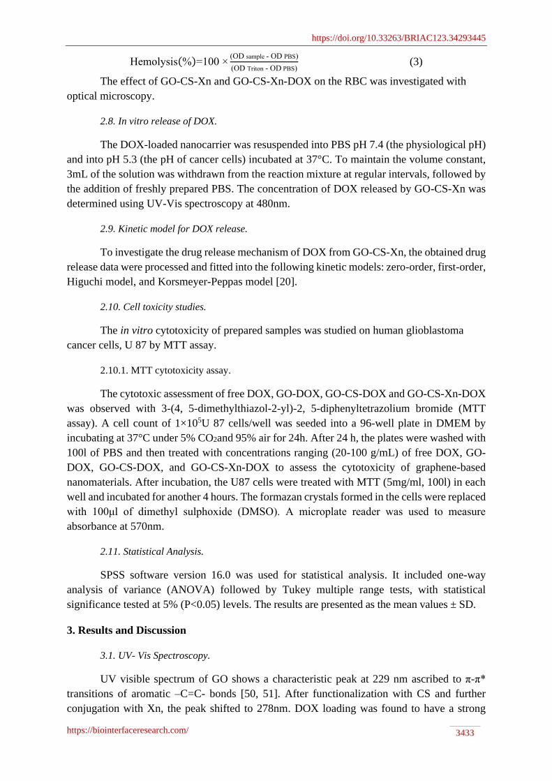
UV visible spectrum of GO shows a characteristic peak at 229 nm ascribed to π-π*
transitions of aromatic –C=C- bonds [50, 51]. After functionalization with CS and further
conjugation with Xn, the peak shifted to 278nm. DOX loading was found to have a strong
https://doi.org/10.33263/BRIAC123.34293445
https://biointerfaceresearch.com/ 3434
absorbance band at 232nm, 288nm, and 480nm, indicating a strong interaction between the
DOX and the nanocarrier system GO-CS-Xn nanoparticles (Figure 1).
Figure 1. UV-Vis Spectra of GO, GO-CS-Xn, and GO-CS-Xn-DOX.
3.2. FT-IR Spectroscopy.
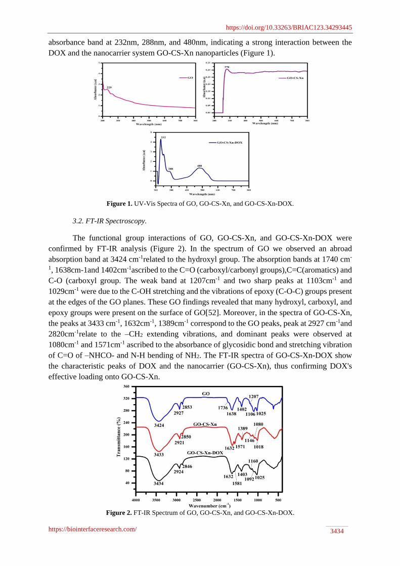
The functional group interactions of GO, GO-CS-Xn, and GO-CS-Xn-DOX were
confirmed by FT-IR analysis (Figure 2). In the spectrum of GO we observed an abroad
absorption band at 3424 cm-1related to the hydroxyl group. The absorption bands at 1740 cm-
1, 1638cm-1and 1402cm-1ascribed to the C=O (carboxyl/carbonyl groups),C=C(aromatics) and
C-O (carboxyl group. The weak band at 1207cm-1 and two sharp peaks at 1103cm-1 and
1029cm-1 were due to the C-OH stretching and the vibrations of epoxy (C-O-C) groups present
at the edges of the GO planes. These GO findings revealed that many hydroxyl, carboxyl, and
epoxy groups were present on the surface of GO[52]. Moreover, in the spectra of GO-CS-Xn,
the peaks at 3433 cm-1, 1632cm-1, 1389cm-1 correspond to the GO peaks, peak at 2927 cm-1and
2820cm-1relate to the –CH2 extending vibrations, and dominant peaks were observed at
1080cm-1 and 1571cm-1 ascribed to the absorbance of glycosidic bond and stretching vibration
of C=O of –NHCO- and N-H bending of NH2. The FT-IR spectra of GO-CS-Xn-DOX show
the characteristic peaks of DOX and the nanocarrier (GO-CS-Xn), thus confirming DOX's
effective loading onto GO-CS-Xn.
Figure 2. FT-IR Spectrum of GO, GO-CS-Xn, and GO-CS-Xn-DOX.
https://doi.org/10.33263/BRIAC123.34293445
https://biointerfaceresearch.com/ 3435
3.3. XRD Spectroscopy.
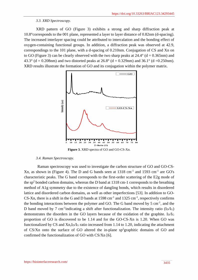
XRD pattern of GO (Figure 3) exhibits a strong and sharp diffraction peak at
10.8°corresponds to the 001 plane, represented a layer to layer distance of 0.82nm (d-spacing).
The increased interlayer spacing could be attributed to intercalation and the bonding effect of
oxygen-containing functional groups. In addition, a diffraction peak was observed at 42.9,
correspondings to the 101 plane, with a d-spacing of 0.210nm. Conjugation of CS and Xn on
to GO (Figure 3) can be clearly observed with the two sharp peaks at 24.4° (d = 0.365nm) and
43.3° (d = 0.208nm) and two distorted peaks at 26.8° (d = 0.329nm) and 36.1° (d =0.250nm).
XRD results illustrate the formation of GO and its conjugation within the polymer matrix.
Figure 3. XRD spectra of GO and GO-CS-Xn.
3.4. Raman Spectroscopy.
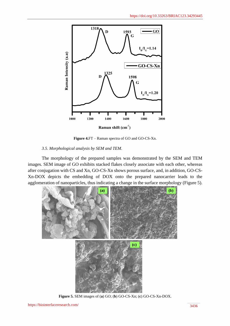
Raman spectroscopy was used to investigate the carbon structure of GO and GO-CS-
Xn, as shown in (Figure 4). The D and G bands seen at 1318 cm-1 and 1593 cm-1 are GO's
characteristic peaks. The G band corresponds to the first-order scattering of the E2g mode of
the sp2 bonded carbon domains, whereas the D band at 1318 cm-1 corresponds to the breathing
method of A1g symmetry due to the existence of dangling bonds, which results in disordered
lattice and disordered carbon domains, as well as other imperfections [53]. In addition to GO-
CS-Xn, there is a shift in the G and D bands at 1598 cm-1 and 1325 cm-1, respectively confirms
the bonding interactions between the polymer and GO. The G band moved by 5 cm-1, and the
D band moved by 7 cm-1indicating a shift after functionalization. The intensity ratio (ID/IG)
demonstrates the disorders in the GO layers because of the oxidation of the graphite. ID/IG
proportion of GO is discovered to be 1.14 and for the GO-CS-Xn is 1.20. When GO was
functionalized by CS and Xn,ID/IG ratio increased from 1.14 to 1.20, indicating the attachment
of CS/Xn onto the surface of GO altered the in-plane sp2graphitic domains of GO and
confirmed the functionalization of GO with CS/Xn [6].
https://doi.org/10.33263/BRIAC123.34293445
https://biointerfaceresearch.com/ 3436
Figure 4.FT – Raman spectra of GO and GO-CS-Xn.
3.5. Morphological analysis by SEM and TEM.
The morphology of the prepared samples was demonstrated by the SEM and TEM
images. SEM image of GO exhibits stacked flakes closely associate with each other, whereas
after conjugation with CS and Xn, GO-CS-Xn shows porous surface, and, in addition, GO-CS-
Xn-DOX depicts the embedding of DOX onto the prepared nanocarrier leads to the
agglomeration of nanoparticles, thus indicating a change in the surface morphology (Figure 5).
Figure 5. SEM images of (a) GO; (b) GO-CS-Xn; (c) GO-CS-Xn-DOX.
https://doi.org/10.33263/BRIAC123.34293445
https://biointerfaceresearch.com/ 3437
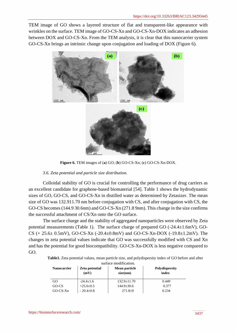
TEM image of GO shows a layered structure of flat and transparent-like appearance with
wrinkles on the surface. TEM image of GO-CS-Xn and GO-CS-Xn-DOX indicates an adhesion
between DOX and GO-CS-Xn. From the TEM analysis, it is clear that this nanocarrier system
GO-CS-Xn brings an intrinsic change upon conjugation and loading of DOX (Figure 6).
Figure 6. TEM images of (a) GO; (b) GO-CS-Xn; (c) GO-CS-Xn-DOX.
3.6. Zeta potential and particle size distribution.
Colloidal stability of GO is crucial for controlling the performance of drug carriers as
an excellent candidate for graphene-based biomaterial [54]. Table 1 shows the hydrodynamic
sizes of GO, GO-CS, and GO-CS-Xn in distilled water as determined by Zetasizer. The mean
size of GO was 132.911.70 nm before conjugation with CS, and after conjugation with CS, the
GO-CS becomes (144.9 30.6nm) and GO-CS-Xn (271.8 9nm). This change in the size confirms
the successful attachment of CS/Xn onto the GO surface.
The surface charge and the stability of aggregated nanoparticles were observed by Zeta
potential measurements (Table 1). The surface charge of prepared GO (-24.4±1.6mV), GO-
CS (+ 25.6± 0.5mV), GO-CS-Xn (-20.4±0.8mV) and GO-CS-Xn-DOX (-19.8±1.2mV). The
changes in zeta potential values indicate that GO was successfully modified with CS and Xn
and has the potential for good biocompatibility. GO-CS-Xn-DOX is less negative compared to
GO.
Table1. Zeta potential values, mean particle size, and polydispersity index of GO before and after
surface modification.
Nanocarrier Zeta potential
(mV)
Mean particle
size(nm)
Polydispersity
index
GO
GO-CS
GO-CS-Xn
-24.4±1.6
+25.6±0.5
- 20.4±0.8
132.9±11.70
144.9±30.6
271.8±9
0.449
0.377
0.234

https://doi.org/10.33263/BRIAC123.34293445
https://biointerfaceresearch.com/ 3438
This is most likely due to the ionization of the carboxylate/ carboxylic group, as well
as the coating of CS and Xn on the GO surface. As a result, these groups are responsible for
the formation of negative charges following DOX loading.3.7. Hemolysis assay.
In vitro hemocompatibility analysis plays a prominent role in the field of biomedical
applications. For intravenous injection and drug delivery, the nanocarrier should have great
blood compatibility. The RBC membranes were incubated with different concentrations of GO-
CS-Xn and GO-CS-Xn-DOX. At a concentration of 100μg/mL; GO-CS-Xn exhibits a
hemolytic effect of 10%, whereas after loading of DOX onto GO-CS-Xn, which brings about
a reasonable change in RBCs toxicity of about 1.4% at the same concentration (Figure 7a and
7b), indicating a low or no toxicity in RBCs makes this nanocarrier more biocompatible and
better suited for drug delivery applications. Optical microscopy images of RBCs were observed
and shown in Figure 7c.
Figure 7. (a) Hemolytic activity of GO-CS-Xn and GO-CS-Xn –DOX; (b) Visually observe the presence of
hemoglobin in the supernatant; (c) optical microscopy images of RBC s with GO-CS-Xn and GO-CS-Xn –
DOX.
3.8. Loading efficiency of DOX.
Table 2 shows the percentage of DOX loaded onto GO and GO-CS-Xn - stacking and
hydrophobic interactions between GO and DOX using a simple mixture and sonication method.
Centrifugation was used to remove the unbound drug, and the loading efficiency of DOX onto
the functionalized GO-based nanocarrier was calculated by measuring the concentration of the
unbound drug-using UV-Vis spectra. According to the data in (Table 2), GO-CS-Xn's loading
efficiency and loading capacity are as high as 73.5% and 81.8%, respectively, when compared
to GO. Apart from these, some surface areas of GO are already occupied by amino
polysaccharide CS and Xn polymer, resulting in high drug loading and efficiency compared
with the GO.
https://doi.org/10.33263/BRIAC123.34293445
https://biointerfaceresearch.com/ 3439
Table 2. Loading capacity and loading efficiency of DOX loaded onto GO and GO-CS-Xn.
Nanocarrier %LC %LE
GO
GO-CS-Xn
48.0
81.8
44.5
73.5
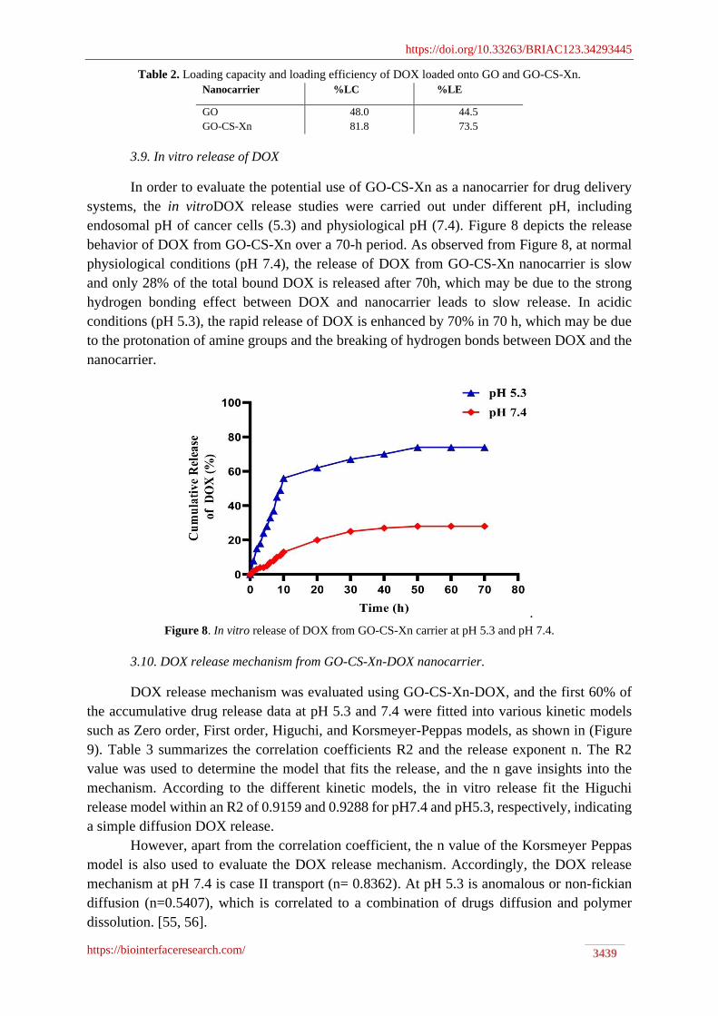
3.9. In vitro release of DOX
In order to evaluate the potential use of GO-CS-Xn as a nanocarrier for drug delivery
systems, the in vitroDOX release studies were carried out under different pH, including
endosomal pH of cancer cells (5.3) and physiological pH (7.4). Figure 8 depicts the release
behavior of DOX from GO-CS-Xn over a 70-h period. As observed from Figure 8, at normal
physiological conditions (pH 7.4), the release of DOX from GO-CS-Xn nanocarrier is slow
and only 28% of the total bound DOX is released after 70h, which may be due to the strong
hydrogen bonding effect between DOX and nanocarrier leads to slow release. In acidic
conditions (pH 5.3), the rapid release of DOX is enhanced by 70% in 70 h, which may be due
to the protonation of amine groups and the breaking of hydrogen bonds between DOX and the
nanocarrier.
.
Figure 8. In vitro release of DOX from GO-CS-Xn carrier at pH 5.3 and pH 7.4.
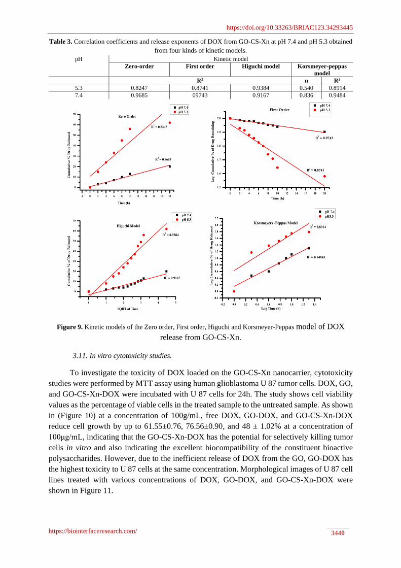
3.10. DOX release mechanism from GO-CS-Xn-DOX nanocarrier.
DOX release mechanism was evaluated using GO-CS-Xn-DOX, and the first 60% of
the accumulative drug release data at pH 5.3 and 7.4 were fitted into various kinetic models
such as Zero order, First order, Higuchi, and Korsmeyer-Peppas models, as shown in (Figure
9). Table 3 summarizes the correlation coefficients R2 and the release exponent n. The R2
value was used to determine the model that fits the release, and the n gave insights into the
mechanism. According to the different kinetic models, the in vitro release fit the Higuchi
release model within an R2 of 0.9159 and 0.9288 for pH7.4 and pH5.3, respectively, indicating
a simple diffusion DOX release.
However, apart from the correlation coefficient, the n value of the Korsmeyer Peppas
model is also used to evaluate the DOX release mechanism. Accordingly, the DOX release
mechanism at pH 7.4 is case II transport (n= 0.8362). At pH 5.3 is anomalous or non-fickian
diffusion (n=0.5407), which is correlated to a combination of drugs diffusion and polymer
dissolution. [55, 56].
https://doi.org/10.33263/BRIAC123.34293445
https://biointerfaceresearch.com/ 3440
Table 3. Correlation coefficients and release exponents of DOX from GO-CS-Xn at pH 7.4 and pH 5.3 obtained
from four kinds of kinetic models.
pH Kinetic model
Zero-order First order Higuchi model Korsmeyer-peppas
model
R2 n R2
5.3 0.8247 0.8741 0.9384 0.540 0.8914
7.4 0.9685 09743 0.9167 0.836 0.9484
Figure 9. Kinetic models of the Zero order, First order, Higuchi and Korsmeyer-Peppas model of DOX
release from GO-CS-Xn.
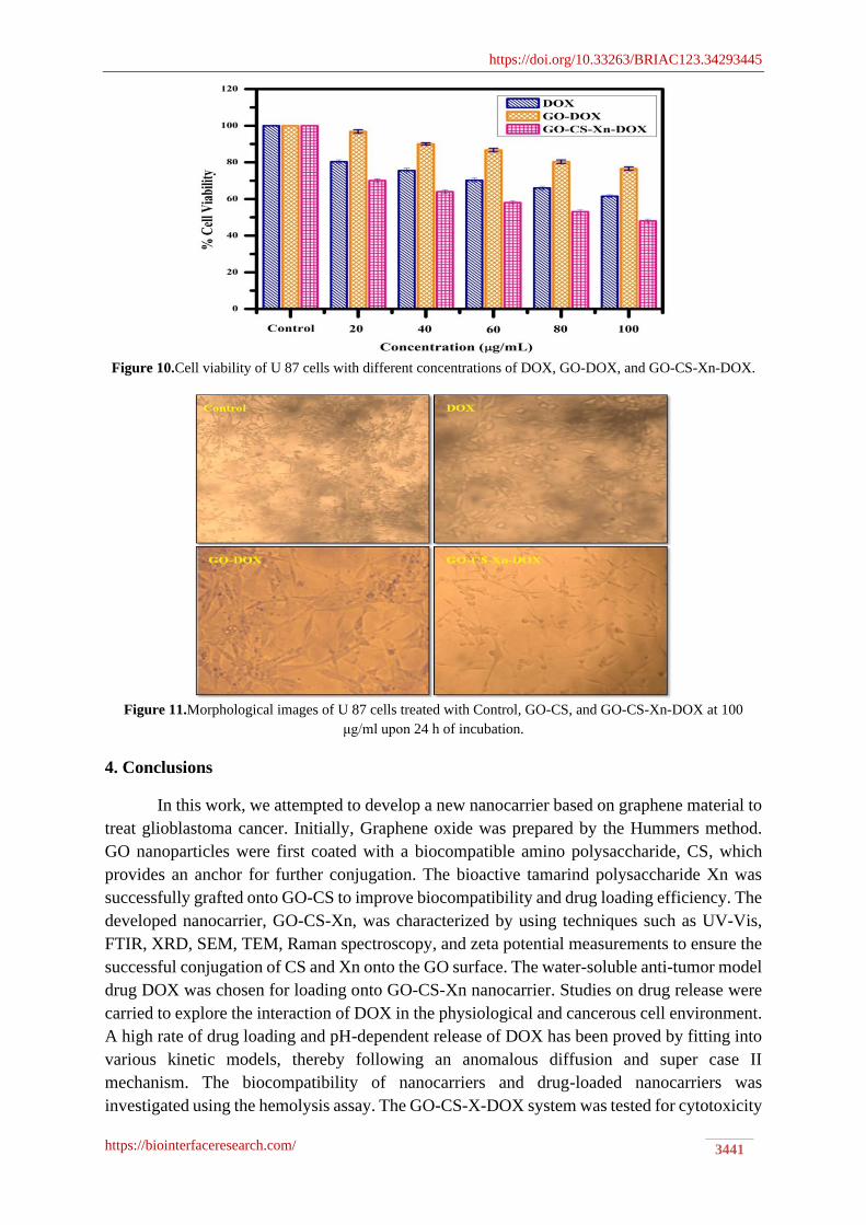
3.11. In vitro cytotoxicity studies.
To investigate the toxicity of DOX loaded on the GO-CS-Xn nanocarrier, cytotoxicity
studies were performed by MTT assay using human glioblastoma U 87 tumor cells. DOX, GO,
and GO-CS-Xn-DOX were incubated with U 87 cells for 24h. The study shows cell viability
values as the percentage of viable cells in the treated sample to the untreated sample. As shown
in (Figure 10) at a concentration of 100g/mL, free DOX, GO-DOX, and GO-CS-Xn-DOX
reduce cell growth by up to 61.55±0.76, 76.56±0.90, and 48 ± 1.02% at a concentration of
100μg/mL, indicating that the GO-CS-Xn-DOX has the potential for selectively killing tumor
cells in vitro and also indicating the excellent biocompatibility of the constituent bioactive
polysaccharides. However, due to the inefficient release of DOX from the GO, GO-DOX has
the highest toxicity to U 87 cells at the same concentration. Morphological images of U 87 cell
lines treated with various concentrations of DOX, GO-DOX, and GO-CS-Xn-DOX were
shown in Figure 11.
https://doi.org/10.33263/BRIAC123.34293445
https://biointerfaceresearch.com/ 3441
Figure 10.Cell viability of U 87 cells with different concentrations of DOX, GO-DOX, and GO-CS-Xn-DOX.
Figure 11.Morphological images of U 87 cells treated with Control, GO-CS, and GO-CS-Xn-DOX at 100
μg/ml upon 24 h of incubation.
4. Conclusions
In this work, we attempted to develop a new nanocarrier based on graphene material to
treat glioblastoma cancer. Initially, Graphene oxide was prepared by the Hummers method.
GO nanoparticles were first coated with a biocompatible amino polysaccharide, CS, which
provides an anchor for further conjugation. The bioactive tamarind polysaccharide Xn was
successfully grafted onto GO-CS to improve biocompatibility and drug loading efficiency. The
developed nanocarrier, GO-CS-Xn, was characterized by using techniques such as UV-Vis,
FTIR, XRD, SEM, TEM, Raman spectroscopy, and zeta potential measurements to ensure the
successful conjugation of CS and Xn onto the GO surface. The water-soluble anti-tumor model
drug DOX was chosen for loading onto GO-CS-Xn nanocarrier. Studies on drug release were
carried to explore the interaction of DOX in the physiological and cancerous cell environment.
A high rate of drug loading and pH-dependent release of DOX has been proved by fitting into
various kinetic models, thereby following an anomalous diffusion and super case II
mechanism. The biocompatibility of nanocarriers and drug-loaded nanocarriers was
investigated using the hemolysis assay. The GO-CS-X-DOX system was tested for cytotoxicity
https://doi.org/10.33263/BRIAC123.34293445
https://biointerfaceresearch.com/ 3442
against U 87 tumor cell line. It exhibited higher cytotoxicity of 51.7% against cell lines. The
current study is a brief screening of bioactive polysaccharides used to synthesize a newer
platform, GO-CS-Xn, for tumor drug delivery. Further research will be conducted to
demonstrate the efficacy of drug delivery via nanocarrier in vivo.
Funding
This research received no external funding.
Acknowledgments
This research has no acknowledgment.
Conflicts of Interest
The authors confirm that this article has no conflict of interest.
References
1. Siegel, R.L.; Miller, K.D.; Jemal, A. Cancer statistics, 2019. CA: A Cancer Journal for Clinicians 2019, 69,
7-34, https://doi.org/10.3322/caac.21551.
2. Liu, B.; Che, C.; Liu, J.; Si, M.; Gong Z, Li, Y.; Yang, G. Fabrication and Antitumor Mechanism of a
Nanoparticle Drug Delivery System: Graphene Oxide/Chitosan Oligosaccharide/γ-Polyglutamic Acid
Composites for Anticancer Drug Delivery. Chemistry Select 2019, 4, 12491–12502,
https://doi.org/10.1002/slct.201903145.
3. Karthika, V.; AlSalhi, M.S.; Devanesan, S.; Gopinath, K.; Arumugam, A.; Govindarajan, M. Chitosan
Overlaid Fe3O4/rGO Nanocomposite for Targeted Drug Delivery, Imaging, and Biomedical
applications. Scientific Reports 2020, 3, 10, 18912, https://doi.org/10.1038/s41598- 020-76015-3.
4. Arias, J.L.; Reddy, L.H.; Couvreur, P. Fe3O4/ Chitosan Nanocomposite for Magnetic Drug Targeting to
Cancer. Journal of Materials Chemistry 2012, 22, 7622–7632, https://doi.org/10.1039/C2JM15339D.
5. Zuchowska, A.; Chudy, M.; Dybko, A.; Brzozka. Z. Graphene as a new material in anticancer therapy-in
vitrostudies. Sensors and Actuators B: Chemical 2017, 243, 152-165,
https://doi.org/10.1016/j.snb.2016.11.105.
6. Borandeh, S.; Abdolmaleki, A.; Abolmaali, S.S.; Tamaddon, A.M. Synthesis, structural and in-vitro
characterization of β-cyclodextrin grafted L-phenylalanine functionalized graphene oxide nanocomposite: A
versatile nanocarrier for pH-sensitive doxorubicin delivery. Carbohydrate Polymers 2018, 201,151-161,
https://doi.org/10.1016/j.carbpol.2018.08.064.
7. Wakaskar, R.R. General overview of lipid-polymer hybrid nanoparticles, dendrimers, micelles, liposomes,
spongosomes and cubosomes. Journal of Drug Targeting 2018, 26, 311-318,
https://doi.org/10.1080/1061186X.2017.1367006.
8. Torres, J.; Liu, Y.; So, S.; Yi, H.; Park, S.; Lee, J.K.; Lim, S.C.; Yun, M. Effects of Surface Modifications to
Single and Multilayer Graphene Temperature Coefficient of Resistance. ACS Appl. Mater. Interfaces 2020,
12, 48890–48898, https://doi.org/10.1021/acsami.0c09621.
9. Liu, W.; Speranza, G. Tuning the Oxygen Content of Reduced Graphene Oxide and Effects on Its Properties.
ACS Omega 2021, 6, 6195–6205, https://doi.org/10.1021/acsomega.0c05578.
10. Patil, T.V.; Patel, D.K.; Dutta, S.D.; Ganguly, K.; Lim, K.T. Graphene Oxide-Based Stimuli-Responsive
Platforms for Biomedical Applications. Molecules, 26, 2797, https://doi.org/10.3390/molecules26092797.
11. Javanbakht, S.; Pooresmaeil, M.; Namazi, H. Green one-pot synthesis of carboxymethylcellulose/Zn-based
metal-organic framework/graphene oxide bio-nanocomposite as a nanocarrier for drug delivery system.
Carbohydr. Polym. 2019, 208, 294–301, https://doi.org/10.1016/j.carbpol.2018.12.066.
12. Morales-Narvaez, E.; Merkoci, A. Graphene Oxide as an Optical Biosensing Platform: A Progress Report.
Adv. Mater. 2019, 31, 1805043, https://doi.org/10.1002/adma.201805043.
https://doi.org/10.33263/BRIAC123.34293445
https://biointerfaceresearch.com/ 3443
13. Yogesh, G.K.; Shuaib, E.P.; Roopmani, P.; Gumpu, M.B.; Krishnan, U.M.; Sastikumar, D. Synthesis,
characterization and bioimaging application of laser-ablated graphene-oxide nanoparticles (nGOs) Diam.
Relat. Mater. 2020, 104, 107733, https://doi.org/10.1016/j.diamond.2020.107733.
14. Martín, C.; Ruiz, A.; Keshavan, S.; Reina, G.; Murera, D.; Nishina, Y.; Fadeel, B.; Bianco, A. A
Biodegradable Multifunctional Graphene Oxide Platform for Targeted Cancer Therapy. Adv. Funct. Mater.
2019, 29, 1901761, https://doi.org/10.1002/adfm.201901761.
15. Khalili, R.; Zarrintaj, P.; Jafari, S.H.; Vahabi, H.; Saeb, M.R. Electroactive poly (p-phenylene sulfide)/r-
graphene oxide/chitosan as a novel potential candidate for tissue engineering. Int. J. Biol. Macromol. 2020,
154, 18–24, https://doi.org/10.1016/j.ijbiomac.2020.03.029.
16. Ahmed, M.K.; Mansou,r S.F.; Al-Wafi, R.; Menazea, A.A. Composition and design of nanofibrous scaffolds
of Mg/Se- hydroxyapatite/graphene oxide @ ε-polycaprolactone for wound healing applications. J. Mater.
Res. Technol. 2020, 9, 7472–7485, https://doi.org/10.1016/j.jmrt.2020.04.094.
17. Anirudhan, T.S.; Chithra Sekhar, V.; Athira, V.S. Graphene oxide based functionalized chitosan
polyelectrolyte nanocomposite for targeted and pH responsive drug delivery. Int J Biol Macromol. 2020, 150,
468-479, https://doi.org/10.1016/j.ijbiomac.2020.02.053.
18. Jafari, Z.; Rad, A.S.; Baharfar, R.; Asghari, S.; Esfahani, MR. Synthesis and application of
chitosan/tripolyphosphate/graphene oxide hydrogel as a new drug delivery system for Sumatriptan Succinate.
J. Mol. Liq. 2020, 315, 113835, https://doi.org/10.1016/j.molliq.2020.113835.
19. Abdelhamid, H.N.; Hussein,K.H. Graphene Oxide as a Carrier for Drug Delivery of Methotrexate.
Biointerface Research in Applied Chemistry 2021, 11, 14726-14735,
https://doi.org/10.33263/BRIAC116.1472614735.
20. Tiwari, H.; Karki, N.; Pal, M.; Basak, S.; Verma, R.K.; Bal, R.; Kandpal, N.D.; Bisht, G.; Sahoo, NG.
Functionalized graphene oxide as a nanocarrier for dual drug delivery applications: The synergistic effect of
quercetin and gefitinib against ovarian cancer cells. Colloids Surf B Biointerfaces 2019 178, 452-459,
https://doi.org/10.1016/j.colsurfb.2019.03.037.
21. Figueroa, T.; Aguayo, C.; Fernández, K. Design and Characterization of Chitosan-Graphene Oxide
Nanocomposites for the Delivery of Proanthocyanidins. International journal of nanomedicine 2020,
15, 1229–1238, https://doi.org/10.2147/IJN.S240305.
22. Pei, X.; Zhu, Z.; Gan, Z.; Chen, J.; Zhang, X.; Cheng, X.;, Wan, Q.;Wang, J. PEGylated nano-graphene oxide
as a nanocarrier for delivering mixed anticancer drugs to improve anticancer activity. Sci zzxRep 2020, 10,
2717, https://doi.org/10.1038/s41598-020-59624-w.
23. Dhanavel, S.; Revathy, T.A.; Sivaranjani, T.; Sivakumar, P.;Palani, V.; Narayanan, A. 5-Fluorouracil and
curcumin co-encapsulated chitosan/reduced graphene oxide nanocomposites against human colon cancer cell
lines. Polym. Bull. 2020, 77, 213–233, https://doi.org/10.1007/s00289-019-02734-x.
24. Pan, H.; Yu, Y.; Li, L.; Liu, B.; Liu, Y. Fabrication and Characterization of Taurine Functionalized Graphene
Oxide with 5-Fluorouracil as Anticancer Drug Delivery Systems. Nanoscale Res Lett 2021, 16, 84,
https://doi.org/10.1186/s11671-021-03541-y.
25. Vinothini, K.; Rajendran, N.K.; Rajan, M.; Ramu, A.; Marraiki, N.; Elgorban, A.M. A magnetic nanoparticle
functionalized reduced graphene oxide-based drug carrier system for a chemo-photodynamic cancer therapy.
N J Chem. 2020, 44, 5265–5277, https://doi.org/10.1039/D0NJ00049C.
26. Zaboli, M.; Raissi, H.; Moghaddam, N.R.; Farzad, F. Probing the adsorption and release mechanisms
of cytarabine anticancer drug on/from dopamine functionalized graphene oxide as a highly efficient drug
delivery system. J. Mol. Liq. 2020, 301, 112458, https://doi.org/10.1016/j.molliq.2020.112458.
27. Makvandi, P.; Ghomi, M.; Ashrafizadeh, M.; Tafazoli, A.; Agarwal, T.; Delfi, M.; Akhtari, J.; Zare, E.N.;
Padil, V.V.T.; Zarrabi, A.; Pourreza, N.; Miltyk, W.; Maiti, T.K. A review on advances in graphene-
derivative / polysaccharide bionanocomposites: Therapeutics,pharmacogenomics and toxicity. Carbohydrate
Polymers 2020, 250, 116952, https://doi.org/10.1016/j.carbpol.2020.116952.
28. Tolvanen, J.; Kilpijarvi, J.; Pitkanen, O.; Hannu, J.; Jantunen, H. Stretchable Sensors with Tunability and
Single Stimuli-Responsiveness through Resistivity Switching Under Compressive Stress. ACS Appl. Mater.
Interfaces 2020, 12, 14433–14442, https://doi.org/10.1021/acsami.0c00023.
29. Song, Y.-Y.; Liu, Y.; Jiang, H.-B.; Xue, J.-Z.; Yu, Z.-P.; Li, S.-Y.; Han, Z.-W.; Ren, L.-Q. Janus Soft
Actuators with On–Off Switchable Behaviors for Controllable Manipulation Driven by Oil.ACS Appl. Mater.
Interfaces 2019, 11, 13742–13751,https://doi.org/10.1021/acsami.8b20061.
https://doi.org/10.33263/BRIAC123.34293445
https://biointerfaceresearch.com/ 3444
30. Abu-Thabit, N.Y.; Hamdy, A.S. Stimuli-responsive Polyelectrolyte Multilayers for fabrication of self-healing
coatings—A review. Surface and Coatings Technology 2016, 303, 406–424,
https://doi.org/10.1016/j.surfcoat.2015.11.020.
31. Chatterje,e S.; Chi-leung Hui, P. Review of Stimuli-Responsive Polymers in Drug Delivery and Textile
Application. Molecules 2019, 24, 2547, https://doi.org/10.3390/molecules24142547.
32. Shu, T.; Hu, L.; Shen, Q.; Jiang, L.; Zhang, Q.; Serpe, M.J. Stimuli-responsive polymer-based systems for
diagnostic applications. J. Mater. Chem. B. 2020, 8, 7042–7061, https://doi.org/10.1039/D0TB00570C.
33. Son, H.; Yoon, C. Advances in Stimuli-Responsive Soft Robots with Integrated Hybrid Materials. Actuators
2020, 9, 115, https://doi.org/10.3390/act9040115.
34. Li, Z.; Yin, Y. Stimuli-Responsive Optical Nanomaterials. Adv. Mater. 2019, 31, 1807061,
https://doi.org/10.1002/adma.201807061.
35. Motiei, M.; Kashanian, S.; Lucia, L.A.; Khazaei, M. Intrinsic parameters for the synthesis and tuned
properties of amphiphilic chitosan drug delivery nanocarriers. Journal of Controlled Release 2017, 260, 213-
225, https://doi.org/10.1016/j.jconrel.2017.06.010.
36. Di Martino, A.; Sedlarik, V. Amphiphilic chitosan-grafted-functionalized polylactic acid based nanoparticles
as a delivery system for doxorubicin and temozolomide co-therapy. International Journal of
Pharmaceutics2014, 474, 134-145, https://doi.org/10.1016/j.ijpharm.2014.08.014.
37. Di Martino, A.; Kucharczyk, P.; Zednik, J.; Sedlarik, V. Chitosan grafted low molecular weight polylactic
acid for protein encapsulation and burst effect reduction. International Journal of Pharmaceutics 2015, 496,
912-921, https://doi.org/10.1016/j.ijpharm.2015.10.017.
38. Tao, F.; Cheng, Y.; Shi, X.; Zheng, H.; Du, Y.; Xiang, W.; Deng, H. Applications of chitin andchitosan
nanofibers in bone regenerative engineering. Carbohydr. Polym. 2020, 230, 115658,
https://doi.org/10.1016/j.carbpol.2019.115658.
39. Moeini, A.; Pedram, P.; Makvandi, P.; Malinconico, M.; d'Ayala, G. Wound healing and antimicrobial effect
of active secondary metabolites in chitosan-based wound dressings: A review. Carbohydr. Polym. 2020, 233,
115839, https://doi.org/10.1016/j.carbpol.2020.115839.
40. Diop, M.; Auberval, N.; Viciglio, A.; Langlois, A.; Bietiger, W.; Mura, C.; Peronet, C.; Bekel, A.; Julien
David, D.; Zhao, M.; Pinget, M.; Jeandidier, N.; Vauthier, C.; Marchioni, E.; Frere, Y.;Sigrist, S. Design,
characterisation, and bioefficiency of insulin-chitosan nanoparticles after stabilisation by freeze-
drying or cross-linking. International Journal of Pharmaceutics 2015, 491, 402-408,
https://doi.org/10.1016/j.ijpharm.2015.05.065.
41. Ramalingam, P.; Ko, Y.T.; Enhanced oral delivery of curcumin from N-trimethyl chitosan surface-modified
solid lipid nanoparticles: pharmacokinetic and brain distribution evaluations. Pharmaceutical research 2015,
32, 389-402, https://doi.org/10.1007/s11095-014-1469-1.
42. Kulkarni, A.D.; Joshi, A.A.; Patil, C.L.; Amale, P.D.; Patel, H.M.; Surana, S.J.; Belgamwar, V.S.; Chaudhari,
K.S.; Pardeshi, C.V.; Xyloglucan: A functional biomacromolecule for drug delivery applications.
International Journal of Biological Macromolecule 2017, 104, 799-812,
https://doi.org/10.1016/j.ijbiomac.2017.06.088.
43. Han, M.; Liu, Y.; Zhang, F.; Sun, D.; Jiang, J. Effect of galactose side-chain on the self-assembly of
xyloglucan macromolecule. Carbohydr. Polym. 2020, 246, 116577,
https://doi.org/10.1016/j.carbpol.2020.116577.
44. Da Silva, A.C.C.; De Almeida, R.R.; Da Cruz Sousa, A.C.; Martinez, F.N.A.; Denardin, J.C.; De Morais,
S.M.; Ricardo, N.M.P.S. Xyloglucan-based hybrid nanocomposite with potential for biomedical applications.
International Journal of Biological Macromolecule 2021, 168, 722-732,
https://doi.org/10.1016/j.ijbiomac.2020.11.128.
45. Chandrakantsing V, Pardeshi.; Abhijeet D, Kulkarni.; Veena S, Belgamwar.; Sanjay J, Surana. Chapter 7 –
Xyloglucan for drug delivery applications. Fundamental Biomaterials: Polymers, Sabu Thomas, Preetha
Balakrishnan, M.S. Sreekala.; In Woodhead Publishing Series inBiomaterials: India 2018,143-169,
https://doi.org/10.1016/B978-0-08-102194-1.00007-4.
46. Hummers, W.S., Offeman, R.E. Preparation of Graphitic Oxide. Journal of the American Chemical Society
1958, 80, 1339-1339, https://doi.org/10.1021/ja01539a017.
47. Marcano, D.C.; Kosynkin, D.V.; Berlin, J.M.; Sinitskii, A.; Sun, Z.; Slesarev, A.; Alemany, L.B.;
Lu,W.;Tour, J.M. Improved synthesis of graphene oxide. ACS Nano 2010, 4, 4806-4814,
https://doi.org/10.1021/nn1006368.
https://doi.org/10.33263/BRIAC123.34293445
https://biointerfaceresearch.com/ 3445
48. Muthukumarasamyvel, T.; Baskar, R.; Chandirasekar, S.; Umamaheswari, K.; Rajendiran, N.Hierarchical
self- assembly of bile-acid-derived dicationic amphiphiles and their toxicity assessment on microbial and
mammalian systems. ACS Applied Materials and Interfaces 2016, 8, 25111– 25126,
https://doi.org/10.1021/acsami.6b08018.
49. Paarakh, M.P.; Jose, P.A.; Setty, C.; Christoper, G. Release kinetics–concepts and applications. International
Journal of Pharmacy Research and Technology 2018, 8, 12-20, https://doi:10.31838/ijprt/08.01.02.
50. Kumar, N.; Srivastava, V.C. Simple Synthesis of Large Graphene Oxide Sheets via Electrochemical
Method Coupled with Oxidation Process. ACS Omega 2018, 3, 10233-10242,
https://doi.org/10.1021/acsomega.8b01283.
51. Su, Z.; Sun, D.; Zhang, L.; He, M.; Jiang, Y.; Millar, B.; Douglas, P.; Mariotti, D.; Maguire, P.; Sun, D.
Chitosan/Silver Nanoparticle/Graphene Oxide Nanocomposites with Multi-Drug Release, Antimicrobial, and
Photothermal Conversion Functions. Materials 2021, 14, 2351, https://doi.org/10.3390/ma14092351.
52. Aliyev, E.; Filiz, V.; Khan, M.M.; Lee, Y.J.; Abetz, C.; Abetz, V. Structural characterization of Graphene
Oxide: surface functional groups and fractionated oxidative debris. Nanomaterials 2019, 9, 1180,
https://doi.org/10.3390/nano9081180.
53. López-Díaz, D.; Delgado-Notario, J.A.; Clericò, V.; Diez, E.; Merchán, M.D.; Velázquez, M.M.;Towards
Understanding the Raman Spectrum of Graphene Oxide: The Effect of the Chemical Composition. Coatings
2020, 10,524, https://doi.org/10.3390/coatings10060524.
54. Shu, M.; Gao, F.; Zeng, M.; Yu, C.; Wang, X.; Huang, R.; Yang, J.; Su, Y.; Hu, N.; Zhou, Z.;, Liu,K.;Yang,
Zhi.;T an, H.; Xu, L. et al. Microwave-Assisted Chitosan-Functionalized Graphene Oxide as Controlled
Intracellular Drug Delivery Nanosystem for Synergistic Antitumour Activity. Nanoscale Res Lett 2021, 16,
75, https://doi.org/10.1186/s11671-021-03525-y.
55. Mhlanga, N.; Ray, S.S. Kinetic models for the release of the anticancer drug doxorubicin from biodegradable
polylactide/metal oxide-based hybrids. International Journal of Biological Macromolecule 2015, 72, 1301-
1307, https://doi.org/10.1016/j.ijbiomac.2014.10.038.
56. Prasanth, A.G.; Kumar, A.S.; Shruthi, B.S.; Subramanian, S. Kinetic study and in vitro drug release studies
of nitrendipine loaded arylamide grafted chitosan blend microspheres. Materials Research Express 2020, 5,
125427, https://doi.org/10.1088/2053-1591/ab5811.