bio-inspired design of multiscale structures for function...
TRANSCRIPT

Nano Today (2011) 6, 155—175
avai lab le at www.sc iencedi rec t .com
journa l homepage: www.e lsev ier .com/ locate /nanotoday
REVIEW
Bio-inspired design of multiscale structures forfunction integration
Kesong Liua, Lei Jianga,b,∗
a Key Laboratory of Bio-inspired Smart Interfacial Science and Technology of Ministry of Education, School of Chemistry andEnvironment, Beihang University, Beijing 100190, PR Chinab Beijing National Laboratory for Molecular Sciences (BNLMS), Key Laboratory of Organic Solids, Institute of Chemistry, ChineseAcademy of Sciences, Beijing 100190, PR China
Received 9 November 2010; received in revised form 25 January 2011; accepted 7 February 2011Available online 5 March 2011
KEYWORDSBiomimetic;Bio-inspired;Multifunctional;
Summary Multiscale structures of biological materials exhibit inherent multifunctional inte-gration. This special biological solution provides some inspiration for scientists and engineers todesign multifunctional artificial materials with multiscale structures. In this review, we focuson recent research progress in some typical biological materials (such as lotus leaves, rice
Multiscale;Wettability;Superhydrophobic;Self-cleaning;
leaves, butterfly wings, water strider legs, insect compound eyes, fish scales, red rose petals,brittlestars, spider silks, nacre, glass sponges, gecko feet, mussels, and others) and the corre-sponding bio-inspired multiscale materials possessing function integration. The challenges andperspectives for bio-inspired design of multifunctional structures in the future are also brieflyaddressed.
ts re
potim
Optical;Mechanical;Anti-reflection
© 2011 Elsevier Ltd. All righ
Introduction
After billions of years of evolution, creatures in nature pos-sess almost perfect structures and functions [1]. Multiscalestructures ranging from nano, micro to macro are character-
istic for biological materials, which play an important rolein achieving structural and functional integrity. In the lastfew decades, a great number of natural materials have beeninvestigated by scientists and engineers from multidisci-∗ Corresponding author at: Key Laboratory of Bio-inspired SmartInterfacial Science and Technology of Ministry of Education, Schoolof Chemistry and Environment, Beihang University, Beijing 100190,PR China. Tel.: +86 10 8262 1396; fax: +86 10 8262 7566.
E-mail address: [email protected] (L. Jiang).
T
Cm[bamb
p
1748-0132/$ — see front matter © 2011 Elsevier Ltd. All rights reserved.doi:10.1016/j.nantod.2011.02.002
served.
linary fields. It was found the inherent multiscale structuref biological materials is not unifunctional but multifunc-ional, i.e., possessing more than one function (functionalntegration). Some representative biological materials withultiscale structures for function integration are listed in
able 1.Should we just be fascinated by what nature provides?
reating multifunctional materials is an eternal goal ofankind. Nature is a school for scientists and engineers
1,36—39]. Learning from nature has long been a source ofio-inspiration for human beings [40—46]. In recent years,
great deal of work has been devoted to fabricatingultiscale structures for function integration through theiomimetic or bio-inspired approach.
In this review article, we summarize recent researchrogress in some typical biological materials with
156 K. Liu, L. Jiang
Table 1 Typical biological materials with function integration.
Biological materials Functions Ref.
Butterfly wing Superhydrophobicity, directional adhesion,structural color, self-cleaning, chemicalsensing capability, fluorescence emissionfunctions
[2—6]
Brittlestar Mechanical and optical functions [7]Cicada wing Anti-reflection, superhydrophobicity [8]Fish scale Drag reduction, superoleophilicity in air,
superoleophobicity in water[9]
Gecko foot Reversible adhesive, superhydrophobicity,self-cleaning
[10]
Lotus leaf Superhydrophobicity, low adhesion,self-cleaning
[11]
Mosquito compound eye Superhydrophobicity, anti-reflection,anti-fogging
[12]
Nacre Mechanical property, structural color [13,14]Peacock feather Structural color, superhydrophobicity [15]Polar bear fur Optical property, thermal insulation [16]Rice leaf Superhydrophobicity, anisotropic wettability [11]Rose petal Superhydrophobicity, structural color, high
adhesion[17—19]
Shark skin Drag reduction, anti-biofouling [20]Spicule Mechanical and fibre-optical properties [21—23]Spider capture silk Water collection ability, mechanical property,
elasticity, stickiness[24—26]
Spider dragline silk Mechanical property, supercontraction,torsional shape memory
[27—34]
Water strider leg Durable and robust superhydrophobicity [35]
fbfinTttswfrmpf
L
Il(isepwm
lchaaaas
gsaoaa(tgacef
unction integration, such as lotus leaves, rice leaves,utterfly wings, water strider legs, insect compound eyes,sh scales, red rose petals, brittlestars, spider silks,acre, glass sponges, gecko feet, mussels, and others.he corresponding biomimetic materials with multifunc-ional properties are also presented. This may be helpfulo stimulate interdisciplinary collaboration of materialcience, chemistry, physics, biology, engineering, etc.,hich is essential for the further discovery of additional
unction of biological materials and rational design and theeproducible construction of bio-inspired multifunctionalaterials. In the final section, we discuss challenges anderspectives for bio-inspired design of multiscale structuresor function integration in the future.
otus leaves and their biomimetic materials
n nature, over millions of years of evolution, many planteaves exhibit special wettability. Among them, the lotusNelumbo nucifera) leaf, better known as the water lily,s one of most promising. The sacred lotus has been a
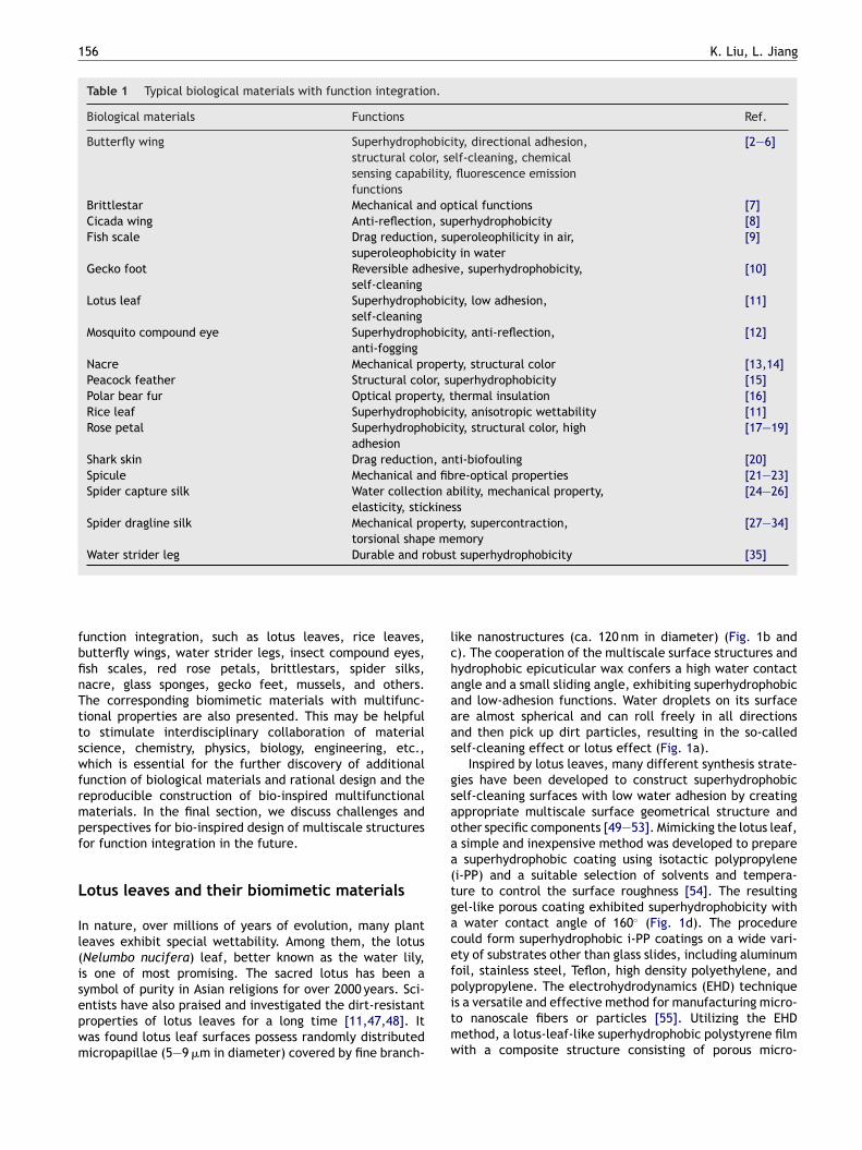
ymbol of purity in Asian religions for over 2000 years. Sci-ntists have also praised and investigated the dirt-resistantroperties of lotus leaves for a long time [11,47,48]. Itas found lotus leaf surfaces possess randomly distributedicropapillae (5—9 �m in diameter) covered by fine branch-pitmw
ike nanostructures (ca. 120 nm in diameter) (Fig. 1b and). The cooperation of the multiscale surface structures andydrophobic epicuticular wax confers a high water contactngle and a small sliding angle, exhibiting superhydrophobicnd low-adhesion functions. Water droplets on its surfacere almost spherical and can roll freely in all directionsnd then pick up dirt particles, resulting in the so-calledelf-cleaning effect or lotus effect (Fig. 1a).
Inspired by lotus leaves, many different synthesis strate-ies have been developed to construct superhydrophobicelf-cleaning surfaces with low water adhesion by creatingppropriate multiscale surface geometrical structure andther specific components [49—53]. Mimicking the lotus leaf,simple and inexpensive method was developed to preparesuperhydrophobic coating using isotactic polypropylene
i-PP) and a suitable selection of solvents and tempera-ure to control the surface roughness [54]. The resultingel-like porous coating exhibited superhydrophobicity withwater contact angle of 160◦ (Fig. 1d). The procedure
ould form superhydrophobic i-PP coatings on a wide vari-ty of substrates other than glass slides, including aluminumoil, stainless steel, Teflon, high density polyethylene, and
olypropylene. The electrohydrodynamics (EHD) techniques a versatile and effective method for manufacturing micro-o nanoscale fibers or particles [55]. Utilizing the EHDethod, a lotus-leaf-like superhydrophobic polystyrene filmith a composite structure consisting of porous micro-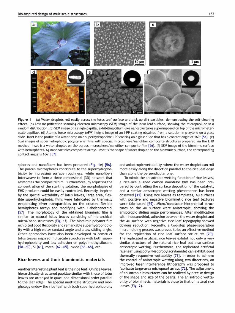
Bio-inspired design of multiscale structures 157
Figure 1 (a) Water droplets roll easily across the lotus leaf surface and pick up dirt particles, demonstrating the self-cleaningeffect. (b) Low magnification scanning electron microscopy (SEM) image of the lotus leaf surface, showing the micropapillae in arandom distribution. (c) SEM image of a single papilla, exhibiting cilium-like nanostructures superimposed on top of the micrometer-scale papillae. (d) Atomic force microscopy (AFM) height image of an i-PP coating obtained from a solution in p-xylene on a glassslide. Inset is the profile of a water drop on a superhydrophobic i-PP coating on a glass slide that has a contact angle of 160◦ [54]. (e)SEM images of superhydrophobic polystyrene films with special microsphere/nanofiber composite structures prepared via the EHD
nofibe sha
amt
apaowwtawtomfTsartti
method. Inset is a water droplet on the porous microsphere/nawith hemispheres/Ag nanoparticles composite arrays. Inset is thcontact angle is 166◦ [57].
spheres and nanofibers has been prepared (Fig. 1e) [56].The porous microspheres contribute to the superhydropho-bicity by increasing surface roughness, while nanofibersinterweave to form a three-dimensional (3D) network thatreinforces the composite film. Furthermore, by adjusting theconcentration of the starting solution, the morphologies ofEHD products could be easily controlled. Recently, inspiredby the special wettability of lotus leaves, large area, flex-ible superhydrophobic films were fabricated by thermallyevaporating silver nanoparticles on the created flexiblehemispheres arrays and modifying with 1-dodecanethiol[57]. The morphology of the obtained biomimic film issimilar to natural lotus leaves consisting of hierarchicalmicro/nano structures (Fig. 1f). The biomimic polymer filmexhibited good flexibility and remarkable superhydrophobic-ity with a high water contact angle and a low sliding angle.Other approaches have also been developed to constructlotus leaves inspired multiscale structures with both super-hydrophobicity and low adhesion on polydimethylsiloxane[58—60], Si [61], metal [62—65], oxide [66—68], etc.
Rice leaves and their biomimetic materials
Another interesting plant leaf is the rice leaf. On rice leaves,hierarchically structured papillae similar with those of lotusleaves are arranged in quasi-one-dimensional order parallelto the leaf edge. The special multiscale structure and mor-phology endow the rice leaf with both superhydrophobicity
foobl
er composite film [56]. (f) SEM image of the biomimic surfacepe of water droplet on the biomimic surface, the corresponding
nd anisotropic wettability, where the water droplet can rollore easily along the direction parallel to the rice leaf edge
han along the perpendicular one.To mimic the anisotropic wetting function of rice leaves,rice-like aligned carbon nanotube film has been pre-
ared by controlling the surface deposition of the catalyst,nd a similar anisotropic wetting phenomenon has beenbserved [11]. Using rice leaves as templates, Au surfacesith positive and negative biomimetic rice leaf texturesere fabricated [69]. Micro/nanoscale hierarchical struc-
ures on the Au surface were anisotropic, showing thenisotropic sliding angle performances. After modificationith 1-decanethiol, adhesion between the water droplet and
he Au surface with negative rice leaf topography showedbvious reduction. Recently, a two-step phase-separationicromolding process was proved to be an effective method
or the replication of rice leaf surface structures [70].he replicated artificial rice leaves exhibit not only a veryimilar structure of the natural rice leaf but also surfacenisotropic wetting. Furthermore, the replicated artificialice leaf using poly(N-isopropylacrylamide) can exhibit goodhermally responsive wettability [71]. In order to achievehe control of anisotropic wetting along two directions, anmproved laser interference lithography was proposed to
abricate large-area micropearl arrays [72]. The adjustmentf anisotropic biosurfaces can be realized by precise designf the shape and size of the pearls. The anisotropic wetta-ility of biomimetic materials is close to that of natural riceeaves (Fig. 2).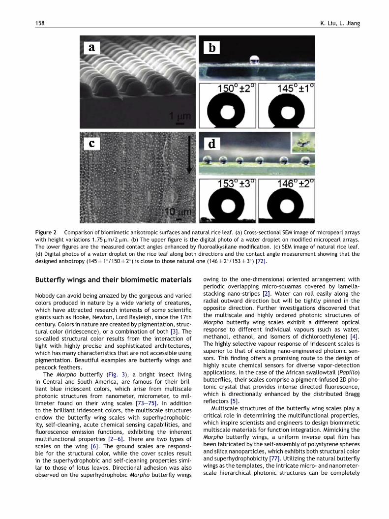
158 K. Liu, L. Jiang
Figure 2 Comparison of biomimetic anisotropic surfaces and natural rice leaf. (a) Cross-sectional SEM image of micropearl arrayswith height variations 1.75 �m/2 �m. (b) The upper figure is the digital photo of a water droplet on modified micropearl arrays.T y flu( h dird l one
B
Ncwgctslwpp
ilplteiflmsbilo
opsrotMrmTsshabtwr
cwmM
he lower figures are the measured contact angles enhanced bd) Digital photos of a water droplet on the rice leaf along botesigned anisotropy (145 ± 1◦/150 ± 2◦) is close to those natura
utterfly wings and their biomimetic materials
obody can avoid being amazed by the gorgeous and variedolors produced in nature by a wide variety of creatures,hich have attracted research interests of some scientificiants such as Hooke, Newton, Lord Rayleigh, since the 17thentury. Colors in nature are created by pigmentation, struc-ural color (iridescence), or a combination of both [3]. Theo-called structural color results from the interaction ofight with highly precise and sophisticated architectures,hich has many characteristics that are not accessible usingigmentation. Beautiful examples are butterfly wings andeacock feathers.
The Morpho butterfly (Fig. 3), a bright insect livingn Central and South America, are famous for their bril-iant blue iridescent colors, which arise from multiscalehotonic structures from nanometer, micrometer, to mil-imeter found on their wing scales [73—75]. In additiono the brilliant iridescent colors, the multiscale structuresndow the butterfly wing scales with superhydrophobic-ty, self-cleaning, acute chemical sensing capabilities, anduorescence emission functions, exhibiting the inherentultifunctional properties [2—6]. There are two types of
cales on the wing [6]. The ground scales are responsi-le for the structural color, while the cover scales resultn the superhydrophobic and self-cleaning properties simi-ar to those of lotus leaves. Directional adhesion was alsobserved on the superhydrophobic Morpho butterfly wings
baaws
oroalkysilane modification. (c) SEM image of natural rice leaf.ections and the contact angle measurement showing that the(146 ± 2◦/153 ± 3◦) [72].
wing to the one-dimensional oriented arrangement witheriodic overlapping micro-squamas covered by lamella-tacking nano-stripes [2]. Water can roll easily along theadial outward direction but will be tightly pinned in thepposite direction. Further investigations discovered thathe multiscale and highly ordered photonic structures oforpho butterfly wing scales exhibit a different optical
esponse to different individual vapours (such as water,ethanol, ethanol, and isomers of dichloroethylene) [4].he highly selective vapour response of iridescent scales isuperior to that of existing nano-engineered photonic sen-ors. This finding offers a promising route to the design ofighly acute chemical sensors for diverse vapor-detectionpplications. In the case of the African swallowtail (Papilio)utterflies, their scales comprise a pigment-infused 2D pho-onic crystal that provides intense directed fluorescence,hich is directionally enhanced by the distributed Bragg
eflectors [5].Multiscale structures of the butterfly wing scales play a
ritical role in determining the multifunctional properties,hich inspire scientists and engineers to design biomimeticultiscale materials for function integration. Mimicking theorpho butterfly wings, a uniform inverse opal film has
een fabricated by the self-assembly of polystyrene spheresnd silica nanoparticles, which exhibits both structural colornd superhydrophobicity [77]. Utilizing the natural butterflyings as the templates, the intricate micro- and nanometer-cale hierarchical photonic structures can be completely

Bio-inspired design of multiscale structures 159
w an
potiirmi
Figure 3 (a) Morpho didius. SEM images of (b) an oblique viedidius [76].
replicated by a alumina coating through a low-temperatureatomic layer deposition process [78]. Furthermore, the alu-mina replicas with a 3D structure possess the inheritedphotonic properties of the natural wing scales and othernovel properties, which can serve as optical waveguides,beam splitters and other building blocks of photonic inte-grated circuits (Fig. 4). Recently, 3D rutile titania-basedstructures with Morpho butterfly wing scale morphologieswere fabricated using a layer-by-layer sol—gel-based depo-
sition technique [79]. Rutile replicas of butterfly scales wereobtained after 40 cycles of exposure of the templates to amixed titanium—tin alkoxide solution and water, followingby calcination.tcc(
Figure 4 Left. Images of the alumina replicas of the butterfly wcoated butterfly wing scales, of which the color changed from orialumina replicas of butterfly wing scales on silicon substrate after thdispersive X-ray spectrum of the alumina replica shown in part b. (scale, where the replica exhibits exactly the same fine structures.Right. ‘‘Waveguide’’ properties of alumina replicas. (a) A dark fieldrows; the inset is an optical image of an individual scale. (b) SEM imaimage shown in part a. (c) SEM image of the bifurcated lamella structshowing the potential application as a beam splitter [78].
d (c) a cross-section of a ground scale of the butterfly Morpho
Another typical example for structural color is theeacock feather. The male peacock in full plumage isne of the most beautiful and spectacular birds inhe natural world, which displays iridescent colors andntricate eye patterns in the tails (Fig. 5a). The tiny,ntricate two-dimensional photonic crystal structure isesponsible for the color production in peacock (Pavouticus) feathers (Fig. 5b and c) [15]. The alterations
n the lattice constant and the number of periods in
he photonic crystal structure can lead to a variation inolor. In addition to the stunning structural color, pea-ock feathers also demonstrate superhydrophobic propertyFig. 5a).ing scales. (a) An optical microscope image of the aluminaginal blue to pink. (b) A low-magnification SEM image of thee butterfly template was completely removed. (c) The energyd) A higher magnification SEM image of an alumina replicated(e) SEM image of two broken rib tips on an alumina replica.optical microscope image of an alumina replica of the lamellage of the replica structure corresponding to optical microscopyure. (d) The corresponding dark field optical microscope image
160 K. Liu, L. Jiang
Figure 5 (a) Iridescent peacock feathers with superhydrophobicity. (b and c) SEM images of barbule structures [15]. (b) Transversec greew
floeiihfncpacp
Wm
Tbsllmaptoh[fmtitfcbl
hh
tbh(be
Im
I[moopo(
mebfwavhdacta
ross section of the cortex under higher magnification for theith the surface keratin layer removed.
Photonic structures in nature provide the inspiration foruture technological applications [75]. For example, theight manipulation process in the beautifully colored wingsf Papilio nireus is strikingly similar to that found in highmission light emitting diodes, which may help scientistsmprove the performance of manmade devices [5]. Recently,nspired by the structural color originating from the specialierarchical structures found in butterfly wings and peacockeathers, a high-resolution multicoloured patterning tech-ique has been developed to achieve artificial structuralolor within seconds by using a single material and flexiblehotonic crystals, where the color is magnetically tunablend lithographically fixable [80]. This controllable structuralolor printing technique has potential applications in forgeryrotection and printing technology.
ater strider legs and their biomimeticaterials
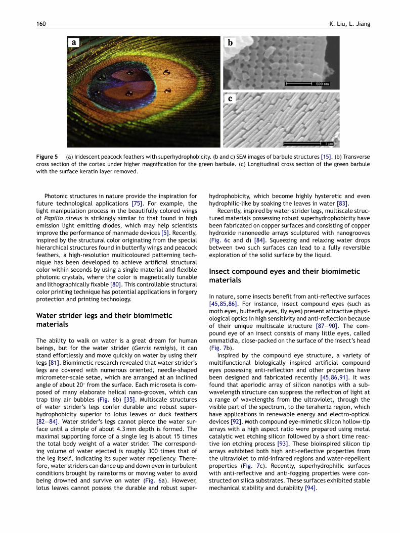
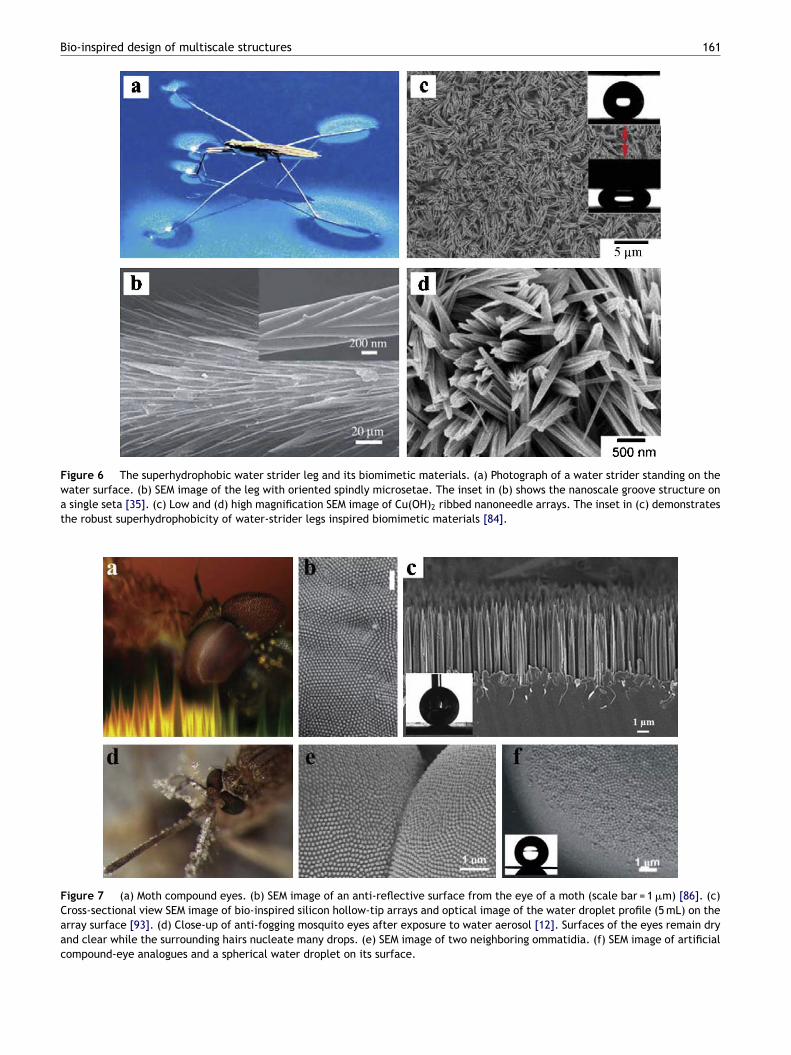
he ability to walk on water is a great dream for humaneings, but for the water strider (Gerris remigis), it cantand effortlessly and move quickly on water by using theiregs [81]. Biomimetic research revealed that water strider’segs are covered with numerous oriented, needle-shapedicrometer-scale setae, which are arranged at an inclined
ngle of about 20◦ from the surface. Each microseta is com-osed of many elaborate helical nano-grooves, which canrap tiny air bubbles (Fig. 6b) [35]. Multiscale structuresf water strider’s legs confer durable and robust super-ydrophobicity superior to lotus leaves or duck feathers82—84]. Water strider’s legs cannot pierce the water sur-ace until a dimple of about 4.3 mm depth is formed. Theaximal supporting force of a single leg is about 15 times
he total body weight of a water strider. The correspond-ng volume of water ejected is roughly 300 times that of
he leg itself, indicating its super water repellency. There-ore, water striders can dance up and down even in turbulentonditions brought by rainstorms or moving water to avoideing drowned and survive on water (Fig. 6a). However,otus leaves cannot possess the durable and robust super-tpwsm
n barbule. (c) Longitudinal cross section of the green barbule
ydrophobicity, which become highly hysteretic and evenydrophilic-like by soaking the leaves in water [83].
Recently, inspired by water-strider legs, multiscale struc-ured materials possessing robust superhydrophobicity haveeen fabricated on copper surfaces and consisting of copperydroxide nanoneedle arrays sculptured with nanogroovesFig. 6c and d) [84]. Squeezing and relaxing water dropsetween two such surfaces can lead to a fully reversiblexploration of the solid surface by the liquid.
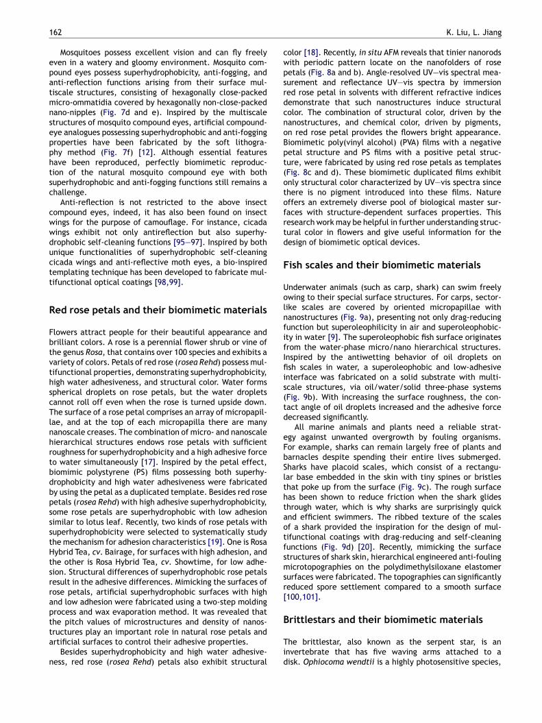
nsect compound eyes and their biomimeticaterials
n nature, some insects benefit from anti-reflective surfaces45,85,86]. For instance, insect compound eyes (such asoth eyes, butterfly eyes, fly eyes) present attractive physi-
logical optics in high sensitivity and anti-reflection becausef their unique multiscale structure [87—90]. The com-ound eye of an insect consists of many little eyes, calledmmatidia, close-packed on the surface of the insect’s headFig. 7b).
Inspired by the compound eye structure, a variety ofultifunctional biologically inspired artificial compound
yes possessing anti-reflection and other properties haveeen designed and fabricated recently [45,86,91]. It wasound that aperiodic array of silicon nanotips with a sub-avelength structure can suppress the reflection of light atrange of wavelengths from the ultraviolet, through the
isible part of the spectrum, to the terahertz region, whichave applications in renewable energy and electro-opticalevices [92]. Moth compound eye-mimetic silicon hollow-tiprrays with a high aspect ratio were prepared using metalatalytic wet etching silicon followed by a short time reac-ive ion etching process [93]. These bioinspired silicon tiprrays exhibited both high anti-reflective properties from
he ultraviolet to mid-infrared regions and water-repellentroperties (Fig. 7c). Recently, superhydrophilic surfacesith anti-reflective and anti-fogging properties were con-tructed on silica substrates. These surfaces exhibited stableechanical stability and durability [94].
Bio-inspired design of multiscale structures 161
Figure 6 The superhydrophobic water strider leg and its biomimetic materials. (a) Photograph of a water strider standing on thewater surface. (b) SEM image of the leg with oriented spindly microsetae. The inset in (b) shows the nanoscale groove structure ona single seta [35]. (c) Low and (d) high magnification SEM image of Cu(OH)2 ribbed nanoneedle arrays. The inset in (c) demonstratesthe robust superhydrophobicity of water-strider legs inspired biomimetic materials [84].
Figure 7 (a) Moth compound eyes. (b) SEM image of an anti-reflective surface from the eye of a moth (scale bar = 1 �m) [86]. (c)Cross-sectional view SEM image of bio-inspired silicon hollow-tip arrays and optical image of the water droplet profile (5 mL) on thearray surface [93]. (d) Close-up of anti-fogging mosquito eyes after exposure to water aerosol [12]. Surfaces of the eyes remain dryand clear while the surrounding hairs nucleate many drops. (e) SEM image of two neighboring ommatidia. (f) SEM image of artificialcompound-eye analogues and a spherical water droplet on its surface.
1
epatmnsepphtsc
cwwductt
R
FbtvthscTlnhrtbdbpssstHtsrraptta
n
cwpsrdcnoBpt(otofrtd
F
UolnfifIfiis(td
eFbSlthtaotfsmsr[
62
Mosquitoes possess excellent vision and can fly freelyven in a watery and gloomy environment. Mosquito com-ound eyes possess superhydrophobicity, anti-fogging, andnti-reflection functions arising from their surface mul-iscale structures, consisting of hexagonally close-packedicro-ommatidia covered by hexagonally non-close-packed
ano-nipples (Fig. 7d and e). Inspired by the multiscaletructures of mosquito compound eyes, artificial compound-ye analogues possessing superhydrophobic and anti-foggingroperties have been fabricated by the soft lithogra-hy method (Fig. 7f) [12]. Although essential featuresave been reproduced, perfectly biomimetic reproduc-ion of the natural mosquito compound eye with bothuperhydrophobic and anti-fogging functions still remains ahallenge.
Anti-reflection is not restricted to the above insectompound eyes, indeed, it has also been found on insectings for the purpose of camouflage. For instance, cicadaings exhibit not only antireflection but also superhy-rophobic self-cleaning functions [95—97]. Inspired by bothnique functionalities of superhydrophobic self-cleaningicada wings and anti-reflective moth eyes, a bio-inspiredemplating technique has been developed to fabricate mul-ifunctional optical coatings [98,99].
ed rose petals and their biomimetic materials
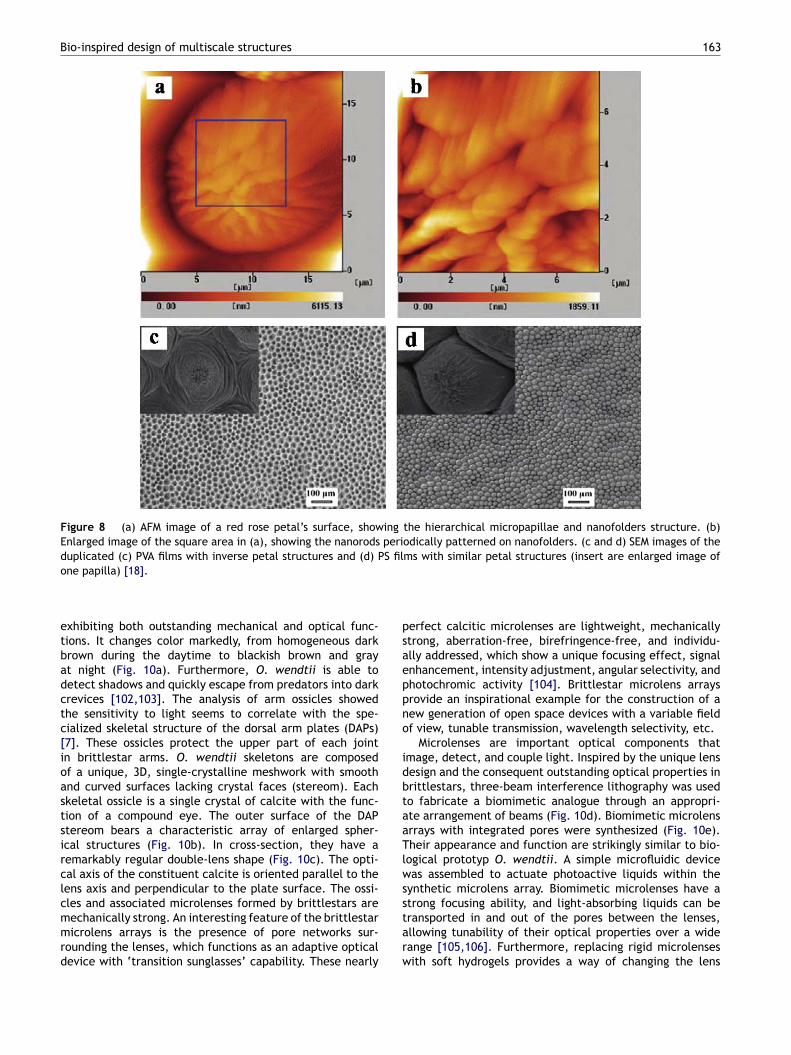
lowers attract people for their beautiful appearance andrilliant colors. A rose is a perennial flower shrub or vine ofhe genus Rosa, that contains over 100 species and exhibits aariety of colors. Petals of red rose (rosea Rehd) possess mul-ifunctional properties, demonstrating superhydrophobicity,igh water adhesiveness, and structural color. Water formspherical droplets on rose petals, but the water dropletsannot roll off even when the rose is turned upside down.he surface of a rose petal comprises an array of micropapil-
ae, and at the top of each micropapilla there are manyanoscale creases. The combination of micro- and nanoscaleierarchical structures endows rose petals with sufficientoughness for superhydrophobicity and a high adhesive forceo water simultaneously [17]. Inspired by the petal effect,iomimic polystyrene (PS) films possessing both superhy-rophobicity and high water adhesiveness were fabricatedy using the petal as a duplicated template. Besides red roseetals (rosea Rehd) with high adhesive superhydrophobicity,ome rose petals are superhydrophobic with low adhesionimilar to lotus leaf. Recently, two kinds of rose petals withuperhydrophobicity were selected to systematically studyhe mechanism for adhesion characteristics [19]. One is Rosaybrid Tea, cv. Bairage, for surfaces with high adhesion, andhe other is Rosa Hybrid Tea, cv. Showtime, for low adhe-ion. Structural differences of superhydrophobic rose petalsesult in the adhesive differences. Mimicking the surfaces ofose petals, artificial superhydrophobic surfaces with highnd low adhesion were fabricated using a two-step moldingrocess and wax evaporation method. It was revealed that
he pitch values of microstructures and density of nanos-ructures play an important role in natural rose petals andrtificial surfaces to control their adhesive properties.Besides superhydrophobicity and high water adhesive-ess, red rose (rosea Rehd) petals also exhibit structural
B
Tid
K. Liu, L. Jiang
olor [18]. Recently, in situ AFM reveals that tinier nanorodsith periodic pattern locate on the nanofolders of roseetals (Fig. 8a and b). Angle-resolved UV—vis spectral mea-urement and reflectance UV—vis spectra by immersioned rose petal in solvents with different refractive indicesemonstrate that such nanostructures induce structuralolor. The combination of structural color, driven by theanostructures, and chemical color, driven by pigments,n red rose petal provides the flowers bright appearance.iomimetic poly(vinyl alcohol) (PVA) films with a negativeetal structure and PS films with a positive petal struc-ure, were fabricated by using red rose petals as templatesFig. 8c and d). These biomimetic duplicated films exhibitnly structural color characterized by UV—vis spectra sincehere is no pigment introduced into these films. Natureffers an extremely diverse pool of biological master sur-aces with structure-dependent surfaces properties. Thisesearch work may be helpful in further understanding struc-ural color in flowers and give useful information for theesign of biomimetic optical devices.
ish scales and their biomimetic materials
nderwater animals (such as carp, shark) can swim freelywing to their special surface structures. For carps, sector-ike scales are covered by oriented micropapillae withanostructures (Fig. 9a), presenting not only drag-reducingunction but superoleophilicity in air and superoleophobic-ty in water [9]. The superoleophobic fish surface originatesrom the water-phase micro/nano hierarchical structures.nspired by the antiwetting behavior of oil droplets onsh scales in water, a superoleophobic and low-adhesive
nterface was fabricated on a solid substrate with multi-cale structures, via oil/water/solid three-phase systemsFig. 9b). With increasing the surface roughness, the con-act angle of oil droplets increased and the adhesive forceecreased significantly.
All marine animals and plants need a reliable strat-gy against unwanted overgrowth by fouling organisms.or example, sharks can remain largely free of plants andarnacles despite spending their entire lives submerged.harks have placoid scales, which consist of a rectangu-ar base embedded in the skin with tiny spines or bristleshat poke up from the surface (Fig. 9c). The rough surfaceas been shown to reduce friction when the shark glideshrough water, which is why sharks are surprisingly quicknd efficient swimmers. The ribbed texture of the scalesf a shark provided the inspiration for the design of mul-ifunctional coatings with drag-reducing and self-cleaningunctions (Fig. 9d) [20]. Recently, mimicking the surfacetructures of shark skin, hierarchical engineered anti-foulingicrotopographies on the polydimethylsiloxane elastomer
urfaces were fabricated. The topographies can significantlyeduced spore settlement compared to a smooth surface100,101].
rittlestars and their biomimetic materials
he brittlestar, also known as the serpent star, is annvertebrate that has five waving arms attached to aisk. Ophiocoma wendtii is a highly photosensitive species,
Bio-inspired design of multiscale structures 163
Figure 8 (a) AFM image of a red rose petal’s surface, showing the hierarchical micropapillae and nanofolders structure. (b)Enlarged image of the square area in (a), showing the nanorods periodically patterned on nanofolders. (c and d) SEM images of the
S fil
psaeppno
idbtaaTlws
duplicated (c) PVA films with inverse petal structures and (d) Pone papilla) [18].
exhibiting both outstanding mechanical and optical func-tions. It changes color markedly, from homogeneous darkbrown during the daytime to blackish brown and grayat night (Fig. 10a). Furthermore, O. wendtii is able todetect shadows and quickly escape from predators into darkcrevices [102,103]. The analysis of arm ossicles showedthe sensitivity to light seems to correlate with the spe-cialized skeletal structure of the dorsal arm plates (DAPs)[7]. These ossicles protect the upper part of each jointin brittlestar arms. O. wendtii skeletons are composedof a unique, 3D, single-crystalline meshwork with smoothand curved surfaces lacking crystal faces (stereom). Eachskeletal ossicle is a single crystal of calcite with the func-tion of a compound eye. The outer surface of the DAPstereom bears a characteristic array of enlarged spher-ical structures (Fig. 10b). In cross-section, they have aremarkably regular double-lens shape (Fig. 10c). The opti-cal axis of the constituent calcite is oriented parallel to thelens axis and perpendicular to the plate surface. The ossi-
cles and associated microlenses formed by brittlestars aremechanically strong. An interesting feature of the brittlestarmicrolens arrays is the presence of pore networks sur-rounding the lenses, which functions as an adaptive opticaldevice with ‘transition sunglasses’ capability. These nearlystarw
ms with similar petal structures (insert are enlarged image of
erfect calcitic microlenses are lightweight, mechanicallytrong, aberration-free, birefringence-free, and individu-lly addressed, which show a unique focusing effect, signalnhancement, intensity adjustment, angular selectivity, andhotochromic activity [104]. Brittlestar microlens arraysrovide an inspirational example for the construction of aew generation of open space devices with a variable fieldf view, tunable transmission, wavelength selectivity, etc.
Microlenses are important optical components thatmage, detect, and couple light. Inspired by the unique lensesign and the consequent outstanding optical properties inrittlestars, three-beam interference lithography was usedo fabricate a biomimetic analogue through an appropri-te arrangement of beams (Fig. 10d). Biomimetic microlensrrays with integrated pores were synthesized (Fig. 10e).heir appearance and function are strikingly similar to bio-
ogical prototyp O. wendtii. A simple microfluidic deviceas assembled to actuate photoactive liquids within the
ynthetic microlens array. Biomimetic microlenses have a
trong focusing ability, and light-absorbing liquids can beransported in and out of the pores between the lenses,llowing tunability of their optical properties over a wideange [105,106]. Furthermore, replacing rigid microlensesith soft hydrogels provides a way of changing the lens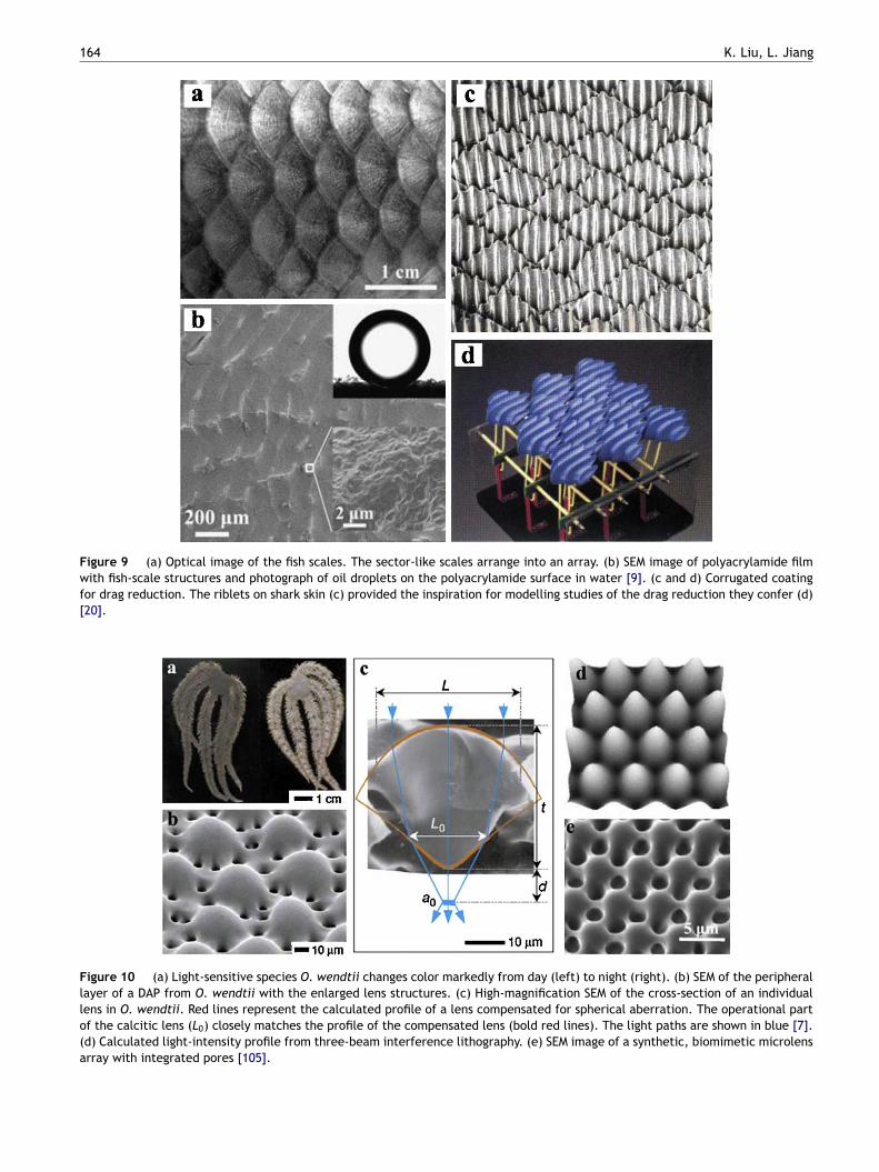
164 K. Liu, L. Jiang
Figure 9 (a) Optical image of the fish scales. The sector-like scales arrange into an array. (b) SEM image of polyacrylamide filmwith fish-scale structures and photograph of oil droplets on the polyacrylamide surface in water [9]. (c and d) Corrugated coatingfor drag reduction. The riblets on shark skin (c) provided the inspiration for modelling studies of the drag reduction they confer (d)[20].
Figure 10 (a) Light-sensitive species O. wendtii changes color markedly from day (left) to night (right). (b) SEM of the peripherallayer of a DAP from O. wendtii with the enlarged lens structures. (c) High-magnification SEM of the cross-section of an individuallens in O. wendtii. Red lines represent the calculated profile of a lens compensated for spherical aberration. The operational partof the calcitic lens (L0) closely matches the profile of the compensated lens (bold red lines). The light paths are shown in blue [7].(d) Calculated light-intensity profile from three-beam interference lithography. (e) SEM image of a synthetic, biomimetic microlensarray with integrated pores [105].
ttatvTbwntfiSonofit(icawistinaldsmne
sssaafslapcRsfiobeptttw
Bio-inspired design of multiscale structures
geometry and refractive index continuously in response toexternal stimuli, resulting in intelligent, multifunctional,tunable optics [107]. Nature provides a whole host of supe-rior multifunctional structures. These biological principleswill improve our current capabilities for the fabrication ofoptical elements and contribute to the construction of novelmicro-scale optical devices.
Spider silks and their biomimetic materials
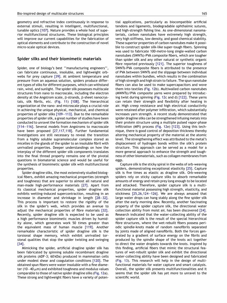
Spider, one of biology’s best ‘‘manufacturing engineers’’,can fabricate continuous, insoluble, and lightweight orb-webs for prey capture [39]. At ambient temperature andpressure from an aqueous solution, spiders produce differ-ent types of silks for different purposes, which can withstandrain, wind, and sunlight. The spider silk possesses multiscalestructures from nano to macroscale, including the electrondensity at the Angstrom scale, �-strands, �-sheet nanocrys-tals, silk fibrils, etc. (Fig. 11) [108]. The hierarchicalorganization at the nano- and microscale plays a crucial rolein achieving the unique physical, mechanical, and chemicalproperties of spider silks [109—113]. Due to the remarkableproperties of spider silk, a great number of studies have beenconducted to unravel the formation mechanism of spider silk[114—116]. Several models from Angstroms to macroscalehave been proposed [27,117,118]. Further fundamentalinvestigations are still necessary to reveal the transitionfrom a highly soluble supramolecular complex stored asmicelles in the glands of the spider to an insoluble fibril withunrivalled properties. Deeper understandings on how theinterplay of the different spider silk components translatesinto the final thread property remains one of the pivotalquestions in biomaterial science and would be useful forthe synthesis of biomimetic spider silk with multifunctionalproperties.
Spider dragline silks, the most extensively studied biolog-ical fibers, exhibit amazing mechanical properties (strengthand toughness) that are superior to almost all natural andman-made high-performance materials [27]. Apart fromits classical mechanical properties, spider dragline silkexhibits wetting-induced supercontraction, resulting in anincrease in diameter and shrinkage in length [28—32].This process is important to restore the rigidity of thesilk in the spider’s web, which provides an avenue toadjust the mechanical properties of fibre materials [32].Recently, spider dragline silk is expected to be used asa high performance biomimetic muscles driven by humid-ity alone, which generates work 50 times greater thanthe equivalent mass of human muscle [119]. Anotherremarkable characteristic of spider dragline silk is thetorsional shape memory, which exhibit unrivalled tor-sional qualities that stop the spider twisting and swinging[34].
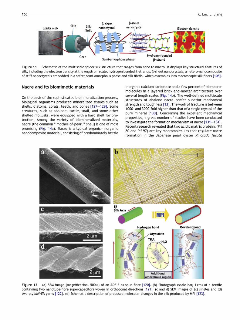
Mimicking the spider, artificial dragline spider silk hasbeen fabricated by spinning soluble recombinant draglinesilk proteins (ADF-3; 60 kDa) produced in mammalian cells
under modest shear and coagulation conditions [120]. Theobtained spun fibers were water insoluble with a fine diame-ter (10—40 �m) and exhibited toughness and modulus valuescomparable to those of native spider dragline silks (Fig. 12a).These strong and lightweight fibers have a variety of poten-(fOss
165
ial applications, particularly as biocompatible artificialendons and ligaments, biodegradable ophthalmic sutures,nd high-strength fishing line. As one-dimensional nanoma-erials, carbon nanotubes have extremely high strength,ery high stiffness, low density, and good chemical stability.hese superior properties of carbon nanotubes make it possi-le to construct spider silk-like super-tough fibers. Spinningas used to fabricate 100-metre-long single-walled carbonanotubes (SWNTs)-PVA composite fibers, which are tougherhan spider silk and any other natural or synthetic organicbre reported previously [121]. The superior toughness ofWNTs-PVA composite fibers is attributed to the presencef PVA between SWNTs and the slippage between individualanotubes within bundles, which results in the combinationf high strength and high strain to failure. The spun nanotubebers can also be used to make supercapacitors and wovehem into textiles (Fig. 12b). Multiwalled carbon nanotubesMWNTs)/PVA composite yarns were prepared by introduc-ng twist during spinning (Fig. 12c and d) [122]. These yarnsan retain their strength and flexibility after heating inir. High creep resistance and high electrical conductivityere retained after polymer infiltration, which substantially
ncreases yarn strength. A recent study demonstrated thatpider dragline silks can be strengthened infusing metals intoheir protein structure using a multiple pulsed vapor-phasenfiltration (MPI) process (Fig. 12e) [123]. Using this tech-ique, there is good control of deposition thickness therebyltering mechanical property of the material at the atomicevel. The strengthening effect was attributed to the metal’sisplacement of hydrogen bonds within the silk’s proteintructure. This approach can be served as a model for aore general approach to enhance the strength and tough-
ess of other biomaterials, such as collagen membranes fromggs.
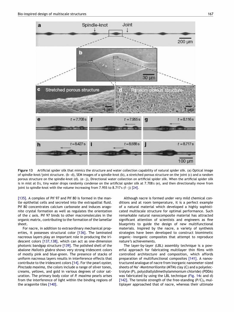
Capture silk is the sticky spiral in the webs of orb-weavingpiders, demonstrating exceptional elasticity [25]. Captureilk is five times as elastic as dragline silk. Orb-weavingpiders rely on sticky capture silks to absorb remarkablemounts of energy and retain prey long enough to be locatednd attacked. Therefore, spider capture silk is a multi-unctional material possessing high strength, elasticity, andtickiness [25,26,124—126]. We are always amazed thatarge water drops can hang stably along the fine spider silkfter the early morning dew. Recently, another fascinatingroperty of the spider capture silk, the directional waterollection ability from moist air, has been discovered [24].esearch indicated that the water-collecting ability of thepider capture silk is the result of the special hierarchicalbre structures, where the wet-rebuilt fibers possess peri-dic spindle-knots made of random nanofibrils separatedy joints made of aligned nanofibrils. Both the forces gen-rated by a gradient of surface energy on the fibrils androduced by the spindle shape of the knots act togethero direct the water droplets towards the knots. Inspired byhis finding, artificial fibers that mimic the structural fea-ures of wet-rebuilt spider silk and exhibit the directionalater-collecting ability have been designed and fabricated
Fig. 13). This research will help in the design of multi-unctional materials for water capture and smart catalysis.verall, the spider silk presents multifunctionalities and iteems that the spider silk has yet more to unravel to thecientific world.
166 K. Liu, L. Jiang
F at ras en boo e and
N
Obscstnpn
imsss1p
Fct
igure 11 Schematic of the multiscale spider silk structure thilk, including the electron density at the Angstrom scale, hydrogf stiff nanocrystals embedded in a softer semi-amorphous phas
acre and its biomimetic materials
n the basis of the sophisticated biomineralization process,iological organisms produced mineralized tissues such ashells, diatoms, corals, teeth, and bones [127—129]. Somereatures, such as abalone, turtle, snail, and some otherhelled mollusks, were equipped with a hard shell for pro-
ection. Among the variety of biomineralized materials,acre (the common ‘‘mother-of-pearl’’ shell) is one of mostromising (Fig. 14a). Nacre is a typical organic—inorganicanocomposite material, consisting of predominately brittleptR8f
igure 12 (a) SEM image (magnification, 500×) of an ADF-3 as-sontaining two nanotube-fibre supercapacitors woven in orthogonawo-ply MWNTs yarns [122]. (e) Schematic description of proposed m
nges from nano to macro. It displays key structural features ofnded �-strands, �-sheet nanocrystals, a hetero-nanocompositesilk fibrils, which assembles into macroscopic silk fibers [108].
norganic calcium carbonate and a few percent of biomacro-olecules in a layered brick-and-mortar architecture over
everal length scales (Fig. 14b). The well-defined multiscaletructures of abalone nacre confer superior mechanicaltrength and toughness [13]. The work of fracture is between000- and 3000-fold higher than that of a single crystal of theure mineral [130]. Concerning the excellent mechanical
roperties, a great number of studies have been conductedo investigate the formation mechanism of nacre [131—134].ecent research revealed that two acidic matrix proteins (Pif0 and Pif 97) are key macromolecules that regulate nacreormation in the Japanese pearl oyster Pinctada fucatapun fibre [120]. (b) Photograph (scale bar, 1 cm) of a textilel directions [121]. (c and d) SEM images of (c) singles and (d)olecular changes in the silk produced by MPI [123].
Bio-inspired design of multiscale structures 167
Figure 13 Artificial spider silk that mimics the structure and water collection capability of natural spider silk. (a) Optical imageof spindle-knot/joint structure. (b—d), SEM images of a spindle-knot (b), a stretched porous structure on the joint (c) and a random
er ctifici8.717
docrsbmson
ecpt
porous structure on the spindle-knot (d). (e—j), Directional watis in mist at 0 s, tiny water drops randomly condense on the arjoint to spindle-knot with the volume increasing from 7.955 to
[135]. A complex of Pif 97 and Pif 80 is formed in the man-tle epithelial cells and secreted into the extrapallial fluid.Pif 80 concentrates calcium carbonate and induces arago-nite crystal formation as well as regulates the orientationof the c axis. Pif 97 binds to other macromolecules in theorganic matrix, contributing to the formation of the lamellarsheet.
For nacre, in addition to extraordinary mechanical prop-erties, it possesses structural color [136]. The laminatednacreous layers play an important role in producing the iri-descent colors [137,138], which can act as one-dimensionphotonic bandgap structure [139]. The polished shell of theabalone Haliotis glabra shows very strong iridescent colorsof mostly pink and blue-green. The presence of stacks ofuniform nacreous layers results in interference effects thatcontribute to the iridescent colors [14]. For the pearl oyster,
Pinctada maxima, the colors include a range of silver tones,creams, yellows, and gold in various degrees of color sat-uration. The primary body color of P. maxima pearls arisesfrom the interference of light within the binding regions ofthe aragonite tiles [140].stw[t
ollection on artificial spider silk. When the artificial spider silkal spider silk at 7.708 s (e), and then directionally move froms (f—j) [24].
Although nacre is formed under very mild chemical con-itions and at room temperature, it is a perfect examplef a natural material which developed a highly sophisti-ated multiscale structure for optimal performance. Suchemarkable natural nanocomposite material has attractedignificant attention of scientists and engineers as finelueprints to guide the design of new multifunctionalaterials. Inspired by the nacre, a variety of synthesis
trategies have been developed to construct biomimeticrganic—inorganic composites that attempt to reproduceature’s achievements.
The layer-by-layer (LBL) assembly technique is a pow-rful approach for fabricating multilayer thin films withontrolled architecture and composition, which affordsreparation of multifunctional composites [141]. A nanos-ructured analogue of nacre from inorganic nanometer-sized
+
heets of Na -Montmorillonite (MTM) clay (C) and a polyelec-rolyte (P), poly(diallyldimethylammonium chloride) (PDDA)as fabricated by using the LBL technique (Fig. 14c and d)142]. The tensile strength of the free-standing (P/C)n mul-iplayer approached that of nacre, whereas their ultimate

168 K. Liu, L. Jiang
Figure 14 (a) Typical digital photograph of the inner nacreous layer of the abalone shell. (b) SEM image of a fracture surface innacre. (c) Schematic illustration of the (P/C)n film structure. The thickness of each clay platelet is 0.9 nm. (d) SEM image of an edgeof a (P/C)100 film [142]. Optical microscopy images of human osteoblasts cultured on a bare glass slide (e) and on a glass slide coatedw ionac 1000c
Ycisfmapw
ntipe
ith (PDDA/MTM/PDDA/Ag)10 (f) for 3 days [143]. (g) Cross-secthitosan hybrid film (3 wt.% chitosan solution, spin coating ato-assembly VCu-NO3/VCo-Al-CO3 = 3:1-chitosan hybrid film [146].
oung modulus was similar to that of lamellar bones. Theseomposites were expected to be used as potential bonemplants. By alternating clay layers with starch-stabilizedilver nanoparticles, nanostructured, hybrid, and multi-unctional PDDA/MTM/PDDA/Ag composites possessing good
echanical properties, strong antibacterial characteristicss well as biocompatibility with human osteoblasts wererepared using a LBL assembly (Fig. 14e and f). Withide variety of materials available, these high strength
ffoL
l SEM image of co-assembly structure of VCu-NO3/VCo-Al-CO3 = 3:1-rpm), the inset images are magnifications. (h) Photograph of
acre-like composites can be expanded to additional func-ionalities [143]. LBL assembly has also proven invaluablen its ability to merge the functionalities of its com-onents. Nacre-like multifunctional materials with bothxcellent mechanical and optical properties were fabricated
rom nano to microscale [144,145]. Recently, a series ofree-standing, strong, transparent, and functional layeredrganic—inorganic hybrid films were fabricated through aBL assembly procedure using layered double hydroxides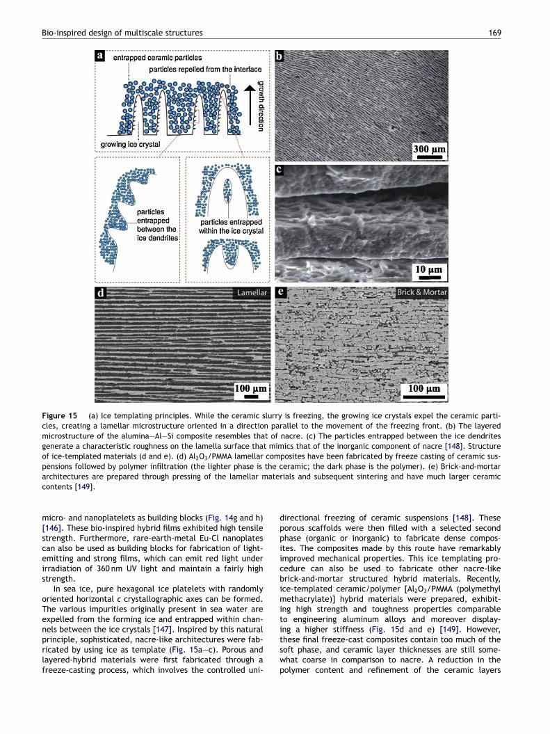
Bio-inspired design of multiscale structures 169
Figure 15 (a) Ice templating principles. While the ceramic slurry is freezing, the growing ice crystals expel the ceramic parti-cles, creating a lamellar microstructure oriented in a direction parallel to the movement of the freezing front. (b) The layeredmicrostructure of the alumina—Al—Si composite resembles that of nacre. (c) The particles entrapped between the ice dendritesgenerate a characteristic roughness on the lamella surface that mimics that of the inorganic component of nacre [148]. Structureof ice-templated materials (d and e). (d) Al2O3/PMMA lamellar composites have been fabricated by freeze casting of ceramic sus-pensions followed by polymer infiltration (the lighter phase is the ceramic; the dark phase is the polymer). (e) Brick-and-mortar
ater
dppiicbimit
architectures are prepared through pressing of the lamellar mcontents [149].
micro- and nanoplatelets as building blocks (Fig. 14g and h)[146]. These bio-inspired hybrid films exhibited high tensilestrength. Furthermore, rare-earth-metal Eu-Cl nanoplatescan also be used as building blocks for fabrication of light-emitting and strong films, which can emit red light underirradiation of 360 nm UV light and maintain a fairly highstrength.
In sea ice, pure hexagonal ice platelets with randomlyoriented horizontal c crystallographic axes can be formed.The various impurities originally present in sea water areexpelled from the forming ice and entrapped within chan-
nels between the ice crystals [147]. Inspired by this naturalprinciple, sophisticated, nacre-like architectures were fab-ricated by using ice as template (Fig. 15a—c). Porous andlayered-hybrid materials were first fabricated through afreeze-casting process, which involves the controlled uni-itswp
ials and subsequent sintering and have much larger ceramic
irectional freezing of ceramic suspensions [148]. Theseorous scaffolds were then filled with a selected secondhase (organic or inorganic) to fabricate dense compos-tes. The composites made by this route have remarkablymproved mechanical properties. This ice templating pro-edure can also be used to fabricate other nacre-likerick-and-mortar structured hybrid materials. Recently,ce-templated ceramic/polymer [Al2O3/PMMA (polymethylethacrylate)] hybrid materials were prepared, exhibit-
ng high strength and toughness properties comparableo engineering aluminum alloys and moreover display-
ng a higher stiffness (Fig. 15d and e) [149]. However,hese final freeze-cast composites contain too much of theoft phase, and ceramic layer thicknesses are still some-hat coarse in comparison to nacre. A reduction in theolymer content and refinement of the ceramic layers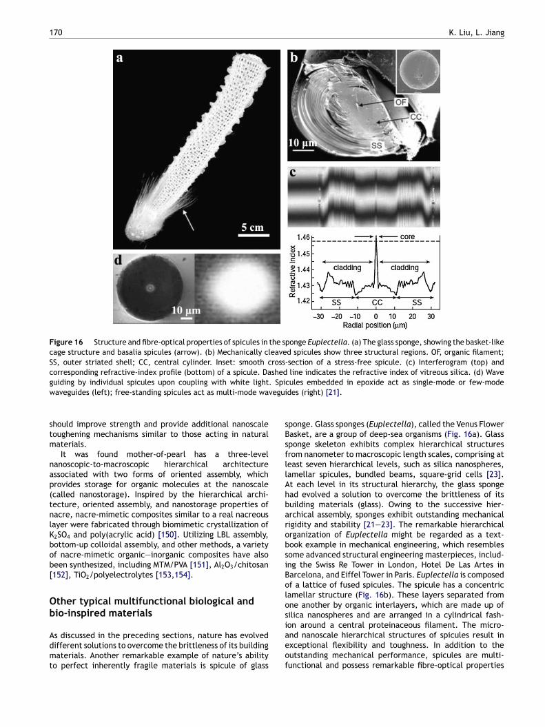
170 K. Liu, L. Jiang
Figure 16 Structure and fibre-optical properties of spicules in the sponge Euplectella. (a) The glass sponge, showing the basket-likecage structure and basalia spicules (arrow). (b) Mechanically cleaved spicules show three structural regions. OF, organic filament;SS, outer striated shell; CC, central cylinder. Inset: smooth cross-section of a stress-free spicule. (c) Interferogram (top) andcorresponding refractive-index profile (bottom) of a spicule. Dashed line indicates the refractive index of vitreous silica. (d) Waveg . Spiw vegui
stm
nap(tnlKbob[
Ob
Admt
sBsfllAhbarobsiBolos
uiding by individual spicules upon coupling with white lightaveguides (left); free-standing spicules act as multi-mode wa
hould improve strength and provide additional nanoscaleoughening mechanisms similar to those acting in naturalaterials.It was found mother-of-pearl has a three-level
anoscopic-to-macroscopic hierarchical architecturessociated with two forms of oriented assembly, whichrovides storage for organic molecules at the nanoscalecalled nanostorage). Inspired by the hierarchical archi-ecture, oriented assembly, and nanostorage properties ofacre, nacre-mimetic composites similar to a real nacreousayer were fabricated through biomimetic crystallization of2SO4 and poly(acrylic acid) [150]. Utilizing LBL assembly,ottom-up colloidal assembly, and other methods, a varietyf nacre-mimetic organic—inorganic composites have alsoeen synthesized, including MTM/PVA [151], Al2O3/chitosan152], TiO2/polyelectrolytes [153,154].
ther typical multifunctional biological andio-inspired materials
s discussed in the preceding sections, nature has evolvedifferent solutions to overcome the brittleness of its buildingaterials. Another remarkable example of nature’s ability
o perfect inherently fragile materials is spicule of glass
iaeof
cules embedded in epoxide act as single-mode or few-modedes (right) [21].
ponge. Glass sponges (Euplectella), called the Venus Flowerasket, are a group of deep-sea organisms (Fig. 16a). Glassponge skeleton exhibits complex hierarchical structuresrom nanometer to macroscopic length scales, comprising ateast seven hierarchical levels, such as silica nanospheres,amellar spicules, bundled beams, square-grid cells [23].t each level in its structural hierarchy, the glass spongead evolved a solution to overcome the brittleness of itsuilding materials (glass). Owing to the successive hier-rchical assembly, sponges exhibit outstanding mechanicaligidity and stability [21—23]. The remarkable hierarchicalrganization of Euplectella might be regarded as a text-ook example in mechanical engineering, which resemblesome advanced structural engineering masterpieces, includ-ng the Swiss Re Tower in London, Hotel De Las Artes inarcelona, and Eiffel Tower in Paris. Euplectella is composedf a lattice of fused spicules. The spicule has a concentricamellar structure (Fig. 16b). These layers separated fromne another by organic interlayers, which are made up ofilica nanospheres and are arranged in a cylindrical fash-
on around a central proteinaceous filament. The micro-nd nanoscale hierarchical structures of spicules result inxceptional flexibility and toughness. In addition to theutstanding mechanical performance, spicules are multi-unctional and possess remarkable fibre-optical properties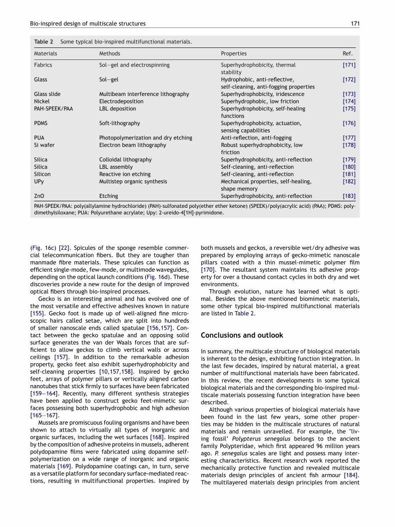
Bio-inspired design of multiscale structures 171
Table 2 Some typical bio-inspired multifunctional materials.
Materials Methods Properties Ref.
Fabrics Sol—gel and electrospinning Superhydrophobicity, thermalstability
[171]
Glass Sol—gel Hydrophobic, anti-reflective,self-cleaning, anti-fogging properties
[172]
Glass slide Multibeam interference lithography Superhydrophobicity, iridescence [173]Nickel Electrodeposition Superhydrophobic, low friction [174]PAH-SPEEK/PAA LBL deposition Superhydrophobicity, self-healing
functions[175]
PDMS Soft-lithography Superhydrophobicity, actuation,sensing capabilities
[176]
PUA Photopolymerization and dry etching Anti-reflection, anti-fogging [177]Si wafer Electron beam lithography Robust superhydrophobicity, low
friction[178]
Silica Colloidal lithography Superhydrophobicity, anti-reflection [179]Silica LBL assembly Self-cleaning, anti-reflection [180]Silicon Reactive ion etching Self-cleaning, anti-reflection [181]UPy Multistep organic synthesis Mechanical properties, self-healing,
shape memory[182]
ZnO Etching Superhydrophobicity, anti-reflection [183]
PAH-SPEEK/PAA: poly(allylamine hydrochloride) (PAH)-sulfonated poly(ether ether ketone) (SPEEK)/poly(acrylic acid) (PAA); PDMS: poly-]-pyr
bpp[ee
msa
C
IitnIbtd
btmif
dimethylsiloxane; PUA: Polyurethane acrylate; Upy: 2-ureido-4[1H
(Fig. 16c) [22]. Spicules of the sponge resemble commer-cial telecommunication fibers. But they are tougher thanmanmade fibre materials. These spicules can function asefficient single-mode, few-mode, or multimode waveguides,depending on the optical launch conditions (Fig. 16d). Thesediscoveries provide a new route for the design of improvedoptical fibers through bio-inspired processes.
Gecko is an interesting animal and has evolved one ofthe most versatile and effective adhesives known in nature[155]. Gecko foot is made up of well-aligned fine micro-scopic hairs called setae, which are split into hundredsof smaller nanoscale ends called spatulae [156,157]. Con-tact between the gecko spatulae and an opposing solidsurface generates the van der Waals forces that are suf-ficient to allow geckos to climb vertical walls or acrossceilings [157]. In addition to the remarkable adhesionproperty, gecko feet also exhibit superhydrophobicity andself-cleaning properties [10,157,158]. Inspired by geckofeet, arrays of polymer pillars or vertically aligned carbonnanotubes that stick firmly to surfaces have been fabricated[159—164]. Recently, many different synthesis strategieshave been applied to construct gecko feet-mimetic sur-faces possessing both superhydrophobic and high adhesion[165—167].
Mussels are promiscuous fouling organisms and have beenshown to attach to virtually all types of inorganic andorganic surfaces, including the wet surfaces [168]. Inspiredby the composition of adhesive proteins in mussels, adherent
polydopamine films were fabricated using dopamine self-polymerization on a wide range of inorganic and organicmaterials [169]. Polydopamine coatings can, in turn, serveas a versatile platform for secondary surface-mediated reac-tions, resulting in multifunctional properties. Inspired byaemmT
imidone.
oth mussels and geckos, a reversible wet/dry adhesive wasrepared by employing arrays of gecko-mimetic nanoscaleillars coated with a thin mussel-mimetic polymer film170]. The resultant system maintains its adhesive prop-rty for over a thousand contact cycles in both dry and wetnvironments.
Through evolution, nature has learned what is opti-al. Besides the above mentioned biomimetic materials,
ome other typical bio-inspired multifunctional materialsre listed in Table 2.
onclusions and outlook
n summary, the multiscale structure of biological materialss inherent to the design, exhibiting function integration. Inhe last few decades, inspired by natural material, a greatumber of multifunctional materials have been fabricated.n this review, the recent developments in some typicaliological materials and the corresponding bio-inspired mul-iscale materials possessing function integration have beenescribed.
Although various properties of biological materials haveeen found in the last few years, some other proper-ies may be hidden in the multiscale structures of naturalaterials and remain unravelled. For example, the ‘liv-
ng fossil’ Polypterus senegalus belongs to the ancientamily Polypteridae, which first appeared 96 million years
go. P. senegalus scales are light and possess many inter-sting characteristics. Recent research work reported theechanically protective function and revealed multiscaleaterials design principles of ancient fish armour [184].he multilayered materials design principles from ancient
1
abcorcusec
todfiwttbowmblnfnatrmi
A
WR2DN2DR
R
72
rmored fish will give clues for improved engineerediomimetic structural materials [185]. Therefore, interdis-iplinary cooperation is necessary for researchers in the areaf science and engineering to further investigate the natu-al materials and discover the new function. The increasingollaboration work would also be useful for the improvednderstanding of multiscale design laws, clarification oftructure-multifunction relationship, extraction of usefulngineering principles, and adaptation of models for practi-al applications.
Nature has learned how to achieve most efficient mul-ifunctional structures, i.e., functional integration. Theptimized biological solution should give us inspiration andesign principles for the construction of multifunctional arti-cial materials with multiscale structures. Most of currentork has still focused on the biomimetic synthesis of mul-
iscale structures inspired by one biological materials. Inhe near future, the following research directions shoulde a growing and vigorous field. (i) To extend the functionf bio-inspired multiscale structures through modificationith functional molecules. (ii) To fabricate novel multiscaleaterials for functional integration inspired by two or moreiological materials [186]. For example, taking advantage ofayered nacre and the marine adhesive of mussels, a novelanostructured composite film was constructed [187]. Theusion of two or more seemingly distinct concepts found inature into a unique composite with excellent functions isn exciting direction for the fabrication of novel multifunc-ional materials. Although the biomimetic and bio-inspiredesearch is in its infancy, it is a rapidly growing and enor-ously promising field, which will become the focus of
nternational competition in the near future.
cknowledgements
e appreciate the financial support of National Basicesearch Program of China (2010CB934700, 2009CB930404,007CB936403), National High Technology Research andevelopment Program of China (2009AA03Z339), Nationalatural Science Foundation of China (20920102036,0974113, 21001013), Specialized Research Fund for theoctoral Program of Higher Education, and the Fundamentalesearch Funds for the Central Universities.
eferences
[1] C. Sanchez, H. Arribart, M.M.G. Guille, Nat. Mater. 4 (2005)277—288.
[2] Y.M. Zheng, X.F. Gao, L. Jiang, Soft Matter 3 (2007) 178—182.[3] P. Vukusic, J.R. Sambles, Nature 424 (2003) 852—855.[4] R.A. Potyrailo, H. Ghiradella, A. Vertiatchikh, K. Dovidenko,
J.R. Cournoyer, E. Olson, Nat. Photon 1 (2007) 123—128.[5] P. Vukusic, I. Hooper, Science 310 (2005) 1151—11151.[6] O. Sato, S. Kubo, Z.Z. Gu, Acc. Chem. Res. 42 (2009) 1—10.[7] J. Aizenberg, A. Tkachenko, S. Weiner, L. Addadi, G. Hendler,
Nature 412 (2001) 819—822.[8] T.L. Sun, L. Feng, X.F. Gao, L. Jiang, Acc. Chem. Res. 38 (2005)
644—652.[9] M.J. Liu, S.T. Wang, Z.X. Wei, Y.L. Song, L. Jiang, Adv. Mater.
21 (2009) 665—669.[10] W.R. Hansen, K. Autumn, Proc. Natl. Acad. Sci. U.S.A. 102
(2005) 385—389.
K. Liu, L. Jiang
[11] L. Feng, S.H. Li, Y.S. Li, H.J. Li, L.J. Zhang, J. Zhai, Y.L. Song,B.Q. Liu, L. Jiang, D.B. Zhu, Adv. Mater. 14 (2002) 1857—1860.
[12] X.F. Gao, X. Yan, X. Yao, L. Xu, K. Zhang, J.H. Zhang, B. Yang,L. Jiang, Adv. Mater. 19 (2007) 2213—2217.
[13] G. Mayer, Science 310 (2005) 1144—1147.[14] T.L. Tan, D. Wong, P. Lee, Opt. Express 12 (2004) 4847—4854.[15] J. Zi, X.D. Yu, Y.Z. Li, X.H. Hu, C. Xu, X.J. Wang, X.H. Liu, R.T.
Fu, Proc. Natl. Acad. Sci. U.S.A. 100 (2003) 12576—12578.[16] T. Stegmaier, M. Linke, H. Planck, Philos. Trans. R. Soc. A 367
(2009) 1749—1758.[17] L. Feng, Y.A. Zhang, J.M. Xi, Y. Zhu, N. Wang, F. Xia, L. Jiang,
Langmuir 24 (2008) 4114—4119.[18] L. Feng, Y.A. Zhang, M.Z. Li, Y.M. Zheng, W.Z. Shen, L. Jiang,
Langmuir 26 (2010) 14885—14888.[19] B. Bhushan, E.K. Her, Langmuir 26 (2010) 8207—8217.[20] P. Ball, Nature 400 (1999) 507—509.[21] V.C. Sundar, A.D. Yablon, J.L. Grazul, M. Ilan, J. Aizenberg,
Nature 424 (2003) 899—900.[22] J. Aizenberg, V.C. Sundar, A.D. Yablon, J.C. Weaver, G. Chen,
Proc. Natl. Acad. Sci. U.S.A. 101 (2004) 3358—3363.[23] J. Aizenberg, J.C. Weaver, M.S. Thanawala, V.C. Sundar, D.E.
Morse, P. Fratzl, Science 309 (2005) 275—278.[24] Y.M. Zheng, H. Bai, Z.B. Huang, X.L. Tian, F.Q. Nie, Y. Zhao,
J. Zhai, L. Jiang, Nature 463 (2010) 640—643.[25] N. Becker, E. Oroudjev, S. Mutz, J.P. Cleveland, P.K. Hansma,
C.Y. Hayashi, D.E. Makarov, H.G. Hansma, Nat. Mater. 2 (2003)278—283.
[26] B.O. Swanson, T.A. Blackledge, C.Y. Hayash, J. Exp. Zool.307A (2007) 654—666.
[27] F. Vollrath, D.P. Knight, Nature 410 (2001) 541—548.[28] R.W. Work, J. Exp. Biol. 118 (1985) 379—404.[29] Z.Z. Shao, F. Vollrath, Polymer 40 (1999) 1799—1806.[30] Z. Shao, F. Vollrath, J. Sirichaisit, R.J. Young, Polymer 40
(1999) 2493—2500.[31] J. Perez-Rigueiro, M. Elices, G.V. Guinea, Polymer 44 (2003)
3733—3736.[32] Y. Liu, Z.Z. Shao, F. Vollrath, Nat. Mater. 4 (2005) 901—905.[33] F.I. Bell, I.J. McEwen, C. Viney, Nature 416 (2002) 37—137.[34] O. Emile, A. Le Floch, F. Vollrath, Nature 440 (2006)
621—1621.[35] X.F. Gao, L. Jiang, Nature 432 (2004) 36—136.[36] T. Douglas, Science 299 (2003) 1192—1193.[37] P. Fratzl, F.G. Barth, Nature 462 (2009) 442—448.[38] J. Aizenberg, P. Fratzl, Adv. Mater. 21 (2009) 387—388.[39] F.G. Omenetto, D.L. Kaplan, Science 329 (2010) 528—531.[40] K. Liu, X. Yao, L. Jiang, Chem. Soc. Rev. 39 (2010) 3240—3255.[41] N. Huebsch, D.J. Mooney, Nature 462 (2009) 426—432.[42] L.P. Lee, R. Szema, Science 310 (2005) 1148—1150.[43] M.J. Liu, Y.M. Zheng, J. Zhai, L. Jiang, Acc. Chem. Res. 43
(2010) 368—377.[44] F. Xia, L. Jiang, Adv. Mater. 20 (2008) 2842—2858.[45] Y.F. Li, J.H. Zhang, B. Yang, Nano Today 5 (2010) 117—127.[46] K. Liu, L. Jiang, Nanoscale 3 (2011),
doi:10.1039/c1030nr00642d.[47] W. Barthlott, C. Neinhuis, Planta 202 (1997) 1—8.[48] C. Neinhuis, W. Barthlott, New Phytol. 138 (1998) 91—98.[49] A. Lafuma, D. Quéré, Nat. Mater. 2 (2003) 457—460.[50] J. Genzer, A. Marmur, MRS Bull. 33 (2008) 742—746.[51] B. Bhushan, Y.C. Jung, K. Koch, Philos. Trans. R. Soc. A 367
(2009) 1631—1672.[52] X. Zhang, F. Shi, J. Niu, Y.G. Jiang, Z.Q. Wang, J. Mater. Chem.
18 (2008) 621—633.[53] X. Yao, Y. Song, L. Jiang, Adv. Mater. 23 (2011),
doi:10.1002/adma.201002689.[54] H.Y. Erbil, A.L. Demirel, Y. Avci, O. Mert, Science 299 (2003)
1377—1380.[55] J.F.L. Duval, F. Gaboriaud, Curr. Opin. Colloid Interf. Sci. 15
(2010) 184—195.
[
[
[[[
[
[
[[
[[
[
[[
[
[
[
[[
[
[
[
[
[
[[[
Bio-inspired design of multiscale structures
[56] L. Jiang, Y. Zhao, J. Zhai, Angew. Chem. Int. Ed. 43 (2004)4338—4341.
[57] M.J. Xu, N. Lu, H.B. Xu, D.P. Qi, Y.D. Wang, S.L. Shi, L.F. Chi,Soft Matter 6 (2010) 1438—1443.
[58] Y.T. Peng, K.F. Lo, Y.J. Juang, Langmuir 26 (2010) 5167—5171.[59] M.H. Jin, X.J. Feng, J.M. Xi, J. Zhai, K.W. Cho, L. Feng, L.
Jiang, Macromol. Rapid Commun. 26 (2005) 1805—1809.[60] B. Cortese, S. D’Amone, M. Manca, I. Viola, R. Cingolani, G.
Gigli, Langmuir 24 (2008) 2712—2718.[61] K. Koch, B. Bhushan, Y.C. Jung, W. Barthlott, Soft Matter 5
(2009) 1386—1393.[62] X.H. Xu, Z.Z. Zhang, J. Yang, Langmuir 26 (2010) 3654—3658.[63] K.S. Liu, M.L. Zhang, J. Zhai, J. Wang, L. Jiang, Appl. Phys.
Lett. 92 (2008) 183103.[64] R. Pogreb, G. Whyman, R. Barayev, E. Bormashenko, D. Aur-
bach, Appl. Phys. Lett. 94 (2009) 221902.[65] K. Zhao, K.S. Liu, J.F. Li, W.H. Wang, L. Jiang, Scr. Mater. 60
(2009) 225—227.[66] M.D. Pei, B. Wang, E. Li, X.H. Zhang, X.M. Song, H. Yan, Appl.
Surf. Sci. 256 (2010) 5824—5827.[67] K.S. Liu, J. Zhai, L. Jiang, Nanotechnology 19 (2008) 165604.[68] C. Sun, Z.Z. Gu, H. Xu, Langmuir 25 (2009) 12439—12443.[69] W.J. Zhao, L.P. Wang, Q.J. Xue, J. Phys. Chem. C 114 (2010)
11509—11514.[70] J. Gao, Y.L. Liu, H.P. Xu, Z.Q. Wang, X. Zhang, Langmuir 25
(2009) 4365—4369.[71] J.A. Gao, Y.L. Liu, H.P. Xu, Z.Q. Wang, X. Zhang, Langmuir 26
(2010) 9673—9676.[72] S.Z. Wu, D. Wu, J. Yao, Q.D. Chen, J.N. Wang, L.G. Niu, H.H.
Fang, H.B. Sun, Langmuir 26 (2010) 12012—12016.[73] S. Kinoshita, S. Yoshioka, Chemphyschem 6 (2005)
1442—1459.[74] A.L. Ingram, A.R. Parker, Philos. Trans. R. Soc. B 363 (2008)
2465—2480.[75] A.R. Parker, Philos. Trans. R. Soc. A 367 (2009) 1759—1782.[76] S. Kinoshita, S. Yoshioka, K. Kawagoe, Proc. R. Soc. Lond. B
269 (2002) 1417—1421.[77] Z.Z. Gu, H. Uetsuka, K. Takahashi, R. Nakajima, H. Onishi, A.
Fujishima, O. Sato, Angew. Chem. Int. Ed. 42 (2003) 894—897.[78] J.Y. Huang, X.D. Wang, Z.L. Wang, Nano Lett. 6 (2006)
2325—2331.[79] M.R. Weatherspoon, Y. Cai, M. Crne, M. Srinivasarao, K.H.
Sandhage, Angew. Chem. Int. Ed. 47 (2008) 7921—7923.[80] H. Kim, J. Ge, J. Kim, S. Choi, H. Lee, H. Lee, W. Park, Y. Yin,
S. Kwon, Nat. Photon 3 (2009) 534—540.[81] D.L. Hu, B. Chan, J.W.M. Bush, Nature 424 (2003) 663—666.[82] P. Ball, Nat. Mater. 8 (2009) 250—1250.[83] J.H. Zhang, X.L. Sheng, L. Jiang, Langmuir 25 (2009)
1371—1376.[84] X. Yao, Q.W. Chen, L. Xu, Q.K. Li, Y.L. Song, X.F. Gao, D.
Quere, L. Jiang, Adv. Funct. Mater. 20 (2010) 656—662.[85] M. Srinivasarao, Chem. Rev. 99 (1999) 1935—1961.[86] A.R. Parker, H.E. Townley, Nat. Nanotechnol. 2 (2007)
347—353.[87] W.H. Miller, G.D. Bernard, J.L. Allen, Science 162 (1968)
760—767.[88] D.G. Stavenga, S. Foletti, G. Palasantzas, K. Arikawa, Proc.
R. Soc. Lond. B 273 (2006) 661—667.[89] G.S. Watson, J.A. Watson, Appl. Surf. Sci. 235 (2004)
139—144.[90] J.Y. Huang, X.D. Wang, Z.L. Wang, Nanotechnology 19 (2008)
025602.[91] K.H. Jeong, J. Kim, L.P. Lee, Science 312 (2006) 557—561.
[92] Y.F. Huang, S. Chattopadhyay, Y.J. Jen, C.Y. Peng, T.A. Liu,Y.K. Hsu, C.L. Pan, H.C. Lo, C.H. Hsu, Y.H. Chang, C.S. Lee,K.H. Chen, L.C. Chen, Nat. Nanotechnol. 2 (2007) 770—774.
[93] Y.F. Li, J.H. Zhang, S.J. Zhu, H.P. Dong, Z.H. Wang, Z.Q. Sun,J.R. Guo, B. Yang, J. Mater. Chem. 19 (2009) 1806—1810.
[
[
[
173
[94] Y.F. Li, J.H. Zhang, S.J. Zhu, H.P. Dong, F. Jia, Z.H. Wang, Z.Q.Sun, L. Zhang, Y. Li, H.B. Li, W.Q. Xu, B. Yang, Adv. Mater. 21(2009) 4731—4734.
[95] W. Lee, M.K. Jin, W.C. Yoo, J.K. Lee, Langmuir 20 (2004)7665—7669.
[96] G.M. Zhang, J. Zhang, G.Y. Xie, Z.F. Liu, H.B. Shao, Small 2(2006) 1440—1443.
[97] G.Y. Xie, G.M. Zhang, F. Lin, J. Zhang, Z.F. Liu, S.C. Mu, Nan-otechnology 19 (2008) 095605.
[98] C.H. Sun, A. Gonzalez, N.C. Linn, P. Jiang, B. Jiang, Appl.Phys. Lett. 92 (2008) 051107.
[99] W.L. Min, B. Jiang, P. Jiang, Adv. Mater. 20 (2008) 3914—3918.100] J.F. Schumacher, M.L. Carman, T.G. Estes, A.W. Feinberg, L.H.
Wilson, M.E. Callow, J.A. Callow, J.A. Finlay, A.B. Brennan,Biofouling 23 (2007) 55—62.
101] J.F. Schumacher, N. Aldred, M.E. Callow, J.A. Finlay, J.A. Cal-low, A.S. Clare, A.B. Brennan, Biofouling 23 (2007) 307—317.
102] G. Hendler, Pszni Mar. Ecol. 5 (1984) 379—401.103] G. Hendler, M. Byrne, Zoomorphology 107 (1987) 261—272.104] J. Aizenberg, G. Hendler, J. Mater. Chem. 14 (2004)
2066—2072.105] S. Yang, G. Chen, M. Megens, C.K. Ullal, Y.J. Han, R. Rapaport,
E.L. Thomas, J. Aizenberg, Adv. Mater. 17 (2005) 435—438.106] S. Yang, C.K. Ullal, E.L. Thomas, G. Chen, J. Aizenberg, Appl.
Phys. Lett. 86 (2005) 201121.107] S. Yang, J. Aizenberg, Mater. Today 8 (2005) 40—46.108] S. Keten, Z.P. Xu, B. Ihle, M.J. Buehler, Nat. Mater. 9 (2010)
359—367.109] D. Porter, F. Vollrath, Adv. Mater. 21 (2009) 487—492.110] M. Heim, D. Keerl, T. Scheibel, Angew. Chem. Int. Ed. 48
(2009) 3584—3596.111] C.J. Fu, Z.Z. Shao, V. Fritz, Chem. Commun. (2009)
6515—6529.112] A. George, S. Ravindran, Nano Today 5 (2010) 254—266.113] M. Heim, L. Römer, T. Scheibel, Chem. Soc. Rev. 39 (2010)
156—164.114] G. Askarieh, M. Hedhammar, K. Nordling, A. Saenz, C. Casals,
A. Rising, J. Johansson, S.D. Knight, Nature 465 (2010)236—238.
115] F. Hagn, L. Eisoldt, J.G. Hardy, C. Vendrely, M. Coles, T.Scheibel, H. Kessler, Nature 465 (2010) 239—242.
116] A. Nova, S. Keten, N.M. Pugno, A. Redaelli, M.J. Buehler, NanoLett. 10 (2010) 2626—2634.
117] H.J. Jin, D.L. Kaplan, Nature 424 (2003) 1057—1061.118] R. Silvers, F. Buhr, H. Schwalbe, Angew. Chem. Int. Ed. 49
(2010) 5410—5412.119] I. Agnarsson, A. Dhinojwala, V. Sahni, T.A. Blackledge, J. Exp.
Biol. 212 (2009) 1990—1994.120] A. Lazaris, S. Arcidiacono, Y. Huang, J.F. Zhou, F. Duguay, N.
Chretien, E.A. Welsh, J.W. Soares, C.N. Karatzas, Science 295(2002) 472—476.
121] A.B. Dalton, S. Collins, E. Munoz, J.M. Razal, V.H. Ebron, J.P.Ferraris, J.N. Coleman, B.G. Kim, R.H. Baughman, Nature 423(2003) 703—1703.
122] M. Zhang, K.R. Atkinson, R.H. Baughman, Science 306 (2004)1358—1361.
123] S.M. Lee, E. Pippel, U. Gosele, C. Dresbach, Y. Qin, C.V. Chan-dran, T. Brauniger, G. Hause, M. Knez, Science 324 (2009)488—492.
124] H.J. Zhou, Y. Zhang, Phys. Rev. Lett. 94 (2005) 028104.125] I. Agnarsson, T.A. Blackledge, J. Zool. 278 (2009) 134—140.126] V. Sahni, T.A. Blackledge, A. Dhinojwala, Nat. Commun. 1
(2010) 19.
127] P. Fratzl, R. Weinkamer, Prog. Mater. Sci. 52 (2007)1263—1334.128] M.A. Meyers, P.Y. Chen, A.Y.M. Lin, Y. Seki, Prog. Mater. Sci.
53 (2008) 1—206.129] M. Cusack, A. Freer, Chem. Rev. 108 (2008) 4433—4454.

1
[
[
[
[[
[
[[[
[
[
[
[
[
[
[
[
[
[
[
[[
[
[
[
[
[
[
[
[185] S.R. Barnes, MRS Bull. 33 (2008) 991—992.
74
130] X.D. Li, W.C. Chang, Y.J. Chao, R.Z. Wang, M. Chang, NanoLett. 4 (2004) 613—617.
131] A.G. Chec, J.H.E. Cartwright, M.G. Willinger, Proc. Natl.Acad. Sci. U.S.A. 106 (2009) 38—43.
132] J.H.E. Cartwright, A.G. Checa, B. Escribano, C.I. Sainz-Diaz,Proc. Natl. Acad. Sci. U.S.A. 106 (2009) 10499—10504.
133] N. Kroger, Science 325 (2009) 1351—1352.134] P.U.P.A. Gilbert, R.A. Metzler, D. Zhou, A. Scholl, A. Doran, A.
Young, M. Kunz, N. Tamura, S.N. Coppersmith, J. Am. Chem.Soc. 130 (2008) 17519—17527.
135] M. Suzuki, K. Saruwatari, T. Kogure, Y. Yamamoto, T.Nishimura, T. Kato, H. Nagasawa, Science 325 (2009)1388—1390.
136] B. Bhushan, Philos. Trans. R. Soc. A 367 (2009) 1445—1486.137] M.R. Snow, A. Pring, Am. Miner. 90 (2005) 1705—1711.138] L. Ghannam, H. Garay, J. Francois, L. Billon, Macromol.
Chem. Phys. 208 (2007) 1469—1479.139] B. Li, J. Zhou, L.T. Li, Q. Li, S. Han, Z.B. Hao, Chin. Sci. Bull.
50 (2005) 1529—1531.140] M.R. Snow, A. Pring, P. Self, D. Losic, J. Shapter, Am. Miner.
89 (2004) 1353—1358.141] X. Zhang, H. Chen, H.Y. Zhang, Chem. Commun. (2007)
1395—1405.142] Z.Y. Tang, N.A. Kotov, S. Magonov, B. Ozturk, Nat. Mater. 2
(2003) 413—418.143] P. Podsiadlo, S. Paternel, J.M. Rouillard, Z.F. Zhang, J.
Lee, J.W. Lee, L. Gulari, N.A. Kotov, Langmuir 21 (2005)11915—11921.
144] P. Podsiadlo, A.K. Kaushik, E.M. Arruda, A.M. Waas,B.S. Shim, J.D. Xu, H. Nandivada, B.G. Pumplin, J.Lahann, A. Ramamoorthy, N.A. Kotov, Science 318 (2007)80—83.
145] P. Podsiadlo, E.M. Arruda, E. Kheng, A.M. Waas, J. Lee, K.Critchley, M. Qin, E. Chuang, A.K. Kaushik, H.S. Kim, Y. Qi,S.T. Noh, N.A. Kotov, ACS Nano 3 (2009) 1564—1572.
146] H.-B. Yao, H.-Y. Fang, Z.-H. Tan, L.-H. Wu, S.-H. Yu, Angew.Chem. Int. Ed. 49 (2010) 2140—2145.
147] M.G. Worster, J.S. Wettlaufer, J. Phys. Chem. B 101 (1997)6132—6136.
148] S. Deville, E. Saiz, R.K. Nalla, A.P. Tomsia, Science 311 (2006)515—518.
149] E. Munch, M.E. Launey, D.H. Alsem, E. Saiz, A.P. Tomsia, R.O.Ritchie, Science 322 (2008) 1516—1520.
150] K. Oaki, H. Imai, Angew. Chem. Int. Ed. 44 (2005) 6571—6575.151] A. Walther, I. Bjurhager, J.M. Malho, J. Pere, J. Ruokolainen,
L.A. Berglund, O. Ikkala, Nano Lett. 10 (2010) 2742—2748.152] L.J. Bonderer, A.R. Studart, L.J. Gauckler, Science 319 (2008)
1069—1073.153] Z. Burghard, A. Tucic, L.R.H. Jeurgens, R.C. Hoffmann, J. Bill,
F. Aldinger, Adv. Mater. 19 (2007) 970—974.154] Z. Burghard, L. Zini, V. Srot, P. Bellina, P.A. van Aken, J. Bill,
Nano Lett. 9 (2009) 4103—4108.155] L.F. Boesel, C. Greiner, E. Arzt, A. del Campo, Adv. Mater. 22
(2010) 2125—2137.156] K. Autumn, Y.A. Liang, S.T. Hsieh, W. Zesch, W.P. Chan,
T.W. Kenny, R. Fearing, R.J. Full, Nature 405 (2000)681—685.
157] K. Autumn, M. Sitti, Y.C.A. Liang, A.M. Peattie, W.R. Hansen,S. Sponberg, T.W. Kenny, R. Fearing, J.N. Israelachvili, R.J.Full, Proc. Natl. Acad. Sci. U.S.A. 99 (2002) 12252—12256.
158] S. Sethi, L. Ge, L. Ci, P.M. Ajayan, A. Dhinojwala, Nano Lett.8 (2008) 822—825.
K. Liu, L. Jiang
[159] A.K. Geim, S.V. Dubonos, I.V. Grigorieva, K.S. Novoselov, A.A.Zhukov, S.Y. Shapoval, Nat. Mater. 2 (2003) 461—463.
[160] L. Qu, L. Dai, Adv. Mater. 19 (2007) 3844—3849.[161] L. Ge, S. Sethi, L. Ci, P.M. Ajayan, A. Dhinojwala, Proc. Natl.
Acad. Sci. U.S.A. 104 (2007) 10792—10795.[162] L.T. Qu, L.M. Dai, M. Stone, Z.H. Xia, Z.L. Wang, Science 322
(2008) 238—242.[163] A. Mahdavi, L. Ferreira, C. Sundback, J.W. Nichol, E.P. Chan,
D.J.D. Carter, C.J. Bettinger, S. Patanavanich, L. Chignozha,E. Ben-Joseph, A. Galakatos, H. Pryor, I. Pomerantseva, P.T.Masiakos, W. Faquin, A. Zumbuehl, S. Hong, J. Borenstein, J.Vacanti, R. Langer, J.M. Karp, Proc. Natl. Acad. Sci. U.S.A.105 (2008) 2307—2312.
[164] H.E. Jeong, J.K. Lee, H.N. Kim, S.H. Moon, K.Y. Suh, Proc.Natl. Acad. Sci. U.S.A. 106 (2009) 5639—5644.
[165] S.G. Park, H.H. Moon, S.K. Lee, J. Shim, S.M. Yang, Langmuir26 (2010) 1468—1472.
[166] S.J. Zhu, Y.F. Li, J.H. Zhang, C.L. Lu, X. Dai, F. Jia, H.N. Gao,B. Yang, J. Colloid Interf. Sci. 344 (2010) 541—546.
[167] J.Y. Peng, P.R. Yu, S.J. Zeng, X. Liu, J.R. Chen, W.J. Xu, J.Phys. Chem. C 114 (2010) 5926—5931.
[168] M.J. Harrington, A. Masic, N. Holten-Andersen, J.H. Waite, P.Fratzl, Science 328 (2010) 216—220.
[169] H. Lee, S.M. Dellatore, W.M. Miller, P.B. Messersmith, Science318 (2007) 426—430.
[170] H. Lee, B.P. Lee, P.B. Messersmith, Nature 448 (2007)338—341.
[171] H.S. Lim, J.H. Baek, K. Park, H.S. Shin, J. Kim, J.H. Cho, Adv.Mater. 22 (2010) 2138—2141.
[172] M. Faustini, L. Nicole, C. Boissiere, P. Innocenzi,C. Sanchez, D. Grosso, Chem. Mater. 22 (2010)4406—4413.
[173] D. Wu, Q.D. Chen, H. Xia, J. Jiao, B.B. Xu, X.F. Lin, Y. Xu, H.B.Sun, Soft Matter 6 (2010) 263—267.
[174] M. Shafiei, A.T. Alpas, Appl. Surf. Sci. 256 (2009) 710—719.[175] Y. Li, L. Li, J.G. Sun, Angew. Chem. Int. Ed. 49 (2010)
6129—6133.[176] B. Pokroy, A.K. Epstein, M.C.M. Persson-Gulda, J. Aizenberg,
Adv. Mater. 21 (2009) 463—469.[177] D. Tahk, T.-i. Kim, H. Yoon, M. Choi, K. Shin, K.Y. Suh, Lang-
muir 26 (2010) 2240—2243.[178] J. Shieh, F.J. Hou, Y.C. Chen, H.M. Chen, S.P. Yang, C.C.
Cheng, H.L. Chen, Adv. Mater. 22 (2010) 597—601.[179] Y.F. Li, J.H. Zhang, S.J. Zhu, H.P. Dong, F. Jia, Z.H. Wang,
Y. Tang, L.A. Zhang, S.Y. Zhang, B. Yang, Langmuir 26 (2010)9842—9847.
[180] X.Y. Li, X. Du, J.H. He, Langmuir 26 (2010) 13528—13534.[181] Y.D. Wang, N. Lu, H.B. Xu, G. Shi, M.J. Xu, X.W. Lin, H.B.
Li, W.T. Wang, D.P. Qi, Y.Q. Lu, L.F. Chi, Nano Res. 3 (2010)520—527.
[182] A.M. Kushner, J.D. Vossler, G.A. Williams, Z.B. Guan, J. Am.Chem. Soc. 131 (2009) 8766—8768.
[183] J. Xiong, S.N. Das, B. Shin, J.P. Kar, J.H. Choi, J.M. Myoung,J. Colloid Interf. Sci. 350 (2010) 344—347.
[184] B.J.F. Bruet, J.H. Song, M.C. Boyce, C. Ortiz, Nat. Mater. 7(2008) 748—756.
[186] H.D. Espinosa, J.E. Rim, F. Barthelat, M.J. Buehler, Prog.Mater. Sci. 54 (2009) 1059—1100.
[187] P. Podsiadlo, Z.Q. Liu, D. Paterson, P.B. Messersmith, N.A.Kotov, Adv. Mater. 19 (2007) 949—955.
Bio-inspired design of multiscale structures
Kesong Liu is currently an associate professorat the School of Chemistry and Environment,Beihang University. He obtained his Ph.D.degree in materials science in 2006 formHarbin Engineering University of China underthe supervision of Prof. Milin Zhang and Prof.Honggang Fu. He then worked as a post-doctoral fellow (2006—2008) with Prof. LeiJiang at the Institute of Chemistry, Chinese
Academy of Sciences (ICCAS). His current sci-entific interests are focused on the design andfabrication of bio-inspired surfaces with special wettability andbio-inspired organic—inorganic composite functional materials withimpressive mechanical properties.
hsm
175
Lei Jiang is currently a professor at ICCAS,and Dean of School of Chemistry and Environ-ment, Beihang University. He received his B.S.degree (1987), M.S. degree (1990), and Ph.D.degree (1994) from Jilin University (TiejinLi’s group). He then worked as a postdoc-toral fellow in Prof. Akira Fujishima’s groupin Tokyo University. In 1996, he worked as asenior researcher in Kanagawa Academy of
Sciences and Technology under Prof. KazuhitoHashimoto. He joined ICCAS in 1999. In 2009,e was elected academician of the Chinese Academy of Sciences. Hiscientific interest is focused on bio-inspired surface and interfacialaterials.