bio imaging navigator - olympus corporation of the … imaging navigator 1 fsx.olympus-global.com...
TRANSCRIPT

fsx.o lympus-global .com
Bio Imaging Navigator
Visit our Web site for on-line demonstration of FSX100.
fsx.o lympus-global .com
All brands are trademarks or registered trademarks of their respective owners.
Specifications are subject to change without any obligation on the part of the manufacturer.
FSX100 is the environmental conscious product according toOLYMPUS's own standards.
Main features of OLYMPUS Eco-products are as follows. • Lead-free and arsenic-free Eco-glass for optics, such as lenses and prisms. • Adoption of cardboard for packing materials without styrene foam for promoting the recycling. * Some accessories are inapplicable.
Please visit our web site for further information:http://www.olympus.co.jp/en/eco-products/
Printed in Japan M1658E-1008B

Bio Imaging Navigator
1 fsx.olympus-global.com fsx.olympus-global.com 2
Innovation - All for the sake of researchers.
Three majorinnovations
The quality of microscope images is increasing everyday thanks to innovative advances in digital imaging technology.
The quality of capturing digital microscopic images is increasing everyday thanks to
innovative advances in digital image technology.
Fundamental in the design of the new Bio Imaging Navigator, FSX100 was ease of use.
By removing complicated microscope adjustments and set up procedures,
the efficiency of research activities is dramatically improved.
Acquiring high quality fluorescence microscope images has never been easier.
Everyone in the Research Laboratory can now realize the value of
fluorescence imaging in their documentation and data publishing work flow.
Please note the ingenuity of this microscope, which allows you to take fluorescence microscopy images easily,
even if you are a first-time user of a microscope.
Every time you click the mouse button, a whole new world of imaging will be opened at your finger tips.
Labor-savingwork flow
Super-easyoperation
Superb imagequality
I N D E X
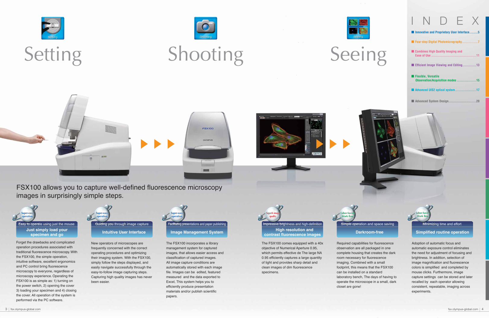
New operators of microscopes are frequently concerned with the correct operating procedures and optimizing their imaging system. With the FSX100, simply follow the steps displayed, and easily navigate successfully through the easy-to-follow image capturing steps. Capturing high quality images has never been easier.
Adoption of automatic focus and automatic exposure control eliminates the need for adjustment of focusing and brightness. In addition, selection of image magnification and fluorescence colors is simplified and completed by mouse clicks. Furthermore, image capture settings can be stored and later recalled by each operator allowing consistent, repeatable, imaging across experiments.
The FSX100 comes equipped with a 40x objective of Numerical Aperture 0.95, which permits effective de The large NA 0.95 efficiently captures a large quantity of light and provides sharp detail and clean images of dim fluorescence specimens.
Required capabilities for fluorescence observation are all packaged in one complete housing that creates the dark room necessary for fluorescence imaging. Combined with a small footprint, this means that the FSX100 can be installed on a standard laboratory bench, The days of having to operate the microscope in a small, dark closet are gone!
FSX100 allows you to capture well-defined fluorescence microscopyimages in surprisingly simple steps.
The FSX100 incorporates a library management system for captured images, that allows easier access and classification of captured images.All image capture conditions are automatically stored with each image file. Images can be edited, featured measured and the data exported to Excel, This system helps you to efficiently produce presentation materials and/or publish scientific papers.
■ Innovative and Proprietary User Interface........5
■ Four-step Digital Photomicrography...............7
■ Combines High Quality Imaging and Ease of Use ..........................................11
■ Efficient Image Viewing and Editing.............13
■ Flexible, Versatile Observation/Acquisition modes ..................15
■ Advanced UIS2 optical system....................17
■ Advanced System Design..........................20
Easy to operate using just the mouse
Just simply load yourspecimen and go
Guiding you through image capture
Intuitive User Interface
Facilitating presentations and paper publishing
Image Management System
Impressive brightness and high-definition
High resolution andcontrast fluorescence images
Simple operation and space saving
Darkroom-free
Minimizing time and effort
Simplified routine operation
Superb imagequality
Labor-SavingWork flow
Super-easyoperation
3 fsx.olympus-global.com fsx.olympus-global.com 4
Setting Shooting Seeing
Super-easyoperation
Super-easyoperation
Labor-SavingWork flow

The user interface is designed for even first time users to smoothly and successfully capture high quality images.Follow the four steps "Mode Selection" → "Selection of start position" → "Framing" → "Imaging." After loading your specimen, there is no need to touch the FSX100 during the above procedure.
Crisp microscope images,consistently captured using only a computer mouse.
Dramatically improved work flow due to a considerable saving of time and la
1.Turn on 2.Adjustment of lampand condenser lenses
3.Selection of observationmethod and filter
4.X-Y stage movementTraditional microscopes
Work flow
1.Turn on 2.Macro imageviewing
3.Framing 4.Image capturing
Clear, high-definition images can be easily captured by simply followingthe step by step instructions displayed one screen at a time.
Step1 Mode Selection Step2 Selection of start position Step3 Framing Step4 Imaging
6.Image capturing5.Focusing tocontrol exposure
Searchingfor thetarget
Adjustmentof unit
Adjustmentof opticsStart Focusing Shooting
5 fsx.olympus-global.com fsx.olympus-global.com 6
bor

FSX100 offers three microscope Observation Modes, 1) Fluorescence / Phase Contrast, 2) Phase Contrast, and 3) Bright Field, and also four Acquisition Modes, 1) Single, 2) Time Lapse, 3) Z-stack, and 4) Stitching, to meet a wide range of applications.
The area selected in Step 2 is displayed and the operator moves a smaller selection window to choose a region for closer examination. By adjusting the size of the selection window, the operator is choosing the magnification at which to capture a high resolution image at Step 4.
This window is where the operator chooses where on the specimen to begin imaging. When the crosshair is placed over the area of interest, the operator clicks the NEXT button and this area is displayed at low magnification and is the starting view for Step 3.The area selected is then shown in the next window at low magnification.
Step1 ModeSelection Step2 Selection of
start position
Step3 Framing
The specimen holder will automatically be identified and the outline of the specimen is displayed. The operator then moves a crosshair to select the location to begin the framing process.
Fluorescence /Phase Contrast
Phase Contrast Bright Field
●Bookmark option [for registration]
Observation mode selection
Displaying the observation range
The wide field of view for macro images allows fast detection of point for image capturing.
Macro image framing
Up to 30 points may be registered on a macro image. The registered points may be read out during the image capturing.
As the crosshair is repositioned around the specimen, a live phase contrast image is displayed.
Displaying the live phase contrast image
An overview image of the entire specimen can be displayed, if desired. This image allows the identification of areas of interest for closer examination.
Mapping option
Acquisition mode selection
Single/Time Lapse/Z-stack/Stitching
Bookmark function
7 fsx.olympus-global.com fsx.olympus-global.com 8
Even first-time users can capture very high qualityimages by simply following these four easy steps.
Designed to provide high quality images with a simple user interface.Even new users can confidently capture beautiful images.
*Stitching mode is to be loaded around April 2009.
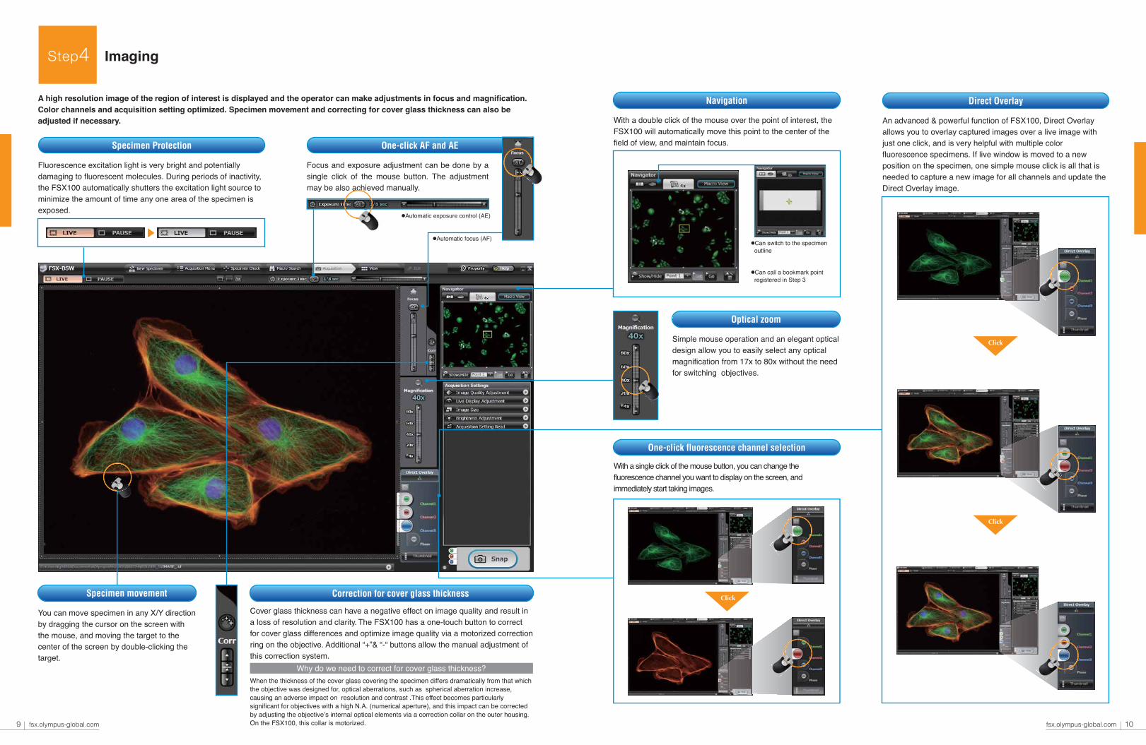
With a double click of the mouse over the point of interest, the FSX100 will automatically move this point to the center of the field of view, and maintain focus.
Fluorescence excitation light is very bright and potentially damaging to fluorescent molecules. During periods of inactivity, the FSX100 automatically shutters the excitation light source to minimize the amount of time any one area of the specimen is exposed.
Focus and exposure adjustment can be done by a single click of the mouse button. The adjustment may be also achieved manually.
●Automatic exposure control (AE)
●Automatic focus (AF)
An advanced & powerful function of FSX100, Direct Overlay allows you to overlay captured images over a live image with just one click, and is very helpful with multiple color fluorescence specimens. If live window is moved to a new position on the specimen, one simple mouse click is all that is needed to capture a new image for all channels and update the Direct Overlay image.
Click
Click
Why do we need to correct for cover glass thickness?
Step4 Imaging
A high resolution image of the region of interest is displayed and the operator can make adjustments in focus and magnification. Color channels and acquisition setting optimized. Specimen movement and correcting for cover glass thickness can also be adjusted if necessary.
Specimen Protection
Navigation
You can move specimen in any X/Y direction by dragging the cursor on the screen with the mouse, and moving the target to the center of the screen by double-clicking the target.
Specimen movement
One-click fluorescence channel selection
Simple mouse operation and an elegant optical design allow you to easily select any optical magnification from 17x to 80x without the need for switching objectives.
Optical zoom
Correction for cover glass thickness
One-click AF and AE
Direct Overlay
With a single click of the mouse button, you can change the fluorescence channel you want to display on the screen, and immediately start taking images.
When the thickness of the cover glass covering the specimen differs dramatically from that which the objective was designed for, optical aberrations, such as spherical aberration increase, causing an adverse impact on resolution and contrast .This effect becomes particularly significant for objectives with a high N.A. (numerical aperture), and this impact can be corrected by adjusting the objective’s internal optical elements via a correction collar on the outer housing. On the FSX100, this collar is motorized.
Click
9 fsx.olympus-global.com fsx.olympus-global.com 10
●Can switch to the specimen outline
●Can call a bookmark point registered in Step 3
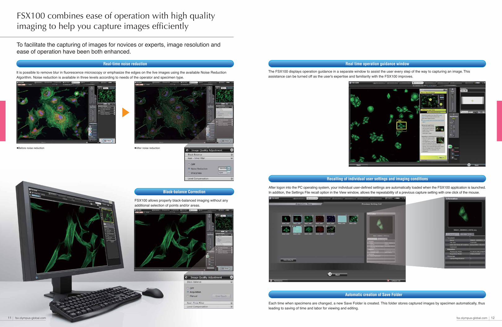
Each time when specimens are changed, a new Save Folder is created. This folder stores captured images by specimen automatically, thus leading to saving of time and labor for viewing and editing.
Automatic creation of Save Folder
It is possible to remove blur in fluorescence microscopy or emphasize the edges on the live images using the available Noise Reduction Algorithm. Noise reduction is available in three levels according to needs of the operator and specimen type.
Real-time noise reduction
After logon into the PC operating system, your individual user-defined settings are automatically loaded when the FSX100 application is launched. In addition, the Settings File recall option in the View window, allows the repeatability of a previous capture setting with one click of the mouse.
Recalling of individual user settings and imaging conditions
FSX100 allows properly black-balanced imaging without any additional selection of points and/or areas.
Black-balance Correction
Real time operation guidance window
●Before noise reduction ●After noise reduction
11 fsx.olympus-global.com fsx.olympus-global.com 12
FSX100 combines ease of operation with high qualityimaging to help you capture images efficiently
To facilitate the capturing of images for novices or experts, image resolution andease of operation have been both enhanced.
View& Edit
ViewWindow
FSX100 software can be installed on separate computers so they can serve as viewing platforms for FSX100 images.
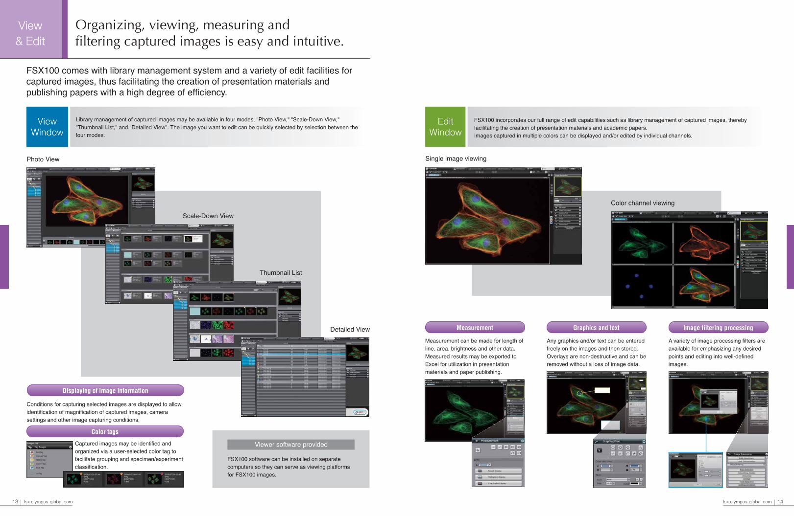
Library management of captured images may be available in four modes, "Photo View," "Scale-Down View," "Thumbnail List," and "Detailed View". The image you want to edit can be quickly selected by selection between the four modes.
Measurement can be made for length of line, area, brightness and other data. Measured results may be exported to Excel for utilization in presentation materials and paper publishing.
A variety of image processing filters are available for emphasizing any desired points and editing into well-defined images.
Viewer software provided
Color tags
Conditions for capturing selected images are displayed to allow identification of magnification of captured images, camera settings and other image capturing conditions.
Displaying of image information
Measurement Graphics and text Image filtering processing
Single image viewingPhoto View
Color channel viewing
Scale-Down View
Thumbnail List
Detailed View
13 fsx.olympus-global.com fsx.olympus-global.com 14
Organizing, viewing, measuring andfiltering captured images is easy and intuitive.
FSX100 comes with library management system and a variety of edit facilities forcaptured images, thus facilitating the creation of presentation materials andpublishing papers with a high degree of efficiency.
EditWindow
FSX100 incorporates our full range of edit capabilities such as library management of captured images, thereby facilitating the creation of presentation materials and academic papers.Images captured in multiple colors can be displayed and/or edited by individual channels.
FSX100 is compatible with slide glass, dia.35 mm glass bottom dish, 96-hole multi-well plate*, plastic Petri dish* and other various specimens.
Observationmode
Versatile observationmodes available for various research purposes.
Acquisitionmode
Versatile acquisition modes available formore efficient laboratory experiments.
Available established three types of microscopy mode toexpand laboratory experiments for different specimens.
Image capturing in various modes is available in the same steps as in thesimple mode, contributing greatly to improved efficiency in laboratory experiments.
For a given specimen, a number of frames can be automatically shot with a certain time interval between each frame once you select the number of shots and the time interval before shooting.
3x3 or 5x5 image montages can be acquired easily using the Stitching Mode
Two specimen holders are provided as standard. One of the holders is intended for slide glass and dia. 35mm glass bottom dish. The other one is for a flat plate to accommodate various shapes of specimens.
●Select the shooting points using the same procedures as in the simple mode, then click on START.
●The time lapse moving image may be integrated into a file of moving images for view/editing through the dedicated viewer.
●Enclose the capturing area with the frame for stitching mode on the macro search screen.●Images of 4x to 20x equivalent magnifications can be captured.
Provided with the three excitation filters, U, B, and G, as standard equipment for fluorescence microscopy through appropriate objectives with high NA and high S/N. Any one optional mirror unit may be additionally loaded for image capturing at the specific wavelength substituting for any one of the three wavelengths.
Fluorescence Time Lapse mode
Z-stack mode
Stitching mode
Compatible with a variety of specimens
Allows phase contrast imaging of cultured specimens and other transparent specimens. The pre-centered design eliminates the need for troublesome center alignment of the phase contrast plate, and facilitates the capture of high-contrast phase contrast images.
Phase contrast
Designed to accommodate the bright field imaging of stained specimens with the color camera with a high degree of color reproducibility. It provides sharp bright field image capturing without any troublesome white balance and other adjustments.
Bright field
Images can be shot in the Z direction while shifting the focal point at a predefined pitch. The extended focus function provides clear, larger focal depth images of specimen with the entire specimen in sharp focus for thick specimens.
●Z-stack movie mages may be integrated into a file of moving images for view/editing through the dedicated viewer.
(1) Select the point for observation using the same procedures as in the normal mode.(2) Specify the upper and lower end positions.(3) Select the pitch for shifting of focal point or the number of shots, then click on START.
15 fsx.olympus-global.com fsx.olympus-global.com 16* Use with optional objective LCAHCN40XPHP
*Stitching mode is to be loaded around April 2009.
High NA objective ideally suited for fluorescence imaging
Optics Advanced UIS2 optical system.
Olympus proprietary UIS2, Opto-Digital Technology,is the foundation inside the FSX100 responsible for the outstanding image quality.
Projection phase contrastProjection phase contrast is adopted for phase contrast microscopy. In the fluorescence microscopy, the phase plate is retracted from the optical path and thus FSX100 enables to maximize fluorescence brightness with the higher NA objectives.
Advanced auto-focusing forfluorescence microscopyAdvanced auto-focusing is incorporated tin all modes to greatly improve the ease of use and free the operator from this time consuming task.
Real-time automatic exposure controlExposure time is automatically changed following the changes in framing and magnification settings to allow capturing of images in the best possible brightness for individual observation modes.
Superior color reproducibilityA high-performance, Olympus designed and manufactured color camera of 12.5 Mega-pixels. Featuring superior color reproducibility and an ISO 1600 sensitivity equivalent, excellent images are easily attained thanks to proprietary Olympus know-how.
Optimal optical system for digital imaging
High transmittance maintained overa wide range of wavelengths
0
20
40
60
80
100
300 400 500 600 700 800 900 1000
Wavelength (nm)T
rans
mis
sion
rat
io (
%)
17 fsx.olympus-global.com fsx.olympus-global.com 18
17x
40x
80x
40x objective with NA0.95FSX100 is equipped with a UIS2 40x objective with the world’s highest Numerical Aperture (NA) 0.95 for a dry system. The objective can efficiently collect weak fluorescence signals and greatly improves the FSX100 sensitivity for low light level specimens.

Fluorescence illumination light source
Darkroom-freeClosing the front cover shields the specimen from ambient light and creates a dark room condition inside the FSX100. Unlike a conventional microscope, fluorescence imaging can now occur with the room lights on, and out in the main laboratory area.
Space savingA clean, smooth outer housing and compact optical design combine to form a small, compact footprint for FSX100. Smaller space requirements, means the FSX100 can be mounted in locations previously too small for a conventional microscope.
Transmitted LED light illumination requiring no lamp changingThe transmitted light source utilized for phase and bright field imaging has a life expectancy of approximately 16,000 hours. Consistent performance over multiple years of use is achieved with this white LED system.
Adoption of long-life fluorescence lampThe pre-centered, 2,000-hour fluorescence excitation lamp system in the FSX100 is designed for stable performance and easy bulb replacement. Intelligent lamp design notifies the user when the bulb is nearing the end of its life cycle.
No Darkroom Needed & Space saving
Image Capture Assistance
Objective correction collarThe 40x NA0.95 objective includes a motorized correction collar that can be adjusted automatically or manually to correct for varying cover glass thicknesses and improve image quality.
Selection between fluorescence filter and projection phase contrast plateFluorescence filter and projection contrast plate are quickly selected in response to the selected observation mode and fluorescence wavelength.
Specimen protection
Field StopThe field stop of the FSX100 is a rectangular aperture that is automatically set to the imaging area of the camera. Fluorescence excitation is potentially damaging to fluorescent molecules, and the automatic adjustment of the field stop area protects photobleach of the specimen outside of the field of the view.
Extended life-span design
19 fsx.olympus-global.com fsx.olympus-global.com 20
Systemdesign
The smooth exterior shape and superior image qualityof the FSX100 communicate "sophisticated performance" and is at home on any laboratory bench.
A clean external design, small footprint and extended life span components combineto allow the FSX100 to be installed in a smaller space and less maintenance thanconventional microscopes.
Bio Imaging Navigator

Specifications Dimensions
Optionally available equipment for expanding applications
Oil-immersion objective Objective for plastic vessels Mirror unit
Fluorescence filter
Item
Observation mode
Acquisition mode
Bookmarks
Automatic focus (AF)
Exposure control
Real-time image processing
Image overlay
White balance, Black balance
Image format
Specimen Protection
Auto image library
Operation tutorial
Settings restore function
Image playback
Image editing
Specimen holder
Standard objectives
Optical zoom
Motorized correction collar
XY stage
Focus range
Transmitted illumination
Transmitted illumination light source
Fluorescence illumination
Fluorescence illumination light source
Fluorescence/Phase contrast filter
Camera type
Imaging sensor
Effective image resolution
Sensitivity
A/D converter
Display frame rate
Interface
OS
Dimensions (mm)
Weight
Operating environment
Input power/Power consumption
Optional equipment
Objectives
Fluorescence mirror unit
Specifications
Fluorescence/Phase contrast, Phase contrast, and Bright field
Single, Time Lapse, Z-stack, and Stitching*1 (fluorescence multi color imaging possible for all)
Up to 30 positions, can be selected and recalled
Automatic during the screen transition with one click operation
Auto (with exposure adjustment ), Manual
Noise reduction (3 adjustment levels) , Sharpness (2 adjustment levels)
Direct overlay function (live image)
Pre-set/Manual, Acquisition/Manual
BMP, JPG, TIFF, AVI
-Automatically shut OFF excitation light when the system is left without any operation for a certain period of time
-Be changeable F.S. automatically set to field of view
Automatically creating a folder every time specimen is changed
Operation guidance display function, user switchable ON/OFF
Multi-user settings storage/reproduction, imaging conditions recording/reproduction functions
Display switching (Photo View, Scale-Down View, Thumbnail List, Detailed View)
Time-lapse / Z-stack dedicated viewer Categorization function with the color tag
4 segmented display dedicated to multi color image, full screen display, print, adding figures and text, scale display, date and time display,
image rotation, image trimming, image size change, image processing filters, RGB color adjustment (16 bits, 8 bits), gray scale conversion,
overlay compensation, RGB adjustment, level adjustment,measurement (possible to export to Excel)
Accepts 1”x3” slide, dia. 35mm dish
Dia.50 mm hole opening , all metal construction
Capture: 40x NA0.95 (17x to 80x with optical zoom)
Macro: 10x NA0.40 (4.2x fixed with optical zoom)
0.42x to 2.0x
with focusing assist
Stroke: 56 × 26mm (with slide glass), 11 × 11 mm (with 35mm dish), 18 × 18mm (with 50mm hole plate)
(Automatic switch by recognizing the specimen holder)
11 mm
Condenser lens NA0.55, Working distance 27 mm, Phase contrast slit
White LED light source, average lifetime 16,000 hours
Fly-eye lens, Field stop (working with optical zoom), ND filter (automatic switching), Shutter
Metal halide lamp (pre-centered design), average lifetime 2,000 hours
U excitation (Ex360-370 Em420-460 DM400) Equivalent to U-MNUA2
B excitation (Ex460-495 Em510-550 DM505) Equivalent to U-MWIBA3
G excitation (Ex3530-550 E475IF DM570) Equivalent to U-MWIG3
Projection phase contrast plate (automatic switching)
Single-panel color CCD pixel shift type
2/3 size (inch), 1.45 mega-pixels (Total number of pixels: 1.5 mega-pixels), Peltier cooling (Max: Ta-10°C)
4080 × 3072 (12.5 megapixels)*2, 2040 × 1536 (3.1 megapixels)*2, 1360 × 1024 (1.4 megapixels),680 × 512 (350,000 pixels),
2 x 2 binning; 680 × 510 (350,000 pixels), 4 × 4 binning; 340 × 250 (85,000 pixels)
Equivalent to ISO 200/400/800/1600
12-bit per channel
Max. 15 frames/sec. (at live image size of 1,360 × 1,024)
IEEE 1394 cable, Proprietary camera cable
Windows Vista Business SP1 (32bit)
Height 476 × Width 388 × Depth 583
Approx 35kg
+10 to +35°C/35 to 80%RH (no condensation),Pollution degree: 2 (in accordance with IEC60664),
Installation (overvoltage) category: II (in accordance with IEC60664)
AC 100-120/220-240V 50/60Hz 2.5A/1.2A
UPLSAPO60xO*3 NA1.35 (For oil immersion) (26x to 120x with optical zoom)
LCACHN40xPHP*4 NA0.55 (For plastic vessel) (17x to 80x with optical zoom)
Allowable additional installation of one set of UIS2 mirror unit (Exclusive use against any other filter sets)
The oil-immersion objective UPLSAPO60XO with NA 1.35 is available for fluorescence photomicrography with higher resolution and higher S/N. It is recommended that this optional objective be used with Olympus low auto-fluorescence immersion oil.
One additional UIS2 mirror unit may be installed as optional accessory.
Tra
nsm
issi
on r
atio
(%
)
Tra
nsm
issi
on r
atio
(%
)
Tra
nsm
issi
on r
atio
(%
)
3000
102030405060708090
100
350 400 450 500 550 600 650 700 750 800
DM400BA420-460BP360-370
3000
10
20
30
40
50
60
70
80
90
100
350 400 450 500 550 600 650 700 750 800
BP460-495 DM505
3000
102030405060708090
100
350 400 450 500 550 600 650 700 750 800
BP530-550 BA575IFBA510-550 DM570
21 fsx.olympus-global.com fsx.olympus-global.com 22
Wavelength (nm) Wavelength (nm) Wavelength (nm)
Ultra violet excitation Green excitationBlue excitation
388 623
628
476
583
(mm)
*1 Stitching mode is to be loaded around April 2009. *2 Phase contrast and Bright field observation mode only. *3 Not available Phase contrast, selection of start position, macro image framing, automatic focus and stitching images*4 Not available ultra violet fluorescence, selection of start position, macro image framing, automatic focus and stitching images.