bio and nanomaterials based on fe3o4
TRANSCRIPT

Molecules 2014, 19, 21506-21528; doi:10.3390/molecules191221506
molecules ISSN 1420-3049
www.mdpi.com/journal/molecules
Review
Bio and Nanomaterials Based on Fe3O4
Jia-Kun Xu 1,*, Fang-Fang Zhang 1,2, Jing-Jing Sun 1, Jun Sheng 1, Fang Wang 1 and Mi Sun 1,*
1 Key Laboratory of Sustainable Development of Marine Fisheries, Ministry of Agriculture,
Yellow Sea Fisheries Research Institute, Chinese Academy of Fishery Sciences, Qingdao 266071,
China; E-Mails: [email protected] (F.-F.Z.); [email protected] (J.-J.S.);
[email protected] (J.S.); [email protected] (F.W.) 2 College of Food Science and Engineering, Ocean University of China, Qingdao 266003, China
* Authors to whom correspondence should be addressed; E-Mails: [email protected] (J.-K.X.);
[email protected] (M.S.); Tel.: +86-532-8583-3961 (M.S.).
External Editor: Alexandru Mihai Grumezescu
Received: 18 November 2014; in revised form: 16 December 2014 / Accepted: 17 December 2014 /
Published: 22 December 2014
Abstract: During the past few years, nanoparticles have been used for various applications
including, but not limited to, protein immobilization, bioseparation, environmental
treatment, biomedical and bioengineering usage, and food analysis. Among all types of
nanoparticles, superparamagnetic iron oxide nanoparticles, especially Fe3O4, have attracted
a great deal of attention due to their unique magnetic properties and the ability of being
easily chemical modified for improved biocompatibility, dispersibility. This review covers
recent advances in the fabrication of functional materials based on Fe3O4 nanoparticles
together with their possibilities and limitations for application in different fields.
Keywords: Fe3O4; surface modification; application
1. Introduction
Owing to the unique properties, such as superparamagnetism, high surface area, large surface-to-volume
ratio, low toxicity, easy separation under external magnetic fields, Fe3O4 nanoparticles have enormous
potential in the fields such as immobilization of biomaterials [1–10], bioseparation [11–15], environmental
treatment [16–23], biomedical and bioengineering usage [24–36], and food analysis [37–44]. Various
fabrication methods have been developed for the synthesis of Fe3O4 nanoparticles, including the
OPEN ACCESS

Molecules 2014, 19 21507
physical methods [45–47], wet chemical preparation methods [48–66] and microbial methods [67–69].
Since the bare Fe3O4 nanoparticles often have poor stability and dispersity, various modification
methods have been exploited to get the soluble and biocompatible Fe3O4 nanoparticles. The resulting
modified Fe3O4 nanoparticles have been extensively used for various applications. In this review, the
traditional and modern methods for synthesis of Fe3O4 nanoparticles are summarized; the methods for
modification of Fe3O4 nanoparticles are also described. Finally, a variety of practical and potential
applications as well as the corresponding limitations of the resulting Fe3O4 nanoparticles are introduced.
2. Methods for Preparation of Fe3O4 Nanoparticles
The outstanding potential of Fe3O4 nanoparticles has stimulated the extensive development of
the synthetic technologies, which could be broadly classified into three categories: physical, chemical
and biological methods. (i) Physical methods, such as electron beam lithography [45], gas-phase
deposition [46], and mechanical techniques [47]. Externally controlled tools like traditional workshop
or microfabrication equipment are often involved in physical methods, where are used to process
materials into the desired shape and order. Although physical methods are easy to perform, it is rather
difficult for them to control the particle size. (ii) Wet chemical preparation methods, such as sol-gel
synthesis [48,49], oxidation method [50,51], reduction method [52], chemical coprecipitation [53,54],
hydrothermal reactions [55,56], solvothermal method [57], thermal decomposition method [58], flow
injection synthesis [59], electrochemical method [60,61], aerosol/vapor phase method [62], sonochemical
decomposition reactions [63,64], supercritical fluid method [65,66], synthesis using nonreactors [67].
In the case of wet chemical preparation methods, relatively less energy was consumed compared with
that of physical methods. Among wet chemical preparation methods, coprecipitation of Fe3+ and Fe2+
salts is a most often employed method to prepare water-borne iron oxide nanoparticles. The size
and morphology of the nanoparticles can be controlled by selectively choosing the reaction media, the
physical parameters of the reaction, such as precursors, reactant concentration, base (NaOH, ammonium
hydroxide, and CH3NH2), ionic strength (N(CH3)4+, CH3NH3+, NH4+, Na+, Li+ and K+), reaction
temperature, pH of the media, and also some other factors [68]. For instance, an increase of the mixing
rate tends to decrease the particle size. Moreover, inlet of nitrogen into the reaction system that protects
against critical oxidation of the magnetite also reduces the particle size when compared to methods
without oxygen removal. However, coprecipitation protocol leads to reduced control of particle shape,
broad distributions of sizes and aggregation of particles. In general, the size distribution of nanoparticles
is an important factor to be considered for a particular application. Some of wet chemical methods can
yield efficient control of the particle size by carefully adjusting the involved parameters, including
sol-gel method, hydrothermal method, flow injection method, electrochemical method, sonochemical
decomposition method, supercritical fluid method and synthesis using nanoreactors. (iii) Microbial
method. Microbial method is an environment friendly nanoparticle formation processes which can
produce 5–90 nm pure magnetite or metal-substituted magnetite without usage of toxic chemicals in
their synthesis process [69–72]. Microbial method represents an advantageous manufacturing technology
with respect to high yield, good reproducibility, and good scalability, as well as low costs and low
energy input, but the fermentation process is rather time-consuming. Table 1 shows the summary of
various methods for preparing Fe3O4 nanoparticles.

Molecules 2014, 19 21508
Table 1. Comparation between methods for synthesis of magnetic nanoparticles [46,47,73].
Methods Advantages Disadvantages
Physical
methods
Electron beam lithography well controlled inter-particle
spacing
expensive and highly complex
machines requiring
Gas-phase deposition easy to perform difficult to control the particle size
Mechanical techniques no chemicals involved highly complex machines requiring
and time-consuming
Wet
chemical
preparation
methods
Sol-gel synthesis precisely controlled in size, aspect
ratio, and internal structure
weak bonding, low wear-resistance,
high permeability
Oxidation method uniform size and narrow size
distribution small-sized ferrite colloids
Reduction method simple high reaction temperature
Chemical coprecipitation simple and efficient
not suitable for the preparation of
high pure, accurate stoichiometric
phase
Hydrothermal reactions easy to control particle size and
shapes
high reaction temperature, high
pressure
Solvothermal method easy to control particle size and
shape high reaction temperature
Thermal decomposition
method
easy to control particle size and
shape involve multiple steps
Flow injection synthesis
good reproducibility and high
mixing homogeneity together with
a precise control of the process
need continuous or segmented
mixing of reagents under a laminar
flow regime in a capillary reactor
Electrochemical method easy to control particle size bad reproducibility
Aerosol/vapor phase method high yields extremely high temperatures
Sonochemical
decomposition reactions narrow particle size distribution mechanism not still understood
Supercritical fluid method efficient control of the particle
size, no organic solvents involved critical pressure and temperature
Synthesis using nanoreactors precisely control the particle size complex condition
Microbial
methods Microbial incubation
environmental friendly, high yield,
good reproducibility, and good
scalability, low cost
time-consuming
3. Modification of Fe3O4 Magnetic Nanoparticles
Because of the high surface energy, the naked Fe3O4 nanoparticles are generally unstable and aggregate
easily, which strongly affects their dispersion into aqueous medium. In addition, Fe3O4 nanoparticles
are highly susceptible to be oxidized to γ-Fe2O3 nanoparticles in the presence of oxygen [74]. To
overcome such limitations, various surface modification methods have been developed to modify the
surface of naked Fe3O4 nanoparticles via loading of other chemicals or biological materials during or
after the synthesis process to improve the dispersibility, stability, biocompatibility and biodegradability
for specific purposes [75–94]. With proper surface modification, the stability, dispersity and

Molecules 2014, 19 21509
biocompatibility of Fe3O4 nanoparticles could be improved, and the oxidation process from Fe3O4
nanoparticles to γ-Fe2O3 nanoparticles could be greatly slowed down.
The common reagents employed for modification of Fe3O4 nanoparticles includes surfactants
(such as oleic acid(OA) [35,75], lauric acid [76], alkane sulfonic acids [77], and alkane phosphonic
acids) [78], polymers (such as polyethylene glycol (PEG) [79], polyvinylpyrrolidone (PVP) [80], poly
(ethylene-co-vinyl acetate) [81], polylactic-co-glycolic acid (PLGA) [82], polyvinyl alcohol (PVA) [83],
polystyrene [84], polyethyleneimine (PEI), and poly(acrylic acid) (PAA) [85]) and natural dispersants
(chitosan [86,87], dextran [88], gelatin [89], polylactic acids [90], starch [91], albumin [92],
liposomes [93], and ethyl cellulose [94]). The methods of modification of Fe3O4 nanoparticles mainly
include physical immobilization, covalent conjugation, and biologically mediated specific interaction.
The advantages and disadvantages of these three immobilization methods are summarized in Table 2.
Table 2. Comparation between different immobilization methods.
Methods Interactions Advantages Disadvantages
Physical
immobilization
physical absorption, electrostatic
interaction, hydrogen bonds, van
der Waals forces, and
hydrophobic interactions
easy to perform and recycle,
no additional coupling reagents
and surface treatment are
required
nonspecificity, the
binding stability is highly
affected by environmental
conditions
Covalent
conjugation covalent interaction
the binding process can be
rationally regulated with specific
functional groups
nonspecificity, the
support can’t be recycled
Biologically
mediated specific
interaction
biologically mediated specific
interaction site-specific
site-selective attachment
is desired
Jadhav et al. prepared oleic acid (OA) functionalized Fe3O4 nanoparticles using modified wet
method, and sodium carbonate was used to improve the biological applicability (Scheme 1) [75]. In
another example, Yang et al. synthesized PEG-coated Fe3O4 nanoparticles via traditional chemical
coprecipitation method, the influence of vapor pressure, molecular weights and amounts of PEG on the
structural and paramagnetic properties of PEG-Fe3O4 NPs were investigated [79]. Fe3O4 nanoparticles
synthesized in sealed environment (S-Fe3O4) displayed much high crystalline quality than that
synthesized in open environment (O-Fe3O4). The calculated average crystalline size of S-Fe3O4 and
O-Fe3O4 is 15.2 nm and 14.5 nm, respectively. Both of the S-Fe3O4 and O-Fe3O4 nanoparticles showed
superparamagnetic properties, and the saturation magnetization for S-Fe3O4 and O-Fe3O4 nanoparticles
is 44 emu/g and 24 emu/g, respectively. The well-dispersed magnetic PEG-Fe3O4 nanoparticles with
better size distribution can be obtained with adding 4 g PEG1000 while sealing the beaker. There were
no significant size change caused by the PEG coating. However, the saturation magnetization of
PEG-Fe3O4 nanoparticles showed an apparent decrease compared to that of bulk material (92 emu/g),
which could be attributed to the surface disorder or spin canting at the surface of nanoparticles. Qu et al.
prepared Fe3O4–chitosan nanoparticles with core-shell structure [87]. Oleic acid (OA) modified Fe3O4
nanoparticles (MN) were firstly prepared by coprecipitation, chitosan was then added to coat on the surface
of the Fe3O4 nanoparticles by physical absorption, and glutaraldehyde was used to crosslink the amino
groups on the chitosan. The saturation magnetization of the Fe3O4–chitosan nanoparticles (30.7 emu/g)

Molecules 2014, 19 21510
was lower than the pristine Fe3O4 nanoparticles (74.3 emu/g), which could be partly attributed to the
existence of the large amount of diamagnetic chitosan in the Fe3O4–chitosan nanoparticles.
Scheme 1. Schematic representation for interaction of oleic acid (OA) modified Fe3O4
nanoparticles with sodium carbonate. OA is chemically bound to Fe3O4 nanoparticles by
the carboxyl head group (-COOH) and the hydrophobic tail group is free, making it
non-dispersible in aqueous medium. The hydrophobic tail in turn interacts with the free
OA via hydrophobic interactions. The formulation is stabilized in aqueous medium by
ionization of the carboxyl head group of free OA by sodium carbonate, wherein Na+
interact by ionic interactions with COO− group of free OA [75].
4. Applications of Fe3O4 Nanoparticles
Due to the unique properties, Fe3O4 nanoparticles appear to be very promising for their applications
in protein immobilization, bioseparation, environments treatment, biomedical and bioengineering
usage, and food analysis.
4.1. Protein Immobilization
Protein immobilization serves as a very effective tool to solve the difficulties encountered in the
catalytic application of free enzymes, such as poor stability and hard recovery. It is of vital importance
to select proper immobilization basis for protein immobilization. Fe3O4 nanoparticles have been intensively
utilized to realize this objective due to its unique magnetic performance, and various practical and
economical biocatalysts with improved stability and reusability have been fabricated based on Fe3O4
nanoparticles, which could be easily separated from the reaction medium in the presence of external
magnetic field [1–10,95–114]. Proteins could be immobilized onto Fe3O4 nanoparticles in the manner
of physical absorption [95–97], covalent bonding [98–104], and bioconjugation [105–107]. Coupling
reagents, such as glutaraldehyde [99–102,108–110], 1-ethyl-3-(3-dimethylaminopropyl) carbodiimide
hydrochloride (EDC) [103,104,111–113] and sodium tripolyphosphate (TPP) [114], are often utilized
to achieve much more stable immobilization via covalent bonding because their functional groups can
interact with both functional groups of the modified magnetic nanoparticles and proteins. For example,
Huang et al. covalently bound glucose oxidase to Fe3O4/silicon dioxide nanoparticles using glutaraldehyde,
resulting in an activity of immobilized glucose oxidase of 4570 U/g at pH 7 and 50 °C [102].
The immobilized glucose oxidase retained 80% of its initial activity after 6 h at 45 °C compared to
only 20% for the free enzyme. After six cycles of repeated use, the immobilized glucose oxidase still
Molecules 2014, 19 21511
maintained 60% of its initial activity; 75% of its initial activity remained after 1 month at 4 °C
compared to 62% for the free enzyme. Hong et al. obtained amine-functionalized magnetic nanogel by
Hoffman degradation of the polyacrylamide (PAM)-coated Fe3O4 nanoparticles. α-Chymotrypsin (CT)
covalently bound to the magnetic nanogel with reactive amino groups by using EDC as coupling
reagent [104]. The binding capacity was determined to be 61 mg enzyme/g nanogel by BCA protein
assay. Specific activity of the immobilized CT was measured to be 0.93 U/(mg min), 59.3% as that of
free CT. The immobilized CT still had a remaining activity of 60% when the reaction temperature rose
to 60 °C while free CT lost all-initial activity. Wu et al. prepared magnetic Fe3O4-chitosan nanoparticles
by cross-linking with TPP, precipitation with NaOH and oxidation with O2 in hydrochloric acid aqueous
phase containing chitosan and Fe(OH)2 [114]. The adsorption capacity of the prepared Fe3O4-chitosan
nanoparticles to lipase was 129 mg/g; and the maximal enzyme activity was 20.02 μmol·min−1·mg−1
(protein), and 55.6% activity was retained at a certain loading amount.
4.2. Bioseparation
Magnetic separation is a commonly used technique for polypeptide/protein separation and cell
separation. Magnetic separation possesses several advantages such as timesaving, gentle, easily
automated, and can be directly used to remove target compounds from crude medium by the simple
application of an external magnetic field. To construct Fe3O4 based composite nanomaterials for
separation, core/shell microspheres are generally fabricated with a Fe3O4 as a core and other functional
materials as a shell [115–123]. Ma et al. synthesized the Fe3O4@mTiO2 microspheres with a well-defined
core/shell structure, the high specific surface area (167.1 m2/g), large pore volume (0.45 cm3/g),
appropriate and tunable pore size (8.6–16.4 nm), and high magnetic susceptibility [123]. The
composite could selectively enrich phosphopeptides from complex mixtures even at a very low molar
ratio of phosphopeptides/non-phosphopeptides (1:1000), large enrichment capacity (as high as 225 mg/g,
over 10 times as that of the Fe3O4@TiO2 microspheres), extreme sensitivity (the detection limit was at
the fmol level), excellent speed (the enrichment can be completed in less than 5 min), and high
recovery of phosphopeptides (as high as 93%).
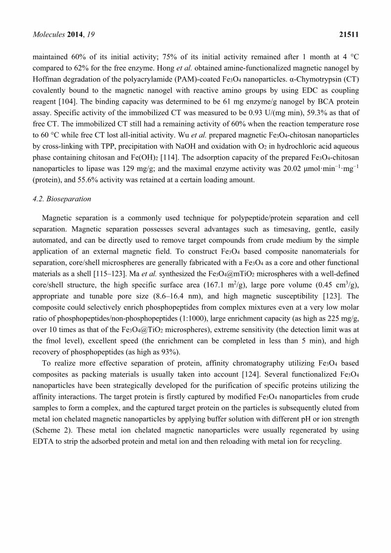
To realize more effective separation of protein, affinity chromatography utilizing Fe3O4 based
composites as packing materials is usually taken into account [124]. Several functionalized Fe3O4
nanoparticles have been strategically developed for the purification of specific proteins utilizing the
affinity interactions. The target protein is firstly captured by modified Fe3O4 nanoparticles from crude
samples to form a complex, and the captured target protein on the particles is subsequently eluted from
metal ion chelated magnetic nanoparticles by applying buffer solution with different pH or ion strength
(Scheme 2). These metal ion chelated magnetic nanoparticles were usually regenerated by using
EDTA to strip the adsorbed protein and metal ion and then reloading with metal ion for recycling.

Molecules 2014, 19 21512
Scheme 2. Illustration of the separation mechanism of affinity chromatography utilizing
Fe3O4 based composite as packing material.
4.3. Environmental Treatment
As a result of rapid industrialization and urbanization, various pollutants particularly those
entering aquatic systems have attracted worldwide concern. The development of efficient and
cost-effective methods for environmental treatment is of primary concern for sustainable economic
and social development. Due to the extremely small particle size, high surface-area-to-volume
ratio, and more important the magnetism, Fe3O4 nanoparticles have been widely used and have
shown promising performance in environments treatment, including pollutant removal and toxicity
mitigation [16–23,125–129]. Proper surface coating cannot only improve the removal capacity and
affinity of the Fe3O4 nanoparticles, but also promote the desorption process. Pollutants generally
adsorb to the surface of Fe3O4 nanoparticles through interactions including physical adsorption,
ion-exchange, chemical bonding (complexation and/or chelation), hydrogen bonds, and van der Wall
forces. Fe3O4/ZrO2/chitosan composite was synthesized and employed for the removal of amaranth and
tartrazine dyes removal, the adsorption capacities of which were 99.6 and 47.3 mg/g for amaranth and
tartrazine dyes, respectively [128]. In another report, Hakami et al. prepared Fe3O4 nanoparticles
functionalized with thiol groups by adding (3-mercaptopropyl) trimethoxysilane on silica-coating
to remove Hg, and the sulfur atoms in thiol moieties served as ligands to bind with soft metal
cation Hg+. Thiourea was added to facilitate desorption of Hg because of the presence of sulfur atoms
(Scheme 3) [129].
Scheme 3. Illustration of the moval and recovery of Hg(II) using thiol-functionalized
mesoporous silica-coated magnetite nanoparticles [129].

Molecules 2014, 19 21513
In the real engineering applications, the strategically utilization of Fe3O4 nanoparticles should consider
the complex environmental conditions such as background ions, humic substances, temperature, and pH.
4.4. Biomedical Usage
Fe3O4 nanoparticles with appropriate surface properties have been widely used for numerous
biomedical and bioengineering applications such as targeted drug delivery, biosensor, magnetic
resonance imaging, hyperthermia, tissue engineering, magnetofection, etc. All these applications require
that these nanoparticles not only possess high magnetization values, but also with narrow particle size
distribution and similar surface topography, so that the particles have uniform physical and chemical
properties. Moreover, the magnetic nanoparticles for biomedical applications should be non-toxic and
biocompatible. In a word, both of the nature and the geometric arrangement of surface coatings on the
nanoparticles have apparent influence on bio-kinetics and bio-distribution of nanoparticles in the body.
4.4.1. Targeted Drug Delivery
Due to the unique capabilities (e.g., superparamagnetism and biocompatibility) and the negligible
side effects, magnetic Fe3O4 nanoparticles with proper surface modification and conjugated targeting
ligands/molecules have become a major research focus for drug delivery applications. Compare to the
conventional, non-targeted methods of drug delivery, magnetic nanoparticles are promising drug
carriers due to the better specificity to the target site and the reduced adverse effects. Drug carried by
magnetic nanoparticles could be concentrated at the desired site to receive much high therapeutic
efficiency. Since the drugs simply physically attached to the nanoparticle surface tend to release
quickly before reaching the final destination, a core-shell structure consisting of a magnetic core and
a shell is preferred in magnetic drug-delivery systems to achieve sufficient drug loading capacity and
good transportation effect [130–138]. For example, Chen et al. prepared Fe3O4@SiO2 core–shell
nanoparticles and grafted a widely used anticancer agent doxorubincin (DOX) to the surface of the
core–shell nanoparticles via an amide bond with the aid of a spacer arm. Most of the conjugated DOX
can release from the nanoparticles within 12 h and the release process prefers low pH conditions.
The saturation magnetization value of the obtained superparamagnetic DOX-grafted Fe3O4@SiO2
core-shell structure nanoparticles was 49.3 emu·g−1, indicating its great potential application in the
treatment of cancer using magnetic targeting drug-delivery technology [136].
4.4.2. Biosensor
Fe3O4 nanoparticles based bioanalytical sensors could be fabricated by coating Fe3O4 nanoparticles
with materials such as a fluorescent one [139,140], a metal [141,142], silica [143,144], or a
polymer [145,146]. Tang et al. developed a practical glucose biosensor by combining the intrinsic
peroxidase-like activity of Fe3O4 nanoparticles and the anti-interference ability of the nafion film.
Glucose oxidase was simply mixed with Fe3O4 nanoparticles and cross-linked on the Pt electrode with
chitosan medium by glutaraldehyde, and then covered with a thin nafion film. The biosensor showed
high sensitivity (11.54 μAcm−2·mM−1), low detection limit (6 × 10⁻⁶ M), and good storage
stability [147]. Liu et al. developed a reusable, single-step system for the detection of specific substrates

Molecules 2014, 19 21514
using oxidase-functionalized Fe3O4 nanoparticles as a bienzyme system and using amplex ultrared
(AU) as a fluorogenic substrate. A composite of poly (diallyldimethylammonium chloride)-modified
Fe3O4 nanoparticles and oxidase was prepared for the quantification of specific substrates through the
H2O2-mediated oxidation of AU. The reaction process was monitored by checking fluorescence
intensity at 587 nm, and the minimum detectable concentrations of glucose, galactose, and choline
were found to be 3, 2, and 20 μM utilizing glucose oxidase-Fe3O4, galactose oxidase-Fe3O4, and
choline oxidase-Fe3O4 composites, respectively (Scheme 4) [148].
Scheme 4. Illustration of Fe3O4 nanoparticles play a peroxidase-like role to detect the
specific substrate in the presence of AU [148].
4.4.3. Magnetic Resonance Imaging
Magnetic resonance imaging (MRI) is a commonly used non-invasive medical imaging technique in
clinical medicine to visualize the structure and function of tissues, which is based on the behavior,
alignment and interaction of protons in the presence of an applied magnetic field [149–153]. For example,
Fan et al. present a relatively simple and scalable approach for preparing poly(poly(ethyleneglycol)
monomethacrylate) (P(PEGMA))-grafted Fe3O4 core–shell nanoparticles with well-controlled
properties using a solvent-free ATRP [153]. The so-obtained P(PEGMA)-grafted nanoparticles have a
uniform hydrodynamic particle size of 36.0 ± 1.2 nm. The morphology and viability of the
macrophage cells cultured in a medium containing 0.2 mg/mL of P(PEGMA)-grafted nanoparticles
were found similar to those of cells cultured without nanoparticles, indicating an absence of significant
cytotoxicity effects. T2-weighted magnetic resonance imaging (MRI) of P(PEGMA)-grafted MNPs
showed that the magnetic resonance signal is enhanced significantly with increasing nanoparticle
concentration in water. The R1 and R2 values (longitudinal and transverse relaxivities) per millimole
Fe, and R2/R1 value of the P(PEGMA)-grafted MNPs were calculated to be 8.8 mM−1·s−1, 140 mM−1·s−1,
and 16, respectively. These results indicate that the P(PEGMA)-grafted nanoparticles have great
potential for application in MRI of specific biotargets.
4.4.4. Hyperthermia
Magnetic induction hyperthermia means the exposition of cancer tissues to an alternating magnetic
field, in which heat is generated due to magnetic hysteresis loss. Cancer cells exposed to magnetic

Molecules 2014, 19 21515
particles will heat up to a temperature higher than 43 °C, at which the cancer cells are destroyed
whereas the normal cells can survive. Much research work using magnetic particles for hyperthermia have
manifested a therapeutic effect on several types of tumors [30–33,154–156]. For example, Ghosh et al.
synthesized Fe3O4 magnetic nanoparticles (MN) capped with either oleic acid (Fe3O4-OA-MN) or
polyethylene glycol (Fe3O4-PEG-MN), which were prepared by a co-precipitation method. The
average particle sizes of the obtained Fe3O4-MN, Fe3O4-OA-MN and Fe3O4-PEG-MN were found to
be 12, 6 and 8 nm, respectively. A 35% increase of killing effect was observed in human breast cancer
cells (MCF7) after Fe3O4-OA-MN treatment, which was further enhanced (65%) in the presence of
induction heating. However, only 5%–10% killing was achieved while Fe3O4-MN or Fe3O4-PEG-MN
was used to treat MCF7 cells after induction heating. The effect of only OA (0.088 mg·mL−1, a
concentration low than that in Fe3O4-OA-MN) or PEG (0.1 mg·mL−1) with/without induction heating
on cell viability experiments indicated that loss of viability by OA was ~75%, which was higher than
1 mg of Fe3O4-OA-MN (35%) alone. However, PEG at this concentration did not show any significant
change in cell toxicity. The same control experiments conducted under induction heating showed
insignificant change in cell viability. These results displayed the surface characteristics of the modified
magnetic nanoparticles (e.g., lipophilicity) greatly influence their hyperthermia applications in cancer
therapy [156].
4.4.5. Tissue Engineering
Tissue engineering is a promising technology for overcoming the organ transplantation crisis, and
the fabricated tissue equivalents may also be used to screen the effects of drugs and toxins [157,158].
It has been a great challenge for scientists and medical experts to fabricate functional organs of the
similar architectures in vitro to the in vivo organs, in which the cells are allocated precisely. To realize
this objective, three-dimensional constructs (scaffolds or hydrogels) functioning similarly as under
in vivo conditions should be firstly built up [159–161]. The cells generally isolated from a tissue
biopsy, cultured in vitro, subsequently seeded into the three dimensional constructs. To achieve an efficient
cell seeding and to enable controlled tissue assembly and complex tissue formation, magnetic force-based
tissue engineering technique is required to provide magneto-responsive features to the cells [162–164].
The inclusion of magnetic particles has no significant effect on the porosity, stability and wetting
properties of the composite scaffolds, making them appropriate for cellular support and cultivation.
For instance, Sapir et al. created a stimulating microenvironment by inserting magnetically responsive
Fe3O4 nanoparticles into a macroporous alginate scaffold, which was suitable for promoting endothelial
cell organization into capillary-like structures in vitro [165].
4.4.6. Magnetofection
Magnetofection rely on the delivery of nucleic acids (e.g., DNA, antisense oligodeoxynucleotides
(AODN), and small interfering ribonucleic acids (siRNA) into the targeted cells in presence of a magnetic
field [46]. The delivery of nucleic acids using viral vectors is called transduction, whereas the delivery
using nonviral vectors is termed transfection. The negatively charged nucleic acids generally interact
with MNPs chemically modified by cationic substances such as PEI or protamine sulfate polymers [166],
which could contribute to the intracellular penetration. The application of an external Fe3O4 magnetic

Molecules 2014, 19 21516
field directs viral or non-viral gene delivery vectors facilitates fast and efficient nucleic acid delivery
towards the target cells [167].
Although substantial progress has been made with creating proper delivery systems for nucleic
acids, our knowledge of the internal operation mechanism inside cells is still unclear, and target delivery
of nucleic acids still has not lived up to its potential clinical application. The processes governing
nucleic acid uptake and delivery are far from being clarified, as well as their intracellular interactions,
intracellular trafficking and the regulation of nucleic acid action inside cells [46].
4.5. Food Analysis
It is of vital importance to accurately analyze food components and food contaminants for ensuring
food safety and quality. Although the frequently employed techniques (e.g., gas chromatography (GC),
culture and colony counting, immunoassay, high-performance liquid chromatography (HPLC), and liquid
chromatography coupled with tandem mass spectrometry (LC-MS/MS)) are of very great usage for
food analysis, most of them are laborious, complex, time-consuming, expensive, and show somewhat
dissatisfying specificity and detectability to some special targets. Magnetic nanoparticles such as Fe3O4
are of special interest for food analysis not only because the unique properties such as low toxicity,
good biocompatibility, large specific surface area, high capacity for charge transfer and convenient
separation from a reaction mixture with an external magnetic field, but also for the rapid, highly
selective and sensitive detection of food contaminants and food components after the proper surface
modification. Fe3O4 nanoparticles are usually involved in detection techniques for food analysis in two
ways: electrode modifier and sample pre-concentrator [37–43,168]. Fe3O4 nanoparticles have been widely
used in many detection techniques for food analysis, including PCR, immunoassay, HPLC, LC-MS/MS,
and optical method. For example, Liu et al. developed a superparamagnetic nano-immunobeads
(SPM-NIBs) by conjugation of Fe3O4 nanoparticles with specific antibodies (Scheme 5). The prepared
SPM-NIBs showed superior colloidal stability and reversible magnetic response to Vibrio parahaemolyticus,
a main foodborne pathogenes from contaminated seafood. About 80% of Vibrio parahaemolyticus
cells could be captured when the concentration of the broth was 103 CFU/mL [41].
Scheme 5. Illustration of the process of target bacteria separation using superparamagnetic
nano-immunobeads (SPM-NIBs) [41].

Molecules 2014, 19 21517
5. Concluding Remarks and Prospects
Due to the unique properties (e.g., superparamagnetism, high surface area, large surface-to-volume
ratio, low toxicity, and easy separation), Fe3O4 nanoparticles have emerged as ideal frame materials
for generating functional materials of different surface architecture, which have already displayed
promising effects in practical applications in protein immobilization, bioseparation, biomedical science,
environmental treatment, and food analysis. Various Fe3O4 based nanoparticles have already realized
their practical applications. However, there are still many Fe3O4 based nanoparticles having not been
scaled up from the laboratory scale into industry-level, and several crucial scientific, technical, and
economical issues still need to be settled. Therefore, more and more efforts are still required to
meet the tremendous demands for advanced materials of modern technology, which might call for
interdisciplinary cooperation of material, chemistry, physics, medicine, and other related disciplines.
Acknowledgments
The authors thank Qingdao Scientific and Technological Achievements Transformation Program
(14-2-4-91-jch), National Natural Science Foundation of China (NSFC) (31200642), Science and
Technology Development Plan of Shandong Province (2014GHY115029), Qingdao Municipal Science
and Technology Plan Project (12-1-4-12-(2)-jch), and Special Scientific Research Funds for Central
Non-profit Institutes, Chinese Academy of Fishery Sciences (2013A1002) for financial support.
The authors would also like to thank Xingguo Liang for reviewing this article.
Author Contributions
J.-K.X.: conception, design, and manuscript preparation; F.-F.Z.: manuscript preparation; J.-J.S.:
manuscript preparation; J.S.: review of the manuscript; F.W.: review of the manuscript; M.S.: review
of the manuscript.
Conflicts of Interest
The authors declare no conflict of interest.
References
1. Laurent, S.; Forge, D.; Port, M.; Roch, A.; Robic, C.; Elst, L.V.; Muller, R.N. Magnetic iron
oxide nanoparticles: Synthesis, stabilization, vectorization, physicochemical characterizations,
and biological applications. Chem. Rev. 2008, 108, 2064–2110.
2. Garcia-Galan, C.; Berenguer-Murcia, A.; Fernandez-Lafuente, R.; Rodrigues, R.C. Potential of
different enzyme immobilization strategies to improve enzyme performance. Adv. Synth. Catal.
2011, 353, 2885–2904.
3. Romdhane, I.B.B.; Romdhane, Z.B.; Gargouri, A.; Belghith, H. Esterification activity and
stability of Talaromyces thermophilus lipase immobilized onto chitosan. J. Mol. Catal. B Enzym.
2011, 68, 230–239.

Molecules 2014, 19 21518
4. Peng, H.P.; Liang, R.P.; Qiu, J.D. Facile synthesis of Fe3O4@Al2O3 core–shell nanoparticles and
their application to the highly specific capture of heme proteins for direct electrochemistry.
Biosens. Bioelectron. 2011, 26, 3005–3011.
5. Wu, J.J.; Zhou, L.L.; Zhang, H.J.; Guo, J.; Mei, X.; Zhang, C.; Yuan, J.Y.; Xing, X.H. Direct affinity
immobilization of recombinant heparinase I fused to maltose binding protein on maltose-coated
magnetic nanoparticles. Biochem. Eng. J. 2014, 90, 170–177.
6. Xu, J.K.; Ju, C.X.; Sheng, J.; Wang, F.; Zhang, Q.; Sun, G.L.; Sun, M. Synthesis and characterization
of magnetic nanoparticles and its application in lipase immobilization. Bull. Korean Chem. Soc.
2013, 34, 2408–2012.
7. Baghayeri, M.; Zare, E.N.; Lakouraj, M.M. Novel superparamagnetic PFu@Fe3O4 conductive
nanocomposite as a suitable host for hemoglobin immobilization. Sens. Actuators B Chem.
2014, 202, 1200–1208.
8. Peng, H.P.; Liang, R.P.; Zhang, L.; Qiu, J.D. Sonochemical synthesis of magnetic core-shell
Fe3O4@ZrO2 nanoparticles and their application to the highly effective immobilization of myoglobin
for direct electrochemistry. Electrochim. Acta 2011, 56, 4231–4236.
9. Xu, L.X.; Kim, M.J.; Kim, K.D.; Cho, Y.H. Kim, H.T. Surface modified Fe3O4 nanoparticles as
a protein delivery vehicle. Colloids Surf. A Physicochem. Eng. Asp. 2009, 350, 8–12.
10. Can, K.; Ozmen, M.; Ersoz.M. Immobilization of albumin on aminosilane modified
superparamagnetic magnetite nanoparticles and its characterization. Colloids Surf. B 2009, 71,
154–159.
11. Mahmoud, M.E.; Ahmed, S.B.; Osman, M.M.; Abdel-Fattah, T.M. A novel composite of
nanomagnetite-immobilized-baker’s yeast on the surface of activated carbon for magnetic solid
phase extraction of Hg (II). Fuel 2015, 139, 614–621.
12. Cui, Y.R.; Hong, C.; Zhou, Y.L.; Li, Y.; Gao, X.M.; Zhang, X.X. Synthesis of orientedly
bioconjugated core/shell Fe3O4@Au magnetic nanoparticles for cell separation. Talanta 2011, 85,
1246–1252.
13. Chen, L.; Guo, C.; Guan, Y.; Liu, H. Isolation of lactoferrin from acid whey by magnetic affinity
separation. Sep. Sci. Technol. 2007, 56, 168–174.
14. Ma, Z.; Liu, X.; Guan, Y.; Liu, H. Synthesis of magnetic silica nanospheres with metal ligands
and application in affinity separation of proteins. Colloids Surf. A Physicochem. Eng. Asp. 2006, 275,
87–91.
15. Hou, Y.H.; Han, X.Y.; Chen, J.; Li, Z.L.; Chen, X.C.; Gai, L.G. Isolation of PCR-ready genomic
DNA from Aspergillus niger cells with Fe3O4/SiO2 microspheres. Sep. Purif. Technol. 2013, 116,
101–106.
16. Liu, X.F.; Lu, X.; Huang, Y.; Liu, C.W.; Zhao, S.L. Fe3O4@ionic liquid@methyl orange
nanoparticles as a novel nano-adsorbent for magnetic solid-phase extraction of polycyclic aromatic
hydrocarbons in environmental water samples. Talanta 2014, 119, 341–347.
17. Mao, J.Y.; Jiang, W.; Gu, J.J.; Zhou, S.; Lu, Y.; Xie, T. Synthesis of P (St-DVB)/Fe3O4 microspheres
and application for oil removal in aqueous environment. Appl. Surf. Sci. 2014, 317, 787–793.
18. Xu, J.; Tang, J.; Baig, S.A.; Lv, X.S.; Xu, X.H. Enhanced dechlorination of 2,4-dichlorophenol
by Pd/Fe—Fe3O4 nanocomposites. J. Hazard. Mater. 2013, 244–245, 628–636.

Molecules 2014, 19 21519
19. Guo, X.Y.; Du, B.; Wei, Q.; Yang, J.; Hu, L.H.; Yan, L.G.; Xu, W.Y. Synthesis of amino
functionalized magnetic graphenes composite material and its application to remove Cr(VI),
Pb(II), Hg(II), Cd(II) and Ni(II) from contaminated water. J. Hazard. Mater. 2014, 278, 211–220.
20. Liu, J.; Zhao, Z.W.; Shao, P.H.; Cui, F.Y. Activation of peroxymonosulfate with magnetic
Fe3O4–MnO2 core–shell nanocomposites for 4-chlorophenol degradation. Chem. Eng. J. 2015, 262,
854–861.
21. Wang, H.; Yuan, X.Z.; Wu, Y.; Chen, X.H.; Leng, L.J.; Wang, H.; Li, H.; Zeng, G.M.
Facile synthesis of polypyrrole decorated reduced graphene oxide–Fe3O4 magnetic composites
and its application for the Cr(VI) removal. Chem. Eng. J. 2015, 262, 597–606.
22. Song, W.C.; Liu, M.C.; Hu, R.; Tan, X.L.; Li, J.X. Water-soluble polyacrylamide coated-Fe3O4
magnetic composites for high-efficient enrichment of U(VI) from radioactive waste water.
Chem. Eng. J. 2014, 246, 268–276.
23. Zhang, Z.Y.; Kong, J.L. Novel magnetic Fe3O4@C nanoparticles as adsorbents for removal of
organic dyes from aqueous solution. J. Hazard. Mater. 2011, 193, 325–329.
24. Souza, K.C.; Ardisson, J.D.; Sousa, E.M.B. Study of mesoporous silica/magnetite systems in
drug controlled release. J. Mater. Sci. Mater. Med. 2009, 20, 507–512.
25. Peng, H.P.; Liang, R.P.; Zhang, L.; Qiu, J.D. Facile preparation of novel core-shell enzyme-Au-
polydopamine-Fe3O4 magnetic bionanoparticles for glucose sensor. Biosens. Bioelectron. 2013, 42,
293–299.
26. Baghayeri, M.; Zare, E.N.; Lakouraj, M.M. A simple hydrogen peroxide biosensor based on a
novel electro-magnetic poly(p-phenylenediamine)@Fe3O4 nanocomposite. Biosens. Bioelectron.
2014, 55, 259–265.
27. Liu, Y.; Yuan, M.; Qiao, L.J.; Guo, R. An efficient colorimetric biosensor for glucose based on
peroxidase-like protein-Fe3O4 and glucose oxidase nanocomposites. Biosens. Bioelectron. 2014,
52, 391–396.
28. Sun, C.; Veiseh, O.; Gunn, J.; Fang, C.; Hansen, S.; Lee, D.; Sze, R.; Ellenbogen, R.G.; Olson, J.;
Zhang, M. In vivo MRI detection of gliomas by chlorotoxin-conjugated superparamagnetic
nanoprobes. Small 2008, 4, 372–379.
29. Liu, H.L.; Ko, S.P.; Wu, J.H.; Jung, M.H.; Min, J.H.; Lee, J.H.; An, B.H.; Kim, Y.K. One-pot
polyol synthesis of monosize PVP-coated sub-5 nm Fe3O4 nanoparticles for biomedical
applications. J. Magn. Magn. Mater. 2007, 310, 815–817.
30. Singh, S.; Barick, K.C.; Bahadur, D. Inactivation of bacterial pathogens under magnetic hyperthermia
using Fe3O4–ZnO nanocomposite. Powder Technol. 2015, 269, 513–519.
31. Sadat, M.E.; Patel, R.; Sookoor, J.; Bud’ko, S.L.; Ewing, R.C.; Zhang, J.M.; Xu, H.; Wang, Y.L.;
Pauletti, G.M.; Mast, D.B.; et al. Effect of spatial confinement on magnetic hyperthermia via
dipolar interactions in Fe3O4 nanoparticles for biomedical applications. Mater. Sci. Eng. C 2014,
42, 52–63.
32. Bai, L.Z.; Zhao, D.L.; Xu, Y.; Zhang, J.M.; Gao, Y.L.; Zhao, L.Y.; Tang, J.T. Inductive heating
property of graphene oxide–Fe3O4 nanoparticles hybrid in an AC magnetic field for localized
hyperthermia. Mater. Lett. 2012, 68, 399–401.

Molecules 2014, 19 21520
33. Shete, P.B.; Patil, R.M.; Thorat, N.D.; Prasad, A.; Ningthoujam, R.S.; Ghosh, S.J.; Pawar, S.H.
Magnetic chitosan nanocomposite for hyperthermia therapy application: Preparation, characterization
and in vitro experiments. Appl. Surf. Sci. 2014, 288, 149–157.
34. Devkota, J.; Mai, T.T.T.; Stojak, K.; Ha, P.T.; Pham, H.N.; Nguyen, X.P.; Mukherjee, P.;
Srikanth, H.; Phan, M.H. Synthesis, inductive heating, and magnetoimpedance-based detection of
multifunctional Fe3O4 nanoconjugates. Sens. Actuators B Chem. 2014, 190, 715–722.
35. Shete, P.B.; Patil, R.M.; Tiwale, B.M.; Pawar, S.H. Water dispersible oleic acid-coated Fe3O4
nanoparticles for biomedical applications. J. Magn. Magn. Mater. 2015, 377, 406–410.
36. Kumari, S.; Singh, R.P. Glycolic acid-g-chitosan–Pt–Fe3O4 nanoparticles nanohybrid scaffold for
tissue engineering and drug delivery. Int. J. Biol. Macromol. 2012, 51, 76–82.
37. Yin, H.S.; Zhou, Y.L.; Meng, X.M.; Tang, T.T.; Ai, S.Y.; Zhu, L.S. Electrochemical behaviour
of Sudan I at Fe3O4 nanoparticles modified glassy carbon electrode and its determination in food
samples. Food Chem. 2011, 127, 1348–1353.
38. Zhao, Y.G.; Cai, M.Q.; Chen, X.H.; Pan, S.D.; Yao, S.S.; Jin, M.C. Analysis of nine food
additives in wine by dispersive solid-phase extraction and reversed-phase high performance liquid
chromatography. Food Res. Int. 2013, 52, 350–358.
39. Zhao, Q.; Wei, F.; Xiao, N.; Yu, Q.W.; Yuan, B.F.; Feng, Y.Q. Dispersive microextraction based
on water-coated Fe3O4 followed by gas chromatography–mass spectrometry for determination of
3-monochloropropane-1, 2-diol in edible oils. J. Chromatogr. A 2012, 1240, 45–51.
40. Mashhadizadeh, M.H.; Amoli-Diva, M.; Shapouri, M.R.; Afruzi, H. Solid phase extraction of
trace amounts of silver, cadmium, copper, mercury, and lead in various food samples based on
ethylene glycol bis-mercaptoacetate modified 3-(trimethoxysilyl)-1-propanethiol coated Fe3O4
nanoparticles. Food Chem. 2014, 151, 300–305.
41. Liu, X.; Zhang, L.; Zeng, J.; Gao, Y.; Tang, Z. Superparamagnetic nano-immunobeads toward
food safety insurance. J. Nanoparticle Res. 2013, 15, 1796.
42. Speroni, F.; Elviri, L.; Careri, M.; Mangia, A. Magnetic particles functionalized with
PAMAM-dendrimers and antibodies: A new system for an ELISA method able to detect Ara
h3/4 peanut allergen in foods. Anal. Bioanal. Chem. 2010, 397, 3035–3042.
43. Chen, J.P.; Zhu, X.S. Ionic liquid coated magnetic core/shell Fe3O4@SiO2 nanoparticles for the
separation/analysis of linuron in food samples. Spectrochim. Acta A 2015, 137, 456–462.
44. Taherimaslak, Z.; Amoli-Diva, M.; Allahyary, M.; Pourghazi, K. Magnetically assisted solid phase
extraction using Fe3O4 nanoparticles combined with enhanced spectrofluorimetric detection for
aflatoxin M1 determination in milk samples. Anal. Chim. Acta 2014, 842, 63–69.
45. Rishton, A.; Lu, Y.; Altman, R.A.; Marley, A.C.; Bian Hahnes, C.; Viswanathan, R.; Xiao, G.;
Gallagher, W.J.; Parkin, S.S.P. Magnetic tunnel junctions fabricated at tenth-micron dimensions
by electron beam lithography. Microelectron. Eng. 1997, 35, 249–252.
46. Reddy, L.H.; Arias, J.L.; Nicolas, j.; Couvreur, P. Magnetic nanoparticles: Design and
characterization, toxicity and biocompatibility, pharmaceutical and biomedical applications.
Chem. Rev. 2012, 112, 5818–5878.
47. Chaudhuri, R.G.; Paria, S. Core/shell nanoparticles: Classes, properties, synthesis mechanisms,
characterization, and applications. Chem. Rev. 2012, 112, 2373–2433.

Molecules 2014, 19 21521
48. Da Costa, G.M.; de Grave, E.; de Bakker, P.M.A.; Vandenberghe, R.E.J. Synthesis and
characterization of some iron oxides by sol-gel method. Solid State Chem. 1994, 113, 405–412.
49. Itoh, H.; Sugimoto, T.J. Systematic control of size, shape, structure, and magnetic properties of
uniform magnetite and maghemite particles. J. Colloid Interface Sci. 2003, 265, 283–295.
50. Amemiya, Y.; Arakaki, A.; Staniland, S.S.; Tanaka, T.; Matsunaga, T. Controlled formation
of magnetite crystal by partial oxidation of ferrous hydroxide in the presence of recombinant
magnetotactic bacterial protein Mms6. Biomaterials 2007, 28, 5381–5389.
51. Vereda, F.; Rodríguez-González, B.; de Vicente, J.; Hidalgo-Álvarez, R.J. Evidence of direct
crystal growth and presence of hollow microspheres in magnetite particles prepared by oxidation
of Fe(OH)2. J. Colloid Interface Sci. 2008, 318, 520–524.
52. Chueh, Y.L.; Lai, M.W.; Liang, J.Q.; Chou, L.J.; Wang, Z.L. Systematic study of the growth of
aligned arrays of a-Fe2O3 and Fe3O4 nanowires by a vapor–solid process. Adv. Funct. Mater.
2006, 16, 2243–2251.
53. Massart, R. Preparation of aqueous magnetic liquids in alkaline and acidic media. IEEE Trans. Magn.
1981, 17, 1247–1248.
54. Estévez, M.; Vargas, S.; Castaño, V.M.; Rodríguez, J.R.; Lobland, H.E.H.; Brostow, W. Novel
wear resistant and low toxicity dental obturation materials. Mater. Lett. 2007, 61, 3025–3029.
55. Khollam, Y.B.; Dhage, S.R.; Potdar, H.S.; Deshpande, S.B.; Bakare, P.P.; Kulkarni, S.D.; Date, S.K.
Microwave hydrothermal preparation of submicron-sized spherical magnetite (Fe3O4) powders.
Mater. Lett. 2002, 56, 571–577.
56. Chen, F.; Gao, Q.; Hong, G.; Ni, J. Synthesis and characterization of magnetite dodecahedron
nanostructure by hydrothermal method. J. Magn. Magn. Mater. 2008, 320, 1775–1780.
57. Li, Y.F.; Jiang, R.L.; Liu, T.Y.; Lv, H.; Zhou, L.; Zhang, X.Y. One-pot synthesis of grass-like
Fe3O4 nanostructures by a novel microemulsion-assisted solvothermal method. Ceram. Int. 2014, 40,
1059–1063.
58. Liu, X.H.; Guo, Y.; Wang, Y.G.; Ren, J.W.; Wang, Y.Q.; Guo, Y.L.; Guo, Y.; Lu, G.Z.; Wang, Y.S.;
Zhang, Z.G. Direct synthesis of mesoporous Fe3O4 through citric acid-assisted solid thermal
decomposition. J. Mater. Sci. 2010, 45, 906–910.
59. Salazar-Alvarez, G.; Muhammed, M.; Zagorodni, A.A. Novel flow injection synthesis of iron
oxide nanoparticles with narrow size distribution. Chem. Eng. Sci. 2006, 61, 4625–4633.
60. Cabrera, L.; Gutierrez, S.; Menendes, N.; Morales, M.P.; Herrasti, P. Magnetite nanoparticles:
Electrochemical synthesis and characterization. Electrochim. Acta 2008, 53, 3436–3441.
61. Marques, R.F.C.; Garcia, C.; Lecante, P.; Ribeiro, J.L.; Noé, L.; Silva, N.J.O.; Amaral, V.S.;
Millan, A.; Verelst, M. Electro-precipitation of Fe3O4 nanoparticles in ethanol. J. Magn.
Magn. Mater. 2008, 320, 2311–2315.
62. Strobel, R.; Pratsinis, S.E. Direct synthesis of maghemite, magnetite and wustite nanoparticles by
flame spray pyrolysis. Adv. Powder Technol. 2009, 20, 190–194.
63. Ghanbari, D.; Salavati-Niasari, M.; Ghasemi-Kooch, M. A sonochemical method for synthesis of
Fe3O4 nanoparticles and thermal stable PVA-based magnetic nanocomposite. J. Ind. Eng. Chem.
2014, 20, 3970–3974.
64. Dang, F.; Enomoto, N.; Hojo, J.; Enpuku, K. Sonochemical synthesis of monodispersed magnetite
nanoparticles by using an ethanol-water mixed solvent. Ultrason. Sonochem. 2009, 16, 649–654.

Molecules 2014, 19 21522
65. Eckert, C.A.; Knutson, B.L.; Debenedetti, P.G. Supercritical fluids as solvents for chemical and
materials processing. Nature 1996, 383, 313–318.
66. Teng Lam, U.; Mammucari, R.; Suzuki, K.; Foster, N.R. Processing of iron oxide nanoparticles
by supercritical fluids. Ind. Eng. Chem. Res. 2008, 47, 599–614.
67. Breulmann, M.; Colfen, H.; Hentze, H.P.; Antonietti, M.; Walsh, D.; Mann, S. Elastic magnets:
Template-controlled mineralization of iron oxide colloids in a sponge-like gel matrix. Adv. Mater.
1998, 10, 237–240.
68. Mahmoudi, M.; Sant, S.; Wang, B.; Laurent, S.; Sen, T. Superparamagnetic iron oxide
nanoparticles (SPIONs): Development, surface modification and applications in chemotherapy.
Adv. Drug Deliver. Rev. 2011, 63, 24–46.
69. Narayanan, K.B.; Sakthivel, N. Biological synthesis of metal nanoparticles by microbes.
Adv. Colloid Interface Sci. 2010, 156, 1–13.
70. Moon, J.W.; Roh, Y.; Lauf, R.J.; Vali, H.; Yeary, L.W.; Phelps, T.J. Microbial preparation of
metal-substituted magnetite nanoparticles. J. Microbiol. Methods 2007, 70, 150–158.
71. Moon, J.W.; Rawn, C.J.; Rondinone, A.J.; Love, L.J.; Roh, Y.; Everett, S.M.; Lauf, R.J.;
Phelps, T.J.J. Large-scale production of magnetic nanoparticles using bacterial fermentation.
Ind. Microbiol. Biotechnol. 2010, 37, 1023–1031.
72. Kolinko, I.; Lohße, A.; Borg, S.; Raschdorf, O.; Jogler, C.; Tu, Q.; Pósfai, M.; Tompa, É.;
Plitzko, J.M.; Brachmann, A.; et al. Biosynthesis of magnetic nanostructures in a foreign organism
by transfer of bacterial magnetosome gene clusters. Nat. Nanotechnol. 2014, 9, 193–197.
73. Xu, J.K.; Sun, J.J.; Wang, Y.J.; Sheng, J.; Wang, F.; Sun, M. Application of iron magnetic
nanoparticles in protein immobilization. Molecules 2014, 19, 11465–11486.
74. Chen, S.; Xu, Z.; Dai, H.; Zhang, S. Facile sythesis and magnetic properties of monodisperse
Fe3O4/silica nanocomposite microspheres with embedded structures via a direct solution-based
route. J. Alloys Compd. 2010, 497, 221–227.
75. Jadhav, V.N.; Prasad, A.I.; Kumar, A.; Mishra, R.; Dhara, S.; Babuc, K.R.; Prajapat, C.L.;
Misra, N.L.; Ningthoujam, R.S.; Pandey, B.N.; et al. Synthesis of oleic acid functionalized Fe3O4
magnetic nanoparticles and studying their interaction with tumor cells for potential hyperthermia
applications. Colloid Surf. B Biointerfaces 2013, 108, 158–168.
76. Mamani, J.B.; Costa-Filho, A.J.; Cornejo, D.R.; Vieira, E.D.; Gamarra, L.F. Synthesis and
characterization of magnetite nanoparticles coated with lauric acid. Mater. Charact. 2013, 81,
28–36.
77. Naeimi, H.; Nazifi, Z.S. A highly efficient nano-Fe3O4 encapsulated-silica particles bearing sulfonic
acid groups as a solid acid catalyst for synthesis of 1,8-dioxo-octahydroxanthene derivatives.
J. Nanoparticle Res. 2013, 15, 1–11.
78. Sahoo, Y.; Pizem, H.; Fried, T.; Golodnitsky, D.; Burstein, L.; Sukenik, C.N.; Markovich, G.
Alkyl phosphonate/phosphate coating on magnetite nanoparticles: A comparison with fatty acids.
Langmuir 2001, 17, 7907–7911.
79. Yang, J.; Zou, P.; Yang, L.; Cao, J.; Sun, Y.; Han, D.; Yang, S.; Wang, Z.; Chen, G.; Wang, B.; et al.
A comprehensive study on the synthesis and paramagnetic properties of PEG-coated Fe3O4
nanoparticles. Appl. Surf. Sci. 2014, 303, 425–432.

Molecules 2014, 19 21523
80. Zhang, Y.; Liu, J.Y.; Ma, S.; Zhang, Y.J.; Zhao, X.; Zhang, X.D.; Zhang, Z.D. Synthesis of
PVP-coated ultra-small Fe3O4 nanoparticles as a MRI contrast agent. J. Mater. Sci. Mater. Med.
2010, 21, 1205–1210.
81. Lee, M.-H.; Thomas, J.L.; Ho, M.-H.; Yuan, C.; Lin, H.-Y. Synthesis of magnetic molecularly
imprinted poly (ethylene-co-vinyl alcohol) nanoparticles and their uses in the extraction and
sensing of target molecules in urine. ACS Appl. Mater. Interfaces 2010, 2, 1729–1736.
82. Grumezescu, V.; Holban, A.M.; Grumezescu, A.M.; SocoL, G.; Ficai, A.; Vasile, B.S.; Truscă, R.;
Bleotu, C.; Lazar, V.; Chifiriuc, C.M.; et al. Usnic acid-loaded biocompatible magnetic PLGA-PVA
microsphere thin films fabricated by MAPLE with increased resistance to staphylococcal
colonization. Biofabrication 2014, 6, 035002.
83. Lee, H.; Lee, E.; Kim, D.K.; Jang, N.K.; Jeong, Y.Y.; Jon, S. Antibiofouling polymer-coated
superparamagnetic iron oxide nanoparticles as potential magnetic resonance contrast agents for
in vivo cancer imaging. J. Am. Chem. Soc. 2006, 128, 7383–7389.
84. Huang, Z.; Tang, F. Preparation, structure, and magnetic properties of polystyrene coated by Fe3O4
nanoparticles. J. Colloid Interface Sci. 2004, 275, 142–147.
85. Calatayud, M.P.; Sanz, B.; Raffa, V.; Riggio, C.; Ibarra, M.R.; Goya, G.F. The effect of surface
charge of functionalized Fe3O4 nanoparticles on protein adsorption and cell uptake. Biomaterials
2014, 35, 6389–6399.
86. Fang, C.L.; Xiong, Z.C.; Qin, H.Q.; Huang, G.; Liu, J.; Ye, M.L.; Feng, S.; Zou, H.F. One-pot
synthesis of magnetic colloidal nanocrystal clusters coated with chitosan for selective enrichment
of glycopeptides. Anal. Chim. Acta 2014, 841, 99–105.
87. Qu, J.M.; Liu, G.; Wang, Y.M.; Hong, R.Y. Preparation of Fe3O4–chitosan nanoparticles used for
hyperthermia. Adv. Powder Technol. 2010, 21, 461–467.
88. Lüdtke-Buzug, K.; Biederer, S.; Sattel, T.; Knopp, T.; Buzug, T.M. Preparation and characterization
of dextran-covered Fe3O4 nanoparticles for magnetic particle imaging. IFMBE Proc. 2009, 22,
2343–2346.
89. Gaihre, B.; Aryal, S.; Khil, M.S.; Kim, H.Y. Encapsulation of Fe3O4 in gelatin nanoparticles:
Effect of different parameters on size and stability of the colloidal dispersion. J. Microencapsul.
2008, 25, 21–30.
90. Lu, R.; Tao, K.; Sun, K.; Dou, H.; Xu, B. Facile synthesis of magnetic microcapsules by synchronous
formation of magnetite nanoparticles. Colloid Polym. Sci. 2010, 288, 353–357.
91. Dung, T.T.; Danh, T.M.; Hoa, L.T.M.; Chien, D.M.; Duc, N.H. Structural and magnetic properties
of starch-coated magnetite nanoparticles. J. Exp. Nanosci. 2009, 4, 259–267.
92. Widder, K.J.; Morris, R.M.; Poore, G.; Howard, D.P., Jr.; Senyei, A.E. Tumor remission in Yoshida
sarcoma-bearing rats by selective targeting of magnetic albumin microspheres containing
doxorubicin. Proc. Natl. Acad. Sci. USA 1981, 78, 579–581.
93. Katagiri, K.; Imai, Y.; Koumoto, K.; Kaiden, T.; Kono, K.; Aoshima, S. Magnetoresponsive
on-demand release of hybrid liposomes formed from Fe3O4 nanoparticles and thermosensitive
block copolymers. Small 2011, 7, 1683–1689.
94. Lu, S.; Cheng, G.; Zhang, H.; Pang, X. Preparation and characteristics of tryptophan-imprinted
Fe3O4/P(TRIM) composite microspheres with magnetic susceptibility by inverse
emulsion–suspension polymerization. J. Appl. Polym. Sci. 2006, 99, 3241–3250.

Molecules 2014, 19 21524
95. Bahrami, A.; Hejazi, P. Electrostatic immobilization of pectinase on negatively charged
AOT-Fe3O4 nanoparticles. J. Mol. Catal. B Enzym. 2013, 93, 1–7.
96. Li, S.K.; Hou, X.C.; Huang, F.Z.; Li, C.H.; Kang, W.J.; Xie, A.J.; Shen, Y.H. Simple and
efficient synthesis of copper(II)-modified uniform magnetic Fe3O4@SiO2 core/shell microspheres.
J. Nanoparticle Res. 2013, 15, 2013.
97. Valdes-Solis, T.; Rebolledo, A.F.; Sevilla, M.; Valle-Vigon, P.; Bomati-Miguel, O.; Fuertes, A.B.;
Tartaj, P. Preparation, characterization, and enzyme immobilization capacities of superparamagnetic
silica/iron oxide nanocomposites with mesostructured porosity. Chem. Mater. 2009, 21, 1806–1814.
98. Ranjbakhsh, E.; Bordbar, A.K.; Abbasi, M.; Khosropour, A.R.; Shams, E. Enhancement of
stability and catalytic activity of immobilized lipase on silica-coated modified magnetite
nanoparticles. Chem. Eng. J. 2012, 179, 272–276.
99. Long, J.; Jiao, A.; Wei, B.; Wu, Z.; Zhang, Y.; Xu, X.; Jin, Z. A novel method for pullulanase
immobilized onto magneticchitosan/Fe3O4 composite nanoparticles by in situ preparation and
evaluation of the enzyme stability. J. Mol. Catal. B Enzym. 2014, 109, 53–61.
100. Wang, J.Z.; Zhao, G.H.; Li, Y.F.; Liu, X.; Hou, P.P. Reversible immobilization of glucoamylase
onto magnetic chitosan nanocarriers. Appl. Microbiol. Biotechnol. 2013, 97, 681–692.
101. Saravanakumar, T.; Palvannan, T.; Kim, D.H.; Park, S.M. Optimized immobilization of peracetic
acid producing recombinantacetyl xylan esterase on chitosan coated-Fe3O4 magnetic nanoparticles.
Process Biochem. 2014, 49, 1920–1928.
102. Huang, J.; Zhao, R.; Wang, H.; Zhao, W.; Ding, L. Immobilization of glucose oxidase on Fe3O4/SiO2
magnetic nanoparticles. Biotechnol. Lett. 2010, 32, 817–821.
103. Sui, Y.; Cui, Y.; Nie, Y.; Xia, G.M.; Sun, G.X.; Han, J.T. Surface modification of magnetite
nanoparticles using gluconic acid and their application in immobilized lipase. Colloids Surf.
B Biointerfaces 2012, 93, 24–28.
104. Hong, J.; Gong, P.J.; Xu, D.M.; Dong, L.; Yao, S.D. Stabilization of α-chymotrypsin by covalent
immobilization on amine-functionalized superparamagnetic nanogel. J. Biotechnol. 2007, 128,
597–605.
105. Liu, H.L.; Sonn, C.H.; Wu, J.H.; Lee, K.-M.; Kim, Y.K. Synthesis of streptavidin-FITC-conjugated
core–shell Fe3O4-Au nanocrystals and their application for the purification of CD4t lymphocytes.
Biomaterials 2008, 29, 4003–4011.
106. Zhang, R.Q.; Nakajima, H.; Soh, N.; Nakano, K.; Masadome, T.; Nagata, K. Sakamoto, K.;
Imato, T. Sequential injection chemiluminescence immunoassay for nonionic surfactants by
using magnetic microbeads. Anal. Chim. Acta 2007, 600, 105–113.
107. Nidumolu, B.G.; Urbina, M.C.; Hormes, J.; Kumar, C.S.; Monroe, W.T. Functionalization of
Gold and Glass Surfaces with Magnetic Nanoparticles Using Biomolecular Interactions.
Biotechnol. Prog. 2006, 22, 91–95.
108. Ibrahim, A.S.S.; Al-Salamah, A.A.; El-Toni, A.M.; El-Tayeb, M.A.; Elbadawi, Y.B.
Cyclodextrin glucanotransferase immobilization onto functionalized magnetic double mesoporous
core–shell silica nanospheres. Electron. J. Biotechnol. 2014, 17, 55–64.
109. Pan, C.; Hu, B.; Li, W.; Sun, Y.; Ye, H.; Zeng, X. Novel and efficient method for immobilization
and stabilization of β-d-galactosidase by covalent attachment onto magnetic Fe3O4–chitosan
nanoparticles. J. Mol. Catal. B Enzym. 2009, 61, 208–215.

Molecules 2014, 19 21525
110. Cui, Y.; Li, Y.; Yang, Y.; Liu, X.; Lei, L.; Zhou, L.; Pan, F. Facile synthesis of amino-silane
modified superparamagnetic Fe3O4 nanoparticles and application for lipase immobilization.
J. Biotechnol. 2010, 150, 171–174.
111. Saiyed, Z.M.; Sharma, S.; Godawat, R.; Telang, S.D.; Ramchand, C.N. Activity and stability
of alkaline phosphatase (ALP) immobilized onto magnetic nanoparticles (Fe3O4). J. Biotechnol.
2007, 131, 240–244.
112. Kuo, C.H.; Liu, Y.C.; Chang, C.M.J.; Chen, J.H.; Chang, C.; Shieh, C.J. Optimum conditions
for lipase immobilization on chitosan-coated Fe3O4 nanoparticles. Carbohydr. Polym. 2012, 87,
2538–2545.
113. Ju, H.Y.; Kuo, C.H.; Too, J.R.; Huang, H.Y.; Twu, Y.K.; Chang, C.M.J.; Liu, Y.C.; Shieh, C.J.
Optimal covalent immobilization of α-chymotrypsin on Fe3O4- chitosan nanoparticles. J. Mol.
Catal. B Enzym. 2012, 78, 9–15.
114. Wu, Y.; Wang, Y.; Luo, G.; Dai, Y. In situ preparation of magnetic Fe3O4-chitosan nanoparticles
for lipase immobilization by cross-linking and oxidation in aqueous solution. Bioresour. Technol.
2009, 100, 3459–3464.
115. He, H.; Yuan, D.H.; Gao, Z.Q.; Xiao, D.L.; He, H.; Dai, H.; Peng, J.; Li, N. Mixed hemimicelles
solid-phase extraction based on ionic liquid-coated Fe3O4/SiO2 nanoparticles for the determination
of flavonoids in bio-matrix samples coupled with high performance liquid chromatography.
J. Chromatogr. A 2014, 1324, 78–85.
116. Sun, J.; Su, Y.; Rao, S.; Yang, Y. Separation of lysozyme using superparamagnetic carboxymethyl
chitosan nanoparticles. J. Chromatogr. B 2011, 879, 2194–2200.
117. Zhang, G.; Cao, Q.; Li, N.; Li, K.; Liu, F. Tris(hydroxymethyl) aminomethane-modified magnetic
microspheres for rapid affinity purification of lysozyme. Talanta 2011, 83, 1515–1520.
118. Liao, M.; Chen, D. Fast and efficient adsorption/desorption of protein by a novel magnetic
nano-adsorbent. Biotechnol. Lett. 2002, 24, 1913–1917.
119. Başar, N.; Uzun, L.; Güner, A.; Denizli, A. Lysozyme purification with dye-affinity beads under
magnetic field. Int. J. Biol. Macromol. 2007, 41, 234–242.
120. Meyer, A.; Hansen, D.B.; Gomes, C.S.G.; Hobley, T.J.; Thomas, O.R.T.; Franzreb, M.
Demonstration of a strategy for product purification by high-gradient magnetic fishing: Recovery
of superoxide dismutase from unconditioned whey. Biotechnol. Prog. 2005, 21, 244–254.
121. Huang, S.; Liao, M.; Chen, D. Fast and efficient recovery of lipase by polyacrylic
acid-coatedmagnetic nano-adsorbent with high activity retention. Sep. Sci. Technol. 2006, 51,
113–117.
122. Sahu, S.K.; Chakrabarty, A.; Bhattacharya, D.; Ghosh, S.K.; Pramanik, P. Single step surface
modification of highly stable magnetic nanoparticles for purification of His-tag proteins.
J. Nanoparticle Res. 2011, 13, 2475–2484.
123. Ma, W.F.; Zhang, Y.; Li, L.L.; You, L.J.; Zhang, P.; Zhang, Y.T.; Li, J.M.; Yu, M.; Guo, J.;
Lu, H.J.; et al. Tailor-made magnetic Fe3O4@mTiO2 microspheres with a tunable mesoporous
anatase shell for highly selective and effective enrichment of phosphopeptides. ACS Nano 2012, 6,
3179–3188.
124. Song, M.M.; Nie, H.L.; Zhou, Y.T.; Zhu, L.M.; Bao, J.Y. Affinity adsorption of bromelain on
reactive red 120 immobilized magnetic composite particles. Sep. Sci. Technol. 2011, 46, 473–482.

Molecules 2014, 19 21526
125. Rao, A.; Bankar, A.; Kumar, A.R.; Gosavi, S.; Zinjarde, S. Removal of hexavalent chromiumions by
Yarrowia lipolytica cells modified with phyto-inspired Fe°/Fe3O4 nanoparticles. J. Contam. Hydrol.
2013, 146, 63–73.
126. Wang, Z.; Wu, D.; Wu, G.; Yang, N.; Wu, A. Modifying Fe3O4 microspheres with rhodamine
hydrazide for selective detection and removal of Hg2+ ion in water. J. Hazard. Mater. 2013, 244–245,
621–627.
127. Cui, R.; Bai, C.; Jiang, Y.; Hu, M; Li, S.; Zhai, Q. Well-defined bioarchitecture for
immobilization of chloroperoxidase on magnetic nanoparticles and its application in dye
decolorization. Chem. Eng. J. 2015, 259, 640–646.
128. Jiang, H.; Chen, P.; Luo, S.; Luo, X.; Tu, X.; Cao, Q.; Zhou, Y.; Zhang, W. Synthesis of
novel biocompatible composite Fe3O4/ZrO2/chitosan and its application for dye removal. J. Inorg.
Organomet. Polym. Mater. 2013, 23, 393–400.
129. Hakami, O.; Zhang, Y.; Banks, C.J. Thiol-functionalised mesoporous silica-coated magnetite
nanoparticles for high efficiency removal and recovery of Hg from water. Water Res. 2012, 46,
3913–3922.
130. Pan, D.; Zhang, H.; Fan, T.; Chen, J.; Duan, X. Nearly monodispersed core–shell structural
Fe3O4@DFUR–LDH submicro particles for magnetically controlled drug delivery and release.
Chem. Commun. 2011, 47, 908–910.
131. Li, L.; Chen, D.; Zhang, Y.; Deng, Z.; Ren, X.; Meng, X.; Tang, F.; Ren, J.; Zhang, L.
Magnetic and fluorescent multifunctional chitosan nanoparticles as a smart drug delivery system.
Nanotechnology 2007, 18, 405102.
132. Li, X.; Huang, X.; Liu, D.; Wang, X.; Song, S.; Zhou, L.; Zhang, H. Synthesis of 3D Hierarchical
Fe3O4/Graphene Composites with High Lithium Storage Capacity and for Controlled Drug Delivery.
J. Phys. Chem. C 2011, 115, 21567–21573.
133. Zhu, Y.; Fang, Y.; Kaskel, S. Folate-Conjugated Fe3O4@SiO2 Hollow Mesoporous Spheres for
Targeted Anticancer Drug Delivery. J. Phys. Chem. C 2010, 114, 16382–16388.
134. Zhu, Y.; Ikoma, T.; Hanagata, N.; Kaskel, S. Rattle-Type Fe3O4@SiO2 Hollow Mesoporous
Spheres as Carriers for Drug Delivery. Small 2010, 6, 471–478.
135. Cao, S.; Zhu, Y.; Ma, M.-Y.; Li, L.; Zhang, L. Hierarchically Nanostructured Magnetic Hollow
Spheres of Fe3O4 and γ-Fe2O3: Preparation and Potential Application in Drug Delivery. J. Phys.
Chem. C 2008, 112, 1851–1856.
136. Chen, F.H.; Gao, Q.; Ni, J.Z. The grafting and release behavior of doxorubincin from Fe3O4@SiO2
core–shell structure nanoparticles via an acid cleaving amide bond: the potential for magnetic
targeting drug delivery. Nanotechnology 2008, 19, 165103.
137. Cai, K.Y.; Li, J.H.; Luo, Z.; Hu, Y.; Hou, Y.H.; Ding, X.W. β-Cyclodextrin conjugated magnetic
nanoparticles for diazepam removal from blood. Chem. Commun. 2011, 47, 7719–7721.
138. Luo, Z.; Cai, K.Y.; Hu, Y.; Li, J.H.; Ding, X.W.; Zhang, B.L.; Xu, D.W.; Yang, W.H.; Liu, P.
Redox-responsive molecular nanoreservoirs for controlled intracellular anticancer drug delivery
based on magnetic nanoparticles. Adv. Mater. 2012, 24, 431–435.
139. Wang, X.; Tao, G.; Meng, Y. A novel CdSe/CdS quantum dot-based competitive
fluoroimmunoassay for the detection of clenbuterol residue in pig urine using magnetic core/shell
Fe3O4/Au nanoparticles as a solid carrier. Anal. Sci. 2009, 25, 1409–1413.

Molecules 2014, 19 21527
140. Yi, X.; Shen, X; Cui, S.; Fan, M.; Li, Y. Fluorescence-Functionalized Magnetic Nanocomposites
as Tracking and Targeting Systems: Their Preparation and Characterizations. Curr. Nanosci.
2011, 7, 563–567.
141. Guo, S.; Dong, S.; Wang, E. A General Route to Construct Diverse Multifunctional Fe3O4/Metal
Hybrid Nanostructures. Chem. Eur. J. 2009, 15, 2416–2424.
142. Yu, C.-M.; Guo, J.-W.; Gu, H.-Y. Direct electrochemical behavior of hemoglobin at surface of
Au@Fe3O4 magnetic nanoparticles. Microchim. Acta 2009, 166, 215–220.
143. Wang, L.; Sun, Y.; Wang, J.; Wang, J.; Yu, A.; Zhang, H.; Song, D. Preparation of surface
plasmon resonance biosensor based on magnetic core/shell Fe3O4/SiO2 and Fe3O4/Ag/SiO2
nanoparticles. Colloids Surf. B Biointerfaces 2011, 84, 484–490.
144. Won, Y.-H.; Aboagye, D.; Jang, H.S.; Jitianu, A.; Stanciu, L.A. Core/shell nanoparticles as
hybrid platforms for the fabrication of a hydrogen peroxide biosensor. J. Mater. Chem. 2010, 20,
5030–5034.
145. Villalonga, R.; Villalonga, M.L.; Díeza, P.; Pingarrón, J.M. Decorating carbon nanotubes with
polyethylene glycol-coated magnetic nanoparticles for implementing highly sensitive enzyme
biosensors. J. Mater. Chem. 2011, 21, 12858–12864.
146. Liu, Z.; Wang, J.; Xie, D.; Chen, G. Polyaniline-Coated Fe3O4 Nanoparticle–Carbon-Nanotube
Composite and its Application in Electrochemical Biosensing. Small 2008, 4, 462–466.
147. Yang, L.; Ren, X.; Tang, F.; Zhang, L. A practical glucose biosensor based on Fe3O4 nanoparticles
and chitosan/nafion composite film. Biosens. Bioelectron. 2009, 25, 889–895.
148. Liu, C.H.; Tseng, W.L. Oxidase-functionalized Fe3O4 nanoparticles for fluorescence sensing of
specific substrate. Anal. Chim. Acta 2011, 703, 87–93.
149. Hong, R.Y.; Feng, B.; Chen, L.L.; Liu, G.H.; Li, H.Z.; Zheng, Y.; Wei, D.G. Synthesis,
characterization and MRI application of dextran-coated Fe3O4 magnetic nanoparticles.
Biochem. Eng. J. 2008, 42, 290–300.
150. Zhou, Z.; Sun, Y.; Shen, J.; Wei, J.; Yu, C.; Kong, B.; Liu, W.; Yang, H.; Yang, S.; Wang, W.
Iron/iron oxide core/shell nanoparticles for magnetic targeting MRI and near-infrared photothermal
therapy. Biomaterials 2014, 35, 7470–7478.
151. Wu, H.; Liu, G.; Zhuang, Y.; Wu, D.; Zhang, H.; Yang, H.; Hu, H.; Yang, S. The behavior after
intravenous injection in mice of multiwalled carbon nanotube/Fe3O4 hybrid MRI contrast agents.
Biomaterials 2011, 32, 4867–4876.
152. Yi, P.; Chen, G.; Zhang, H.; Tian, F.; Tan, B.; Dai, J.; Wang, Q.; Deng, Z. Magnetic resonance
imaging of Fe3O4@SiO2-labeled human mesenchymal stem cells in mice at 11.7 T. Biomaterials
2013, 34, 3010–3019.
153. Fan, Q.L.; Neoh, K.G.; Kang, E.T.; Shuter, B.; Wang, S.C. Solvent-free atom transfer radical
polymerization for the preparation of poly(poly(ethyleneglycol) monomethacrylate)-grafted
Fe3O4 nanoparticles: Synthesis, characterization and cellular uptake. Biomaterials 2007, 28,
5426–5436.
154. Luderer, A.A.; Borrelli, N.F.; Panzarino, J.N.; Mansfield, G.R.; Hess, D.M.; Brown, J.L.;
Barnett, E.H. Glass-ceramic-mediated, magneticfield-induced localized hyperthermia: Response
of a murine mammary carcinoma. Radiat. Res. 1983, 94, 190–198.

Molecules 2014, 19 21528
155. Chan, D.C.F.; Kirpotin, D.B.; Bunn, P.A., Jr. Synthesis and evaluation of colloidal magnetic iron
oxides for the site specific radiofrequency-induced hyperthermia of cancer. J. Magn. Magn. Mater.
1993, 122, 374–378.
156. Ghosh, R.; Pradhan, L.; Devi, Y.P.; Meena, S.S.; Tewari, R.; Kumar, A.; Sharma, S.; Gajbhiye, N.S.;
Vatsa, R.K.; Pandey, B.N.; et al. Induction heating studies of Fe3O4 magnetic nanoparticles
capped with oleic acid and polyethylene glycol for hyperthermia. J. Mater. Chem. 2011, 21,
13388–13398.
157. Langer, R.; Vacanti, J.P. Tissue engineering. Science 1993, 260, 920–926.
158. Griffith, L.G.; Naughton, G. Tissue engineering-current challenges and expanding opportunities.
Science 2002, 295, 1009–1014.
159. Santo, V.E.; Rodrigues, M.T.; Gomes, M.E. Contributions and future perspectives on the use of
magnetic nanoparticles as diagnostic and therapeutic tools in the field of regenerative medicine.
Expert Rev. Mol. Diagn. 2013, 13, 553–566.
160. Lee, E.A.; Yim, H.; Heo, J.; Kim, H.; Jung, G.; Hwang, N.S. Application of magnetic nanoparticle
for controlled tissue assembly and tissue engineering. Arch. Pharm. Res. 2014, 37, 120–128.
161. Sensenig, R.; Sapir, Y.; MacDonald, C.; Cohen, S.; Polyak, B. Magnetic nanoparticle-based
approaches to locally target therapy and enhance tissue regeneration in vivo. Nanomedicine 2012, 7,
1425–1442.
162. Ino, K.; Ito, A.; Honda, H. Cell patterning using magnetite nanoparticles and magnetic force.
Biotechnol. Bioeng. 2007, 97, 1309–1317.
163. Ito, H.; Kato, R.; Ino, K.; Honda, H. Magnetic manipulation device for the optimization of cell
processing conditions. J. Biosci. Bioeng. 2010, 109, 182–188.
164. Yamamoto, Y.; Ito, A.; Fujita, H.; Nagamori, E.; Kawabe, Y.; Kamihira, M. Functional evaluation
of artificial skeletal muscle tissue constructs fabricated by a magnetic force-based tissue engineering
technique. Tissue Eng. Part A 2011, 17, 107–114.
165. Sapir, Y.; Cohen, S.; Friedman, G.; Polyak, B. The promotion of in vitro vessel-like organization
of endothelial cells in magnetically responsive alginate scaffolds. Biomaterials 2012, 33, 4100–4109.
166. Morishita, N.; Nakagami, H.; Morishita, R.; Takeda, S.; Mishima, F.; Terazono, B.; Nishijima, S.;
Kaneda, Y.; Tanaka, N. Magnetic nanoparticles with surface modification enhanced gene delivery
of HVJ-E vector. Biochem. Biophys. Res. Commun. 2005, 334, 1121–1126.
167. Ino, K.; Kawasumi, T.; Ito, A.; Honda, H. Plasmid DNA transfection using magnetite cationic
liposomes for construction of multilayered gene-engineered cell sheet. Biotechnol. Bioeng.
2008, 100, 168–176.
168. Cao, M.; Li, Z.; Wang, J.; Ge, W.; Yue, T.; Li, R. Food related applications of magnetic iron
oxide nanoparticles: Enzyme immobilization, protein purification, and food analysis. Trends Food
Sci. Technol. 2012, 27, 47–56.
Sample Availability: Not available.
© 2014 by the authors; licensee MDPI, Basel, Switzerland. This article is an open access article
distributed under the terms and conditions of the Creative Commons Attribution license
(http://creativecommons.org/licenses/by/4.0/).