bin1srchomology3domainactsasascaffoldformyofiber · pdf...
TRANSCRIPT

Bin1 Src Homology 3 Domain Acts as a Scaffold for MyofiberSarcomere Assembly*□S
Received for publication, June 9, 2009, and in revised form, July 10, 2009 Published, JBC Papers in Press, July 26, 2009, DOI 10.1074/jbc.M109.029538
Pasan Fernando‡1, Jacqueline S. Sandoz§, Wen Ding¶, Yves de Repentigny§, Steve Brunette‡, John F. Kelly¶,Rashmi Kothary§, and Lynn A. Megeney‡2
From ‡The Sprott Center for Stem Cell Research, Regenerative Medicine Program, and §Ottawa Hospital Research Institute,Ottawa, Ontario K1H 8L6 and ¶Institute for Biological Sciences, National Research Council, Ottawa, Ontario K1A 0R6, Canada
In skeletal muscle development, the genes and regulatory fac-tors that govern the specificationofmyocytes arewell described.Despite this knowledge, themechanisms that regulate the coor-dinated assembly of myofiber proteins into the functional con-tractile unit or sarcomere remain undefined. Here we exploredthe hypothesis thatmodular domain proteins such as Bin1 coor-dinate protein interactions to promote sarcomere formation.We demonstrate that Bin1 facilitates sarcomere organizationthrough protein-protein interactions as mediated by the Srchomology 3 (SH3) domain.We observed a profound disorder inmyofiber size and structural organization in a murine modelexpressing the Bin1 SH3 region. In addition, satellite cell-de-rived myogenesis was limited despite the accumulation of skel-etalmuscle-specific proteins.Our experiments revealed that theBin1 SH3 domain formed transient protein complexes withboth actin and myosin filaments and the pro-myogenic kinaseCdk5.Bin1 also associatedwith aCdk5phosphorylationdomainof titin. Collectively, these observations suggest that Bin1 dis-plays protein scaffold-like properties and binds with sarcomericfactors important in directing sarcomere protein assembly andmyofiber maturation.
Skeletal muscle differentiation is a highly orchestrated phe-nomenon. The transition from cycling myoblasts to maturemyofibers is dependent on a coordinated response involvingup-regulation of muscle-specific transcription factors, engage-ment of a defined gene expression program, followed by anordered assembly of muscle structural proteins to form thebasic contractile units known as sarcomeres. The keymoleculargenetic features of this skeletal muscle differentiation programare well understood (1–3). Nevertheless, the regulatory net-works that control and integrate sarcomeric assembly in devel-oping myofibers remain comparatively unknown.The sarcomere is composed of thick myosin and thin actin
myofilaments together with the giant sarcomeric proteins titinand nebulin. The actin and myosin filaments are anchored at
the Z-line and M-line, respectively. Titin has been coined a“molecular ruler” of the thick filament because it mediates anordered and repetitive series of interactions with myosin andwith several proteins at the Z- and M-lines that include thesarcomeric protein complex (4, 5). Similarly, nebulin has alsobeen coined the molecular ruler of the thin filament for itsordered assembly of actin (6, 7), and recent evidence indicatesthat nebulin also mediates protein interactions of the sar-comere (reviewed in Refs. 4, 8).The large number of protein interactions that initiate and
establish the mature sarcomere implies that one or more pro-tein structural motifs may be critical to the assembly process.Surprisingly, many of these proteins contain Src homology 3domains (SH3),3 a well characterized protein-protein interac-tion domain (reviewed in Refs. 9, 10). Titin contains numerousSH3 domains, many of which affect its function. Similarly,nebulin function and incorporation into the mature sarcomereappear to be dependent on an endogenous SH3 domain (4,11–13). Titin-associated proteins such as obscurin have SH3motifs that appear to modulate the G-protein-coupled signaltransduction pathways. Notably, a stretch of prolines repre-sentative of an SH3 binding region resides within the Rhoguanine nucleotide exchange factor domain of obscurin (14,15). These observations suggest that SH3 adaptor protein(s)play a pivotal role in the construction and stabilization of thesarcomere.Once assembled, the sarcomeremust be stabilizedwith other
structures in the developingmyofiber. Paramount among thesecomponents is the sarcolemma/t-tubule system. The sarco-lemma is a highly specialized membrane with numerous invo-lutions (t-tubules) that couple the external signal for contrac-tion to the basic contractile unit, the sarcomere. As such, it isreasonable to hypothesize that sarcomere assembly and sar-colemmal biogenesis may be facilitated by an overlapping set ofproteins. However, invoking such amodel will be dependent onthe identification of a modular protein that utilizes distinctdomains to influence each of these disparate activities. Withinthis context, one candidate factor that has emerged is the tumorsuppressor protein Bin1 (bridging integrator protein 1). Bin1was initially characterized as a c-Myc interacting protein, capa-ble of repressing c-Myc transcriptional activation (16, 17). Bin1retains distinct modular features that include a mid-body
* This work was supported in part by grants from the Canadian Institutes ofHealth Research (to L. A. M. and R. K.) and the Muscular Dystrophy Associ-ation (to L. A. M.).
□S The on-line version of this article (available at http://www.jbc.org) containssupplemental Table 1 and Fig. 1.
1 Supported by a post-doctoral fellowship from the Heart and Stroke Foun-dation. To whom correspondence should be addressed: MDS Nordion,University of Ottawa Heart Institute, 40 Ruskin St., Ottawa, Ontario N K1Y4W7, Canada. E-mail: [email protected].
2 Holds a Mach Gaennslen Chair in Cardiac Research.
3 The abbreviations used are: SH3, Src homology 3; HA, hemagglutinin; dpc,days post-coitum; GFP, green fluorescent protein; MS/MS, tandem massspectrometry; LC, liquid chromatography; PI, propidium iodide.
THE JOURNAL OF BIOLOGICAL CHEMISTRY VOL. 284, NO. 40, pp. 27674 –27686, October 2, 2009© 2009 by The American Society for Biochemistry and Molecular Biology, Inc. Printed in the U.S.A.
27674 JOURNAL OF BIOLOGICAL CHEMISTRY VOLUME 284 • NUMBER 40 • OCTOBER 2, 2009
by guest on May 26, 2018
http://ww
w.jbc.org/
Dow
nloaded from

c-Myc binding domain, a C-terminal SH3 domain with uniquestructural features not shared with SH3 regions of sequence-related proteins, and an N-terminal domain (referred to asthe BAR domain) with sequence similarity to a larger family ofsynaptic vesicle/clathrin-interacting factors, exemplified by theneuron-enriched protein amphiphysin (18–20).Bin1 has been implicated in regulating striated muscle func-
tion across a variety of model systems. Overexpression of Bin1in a myoblast cell line inhibits cell growth and results in a morerapid onset of differentiation following growth factor with-drawal (21). Generation of mice with a null mutation in bin1leads to a severe disruption in cardiomyocyte function throughan undetermined mechanism (22). Null mutations of the Dro-sophila bin1 homologue have revealed that Bin1 is required formaintenance of excitation-contraction coupling in skeletalmuscle (23, 24). Regulation of the contractile response wasattributed to an ability of the Bin1 BAR domain to enhancesarcolemmalmembrane curvature, influencing t-tubule assem-bly and maturation (25). Interestingly, a recent study has dem-onstrated that patients suffering from centronuclear myopathyhave homozygous mutations in bin, at the regions encodingeither the BAR or SH3 domain (26). A representative tissueculture model of the individual BAR domain or SH3 domainmutations displayed obvious defects in membrane tubulationevents. This myopathy also displayed a phenotype consistentwith sarcomere disorder, yet the significance of the Bin1 BARand SH3 domains was not tested in this regard.Here we explored the hypothesis that distinct modular
domains of Bin1 separately influence the key events associatedwith skeletal muscle differentiation. Specifically, we proposethat the Bin1 SH3 domain is a sarcomere-organizing protein.We demonstrate that transgenic overexpression of the Bin1SH3 domain results in a profound perturbation of skeletalmus-cle ultrastructure, characterized by increased myofiber sizewith sarcomeric disorganization. This phenotypic outcomederives in part from a disruption in the endogenous interac-tion between the Bin1 SH3 domain and a number of con-tractile proteins, including sarcomeric actin and myosin.The sarcomere disruption in this model is also influenced bya loss in the endogenous interaction between the Bin1 SH3domain and Cdk5, a pro-myogenic kinase that promotes sar-comeric assembly in part by phosphorylating a serine-responsive region of titin. Collectively, these observationssuggest that Bin1 is a crucial adaptor protein that acts topromote skeletal muscle differentiation through domain-specific assembly of the mature sarcomere.
EXPERIMENTAL PROCEDURES
Transgenic Animals—The SH3 region (encoding aminoacids 361–434) of the murine bin1 gene (gene identifier) wasproduced through PCR amplification. To direct systemicexpression of the Bin1 SH3 construct, the sequence-verifiedPCRproductwas cloned in-frame into the pCAGGS expressionvector containing a modified cytomegalovirus enhancer/pro-moter with a rabbit �-globin poly(A) (27, 28). Injection andderivation of transgenicmice were performed as described pre-viously (29). A total of 12 founder lines were generated, ofwhich 5 were subject to further characterization.
Histological Analyses and Electron Microscopy—Tissueswere removed and fixed in 10% formalin for 4–5 days (skeletalmuscle, heart, mammary gland, prostate, skin, liver, lung,brain). Fixed tissues were then embedded in paraffin, sectionedat 10 �m, stained, and counter-stained with hematoxylin andeosin. Muscle fiber diameters were assessed on hindlimb mus-cle groups frommultiple founder lines, with aminimumof fourindividual muscles measured per group.Ultrastructure examination of wild type and bin1SH3 gas-
trocnemius was performed as described previously (30).Longitudinal semi-thin (0.3–0.5 �m) sections were used tovisualize the muscle ultrastructure under an acceleratingvoltage of 100 kV.Whole Mount in Situ Hybridization—Embryos were col-
lected between 8.5 and 12.5 days post-coitum (dpc) and pre-pared as described previously (31) A 5�-portion of the murinebin1 cDNAwas used as a probe for wholemount in situ hybrid-izations. In situ detection of the pCAGGS transgene was per-formed with a riboprobe generated against the full-length rab-bit �-globin poly(A) region. The myogenin riboprobe used wasas described previously (32). Digoxigenin-labeled sense andantisense riboprobes were generated as per the manufacturer’sinstructions (Roche Applied Science). For in situ hybridization,murine embryos were treated as described (31).Cell Culture—Skeletal myoblast c2c12 cells were cultured as
described previously (33). All reagents for c2c12 culture wereobtained from Invitrogen. Primary myoblast cell lines (musclesatellite cells) were isolated as described previously (33).Immunocytochemistry—Immunolocalization of skeletal
muscle differentiation markers in cultured cells was performedas described previously (33). Cells were fixed in 4% paraform-aldehyde and stained with anti-myosin heavy chain MF20hybridoma and visualized by counterstainingwith a fluoresceinisothiocyanate-conjugated secondary antibody.Adenoviral Constructs and Infection—Adenoviral expression
vectors were generated using the AdEasy adenoviral vector sys-tem (Stratagene). TheAdHA-SH3-hrGFP-2 constructwas gen-erated using the bin1 SH3 region (exons 15 and 16). The KSPregion of titin (cDNA gift from Siegfried Labeit, Mannheim,Germany) was used to generate the AdHA-TiKSP-GFP.Flow Cytometry/Cell Viability Assays—Annexin-V-pro-
pidium iodide (PI) analyses of apoptotic and necrotic cellswere performed using the annexin-V-FLUOS staining kit(Roche Applied Science) on a Beckman-Coulter ALTRAflow cytometer.Cell growth assays were evaluated in both cycling cells and in
cells after 2 days in low serum media (differentiation media)using a 5-bromo-2-deoxyuridine cell proliferation assay kit(Roche Applied Science). Myoblast fusion was evaluated bydetermining the number of myosin heavy chain-positive myo-tubes containing two or more nuclei relative to the total num-ber of myosin heavy chain-positive myotubes.Immunoblot Analysis—Cell and tissue protein lysates were
prepared in modified radioimmunoprecipitation assay(RIPA) buffer (33). Immunoblots assays were performed asdescribed previously (33) using antibodies against myoge-nin, p38, MEF2C, skeletal �-actin and myosin, Cdk5, and
Bin1 Mediates Sarcomere Assembly
OCTOBER 2, 2009 • VOLUME 284 • NUMBER 40 JOURNAL OF BIOLOGICAL CHEMISTRY 27675
by guest on May 26, 2018
http://ww
w.jbc.org/
Dow
nloaded from

Bin199D (all from Santa Cruz Biotechnology), myosin heavychain MF20 hybridoma, and M-cadherin and HA tag (Sigma).Size Exclusion Chromatography—Lysates from c2c12 cul-
tured cells were prepared as above, and 800–1000 �g of totalcell lysate was loaded onto an equilibrated Superose 6HR 10/30column. Samples were run in 0.05 M phosphate buffer with 0.15M NaCl using an AKTA 10 Explorer FPLC.Co-immunoprecipitation, GST Pulldown, and Kinase Activ-
ity Assays—Co-immunoprecipitation assayswere performed asdescribed previously (33). GST-pulldown assays were per-formed using recombinant GST and GST-SH3 purified on glu-tathione beads with 150 �g of total protein from tissue culturelysates. Protein kinase analyses were performed as describedpreviously (33).
Mass Spectrometry—A two-di-mensional nano-liquid chromatog-raphy MS/MS approach was usedto identify Bin1 SH3-interactingproteins. Following GST pulldownassays, the tagged protein com-plexes were subjected to on-beadtryptic digests, and peptides werebound to a cation exchange columnon a CapLC capillary LC system(Waters). Peptides were eluted witha gradient ammonium acetate solu-tion (2, 5, 10, 25, 50 100, and 200mM) and separated through a C18reverse phase column followed byelectrospray ionization and quadru-pole/time-of-flight MS/MS on aQ-TOF Ultima hybrid mass spec-trometer. Peptide MS/MS spectrawere searched against the NCBInrdata base using the MASCOTsearching algorithm.
RESULTS
Themodular domain structure ofBin1 indicated that this proteinmayserve as an ideal adaptor protein forthe assembly and maintenance ofthe mature myofiber. However, thepost-natal distribution of Bin1 hasbeen reported to be ubiquitous withan enriched expression pattern inneural and cardiac tissues and inadult skeletal muscle (16, 18, 34).Therefore, to address the functionof Bin1 in skeletal muscle as a prob-able maturation regulatory factor,we examined its distribution duringmurine embryogenesis using anantisense riboprobe specific to themurine bin1 cDNA (16).bin1 expression was not detecta-
ble in developing embryos prior to8.5 dpc. By 10–10.5 dpc, bin1 tran-
scripts were readily visible within the branchial arches, somites,and the ependymal region of the developing forebrain andhindbrain (Fig. 1,A–D). Somites represent the tissue reservoirsfromwhichmost skeletalmuscle is derived. Themyogenic stemcells originating from the somites provide the myoblast poolfrom which all trunk and limb musculature arises, whereas thebranchial arch myoblasts contribute to components of headmusculature (35). Histological sections of embryos at 10.5 dpcconfirmed that bin1 expression was concentrated in the differ-entiated regions of the somite (myotome portion) and ependy-mal layers of the developing brain (Fig. 1,C andD). In addition,embryos revealed elevated bin1 expression in both the epicar-dial region of ventricular trabeculae and within the developingoptic cup (data not shown), although the predominant expres-
FIGURE 1. Developmental expression of bin1. A, bin1 is accumulated within the somites (som), branchialarches (ba), and regions within the developing hindbrain (hb) and forebrain (fb) at 10.5 dpc. t, tailbud; aer, apicalectodermal ridge; flb, forelimb bud. B, larger concentration of bin1 is detected in regions of differentiatingskeletal muscle at 10.5 dpc, including the somites (som) and branchial arches (ba). Limited staining is alsoobserved in the apical ectodermal ridge (aer) of the developing forelimb bud (flb) and in regions of the hind-brain (hb). C, transverse sections of the embryo at 10 dpc reveal that bin1 expression is limited to the ependy-mal layers of the developing hindbrain/rhombencephalon (rhc) (arrows) and forebrain/telencephalon (tc)(arrows). D, bin1 expression in 10.5 dpc embryos is restricted to a somite subdomain (arrow) in which myogenicdifferentiation has been initiated. E and F, enhanced expression of bin1 in early stage muscle differentiation at12.5 dpc. F, bin1 expression is elevated in the developing forelimb and shoulder girdle muscles (arrows). bin1expression declines in areas of more established muscle development such as those within the body-wall(arrowhead).
Bin1 Mediates Sarcomere Assembly
27676 JOURNAL OF BIOLOGICAL CHEMISTRY VOLUME 284 • NUMBER 40 • OCTOBER 2, 2009
by guest on May 26, 2018
http://ww
w.jbc.org/
Dow
nloaded from

sion appeared to be within the skeletal muscle primordia. Atlater stages of embryogenesis (12.5 dpc), the expression of bin1remained elevated within tissues of the skeletal muscle lineage,including the developing pre-muscle masses of the limbs, head,and scapular regions (Fig. 1E). However, closer examination ofthese embryos revealed a divergence in the skeletal muscleexpression pattern of bin1 in that the highest expression wasfound in regions where differentiation had recently initiatedsuch as in limb and scapular muscle groups (Fig. 1, E and F).Conversely, bin1 expression declined in areas of more estab-lished muscle development, such as the primordia of body wallmuscle at 12.5 dpc (Fig. 1F).The enriched expression of bin1 in regions specific to skeletal
muscle development suggested a site-specific function for thisprotein. Interestingly, accumulation of differentially splicedbin1 variants in neural, cardiac, testicular, and other tissuesmay also suggest site-specific functions of bin1 (reviewed inRef.20). Indeed, other members of the BAR domain family havebeen assigned a variety of cellular functions, including tran-scription, apoptosis, endocytosis, tumor suppression, and cellgrowth control (reviewed in Ref. 20). Conceivably, such diver-gent roles for Bin1 may be explained by invoking a modelwhereby separable domains of the Bin1 protein managedomain-specific cellular activities. Therefore, to directly testthis hypothesis, we created a transgenic strain that overex-pressed the SH3 domain of Bin1. The SH3 domain remains themost conserved feature of all the Bin1 splice variants (36).Moreover, the Bin1 SH3 domain appears to be structurallyunique across a wide variety of SH3motifs suggesting a distinctor limited function for this region of the Bin1 protein (19, 36,37). As such, we anticipated that the overexpression of thisdomain would disrupt endogenous Bin1 interactions andthereby impair Bin1 function with little to no effect on otherSH3-containing proteins. We utilized a ubiquitously expressedtransgene (pCAGGS expression vector) as a targeted deliverysystem for overexpression of the Bin1 SH3 domain (27) (Fig.2A). The enhancer promoter combination of this transgene hasfrequently demonstrated a global expression pattern in trans-genic mice (28). Importantly, the use of a ubiquitous promoterallowed us to address whether the SH3 domain of Bin1 per-formed tissue-specific functions or whether this region of theBin1 protein conveyed non-muscle cellular functions, in addi-tion to the anticipated role in skeletal muscle.We performed numerous rounds of pronuclear injections to
collect and analyze founder embryos (transient transgenics)that might display profound changes in vivo and create founderlines for breeding to homozygosity. As expected, a ubiquitousexpression of Bin1 SH3 gene product was detected in embryosat 10.5 days with a riboprobe designed against the �-globinpoly(A) sequence from the pCAGGS targeting construct (Fig.2B). Transgenic mice expressing the SH3 region (bin1SH3)were characteristically smaller than littermate controls (Fig.2C). Despite the ubiquitous expression of bin1SH3 in youngpups, we observed an enriched accumulation of the Bin1 SH3transgene in both the heart and skeletal muscles of adult mice.The pronounced expression of the SH3 domain in skeletalmus-clemay indicate a bias for expression of the transgene/enhancer
combination in this tissue type. Nevertheless, robust expres-sion was noted in several other cell types.To investigate putative pathological effects caused by the
overexpression of the Bin1 SH3 domain, we performed anextensive histologic examination of the Bin1 SH3 gene productin mice. The loss of Bin1 function has been associated withmalignant transformation in a variety of tissues and cells (38–42). In addition, homozygous ablation of bin1 resulted in lungandmammary carcinoma aswell as severe cardiomyopathy (22,43, 44), suggesting that altered expression of the Bin1 SH3domain may result in pleitrophic pathology. We inspected theprogeny of five independent bin1SH3 transgenic lines (�200mice) during the natural lifespan of these animals. Overt tumorformation was never observed in any progeny even up to 1 yearof age. Moreover, with the exception of skeletal muscle, histo-logic inspection of target organs in aged mice showed normalcellular architecturewith no evidence of hyperplasia despite therobust expression of bin1SH3 (Fig. 3A). For example, sectionsof heart, lung, liver, prostate, and skin revealed no overt alter-ation in cellular architecture or in gross morphology (Fig. 3A).As noted above, the absence of pathology in these tissues is in
contrast to studies demonstrating tumor formation with bin1disruption. Bin1 and its homologue in yeast have also beenimplicated as integral signal components in apoptotic pro-grams (38, 42, 45–47). To further evaluate the consequences ofBin1 SH3 overexpression on cellular homeostasis, we per-formed comparative whole mount terminal dUTP nick-endlabeling assays on both founder and bin1SH3 homozygous ani-mals. Terminal dUTP nick-end labeling positive regions werenoted in the otic vesicles, somites, and apical ectodermal ridgein all embryos examined. However, the degree of apoptosisassociated during development did not vary between thebin1SH3 and wild type littermates (Fig. 3B). These results sug-gest that the overexpression of the SH3domain of Bin1 does not
FIGURE 2. Transgenic overexpression of the Bin1 SH3 domain. A, sche-matic representation of Bin1 showing the relative positions of the Bin1-Am-phiphysin-RSV (BAR) domain, Myc-binding domain (MBD), and the Src homol-ogy 3 (SH3) domain. A schematic representation of the pCAGGS bin1SH3transgene used to generate bin1SH3 mice is also shown. The SH3 domain isflanked 5� with the sequences from murine cytomegalovirus (CMV) and rabbitAG (AG) promoter and 3� with the rabbit �-globin poly(A) tail. B, bin1SH3expression is ubiquitously high in the transgenic (tg) embryo (left) and absentin the wild type (wt) embryo (right) at 10.5 dpc. C, wild type mouse (top) isshown relative to a smaller bin1SH3 transgenic littermate at post-embryonicday 14. D, bin1SH3 transgene was highly expressed in adult skeletal muscle(sm) and heart (h). lv, liver; b, brain; s, skin; p, prostate, lg, lung.
Bin1 Mediates Sarcomere Assembly
OCTOBER 2, 2009 • VOLUME 284 • NUMBER 40 JOURNAL OF BIOLOGICAL CHEMISTRY 27677
by guest on May 26, 2018
http://ww
w.jbc.org/
Dow
nloaded from

Bin1 Mediates Sarcomere Assembly
27678 JOURNAL OF BIOLOGICAL CHEMISTRY VOLUME 284 • NUMBER 40 • OCTOBER 2, 2009
by guest on May 26, 2018
http://ww
w.jbc.org/
Dow
nloaded from

impede the normal apoptotic process that has been previouslycharacterized in other models of Bin1 perturbation (26, 39, 43,44, 47, 48).Myoblasts isolated from the skeletal muscles of both wild
type and bin1SH3mice were plated at equal concentrations inhigh serum conditions. Differences in the growth rates of eitherculture were not observed (Fig. 3C). Additionally, the degree ofDNA replication was comparable between wild type and SH3overexpressing myoblasts in both growth and differentiationconditions (Fig. 3D). To address potential alterations in apopto-sis, we employed a comparative fluorescence-activated cellsorter analysis using propidium iodide (PI) staining, coupledwith annexin V during low serum induction of differentiation.During the differentiation time course, late apoptotic events(PI � annexinV/PI co-stained cells) appeared in a relativelysimilar frequency in both wild type and bin1SH3 cultures (Fig.3E). Taken together, our results indicate that the bin1SH3transgene does not impact cellular apoptosis and has nodemonstrable effect on cell cycle kinetics.Histologic examination of the skeletal musculature from
bin1SH3 mice revealed significant differences in the myofiberstructure compared with wild type littermates. For example,myofibers from bin1SH3 muscle had visibly increased diame-ters and distorted myofiber structure (Fig. 4, A and B). Weemployed a transverse tubule stain using potassium ferricya-nide (30) to examine the ultrastructural organization of skeletalmuscle from wild type and bin1SH3 mice. The I-band andz-disk from transgenic muscles were strikingly wider andappeared more disseminated than myofibers from wild typecounterparts (Fig. 4, G and H). Closer comparison exposed adistinctivemisalignment of the z-disk in the transgenicmuscles(Fig. 4H, arrowheads). Importantly, the t-tubule-specific staindid not reveal any overt differences in the t-tubule structure ofwild type and bin1SH3 mice suggesting that the SH3 domainmediates a sarcomere-specific function.At a gross morphology level, the tibialis anterior and soleus
muscles from bin1SH3 mice were significantly larger in cross-sectional area than in wild type littermates (Fig. 4E). Addition-ally, pronounced enlargements in individual fiber diameterswere evident in the gastrocnemius, tibialis anterior, and soleusmuscles of bin1SH3 mice (Fig. 4, C, D, and F). These musclesrepresent tissues with a broad metabolic range and cellularphysiology, suggesting that Bin1 exerted a dominant effect onskeletal muscle cyto-structure independent of metabolic orfiber-type specificity.Given the early expression pattern of bin1, it is reasonable to
assume that the Bin1 SH3 domain may affect sarcomere struc-ture indirectly by alteringmyogenic gene activation and proteinaccumulation. Therefore, to discriminate direct versus indirect
effects of Bin1 on myofiber maturation, we examined theexpression of key myogenic regulatory factors. In 12.5 dpcembryos, whole mount in situ analysis revealed significantaccumulation of myogenin message within the somites and atthe forelimb buds of wild type litters (Fig. 5A, panels a and c).Although myogenin expression appeared to be somewhatattenuated in bin1SH3 embryos, the overall expression patternwas conserved (Fig. 5A, panels b and d).To further investigate the effect of the Bin1 SH3 domain on
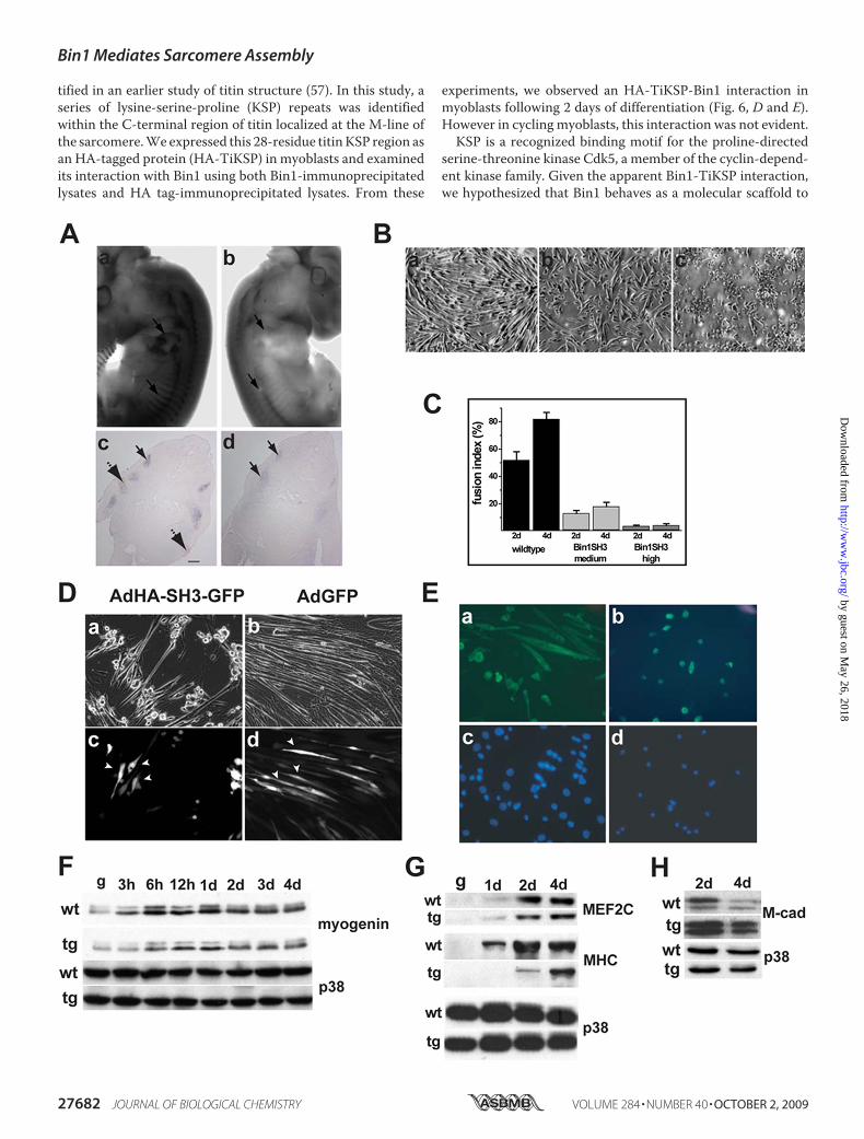
skeletal myogenesis, we isolated satellite cells from the hind-limbs of wild type and bin1SH3 mice. These cells displayedcomparable growth kinetics during proliferation. However,induction of differentiation resulted in strikingly disparatemorphologies between wild type and transgenic myoblast cul-tures. After 4 days in low serum,wild typemyoblasts underwentrobust differentiation and displayed elongated and arrayedextensions containingmultiple nuclei (Fig. 5,B, panel a, andC).In contrast, high expressing bin1SH3 myoblasts displayed anappropriate increase in myosin heavy chain expression, yet didnot form orderedmyotube structures (Fig. 5, B, panel c, andC).Interestingly, myoblasts from a transgenic line with moderateexpression of bin1SH3 did not form stable myotubes to thesame degree as the wild type cells, but the myogenic phenotypehad progressed further than the bin1SH3 high expressing cells(Fig. 5, B, panel b, and C). The limitation in myotube structureand patterning was not a simple temporal delay becauseextended low serum exposure (�7 days) did not result in robustmyotubes in transgenic cultures (data not shown). These exper-iments confirmed our observations that bin1SH3 expressionaffects the morphological characteristics of myoblast differen-tiation in a dose-dependent manner.To further determine SH3-specific effects, we employed the
c2c12 myoblast cell line. These cells were infected with adeno-virus expressing either GFP (Ad-IRES-GFP) or the SH3 domainof Bin1 (AdGFP-IRES-SH3), maintained in low serum mediafor 4 days, and then examined for GFP expression (Fig. 5D).GFP-positive cells that also expressed the Bin1 SH3 domainremained largely mononuclear and did not efficiently formmyotubes, whereas uninfected cells (GFP-negative) differenti-ated normally. This was in contrast to c2c12 cells infected withadenovirus-GFP in which bothGFP-positive andGFP-negativecells consistently formed robust multinucleatedmyotubes (Fig.5D, right panels). Trypan blue dye exclusion analyses did notindicate cellular toxicity with either Ad-IRES-GFP or AdGFP-IRES-SH3 (�3% of total infected cells) through a range ofadenoviral infections (0–500 infectious particles/cell) (supple-mental Fig. 1). These experiments verified that structural defi-ciencies in myotube formation are attributable to the SH3domain of Bin1.
FIGURE 3. Pathological examination of the bin1SH3 genotype. A, hematoxylin/eosin staining of the heart (panels a and b), lung (panels c and d), liver (panelse and f), prostate (panels g and h), and skin (panels i and j) showed no anomalies in cell morphology and tissue density in wild type (WT) and bin1SH3 mice. Barrepresents 100 �m. B, whole mount tunnel assays for apoptosis show no differences in cell death at 9.5 dpc between wild type (panel a) and bin1SH3 (panel b)embryos at the otic vesicles (arrows). A comparable degree of apoptosis is also observed within the somites (arrowheads) and along the apical ectodermal ridgein both wild type (panel c) and bin1SH3 (panel d) embryos. C, growth kinetics of wild type (wt) and bin1SH3 myoblasts held in growth medium were similar. n �9 animals per culture for each wild type and bin1SH3. D, cells were incubated in growth medium, and cell proliferation was examined using 5-bromo-2-deoxyuridine (BrdU). n � 9 animals per culture for each wild type (WT) and bin1SH3. E, annexin-V/propidium iodide (PI) apoptosis analyses of wild type andbin1SH3 myoblasts during incubation in low serum media. Both wild type and bin1SH3 myoblasts had comparable levels of apoptosis at the indicated timepoints. n � 9 animals per culture for each wild type and bin1SH3.
Bin1 Mediates Sarcomere Assembly
OCTOBER 2, 2009 • VOLUME 284 • NUMBER 40 JOURNAL OF BIOLOGICAL CHEMISTRY 27679
by guest on May 26, 2018
http://ww
w.jbc.org/
Dow
nloaded from

Bin1 Mediates Sarcomere Assembly
27680 JOURNAL OF BIOLOGICAL CHEMISTRY VOLUME 284 • NUMBER 40 • OCTOBER 2, 2009
by guest on May 26, 2018
http://ww
w.jbc.org/
Dow
nloaded from
The apparent lack of myotube formation after low serumincubation in bin1SH3 cultures prompted a more thoroughexamination of muscle-specific transcription factors in thesecells. After 2 days in low serum media, both wild type andbinSH3 myoblasts showed early accumulation of the terminaldifferentiationmarkers such as myosin heavy chain, myogenin,and MEF2C (Fig. 5, E–G), further confirming our previousimmunocytochemical results and suggesting that the Bin SH3domain did not impact the differentiation program per se (Fig.5, F and G).Myoblast fusion is a distinct feature of muscle cells. The
trans-membrane glycoprotein muscle cadherin (M-cadherin)is a member of the cadherin family of adhesion receptor mole-cules and has a role in myoblast fusion. M-cadherin is normallyexpressed at high levels during myoblast differentiation andrapidly declines followingmyoblast fusion (49–51). The lack ofelongated, multinucleated myotubes observed in our ex vivocultures of wild type and bin1SH3 myoblasts prompted us tocompare the pattern of M-cadherin protein in these cells. Anotable level ofM-cadherin was present in both cultures after 2days following induction of muscle differentiation. However,M-cadherin levels declined after 4 days in wild type cells butremained substantially elevated in bin1SH3 cells (Fig. 5H).These results suggested that at least in cultured cells, M-cad-herin is improperly regulated in bin1SH3 myoblasts. Intrigu-ingly, fusion did not appear to be perturbed in bin1SH3musclesin vivo despite the presence of disrupted sarcomeric structures.Although we cannot directly account for this inconsistency, itwould seem likely that other myoblast fusion-related factorsplay a role in vivo (52). Therefore, we cannot irrefutably con-clude thatM-cadherin regulation is directly influenced by Bin1.However, the significance of Bin1 in prompting adhesion-de-pendent signals that influence cell fate has been noted previ-ously (53).The modular structure of Bin1 implicates its participation in
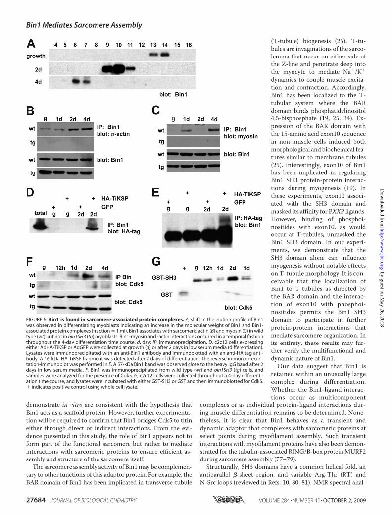
protein-protein interactions. Furthermore, the phenotypic out-come from overexpression of the Bin1 SH3 domain in bothcultured cells and transgenic animals suggested that Bin1 asso-ciates with protein(s) critical for assembly of the musclesarcomere, at least in part through the SH3 region. In a develop-mental context, it is conceivable that a protein involved inmyo-structural assembly would form transient protein complexeswith myocyte proteins. To begin to examine Bin1-dependentprotein interactions duringmyogenesis, we analyzed the nativesize of Bin1 in differentiating c2c12 cells using gel filtrationchromatography.With a Bin1-specific antibody probed againsta series of gel filtration fractions, we observed a shift in theelution of Bin1 protein by immunoblot analyses (Fig. 6A). Inthese experiments, elution at a lower volume (fraction number)indicated a larger size in the native protein. Bin1 was observed
in fractions 13–14 from lysates derived from cyclingmyoblasts.After 2 and 4 days of low serum induction of differentiation, theprospective Bin1 complex was found in fractions 9–11 and6–7, respectively, signifying a substantial increase in the size ofBin1. Interestingly, Bin1 did not seem to undergo dynamicstructural changes in bin1SH3 myoblasts. In an earlier study,alternative splicing led to higher molecular weight isoforms ofBin1 in differentiated c2c12 cells (�65-kDa growth to 68–70-kDa differentiation) (21). In our experiments, we found similarisoforms in 4- day differentiated cultures using the same anti-Bin1 antibody (99D). However, the elution profile from thesegel filtration experiments still designated Bin1 as a largemolec-ular weight protein of variable size. Taken together these datasuggest that the size of Bin1 and presumably Bin1-associatedprotein complexes increased in mass during early c2c12 differ-entiation. Moreover, a deviation in the native size of Bin1(�67–440 kDa during growth to �700 kDa during differentia-tion) suggested that Bin1 engages transient protein interactionsduring the process of myoblast differentiation.Clearly, the dynamic range in themolecular weight of Bin1 is
attributable in part to the SH3 interaction domain. In a prelim-inary LC-MS/MS screen of Bin1 SH3-interacting proteins, weidentified skeletal muscle myosin and sarcomeric actin as asso-ciated proteins within a Bin1-SH3 complex. To further definethe scope ofmyofilament protein interactionswith Bin1, a tem-poral analysis of the actin-Bin1 association was examined inwild type and bin1SH3 cultured myoblasts. Using an �-sarco-meric actin-specific antibody, we observed a positive Bin1-ac-tin interaction in differentiating wild type myoblasts but not inbin1SH3 transgenic myoblasts (Fig. 6B). Transient interactionsbetween actin and Bin1 occurred at a 4-day post low seruminduction of differentiation. A similar analysis was undertakento verify the degree of Bin1-myosin interaction. As with actin, amyosin-Bin1 interaction was only evident in differentiated wildtypemyoblasts. However, themost pronounced degree ofmyo-sin interaction occurred after 1 day and again after 4 days ofdifferentiation (Fig. 6C). These results further corroborate theLC-MS/MS findings and suggest that the Bin1 SH3 domaininteracts with actin and myosin in a temporal manner duringmyoblast differentiation.At the sarcomere, actin and myosin are in close proximity
with the giant myofilament protein titin. Titin has been exten-sively characterized and is known to contain several key fea-tures required for muscle force generation (54, 55). Titin scaf-folds numerous myofibrillar proteins, including �-actinin,myomesin, telethonin, components of the thick filament, andseveral signaling proteins (5, 56). The loss of functional titingives rise to disordered actin-myosin arrangement and ultimateloss of myofilament assembly. Interestingly, a region importantformuscle development andmyoblast differentiationwas iden-
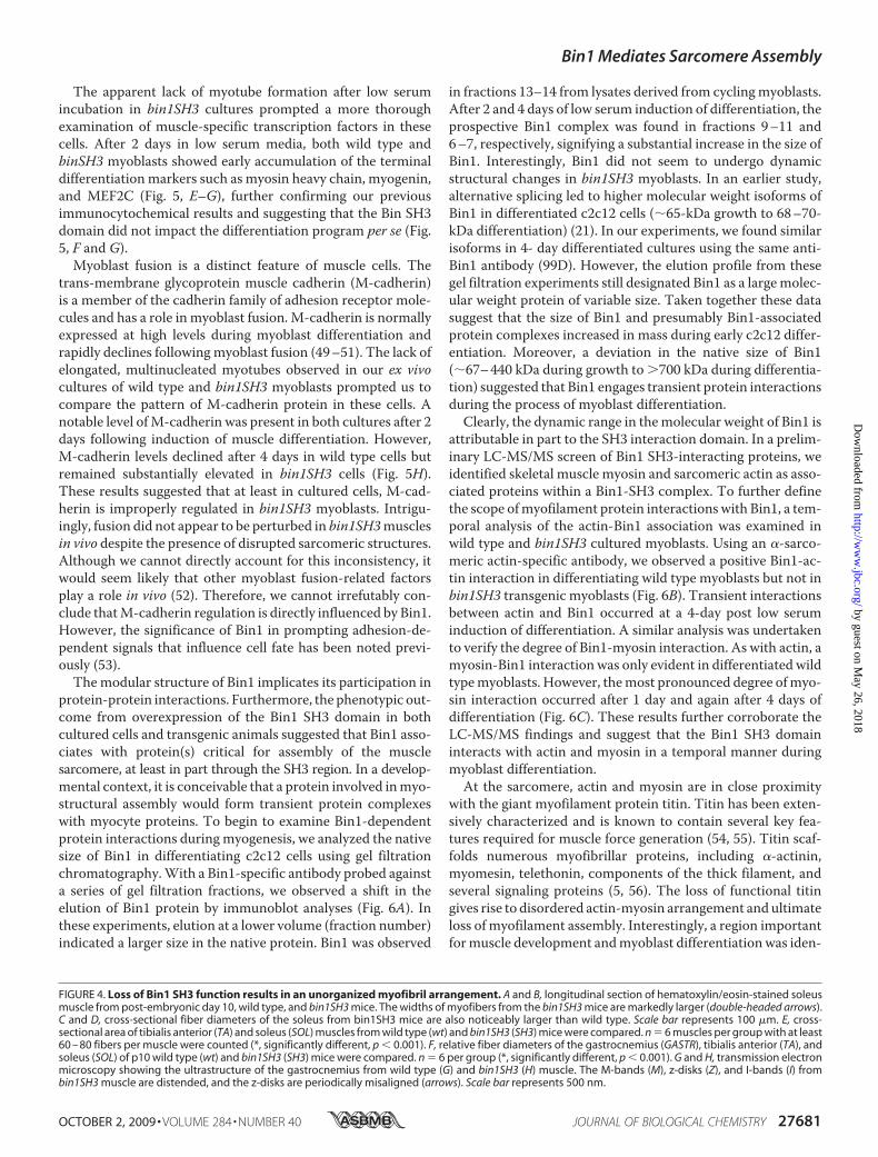
FIGURE 4. Loss of Bin1 SH3 function results in an unorganized myofibril arrangement. A and B, longitudinal section of hematoxylin/eosin-stained soleusmuscle from post-embryonic day 10, wild type, and bin1SH3 mice. The widths of myofibers from the bin1SH3 mice are markedly larger (double-headed arrows).C and D, cross-sectional fiber diameters of the soleus from bin1SH3 mice are also noticeably larger than wild type. Scale bar represents 100 �m. E, cross-sectional area of tibialis anterior (TA) and soleus (SOL) muscles from wild type (wt) and bin1SH3 (SH3) mice were compared. n � 6 muscles per group with at least60 – 80 fibers per muscle were counted (*, significantly different, p � 0.001). F, relative fiber diameters of the gastrocnemius (GASTR), tibialis anterior (TA), andsoleus (SOL) of p10 wild type (wt) and bin1SH3 (SH3) mice were compared. n � 6 per group (*, significantly different, p � 0.001). G and H, transmission electronmicroscopy showing the ultrastructure of the gastrocnemius from wild type (G) and bin1SH3 (H) muscle. The M-bands (M), z-disks (Z), and I-bands (I) frombin1SH3 muscle are distended, and the z-disks are periodically misaligned (arrows). Scale bar represents 500 nm.
Bin1 Mediates Sarcomere Assembly
OCTOBER 2, 2009 • VOLUME 284 • NUMBER 40 JOURNAL OF BIOLOGICAL CHEMISTRY 27681
by guest on May 26, 2018
http://ww
w.jbc.org/
Dow
nloaded from
tified in an earlier study of titin structure (57). In this study, aseries of lysine-serine-proline (KSP) repeats was identifiedwithin the C-terminal region of titin localized at the M-line ofthe sarcomere.We expressed this 28-residue titinKSP region asan HA-tagged protein (HA-TiKSP) in myoblasts and examinedits interaction with Bin1 using both Bin1-immunoprecipitatedlysates and HA tag-immunoprecipitated lysates. From these
experiments, we observed an HA-TiKSP-Bin1 interaction inmyoblasts following 2 days of differentiation (Fig. 6, D and E).However in cyclingmyoblasts, this interaction was not evident.KSP is a recognized binding motif for the proline-directed
serine-threonine kinase Cdk5, a member of the cyclin-depend-ent kinase family. Given the apparent Bin1-TiKSP interaction,we hypothesized that Bin1 behaves as a molecular scaffold to
Bin1 Mediates Sarcomere Assembly
27682 JOURNAL OF BIOLOGICAL CHEMISTRY VOLUME 284 • NUMBER 40 • OCTOBER 2, 2009
by guest on May 26, 2018
http://ww
w.jbc.org/
Dow
nloaded from

mediate phosphorylation at the titin-KSP region by Cdk5 dur-ing myogenesis. Therefore, we examined the extent of Bin1-Cdk5 association in wild type and Bin1 SH3-overexpressingmyoblasts. Cdk5 was present in Bin1-immunoprecipitatedlysates from wild type cells after 1, 2, and 4 days of differentia-tion (Fig. 6F). However, under similar conditions Cdk5 wasabsent in bin1SH3myoblasts. To determine whether this inter-action was mediated by the SH3 domain of Bin1, we conducteda series binding assays from myoblast lysates. Interestingly,Cdk5 was present at growth and after differentiation, althoughthe most robust association was observed after 2 days of differ-entiation (Fig. 6G). Taken together, our results suggest thatBin1 associates with Cdk5 in a temporal manner. The degree ofassociation may be important for mediating myogenicallydependent phosphorylation of titin within the KSP region.
DISCUSSION
Although the molecular genetic mechanisms of muscle dif-ferentiation and specification have been elucidated, the assem-bly of a functional myofiber remains to be defined. Here wedemonstrate that the SH3 domain of Bin1 mediates the assem-bly and organization of the skeletal muscle sarcomere throughassociations with actin andmyosin filaments. Transgenic over-expression of the Bin1 SH3 domain resulted in mis-expressionof skeletalmuscle-specific proteins with consequent disruptionof muscle fiber size and ultrastructural organization. We pro-pose that SH3-mediated interactions with actin, myosin, andCdk5 allow Bin1 to direct the assembly and organization of theskeletal muscle sarcomere.Bin1 is a protein with diverse cellular functions. Early reports
classed Bin1 as a Myc-interacting protein with tumor suppres-sor properties. and more recent experiments have verified thisfunction (16, 43, 44, 48, 58). In these studies, high expression ofBin1 was identified in skeletal muscle found near actin fila-ments (16, 18, 34). Localization of Bin1 at the t-tubules of stri-ated muscle implicated it as a modulator of membrane curva-ture thatwasmediated through interactions of theBARdomain(25, 34). To date, the role of the SH3 domain has remained amajor gap in the understanding of Bin1 function. Our observa-tions suggest that theBin1 SH3domain provides a skeletalmus-cle-specific role for an otherwise ubiquitous protein that main-tains a variety of cellular functions.
In this study, we anticipated that endogenous SH3-mediatedBin1 functions would be suppressed by dominant expression ofthe SH3 transgene. In eukaryotes, SH3 domains share a signif-icant degree of sequence similarity and mediate protein inter-actions by binding proline-rich sequences with a core PXXPmotif (whereX represents any amino acid) (59, 60). Despite thisgenerality, there are numerous examples of SH3 domains thatbind to alternate sequence motifs on target proteins (61–65).Although the atomic resolution of the Bin1 SH3 region witheither actin or myosin was not determined in this study, it islikely that these proteins associate through noncanonical SH3-ligand recognition mechanisms. In our experiments, we dem-onstrate that the SH3 domain of Bin1 interacts with both actinand myosin and that these interactions occur in a temporallysensitive fashion at early stages of myoblast differentiation.During myogenesis, sufficient and sequential activation of
muscle-specific genes is a requisite for assembling the musclecyto-architecture. The enlarged fiber diameter and irregularultrastructure of Bin1 SH3 murine skeletal muscle clearly indi-cated defects in muscle sarcomere organization. Remarkably,the net accumulation of muscle-specific proteins in the SH3expressing animals was not affected. Despite an adequateexpression of myogenic proteins, the functional componentsare seemingly unable to assemble into ordered units of themyo-fibril structure. To date, knowledge of sarcomere assembly hasbeen generally limited to large proteins of the sarcomere com-plex such as obscurin (14, 15, 66–72).In our experiments, we demonstrate that endogenous Bin1
associates with the C-terminal KSP region of titin that is highlyphosphorylated during skeletal muscle differentiation (57, 73).KSP is a substrate-binding motif for cyclin-dependent kinase 5(Cdk5) (74–76). In addition to interactions with the titin-KSPregion, we also demonstrate Bin1 interactions with Cdk5. Thisassociation is more prominent during the early phases of mus-cle differentiation, and importantly, this interaction is attenu-ated in muscle lysates from Bin1 SH3 animals.The in vivo phosphorylation of titin, as directed by Cdk5, is
required for proper myofibril arrangement and sarcomereorganization. These proteins form a core scaffold bindingregion and anchor with other proteins in a multicomponentcomplex. The Bin1-Cdk5 and Bin1-titin interactions that we
FIGURE 5. Bin1 SH3 domain affects myoblast fusion. A, whole mount in situ hybridization of a wild type 12-dpc embryo (panel a) demonstrates significantaccumulation of myogenin message within the somites and at the forelimb buds (arrows). Similar areas of active myogenesis in the bin1SH3 embryo (panel b)have noticeably absent levels of myogenin message. Cross-sectional analysis of a wild type embryo (panel c) confirms the presence of myogenin messagewithin the somites (solid arrow) and forelimb buds (broken arrow). A similar analysis of a bin1SH3 embryo (panel d) shows a very low accumulation of myogeninmessage within the somites and nondetectable levels of myogenin at the forelimb buds. Scale bar represents 100 �m. B, primary myoblasts isolated from wildtype (panel a) muscle differentiate into elongated myotubes after 48 h. A marked reduction in myotube pattern was noted in myoblasts isolated from a lowexpressing bin1SH3 muscle (panel b). Myoblasts isolated from a high expressing bin1SH3 mice (panel c) failed to elongate and fuse. C, higher percentage of cellscontaining two or more nuclei within a myosin heavy chain positive cell was observed in wild type myoblasts compared with both myoblasts from low and highexpressing bin1SH3 mice following incubation in low serum media. D, c2c12 myoblasts were infected with AdHA-SH3-GFP (left panel) or AdGFP control (rightpanel) and incubated with low serum for 4 days. Bright field micrographs (panels a and b) were compared with GFP fluorescence (panels c and d). SH3GFP-positive cells (panel c showing cells indicated by arrowheads) remained unicellular and did not fuse, whereas SH3-GFP-negative cells were elongated. BothGFP-positive and GFP-negative cells were elongated and visibly differentiated (panel b compared with panel d). Arrows in panel d show GFP-infected cells thatwere elongated. E, myosin heavy chain expression in wild type (panels a and c) and bin1SH3 (panels b and d) myoblasts after 2 days in low serum medium.Myoblasts from Bin1 SH3-expressing mice show an accumulation of myosin heavy chain but do not elongate and fuse to form mature myotubes. Nuclei werevisualized using 4�,6-diamidino-2-phenylindole staining (panels c and d). F, myogenin levels remained relatively similar in both wild type (wt) and transgenic(tg) myoblasts after early and prolonged low serum exposure. Loading was assessed using p38�. d, day. G, MEF2C was also present in both wild type andtransgenic myoblasts after 4 days in low serum media. Myosin heavy chain accumulated more slowly and to a lesser extent in transgenic myoblasts. H, after 4days in low serum conditions, wild type M-cad levels decreased indicating that these cells had fused. M-cad levels remained elevated in transgenic myoblastsat 4 days.
Bin1 Mediates Sarcomere Assembly
OCTOBER 2, 2009 • VOLUME 284 • NUMBER 40 JOURNAL OF BIOLOGICAL CHEMISTRY 27683
by guest on May 26, 2018
http://ww
w.jbc.org/
Dow
nloaded from
demonstrate in vitro are consistent with the hypothesis thatBin1 acts as a scaffold protein. However, further experimenta-tion will be required to confirm that Bin1 bridges Cdk5 to titineither through direct or indirect interactions. From the evi-dence presented in this study, the role of Bin1 appears not toform part of the functional sarcomere but rather to mediateinteractions with sarcomeric proteins to ensure efficient as-sembly and structure of the sarcomere itself.The sarcomere assembly activity of Bin1may be complemen-
tary to other functions of this adaptor protein. For example, theBAR domain of Bin1 has been implicated in transverse-tubule
(T-tubule) biogenesis (25). T-tu-bules are invaginations of the sarco-lemma that occur on either side ofthe Z-line and penetrate deep intothe myocyte to mediate Na�/K�
dynamics to couple muscle excita-tion and contraction. Accordingly,Bin1 has been localized to the T-tubular system where the BARdomain binds phosphatidylinositol4,5-bisphosphate (19, 25, 34). Ex-pression of the BAR domain withthe 15-amino acid exon10 sequencein non-muscle cells induced bothmorphological and biochemical fea-tures similar to membrane tubules(25). Interestingly, exon10 of Bin1has been implicated in regulatingBin1 SH3 protein-protein interac-tions during myogenesis (19). Inthese experiments, exon10 associ-ated with the SH3 domain andmasked its affinity for PXXP ligands.However, binding of phosphoi-nositides with exon10, as wouldoccur at T-tubules, unmasked theBin1 SH3 domain. In our experi-ments, we demonstrate that theSH3 domain alone can influencemyogenesis without notable effectson T-tubule morphology. It is con-ceivable that the localization ofBin1 to T-tubules as directed bythe BAR domain and the interac-tion of exon10 with phosphoi-nositides permits the Bin1 SH3domain to participate in furtherprotein-protein interactions thatmediate sarcomere organization. Inits entirety, these results may fur-ther verify the multifunctional anddynamic nature of Bin1.Our data suggest that Bin1 is
retained within an unusually largecomplex during differentiation.Whether the Bin1-ligand interac-tions occur as multicomponent
complexes or as individual protein-ligand interactions dur-ing muscle differentiation remains to be determined. None-theless, it is clear that Bin1 behaves as a transient anddynamic adaptor that complexes with sarcomeric proteins atselect points during myofilament assembly. Such transientinteractions with myofilament proteins have also been demon-strated for the tubulin-associated RING/B-box proteinMURF2during sarcomere assembly (77–79).Structurally, SH3 domains have a common helical fold, an
antiparallel �-sheet region, and variable Arg-Thr (RT) andN-Src loops (reviewed in Refs. 10, 80, 81). NMR spectral anal-
FIGURE 6. Bin1 is found in sarcomere-associated protein complexes. A, shift in the elution profile of Bin1was observed in differentiating myoblasts indicating an increase in the molecular weight of Bin1 and Bin1-associated protein complexes (fraction � 1 ml). Bin1 associates with sarcomeric actin (B) and myosin (C) in wildtype (wt) but not in bin1SH3 (tg) myoblasts. Bin1-myosin and -actin interactions occurred in a temporal fashionthroughout the 4-day differentiation time course. d, day; IP, immunoprecipitation. D, c2c12 cells expressingeither AdHA-TiKSP or AdGFP were collected at growth (g) or after 2 days in low serum media (differentiation).Lysates were immunoprecipitated with an anti-Bin1 antibody and immunoblotted with an anti-HA tag anti-body. A 16-kDa HA-TiKSP fragment was detected after 2 days of differentiation. The reverse immunoprecipi-tation-immunoblot was performed in E. A 57-kDa Bin1 band was observed close to the heavy IgG band after 2days in low serum media. F, Bin1 was immunoprecipitated from wild type (wt) and bin1SH3 (tg) cells, andsamples were analyzed for the presence of Cdk5. G, c2c12 cells were collected throughout a 4-day differenti-ation time course, and lysates were incubated with either GST-SH3 or GST and then immunoblotted for Cdk5.� indicates positive control using whole cell lysate.
Bin1 Mediates Sarcomere Assembly
27684 JOURNAL OF BIOLOGICAL CHEMISTRY VOLUME 284 • NUMBER 40 • OCTOBER 2, 2009
by guest on May 26, 2018
http://ww
w.jbc.org/
Dow
nloaded from

ysis and crystal resolution of the Bin1 SH3 structure indicatedfeatures that were very unique among SH3 domains that mayenhance Bin1 SH3 binding specificity (36, 37). Accordingly, weobserved unambiguous anomalies in only the skeletal muscula-ture of Bin1 SH3-expressing animals. Interestingly, homozy-gous deletion of Bin1 gave rise to substantial cardiomyopathy(22), an effect not observed in this study where only the SH3domain was targeted. Furthermore, we did not observe the pre-viously reported tumorigenic or cell cycle effects associatedwith the disruption of Bin1 activity (43, 44, 48). The compart-mentalization and spatial distribution of binding partners arelikely to contribute to the function and specificity of the Bin1SH3 as is the case for other proteins with modular interactiondomains (reviewed in Refs. 10, 59, 82).Although Bin1 does not form part of the functional sar-
comere itself, we propose that Bin1 serves a role in sarcomereassembly by mediating interactions with sarcomeric proteins.This is in conjunction with the other functions of Bin1 asimposed by its modular protein regions, namely theMyc-bind-ing domain and the BAR domain. Our data also suggest thatBin1 is retained within an unusually large protein complex dur-ing myogenesis. Taken together, these observations and theresults of this study suggest that myofiber assembly is depend-ent on transient protein interactions that guide and positionsarcomeric proteins at the appropriate time and place.
Acknowledgments—We thank Kim Balazsi and Beata Pekalska forskilled technical assistance and Drs. Jeffrey Dilworth and David Pick-etts for helpful discussions.
REFERENCES1. Sabourin, L. A., and Rudnicki, M. A. (2000) Clin. Genet. 57, 16–252. Buckingham,M., Bajard, L., Chang, T., Daubas, P., Hadchouel, J., Meilhac,
S., Montarras, D., Rocancourt, D., and Relaix, F. (2003) J. Anat. 202,59–68
3. Zhao, P., and Hoffman, E. P. (2004) Dev. Dyn. 229, 380–3924. Au, Y. (2004) Cell. Mol. Life Sci. 61, 3016–30335. Lange, S., Ehler, E., and Gautel, M. (2006) Trends Cell Biol. 16, 11–186. Labeit, S., Gibson, T., Lakey, A., Leonard, K., Zeviani, M., Knight, P.,
Wardale, J., and Trinick, J. (1991) FEBS Lett. 282, 313–3167. McElhinny, A. S., Schwach, C., Valichnac, M., Mount-Patrick, S., and
Gregorio, C. C. (2005) J. Cell Biol. 170, 947–9578. Sinz, A., and Wang, K. (2001) Biochemistry 40, 7903–79139. Kaneko, T., Li, L., and Li, S. S. (2008) Front. Biosci. 13, 4938–495210. Li, S. S. (2005) Biochem. J. 390, 641–65311. Ma, K., Forbes, J. G., Gutierrez-Cruz, G., and Wang, K. (2006) J. Biol.
Chem. 281, 27539–2755612. Ma, K., and Wang, K. (2002) FEBS Lett. 532, 273–27813. Politou, A. S., Millevoi, S., Gautel, M., Kolmerer, B., and Pastore, A. (1998)
J. Mol. Biol. 276, 189–20214. Young, P., Ehler, E., and Gautel, M. (2001) J. Cell Biol. 154, 123–13615. Bang,M. L., Centner, T., Fornoff, F., Geach, A. J., Gotthardt, M.,McNabb,
M., Witt, C. C., Labeit, D., Gregorio, C. C., Granzier, H., and Labeit, S.(2001) Circ. Res. 89, 1065–1072
16. Sakamuro, D., Elliott, K. J., Wechsler-Reya, R., and Prendergast, G. C.(1996) Nat. Genet. 14, 69–77
17. Elliott, K., Sakamuro, D., Basu, A., Du, W., Wunner, W., Staller, P.,Gaubatz, S., Zhang, H., Prochownik, E., Eilers, M., and Prendergast, G. C.(1999) Oncogene 18, 3564–3573
18. Wechsler-Reya, R., Sakamuro, D., Zhang, J., Duhadaway, J., and Prender-gast, G. C. (1997) J. Biol. Chem. 272, 31453–31458
19. Kojima, C., Hashimoto, A., Yabuta, I., Hirose, M., Hashimoto, S., Kanaho,
Y., Sumimoto, H., Ikegami, T., and Sabe, H. (2004) EMBO J. 23,4413–4422
20. Ren, G., Vajjhala, P., Lee, J. S., Winsor, B., and Munn, A. L. (2006)Micro-biol. Mol. Biol. Rev. 70, 37–120
21. Wechsler-Reya, R. J., Elliott, K. J., and Prendergast, G. C. (1998)Mol. Cell.Biol. 18, 566–575
22. Muller, A. J., Baker, J. F., DuHadaway, J. B., Ge, K., Farmer, G., Donover,P. S., Meade, R., Reid, C., Grzanna, R., Roach, A. H., Shah, N., Soler, A. P.,and Prendergast, G. C. (2003)Mol. Cell. Biol. 23, 4295–4306
23. Razzaq, A., Robinson, I.M.,McMahon,H. T., Skepper, J. N., Su, Y., Zelhof,A. C., Jackson, A. P., Gay, N. J., and O’Kane, C. J. (2001) Genes Dev. 15,2967–2979
24. Zelhof, A. C., Bao, H., Hardy, R. W., Razzaq, A., Zhang, B., and Doe, C. Q.(2001) Development 128, 5005–5015
25. Lee, E., Marcucci, M., Daniell, L., Pypaert, M., Weisz, O. A., Ochoa, G. C.,Farsad, K.,Wenk,M. R., andDeCamilli, P. (2002) Science 297, 1193–1196
26. Nicot, A. S., Toussaint, A., Tosch, V., Kretz, C., Wallgren-Pettersson, C.,Iwarsson, E., Kingston,H., Garnier, J.M., Biancalana, V., Oldfors, A.,Man-del, J. L., and Laporte, J. (2007) Nat. Genet. 39, 1134–1139
27. Niwa, H., Yamamura, K., and Miyazaki, J. (1991) Gene 108, 193–19928. Lobe, C. G., Koop, K. E., Kreppner, W., Lomeli, H., Gertsenstein, M., and
Nagy, A. (1999) Dev. Biol. 208, 281–29229. Guy, L. G., Kothary, R., DeRepentigny, Y., Delvoye, N., Ellis, J., andWall, L.
(1996) EMBO J. 15, 3713–372130. Franzini-Armstrong, C. (1991) Dev. Biol. 146, 353–36331. Sassoon, D., Lyons, G., Wright, W. E., Lin, V., Lassar, A., Weintraub, H.,
and Buckingham, M. (1989) Nature 341, 303–30732. Wilkinson, D. G., and Nieto, M. A. (1993) Methods Enzymol. 225,
361–37333. Fernando, P., Kelly, J. F., Balazsi, K., Slack, R. S., andMegeney, L. A. (2002)
Proc. Natl. Acad. Sci. U.S.A. 99, 11025–1103034. Butler, M. H., David, C., Ochoa, G. C., Freyberg, Z., Daniell, L., Grabs, D.,
Cremona, O., and De Camilli, P. (1997) J. Cell Biol. 137, 1355–136735. Buckingham, M. (2006) Curr. Opin. Genet. Dev. 16, 525–53236. Owen, D. J.,Wigge, P., Vallis, Y., Moore, J. D., Evans, P. R., andMcMahon,
H. T. (1998) EMBO J. 17, 5273–528537. Pineda-Lucena, A., Ho, C. S., Mao, D. Y., Sheng, Y., Laister, R. C., Mu-
handiram, R., Lu, Y., Seet, B. T., Katz, S., Szyperski, T., Penn, L. Z., andArrowsmith, C. H. (2005) J. Mol. Biol. 351, 182–194
38. Tajiri, T., Liu, X., Thompson, P. M., Tanaka, S., Suita, S., Zhao, H., Maris,J. M., Prendergast, G. C., and Hogarty, M. D. (2003) Clin. Cancer Res. 9,3345–3355
39. Ge, K., Minhas, F., Duhadaway, J., Mao, N. C., Wilson, D., Buccafusca, R.,Sakamuro, D., Nelson, P., Malkowicz, S. B., Tomaszewski, J., and Prender-gast, G. C. (2000) Int. J. Cancer 86, 155–161
40. Hogarty, M. D., Liu, X., Thompson, P. M., White, P. S., Sulman, E. P.,Maris, J. M., and Brodeur, G. M. (2000)Med. Pediatr. Oncol. 35, 559–562
41. Galderisi, U., Di Bernardo, G., Cipollaro,M., Jori, F. P., Piegari, E., Cascino,A., Peluso, G., and Melone, M. A. (1999) J. Cell. Biochem. 74, 313–322
42. Ge, K., DuHadaway, J., Du, W., Herlyn, M., Rodeck, U., and Prendergast,G. C. (1999) Proc. Natl. Acad. Sci. U.S.A. 96, 9689–9694
43. Chang, M. Y., Boulden, J., Katz, J. B., Wang, L., Meyer, T. J., Soler, A. P.,Muller, A. J., and Prendergast, G. C. (2007) Cancer Res. 67, 7605–7612
44. Chang,M. Y., Boulden, J., Sutanto-Ward, E., Duhadaway, J. B., Soler, A. P.,Muller, A. J., and Prendergast, G. C. (2007) Cancer Res. 67, 100–107
45. Ramalingam, A., Farmer, G. E., Stamato, T. D., and Prendergast, G. C.(2007) Cell Cycle 6, 1914–1918
46. Telfer, J. F., Urquhart, J., and Crouch, D. H. (2005) Cell. Signal. 17,701–708
47. Elliott, K., Ge, K., Du, W., and Prendergast, G. C. (2000) Oncogene 19,4669–4684
48. Muller, A. J., DuHadaway, J. B., Donover, P. S., Sutanto-Ward, E., andPrendergast, G. C. (2004) Cancer Biol. Ther. 3, 1236–1242
49. Charrasse, S., Comunale, F., Fortier, M., Portales-Casamar, E., Debant, A.,and Gauthier-Rouviere, C. (2007)Mol. Biol. Cell 18, 1734–1743
50. Wrobel, E., Brzoska, E., and Moraczewski, J. (2007) Eur. J. Cell Biol. 86,99–109
51. Zeschnigk, M., Kozian, D., Kuch, C., Schmoll, M., and Starzinski-Powitz,
Bin1 Mediates Sarcomere Assembly
OCTOBER 2, 2009 • VOLUME 284 • NUMBER 40 JOURNAL OF BIOLOGICAL CHEMISTRY 27685
by guest on May 26, 2018
http://ww
w.jbc.org/
Dow
nloaded from

A. (1995) J. Cell Sci. 108, 2973–298152. Hollnagel, A., Grund, C., Franke, W. W., and Arnold, H. H. (2002) Mol.
Cell. Biol. 22, 4760–477053. Duhadaway, J., Rowe, F., Elliott, K., Mao, N. C., and Prendergast, G. C.
(1999) Cell Adhes. Commun. 7, 99–11054. Fukuda, N., Granzier, H. L., Ishiwata, S., and Kurihara, S. (2008) J. Physiol.
Sci. 58, 151–15955. Granzier, H. L., and Labeit, S. (2005) Adv. Protein Chem. 71, 89–11956. Clark, K. A., McElhinny, A. S., Beckerle, M. C., and Gregorio, C. C. (2002)
Annu. Rev. Cell Dev. Biol. 18, 637–70657. Gautel, M., Leonard, K., and Labeit, S. (1993) EMBO J. 12, 3827–383458. DuHadaway, J. B., Sakamuro, D., Ewert, D. L., and Prendergast, G. C.
(2001) Cancer Res. 61, 3151–315659. Mayer, B. J. (2001) J. Cell Sci. 114, 1253–126360. Kay, B. K., Williamson, M. P., and Sudol, M. (2000) FASEB J. 14, 231–24161. Moncalian, G., Cardenes, N., Deribe, Y. L., Spínola-Amilibia, M., Dikic, I.,
and Bravo, J. (2006) J. Biol. Chem. 281, 38845–3885362. Tian, L., Chen, L.,McClafferty, H., Sailer, C. A., Ruth, P., Knaus, H. G., and
Shipston, M. J. (2006) FASEB J. 20, 2588–259063. Jia, C. Y., Nie, J., Wu, C., Li, C., and Li, S. S. (2005)Mol. Cell. Proteomics 4,
1155–116664. Kami, K., Takeya, R., Sumimoto, H., and Kohda, D. (2002) EMBO J. 21,
4268–427665. Barnett, P., Bottger, G., Klein, A. T., Tabak, H. F., and Distel, B. (2000)
EMBO J. 19, 6382–639166. Armani, A., Galli, S., Giacomello, E., Bagnato, P., Barone, V., Rossi, D., and
Sorrentino, V. (2006) Exp. Cell Res. 312, 3546–355867. Kontrogianni-Konstantopoulos, A., Catino, D. H., Strong, J. C., Sutter, S.,
Borisov, A. B., Pumplin, D. W., Russell, M. W., and Bloch, R. J. (2006)
FASEB J. 20, 2102–211168. Bagnato, P., Barone, V., Giacomello, E., Rossi, D., and Sorrentino, V. (2003)
J. Cell Biol. 160, 245–25369. Kontrogianni-Konstantopoulos, A., Jones, E. M., Van Rossum, D. B., and
Bloch, R. J. (2003)Mol. Biol. Cell 14, 1138–114870. Bowman, A. L., Catino, D. H., Strong, J. C., Randall, W. R., Kontrogianni-
Konstantopoulos, A., and Bloch, R. J. (2008)Mol. Biol. Cell. 19, 3782–379271. Bang,M. L., Gregorio, C., and Labeit, S. (2002) J. Struct. Biol. 137, 119–12772. Nave, R., Furst, D. O., and Weber, K. (1990) FEBS Lett. 269, 163–16673. Maruyama, K., Endo, T., Kume, H., Kawamura, Y., Kanzawa, N., Kimura,
S., Kawashima, S., and Maruyama, K. (1994) J. Biochem. 115, 147–14974. Sharma, P., Steinbach, P. J., Sharma, M., Amin, N. D., Barchi, J. J., Jr., and
Pant, H. C. (1999) J. Biol. Chem. 274, 9600–960675. Shetty, K. T., Link, W. T., and Pant, H. C. (1993) Proc. Natl. Acad. Sci.
U.S.A. 90, 6844–684876. Hellmich, M. R., Pant, H. C., Wada, E., and Battey, J. F. (1992) Proc. Natl.
Acad. Sci. U.S.A. 89, 10867–1087177. Spencer, J. A., Eliazer, S., Ilaria, R. L., Jr., Richardson, J. A., andOlson, E. N.
(2000) J. Cell Biol. 150, 771–78478. Pizon, V., Iakovenko, A., VanDerVen, P. F., Kelly, R., Fatu, C., Furst, D.O.,
Karsenti, E., and Gautel, M. (2002) J. Cell Sci. 115, 4469–448279. McElhinny, A. S., Perry, C. N., Witt, C. C., Labeit, S., and Gregorio, C. C.
(2004) J. Cell Sci. 117, 3175–318880. Yu, H., Rosen, M. K., Shin, T. B., Seidel-Dugan, C., Brugge, J. S., and
Schreiber, S. L. (1992) Science 258, 1665–166881. Feng, S., Kasahara, C., Rickles, R. J., and Schreiber, S. L. (1995) Proc. Natl.
Acad. Sci. U.S.A. 92, 12408–1241582. Ladbury, J. E., and Arold, S. (2000) Chem. Biol. 7, R3–R8
Bin1 Mediates Sarcomere Assembly
27686 JOURNAL OF BIOLOGICAL CHEMISTRY VOLUME 284 • NUMBER 40 • OCTOBER 2, 2009
by guest on May 26, 2018
http://ww
w.jbc.org/
Dow
nloaded from

John F. Kelly, Rashmi Kothary and Lynn A. MegeneyPasan Fernando, Jacqueline S. Sandoz, Wen Ding, Yves de Repentigny, Steve Brunette,Bin1 Src Homology 3 Domain Acts as a Scaffold for Myofiber Sarcomere Assembly
doi: 10.1074/jbc.M109.029538 originally published online July 26, 20092009, 284:27674-27686.J. Biol. Chem.
10.1074/jbc.M109.029538Access the most updated version of this article at doi:
Alerts:
When a correction for this article is posted•
When this article is cited•
to choose from all of JBC's e-mail alertsClick here
Supplemental material:
http://www.jbc.org/content/suppl/2009/07/26/M109.029538.DC1
http://www.jbc.org/content/284/40/27674.full.html#ref-list-1
This article cites 82 references, 41 of which can be accessed free at
by guest on May 26, 2018
http://ww
w.jbc.org/
Dow
nloaded from