bimaxillary protrusion with missing lower first...
TRANSCRIPT

20
IJOI 44 iAOI CASE REPORT
Bimaxillary Protrusion with Missing Lower First Molar and Upper Premolar:
Asymmetric Extractions, Anchorage Control and Interproximal Reduction
Abstract A 38-year-old female presented with a Class I bimaxillary protrusion, complicated by asymmetric anterior spacing in both arches. Early loss of a lower right (LR) � rst molar resulted in mesial tipping of adjacent molars, a unilateral excessive curve of Spee, and an atrophic ridge. The upper left (UL) second premolar was missing and there was extensive subgingival calculus. Following periodontal scaling, additional extractions were needed to correct the protrusion, so the most compromised teeth in the a� ected quadrants were selected: upper right (UR) � rst premolar with cervical abrasion, a super-erupted maxillary left third molar, and a lower left (LL) � rst molar with extensive caries. The asymmetric extraction spaces required careful management of anchorage to retract the anterior segments without canting the occlusal plane and/or producing a midline deviation. After 34 months of active treatment, the partially edentulous compensated malocclusion with a discrepancy index (DI) of 18 was treated to an acceptable cast-radiograph evaluation score of 23. The facial profile was corrected by retraction of the lips, and the dental esthetics were improved with space closure, symmetrical alignment, and coincidence of the midlines. (Int J Orthod Implantol 2016;44:20-41)
Key words:Adult orthodontic treatment, complex asymmetric malocclusion, bimaxillary protrusion, atrophic edentulous spaces, extraction of compromised teeth, asymmetric mechanics, midline diastema
Introduction
The mandibular fi rst molars are typically the fi rst permanent teeth to erupt, at about age 6 years. They are very important for the occlusal function and normal development of the dentition, but they are at high risk for early loss due to caries.1,2 The enamel of the fi rst molars develops during the infant and toddler period (<3yrs of age), which is a common interval for illnesses with high fever. Up to 20% of mandibular fi rst year molars erupt with enamel hypomineralization defects, that render the teeth highly susceptible to caries and early loss in <2yr after they erupt.3,4 Generalized oral hygiene negligence may result in rampant caries of permanent and deciduous teeth, but the isolated loss of permanent fi rst molars is usually related to molar-incisor hypomineralization (MIH).1-4 Since the permanent fi rst molars are centric stops in occlusion during the late transition stage of dental eruption (age 10-12yr), severe acquired malocclusions may occur.1,2 Restoring atrophic spaces in a compromised permanent dentition is challenging, so orthodontic space closure or opening sites for implants is often preferable if the periodontium is healthy.1,5

21
Asymmetric Extractions IJOI 44
█ Fig. 1: Pre-treatment facial and intraoral photographs
Dr. Chi Huang,Resident, Beethoven Orthodontic Center
Editor, International Journal of Orthodontics & Implantology (Left)
Dr. Chuanwei Su,Director, Newton's Implant Center
Associate Editor, International Journal of Orthodontics & Implantology (Center left)
Dr. Chris Chang, Founder, Beethoven Orthodontic Center
Publisher, International Journal of Orthodontics & Implantology (Center right)
Dr. W. Eugene Roberts,Editor-in-chief, International Journal of Orthodontics & Implantology (Right)

22
IJOI 44 iAOI CASE REPORT
█ Fig. 2: Frontal oral view for smile evaluation.
Closing first molar extraction spaces in adults may be complicated by mesial-tipped second molars and atrophic alveolar ridges, particularly if the teeth were lost early (<age 8yr).1,2,5 Correcting edentulous spaces is a common request for adults. However, loss of bone in the vertical and buccolingual dimensions results in narrow atrophic edentulous spaces.6,7 Protracting second molars to close atrophic ridges requires extensive osteogenesis (anabolic bone
modeling) to expand the ridge and thicken the cortical plates.8,9 Furthermore, when wide tooth roots are protracted through narrow alveolar ridges, enhanced anchorage may be required.10-12 Evaluate all teeth in the arch and prioritize the degree of compromise due to restorative, endodontic and/or periodontal problems. If extractions are required to manage a malocclusion, good clinical practice is to select the most compromised teeth, even if that approach results in asymmetric spaces. Closing asymmetric extraction spaces can result in canting of the occlusal plane and midline discrepancies.12,13 Closure of edentulous spaces may produce a desirable result,14-17 but complex mechanics and temporary anchorage devices (TADs) are often required to maintain or correct symmetry.18-23
The aim of this case report is to investigate the etiology of a complex malocclusion as a guide for developing a relatively conservative, extraction approach for resolving the malocclusion, while also eliminating problem teeth. Cost effective oral rehabilitation was an important service for the current patient, who required orthodontics
to manage a part ia l ly edentulous, acquired malocclusion with a Discrepancy Index (DI) score of 18.
Diagnosis and Etiology
A 38-year-old woman sought orthodontic evaluation with concerns about missing teeth, an unesthetic anterior dentition, prominent lower incisors and protrusive lips (Fig. 1). There was no contributing medical history, but she had a long history of limited, restorative dental care. Extra-oral evaluation with the lips closed showed a symmetric bimaxillary protrusion with coincident dental and facial midlines. Upon smiling her dentition was unattractive due to an end-to-end incisal relationship, occlusal cant (more inferior on the right side), irregular spacing in the anterior segments, and intermaxillary midline diastemas (Figs. 1 and 2).
Intraoral examination revealed three missing teeth: LR first molar, LL third molar, and the UL
23
Asymmetric Extractions IJOI 44
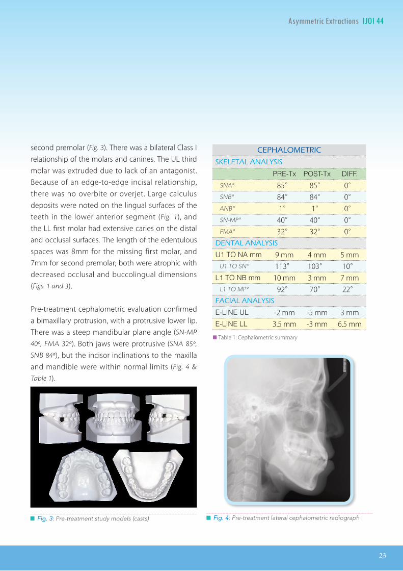
█ Fig. 3: Pre-treatment study models (casts) █ Fig. 4: Pre-treatment lateral cephalometric radiograph
second premolar (Fig. 3). There was a bilateral Class I relationship of the molars and canines. The UL third molar was extruded due to lack of an antagonist. Because of an edge-to-edge incisal relationship, there was no overbite or overjet. Large calculus deposits were noted on the lingual surfaces of the teeth in the lower anterior segment (Fig. 1), and the LL fi rst molar had extensive caries on the distal and occlusal surfaces. The length of the edentulous spaces was 8mm for the missing first molar, and 7mm for second premolar; both were atrophic with decreased occlusal and buccolingual dimensions (Figs. 1 and 3).
Pre-treatment cephalometric evaluation confirmed a bimaxillary protrusion, with a protrusive lower lip. There was a steep mandibular plane angle (SN-MP
40º, FMA 32º). Both jaws were protrusive (SNA 85º,
SNB 84º), but the incisor inclinations to the maxilla and mandible were within normal limits (Fig. 4 &
Table 1).
CEPHALOMETRIC
SKELETAL ANALYSIS
PRE-Tx POST-Tx DIFF.
SNA° 85° 85° 0° SNB° 84° 84° 0° ANB° 1° 1° 0° SN-MP° 40° 40° 0°FMA° 32° 32° 0°
DENTAL ANALYSIS
U1 TO NA mm 9 mm 4 mm 5 mm U1 TO SN° 113° 103° 10°
L1 TO NB mm 10 mm 3 mm 7 mm L1 TO MP° 92° 70° 22°
FACIAL ANALYSIS
E-LINE UL -2 mm -5 mm 3 mm E-LINE LL 3.5 mm -3 mm 6.5 mm
█ Table 1: Cephalometric summary
24
IJOI 44 iAOI CASE REPORT
█ Fig. 6: A facial intraoral photograph shows asymmetry as the patient opens the mandible, apparently due to the interference of the extruded UL third molar.
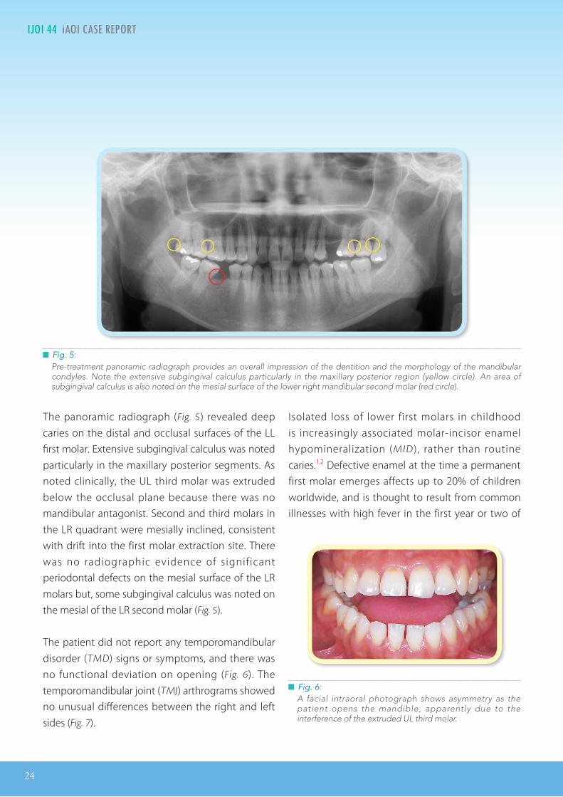
The panoramic radiograph (Fig. 5) revealed deep caries on the distal and occlusal surfaces of the LL fi rst molar. Extensive subgingival calculus was noted particularly in the maxillary posterior segments. As noted clinically, the UL third molar was extruded below the occlusal plane because there was no mandibular antagonist. Second and third molars in the LR quadrant were mesially inclined, consistent with drift into the first molar extraction site. There was no radiographic evidence of signif icant periodontal defects on the mesial surface of the LR molars but, some subgingival calculus was noted on the mesial of the LR second molar (Fig. 5).
The patient did not report any temporomandibular disorder (TMD) signs or symptoms, and there was no functional deviation on opening (Fig. 6). The temporomandibular joint (TMJ) arthrograms showed no unusual differences between the right and left sides (Fig. 7).
█ Fig. 5: Pre-treatment panoramic radiograph provides an overall impression of the dentition and the morphology of the mandibular condyles. Note the extensive subgingival calculus particularly in the maxillary posterior region (yellow circle). An area of subgingival calculus is also noted on the mesial surface of the lower right mandibular second molar (red circle).
Isolated loss of lower first molars in childhood is increasingly associated molar-incisor enamel hypomineralization (MID), rather than routine caries.1,2 Defective enamel at the time a permanent first molar emerges affects up to 20% of children worldwide, and is thought to result from common illnesses with high fever in the first year or two of

25
Asymmetric Extractions IJOI 44
█ Fig. 7: Pre-treatment temporomandibular joint (TMJ) direct arthrograms are shown from the left: R TMJ closed, R TMJ open, L TMJ open, and L TMJ closed.
life.3,4 When the affected first molars enter the oral cavity, they are susceptible to catastrophic caries, resulting in extraction during the early mixed dentition period (6-8yr). Early loss of lower first molars is often a developmental problem because there is no posterior stop in occlusion when the adjacent second primary molar is lost. This occlusal instability can result in functional shifts such as anterior crossbite, a deep curve of Spee on the aff ected side, and/or facial asymmetry (Figs. 2 and 3).1,2
The Amer ican Board of Orthodont ic (AB O ) discrepancy index was 18 points, as shown in the supplementary worksheet 1.
Treatment Objectives
The objectives in order of priority were:
1. Restorative: Restore all caries as needed, evaluate compromised teeth.
2. Periodontal: Remove all calculus, pre-orthodontics preparation as needed.
3. Orthodontics: Retract protrusive lips to correct bimaxillary protrusion.
• Maintain maxillary and mandibular orientation in three dimensions (3D).
• Extract three compromised teeth: UR fi rst premolar because of cervical abrasion, super-erupted UL third molar, and deeply-decayed LL fi rst molar.
• Use a full fi xed appliance to level and align both dental arches.
• Upright and protract mandibular second molars to substitute for missing fi rst molars.
• Diff erential retraction of upper and lower incisors to correct the edge-to-edge bite.
• Asymmetric space closure to minimize iatrogenic midline discrepancies.
• Finishing: optimize alignment with bracket repositioning and archwire adjustments.
Treatment Alternatives
Because of the asymmetric extraction spaces, retracting the incisors r isked occlusal plane canting and/or midline deviation. The patient was prospectively warned about these potential side effects, but was also informed that a 4mm midline deviation is clinically acceptable. She agreed to the use of OrthoBoneScrew® (2x12mm, Newton’s A Ltd,
Hsinchu City, Taiwan) anchorage if needed.

26
IJOI 44 iAOI CASE REPORT
Treatment Progress
After the initial restorative and periodontal care was completed, three compromised teeth were extracted: UR first premolar, UL third molar and LL first molar. A .022” Damon Q® (Ormco, Glendora,
CA) fixed appliance was bonded on all permanent teeth and high torque brackets were selected for the maxillary incisors and canines. Standard torque brackets were used on the entire mandibular arch. The upper arch was leveled and aligned with the following wire sequence: .014” CuNiTi, .014x.025” CuNiTi, .017x.025” TMA and .016x.025” SS. The corresponding lower arch sequence was .014” CuNiTi, .018” CuNiTi, .014x.025” CuNiTi, .017x.025” TMA and .016x.025” SS.
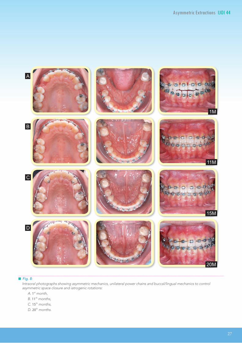
In the first month of active treatment, posterior bite turbos were constructed with Fuji II type II glass ionomer cement (GC America, Alsip IL) on the occlusal surfaces of the mandibular second molars. Bilateral bite turbos were effective for opening the bite, reducing occlusal interferences, preventing functional debonding of molar tubes in the lower arch, as well as for facilitating overjet and overbite correction (Fig. 8A).
In the eighth month, inter-proximal reduction (IPR) of all incisors was performed as needed to optimize the shape of the crowns, and to facilitate correction of root inclinations, as monitored with panoramic radiography. IPR improved the tooth size ratio, changed triangular shapes of incisors to a more esthetic rectangular form, corrected dark interproximal triangles, and provided space for correction of the overjet. To help balance
the anchorage value of the asymmetric upper extractions sites, a power-chain was applied from the UL first premolar to the adjacent first molar to retract the premolar, to help balance the asymmetry of upper posterior anchorage (Fig. 8B).
In the eleventh month of treatment a positive overjet was achieved (Fig. 8B). Diff erential activation of space closure was an attempt to equalize the size of the bilateral spaces as much as possible, (Fig. 8B) before initiating bilateral mechanics to retract the anterior segments (Fig. 8C). To enhance space closure effi ciency, lingual buttons were bonded bilaterally in all four quadrants to control rotations and prevent binding on the labial archwire (Figs. 8B-D).
In the fifteenth month, the lower dental midline deviated ~2mm to the right (Fig. 8C). The space closure force applied to the right posterior segment was decreased until the midline deviation was corrected by additional space closure in the left quadrant. Twenty months into treatment about half of the midline discrepancy was corrected (Fig. 8D), and there was additional space in the LL quadrant to complete the process by the end of active treatment (Fig. 9).
As third order alignment was corrected with the rectangular TMA and SS archwires, symmetric Class II elastics (Fox, 3.5oz) were applied from the mandibular second molar to the maxillary canine bilaterally. As the spaces were closed, a bilateral posterior crossbite tendency was noted. In the last stage of treatment, the .016x.025” stainless steel archwires were expanded in the upper arch and constricted for the lower arch. To supplement these mechanics,
27
Asymmetric Extractions IJOI 44
█ Fig. 8: Intraoral photographs showing asymmetric mechanics, unilateral power chains and buccal/lingual mechanics to control asymmetric space closure and iatrogenic rotations:
A. 1st month, B. 11th months, C. 15th months, D. 20th months.
A
C
B
D
1M
11M
15M
20M

28
IJOI 44 iAOI CASE REPORT
█ Fig. 9: Post-treatment facial and intraoral photographs
posterior bite turbos and cross-elastics were used to facilitate the correction of the lingual crossbites. The occlusion was fi nished with detailing adjustments.
After thirty-four months of active treatment, all appliances were removed. Retention was provided with maxillary and mandibular clear overlay retainers.
Treatment Results
Facial esthetics were improved by retracting the lips to achieve a more harmonious profi le. The maxillary anterior segment was well aligned with an appropriate smile arc, so the lower teeth were no longer visible

29
Asymmetric Extractions IJOI 44
█ Fig. 10: Post-treatment panoramic radiograph showing adequate alignment and space closure in all four quadrants.
when smiling. Overall the face and smile line presented a more youthful appearance (Fig. 9). The dentition was well aligned with closure of all anterior spaces (Fig. 10) and the black triangles were eliminated. However, these favorable corrections significantly decreased the arch circumference of the maxillary anterior segment, so it was necessary to decrease the axial inclination of the lower incisors 22º to compensate for the tooth size problem, in order to achieve a positive overjet (Fig. 11). Post-
treatment TMJ arthrograms were within normal limits (Fig. 12) and there were no signs or symptoms of TMD. The atrophic edentulous spaces were completely closed by protraction of adjacent molars (Figs. 9, 10 and 13). The patient was quite satisfied with the result.
The post-treatment panoramic fi lm revealed modest external apical root resorption (EARR) as evidenced by slight blunting of the maxillary incisors (Fig. 10). This appeared to be an insignifi cant clinical fi nding because all aff ected teeth were still vital and mobility was within normal limits (WNL). Long term follow-up was advised to monitor parafunction.
The super imposed cephalometr ic t rac ings show that the maxillary molars were protracted (moved anteriorly) ~3mm, while the incisors were tipped l ingually ~5mm and intruded ~3mm. The mandibular incisors were tipped lingually ~10mm and the second molars were up-righted and protracted to substitute for the missing first molars. Both upper and lower lips were retracted, but no mandibular rotation was noted in the cephalometrics (Fig. 13).
█ Fig. 11: Post-treatment cephalometric radiograph
█ Fig. 12: Post-treatment TMJ arthrograms in the same sequence as before.
30
IJOI 44 iAOI CASE REPORT
█ Fig. 13: Superimposed cephalometric tracings indicate the upper and lower incisors were tipped lingually. The maxillary incisors were also intruded. The lower 2nd molars were up-righted and protracted to substitute for the lower 1st molars. Upper and lower lips have been retracted.
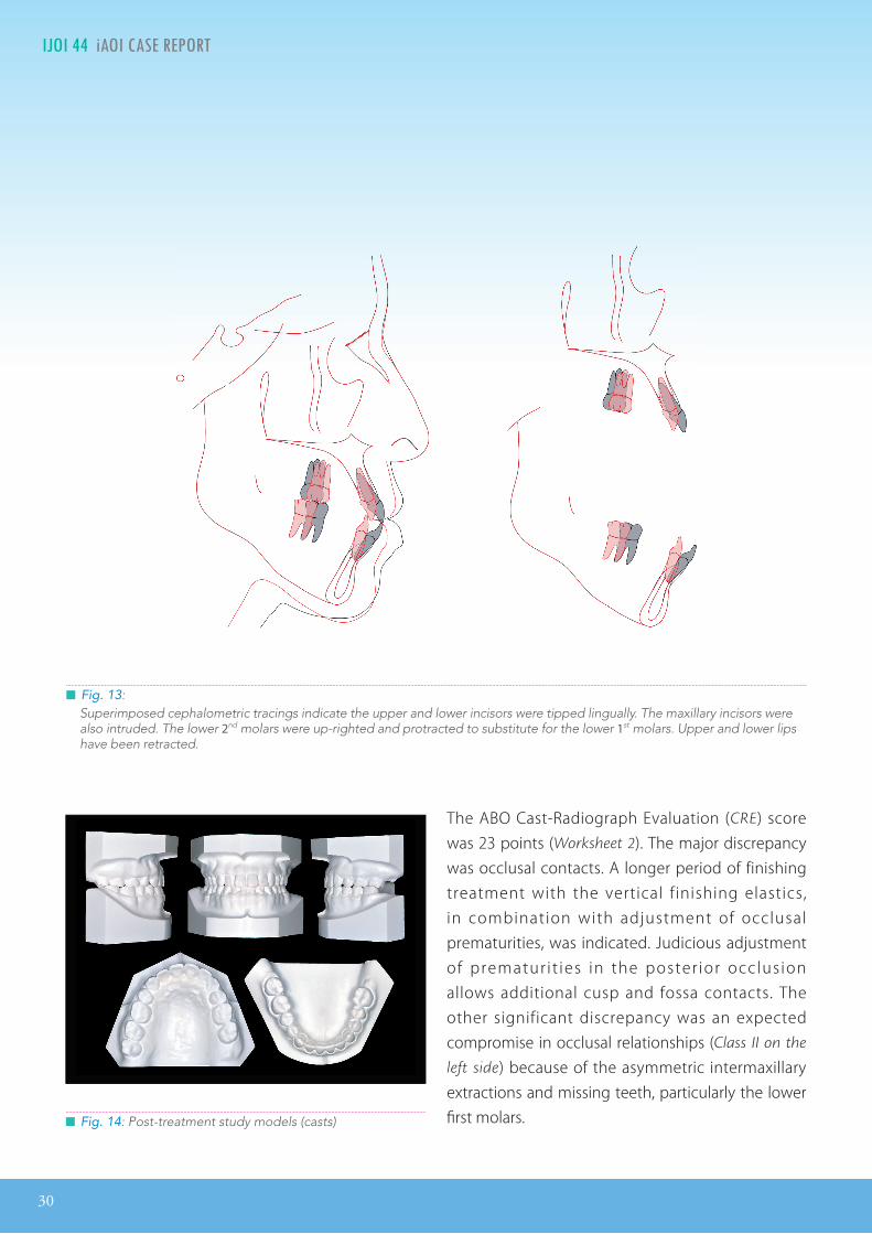
█ Fig. 14: Post-treatment study models (casts)
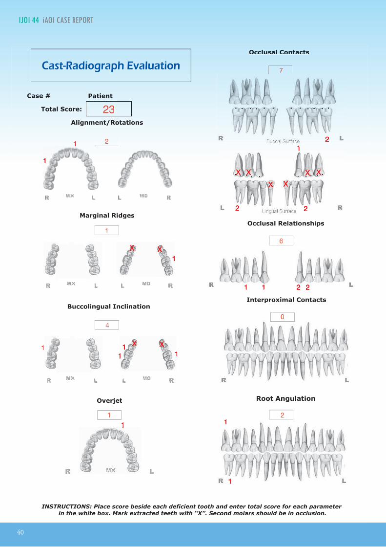
The ABO Cast-Radiograph Evaluation (CRE) score was 23 points (Worksheet 2). The major discrepancy was occlusal contacts. A longer period of finishing treatment with the vertical finishing elastics, in combination with adjustment of occlusal prematurities, was indicated. Judicious adjustment of prematur i t ies in the poster ior occlus ion allows additional cusp and fossa contacts. The other significant discrepancy was an expected compromise in occlusal relationships (Class II on the
left side) because of the asymmetric intermaxillary extractions and missing teeth, particularly the lower fi rst molars.

31
Asymmetric Extractions IJOI 44
Discussion
1. Early loss of permanent � rst molars
The current case report is part of a series of >100 challenging clinical cases published in last fi ve years in News and Trends in Orthodontics (NTO) and the subsequent publication International Journal of Orthodontics and Implantology (IJOI) (http://iaoi.
pro/archive/journal). The isolated loss of one or both mandibular fi rst molars is a prominent feature in the etiology of complex, acquired malocclusions. Two recent reports in IJOI1,2 have discussed the critical role of lower first molars in occlusal development, during the late transitional occlusion (~age 10-
12yr). The present patient (Fig. 1) fits the pattern. She presented with a missing lower first molar and demonstrates the signs of unilateral occlusal collapse that occurs in the early permanent dentition: unilateral deep curve of Spee (Fig. 3) and mesial tipping of second and third molars into the extraction site (Fig. 5).
There is a large literature indicating that the early loss of permanent first molars is associated with a variety of acquired malocclusions3,4 that occur after the adjacent deciduous second molars exfoliate. Although permanent molars may be lost to caries at any age, there is an emerging recognition that this particular developmental problem is commonly related to as MIH, a worldwide problem with a prevalence of 10-22%.3,4 MIH is a dental development problem related to enamel defects associated with the illness of a child <3 years of age. Prolonged and sustained fever is a common occurrence for young children affl icted with maladies, such as exanthemata, respiratory infection
or otitis media. Clinical data have long been consistent with a deleterious effect on the enamel formation of permanent teeth developing at that time, particularly the permanent central incisors and fi rst molars.1-4,24 Febrile conditions are known to disrupt enamel formation in mammals both in vivo24 and in vitro,25 Enamel defects render the teeth highly susceptible to caries as soon as they erupt (~age
6-7yr).
If the incisors are affected, the parents usually notice the problem and seek treatment. However, molar hypomineralization is not usually recognized until the crown of the first molar is destroyed and the child has a toothache. The usual diagnosis is “bombed-out caries” and the only viable treatment is extraction of the permanent first molar, leaving second deciduous molar as the sole posterior occlusal stop by ~age 8yr. There are usually no further problems until the late transitional stage of occlusal development when the second deciduous molars are exfoliated. In the absence of the lower fi rst molar, there is an occlusal collapse, because there is no posterior occlusal stop on the aff ected side. Prior to the eruption of the succedaneous premolar, the dental compensation results in a typical acquired malocclusion: mesially tipped second molars and a deep curve of Spee. The problem may be symmetric or asymmetric and can even result in a functional retrusion of the mandible.1-4
Permanent maxillary fi rst molars are also susceptible to MIH, but their isolated early loss is not as damaging to occlusal development, if the ipsilateral lower first molar is still present. In a Class I molar relationship, the early loss of a maxillary first molar

32
IJOI 44 iAOI CASE REPORT
does not eliminate the posterior centric stop because the lower fi rst molar continues to occlude with the maxillary second deciduous molar. By the time the second deciduous molar exfoliates, the maxillary second molar is usually in occlusion because the development of adjacent molars is accelerated by the extraction of the fi rst molar.26
Another problem associated with early loss of permanent lower fi rst molars is disuse atrophy of the edentulous space resulting in an atrophic ridge.5,8 If the periodontium of the adjacent teeth is healthy, atrophic ridges can be closed orthodontically but the biomechanics and anchorage requirements are challenging.5,8,9
2. Closing atrophic molar space
The mandibular atrophic ridge is usually described as a “knife-edge” ridge on the lingual aspect because the process of disuse atrophy preferentially resorbs the occlusal and buccal aspects of the edentulous ridge.5 This process results in dense, thin alveolar process that is composed of two relatively thick cortical bone plates, connected with coarse trabecular bone. Lower molars can be protracted through atrophic ridges if the periodontium is healthy,9,10 but force should be very light, <100cN (~100g) to control lateral root resorption where the PDL engages the thin but dense atrophic alveolar ridge.5 Widening the osseous ridge ahead of a moving tooth requires anabolic, bone modeling in the subperiosteal region,10 which may be more difficult to achieve with atypical, asymmetric extraction spaces.11-13 Mandibular molars have wide roots that are very effective for inducing anabolic modeling of a
edentulous space, and produce dense cortical bone between the roots of the molars.14 Despite these challenging tooth movement conditions, several case reports have documented ≥10mm mandibular molar protraction into atrophic first molar spaces with and without TADs for anchorage.12-15
For the present patient, the asymmetric extraction spaces, atrophic ridges and differential anchorage requirements (Figs. 1, 3 and 5) resulted in variable rates of space closure in each quadrant (Fig. 8). Careful management of the mechanics resulted in a relatively symmetrical outcome (Fig. 9). Closing space with sliding mechanics on SS rectangular wires was facilitated by balancing lingual and buccal forces to prevent binding of the archwire due to molar mesial-in rotation.16 As the asymmetric spaces were closed, a lack of progressive archwire coordination was manifest as a tendency for a bilateral posterior cross-bite, which required additional treatment time. In retrospect, it would have been wise to adjust archwire widths and use cross-elastics as soon as the cross-bite tendency was detected.
3. Atypical extraction
Closing asymmetric molar spaces that are also atrophic is a challenge that can result in occlusal canting and midline deviation.6,7 Maintaining the midlines and avoiding occlusal canting for the present patient was an important accomplishment. Midl ine discrepancies are among the most complex and difficult problems for orthodontists to manage.17-20 Effective management requires careful examination, precise diagnosis, and a comprehensive treatment plan.18-23 Choosing the

33
Asymmetric Extractions IJOI 44
most esthetic and functional midline is an important fundamental for achieving adequate symmetry. Precise defi nitions are required:
• Symmetry: equality or correspondence in form
of parts, distributed around a center on an axis, at the two extreme of poles, or on the two opposite sides of the body.27
• Facial midline: clinically, the patient’s facial
midline is defined by the center of the philtrum and the nadir of the cupid’s bow of the upper lip.23 The orientation of the nose is also an important consideration.
• Dental midlines: the location of contact between
the mesial surfaces of the central incisors in either arch.27
Facial and dental midline coincidence involves skeletal, dental and functional symmetry,27 and is usually expressed in a pleasing smile.28 Orthodontists should differentially diagnose the etiology of a midline discrepancy. Skeletal, dental, functional components can be present alone or combination (Fig. 15) as defi ned below:18,22,23,29
• Skeletal problem: Panoramic radiographs (Figs.
5 and 10) and temporomandibular joint (TMJ) arthrograms (Figs. 7 and 12) compare the condylar shape and morphology as well as measure the difference between the right and left condylar necks, sigmoid notches, and vertical rami. This is a method for diagnosing the morphologic etiology of skeletal asymmetry.18,22,23 Transverse
skeletal problems and occlusal plane canting are evaluated with imaging or face-bow transfers.23 Trauma that results in asymmetry may have a delayed onset. Minor traumas in childhood may seem insignificant, but they gradually become more evident, especially a deviation of the mandible.18,23
• Dental problem: Even though the skeletal base
is symmetrical, different tooth size proportions for the right compared to the left side may result in a midline discrepancy.23,29 Extraction of teeth may result in tipping of adjacent teeth, with a dental midline shift toward the extraction side.29 Furthermore, tooth agenesis, delayed root development and paths of eruption may also result in midline discrepancies.29,30
• Functional problem : A functional shift is
diagnosed when there is a discrepancy between the centric relation and maximum intercuspal position (Fig. 6).22,23 A functional shift may reflect an occlusal problem like premature contacts.29 Ideally dental midlines should be coincident with their respective skeletal base to minimize occlusal interference.23 Other functional habits like thumb sucking, asymmetrical or unilateral chewing habits, and/or masticatory muscle hypertrophy can contribute to facial asymmetry and midline problems.29
Establishing a realistic treatment plan for the often complex interactions benefits from a problem analysis method such as described in Fig. 15. The varying approaches to the problem(s) were

34
IJOI 44 iAOI CASE REPORT
discussed with the present patient (Fig. 1), prior to formulating the treatment plan.
1. The patient was willing to accept a modest midline deviation
Perfect facial symmetry is a theoretical concept that seldom exists in nature.23,29 A 4mm dental to facial midline discrepancy is undetectable for most patients.31,32 Orthodontists should attempt to eliminate midline discrepancies for the optimization of esthetics and as a guide for functional alignment of the dentition and jaws.33-37 Kokich, et al.38 asked, is it necessary to correct subtle variations if they are undetectable to the average patient? Insisting on correction of every midline discrepancy is not indicated because it can considerably increase the complexity and duration of treatment. The esthetic impact of
the dentition is greater in a mouth-only view compared to a full-facial view,39 so many previous midline studies are biased. It is important for the patient and the clinician to avoid focusing on the oral view for a deviation that is hardly detectable in the full-facial view. Janson, et al.40 conducted an systemic review and concluded that up to a 2.2mm midline deviation is usually acceptable. To avoid misunderstandings later it is very important to discuss probable outcomes of treatment to understand the expectations of the patient. Attempting to manage the esthetic concerns of an unreasonable patient poses a high risk for failure. The present patient (Fig. 1) was informed that a modest midline discrepancy was likely because of the asymmetric extraction pattern. She accepted this possibility as part of the informed consent to begin treatment.
█ Fig. 15: As a clinical diagnosis and treatment planning exercise, three circles are partially superimposed to demonstrate the interaction of the skeletal (red) functional (purple), and dental (green) problems. The usual clinical responses are shown for multiple camouflage approaches for the dental problem (No. 1-5 in green), one functional solution (No. 6 in purple), and an additional option for a skeletal problem (No. 7 in red).
7. Orthognathic Surgery
Camou� age Approach 1. Suggest the patient accept a mild midline deviation 1. Suggest the patient accept a mild midline deviation 1. Suggest the patient accept a mild midline deviation 1. Suggest the patient accept a mild midline deviation 2. Design unusual mechanics in advance 3. Reduce enamel thickness and space closure 4. Use bone screws for skeletal anchorage 5. Plan space distribution and prosthetics
Dental Problem
6. Habits correction and muscle training
Functional Problem
Skeletal Problem
35
Asymmetric Extractions IJOI 44
2. Design asymmetric mechanics in advance
Symmetric mechanics are designed to maintain symmetry, but may result in asymmetry or even worsen the condition. All mechanics should be designed with the potential for modifications, as needed during treatment. Achieving optimal esthetics requires a prospective treatment plan focusing on the defined objectives.37,40,41 If patients are asymmetric prior to treatment, special mechanics are indicated such as asymmetric arch shape, interarch elastics, a r c hw i r e ad j u s tmen t s , o r d i f f e r en t i a l anchorage.40-42 Unilateral activation for space closure may be effective for midline control in asymmetrical dental arches.13,17,29 For the present patient, a good outcome was achieved by differential force control in each quadrant (Figs. 9-11). This type of asymmetric mechanics is readily managed in routine clinical practice.
3. Interproximal reduction and intermaxillary elastics
Bilateral tooth size discrepancies can result in a lack of upper and lower midline coincidence.30 Midline correction is a challenge when no space remains, particularly at the end of the treatment.40,42-44 Correcting tooth size proportions by interproximal reduction (IPR) also creates space for diagonal elastic traction and dark triangle correction.43,44 It is important to monitor the axial inclinations when planning and performing IPR to make sure the enamel reduction and subsequent space closure will result in roots that are parallel.32 For the present patient, IPR was effective for both dark triangle
and tooth size correction without compromising the axial inclinations of the roots (Figs. 9-11).
4. Orthodontic bone screw for anchorage
Skeletal anchorage (TADs) can be used as a form of asymmetric mechanics7-10 as well as to apply intrusive force for controlling the vertical relationships of the dentition. The two main side eff ects of atypical extraction patterns are midline deviation and canting of the occlusal plane. Both of these potential problems can be corrected using TADs.45,46 A clinically challenging scenario is when the upper dental midline is coincident with the facial midline, and the asymmetry is isolated in the lower arch. Using a well-positioned upper arch as anchorage for intermaxillary elastics to correct lower arch alignment may result in an esthetic compromise. TADs are effective skeletal anchor units for diagonal elastics, which are effective mechanics for midline alignment, particularly in combination with IPR.42-46 The present patient agreed to the use of TADs, if needed to off set the eff ects of asymmetric space closure.
5. Space distribution and prostheses design
Orthodontists should prospectively consider all aspects of the treatment required for a desired restoration of esthetics and function.29 A well-planned comprehensive treatment plan may involve a digital smile design and/or implant placement. In addition, prosthetic restoration of dental morphology is a critical consideration, in combination with orthodontic space management, for achieving a satisfactory outcome.15-17

36
IJOI 44 iAOI CASE REPORT
6. Correcting habits and muscle training
Kondo47,48 carefully manages the functional aspects of dentofacial orthopedic treatment. Even skeletal asymmetric malocclusions, that usually require orthognathic surgery, can be managed with muscle training. The method is effective for functional shift corrections that enhance the long term stability of Class III open bite malocclusions, treated with and without surgery.47 Orthodontic treatment can be combined with asymmetric cervical and masticatory muscle corrections, for managing Class III malocclusion with lateral deviation of the mandible, as well as a severely asymmetric condyle and ramus.48 These reports indicate the importance of effectively managing functional problems for facilitating orthodontic treatment.
7. Orthognathic surgery
There are limitations for orthodontic correction combined with prosthodontic camoufl age,10,15-18 and orthognathic surgery may be indicated for correcting the asymmetry.49,50 If a patient is focused on a complete correction of complex, asymmetric midline problems, orthognathic surgery may be the only viable option.
Conclusions
This case report demonstrates that a relatively simple application of asymmetric extractions and biomechanics was effective for managing a complex malocclusion with bimaxillary protrusion
and atrophic extraction sites. Careful design and monitoring of the asymmetric mechanics resulted in an optimal correction that was satisfying for the patient and the clinician. Midline control was maintained without resorting to TADs. For complex malocclusions, it is wise to plan additional anchorage options with the patient to insure that treatment objectives are met.
Acknowledgment
Thanks to Mr. Paul Head for proofreading this article.

37
Asymmetric Extractions IJOI 44
References
1. Yeh HY, Chang CH, Roberts WE. Conservative treatment of periodontally compromised class III malocclusion complicated by early loss of lower first molars. Int J Orthod Implantol 2016;42:44-59.
2. Chang MJ, Chang CH, Roberts WE. Acquired malocclusion due to early loss of permanent first molars: OBS-anchored orthodontics and implant-supported prostheses. Int J Orthod Implantol 2016;42:20-41.
3. Pitiphat W, Savisit R, Chansamak N, Subarnbhesaj A. Molar incisor hypomineralization and dental caries in 6-7 year old Thai children. Pediatric Dentistry 2014;36(7):478-82.
4. Wuollet E, Laisi S, Salmela E, Ess A, Alaluusua S. Background factors of molar-incisor hypomineralization in a group of Finnish children. Acts Odontol Scand 2014;72(8):963-9.
5. Chiu GSC, Chang CH, Roberts WE. Interdiscipl inary treatment for a compensated class II partially edentulous malocclusion: orthodontic creation of an posterior implant site. Am J Orthod Dentofacial Orthop 2016 (Submitted).
6. Ostler MS, Kokich VG. Alveolar ridge changes in patients congenitally missing mandibular second premolars. J Prosthet Dent 1994;71:144–9.
7. Kokich VG, Kokich VO. Congenitally missing mandibular second premolars: Clinical options. Am J Orthod Dentofacial Orthop 2006;130(4):437–44.
8. Roberts WE, Nelson CL, Goodacre CJ. Rigid implant anchorage to close a mandibular first molar extraction site. J Clin Orthod 1994;28(12):693–704.
9. Roberts WE, Marshall KJ, Mozsary PG. Rigid endosseous implant utilized as anchorage to protract molars and close an atrophic extraction site. Angle Orthod 1990;60(2):135–52.
10. Roberts WE. Bone physiology, metabolism, and biomechanics in orthodontic practice. In: Orthodontics: Current Principles and Techniques. 5th ed. St. Louis: Mosby; 2012. pp. 287–343.
11. Shastri D, Tandon P, Nagar A. Atypical extractions in adult treatment. J Clin Orthod 2015 May;49(5):312-8.
12. Chung K, Choo H, Lee J, Kim S. Atypical orthodontic extraction pattern managed by differential en-masse retraction against a temporary skeletal anchorage device in the treatment of bimaxillary protrusion. Am J Orthod Dentofacial Orthop 2011;140(3):423–32.
13. Tayer BH. The asymmetric extraction decision. Angle Orthod 1992;62(4):291–7.
14. Roberts WE, Arbuckle GR , Analoui M. Rate of mesial translation of mandibular molars using implant-anchored mechanics. Angle Orthod 1996;66(5):331–8.
15. Stepovich ML. A Clinical Study on Closing Edentulous Spaces in the Mandible. Angle Orthod 1979;49(4):227–33.
16. Hom BM, Turley PK . The effects of space closure of the mandibular first molar area in adults. Am J Orthod 1984;85(6):457–69.
17. Baik UB, Chun YS, Jung MH, Sugawara J. Protraction of mandibular second and third molars into missing first molar spaces for a patient with an anterior open bite and anterior spacing. Am J Orthod Dentofacial Orthop 2012;141(6):783–95.
18. C ozzani M, Mazzott a L , R i nchusxe DJ , C ozzani P. Asymmetri ca l Mand ibular Molar Protraction with Conventional Mechanics. J Clin Orthod 2015 May;49(5):304-11.
19. Mimura H. Protraction of mandibular second and third molars assisted by partial corticision and miniscrew anchorage. Am J Orthod Dentofacial Orthop 2013;144(2):278–89.
20. Nagaraj K, Upadhyay M, Yadav S. Titanium screw anchorage for protraction of mandibular second molars into first molar extraction sites. Am J Orthod Dentofacial Orthop 2008;134(4):583–91.
21. Kravitz ND, Jolley T. Mandibular molar protraction with temporary anchorage devices. J Clin Orthod 2008;42(6):351–5; quiz 340.
22. Saga AY, Maruo IT, Maruo H, Filho OG, Tanaka OM. Clinical challenges in treating a patient with deviated dental midlines and delayed root development of the mandibular left second premolar. Am J Orthod Dentofacial Orthop 2009;135(4):103–12.
23. Cheong Y, Lo L. Facial asymmetry: etiology, evaluation, and management. Chang Gung Med J 2011;34(4):341–51.
24. Tung K, Fujita H, Yamashita Y, Takagi Y. Effect of turpentine-induced fever during the enamel formation of rat incisor. Arch Oral Biol 2006;51(6):464-70.
25. Ryynänen H, Sahlberg C, Lukinmaa PL, Alaluusua S. The effect of high temperature on the development of mouse dental enamel in vitro. Arch Oral Biol 2014;59(4):400-6.
26. Halicioglu K, Toptas O, Akkas I, Celikoglu M. Permanent first molar extraction in adolescents and young adults and its effect on the development of third molar. Clin Oral Investing 2014;18(5):1489-94.

38
IJOI 44 iAOI CASE REPORT
27. Stedman TL. Stedman’s Medical Dictionary. Baltimore : Williams & Wilkins Co; 1966.
28. Ker AJ, Chan R, Fields HW, Beck M, Rosenstiel S. Esthetics and smile characteristics from the layperson’s perspective: a c ompute r- b a s e d sur vey s tu dy. J Am D e nt Ass o c 2008;139(10):1318–27.
29. Burstone CJ. Diagnosis and treatment planning of patients with asymmetries. Semin Orthod 1998;4(3):153–64.
30. Major M. Ash SJN. Wheeler’s Dental Anatomy, Physiology and Occlusion. 8th ed. Elsevier Health Sciences; 2003.
31. Joondeph DR . Mysteries of asymmetries. Am J Orthod Dentofacial Orthop 2000;117(5):577–9.
32. Bishara SE, Burkey PS, Kharouf JG. Dental and facial asymmetries: a review. Angle Orthod 1994;64(2):89–98.
33. Kilic N, Kiki A, Oktay H. Condylar asymmetry in unilateral posterior crossbite patients. Am J Orthod Dentofacial Orthop 2008;133(3):382–7.
34. Pirttiniemi PM. Associations of mandibular and facial asymmetries-a review. Am J Orthod Dentofacial Orthop 1994;106(2):191–200.
35. Al-Khateeb SN, Abu Alhaija ESJ. Tooth size discrepancies and arch parameters among different malocclusions in a Jordanian sample. Angle Orthod 2006;76(3):459–65.
36. Cağlaroğlu M, Kilic N, Erdem A. Effects of early unilateral first molar extraction on skeletal asymmetry. Am J Orthod Dentofacial Orthop 2008;134(2):270–5.
37. Johnston CD, Burden DJ, Stevenson MR. The influence of dental to facial midline discrepancies on dental attractiveness ratings. Eur J Orthod 1999;21(5):517–22.
38. Kokich VO, Kokich VG, Kiyak HA. Perceptions of dental professionals and laypersons to altered dental esthetics: Asymmetric and symmetric situations. Am J Orthod Dentofacial Orthop 2006;130(2):141–51.
39. Williams RP, Rinchuse DJ, Zullo TG. Perceptions of midline deviations among different facial types. Am J Orthod Dentofacial Orthop 2014;145(2):249–55.
40. Janson G, Branco NC, Fernandes TMF, Sathler R , Garib D, Lauris JRP. Influence of orthodontic treatment, midline position, buccal corridor and smile arc on smile attractiveness. Angle Orthod 2011;81(1):153–61.
41. Flores-Mir C, Silva E, Barriga MI, Lagravère MO, Major PW. Lay person’s perception of smile aesthetics in dental and facial views. J Orthod 2004;31(3):204–9.
42. Kusnoto J, Evans CA, BeGole EA, Obrez A. Orthodontic correction of transverse arch asymmetries. Am J Orthod Dentofacial Orthop 2002;121(1):38–45.
43. Jerrold L, Lowenstein LJ, York N. The midline : Diagnosis and treatment. Am J Orthod Dentofacial Orthop 1990;97(6):453–62.
44. Van Steenbergen E, Nanda R. Biomechanics of orthodontic correction of dental asymmetries. Am J Orthod Dentofacial Orthop 1995;107(6):618–24.
45. Takano-Yamamoto T, Kuroda S. Titanium screw anchorage for correction of canted occlusal plane in patients with facial asymmetry. Am J Orthod Dentofacial Orthop 2007;132(2):237–42.
46. Kang Y, Nam J, Park Y. Use of rhythmic wire system with miniscrews to correct occlusal-plane canting . Am J Orthod Dentofacial Orthop 2010;137(4):540–7.
47. Kondo E, Aoba TJ. Nonsurgical and nonextraction treatment of skeletal Class III open bite: its long-term stability. Am J Orthod Dentofacial Orthop 2000;117(3):267–87.
48. Kondo E. Features and treatment of skeletal class III malocclusion with severe lateral mandibular shift and asymmetric vertical dimension. World J Orthod 2004;5(1):9–24.
49. Hashimoto T, Fukunaga T, Kuroda S, Sakai Y, Yamashiro T, Takano-Yamamoto T. Mandibular deviation and canted maxillary occlusal plane treated with miniscrews and intraoral vertical ramus osteotomy: Functional and morphologic changes. Am J Orthod Dentofacial Orthop 2009;136(6):868–77.
50. Jeon YJ, Kim YH, Son WS, Hans MG. Correction of a canted occlusal plane with miniscrews in a patient with facial asymmetry. Am J Orthod Dentofacial Orthop 2006;130(2):244–52.

39
Asymmetric Extractions IJOI 44
1
1 pts.
0
4
0
0
0
0
1
2 4
0
0
6
2 2
2 4Atrophic ridge, asymmetric anchorage
6
18
Discrepancy Index Worksheet
40
IJOI 44 iAOI CASE REPORT
Total Score:
Case # Patient
2
11
40
1
7
6
2
Alignment/Rotations
Marginal Ridges
Buccolingual Inclination
Overjet
Occlusal Contacts
Occlusal Relationships
Interproximal Contacts
INSTRUCTIONS: Place score beside each deficient tooth and enter total score for each parameter in the white box. Mark extracted teeth with “X”. Second molars should be in occlusion.
23
Root Angulation
1
1
1
X X1
1 X X11
111
11
2
X XX X X X
22
11
22221111
11
1
Cast-Radiograph Evaluation

41
Asymmetric Extractions IJOI 44
12 35 4
4
1 2
3
5
1
2
34 6
12 34
56
12 35 4
4
1 2
3
5
1
2
34 6
12 34
56 12 3
5 4
4
1 2
3
5
1
2
34 6
12 34
56
1. Pink Esthetic Score
IBOI Pink & White Esthetic Score (Before Surgical Crown Lengthening)
Total Score: = 2
2. White Esthetic Score ( for Micro-esthetics )
12 35 4
4
1 2
3
5
1
2
34 6
12 34
56
1. M & D Papilla 0 1 2
2. Keratinized Gingiva 0 1 2
3. Curvature of Gingival Margin 0 1 2
4. Level of Gingival Margin 0 1 2
5. Root Convexity ( Torque ) 0 1 2
6. Scar Formation 0 1 2
1. Midline 0 1 2
2. Incisor Curve 0 1 2
3. Axial Inclination (5°, 8°, 10°) 0 1 2
4. Contact Area (50%, 40%, 30%) 0 1 2
5. Tooth Proportion (1:0.8) 0 1 2
6. Tooth to Tooth Proportion 0 1 2
Total = 0
Total = 2