benign breast problems. introduction anatomy structure of the breast classification initial...
TRANSCRIPT

Benign Breast Problems
Introduction
Anatomy
Structure of the breast
Classification
Initial approach to breast problems
Diagnostic workup
Conclusion
References
Outline
Introduction
Breast problems are a major reason why women visit the
primary care physician
Breast diseases in women constitute a spectrum of benign and
malignant disorders
The most common breast problems for which women consult a
physician are breast pain, nipple discharge and a palpable
mass.
Benign breast lesion is a non-cancerous lesion. According to
American Cancer society , when tissue biopsy is examined
under the microscope, nine out of every 10 women will have
some type of abnormality
AAFP journal , April 15, 2000. Volume 61/ No. 8
Anatomy
The breast is a modified sweat gland with a mass of glandular, fatty
and fibrous tissues on the pectoralis muscles in the chest wall
It is attached to the chest wall by fibrous strands called coopers
ligaments
The glandular tissues of the breast consist of lobules, lobes and
ducts
Fatty and fibrous tissues surround the milk producing system
( lobules and ducts)
Anatomy
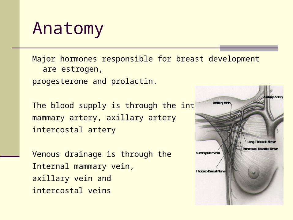
Major hormones responsible for breast development are estrogen,
progesterone and prolactin.
The blood supply is through the internal
mammary artery, axillary artery
intercostal artery
Venous drainage is through the
Internal mammary vein,
axillary vein and
intercostal veins
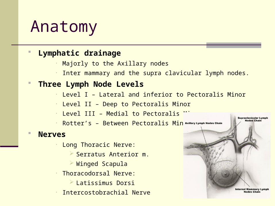
Anatomy
Lymphatic drainage• Majorly to the Axillary nodes• Inter mammary and the supra clavicular lymph nodes.
Three Lymph Node Levels• Level I – Lateral and inferior to Pectoralis Minor• Level II – Deep to Pectoralis Minor• Level III – Medial to Pectoralis Minor• Rotter’s – Between Pectoralis Minor & Major
Nerves• Long Thoracic Nerve:
Serratus Anterior m.
Winged Scapula• Thoracodorsal Nerve:
Latissimus Dorsi• Intercostobrachial Nerve
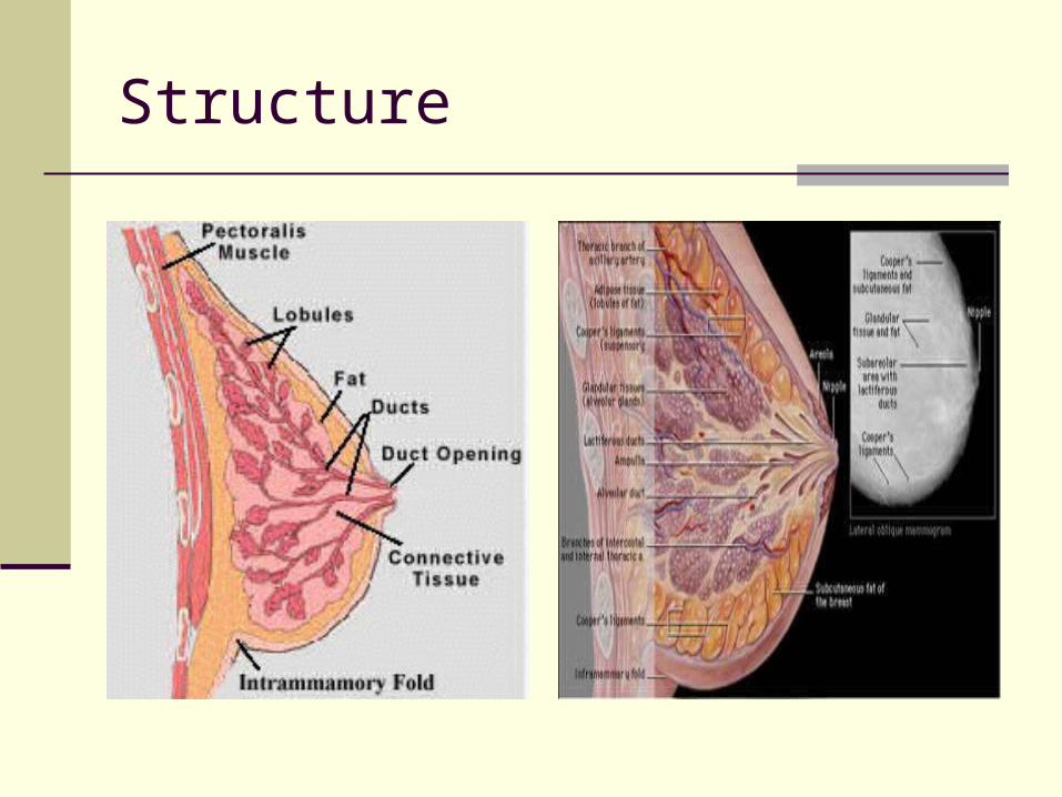
Structure
Classification Based On Histologic Types
Non Proliferative Lesion Simple Cyst Complex cyst
Proliferative Lesions – Without Atypia Ductal hyperplasia Fibroadenoma Intraductal papilloma Sclerosing Adenoma Radial Scars
Atypical Hyperplasia Atypical ductal hyperplasia Atypical lobular hyperplasia
Schnitt, SJ. Benign breast disease and breast cancer risk: morphology and beyond. Am J surg pathology 2003;27:836
Classification Based On Clinical Features
Mastalgia Cyclic Non Cyclic
Tumors and Masses Nodularity or glandular Cysts Galactoceles Fibroadenoma Sclerosing Adenosis Lipoma Harmatoma Diabetic Mastopathy Cystosarcoma Phylloides
AAFP journal , April 15, 2000. Volume 61/ No. 8
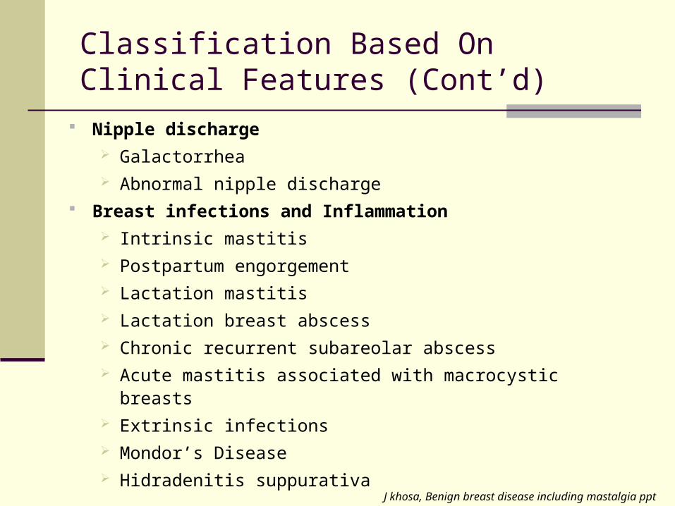
Nipple discharge Galactorrhea Abnormal nipple discharge
Breast infections and Inflammation Intrinsic mastitis Postpartum engorgement Lactation mastitis Lactation breast abscess Chronic recurrent subareolar abscess Acute mastitis associated with macrocystic breasts Extrinsic infections Mondor’s Disease Hidradenitis suppurativa
Classification Based On Clinical Features (Cont’d)
J khosa, Benign breast disease including mastalgia ppt

Classification Lesions with Increased Risk of Ca
Ductal hyperplasia Sclerosing adenosis Complex fibroadenomas Atypical hyperplasia Radial scars
Micheal S sabel .Overview of benign breast disease. Uptodate 2008, November 14
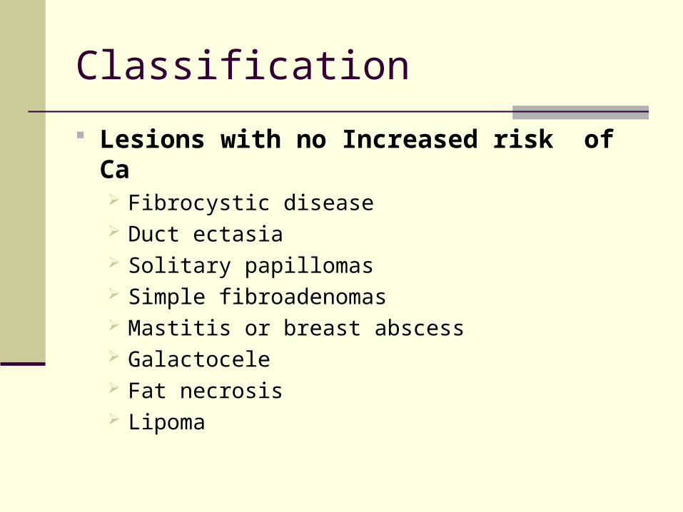
Classification
Lesions with no Increased risk of Ca Fibrocystic disease Duct ectasia Solitary papillomas Simple fibroadenomas Mastitis or breast abscess Galactocele Fat necrosis Lipoma
Breast Pain (Mastalgia)
Most common breast symptom for which women
consult the physician
More common in premenopausal women than in
post menopausal women
Can be cyclical (physiological) or non cyclical
Micheal S Sabel. Initial approach to the woman with breast problems. http://uptodateonline.com 2008, November 6
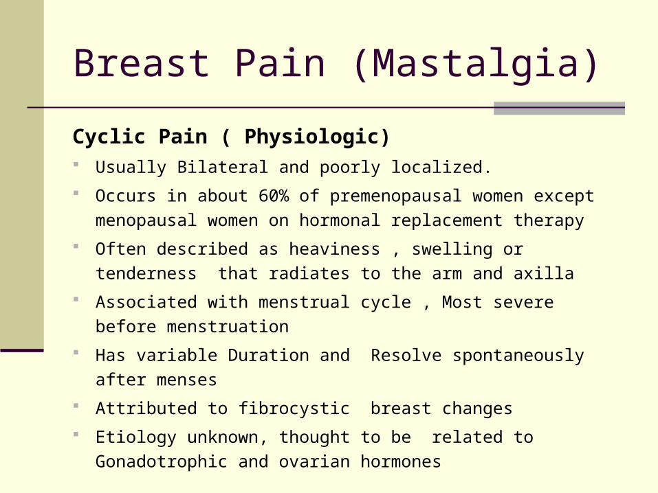
Breast Pain (Mastalgia)
Cyclic Pain ( Physiologic) Usually Bilateral and poorly localized.
Occurs in about 60% of premenopausal women except
menopausal women on hormonal replacement therapy
Often described as heaviness , swelling or tenderness that
radiates to the arm and axilla
Associated with menstrual cycle , Most severe before
menstruation
Has variable Duration and Resolve spontaneously after menses
Attributed to fibrocystic breast changes
Etiology unknown, thought to be related to Gonadotrophic and
ovarian hormones
Mastalgia
Non-Cyclic Pain Most common in women 40 to 50 yrs of age
Often unilateral
Usually described as sharp, burning pain localized in the breast
Occasionally secondary to the presence of Fibroadenoma and
or cyst
Menstrual irregularity, emotional stress, trauma, , scars from
previous biopsies and medications have been associated
Micheal S Sabel. Initial approach to the woman with breast problems. http://uptodateonline.com 2008, November 6
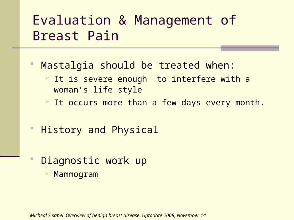
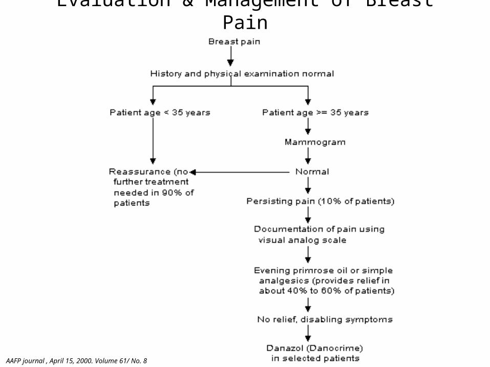
Evaluation & Management of Breast Pain
Mastalgia should be treated when: It is severe enough to interfere with a woman’s life style It occurs more than a few days every month.
History and Physical
Diagnostic work up Mammogram
Micheal S sabel .Overview of benign breast disease. Uptodate 2008, November 14
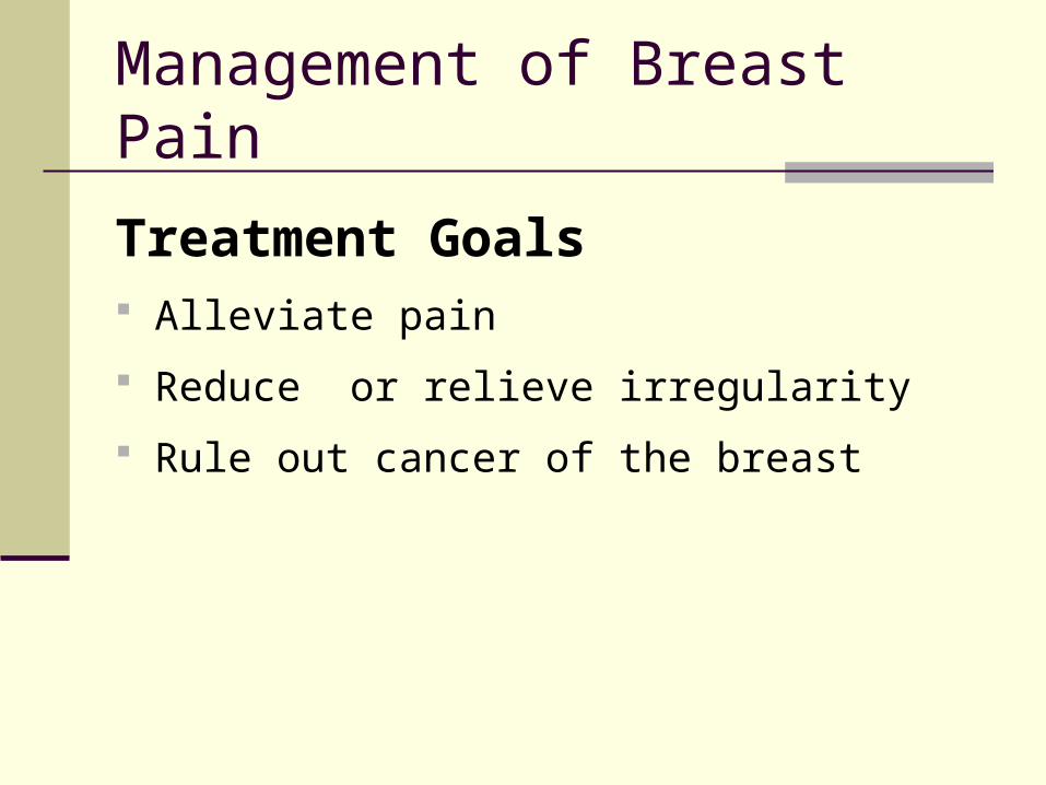
Management of Breast Pain
Treatment Goals Alleviate pain
Reduce or relieve irregularity
Rule out cancer of the breast

Management of Breast Pain
Diet and Lifestyle Modification Elimination of Methylxanthines, Caffeine and
Chocolates Reassurance Supportive Bra Low fat and high complex carbohydrate Vitamin E supplementation Evening Primrose oil
Micheal S sabel .Overview of benign breast disease. Uptodate 2008, November 14

Management of Breast Pain
Pharmacological Treatment NSAIDs OCPs Danazol 100- 400mg per day 75% of women with non cyclic pain will be symptom free SE: Weight gain , menstrual irregularity , acne , hirsutism Tamoxifen 10mg Bromocriptine – prolactin antagonist Surgery has no role in management of breast pain
Micheal S sabel .Overview of benign breast disease. Uptodate 2008, November 14
Evaluation & Management of Breast Pain
AAFP journal , April 15, 2000. Volume 61/ No. 8
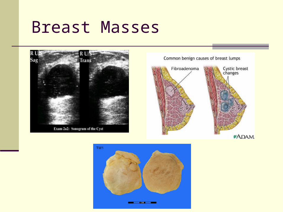
Breast Masses
Normal glandular tissue of the breast is nodular
This is a general pattern or consistency of the breast which include persistent lumpiness or nodularity which is generally not abnormal when it is related to the menstrual cycle.
Dominant masses are characterized by persistence throughout the menstrual cycle
Cystic Breast Mass Common cause of dominant breast mass
May occur at any age, but uncommon in post menopausal
women
Fluctuates with menstrual cycle
Well demarcated from the surrounding tissue
Characteristically firm and mobile
May be tender
Difficult to differentiate from solid mass
Breast Masses: Cysts
Micheal S sabel .Overview of benign breast disease. Uptodate 2008, November 14

Fibrocystic Breast Disease Most common of all benign breast disease Most common between ages 20- 50 50% of women with Fibrocystic changes have clinical
symptoms 53% have histologic changes Believed to be associated the Imbalance of progesterone
and estrogen. May present with bilateral cyclic pain, breast swelling,
palpable mass and heaviness
Breast Masses: Cysts
Fibrocystic Breast Disease
Physical Examination Tenderness Increased engorgement and more dense breast Increased lumpiness / glandular Occasional spontaneous nipple discharge
Micheal Sabel .Overview of benign breast disease. Uptodate 2008, November 14
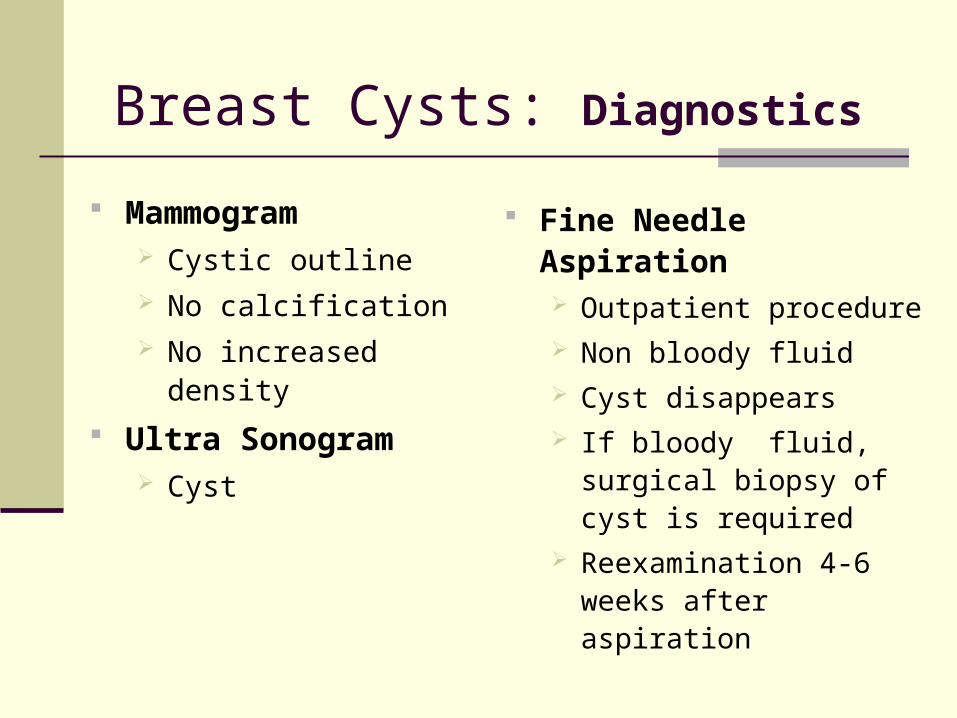
Breast Cysts: Diagnostics
Mammogram Cystic outline No calcification No increased density
Ultra Sonogram Cyst
Fine Needle Aspiration
Outpatient procedure Non bloody fluid Cyst disappears If bloody fluid, surgical
biopsy of cyst is required Reexamination 4-6 weeks
after aspiration
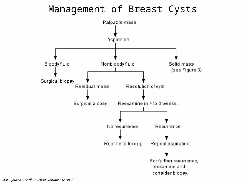
Management of Breast Cysts
AAFP journal , April 15, 2000. Volume 61/ No. 8
Breast Masses
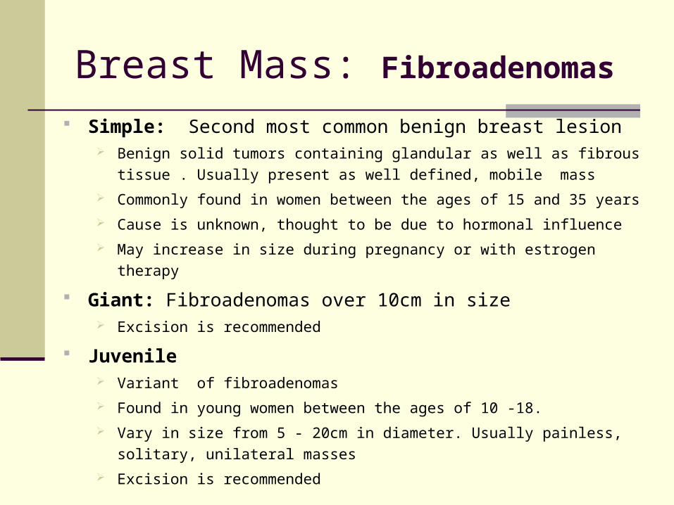
Breast Mass: Fibroadenomas
Simple: Second most common benign breast lesion Benign solid tumors containing glandular as well as fibrous tissue . Usually
present as well defined, mobile mass
Commonly found in women between the ages of 15 and 35 years
Cause is unknown, thought to be due to hormonal influence
May increase in size during pregnancy or with estrogen therapy
Giant: Fibroadenomas over 10cm in size Excision is recommended
Juvenile Variant of fibroadenomas
Found in young women between the ages of 10 -18.
Vary in size from 5 - 20cm in diameter. Usually painless, solitary, unilateral
masses
Excision is recommended
Breast Mass: Fibroadenomas (Cont’d)
Complex Complex fibroadenomas contain other proliferative changes
such as sclerosing adenosis, duct epithelial Hyperplasia,
epithelial calcification.
Associated with slightly increased risk of cancer
Dupont, WD page, DL, parl, FF, et al. Long term risk cancer in women with fIbroadenoma. NEJM 1994;331:10Carty, NJ, Carter, c, Rubin, C et al management of fibroadenoma of the breast. Annals of royal college of surgeon England 1995:77:127Micheal S sabel .Overview of benign breast disease. Uptodate 2008, November 14
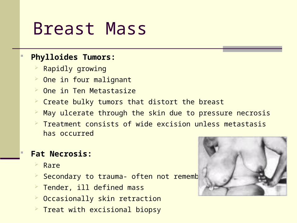
Phylloides Tumors: Rapidly growing One in four malignant One in Ten Metastasize Create bulky tumors that distort the breast May ulcerate through the skin due to pressure necrosis Treatment consists of wide excision unless metastasis has occurred
Fat Necrosis: Rare Secondary to trauma- often not remembered Tender, ill defined mass Occasionally skin retraction Treat with excisional biopsy
Breast Mass

Breast Mass Galactocele
Milk filled cyst from over distension of a lactiferous duct.
Presents as a firm non tender mass in the breast,
Commonly in upper quadrants beyond areola.
Diagnostic aspiration is often curative.
Duct ectasia:
Generally found in older women.
Dilatation of the subareolar ducts can occur.
A palpable retroareolar mass, nipple discharge,
or retraction can be present.
Tx involves excision of area
Breast Mass
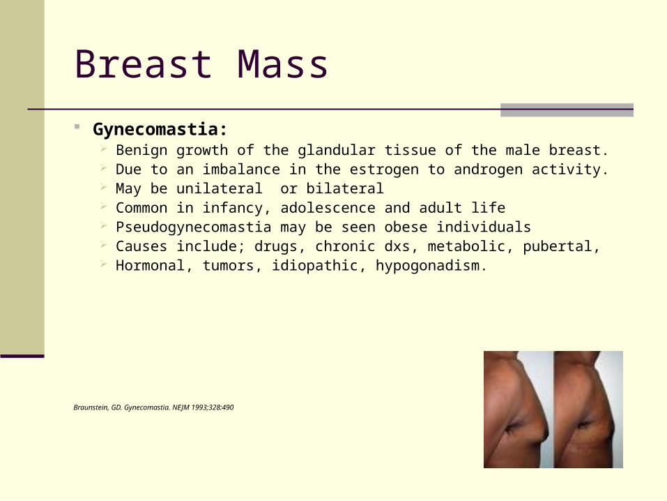
Gynecomastia: Benign growth of the glandular tissue of the male breast. Due to an imbalance in the estrogen to androgen activity. May be unilateral or bilateral Common in infancy, adolescence and adult life Pseudogynecomastia may be seen obese individuals Causes include; drugs, chronic dxs, metabolic, pubertal, Hormonal, tumors, idiopathic, hypogonadism.
Braunstein, GD. Gynecomastia. NEJM 1993;328:490
Nipple Discharge
Majority of causes are benign Most common cause is lactational Overstimulation also common Prolactin secreting tumors Hypothyroidism Drugs Intraductal and other carcinomas Unilateral, spontaneous, bloody discharge is
suspicious
Nipple Discharge
Intraductal Papilloma Benign growth within ductal system Presents as bloody nipple discharge Excision is the only way to differentiate from
carcinoma
Galactorrhea Bilateral milky discharge Obtain prolactin level, TSH level
Nipple Discharge
Good history
Prolactin & TSH levels
Mammogram
Decrease stimulation
Breast Inflammation & Infections
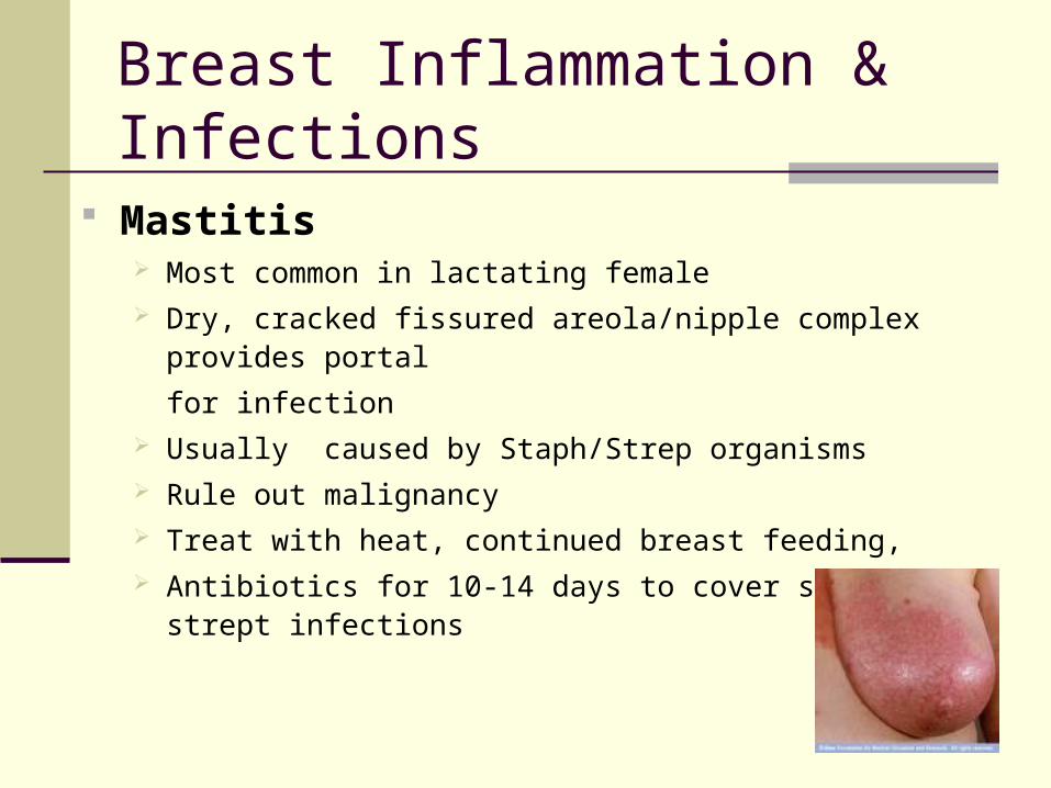
Mastitis Most common in lactating female Dry, cracked fissured areola/nipple complex provides portal
for infection Usually caused by Staph/Strep organisms Rule out malignancy Treat with heat, continued breast feeding, Antibiotics for 10-14 days to cover staph and strept infections
Breast Inflammation & Infections
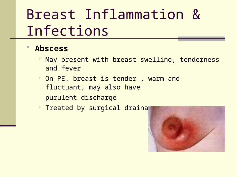
Abscess May present with breast swelling, tenderness and fever On PE, breast is tender , warm and fluctuant, may also have
purulent discharge Treated by surgical drainage
Breast Inflammation & Infections
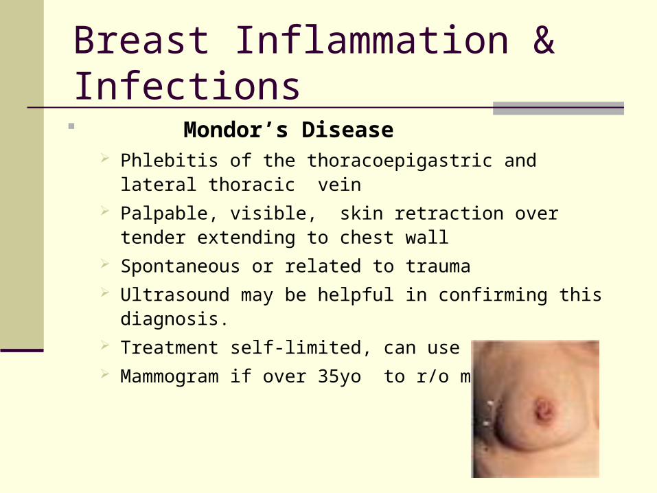
Mondor’s Disease Phlebitis of the thoracoepigastric and lateral thoracic vein Palpable, visible, skin retraction over tender extending to
chest wall Spontaneous or related to trauma Ultrasound may be helpful in confirming this diagnosis. Treatment self-limited, can use NSAIDs Mammogram if over 35yo to r/o malignancy
Breast Inflammation & Infections
Chronic Subareolar Abscess Occurs at base of lactiferous duct, and squamous
metaplasia of duct may occur. Sinus tract to areola develops Treatment requires complete excision of sinus tract Recurrence is common
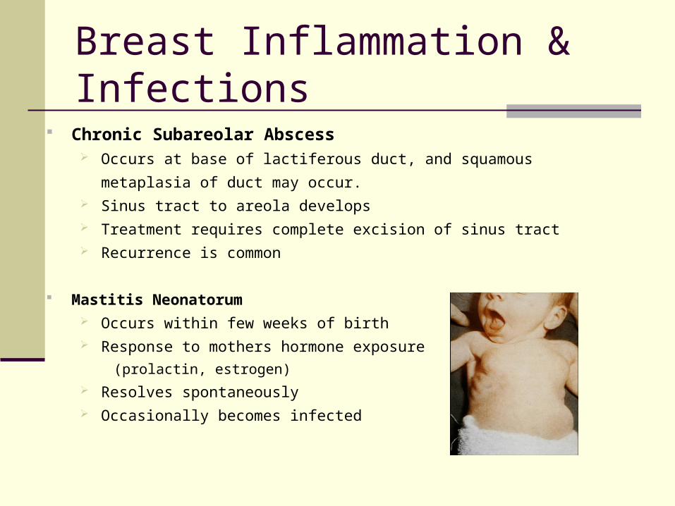
Mastitis Neonatorum Occurs within few weeks of birth Response to mothers hormone exposure
(prolactin, estrogen)
Resolves spontaneously Occasionally becomes infected
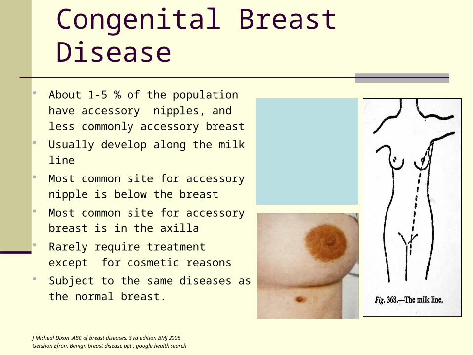
About 1-5 % of the population have
accessory nipples, and less
commonly accessory breast
Usually develop along the milk line
Most common site for accessory
nipple is below the breast
Most common site for accessory
breast is in the axilla
Rarely require treatment except for
cosmetic reasons
Subject to the same diseases as the
normal breast.
J Micheal Dixon .ABC of breast diseases. 3 rd edition BMJ 2005
Gershon Efron. Benign breast disease ppt , google health search
Congenital Breast Disease
Approach to Breast Problems
History Age Family history (Cancer) Onset Duration Discharge Frequency Lump , Nodules Trauma Menstruation (menarche, menopause, contraceptives) Pain
Inspection Symmetry Skin / Nipple Change Bulges / Retractions
Palpation Breast
Axilla
Supraclavicular
Approach to Breast Problems
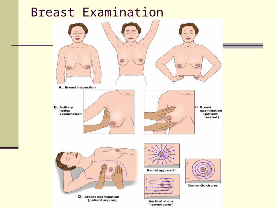
Breast Examination The breast examination starts with inspection of both breast
Sitting up with arms in relaxed position,
Both arms raised over the head
Hands on the hips
Complete regional lymph node examination while patient is in the sitting
position.
Bimanual may be done while patient is still in the sitting position, useful in
patient with large pendulous breast
Complete with the patient in a supine position, with the arms raised above
the head, breast exam can be accomplished with either concentric circles,
radial approach, or vertical strip approach
Areas examined should extend from the clavicle superiorly to the rib cage
inferiorly and from the sternum medially to the mid axillary line laterally
Micheal S Sabel. Initial approach to the woman with breast problems. http://uptodateonline.com 2008, November 6
Breast Examination
Diagnostic Work Up
Ultrasound
Mammography
FNA vs. Core Biopsy
Incisional biopsy
Excisional biopsy
Cyst aspiration
MRI
Ultrasonography: First diagnostic test of choice to differentiate a
cystic mass from a solid mass
Mammogram: Not routinely done in women younger than 35yo,
however not inappropriate in a suspicious mass in younger women
Digital mammography is superior to conventional
A normal mammogram at any age does not eliminate the
need for further evaluation of a suspicious mass.
MRI: Not indicated for the work up of undiagnosed mass. Reserved
for diagnostic dilemmas and should be used with discretion due to
false positive results
FNA: Useful for cystic lesions. If lesion is completely drained and
fluid not bloody or cloudy, no further evaluation needed
Diagnostic Work Up
Micheal S Sabel. Initial approach to the woman with breast problems. http://uptodateonline.com 2008, November 6
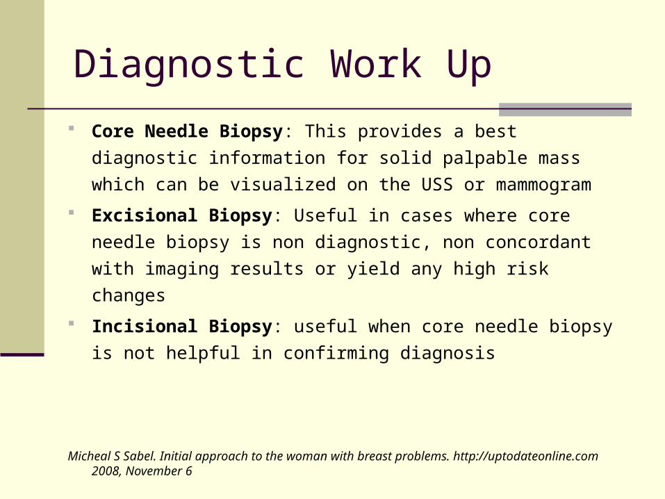
Diagnostic Work Up
Core Needle Biopsy: This provides a best diagnostic
information for solid palpable mass which can be visualized on
the USS or mammogram
Excisional Biopsy: Useful in cases where core needle biopsy
is non diagnostic, non concordant with imaging results or yield
any high risk changes
Incisional Biopsy: useful when core needle biopsy is not
helpful in confirming diagnosis
Micheal S Sabel. Initial approach to the woman with breast problems. http://uptodateonline.com 2008, November 6
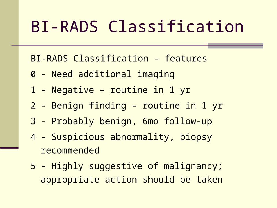
BI-RADS Classification
BI-RADS Classification – features
0 - Need additional imaging
1 - Negative – routine in 1 yr
2 - Benign finding – routine in 1 yr
3 - Probably benign, 6mo follow-up
4 - Suspicious abnormality, biopsy recommended
5 - Highly suggestive of malignancy; appropriate action
should be taken
Conclusion
Benign breast problems account for the majority of
breast problems seen in women
Breast complaints need careful assessment with
thorough history and physical as well as diagnostic
work up if indicated
Women with breast problems can present with a
mass, pain, nipple discharge or skin changes. They
can also be asymptomatic
It is important to rule out breast cancer
Micheal S Sabel. Initial approach to the woman with breast problems. http://uptodateonline.com 2008, November 6
References
1. AAFP journal , April 15, 2000. Volume 61/ No. 82 Schnitt, SJ. Benign breast disease and breast cancer risk:
morphology and beyond. Am J surg pathology 2003;27:836
3.J khosa, Benign breast disease including mastalgia ppt4. Dupont, WD page, DL, parl, FF, et al. Long term risk cancer in
women with fIbroadenoma. NEJM 1994;331:105. Carty, NJ, Carter, c, Rubin, C et al management of fibroadenoma
of the breast. Annals of royal college of surgeon England 1995:77:127
6. Micheal S sabel .Overview of benign breast disease. Uptodate 2008, November 14
7. J Micheal Dixon .ABC of breast diseases. 3 rd edition BMJ 20058.Gershon Efron. Benign breast disease ppt , google health search9. Micheal S Sabel. Initial approach to the woman with breast
problems. http://uptodateonline.com 2008, November 610. Braunstein, GD. Gynecomastia. NEJM 1993;328:490