behavioral and biochemical responses to morphine related ...epubs.surrey.ac.uk/185381/1/bjp paper...
TRANSCRIPT

Br. J Pharmacol.
Behavioral and biochemical responses to morphine related to its
addictive properties are altered in adenosine A2A receptor knockout
mice
A Castañé1#, L Wells3, G Soria1#, S Hourani3, C Ledent2, I Kitchen3, J Opacka-Juffry4,
R Maldonado1 and O Valverde1*
Running Tittle: morphine addictive properties in A2A knockout mice
1Departament de Ciències Experimentals i de la Salut. Universitat Pompeu Fabra,
Barcelone Biomedical Research Park. C/Dr. Aiguader 88, 08003 Barcelona, Spain 2IRIBHM, Université Libre de Bruxelles, B-1070 Bruxelles, Belgium 3School of Biomedical and Molecular Sciences, University of Surrey, Guildford,
Surrey. GU2 4JA, UK 4School of Human and Life Sciences, Roehampton University, Holybourne Avenue,
London, SW12 4JD, UK.
*Corresponding author:
Olga Valverde MD PhD Grup de Recerca de Neurobiologia del Comportament Departament de Ciències Experimentals i de la Salut Universitat Pompeu Fabra Barcelone Biomedical Research Park Dr. Aiguader, 88 08003 Barcelone, Spain [email protected] #Present address:
A.C.: Department of Neurochemistry and Neuropharmacology, Institut
d’Investigacions Biomèdiques de Barcelona (CSIC), IDIBAPS. Rosselló, 161, 6th
floor. 08036 Barcelona, Spain. G.S.: Department of Cerebral Ischemia and
Neurodegeneration, Institut d’Investigacions Biomèdiques de Barcelona (CSIC),
IDIBAPS. Rosselló, 161, 6th floor. 08036 Barcelona, Spain.

Castañé et al.
2
Abstract
Background and purpose. Purinergic system through the A2A adenosine receptor
regulates addiction induced by different drugs of abuse. The aim of the present study
was to investigate the specific role of A2A adenosine receptors in behavioral and
neurochemical morphine responses related to its addictive properties.
Experimental approach. Mice lacking A2A adenosine receptors and wild type littermates
were used to evaluate behavioral responses induced by morphine. Antinociception was
assessed using the tail-immersion and the hot-plate tests. Place conditioning paradigms
were used to evaluate the rewarding effects of morphine and the dysphoric responses of
morphine withdrawal. Microdialysis studies were carried out to evaluate changes in the
extracellular levels of dopamine in the nucleus accumbens of A2A knockout mice after
morphine administration.
Key results. The acute administration of morphine induced a similar enhancement of
locomotor activity and antinociceptive responses in both genotypes. However, the
rewarding effects induced by morphine were completely blocked in A2A knockout mice.
Besides, naloxone did not induce place aversion in animals lacking the A2A adenosine
receptors.
Conclusions and implications. Our findings demonstrate the relevant role played by A2A
adenosine receptors in the addictive properties of morphine. Both, rewarding and
aversive effects associated to abstinence were abolished in A2A knockout mice,
supporting a differential role of the A2A adenosine receptor in somatic and motivational
effects of morphine addiction. This study provides evidence about the role of A2A
adenosine receptor as a general modulator of the addictive phenomenon.

Castañé et al.
3
Keywords: knockout mice, A2A adenosine receptors, dopamine, place conditioning,
microdialysis, reward, morphine, purinergic system.
Abbreviations:
Central nervous system, CNS; dopamine, DA; high-performance liquid
chromatography, HPLC; intraperitoneal, i.p.; knockout, KO; nucleus accumbens, NAc;
subcutaneous, s.c.; wild-type, WT.
Castañé et al.
4
Introduction
Adenosine is an endogenous purine nucleoside, which acts as a neuromodulator
in the central nervous system (CNS), and regulates a wide range of pathophysiological
processes, such as epilepsies, sleep disorders, pain, and drug addiction (Hack &
Christie, 2003). The effects of adenosine are mediated through the activation of four
receptor types: A1, A2A, A2B and A3 (Fredholm et al., 2005). A2A adenosine receptors are
found at high concentrations in brain areas involved in the control of motivational
responses including the olfactory tubercle, the striatum and the nucleus accumbens
(NAc) (Moreau & Huber, 1999).
The generation of knockout (KO) mice with complete and specific inactivation
of the A2A receptor (Ledent et al., 1997) provides a useful genetic model to investigate
the role of these receptors on addictive processes. We have previously identified that
A2A gene deletion in mice modifies the behavioral effects induced by different drugs of
abuse. Thus, nicotine-induced conditioned place preference was suppressed in A2A KO
mice (Castañé et al., 2006). Additionally, both rewarding and aversive effects of Δ9-
tetrahydrocannabinol were reduced, whereas the expression of rimonabant-precipitated
Δ9-tetrahydrocannabinol withdrawal was reported to be attenuated in A2A KO mice
(Soria et al., 2004). Besides, the reinforcing effects of cocaine decreased in these
mutants in the self-administration paradigm (Soria et al., 2006).
Dopamine (DA) neurotransmission in the mesolimbic system plays a crucial role
in reward processes and other addictive related behaviors (Koob, 1996; Di Chiara,
2002). Interestingly, a striatal hypodopaminergic activity was shown in A2A KO mice
(Dassesse et al., 2001) which could account for the attenuated reward effects induced by
psychostimulants observed in KO mice. In the striatum, A2A adenosine receptors are

Castañé et al.
5
expressed on GABAergic striatopallidal neurons and are co-localized with D2 DA
receptors (Ferré et al., 1997). Adenosine appears to regulate DA neurotransmission
through antagonistic interactions between adenosine A1/DA D1 receptors and adenosine
A2A/DA D2 receptors (Franco et al., 2000).
Interactions between the purinergic and opioid systems have also been reported.
Thus, A2A adenosine receptors regulate proenkephalin gene expression in the striatum
(Fink et al., 1992; Schiffmann & Vanderhaeghen, 1993). A reduction of [3H]deltorphin-
I binding to delta receptors and an increase in [3H]Cl-977 binding to kappa receptors
were observed in the spinal cord of A2A adenosine receptors KO mice (Bailey et al.,
2002) associated with functional changes in opioid antinociception. Interestingly,
adenosine has been suggested to regulate pharmacological responses induced by
opioids. Thus, the spinal antinociceptive effects of morphine seem to be mediated, at
least in part, by the release of endogenous adenosine and subsequent activation of A1
and A2 receptors (Sweeney et al., 1987; 1991). Adenosine also participates in the
development of opioid physical dependence since the blockade of adenosine
metabolism by adenosine kinase inhibitors (Kaplan & Coyle, 1998) and the
administration of adenosine agonists (Kaplan & Sears, 1996) decrease the severity of
morphine abstinence, whereas adenosine antagonists increase the expression of
withdrawal symptoms in rats (Salem & Hope, 1997). In agreement, we have shown that
the severity of morphine withdrawal was increased in mice lacking A2A adenosine
receptors (Berrendero et al., 2003; Bailey et al., 2004). However, the involvement of
A2A adenosine receptors in the motivational effects induced by opioids remains to be
clarified.
The aim of the present study was to investigate the specific role of A2A
adenosine receptors in morphine-induced behavioral and neurochemical responses

Castañé et al.
6
related to its addictive properties. For this purpose, we have evaluated the acute
locomotor and antinociceptive effects induced by morphine in mice lacking A2A
receptors. In addition, the rewarding properties of morphine and the aversive effects
associated with morphine withdrawal were evaluated in these mutant mice by using the
place conditioning paradigm. Finally, in vivo microdialysis studies were performed to
assess if the acute effects of morphine on the extracellular levels of DA in the NAc were
modified in these KO animals.

Castañé et al.
7
Material and Methods
Animals
Mice lacking A2A adenosine receptors were generated as previously reported
(Ledent et al., 1997). In order to homogenize the genetic background of the mice, the
first generation heterozygotes were bred for 30 generations on a CD1 background
(Charles River, France) with selection for the mutant A2A gene at each generation.
Beginning with the 30th generation of backcrossed mice, heterozygote-heterozygote
matings of A2A KO mice produced wild-type (WT) and KO littermates for subsequent
experiments. Breeding couples were periodically renovated by crossing heterozygote
mice with WT CD1 females (Charles River, France) in order to maintain a genetically
diverse outbred background.
Fourteen weeks old male A2A KO mice and WT littermates (30-35 g) were housed five
per cage in temperature (21 ± 1ºC) and humidity (55 ± 10 %) controlled rooms, with a
12-h light/12-h dark cycle (light between 8:00 AM and 8:00 PM). For the microdialysis
experiments, animals were housed three per cage. Food and water were available ad
libitum during all experiments except during the exposure to the different behavioral
paradigms. Mice were handled for one week before starting the experiments. Animal
procedures were conducted in accordance with the guidelines of the UK Animals Act
1986 (Scientific Procedures), the guidelines of the European Communities Directive
86/609/EEC regulating animal research and for the behavioral experiments performed in
the laboratory of Barcelona approved by the local ethical committee (CEEA-PRBB).
All experiments were performed under blind conditions.

Castañé et al.
8
Drugs
Morphine used for behavioral studies was obtained from Ministerio de Sanidad y
Consumo (Madrid, Spain). Morphine and cocaine for microdialysis experiments were
purchased in Sigma Chemical Co (Dorset, U.K.). Naloxone, was purchased from Sigma
Chemical Co. (Barcelona, Spain).
All the compounds were dissolved in sterile 0.9 % physiological saline.
Acute effects induced by morphine
Locomotor activity responses induced by an acute subcutaneous (s.c.) injection
of morphine (5 and 10 mg/kg, s.c.) or vehicle were evaluated by using locomotor
activity boxes (9 x 20 x 11 cm) (Imetronic, Bordeaux, France). The boxes were
provided with two lines of photocells, one 2 cm above the floor to measure horizontal
activity, and the other located 6 cm above the floor to measure vertical activity (rears),
in a low luminosity environment (5 lux). Mice were habituated to the locomotor cages
during 30 min for 3 consecutive days. On the day 4, mice were placed in the locomotor
activity boxes immediately after morphine (5 and 10 mg/kg, s.c.) or vehicle injection,
and locomotor activity was recorded during 30 min.
Antinociceptive effects induced by an acute administration of morphine (5 and
10 mg/kg, s.c.) or vehicle were evaluated 30 min after the injection by using the tail-
immersion test, as previously described (Simonin et al., 1998). The latency to a rapid
tail-flick in the bath (50 ± 0.5ºC) was registered with a cut-off latency of 15 s in order to
prevent tissue damage. Subsequently, the hot-plate test (Eddy & Leimbach, 1953) was
performed 31 min after morphine (5 and 10 mg/kg, s.c.) or vehicle injection in the same
experimental sequence. A glass cylinder was used to maintain the heated surface of the
plate, which was kept at a temperature of 52 ± 0.5º C (Columbus Instruments,
Castañé et al.
9
Columbus Ohio, USA). The nociceptive threshold was evaluated measuring the licking
and the jumping responses, and a cut-off of 240 s was used to prevent tissue damage.
Morphine-induced conditioned place preference
The rewarding effects of morphine were evaluated using the conditioned place
preference paradigm, as previously described (Maldonado et al., 1997). The apparatus
consisted of two main square conditioning compartments (15 x 15 x 15 cm), with
differences in texture and colors, separated by a triangular central area (Matthes et al.,
1996). The light intensity within the conditioning chambers was 30 lux. During the pre-
conditioning phase, drug-naive mice were placed in the middle of the central area and
had free access to both compartments of the apparatus for 18 min. The time spent in
each compartment was recorded by computerized monitoring software (Videotrack;
View Point, Lyon, France). During the conditioning phase, mice received alternating
injections of morphine (5 or 10 mg/kg, s.c.) or vehicle and were immediately confined
into one of the two conditioning compartments during 20 min. Three pairings were
carried out with morphine and three pairings with vehicle on alternate days. Treatments
were counterbalanced as closely as possible between compartments. Control animals
received vehicle every day. The post-conditioning phase was conducted exactly as the
pre-conditioning phase, i.e. free access to each compartment for 18 min.
Conditioned place aversion to morphine withdrawal
Dysphoric effects of morphine withdrawal were investigated using the
conditioned place aversion paradigm, as previously reported (Valverde et al., 1996).
Naloxone-induced place aversion in morphine-dependent mice was evaluated on the
same apparatus described for the previous experiment. The pre-conditioning phase (day
Castañé et al.
10
1) was performed in the same way as in the place preference experiment and animals
were free of drugs. The day after the pre-conditioning phase was conducted, opioid
dependence was induced by intraperitoneal (i.p.) administration of increasing doses of
morphine (from 20 to 100 mg/kg, i.p.) twice a day at 10:00 and 19:00 h during 7
consequtive days; 20 and 20 mg/kg on day 2; 40 and 40 mg/kg on day 3; 60 and 60
mg/kg on day 4; 80 and 80 mg/kg on day 5; 100 and 100 mg/kg on day 6, 7 and 8. A
control group of animals received saline by using the same injection schedule. On day
7, animals received the morning morphine (100 mg/kg, i.p.) or saline injection, and 2h
later naloxone (0.05 or 0.1 mg/kg, s.c.) was administered and the animal was confined
in the corresponding compartment during 15 min. On day 8, 2 h after morphine (100
mg/kg, i.p.) or saline injection, animals received saline and were then confined in the
other compartment. On day 9, animals did not receive any treatment. The post-
conditioning phase (day 9) was performed in the same way as in the pre-conditioning
phase (free access to both compartments of the apparatus during 18 min).
Microdialysis studies
Surgery
One day prior to microdialysis, mice were anaesthetized with isofluorane (3.5-
4.5%) and a microdialysis guide cannula (CMA 7, CMA microdialysis, Solna, Sweden)
was stereotaxically implanted targeting the NAc. Each animal was placed on a heated
mat, supported in a stereotaxic frame and a small bore hole drilled in the skull. A
microdialysis guide cannula was implanted at coordinates relative to bregma and skull
(anterior-posterior: +1.5 mm, laterial: -0.9 mm, depth: -4.0 mm) targeting the left NAc
core-shell border. The cannula was secured to the skull with a single anchor screw and
dental acrylic cement. Animals were allowed to recover until they regained their

Castañé et al.
11
righting reflex, and then housed individually for subsequent microdialysis.
Microdialysis was carried out in freely-moving mice. Animals were equilibrated in a
locomotor cage (25.4 x 25.4 x 40.64 cm) 1 h prior to the start of microdialysis. A
microdialysis probe (CMA/7/7/1) was connected to a microdialysis system (CMA/120)
and perfused with artificial cerebrospinal fluid (aCSF: NaCl, 145 mM; KCL, 2.8 mM;
CaCl2, 1.2 mM, MgCl2, 1.2 mM). Under light and brief isofluorane anesthesia the probe
was gently inserted into the guide cannula. The final depth of the probe relative to the
skull was -5.0 mm. The probe was perfused with aCSF at a flow rate of 1 μl/min,
dialysate collected for 2 h and discarded to ensure a stable basal DA level.
Subsequently, five consecutive dialysate fractions (F3-7) were collected at intervals of
20 min to monitor basal DA release. Animals were then injected with 0.9% saline (5
ml/kg, s.c.), morphine (20 mg/kg, s.c.) or cocaine (20 mg/kg, s.c). Sample fractions
were collected at 20 min intervals for 120 min (F8-13). All sample fractions were
collected directly into 35 μl of mobile phase (NaH2PO4, 0.05 M; OSA, 0.8 mM; EDTA,
0.1 mM; methanol 10% (v/v), adjusted to pH 3.3 with orthophosphoric acid) and frozen
immediately in dry ice. After the completion of the microdialysis procedure, mice were
killed by cervical dislocation, the brains were removed and frozen in isopentane with
dry ice (-20ºC to -30ºC) for verification of probe placement by histological examination.
Histology
Coronal cryostat sections (20 μm) were cut from each brain using a cryostat
(Zeiss Microm 505E). Probe placement was checked visually using hematoxilin and
eosin histological staining. Only data obtained from animals with correct probe tracts
were used for analysis.
Dopamine Analysis

Castañé et al.
12
DA concentration in the dialysate was determined by high-performance liquid
chromatography (HPLC) with electrochemical detection. The HPLC system consisted
of an ESA582 pump, ESA542 refrigerated automatic sampler, ESA5020 screening
guard cell (E = +400 mV), ESA5014B dual potential coulometric microdialysis cell,
CoulochemII electrochemical detector (ESA Analytical Ltd, UK) and a Waters
spherisorb ODS2 (100 mm x 4.6 mm x 5 μm) analytical column (Waters Ltd, UK)
protected by a guard column (Phenomenex, UK). The mobile phase (see surgery
section), filtered through a 0.2 μm nylon membrane and degassed, was pumped at a
flow rate of 1 ml/min. All reagents used for the mobile phase were of analytical grade.
DA was detected on a dual porous graphite electrode system (first electrode reduction
potential E1 = -70 mV, working electrode oxidation potential E2 = +340 mV). Under
these conditions DA had a retention time of 9.2 min with a detection limit of 0.2 nM.
Dialysate DA levels were quantified by external standard curve calibration, using peak
area for quantification; data were not corrected for probe recovery.
Statistical analysis
Acute effects of morphine were compared by using two-way ANOVA (genotype
and treatment as factors of variation) between subjects, followed by one-way ANOVA
and Dunnet post-hoc comparisons when required. For the conditioned place preference
and place aversion experiments, paired two-tailed Student's t-tests were made between
the post-conditioning and pre-conditioning time spent in the drug paired compartment.
Data from the microdialysis study (as absolute values) were analysed using a three-way
ANOVA with fraction (F), genotype (G) and treatment (T) as between factors of
variation.

Castañé et al.
13
Results
Acute effects induced by morphine
The acute administration of morphine (5 and 10 mg/kg) induced a similar
enhancement of locomotor activity and antinociceptive responses in the tail-immersion
and hot-plate tests in both genotypes. Two-way ANOVA revealed treatment effects in
all the acute responses, but not interaction between treatment and genotype (Table 1).
One-way ANOVA calculated for ambulatory movements (Fig 1a) showed a significant
effect of treatment in WT (F [2.29] = 59.414; p < 0.01) and KO mice (F [2.29] =
19.326; p < 0.01). Post-hoc analysis showed differences in morphine-treated WT and
KO mice at the dose of 5 (p < 0.01 for WT; p < 0.05 for KO) and 10 mg/kg (p < 0.01 in
all the cases) when compared to saline-treated mice. One-way ANOVA calculated for
total horizontal activity (Fig 1b) showed a significant effect of treatment in WT (F
[2.29] = 26.954; p < 0.01) and KO mice (F [2.29] = 12.541; p < 0.01). Post-hoc analysis
showed differences in morphine-treated WT and KO mice at the dose of 10 mg/kg (p <
0.01) when compared to saline-treated mice. One-way ANOVA calculated for vertical
activity (Fig 1c) showed a significant effect of treatment in WT (F [2.28] = 11.487; p <
0.01) and KO mice (F [2.29] = 4.188; p < 0.05). Post-hoc analysis showed differences
in morphine-treated WT mice at the doses of 5 and 10 mg/kg (p < 0.01), as well as in
morphine-treated KO mice at the dose of 10 mg/kg (p < 0.05) when compared to saline-
treated mice. No difference was revealed between genotypes.
The antinociceptive effects of morphine (5 and 10 mg/kg) were evaluated in A2A
KO mice and WT littermates by using the tail-immersion (tail withdrawal latency) and
the hot-plate test (licking and jumping responses). Two-way ANOVA for the responses
in the tail-immersion test reveled a treatment effect, without genotype effect and not
interaction between treatment and genotype (Table 1). One-way ANOVA calculated for

Castañé et al.
14
tail withdrawal latencies showed a significant effect of treatment in WT (F [2.28] =
42.652; p < 0.01) and KO mice (F [2.29] = 21.192; p < 0.05). Post-hoc analysis showed
differences in morphine-treated WT and KO mice at the doses of 5 (p < 0.05 for KO
mice; p < 0.01 for WT) and 10 mg/kg (p < 0.01 in both genotypes) when compared to
saline-treated mice (Fig 2a). Two-way ANOVA calculated for the licking latency in the
hot-plate test showed a treatment effect, but not interaction between treatment and
genotype (Table 1). One-way ANOVA showed a significant effect of treatment in KO
mice (F [2.29] = 5.963; p < 0.01), but not in WT animals (F [2.29] = 3.019; n.s.). Post-
hoc analysis showed differences in morphine-treated KO mice at the dose of 10 mg/kg
(p < 0.05) when compared to saline-treated mice (Fig 2b). Two-way ANOVA
calculated for the jumping responses in the hot-plate showed a treatment effect, but not
genotype effect and not interaction between treatment and genotype (Table 1).
Subsequent one-way ANOVA showed a significant effect of treatment in WT (F [2.29]
= 287.530; p < 0.01) and KO mice (F [2.29] = 221.705; p < 0.01). Post-hoc analysis
showed differences in morphine-treated WT and KO mice at the doses of 5 and 10
mg/kg (p < 0.01 in all the cases) when compared to saline-treated mice (Fig 2c).
Morphine-induced conditioned place preference
Rewarding responses induced by morphine (5 and 10 mg/kg) were evaluated in
WT and A2A KO mice using the place conditioning paradigm. One-way ANOVA
revealed a similar time spent in the drug-associated compartment during the pre-
conditioning phase in the different groups (F [5.86] = 1.160; n.s.), ensuring the use of an
unbiased procedure (Fig 3). A significant rewarding effect of morphine was observed in
WT at the both doses of morphine used (5 and 10 mg/kg), but not in mice lacking the
A2A adenosine receptor. Accordingly, WT mice conditioned with 5 (t [1.9] = -6.903, p <

Castañé et al.
15
0.01) and 10 mg/kg (t [1.11] = -3.186; p < 0.01) of morphine spent significantly more
time in the drug-associated compartment during the post-conditioning phase than during
the pre-conditioning phase. In contrast, A2A KO mice receiving 5 and 10 mg/kg of
morphine spent the same time in the drug-associated compartment during both phases
(Fig 3).
Naloxone-induced place aversion in morphine-dependent mice
Naloxone-induced aversive effects in morphine-dependent mice were measured
by using the place conditioning paradigm. One-way ANOVA revealed a similar time
spent in the drug-associated compartment during the pre-conditioning phase in the
different groups (F [11,153] = 0.233; n.s.), ensuring the use of an unbiased procedure
(Fig.4). Naloxone 0.1 mg/kg (s.c.) induced a conditioned place aversion in morphine-
dependent WT mice, as revealed by a significant decrease of the time spent in the drug-
associated compartment during the post-conditioning vs. the pre-conditioning phase (t
[1.13] = 4.410, p < 0.01). In contrast, morphine-dependent A2A KO receiving such a
dose of naloxone spent the same time in the drug-associated compartment during both
phases. The administration of a lower dose of naloxone (0.05 mg/kg) did not induce
aversive responses in morphine-dependent WT nor in KO animals (Fig 4).
Absence of morphine-induced effects on extracellular levels of dopamine in the
nucleus accumbens
The extracellular levels of DA in the NAc were assessed by microdialysis in
freely-moving WT and adenosine A2A receptor KO mice (n = 5 for saline-treated WT
mice, n = 6 for saline-treated KO mice, n = 6 for morphine-treated WT mice, n = 7 for
morphine-treated KO mice). Basal extracellular DA levels in the NAc measured over

Castañé et al.
16
five dialysate fractions (F3-7) were comparable in WT (1.29 ± 0.32 nM, n = 11) and KO
mice (1.08 ± 0.16 nM, n = 13) (Fig 5; genotype; P = 0.450, n.s.). Administration of
morphine (20 mg/kg, s.c.) had no effect on extracellular DA levels in the NAc of either
treated WT or KO mice compared to saline treated groups (Fig 5) despite inducing the
marked behavioral responses of hyperlocomotion and straub tail (data not shown).
Further studies using different probe target co-ordinates (left lateral shell of NAc
anterior-posterior: +0.98 mm, laterial: -1.7 mm, depth: -5.0 mm) or prolonged time
between microdialysis surgery and the day of the experiment (5 day protocol; day 1,
implant cannula, day 3, implant probe, day 5, commence experiment) at two doses of
morphine (10 mg/kg and 20 mg/kg) also failed to show any effect of morphine
administration on extracellular DA in the NAc of WT or KO mice (data not shown).
However, under the same experimental conditions, administration of cocaine (20 mg/kg,
s.c.) to WT and KO mice produced a significant increase in extracellular DA levels in
the NAc compared to saline-treated groups (treatment; P < 0.001) confirming method
validation. The maximal increase of accumbal DA induced by cocaine in WT and KO
mice was comparable between genotypes (268.3 % and 293.3 %, respectively)
(manuscript in preparation).

Castañé et al.
17
Discussion
This study evaluates the participation of A2A receptors in behavioral and
neurochemical responses related to morphine addictive effects, including reward and the
dysphoric effects associated to naloxone precipitated morphine withdrawal. We also
investigated acute responses of morphine on nociception and locomotion to assure that
motivational and neurochemical responses observed were not influenced by changes in
acute morphine effects. Mice lacking A2A receptors exhibit similar acute responses after
morphine administration compared to WT littermates. However, both rewarding and
aversive effects associated to morphine abstinence were completely abolished in mice
lacking the A2A receptors.
Deletion of A2A receptors did not modify acute effects induced by morphine. A
hypoalgesic phenotype was previously reported, but the thermal stimulus was stronger
than that used in the present study (Ledent et al., 1997). Thus, changes in basal
nociceptive responses in A2A KO mice are not robust when using the tail-immersion test
at 50ºC, and several studies now suggest that the hypoalgesic phenotype is dependent on
the intensity of the stimulus used (for review see Ferré et al., 2007). Our data
demonstrate that A2A receptors do not participate in the acute effects induced by
morphine.
Morphine-induced rewarding responses were abolished in A2A KO mice. A2A
receptors are mainly located in striatal neurons where they interact with multiple
neurotransmitter systems, being coexpressed with postsynaptic receptors in GABAergic
neurons (Fink et al., 1992). Adenosine regulates DA transmission through antagonistic
interactions of A2A/dopamine D2 receptors (Franco et al., 2000), and this interaction
also modulates glutamate release and thereby affects the striatal neuronal output (Tozzi

Castañé et al.
18
et al., 2007). The regulation of A2A receptors on DA transmission is pertinent to the
behavioral findings of the present study. Hence, the neurotransmitter DA has been
widely implicated as a key regulator of the pharmacological actions and rewarding
properties of drugs of abuse (Berridge & Robinson 1998; Spanagel & Weiss 1999;
Wise, 2004). In addition, DARPP-32, which plays an obligatory role in dopaminergic
transmission, was reported to be altered in mice lacking A2A receptors (Svenningsson et
al., 2000), and a large body of evidence support a key role for DARPP-32 dependent
signaling in mediating the action of multiple drugs of abuse, including caffeine and
morphine (Svenningsson et al., 2005). Mu-opioid receptor agonists, such as morphine,
have been reported to increase DA release preferentially in the NAc by the disinhibition
of γ-aminobutyric acid neurons (Di Chiara & Imperato 1988; Johnson & North 1992).
Microdialysis experiments in freely-moving WT and adenosine A2A receptor KO mice
revealed no difference between genotypes in basal or saline-evoked extracellular DA
levels in the NAc in comparison to previous reports of hypodopaminergic activity in the
striatum of KO mice (Dassesse et al., 2001). Despite producing recognized behavioral
responses of hyperlocomotion and straub tail, (Rethy et al., 1971; Gupta et al., 1988)
morphine up to 20 mg/kg failed to increase extracellular DA release in either the core or
shell of the NAc. In contrast both genotypes displayed a robust and reliable increase in
accumbal DA in response to cocaine making clear that these mice do respond to
dopamimetic agents. Others have shown morphine-induced DA release in a C57/B6
mouse strain (Chefer et al., 2003) and the lack of a DA response in the current study
might be attributable to the outbred background strain of these mice (CD1) as previous
studies have suggested that differences in the background strain of experimental models
can result in a variable sensitivity to drugs of abuse (Barbaccia et al., 1981; Shoaib et
al., 1995; He & Shippenberg 2000; Fadda et al., 2005). Interestingly the CD1 strain has

Castañé et al.
19
been previously characterized as ethanol-avoiding (Short et al., 2006). This suggests
that the CD1 strain may have a differential sensitivity to drugs of abuse explaining its
weak response to the neurochemical effects of morphine in the NAc.
It is known that mu-opioid receptors are responsible for morphine-induced
motivational responses (Matthes et al., 1996). Until now, no study has investigated the
role of A2A receptors related to aversive responses developed during morphine
withdrawal. Interestingly, our findings demonstrate that the absence of A2A receptors
impair the aversive responses exhibited during abstinence. As we have shown no
significant changes in mu-opioid receptor binding in brains from naloxone-precipitated
withdrawn A2A KO mice (Bailey et al., 2004), and a lack of change in mu-opioid
receptor binding also in naive A2A KO mice (Bailey et al., 2002), compensatory changes
in the expression of opioid receptors in A2A KO mice are unlikely to account for the
absence of dysphoric effects related to abstinence. However, a significant increase in the
level of mu-opioid receptor-stimulated [35S]GTPγS binding was reported in the NAc
but not in other brain structures of morphine abstinent mutant mice (Bailey et al., 2004).
It is therefore possible that the increase of mu-opioid receptor G-protein activity in the
NAc might be a compensatory mechanism undertaken to increase levels of extracellular
DA that are extremely low in A2A KO mice during the morphine withdrawal syndrome.
This increase in DA neurotransmission, through the enhancement of intracellular
transduction mechanisms associated with mu-opioid receptor activation, could be
responsible for the lack of dysphoric effects observed during morphine withdrawal in
A2A KO mice and could progressively appear as an adaptive process whilst physical
dependence is developed.
Motor and motivational responses to opioids have been reported to be closely
related (Salamone, 1996). In our case, the absence of place preference and place

Castañé et al.
20
aversion were not dependent on the locomotor impairment of A2A KO mice as these
animals preserve the motor response to morphine (Fig 1). In a previous study, using a
similar procedure to induce morphine dependence, we reported an enhancement in the
physical expression of the morphine withdrawal syndrome in mice lacking A2A
receptors as revealed by a higher global withdrawal score in KO morphine-dependent
mice (Berrendero et al., 2003; Bailey et al., 2004). In agreement with these findings,
pharmacological studies showed that A2A selective adenosine receptor agonists
decreased the incidence of some morphine withdrawal signs, whereas the administration
of adenosine antagonists produced opposite responses (Salem & Hope, 1997; Zarrindast
et al., 1999). In addition, electrophysiological studies have reported that the non-
selective adenosine antagonist caffeine enhances the electrical activity recorded in the
nucleus paragigantocellularis (Khalili et al., 2001), a brain structure which participates
in the expression of the somatic expression of opioid withdrawal syndrome (Rasmussen
& Aghajanian 1989).
Our results demonstrate a clear dissociation between the mechanisms involved
in the motivational properties of opiates, which are related to their addictive capacities,
and the somatic signs of naloxone-precipitated withdrawal syndrome, which reveal the
physical component of opiate dependence. Thus, adenosine A2A receptors appear to be
crucial for rewarding and aversive effects of opiates and for the expression of the
physical opiate dependence, modulating the motivational effects and somatic abstinence
in opposite directions. Our findings also support the belief that receptors involved in
addictive behaviors induced by prototypic drugs require activation of A2A adenosine
receptors, just as it has been shown for ethanol operant self-administration (Arolfo et
al., 2004). Further, we have also demonstrated that A2A KO mice elicit a decreased rate
of self-administration and motivation for cocaine and decrease the rewarding effects of

Castañé et al.
21
nicotine and Δ9-tetrahydrocannabinol (Soria et al., 2004; Castañé et al., 2006; Soria et
al., 2005). The current findings now show evidence that A2A adenosine receptors are
also required for the expression of morphine rewarding responses and the negative
motivational effects related to morphine withdrawal syndrome. We conclude that
adenosine acting through the A2A adenosine receptors represent a general system for
modulating addictive behaviors and support the possibility that pharmacological
manipulation of these receptors may represent a new target in the management of drug
addiction.
Acknowledgements: This study was supported by grants from Spanish MEC
(SAF2004/0568 and SAF2007/60249) and MSC (PNSD-2006), European Communities
(GENADDICT LSHM-CT-2004-005166). The Interuniversity Attraction Poles
Programme (P6-14) - Belgian State - Belgian Science Policy, the Fondation Médicale
Reine Elisabeth, and the Fonds National Belge de la Recherche Scientifique. Authors
kindly acknowledge Dr. Don Fisher (Roehampton University) for his assistance with
HPLC-EC analysis.
Castañé et al.
22
Legends of the Figures
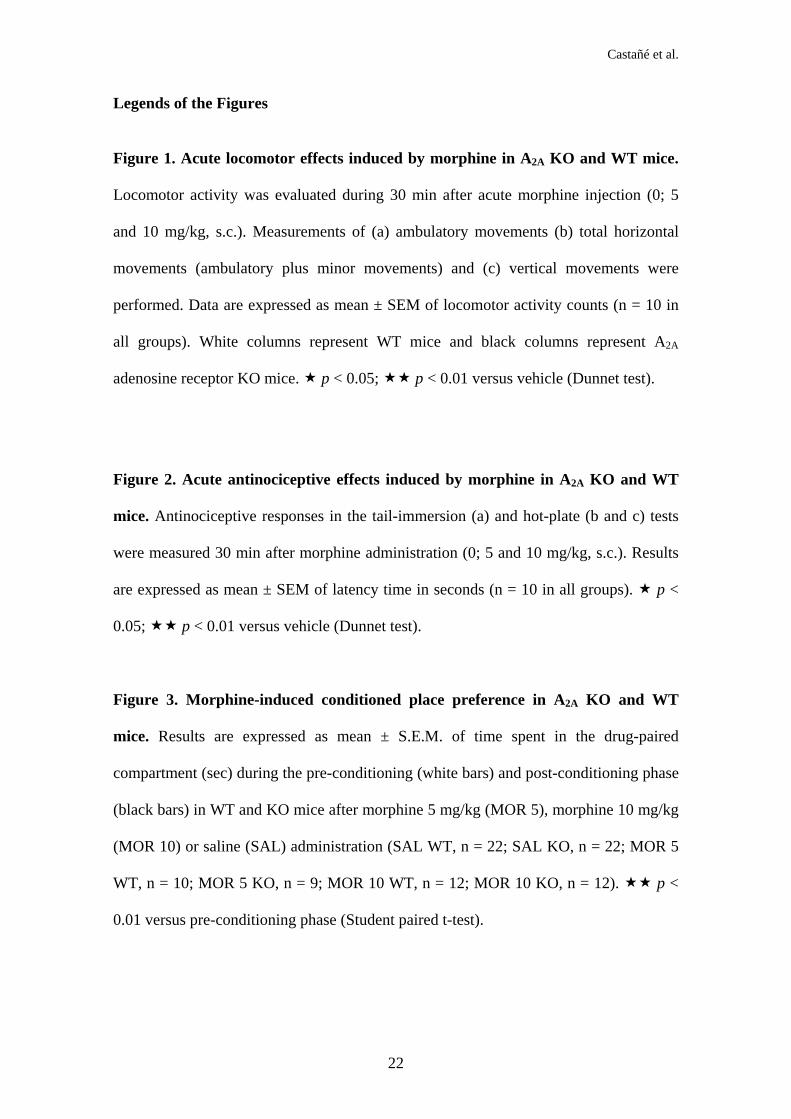
Figure 1. Acute locomotor effects induced by morphine in A2A KO and WT mice.
Locomotor activity was evaluated during 30 min after acute morphine injection (0; 5
and 10 mg/kg, s.c.). Measurements of (a) ambulatory movements (b) total horizontal
movements (ambulatory plus minor movements) and (c) vertical movements were
performed. Data are expressed as mean ± SEM of locomotor activity counts (n = 10 in
all groups). White columns represent WT mice and black columns represent A2A
adenosine receptor KO mice. p < 0.05; p < 0.01 versus vehicle (Dunnet test).
Figure 2. Acute antinociceptive effects induced by morphine in A2A KO and WT
mice. Antinociceptive responses in the tail-immersion (a) and hot-plate (b and c) tests
were measured 30 min after morphine administration (0; 5 and 10 mg/kg, s.c.). Results
are expressed as mean ± SEM of latency time in seconds (n = 10 in all groups). p <
0.05; p < 0.01 versus vehicle (Dunnet test).
Figure 3. Morphine-induced conditioned place preference in A2A KO and WT
mice. Results are expressed as mean ± S.E.M. of time spent in the drug-paired
compartment (sec) during the pre-conditioning (white bars) and post-conditioning phase
(black bars) in WT and KO mice after morphine 5 mg/kg (MOR 5), morphine 10 mg/kg
(MOR 10) or saline (SAL) administration (SAL WT, n = 22; SAL KO, n = 22; MOR 5
WT, n = 10; MOR 5 KO, n = 9; MOR 10 WT, n = 12; MOR 10 KO, n = 12). p <
0.01 versus pre-conditioning phase (Student paired t-test).

Castañé et al.
23
Figure 4. Naloxone-induced conditioned place aversion in morphine-dependent
A2A KO and WT mice. Results are expressed as mean ± S.E.M. of time spent in the
drug-paired compartment (sec) during the pre-conditioning (white bars) and post-
conditioning phase (black bars) in saline (SAL) and morphine (MOR) treated WT and
KO mice after saline (SAL), naloxone 0.05 mg/kg (NAL 0.05) or naloxone 0.1 mg/kg
(NAL 0.1) administration (SAL-SAL WT, n = 11; SAL-SAL KO, n = 13; SAL-NAL
0.05 WT, n = 12; SAL-NAL 0.05 KO, n = 13; SAL-NAL 0.1 WT, n = 10; SAL-NAL
0.1 KO, n = 11; MOR-SAL WT, n = 11; MOR-SAL KO, n = 13; MOR-NAL 0.05 WT,
n = 14; MOR-NAL 0.05 KO, n = 14; MOR-NAL 0.1 WT, n = 14; MOR-NAL 0.1 KO,
n = 15). p < 0.01 versus pre-conditioning phase (Student paired t-test).
Figure 5. Effects of the systemic morphine administration on dialysate DA
concentrations in the NAc of A2A KO and WT mice. Morphine (20 mg/kg, s.c.) did
not increase DA output in the NAc of WT (vehicle, SAL n = 5; morphine, MOR n = 6)
and KO mice (vehicle, SAL n = 6; morphine, MOR n = 7) in any dialysis fraction (F8-
F13). Data are percentages of pre-treatment (basal) values and are given as mean +
SEM values (see methods section for details).

Castañé et al.
24
References
Arolfo MP, Yao L, Gordon AS, Diamond I, Janak PH (2004). Ethanol operant self-
administration in rats is regulated by adenosine A2 receptors. Alcohol Clin Exp Res 28:
1308-1316.
Bailey A, Davis L, Lesscher HM, Kelly MD, Ledent C, Hourani SM, Kitchen I (2004).
Enhanced morphine withdrawal and micro-opioid receptor G-protein coupling in A2A
adenosine receptor KO mice. J Neurochem 88: 827-834.
Bailey A, Ledent C, Kelly M, Hourani SM, Kitchen I (2002). Changes in spinal delta
and kappa opioid systems in mice deficient in the A2A receptor gene. J Neurosci 22:
9210-9220.
Barbaccia ML, Reggiani A, Spano PF, Trabucchi M (1981). Ethanol-induced changes
of dopaminergic function in three strains of mice characterized by a different population
of opiate receptors. Psychopharmacology 74: 260-262.
Berrendero F, Castañé A, Ledent C, Parmentier M, Maldonado R, Valverde O (2003).
Increase of morphine withdrawal in mice lacking A2a receptors and no changes in
CB1/A2a double KO mice. Eur J Neurosci 17 : 315-324.
Berridge KC, Robinson TE (1998). What is the role of dopamine in reward: hedonic
impact, reward learning, or incentive salience?. Brain Res Brain Res Rev 28: 309-369.

Castañé et al.
25
Castañé A, Soria G, Ledent C, Maldonado R, Valverde O (2006). Attenuation of
nicotine-induced rewarding effects in A2A knockout mice. Neuropharmacology 51:
631-640.
Chefer VI, Kieffer BL, Shippenberg TS (2003). Basal and morphine-evoked
dopaminergic neurotransmission in the nucleus accumbens of MOR- and DOR-KO
mice. Eur J Neurosci 18: 1915-1922.
Dassesse D, Massie A, Ferrari R, Ledent C, Parmentier M, Arckens L, Zoli M,
Schiffmann SN (2001). Functional striatal hypodopaminergic activity in mice lacking
adenosine A(2A) receptors. J Neurochem 78: 183-198.
Di Chiara G (2002). Nucleus accumbens shell and core dopamine: differential role in
behavior and addiction. Behav Brain Res 137: 75-114.
Di Chiara G, Imperato A (1988). Drugs abused by humans preferentially increase
synaptic dopamine concentrations in the mesolimbic system of freely moving rats. Proc
Natl Acad Sci U S A 85: 5274-5278.
Eddy NB, Leimbach D (1953). Synthetic analgesics. II. Dithienylbutenyl- and
dithienylbutylamines. J Pharmacol Exp Ther 107: 385-393.
Fadda P, Scherma M, Collu M, Fratta W (2005). Dopamine and serotonin release in
dorsal striatum and nucleus accumbens is differentially modulated by morphine in
DBA/2J and C57BL/6J mice. Synapse 56: 29-38.
Castañé et al.
26
Ferré S, Fredholm BB, Morelli M, Popoli P, Fuxe K (1997). Adenosine-dopamine
receptor-receptor interactions as an integrative mechanism in the basal ganglia. Trends
Neurosci 20: 482-487.
Ferré S, Diamond I, Goldberg SR, Yao L, Hourani SMO, Huang ZL, Urade Y, Kitchen
I (2007). Adenosine A2A receptors in ventral striatum, hypothalamus and nociceptive
circuitry. Implications for drug addiction, sleep and pain. Prog Neurobiol 83: 332-347.
Fink JS, Weaver DR, Rivkees SA, Peterfreund RA, Pollack AE, Adler EM, Reppert SM
(1992). Molecular cloning of the rat A2 adenosine receptor: selective co-expression
with D2 dopamine receptors in rat striatum. Brain Res Mol Brain Res 14: 186-195.
Franco R, Ferré S, Agnati L, Torvinen M, Gines S, Hillion J, Casado V, Lledo P, Zoli
M, Lluis C, Fuxe K (2000). Evidence for adenosine/dopamine receptor interactions:
indications for heteromerization. Neuropsychopharmacology 23: S50-S59.
Fredholm BB, Chen JF, Cunha RA, Svenningsson P, Vaugeois JM (2005). Adenosine
and brain function. Int Rev Neurobiol 63: 191-270.
Gupta ML, Nath R, Gupta TK, Gupta GP (1988). A study of central neurotransmitter
mechanisms in morphine-induced 'Straub reaction' in mice: role of central dopamine
receptors. Clin Exp Pharmacol Physiol 15: 727-732.

Castañé et al.
27
Hack SP, Christie MJ (2003). Adaptations in adenosine signaling in drug dependence:
therapeutic implications. Crit Rev Neurobiol 15: 235-274.
He M, Shippenberg TS (2000). Strain differences in basal and cocaine-evoked
dopamine dynamics in mouse striatum. J Pharmacol Exp Ther 293: 121-127.
Johnson SW, North RA (1992). Two types of neurone in the rat ventral tegmental area
and their synaptic inputs. J Physiology 450: 455-468.
Kaplan GB, Coyle TS (1998). Adenosine kinase inhibitors attenuate opiate withdrawal
via adenosine receptor activation. Eur J Pharmacol 362: 1-8.
Kaplan GB, Sears MT (1996). Adenosine receptor agonists attenuate and adenosine
receptor antagonists exacerbate opiate withdrawal signs. Psychopharmacology 123: 64-
70.
Khalili M, Semnanian S, Fathollahi Y (2001). Caffeine increases paragigantocellularis
neuronal firing rate and induces withdrawal signs in morphine-dependent rats. Eur J
Pharmacol 412: 239-245.
Koob GF (1996). Hedonic valence, dopamine and motivation. Mol Psychiatry 1: 186-
189.
Ledent C, Vaugeois JM, Schiffmann SN, Pedrazzini T, El Yacoubi M, Vanderhaeghen
JJ, Costentin J, Heath JK, Vassart G, Parmentier M (1997). Aggressiveness, hypoalgesia

Castañé et al.
28
and high blood pressure in mice lacking the adenosine A2a receptor. Nature 388: 674-
678.
Maldonado R, Saiardi A, Valverde O, Samad TA, Roques BP, Borrelli E (1997).
Absence of opiate rewarding effects in mice lacking dopamine D2 receptors. Nature
388: 586-589.
Matthes HW, Maldonado R, Simonin F, Valverde O, Slowe S, Kitchen I, Befort K,
Dierich A, Le Meur M, Dolle P, Tzavara E, Hanoune J, Roques BP, Kieffer BL (1996).
Loss of morphine-induced analgesia, reward effect and withdrawal symptoms in mice
lacking the mu-opioid-receptor gene. Nature 383: 819-823.
Moreau JL, Huber G (1999). Central adenosine A(2A) receptors: an overview. Brain
Res Brain Res Rev 31: 65-82.
Rasmussen K, Aghajanian GK (1989). Withdrawal-induced activation of locus
coeruleus neurons in opiate-dependent rats: attenuation by lesions of the nucleus
paragigantocellularis. Brain Res 505: 346-350.
Rethy CR, Smith CB, Villarreal JE (1971). Effects of narcotic analgesics upon the
locomotor activity and brain catecholamine content of the mouse. J Pharmacol Exp
Ther 176: 472-479.

Castañé et al.
29
Salamone JD (1996). The behavioral neurochemistry of motivation: methodological and
conceptual issues in studies of the dynamic activity of nucleus accumbens dopamine. J
Neurosci Methods 64: 137-149.
Salem A, Hope W (1997). Effect of adenosine receptor agonists and antagonists on the
expression of opiate withdrawal in rats. Pharmacol Biochem Behav 57: 671-679.
Schiffmann SN, Vanderhaeghen JJ (1993). Adenosine A2 receptors regulate the gene
expression of striatopallidal and striatonigral neurons. J Neurosci 13: 1080-1087.
Simonin F, Valverde O, Smadja C, Slowe S, Kitchen I, Dierich A, Le Meur M, Roques
BP, Maldonado R, Kieffer BL (1998). Disruption of the kappa-opioid receptor gene in
mice enhances sensitivity to chemical visceral pain, impairs pharmacological actions of
the selective kappa-agonist U-50,488H and attenuates morphine withdrawal. EMBO J
17: 886-897.
Shoaib M, Spanagel R, Stohr T, Shippenberg TS (1995). Strain differences in the
rewarding and dopamine-releasing effects of morphine in rats. Psychopharmacology
117: 240-247
Short JL, Drago J, Lawrence AJ (2006). Comparison of ethanol preference and
neurochemical measures of mesolimbic dopamine and adenosine systems across
different strains of mice. Alcohol Clin Exp Res 30: 606-620.

Castañé et al.
30
Spanagel R, Weiss F (1999). The dopamine hypothesis of reward: past and current
status. Trends Neurosci 22: 521-527.
Soria G, Castañé A, Berrendero F, Ledent C, Parmentier M, Maldonado R, Valverde O
(2004). Adenosine A2A receptors are involved in physical dependence and place
conditioning induced by THC. Eur J Neurosci 20: 2203-2213.
Soria G, Castañé A, Ledent C, Parmentier M, Maldonado R, Valverde O (2006). The
reinforcing efficacy of cocaine is diminished in mice lacking A2A adenosine receptors.
Neuropsychopharmacology 31: 978-987.
Svenningsson P, Lindskog M, Ledent C, Parmentier M, Greengard P, Fredholm BB,
Fisone G (2000). Regulation of the phosphorylation of the dopamine- and cAMP-
regulated phosphoprotein of 32 kDa in vivo by dopamine D1, dopamine D2, and
adenosine A2A receptors. Proc Natl Acad Sci U S A 97: 1856-1860.
Svenningsson P, Nairn AC, Greengard P (2005). DARPP-32 mediates the actions of
multiple drugs of abuse. AAPS J 7: E353-360.
Sweeney MI, White TD, Jhamandas KH, Sawynok J (1987). Morphine releases
endogenous adenosine from the spinal cord in vivo. Eur J Pharmacol 141: 169-170.
Sweeney MI, White TD, Sawynok J (1991). Intracerebroventricular morphine releases
adenosine and adenosine 3',5'-cyclic monophosphate from the spinal cord via a
serotonergic mechanism. J Pharmacol Exp Ther 259: 1013-1018.

Castañé et al.
31
Tozzi A, Tscherter A, Belcastro V, Tantucci M, Costa C, Picconi B, Centonze D,
Calabresi P, Borsini F (2007). Interaction of A2A adenosine and D2 dopamine receptors
modulates corticostriatal glutamatergic transmission. Neuropharmacology 53: 783-789.
Valverde O, Tzavara E, Hanoune J, Roques BP, Maldonado R (1996). Protein kinases
in the rat nucleus accumbens are involved in the aversive component of opiate
withdrawal. Eur J Neurosci 8: 2671-2678.
Wise RA (2004). Dopamine, learning and motivation. Nat Rev Neurosci 5: 483-494.
Zarrindast MR, Naghipour B, Roushan-zamir F, Shafaghi B (1999). Effects of
adenosine receptor agents on the expression of morphine withdrawal in mice. Eur J
Pharmacol 369: 17-22.

Table 1. Two-way ANOVA for acute locomotor and antinociceptive responses induced by morphine in A2A adenosine receptor.
Two-way ANOVA with treatment (T) and genotype (G) as factors of variation. See Materials and Methods for details.
Locomotor activity Antinociception
Ambulatory Total horizontal Vertical Tail-immersion Hot-plate: Licking Hot-plate: jumping F p F p F p F p F p F p
Treatment F(2.54) = 63.672 0.01 F(2.54) = 35.977 0.01 F(2.53) = 14.704 0.01 F(2.53) = 50.431 0.01 F(2.54) = 9.785 0.01 F(2.54) = 502.15 0.01
Genotype F(1.54) = 9.526 0.01 F(1.54) = 17.606 0.05 F(1.53) = 4.523 0.05 F(1.53) = 1.884 n.s. F(1.54) = 0.083 n.s. F(1.54) = 1.164 n.s.
TxG F(2.54) = 0.523 n.s. F(2.54) = 0.587 n.s. F(2.53) = 1.749 n.s. F(2.53) = 0.930 n.s. F(2.54) = 0.519 n.s. F(2.54) = 0.291 n.s.

Wild-type A2a knockout
Figure 1. Castañé et al.
0
100
200
300
400
Cou
nts
0 5 10
a) Ambulatory movements
0
200
400
600
800
Cou
nts
0 5 10
b) Total horizontal activity
0
100
200
300
400
Cou
nts
0 5 10
b) Vertical activity

Figure 2. Castañé et al.
Wild-type A2a knockout
0255075
100125150
Late
ncy
(sec
)
b) Hot-plate: licking
0 5 10
02468
1012
0 5 10
Late
ncy
(sec
)
a) Tail-immersion
050
100150200250300
Late
ncy
(sec
)
b) Hot-plate: jumping
0 5 10

Figure 3. Castañé et al.
MOR 10
0
100
200
300
400
500
600
700
SAL MOR 5
Tim
e sp
enti
n dr
ug p
aire
dco
mpa
rtm
ent(
sec)
WT KO WT KO WT KO
Pre-conditioning phase Post-conditioning phase

Tim
e sp
enti
n dr
ug p
aire
dco
mpa
rtm
ent (
sec)
Pre-conditioning phase Post-conditioning phase
Figure 4. Castañé et al.
WT KO
SAL-SAL
WT KO
SAL-NAL 0.05
WT KO
SAL-NAL 0.1
WT KO
MOR-SAL
WT KO
MOR-NAL 0.05
WT KO
MOR-NAL 0.1
0
100
200
300
400
500
600
700

Figure 5. Castañé et al.
Basal F8 F9 F10 F11 F12 F1350
100
150
200
250
WT SAL WT MORKO SAL KO MOR
Fraction
Dia
lysa
teD
A
(% o
f bas
al v
alue
s)