bats carry pathogenic hepadnaviruses antigenically related to … hepadnaviral... · bats carry...
TRANSCRIPT

Bats carry pathogenic hepadnaviruses antigenicallyrelated to hepatitis B virus and capable of infectinghuman hepatocytesJan Felix Drexlera,1, Andreas Geipelb,1, Alexander Königb, Victor M. Cormana, Debby van Rielc, Lonneke M. Leijtenc,Corinna M. Bremerb, Andrea Raschea, Veronika M. Cottontaild,e, Gael D. Magangaf, Mathias Schlegelg,Marcel A. Müllera, Alexander Adamh, Stefan M. Klosed, Aroldo José Borges Carneiroi, Andreas Stöckerj,Carlos Roberto Frankei, Florian Gloza-Rauscha,k, Joachim Geyerl, Augustina Annanm, Yaw Adu-Sarkodien,Samuel Oppongn, Tabea Bingera, Peter Vallod,o, Marco Tschapkad,e, Rainer G. Ulrichg, Wolfram H. Gerlichb, Eric Leroyf,p,Thijs Kuikenc, Dieter Glebeb,1,2, and Christian Drostena,1,2
aInstitute of Virology, University of Bonn Medical Centre, 53127 Bonn, Germany; bInstitute of Medical Virology, Justus Liebig University, 35392 Giessen,Germany; cDepartment of Viroscience, Erasmus Medical Center, 3000 CA, Rotterdam, The Netherlands; dInstitute of Experimental Ecology, University of Ulm,89069 Ulm, Germany; eSmithsonian Tropical Research Institute, Balboa Ancón, Republic of Panamá; fCentre International de Recherches Médicales deFranceville, BP 769 Franceville, Gabon; gFriedrich-Loeffler-Institut, Institute for Novel and Emerging Infectious Diseases, 17493 Greifswald-Insel Riems,Germany; hInstitute of Pathology, University of Cologne Medical Centre, 50937 Cologne, Germany; iSchool of Veterinary Medicine, Federal University ofBahia, 40.170-110, Salvador, Brazil; jInfectious Diseases Research Laboratory, University Hospital Prof. Edgard Santos, Federal University of Bahia, 40.110-060,Salvador, Brazil; kNoctalis, Centre for Bat Protection and Information, 23795 Bad Segeberg, Germany; lInstitute of Pharmacology and Toxicology, Justus LiebigUniversity, Biomedical Research Center, 35392 Giessen, Germany; mKumasi Centre for Collaborative Research in Tropical Medicine (KCCR), Kumasi, Ghana;nFaculty of Renewable Natural Resources,Kwame Nkrumah University of Science and Technology, Kumasi, Ghana; oInstitute of Vertebrate Biology, Academyof Sciences of the Czech Republic, 60 365 Brno, Czech Republic; and pInstitut de Recherche pour le Développement (IRD), Unité Mixte de Recherche 224(MIVEGEC), IRD/Centre National de la Recherche Scientifique/Universite Montpellier 1, 34032 Montpellier, France
Edited by Robert A. Lamb, Northwestern University, Evanston, IL, and approved August 2, 2013 (received for review April 29, 2013)
The hepatitis B virus (HBV), family Hepadnaviridae, is one of mostrelevant human pathogens. HBV origins are enigmatic, and nozoonotic reservoirs are known. Here, we screened 3,080 specimensfrom 54 bat species representing 11 bat families for hepadnaviralDNA. Ten specimens (0.3%) from Panama and Gabon yieldedunique hepadnaviruses in coancestral relation to HBV. Full genomesequencing allowed classification as three putative orthohepadna-virus species based on genome lengths (3,149–3,377 nt), presenceof middle HBV surface and X-protein genes, and sequence distancecriteria. Hepatic tropism in bats was shown by quantitative PCRand in situ hybridization. Infected livers showed histopathologicchanges compatible with hepatitis. Human hepatocytes trans-fected with all three bat viruses cross-reacted with sera againstthe HBV core protein, concordant with the phylogenetic related-ness of these hepadnaviruses and HBV. One virus from Urodermabilobatum, the tent-making bat, cross-reacted with monoclonalantibodies against the HBV antigenicity determining S domain.Up to 18.4% of bat sera contained antibodies against bat hepad-naviruses. Infectious clones were generated to study all threeviruses in detail. Hepatitis D virus particles pseudotyped with sur-face proteins of U. bilobatum HBV, but neither of the other twoviruses could infect primary human and Tupaia belangeri hepato-cytes. Hepatocyte infection occurred through the human HBV re-ceptor sodium taurocholate cotransporting polypeptide but couldnot be neutralized by sera from vaccinated humans. Antihepadna-viral treatment using an approved reverse transcriptase inhibitorblocked replication of all bat hepadnaviruses. Our data suggestthat bats may have been ancestral sources of primate hepadnavi-ruses. The observed zoonotic potential might affect conceptsaimed at eradicating HBV.
evolution | zoonosis | virome | metagenomics | reverse genetics
More than 40% of the human population has been infectedwith the hepatitis B virus (HBV), giving rise to 240 million
chronic HBV carriers and ca. 620,000 HBV-associated deathsannually (1). A prophylactic vaccine containing the small HBVgenotype A2 surface antigen (SHB) is part of the worldwideExpanded Program on Immunization. Because of the generalsuccess of SHBs-based vaccination, global eradication of HBVhas been considered achievable (2, 3). Potential for the virus to
be eradicated is supported by the fact that there are no knownanimal reservoirs. However, recent studies addressing the dis-tribution of pathogens related to human viruses in wild animals,including mumps- and measles-related viruses in bats, have un-covered surprising putative novel reservoirs for human-patho-genic viruses (4).
Significance
Hepatitis B virus (HBV) is the prototype hepadnavirus; 40% ofhumans have current or past infection. In a global investigationof viral diversity in bats, we discovered three unique hep-adnavirus species. The relatedness of these viruses to HBVsuggests that bats might constitute ancestral sources of pri-mate hepadnaviruses. Infection patterns in bats resembledhuman infection with HBV. After resurrection from bat tissues,pseudotyped viruses carrying surface proteins of one bathepadnavirus could infect human liver cells. HBV vaccination isprobably not protective against these viruses, but viral repli-cation could be blocked by a reverse transcriptase inhibitorused as an anti-HBV drug in humans. The potential of bathepadnaviruses to infect humans should be considered inprograms aimed at eradicating HBV.
Author contributions: J.F.D., D.G., and C.D. designed research; J.F.D., A.G., A.K., V.M.Corman, D.v.R., L.M.L., C.M.B., A.R., M.A.M., A. Adam, A.J.B.C., A.S., and T.K. performedresearch; V.M. Cottontail, G.D.M., M.S., S.M.K., C.R.F., F.G.-R., J.G., A. Annan, Y.A.-S., S.O.,T.B., P.V., M.T., R.G.U., W.H.G., E.L., and T.K. contributed new reagents/analytic tools;J.F.D., A.G., A.K., V.M. Corman, D.v.R., L.M.L., C.M.B., T.K., and D.G. analyzed data; J.F.D.,D.G., and C.D. wrote the paper; V.M. Cottontail performed field work (Panama); G.D.M.,P.V., and E.L. performed field work (Gabon); S.M.K. performed field work (Australia/Papua New Guinea); A.J.B.C. and C.R.F. performed field work (Brazil); F.G.-R. performedfield work (Germany); and A. Annan, Y.A.-S., S.O., and T.B. performed field work (Ghana).
The authors declare no conflict of interest.
This article is a PNAS Direct Submission.
Data deposition: The sequences reported in this paper has been deposited in the GenBankdatabase (accession nos. KC790373–KC790381).1J.F.D., A.G., D.G., and C.D. contributed equally to this work.2To whom correspondence may be addressed. E-mail: [email protected] or [email protected].
This article contains supporting information online at www.pnas.org/lookup/suppl/doi:10.1073/pnas.1308049110/-/DCSupplemental.
www.pnas.org/cgi/doi/10.1073/pnas.1308049110 PNAS Early Edition | 1 of 6
MICRO
BIOLO
GY

HBV is the prototype species of the familyHepadnaviridae, whichcomprises two genera: the genus Orthohepadnavirus associatedwith mammals and the genus Avihepadnavirus associated withbirds. Phylogenetic studies suggested the presence of HBV inhumans for at least 15,000 y (5). Recent analyses of avihepadna-viral sequences integrated into the genomes of several avian spe-cies suggested a much older origin, dating back at least 19 milliony (6). No HBV genomic elements have so far been found inhumans or other primates, preventing more precise estimates ofthe origins of primate HBV (i.e., human and nonhuman primateviruses). HBV strains can be divided into nine strictly human-associated genotypes (A–I). Additional strains outside some ofthose human-specific clades are known in chimpanzees, gorillas,gibbons, and orangutans (7). With sporadic exceptions (8), theseprimate HBV strains do not infect humans. The closest relativeto human or ape viruses has been found in captive woollymonkeys (Lagothrix lagotricha), a South American nonhumanprimate species (9). There are only three nonprimate ortho-hepadnaviruses, all being even less closely related to HBV.These viruses include woodchuck HBV from Eastern wood-chucks (Marmota monax), Californian ground squirrel (Oto-spermophilus beecheyi) HBV, and arctic squirrel (Spermophilusparryi) HBV (10). These rodent hosts are endemic in circum-scribed areas of North America, and their viruses are highly host-specific and cannot infect human hepatocytes (11, 12).Within the ∼5,500 known terrestrial species of mammals,
about 20% are bats. Close relatives of pathogenic human viruseshave been described in bats over the last years, including SevereAcute Respiratory Syndrome (SARS) and Middle East Re-spiratory Syndrome (MERS)-related coronaviruses (CoV) aswell as filoviruses, such as Ebola- and Marburgvirus (13, 14).Among the multiple factors that facilitate virus evolution withinand transmission from bats are their longevity, migratory activity,large and dense roosting communities, and close social in-teraction (14). We have analyzed earlier the role of bats in theevolution of pathogenic viruses using very large globally andphylogenetically comprehensive samples of animals (4, 15). Inthis study, we detected highly diversified bat hepadnavirusescapable of infecting human hepatocytes through the HBV-spe-cific human receptor but not neutralized by SHBs vaccine-induced antibodies.
ResultsHBV Detection. Bats were sampled between 2002 and 2011 inPanama, Brazil, Gabon, Ghana, Germany, Papua New Guinea,and Australia (Fig. 1). These specimens represented 54 differentspecies and 11 of 18 extant bat families (Table S1). Serum andliver specimens from 3,080 individual bats were individuallytested using two broadly reactive and highly sensitive nestedPCR assays. The sensitivities of these PCR assays at 95%probability of detection were 41.3 (95% confidence interval =29.7–75.9) and 64.7 (95% confidence interval = 47.3–112.9) in-ternational units/mL blood (Fig. S1 and Table S2). Liver speci-mens only were available for all 199 bats sampled in Brazil, andall other 2,881 specimens were sera. In 10 of 3,080 specimens(0.3%; all sera), HBV-related sequences were detected. Positivespecimens stemmed from three different bat species. AmongNew World bats, 5 of 54 (9.3%) Uroderma bilobatum specimensfrom Panama tested positive. Among Old World bats, 4 of 51Hipposideros cf. ruber specimens (7.9%) and 1 of 16 Rhinolophusalcyone specimens (6.3%) from Gabon contained HBV-likesequences. Fig. 1 shows the distribution areas of these bat speciesin gray.
Genome Organization. Full virus genome sequences were deter-mined from all positive specimens of H. cf. ruber, the single spec-imen of R. alcyone, and four specimens of U. bilobatum (GenBankaccession nos. KC790373–KC790381). The bat viruses formedthree different lineages on preliminary phylogenetic inspection.Viruses from H. cf. ruber were collectively termed roundleafbat HBV (RBHBV), the virus from R. alcyone was designated
horseshoe bat HBV (HBHBV), and viruses from U. bilobatum werecollectively termed tent-making bat HBV (TBHBV). Virus des-ignations were chosen according to the designation of other non-human hepadnaviruses based on the common names of their hosts[e.g., Woolly monkey HBV (WMHBV)], and they are detailed inFig. S2A. The genome organization and size of the putative openreading frames (ORFs) were compared with all other knownhepadnaviruses (Fig. S2B). RBHBV genomes comprised 3,368 nt,with a total of 12–18 (0.4–0.5%) nucleotide exchanges betweeneach other. HBHBV comprised a genome of 3,377 nt. TheTBHBV genomes comprised 3,149 nt, which varied by 4–74 (0.1–2.3%) nucleotide exchanges from each other. RBHBV andHBHBV diverged by 19% of their genomic nucleotide sequencefrom each other and 39% of their genomic nucleotide sequencefrom TBHBV. All bat viruses varied in their nucleotide sequencesby at least 35% from sequences of any known hepadnavirus(Tables S3 and S4). As in all orthohepadnaviruses, the uniquebat viruses contained four ORFs identifiable as the surface (S),polymerase (P), core (C), and X-ORFs (Fig. S2C). The positionof all ORFs in bat hepadnaviruses was similar to ORFs of theknown members of the Orthohepadnavirus genus but clearlydistinct from ORFs of duck hepatitis B virus, the prototypeavihepadnavirus. The sizes of all predicted ORFs compared withhomologs in prototype hepadnaviruses are shown in Fig. S2B.Details for the comparison of translated amino acid sequences ofeach predicted virus protein are provided in Tables S3 and S4.The surface (S) protein genes encoded in the large open readingframe of all newly discovered hepadnaviral genomes containeda preS1, a preS2, and the S domain. The preS1 domain containedan N-myristoylation signal necessary for myristoylation at glycine-2of preS1. Typical N-glycosylation sites within the preS2 and Sdomains were conserved, similar to HBV. Within the predictedantigenic SHBs loop, all eight essential cysteins for viral assem-bly, secretion, and infectivity (16) were present. Other than ORForganization, HBV and the bat hepadnaviruses also shared asimilar location of the direct repeat (DR) sequences DR1 andDR2 involved in genome replication. In addition, secondarystructure prediction highlighted the structural similarities be-tween HBV and the bat hepadnaviruses in their e-loops, whichserve as templates for the priming of reverse transcription of pre-genomic RNA in all hepadnaviruses (Fig. S2D).According to a Bayesian phylogenetic analysis based on full
ortho- and avihepadnavirus genomes, bat hepadnaviruses clustered
Fig. 1. Sampling sites and distribution of HBV-positive bat species. Sam-pling sites of HBV-positive bats are in red, and other sites are in yellow. Nextto sites, the number of sampled species and specimens per family are given.Red, positive bat species; gray, distribution of positive bats.
2 of 6 | www.pnas.org/cgi/doi/10.1073/pnas.1308049110 Drexler et al.
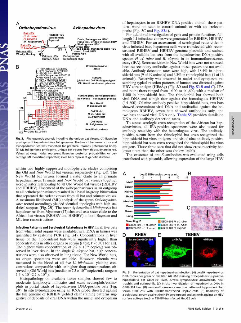
within two highly supported monophyletic clades comprisingthe Old and New World bat viruses, respectively (Fig. 2A). TheNew World bat viruses formed a sister clade to all primatehepadnaviruses. Primate and New World bat viruses togetherwere in sister relationship to all Old World bat viruses (RBHBVand HBHBV). Placement of the avihepadnaviruses as an outgroupto all orthohepadnaviruses resulted in a basal in-group bifurcationthat separated the rodent viruses from all bat and primate viruses.A maximum likelihood (ML) analysis of the genus Orthohepadna-virus rooted accordingly yielded identical topologies with high sta-tistical support (Fig. 2B). The recently described Miniopterus bathepadnavirus fromMyanmar (17) clustered as a sister clade to theAfrican bat viruses (RBHBV and HBHBV) in both Bayesian andML tree reconstructions.
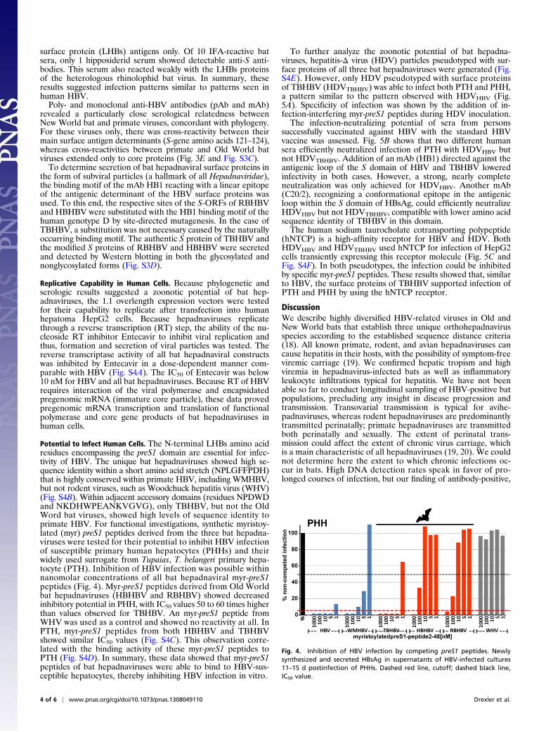
Infection Patterns and Serological Relatedness to HBV. In all five batsfrom which solid organs were available, viral DNA in tissues wasquantified by real-time PCR (Fig. 3A). Concentrations in livertissue of the hipposiderid bats were significantly higher thanconcentrations in other organs or serum (t test, P < 0.01 for all).The highest virus concentration of 2.2 × 10¹² copies/g was ob-served in liver tissue. In the single R. alcyone bat, high concen-trations were also observed in lung tissue. For New World bats,no organ specimens were available. However, viremia wasmeasured in the blood of all five U. bilobatum, yielding con-centrations comparable with or higher than concentrations ob-served in Old World bats (median = 7.3 × 10¹⁰ copies/mL; range =1.4 × 10⁶–2.7 × 10¹¹).Histopathology on available tissue samples showed few to
moderate lymphocyte infiltrates and scant neutrophils/eosino-phils in portal triads of hepadnavirus DNA-positive bats (Fig.3B). In situ hybridization using an RNA probe directed againstthe full genome of RBHBV yielded clear staining patterns sug-gestive of deposits of viral DNA within the nuclei and cytoplasm
of hepatocytes in an RBHBV DNA-positive animal; these pat-terns were not seen in control animals or with an irrelevantprobe (Fig. 3C and Fig. S3A).For additional investigation of gene and protein functions, full-
length 1.1 infectious clones were generated for RBHBV, HBHBV,and TBHBV. For an assessment of serological reactivities invirus-infected bats, hepatoma cells were transfected with recon-structed RBHBV and HBHBV genome plasmids and stainedwith all available bat sera from the hepadnavirus DNA-positivespecies H. cf. ruber and R. alcyone in an immunofluorescenceassay (IFA). Seroreactivities in NewWorld bats were not assessed,because secondary antibodies against these species are not avail-able. Antibody detection rates were high, with 18.4% in hippo-siderid bats (9 of 49 animals) and 6.3% in rhinolophid bats (1 of 16animals). Reactivity was observed in nuclei and cytoplasm, re-sembling typical reaction patterns of human sera directed againstHBV core antigen (HBcAg) (Fig. 3D and Fig. S3 B and C). IFAend-point titers ranged from 1:100 to 1:1,600, with a median of1:400 in hipposiderid bats. The rhinolophid bat showed bothviral DNA and a high titer against the homologous HBHBV(1:1,600). Of nine antibody-positive hipposiderid bats, two batsshowed concomitant viral DNA and antibodies against the ho-mologous RBHBV, seven bats showed antibodies only, andtwo bats showed viral DNA only. Table S5 provides details onDNA and antibody detection rates.To assess serologic cross-recognition of the African bat hep-
adnaviruses, all IFA-positive specimens were also tested forantibody reactivity with the heterologous virus. The antibody-positive serum from the rhinolophid bat cross-recognized thehipposiderid bat virus antigens, and six of nine antibody-positivehipposiderid bat sera cross-recognized the rhinolophid bat virusantigens. Those three sera that did not show cross-reactivity hadlower titers than the other sera (below 1:400).The existence of anti-S antibodies was evaluated using cells
transfected with plasmids, allowing expression of the large HBV
Fig. 2. Phylogenetic analysis including the unique bat viruses. (A) Bayesianphylogeny of Hepadnaviridae full genomes. The branch between ortho- andavihepadnaviruses was truncated for graphical reasons (interrupted lines).(B) ML full genome phylogeny. Unique bat viruses from this study are in red.Values at deep nodes represent Bayesian posterior probabilities or per-centage ML bootstrap replicates; scale bars represent genetic distance.
Fig. 3. Presentation of bat hepadnavirus infection. (A) Log10 hepadnavirusDNA copies per gram or milliliter. (B) H&E staining of hepadnavirus-positivehipposiderid bat GB09-301 liver. Arrow, lymphocytes; arrowheads, neu-trophils and eosinophils. (C) In situ hybridization of hepadnavirus DNA inGB09-301 liver. (D) Immunofluorescence reaction pattern of hipposiderid batserum GB09-256 with RBHBV-transfected HepG2 cells. (E) Reactivity ofa polyclonal serum against the HBV core (green) and an mAb against an HBVsurface epitope (red) in TBHBV-transfected HepG2 cells.
Drexler et al. PNAS Early Edition | 3 of 6
MICRO
BIOLO
GY
surface protein (LHBs) antigens only. Of 10 IFA-reactive batsera, only 1 hipposiderid serum showed detectable anti-S anti-bodies. This serum also reacted weakly with the LHBs proteinsof the heterologous rhinolophid bat virus. In summary, theseresults suggested infection patterns similar to patterns seen inhuman HBV.Poly- and monoclonal anti-HBV antibodies (pAb and mAb)
revealed a particularly close serological relatedness betweenNew World bat and primate viruses, concordant with phylogeny.For these viruses only, there was cross-reactivity between theirmain surface antigen determinants (S-gene amino acids 121–124),whereas cross-reactivities between primate and Old World batviruses extended only to core proteins (Fig. 3E and Fig. S3C).To determine secretion of bat hepadnaviral surface proteins in
the form of subviral particles (a hallmark of all Hepadnaviridae),the binding motif of the mAb HB1 reacting with a linear epitopeof the antigenic determinant of the HBV surface proteins wasused. To this end, the respective sites of the S-ORFs of RBHBVand HBHBV were substituted with the HB1 binding motif of thehuman genotype D by site-directed mutagenesis. In the case ofTBHBV, a substitution was not necessary caused by the naturallyoccurring binding motif. The authentic S protein of TBHBV andthe modified S proteins of RBHBV and HBHBV were secretedand detected by Western blotting in both the glycosylated andnonglycosylated forms (Fig. S3D).
Replicative Capability in Human Cells. Because phylogenetic andserologic results suggested a zoonotic potential of bat hep-adnaviruses, the 1.1 overlength expression vectors were testedfor their capability to replicate after transfection into humanhepatoma HepG2 cells. Because hepadnaviruses replicatethrough a reverse transcription (RT) step, the ability of the nu-cleoside RT inhibitor Entecavir to inhibit viral replication andthus, formation and secretion of viral particles was tested. Thereverse transcriptase activity of all bat hepadnaviral constructswas inhibited by Entecavir in a dose-dependent manner com-parable with HBV (Fig. S4A). The IC50 of Entecavir was below10 nM for HBV and all bat hepadnaviruses. Because RT of HBVrequires interaction of the viral polymerase and encapsidatedpregenomic mRNA (immature core particle), these data provedpregenomic mRNA transcription and translation of functionalpolymerase and core gene products of bat hepadnaviruses inhuman cells.
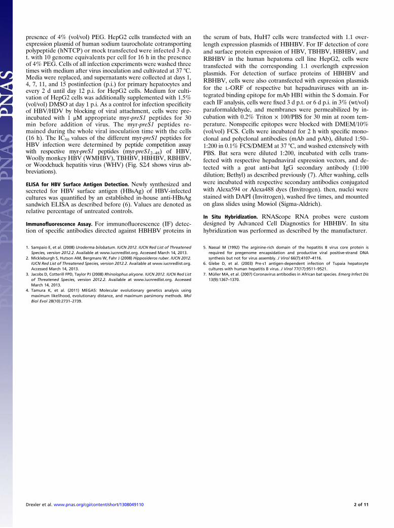
Potential to Infect Human Cells. The N-terminal LHBs amino acidresidues encompassing the preS1 domain are essential for infec-tivity of HBV. The unique bat hepadnaviruses showed high se-quence identity within a short amino acid stretch (NPLGFFPDH)that is highly conserved within primate HBV, including WMHBV,but not rodent viruses, such as Woodchuck hepatitis virus (WHV)(Fig. S4B). Within adjacent accessory domains (residues NPDWDand NKDHWPEANKVGVG), only TBHBV, but not the OldWord bat viruses, showed high levels of sequence identity toprimate HBV. For functional investigations, synthetic myristoy-lated (myr) preS1 peptides derived from the three bat hepadna-viruses were tested for their potential to inhibit HBV infectionof susceptible primary human hepatocytes (PHHs) and theirwidely used surrogate from Tupaias, T. belangeri primary hepa-tocyte (PTH). Inhibition of HBV infection was possible withinnanomolar concentrations of all bat hepadnaviral myr-preS1peptides (Fig. 4). Myr-preS1 peptides derived from Old Worldbat hepadnaviruses (HBHBV and RBHBV) showed decreasedinhibitory potential in PHH, with IC50 values 50 to 60 times higherthan values observed for TBHBV. An myr-preS1 peptide fromWHV was used as a control and showed no reactivity at all. InPTH, myr-preS1 peptides from both HBHBV and TBHBVshowed similar IC50 values (Fig. S4C). This observation corre-lated with the binding activity of these myr-preS1 peptides toPTH (Fig. S4D). In summary, these data showed that myr-preS1peptides of bat hepadnaviruses were able to bind to HBV-sus-ceptible hepatocytes, thereby inhibiting HBV infection in vitro.
To further analyze the zoonotic potential of bat hepadna-viruses, hepatitis-Δ virus (HDV) particles pseudotyped with sur-face proteins of all three bat hepadnaviruses were generated (Fig.S4E). However, only HDV pseudotyped with surface proteinsof TBHBV (HDVTBHBV) was able to infect both PTH and PHH,a pattern similar to the pattern observed with HDVHBV (Fig.5A). Specificity of infection was shown by the addition of in-fection-interfering myr-preS1 peptides during HDV inoculation.The infection-neutralizing potential of sera from persons
successfully vaccinated against HBV with the standard HBVvaccine was assessed. Fig. 5B shows that two different humansera efficiently neutralized infection of PTH with HDVHBV butnot HDVTBHBV. Addition of an mAb (HB1) directed against theantigenic loop of the S domain of HBV and TBHBV loweredinfectivity in both cases. However, a strong, nearly completeneutralization was only achieved for HDVHBV. Another mAb(C20/2), recognizing a conformational epitope in the antigenicloop within the S domain of HBsAg, could efficiently neutralizeHDVHBV but not HDVTBHBV, compatible with lower amino acidsequence identity of TBHBV in this domain.The human sodium taurocholate cotransporting polypeptide
(hNTCP) is a high-affinity receptor for HBV and HDV. BothHDVHBV and HDVTBHBV used hNTCP for infection of HepG2cells transiently expressing this receptor molecule (Fig. 5C andFig. S4F). In both pseudotypes, the infection could be inhibitedby specific myr-preS1 peptides. These results showed that, similarto HBV, the surface proteins of TBHBV supported infection ofPTH and PHH by using the hNTCP receptor.
DiscussionWe describe highly diversified HBV-related viruses in Old andNew World bats that establish three unique orthohepadnavirusspecies according to the established sequence distance criteria(18). All known primate, rodent, and avian hepadnaviruses cancause hepatitis in their hosts, with the possibility of symptom-freeviremic carriage (19). We confirmed hepatic tropism and highviremia in hepadnavirus-infected bats as well as inflammatoryleukocyte infiltrations typical for hepatitis. We have not beenable so far to conduct longitudinal sampling of HBV-positive batpopulations, precluding any insight in disease progression andtransmission. Transovarial transmission is typical for avihe-padnaviruses, whereas rodent hepadnaviruses are predominantlytransmitted perinatally; primate hepadnaviruses are transmittedboth perinatally and sexually. The extent of perinatal trans-mission could affect the extent of chronic virus carriage, whichis a main characteristic of all hepadnaviruses (19, 20). We couldnot determine here the extent to which chronic infections oc-cur in bats. High DNA detection rates speak in favor of pro-longed courses of infection, but our finding of antibody-positive,
Fig. 4. Inhibition of HBV infection by competing preS1 peptides. Newlysynthesized and secreted HBsAg in supernatants of HBV-infected cultures11–15 d postinfection of PHHs. Dashed red line, cutoff; dashed black line,IC50 value.
4 of 6 | www.pnas.org/cgi/doi/10.1073/pnas.1308049110 Drexler et al.

DNA-negative Hipposideros bats together with a high seropre-valence of 18.4% indicate that bats can probably clear the in-fection. Detection of DNA-positive, antibody-negative bats indicatessampling before seroconversion, whereas concomitant detectionof DNA and antibodies indicates delayed clearance just like inhumans. Also, the lack of detectable anti-LHBs antibodies in allbut one bat resembles human disease, because these antibodiescan be precipitated by high concentrations of HB antigen in serum.The possibility of a bat origin of primate hepadnaviruses
enables speculations regarding their evolution. Primate HBV hasonly been detected in one monophyletic ape taxon, the Homi-noidea superfamily, as well as the rather distantly related Woollymonkey, a New World primate (7, 9). There is complete absenceof detection in cercopithecoid monkeys (the Old World monkeysister clade to the Hominoidea) as well as lower nonsimiiformmonkeys. This absence leaves doubts regarding virus–hostcosegregation in primates and suggests a direct acquisition ofHBV as a split-off from the stem lineage leading up to WMHBVby primates (21). It should be noted that WMHBV has neverbeen redetected in wild or captive animals, and serologicalstudies have failed to detect antibodies against HBV in otherNew World monkeys (7). WMHBV could, thus, have been ac-quired in captivity, and its actual host could have been either anOld or New World mammal. Of note, this animal could havebeen a bat, but the stem lineage leading up to primate viruses couldalso have been acquired from any other (probably mammalian)source.Throughout Africa and to a much lesser extent, the Neo-
tropical ecozone, the consumption of bats as wild game byhumans is common practice (22). We have, therefore, performedin-depth studies of the zoonotic potential of the described bathepadnaviruses using several well-established techniques. It hasbeen shown that infectivity and host tropism of HBV is de-termined by highly conserved amino acids of the preS1 domainand its myristic acid (19, 23). In contrast to rodent hep-adnaviruses, all bat hepadnaviruses showed nearly completeconservation of this relevant domain, and its functional impor-tance was reflected in the potential of myr-preS1 peptides tocompete with infection, similar to HBV in vitro and in vivo (24,25). However, we observed marked differences between primaryhuman (PHH) and Tupaia (PTH) hepatocyte cultures. Althoughboth cell culture systems showed similar susceptibilities to human
serum-derived HBV infection, PHH revealed that preS1 peptidesfrom TBHBV, but not HBHBV or RBHBV, reached IC50 valuessimilar to HBV peptides. The inhibitory potential of myr-preS1peptides depends on their ability to block interaction of HBVwith the newly discovered HBV receptor NTCP (26). Althoughthe Tupaia and hNTCP sequences are very similar (26), ourresults suggest that PHH should be used in binding and entrystudies to accurately evaluate the zoonotic potential of newlydiscovered hepadnaviruses.Analysis of infection competence using HDV pseudotypes
suggested that only TBHBV surface proteins were capable ofmediating infection similar to HBV. The incompatibility ofRBHBV and HBHBV proteins was remarkable given the clearability of their preS1 domains to bind hepatocytes and inhibitHBV infection. It should be mentioned that the work by Gudimaet al. (27) reported infection of PHH, even with HDV particlespseudotyped with surface proteins from WHV (HDVWHV), al-though WHV itself does not infect humans or chimpanzees.Gudima et al. (27) concluded that HDVWHV must infect PHHusing a different receptor, because preS1 peptides from WHV,but not HBV, could inhibit HDVWHV infection. In contrast tothat study, we show that HDVTBHBV and HDVHBV could bothbe inhibited by preS1 peptides of TBHBV and HBV with similarefficiencies. Furthermore, both HDVTBHBV and HDVHBV usedthe same HBV-specific receptor (hNTCP). Because HBHBVand RBHBV are replication-competent after transfection intohuman hepatoma cell lines, their restriction point might be thepresence of a second (co-) receptor involved in HDV/HBVinfection that might be highly species-specific. Characterizationof the NTCP molecules of these two bat species might help elu-cidate whether HBHBV and RBHBV also use NTCP for hepa-tocyte entry.Other than the preS1 domain, the antigenic loop of the S
domain is another independent determinant for HDV and HBVinfectivity (16, 28). Among all bat hepadnaviruses, only TBHBVshared exactly the essential sequence C(R/K)TC within the an-tigenic loop with HBV and reacted with the mAb HB1 directedagainst this domain. These differences and others within the Sdomain might explain why TBHBV, but not the other bat hep-adnaviruses, could infect PHH.It should be mentioned that there are several limitations to
our efforts to assess zoonotic potential. For instance, WMHBVcan efficiently infect other New World primates but not OldWorld primates in vivo (29), whereas recombinant HDVWMHBVviruses can infect human and chimpanzee primary hepatocytes(30). One might, therefore, argue that our surrogate assays maynot properly reflect the in vivo situation. However, additionalproof of zoonotic potential would require inoculation of chim-panzees, which is not ethical given the expected severity of in-fection. Severe combined immunodeficiency mice transgenic forurokinase-type plasminogen activator (uPA/SCID) mice engraf-ted with human hepatocytes could serve as alternative models(31), but they would have to be validated by comparisons withchimpanzees before application on TBHBV, which is still dis-tantly related to HBV and thus, might behave differently in thismodel. Another limitation arises from the use of recombinantHDV/bat hepadnaviruses for infection and inhibition studies.However, because the recovery of homologous recombinantorthohepadnaviruses in sufficient amount is technically difficult,recombinant HDV/hepadnaviruses are widely accepted as sur-rogates (19, 27, 30).In conclusion, among the three unique hepadnavirus species
described in this report, we have evidence for a zoonotic po-tential for one of them, the New World bat-associated TBHBV.The lack of neutralization of this virus by high-titered anti-HBssera from vaccinated individuals matches the observation ofoccasional failure of the standard vaccine, even against heter-ologous human HBV genotypes (32). Elimination of HBV fromglobal circulation in humans is conceivable within several gen-erations (3), but a revised vaccine formulation, including fullLHBs or at least the critical preS1 domain, could become
Fig. 5. Zoonotic potential of unique bat hepadnaviruses. (A) Infection withHDVHBV or HDVTBHBV. Presence (+) and absence (−) of specific myr-preS1peptide inhibitors. ge, HDV genome equivalent 12 d postinfection. (B) Lackof protection by antisera from HB-vaccinated persons. C20/2 and HB1, mAbsagainst the HBV surface; IgG, nonspecific mAb; NS, serum of a non-HepB–vaccinated person. (C) HBV and TBHBV use hNTCP for infection. HepG2 cellsexpressing hNTCP (+) or a control (−) were incubated with HDVHBV orHDVTBHBV with (+) or without (−) inhibitors. *P < 0.05, **P < 0.02; t test.Cutoff, dashed red line.
Drexler et al. PNAS Early Edition | 5 of 6
MICRO
BIOLO
GY

necessary to that end (33). Future vaccination concepts mightalso have to integrate considerations of the zoonotic potential ofprimate and nonprimate hepadnaviruses (2). It is unclearwhether bat hepadnaviruses impose an ongoing risk of zoonotichuman infections, but it should be considered that these virusesare genetically sufficiently distinct from HBV to go undetectedin routine serological and molecular screening programs. Ofnote, access of humans to such HBV routine diagnostic pro-grams cannot be deemed likely in the remote tropical areasfrom which these bats were sampled, highlighting the need forscreening of human and nonhuman primate sera from theseareas by broadly reactive diagnostic methods. Whereas apepopulations are decreasing because of habitat exploitation ona global scale, bats can adapt to anthropogenic influence inmultiple ways, leading to modifications of social structure,pathogen richness, and exposure to humans. We are only be-ginning to understand the role of bats as reservoirs of zoonoticviruses, emphasizing the importance of viral surveillance andthe integration of ecological concepts into infectious diseaseepidemiology (34).
Materials and MethodsSampling, Hepadnavirus Detection, and Characterization. Bats were caught asdescribed previously (4). Permits are given in SI Materials and Methods.
Purification, detection, and characterization of viral DNA were done as de-scribed previously and in SI Materials and Methods (4, 15, 35). Phylogeneticanalyses were done using MrBayes (36) and PhyML (37).
HBV Constructs and Infection Experiments. Overlength constructs (1.1), surfaceexpression vectors, and HDV pseudoparticles were generated as describedpreviously (38–40). PHH and PTH infection, HBsAg detection, and infectionof cells expressing the hNTCP were done as described before (41) and in SIMaterials and Methods.
ACKNOWLEDGMENTS. We thank Monika Eschbach-Bludau, Sebastian Brünink,Tobias Bleicker, Thomas Kruppa, Mathieu Bourgarel, Vanessa Rüsseler, andSigrun Broehl for technical assistance; Barbara Döring for the hNTCP-FLAGconstruct; Micha Nübling for HBV standards; and Aurelija Zvirbliene for mAbHB1. This study was funded by German Research Foundation (DFG) SPP 1596Grants DR 810/1-1 (to J.F.D., M.T., and D.G.), GL 595/4-1 (to J.F.D., M.T., andD.G.), TS 81/6-1 (to J.F.D., M.T., and D.G.), DR 772/3-1 (to C.D.), and KA1241/18-1 (to M.T.); Brazilian Foundation for Research Support of the State ofBahia Grant SUS0038/2007 (to J.F.D. and C.R.F.); European Union FP7 ProjectsEuropean Management Platform for Emerging and Re-Emerging InfectiousDisease Entities Grant 223498; Europan Virus Archive Grant 228292 (to C.D.);Anticipating the Global Onset of Novel Epidemics Grant 278976 (to C.D.);and German Federal Ministry of Education and Research (BMBF) Grant01KI1016D (to C.D.). The subproject in Panama was supported by the Smith-sonian Tropical Research Institute and a personal scholarship of the GermanNational Academic Foundation (to V.M. Cottontail).
1. Ott JJ, Stevens GA, Groeger J, Wiersma ST (2012) Global epidemiology of hepatitis Bvirus infection: New estimates of age-specific HBsAg seroprevalence and endemicity.Vaccine 30(12):2212–2219.
2. Chen DS (2010) Toward elimination and eradication of hepatitis B. J GastroenterolHepatol 25(1):19–25.
3. Alter HJ (2012) To have B or not to have B: Vaccine and the potential eradication ofhepatitis B. J Hepatol 57(4):715–717.
4. Drexler JF, et al. (2012) Bats host major mammalian paramyxoviruses. Nat Commun3:796.
5. Paraskevis D, et al. (2013) Dating the origin and dispersal of hepatitis B virus infectionin humans and primates. Hepatology 57(3):908–916.
6. Gilbert C, Feschotte C (2010) Genomic fossils calibrate the long-term evolution ofhepadnaviruses. PLoS Biol 8(9):e1000495.
7. Starkman SE, MacDonald DM, Lewis JC, Holmes EC, Simmonds P (2003) Geographicand species association of hepatitis B virus genotypes in non-human primates. Vi-rology 314(1):381–393.
8. Tatematsu K, et al. (2009) A genetic variant of hepatitis B virus divergent from knownhuman and ape genotypes isolated from a Japanese patient and provisionally as-signed to new genotype J. J Virol 83(20):10538–10547.
9. Lanford RE, Chavez D, Brasky KM, Burns RB, 3rd, Rico-Hesse R (1998) Isolation ofa hepadnavirus from the woolly monkey, a New World primate. Proc Natl Acad SciUSA 95(10):5757–5761.
10. Simmonds P (2001) The origin and evolution of hepatitis viruses in humans. J GenVirol 82(Pt 4):693–712.
11. Wang BJ, et al. (2011) Establishing a new animal model for hepadnaviral infection:Susceptibility of Chinese Marmota-species to woodchuck hepatitis virus infection.J Gen Virol 92(Pt 3):681–691.
12. Trueba D, et al. (1985) Transmission of ground squirrel hepatitis virus to homologousand heterologous hosts. Hepatology 5(3):435–439.
13. Annan A, et al. (2013) Human betacoronavirus 2c EMC/2012-related viruses in bats,Ghana and Europe. Emerg Infect Dis 19(3):456–459.
14. Luis AD, et al. (2013) A comparison of bats and rodents as reservoirs of zoonotic vi-ruses: Are bats special? Proc Biol Sci 280(1756):20122753.
15. Drexler JF, et al. (2012) Bats worldwide carry hepatitis E virus-related viruses that forma putative novel genus within the family Hepeviridae. J Virol 86(17):9134–9147.
16. Salisse J, Sureau C (2009) A function essential to viral entry underlies the hepatitis Bvirus “a” determinant. J Virol 83(18):9321–9328.
17. He B, et al. (2013) Hepatitis virus in long-fingered bats, myanmar. Emerg Infect Dis19(4):638–640.
18. Schaefer S (2007) Hepatitis B virus taxonomy and hepatitis B virus genotypes. World JGastroenterol 13(1):14–21.
19. Glebe D, Urban S (2007) Viral and cellular determinants involved in hepadnaviralentry. World J Gastroenterol 13(1):22–38.
20. Liang TJ (2009) Hepatitis B: The virus and disease. Hepatology 49(5 Suppl):S13–S21.21. Fares MA, Holmes EC (2002) A revised evolutionary history of hepatitis B virus (HBV).
J Mol Evol 54(6):807–814.22. Mickleburgh S, Waylen K, Racey P (2009) Bats as bushmeat: A global review. Oryx
43(2):217–234.
23. Glebe D (2006) Attachment sites and neutralising epitopes of hepatitis B virus. Mi-nerva Gastroenterol Dietol 52(1):3–21.
24. Gripon P, Cannie I, Urban S (2005) Efficient inhibition of hepatitis B virus infection byacylated peptides derived from the large viral surface protein. J Virol 79(3):1613–1622.
25. Volz T, et al. (2013) The entry inhibitor Myrcludex-B efficiently blocks intrahepaticvirus spreading in humanized mice previously infected with hepatitis B virus.J Hepatol 58(5):861–867.
26. Yan H, et al. (2012) Sodium taurocholate cotransporting polypeptide is a functionalreceptor for human hepatitis B and D virus. Elife 1:e00049.
27. Gudima S, et al. (2008) Primary human hepatocytes are susceptible to infection byhepatitis delta virus assembled with envelope proteins of woodchuck hepatitis virus.J Virol 82(15):7276–7283.
28. Le Duff Y, Blanchet M, Sureau C (2009) The pre-S1 and antigenic loop infectivitydeterminants of the hepatitis B virus envelope proteins are functionally independent.J Virol 83(23):12443–12451.
29. Lanford RE, Chavez D, Barrera A, Brasky KM (2003) An infectious clone of woollymonkey hepatitis B virus. J Virol 77(14):7814–7819.
30. Barrera A, Guerra B, Lee H, Lanford RE (2004) Analysis of host range phenotypes ofprimate hepadnaviruses by in vitro infections of hepatitis D virus pseudotypes. J Virol78(10):5233–5243.
31. Sa-Nguanmoo P, et al. (2011) Cross-species transmission of gibbon and orangutanhepatitis B virus to uPA/SCID mice with human hepatocytes. Virus Res 158(1–2):209–215.
32. Tacke F, et al. (2007) Acute hepatitis B virus infection by genotype F despite successfulvaccination in an immune-competent German patient. J Clin Virol 38(4):353–357.
33. Niedre-Otomere B, et al. (2012) Recombinant Semliki Forest virus vectors encodinghepatitis B virus small surface and pre-S1 antigens induce broadly reactive neutral-izing antibodies. J Viral Hepat 19(9):664–673.
34. Drosten C (2013) Virus ecology: A gap between detection and prediction. EmergMicrobes Infect 2:e31.
35. Drexler JF, et al. (2009) A novel diagnostic target in the hepatitis C virus genome. PLoSMed 6(2):e31.
36. Ronquist F, Huelsenbeck JP (2003) MrBayes 3: Bayesian phylogenetic inference undermixed models. Bioinformatics 19(12):1572–1574.
37. Guindon S, et al. (2010) New algorithms and methods to estimate maximum-likeli-hood phylogenies: Assessing the performance of PhyML 3.0. Syst Biol 59(3):307–321.
38. Nassal M (1992) The arginine-rich domain of the hepatitis B virus core protein is re-quired for pregenome encapsidation and productive viral positive-strand DNA syn-thesis but not for virus assembly. J Virol 66(7):4107–4116.
39. Glebe D, et al. (2005) Mapping of the hepatitis B virus attachment site by use of in-fection-inhibiting preS1 lipopeptides and tupaia hepatocytes. Gastroenterology129(1):234–245.
40. Kuo MY, Chao M, Taylor J (1989) Initiation of replication of the human hepatitis deltavirus genome from cloned DNA: Role of delta antigen. J Virol 63(5):1945–1950.
41. Glebe D, et al. (2003) Pre-s1 antigen-dependent infection of Tupaia hepatocyte cul-tures with human hepatitis B virus. J Virol 77(17):9511–9521.
6 of 6 | www.pnas.org/cgi/doi/10.1073/pnas.1308049110 Drexler et al.

Supporting InformationDrexler et al. 10.1073/pnas.1308049110SI Materials and MethodsWildlife Permits and Ethics Clearances for Bat Sampling and SpecimenTransfer. All animals were handled according to the EuropeanUnion Council Directive 86/609/EEC for the protection of ani-mals. Serum specimens were collected without killing any animalsin Panama, Germany, Papua NewGuinea, and Australia. In Brazil,Ghana, and Gabon, animals were dissected, and organ sampleswere additionally obtained under strict anesthesia of animals.Workin Brazil was supported by the Bahia state program for rabiescontrol executed by ADAB (Agência Estadual de Defesa Agro-pecuária da Bahia). Individual permit numbers were Panama[Research-Permit STRI: STRI2563 (PI VC)-IACUC 100316–1001-18/Research-Permit ANAM: SE/A-68–11/Ethics-Permit:IACUC 100316–1001-18/Export Permits: SEX/A-30–11, SEX/A-55–11, SEX/A-81–10, SEX-A-26-10]; Ghana [ResearchPermit: 2008–2010 (A04957)/Ethics-Permit: CHRPE49/09/CITES/Export-Permit: State Agreement between Ghana and Hamburg(BNI)]; Australia [Research Permit: S11828 and S11762/Ethics-Permit: TRIM 01/1118(2), TRIM 06/3569, and University ofQueensland/Animal Ethics Committee SIB600/05/DEST/Export-Permit: DE201-12]; Papua New Guinea (Ethics-Permit: PNG/NatMus/2002/Export-Permit: Conducted by Papua New GuineaNational Museum); Gabon (Ethics-Permit: 00021/MEFEPA/SG/DGEF/DFC); Germany (Ethics-Permit: LANU 314/5327.74.1.6);and Brazil (Sampling permit IBAMA 15304–1; no specimens wereexported).
Distribution of Hepadnavirus-Positive Bat Species. Distribution wasadapted from the International Union for Conservation of Nature(1–3).
In Silico Analyses. Alignments were generated using MEGA5 (4).For MrBayes, 4 million generations were sampled every 100steps, resulting in 40,000 trees; 25% of replicates were discardedas burn in. Maximum likelihood analyses used 1,000 bootstrapreplicates. The HKY nucleotide substitution matrix was used forboth. Statistical analyses were done using SPSS V20 (IBM).
Hepadnavirus Infectious Clones. For expression of hepadnavirusesin cell culture, the vector pCH-9/3091 was used that contains a 1.1overlength hepatitis B virus (HBV) genome (genotype D) underthe control of a human cytomegalovirus immediate early pro-moter (5) by substitution of pCH-9/3091 with an 1.1 overlengthgenome of the respective bat hepadnaviral genome [tent-makingbat HBV (TBHBV), GenBank accession no. KC790379; horse-shoe bat HBV (HBHBV), GenBank accession no. KC790377;roundleaf bat HBV (RBHBV), GenBank accession no. KC790374](Fig. S2A shows nonabbreviated virus designations). Vectors forexpression of surface proteins (large antigen open readingframe, L-ORF) under the control of natural hepadnaviralpromoters contained a 5′-truncated version of the respective 1.1overlength construct starting around 500 bp upstream of thecorresponding L-ORF start codon (pCH9-3091-HBV-L, 506 bp;pCH9-TBHBV-L, 501 bp; pCH9-RBHBV-L, 519 bp; pCH9-HBHBV-L, 517 bp) in a pCH9-3091 backbone without humancytomegalovirus immediate early promoter. Insertion of thebinding motif for mAb HB1 (amino acids 121–125 CRTCTT ofHBV S domain) was done by substitution of the respectiveamino acids in the S domain of RBHBV (amino acids 119–124CASCTI) and HBHBV (amino acids 119–124 CTSCTI) usingsite-directed mutagenesis.
Cell Lines and Transfection.Human hepatoma cell lines HuH7 andHepG2 were cultivated in DMEM with 10% (vol/vol) FCS (In-vitrogen). Cells were transiently transfected with hepadnaviral1.1 overlength expression plasmids at 80% confluence in 10-cmdishes using FuGene HD (Promega) according to the manu-facturer’s protocol; 1 d after transfection, cells were washed, andnew medium was added. Cells and/or supernatants were used forassays 3 d posttransfection (p.t.) unless stated otherwise.
Production of Hepatitis-Δ Virus Pseudoparticles. For production ofhepatitis-Δ virus (HDV) pseudoparticles, HuH7 cells were co-transfected with equimolar amounts of HDV plasmid pSVLD3.After transfection, cells were cultivated in DMEM/2% (vol/vol)FCS. Medium was changed every 3 d. Secreted HDV was quanti-fied by determining genome equivalents in the supernatant bya specific RT-PCR at days 6 and 9. Supernatants from day 6 today 9 were harvested at days 6, 7, 8, and 9 and used for infectionassays after concentration using membrane ultrafiltration (Vi-vaspin, MWCO 10 000; GE Healthcare) according to the man-ufacturer’s instructions.
SDS/PAGE and Immunoblotting. Original or concentrated (10×)supernatants from cells transfected with the respective surfaceprotein expression vectors were pretreated with NuPAGE LDSSample Buffer (Invitrogen) together with 8% (wt/vol) DTT andincubated at 70 °C for 10 min. Samples were analyzed on a 12%(vol/vol) precast polyacrylamide gel (Invitrogen). Immunoblot-ting was done as previously described with mAb HB1 [1 μg/μL in3% (wt/vol) low-fat milk powder in PBS] as the primary anti-body, donkey anti-mouse antibody conjugated with alkalinephosphatase (AP; 1:10,000 in 3% low-fat milk powder in PBS;Dianova) as secondary antibody, and Immobilon Westernchemiluminescence AP Substrate (Merck Millipore).
Purification and Detection of Viral RNA/DNA from Cells. Purificationof nucleic acids from supernatants of cells transfected with HBV/HDV expression plasmids was done using the High Pure ViralNucleic Acid Kit (Roche) according to the manufacturer’s pro-tocol. Purified RNA from transfections with HDV genomeplasmid was additionally digested with RNase-Free DNase I(Thermo Scientific) for 1 h at 37 °C. HDV RNA from infectedcells was purified using the Nucleospin 8 RNAII Kit (Macherey-Nagel) according to the manufacturer’s instructions. Quantifi-cation of HDV genomes was done by a specific quantitative one-step RT-PCR (verso one-step RT-PCR Kit; Thermo Scientific)using the primers and probes listed in Table S2. Quantificationof hepadnavirus DNA was done by specific discriminative SYBRgreen quantitative PCR using ABsolute qPCR SYBR Green-Mix(Thermo Scientific) and the virus-specific primers listed in Table S2.
Myristoylated Hepadnaviral PreS1 Peptides. Chemical synthesis ofcarboxyterminally strep-tagged (amino acid WSHPQFEK), N-terminally myristoylated hepadnaviral preS1 peptides spanningamino acid residues 2–48 (myr-preS12–48) was performed byBiosynthesis Inc.
Primary Human Hepatocytes. Primary human hepatocytes (PHHs)were obtained from Primacyt Cell Culture Technology GmbH.
HDV Pseudotype and HBV Infection Assays. For Tupaias belangeriprimary hepatocyte (PTH) and PHH infections with cell culture-derived HDV pseudoparticles, cells were incubated for 16 h atday 3 postseeding with five genome equivalents per cell in the
Drexler et al. www.pnas.org/cgi/content/short/1308049110 1 of 11
presence of 4% (vol/vol) PEG. HepG2 cells transfected with anexpression plasmid of human sodium taurocholate cotransportingpolypeptide (hNTCP) or mock transfected were infected 3 d p.t. with 10 genome equivalents per cell for 16 h in the presenceof 4% PEG. Cells of all infection experiments were washed threetimes with medium after virus inoculation and cultivated at 37 °C.Media were replaced, and supernatants were collected at days 1,4, 7, 11, and 15 postinfection (p.i.) for primary hepatocytes andevery 2 d until day 12 p.i. for HepG2 cells. Medium for culti-vation of HepG2 cells was additionally supplemented with 1.5%(vol/vol) DMSO at day 1 p.i. As a control for infection specificityof HBV/HDV by blocking of viral attachment, cells were pre-incubated with 1 μM appropriate myr-preS1 peptides for 30min before addition of virus. The myr-preS1 peptides re-mained during the whole viral inoculation time with the cells(16 h). The IC50 values of the different myr-preS1 peptides forHBV infection were determined by peptide competition assaywith respective myr-preS1 peptides (myr-preS12–48) of HBV,Woolly monkey HBV (WMHBV), TBHBV, HBHBV, RBHBV,or Woodchuck hepatitis virus (WHV) (Fig. S2A shows virus ab-breviations).
ELISA for HBV Surface Antigen Detection. Newly synthesized andsecreted for HBV surface antigen (HBsAg) of HBV-infectedcultures was quantified by an established in-house anti-HBsAgsandwich ELISA as described before (6). Values are denoted asrelative percentage of untreated controls.
Immunofluorescence Assay. For immunofluorescence (IF) detec-tion of specific antibodies directed against HBHBV proteins in
the serum of bats, HuH7 cells were transfected with 1.1 over-length expression plasmids of HBHBV. For IF detection of coreand surface protein expression of HBV, TBHBV, HBHBV, andRBHBV in the human hepatoma cell line HepG2, cells weretransfected with the corresponding 1.1 overlength expressionplasmids. For detection of surface proteins of HBHBV andRBHBV, cells were also cotransfected with expression plasmidsfor the L-ORF of respective bat hepadnaviruses with an in-tegrated binding epitope for mAb HB1 within the S domain. Foreach IF analysis, cells were fixed 3 d p.t. or 6 d p.i. in 3% (wt/vol)paraformaldehyde, and membranes were permeabilized by in-cubation with 0.2% Triton × 100/PBS for 30 min at room tem-perature. Nonspecific epitopes were blocked with DMEM/10%(vol/vol) FCS. Cells were incubated for 2 h with specific mono-clonal and polyclonal antibodies (mAb and pAb), diluted 1:50–1:200 in 0.1% FCS/DMEM at 37 °C, and washed extensively withPBS. Bat sera were diluted 1:200, incubated with cells trans-fected with respective hepadnaviral expression vectors, and de-tected with a goat anti-bat IgG secondary antibody (1:100dilution; Bethyl) as described previously (7). After washing, cellswere incubated with respective secondary antibodies conjugatedwith Alexa594 or Alexa488 dyes (Invitrogen). then, nuclei werestained with DAPI (Invitrogen), washed five times, and mountedon glass slides using Mowiol (Sigma-Aldrich).
In Situ Hybridization. RNAScope RNA probes were customdesigned by Advanced Cell Diagnostics for HBHBV. In situhybridization was performed as described by the manufacturer.
1. Sampaio E, et al. (2008) Uroderma bilobatum. IUCN 2012. IUCN Red List of ThreatenedSpecies, version 2012.2. Available at www.iucnredlist.org. Accessed March 14, 2013.
2. Mickleburgh S, Hutson AM, Bergmans W, Fahr J (2008) Hipposideros ruber. IUCN 2012.IUCN Red List of Threatened Species, version 2012.2. Available at www.iucnredlist.org.Accessed March 14, 2013.
3. Jacobs D, Cotterill FPD, Taylor PJ (2008) Rhinolophus alcyone. IUCN 2012. IUCN Red Listof Threatened Species, version 2012.2. Available at www.iucnredlist.org. AccessedMarch 14, 2013.
4. Tamura K, et al. (2011) MEGA5: Molecular evolutionary genetics analysis usingmaximum likelihood, evolutionary distance, and maximum parsimony methods. MolBiol Evol 28(10):2731–2739.
5. Nassal M (1992) The arginine-rich domain of the hepatitis B virus core protein isrequired for pregenome encapsidation and productive viral positive-strand DNAsynthesis but not for virus assembly. J Virol 66(7):4107–4116.
6. Glebe D, et al. (2003) Pre-s1 antigen-dependent infection of Tupaia hepatocytecultures with human hepatitis B virus. J Virol 77(17):9511–9521.
7. Müller MA, et al. (2007) Coronavirus antibodies in African bat species. Emerg Infect Dis13(9):1367–1370.
Drexler et al. www.pnas.org/cgi/content/short/1308049110 2 of 11

Fig. S1. Probit regression analyses of nested PCR assays used for hepadnavirus detection; 200 μL second World Health Organization international HBV ref-erence standard containing 1 million international units (IUs) per milliliter (product code: 97/750; National Institute for Biological Standards and Control) wereextracted using the MinElute Spin Kit (Qiagen) with an elution volume of 100 μL. The assay protocols and individual primer sequences are described in Table S2.A shows the first PCR assay, including primer HBV-F248. B shows the second assay, including primer HBVall-F1364, which allows detection of ortho- andavihepadnaviruses. Each plot depicts the observed proportion of positive results from eight parallel experiments at each dilution step as well as the derivedpredicted proportion of positive results at a given DNA input concentration. The black line shows the prediction, and the gray lines show the 95% confidencelimits of the prediction.
Drexler et al. www.pnas.org/cgi/content/short/1308049110 3 of 11

Fig. S2. Genomic features of newly discovered bat hepadnaviruses. A shows the abbreviations used for hepadnaviruses throughout the text in accordancewith the current taxonomic proposals of the International Committee on Taxonomy of Viruses. B shows the sizes of the genomes and predicted ORFs of bat andprototype hepadnaviruses. *Duck hepatitis B virus (DHBV) as a typical representative of the genus Avihepadnavirus does not have a preS1 or preS2 domain butdoes have a preS domain and lacks the X-ORF. **Genotype G WT does not have an intact preC-ORF because of two stop codons. Human HBV genotypes havegenomes of 3,182–3,248 nt. The genomes of WMHBV and TBHBV, both from hosts endemic in the New World, showed similar lengths of 3,179 and 3,149 nt,respectively. The two Old World viruses, RBHBV and HBHBV, had the largest genome lengths of all known hepadnaviruses, with 3,377–3,368 nt, because of anunusually large preS1 domain-encoding sequence containing 177 and 174 codons, respectively. The genomes of these bat viruses were most similar to WHV,with a genome length of 3,308 nt and a preS1 domain of 144 aa. C shows the genome organization of HBV (genotype D; GenBank accession no. AB126581),DHBV (GenBank accession no. HQ132730), RBHBV/HBHBV (shown together because of high similarity), and TBHBV. DR1/2, direct repeat 1/2; rcDNA, relaxedcircular DNA; (+) and (−), orientation of DNA strands. The unknown length of the (+) strand in the unique bat hepadnaviruses is depicted in analogy to humanHBV. The orthohepadnavirus S-ORF has the potential to encode three cocarboxyterminal surface proteins, putatively encoding large, medium, and smallhepatitis B surface proteins. The S domain is present in all three surface proteins, whereas M contains an additional aminoterminal domain, termed preS2. TheL protein contains preS1/preS2 and the S domain. The P-ORF overlaps with the S- and C-ORFs, covering about two-thirds of the genome. The C-ORF containsa functional precore region encoding a signal peptide for optional secretion of the core protein, like in all WT hepadnaviruses. D shows RNA secondarystructure predictions of the e-structures of the respective sequences with S fold at 37 °C, 1 M NaCl, and no divalent cations (1). HBV and TBHBV e-loops werevery similar, whereas RBHBV and HBHBV differed in their loop structure by an additional unpaired region. Bat hepadnavirus and HBV e-signals included thestart codon (AUG) of the C-ORF (highlighted in pink) and showed the same nucleotide sequence in the unpaired region for the priming of the minus strand
Legend continued on following page
Drexler et al. www.pnas.org/cgi/content/short/1308049110 4 of 11

(UUC; highlighted in light blue). All secondary structures predicted for orthohepadnaviruses yielded similar free energy values ranging from 27.8 to 29.7 kcal/mol compared with 17.2 in DHBV. The predicted stability of the structures is shown in kilocalories per mol.
1. Chan CY, Lawrence CE, Ding Y (2005) Structure clustering features on the Sfold Web server. Bioinformatics 21(20):3926–3928.
Fig. S3. Antigenic and histologic features of the unique bat hepadnaviruses. A shows detection of bat hepadnavirus DNA and mRNA by in situ hybridization(ISH) at a low and high magnification. (Magnification: Upper, 200×; Lower, 1,000×.) (Left) In a PCR-positive bat, a positive signal was observed in hepatocytes(arrow) surrounding the portal triads using hepadnavirus-specific probes. (Center) No signal was observed in serial tissue sections using an irrelevant controlprobe against a rodent hepacivirus (1). (Right) In a PCR-negative bat, no signal was observed using the hepadnavirus-specific probe. B shows (Left) recognitionof Hipposideros bat hepadnavirus RBHBV antigens by three different Hipposideros bat sera and (Right) cross-recognition of RBHBV antigens by polyclonalcontrol anti-HBc sera. C shows recognition of HBV, TBHBV, HBHBV, or RBHBV in transfected HepG2 cells by polyclonal anti-HBcAg (HBV core antigen) anti-bodies (green) and monoclonal anti-HBsAg antibody HB1 (red). For detection of HBHBsAg and RBHBsAg, expression plasmids for appropriate surface proteinswith inserted HB1 binding epitope were cotransfected. D shows a Western blot with mAb HB1 reacting with a linear epitope of the antigenic determinant ofthe HBV surface proteins. For detection of RBHBV and HBHBV, the HB1 epitope of HBV genotype D was introduced in the corresponding region of the S-ORF.LHB, large HBV surface proteins; MHB, medium HBV surface proteins; SHB, small HBV surface proteins.
1. Drexler JF, et al. (2013) Evidence for novel hepaciviruses in rodents. PLoS Pathog 9(6):e1003438.
Drexler et al. www.pnas.org/cgi/content/short/1308049110 5 of 11

Fig. S4. Molecular features of bat hepadnaviruses. A shows the inhibition of bat hepadnavirus replication in hepatoma cells by the reverse transcriptase inhibitorEntecavir. *Replicationwasmeasured in the supernatant of cells 3 d p.t. by quantitativeHBV PCR relative to control cells.B shows an alignment of the aminoterminalpreS1 sequence.Myr-preS1 peptides amino acids 2–48 of indicated hepadnaviral species were used (red, bat hepadnaviruses). IC50 valueswere of the respectivemyr-preS1 peptide on HBV infection using PTH or PHH. Binding, percent binding of the respectivemyr-preS1 peptide to PTH. Essential and accessory domains of the HBVpreS1 domain important for virus–receptor interaction and inhibitory effect of myr-preS1 peptides are indicated (1). C shows inhibition of HBV infection by com-peting preS1 peptides of the different bat viruses: HBV, WMHBV, and WHV. Inhibition was analyzed by measuring newly synthesized and secreted HBsAg withinsupernatants of HBV-infected cultures from day 11 to day 15 p.i. from three experiments on PTH. Dashed red line, cutoff; dashed black line, IC50.D shows binding ofpreS1 peptides from different hepadnaviruses to PTH. Cells were precooled on ice and incubated with different strep-tagged myr-preS1 peptides 2–48 (5,000 nM)fromHBV,WMHBV, TBHBV, HBHBV, RBHBV, orWHV. (Left) After normalization to cell counts, relative binding potential of each peptidewas adjusted to binding ofhuman myr-preS1 peptides. By using specific mouse antistrep antibody and Alexa594-conjugated anti-mouse antibodies, membrane binding was detected byconfocal imaging (red). (Right) Alexa594 Pixel Sum of five overview pictures with 14–75 nuclei from each duplicate was detected by Leica LAS-AF software in threeexperiments. E shows the generation of pseudotyped HDV particles. The human hepatoma cell line HuH7was cotransfected with plasmid pSVLD3 (for transcriptionof a complete replication-competent HDV genome) and expression plasmids for all three surface proteins of the respective hepadnaviruses or a control vector.Medium was changed every 3 d, and HDV genome equivalents of secreted pseudovirions were quantified at days 6 and 9 p.t. Supernatants taken on day 9 wereconcentrated and used for infection. All used orthohepadnaviral surface proteins showed the amino acid sequence responsible for interaction with Δ-antigen(IWMMWYW), especially the three essential tryptophan residues. F shows staining of hepatitis-Δ antigen in HepG2 cells transfected with hNTCP and infected withHBV and TBHBV HDV pseudotypes. Cells were fixed at day 6 p.i. and stained for appearance of Δ-antigen (red). Cell nuclei were stained with DAPI (blue).
1. Glebe D, et al. (2005) Mapping of the hepatitis B virus attachment site by use of infection-inhibiting preS1 lipopeptides and tupaia hepatocytes. Gastroenterology 129(1):234–245.
Drexler et al. www.pnas.org/cgi/content/short/1308049110 6 of 11

Table S1. Sample characteristics
Order-family and species No. of samples PCR positive (%) Sampling site (y)
Chiroptera-PteropodidaeDobsonia praedatrix 9 PNG (2002)Eidolon helvum 348 GHA (2009, 2010)Epomops franquetti 100 GAB (2009)Hypsignathus monstrosus 100 GAB (2009)Melonycteris melanops 7 PNG (2002)Micropterus pusillus 100 GAB (2009)Myonycteris torquata 100 GAB (2009)Pteropus alecto 3 AUS (2006)Pteropus poliocephalus 24 AUS (2006)Rousettus aegyptiacus 204 GAB (2009)Rousettus amplexicaudatus 1 PNG (2002)
Chiroptera-EmballonuridaeColeura afra 14 GAB (2009)Saccopteryx bilineata 14 PAN (2010, 2011)Saccopteryx leptura 3 PAN (2010, 2011)
Chiroptera-HipposideridaeHipposideros cf. ruber 51 4 (7.9) GAB (2009*)Hipposideros gigas 129 GAB (2009)
Chiroptera-MolossidaeMolossus currentium 10 BRA (2009)Molossus rufus 17 BRA (2009)Molossus molossus 2 BRA (2009), PAN (2010, 2011)
Chiroptera-MormoopidaePteronotus parnellii 53 PAN (2010, 2011)
Chiroptera-NatalidaeNatalus stramineus 1 PAN (2010, 2011)
Chiroptera-NoctilionidaeNoctilio leporinus 11 PAN (2005, 2010, 2011)
Chiroptera-PhyllostomidaeArtibeus jamaicensis 981 PAN (2005, 2010, 2011)Artibeus lituratus 94 PAN (2005, 2010, 2011)Artibeus phaeotis 10 PAN (2010, 2011)Artibeus watsoni 16 PAN (2010, 2011)Carollia brevicauda 104 BRA (2009)Carollia castanea 30 PAN (2010, 2011)Carollia perspicillata 181 BRA (2009), PAN (2010, 2011)Carollia spec. 15 BRA (2009)Chiroderma villosum 2 PAN (2010, 2011)Desmodus rotundus 31 BRA (2009), PAN (2010, 2011)Glossophaga sorcina 27 BRA (2009), PAN (2010, 2011)Lonchorhina aurita 1 BRA (2009)Lophostoma brasiliense 4 PAN (2010, 2011)Lophostoma silvicolum 17 PAN (2010, 2011)Micronycteris hirsuta 3 PAN (2010, 2011)Micronycteris microtis 9 PAN (2010, 2011)Micronycteris minuta 1 PAN (2010, 2011)Mimon crenulatum 4 PAN (2010, 2011)Phylloderma stenops 5 PAN (2010, 2011)Phyllostomus discolor 10 PAN (2010, 2011)Phyllostomus hastatus 16 PAN (2010, 2011)Platyrrhinus helleri 6 PAN (2010, 2011)Tonatia saurophila 14 PAN (2010, 2011)Trachops cirrhosus 18 BRA (2009), PAN (2010, 2011)Uroderma bilobatum 54 5 (9.3) PAN (2010*, 2011*)Vampyressa pusilla 3 PAN (2010, 2011)Vampyrodes caraccioli 4 PAN (2010, 2011)
Chiroptera-RhinolophidaeRhinolophus alcyone 16 1 (6.3) GAB (2009*)
Chiroptera-ThyropteridaeThyroptera tricolor 3 PAN (2010, 2011)
Chiroptera-VespertilionidaeMiniopterus inflatus 51 GAB (2009)
Drexler et al. www.pnas.org/cgi/content/short/1308049110 7 of 11

Table S1. Cont.
Order-family and species No. of samples PCR positive (%) Sampling site (y)
Myotis daubentonii 42 GER (2009)Myotis nigricans 5 PAN (2010, 2011)Rhogeessa tumida 2 PAN (2010, 2011)
Total (11 families, 54 species) 3,080 10 (0.3)
AUS, Australia; BRA, Brazil; GAB, Gabon; GER, Germany; PAN, Panama; PNG, Papua New Guinea.*Positive sampling sites/years
Table S2. Oligonucleotides used for PCR screening and virus quantification
Genomic target region andoligonucleotide identity Sequence (5′–3′) Polarity Assay used*
Surface/polymerase†
HBV-F248 CTAGATTBGTGGTGGACTTCTCTCA + Screening PCR first/second roundHBV-R397 GATARAACGCCGCAGATACATCCA − Screening PCR second roundHBV-R450a TCCAGGAGAACCAAYAAGAAAGTGA − Screening PCR first roundHBV-R450b TCCAGGAGAACCAAYAAGAAGATGA − Screening PCR first round
Surface/polymerase‡
HBVall-F1364 CTAGATTSGTGGTGGAYTTCTCTC + Screening PCR first roundHBVall-F1372 GTGGTGGAYTTCTCTCAGTTYTC + Screening PCR second roundHBVall-R1620a GAGAAAMGGRCTGAGRCCSACTCCCAT − Screening PCR first roundHBVall-R1620b GAGAAAMGGRGAGAGRCCSACTCCCAT − Screening PCR first roundHBVall-R1610a CTGAGRCCSACTCCCATWGG − Screening PCR second roundHBVall-R1610b GAGAGRCCSACTCCCATWGG − Screening PCR second round
Surface/polymerase/X/Core/HDVGabonHBV-F TCTGCGGCGTTTTATCATATACC + Real-time PCR-based virus quantificationGabonHBV-P FAM-TCCTGCTGCTATGCCTCATCTTCTTGTTG-BHQ1 +GabonHBV-R CCCCTCCAGTCCAGGAGAA −HBV-PBP10-rtF GTGTCTGCGGCGTTTTATCA +HBV-PBP10-rtP FAM-ACCTCTTGTTCCTGCTGCTAGTCCTCACCT-BHQ1 +HBV-PBP10-rtR CCAGTCCAGGAGAACCAACAA −HDV2s TCCAGAGGACCCCTTCA +HDV2as CCGGGATAAGCCTCACT −HDV-Probe FAM-AGACCGAAGCGAGGAGGAAAGCA-TAMRA +HBV-fw ACTAGGAGGCTGTAGGCATA +HBV-rev AGACTCTAAGGCTTCCCG −TBHBV-fw ATGATTAACAGGGAGGTGG +TBHBV-rev CCACACATAATCAAGTGCC −RBHBV-fw CATATGTATTAGGAGGCTGTAGG +RBHBV-rev CTTCTTCATAGAGAGCTGTGG −HBHBV-fw CATATGTACTAGGAGGCTGTAGG +HBHBV-rev TTCCTCATATAAGGCCACC −
Sensitivity of the screening assays was determined using 200 μL second World Health Organization international HBV reference standard containing 1million IU/mL (product code: 97/750; National Institute for Biological Standards and Control) extracted with the MinElute Spin Kit (Qiagen) and an elutionvolume of 100 μL. The sensitivity of the PCR assay using HBV-F248 at a 95% probability of detection was 41.3 IU/mL blood (95% confidence interval = 29.7–75.9). The sensitivity of the PCR assay using HBVall-F1364 at a 95% probability of detection was 64.7 IU/mL (95% confidence interval = 47.3–112.9). B, C/G/T;BHQ, black hole quencher; FAM, 6-carboxy-fluorescein; M, A/C; R, G/A; S, G/C; W, A/T; Y, C/T.*First-round 25-μL PCR reactions used the Platinum Taq Kit (Invitrogen) with 5 μL DNA, 400 nM each first-round primers or an equimolar mix of primers of thesame polarity, 1 μg BSA, 0.2 mM each dNTP, and 2.5 mM MgCl2. Second-round 50-μL P. Taq reactions were carried out as above and used 1 μL first-round PCRproduct. PCR reactions were carried out using a touchdown protocol at 95 °C for 3 min, 10 cycles of 15 s at 94 °C and 20 s at 64 °C with a decrease of 1 °C percycle, and extension at 72 °C for 45 s followed by another 40 cycles at 54 °C annealing temperature.†Numbered after HBV genotype E (GenBank accession no. X75664).‡Numbered after the polymerase of ape HBV (GenBank accession no. NC_001344).
Drexler et al. www.pnas.org/cgi/content/short/1308049110 8 of 11

Table S3. Full genome nucleotide distances between the unique bat and prototype hepadnaviruses
[1] [2] [3] [4] [5] [6] [7] [8] [9]
HBV (New World genotypes) [1] 8.4HBV (Old World/global genotypes) [2] 13.3–14.7 7.4–12.6WMHBV (New World nonhuman primate) [3] 22.7–23 21.7–22.6 0Ape HBV (Old World nonhuman primate HBV) [4] 12.9–13.5 9.3–12.4 21.2–22.2 5.9–9.8TBHBV (New World bat hepadnavirus) [5] 38.1–38.8 38.8–39.5 39–39.3 38.5–39.5 0.1–2.3RBHBV (Old World bat hepadnavirus) [6] 34.9–35.3 34.9–36 35.7–35.8 35–35.4 40.1–40.4 0.4–0.5HBHBV (Old World bat hepadnavirus) [7] 35.1–35.7 34.3–35.6 35.4–35.4 34.9–35.6 39.6–39.8 18.9–19.1 0GSHV, ASHV, WHV (New World rodent
hepadnaviruses)[8] 36.5–37.8 36.3–38.1 36.6–37.8 36.1–37.5 40.3–41.3 36.3–37.2 29.7–30 14.8–16
SGHBV (Avihepadnavirus) [9] 55.9–56.4 55.9–56.9 57.1–57.1 55.8–56.2 58.5–58.8 58.4–58.5 57.9–57.9 57.1–57.2 0
The Old World bat hepadnaviruses RBHBV and HBHBV were most closely related, with 19% genomic nucleotide sequence distance. This Old World bathepadnavirus clade was equidistant from the New World rodent virus clade containing WHV, GSHV, and ASHV and the primate HBV clade, with 35% and 36%nucleotide exchanges, respectively. The sequence distance between bat hepadnaviruses of the Old World and New World was 39%. The genomic distance ofthe NewWorld bat hepadnavirus TBHBV to primate HBV was 37–39%, and its distance to the NewWorld rodent viruses was 40%. For comparison, the distancebetween the rodent hepadnaviruses and the primate HBV clade was 36–38%. ASHV, Arctic Squirrel Hepatitis Virus; GSHV, Ground Squirrel Hepatitis Virus.
Drexler et al. www.pnas.org/cgi/content/short/1308049110 9 of 11

Table S4. Maximum/minimum amino acid distances between ORFs of the unique bathepadnaviruses and prototype hepadnaviruses
ecnatsiddicaonimamuminiMecnatsiddicaonimamumixaM[1] [2] [3] [4] [5] [6] [7] [8] [9] [1] [2] [3] [4] [5] [6] [7] [8] [9]
Core [1] 0.9 0.9 [2] 10.8 11.8 4.7 2.4 [3] 11.4 13.9 0 10.9 9.9 0 [4] 6.6 10.3 10 4.2 4.2 4.2 8.5 0.9 [5] 42.7 44.3 44.6 44.9 0 42.7 42.9 44.6 44.3 0 [6] 29.9 33.3 31.9 30.8 43 0 28.9 28 31.9 29.4 43 0 [7] 32.7 35.4 34.3 33.2 40.9 15.7 0 31.8 30.3 34.3 32.2 40.9 15.7 0 [8] 30.8 34.5 31.8 31.3 44 27.8 21.7 9.2 28.9 28 31 28.4 42.4 24.7 21.2 7.8 [9] 68.6 70.2 68.5 69.8 70.2 70.1 70.7 71.7 0 68 68 68.5 68 70.2 70.1 70.7 69.9 0 LHBs [1] 4.2 4.2 [2] 19 17 16.7 6.7 [3] 27.2 27.2 0 26.5 25.2 0 [4] 19 16.2 25.7 12.6 15.7 9.8 24.2 5.1 [5] 44.4 46.8 46.2 45.7 2.4 43.6 44.1 45.9 44.4 0.3 [6] 45 45 43.9 43.2 50.3 0.4 44.2 42.9 43.9 41.6 50 0 [7] 44.5 45 44.5 43.7 48.8 27.8 0 43.7 42.9 44.5 42.9 48 27.8 0 [8] 50.9 53.8 51.6 51.8 55.7 45.4 40.7 17.3 49.9 49.2 50.3 48.7 52.5 44.3 40.7 13.2 [9] 68.2 68.9 70.1 67.7 72.8 69.2 69.2 72.6 0 67.9 67.2 70.1 67 72.1 68.8 69.2 71.6 0 Pol [1] 8.9 8.9 [2] 16.8 15.8 15.3 8 [3] 29.7 29.8 0 29.5 28.2 0 [4] 16.8 14.4 29.4 12.6 14.8 11.1 28.8 7.1 [5] 46.1 47.6 48 46.9 3.1 45.5 45.6 47.6 45.4 0.1 [6] 44.3 45.1 44.1 44.3 46.5 1.1 43.7 42.9 43.9 43.4 45.9 0.6 [7] 44.6 45.2 44 44.6 46.4 22 0 43.8 43.2 44 44 46.2 21.8 0 [8] 47.6 49.2 48 48.3 48.7 45.4 36.5 23.4 45.5 45.5 46.4 45.5 48.3 43.9 36 20.4 [9] 66.8 68.6 67.9 67.5 70.1 69.2 70.3 69.5 0 65.9 66.8 67.9 66.3 69.8 69.1 70.3 69.2 0 X [1] 14.9 14.9 [2] 24.7 22.7 15.6 7.1 [3] 33.3 34.7 0 32.7 31.3 0 [4] 18.2 24 34 15.6 14.3 10.4 30 9.7 [5] 53.8 57.7 57.6 57.7 4.4 50.8 51.5 56.1 51.5 0 [6] 48.6 51.4 44.3 51.4 53.8 0.7 48.6 46.4 43.6 49.3 52.3 0 [7] 55.1 53.6 47.1 52.2 56.1 20.6 0 52.2 49.3 47.1 50 54.5 19.9 0 [8] 56.6 57.4 57.4 58.8 63.1 51.4 43.3 31.2 52.5 50 50 50.7 54.6 48.6 41.1 27.5 [9] 81 79.8 77.9 79.8 81.9 82.8 86.2 88.4 0 77.4 76.2 77.9 76.2 80.7 82.8 86.2 83.7 0
Distances are shown in a heat map fashion, with shades of green representing lowest distancesand shades of red representing highest distances. Groups 5, 6, and 7 contain bat hepadnaviruses.Comparisons within one group excluded tautological analyses of individual viruses with themselves,unless there was only one virus compared, to avoid empty fields. Old World bat hepadnaviruseswere generally more related to the primate HBV clade than TBHBV. Minimal amino acid distancesbetween RBHBV/HBHBV and any primate HBV ranged from 28.0% in the core to 43.6% in the Xprotein. For TBHBV, this range was from 42.7% in the core to 50.8% in the X protein. In analogy tosimilar genome lengths, ORFs of the Old World bat hepadnaviruses and specifically, HBHBV wereclosely related to the rodent hepadnaviruses, with minimal amino acid distances ranging from21.2% in the core protein to 41.1% in the X protein. GenBank accession numbers of analyzed viruseswere [1], New World HBV (Genotypes F and H): X69798 and AY090454; [2], Old World/global HBV(Genotypes A–E, G, I, and J): AM282986, AB126581, AF241409, AB117758, AB486012, AB205192,D23678, and AB056513; [3], New World nonhuman primate (WMHBV): AF046996; [4], Old World non-human primate (gorilla, Chimpanzee, orangutan, and gibbon): AF193863, AJ131568, AF305327, andAJ131567; [5], New World bat hepadnavirus TBHBV from Uroderma bilobatum; [6], Old World bathepadnavirus RBHBV from Hipposideros cf. ruber; [7], Old World bat hepadnavirus HBHBV fromRhinolophus alcyone; [8], New World rodent hepadnaviruses (GSHV, ASHV, and WHV):NC_004107, NC_001484, and U29144; and [9], Duck HBV: NC_001344; in X snow goose HBV:AF111000. Snow goose HBV was selected, because it showed the longest X-like ORF within all avi-hepadnaviruses and an ATG start codon, which is not present in all homologous sequences of avihe-padnaviruses.
Drexler et al. www.pnas.org/cgi/content/short/1308049110 10 of 11

Table S5. Association between bat hepadnavirus DNA and antibody detection
Serum identity Hepadnavirus DNA* Copies/mL† IFA‡
IFA end-pointtiter
IFA cross-reactivity§
IFA anti-LHBsreactivity{
IFA anti-LHBs cross-reactivity║
Hipposiderid bat GB/09/251 Negative Positive 1:400 Yes No No281 Negative Positive 1:800 Yes No No293 Negative Positive 1:100 No No No341 Negative Positive 1:200 No No No352 Negative Positive 1:100 No No No359 Negative Positive 1:400 Yes No No385 Negative Positive 1:800 Yes Yes Yes256 Positive 1.54E+05 Positive 1:400 Yes No No262 Positive 5.46E+05 Positive 1:1,600 Yes No No301 Positive 8.24E+08 Negative303 Positive 6.45E+05 Negative
Rhinolophid bat GB/09/403 Positive 3.26E+03 Positive 1:1,600 Yes No No
*In any PCR screening assay.†In strain-specific quantitative real-time PCR assays.‡Immunofluoresence assay (IFA) -detecting antibodies against the full homologous virus in bat sera.§IFA against the full heterologous virus.{IFA against the LHBs of the homologous virus.║IFA against the LHBs of the heterologous virus.
Drexler et al. www.pnas.org/cgi/content/short/1308049110 11 of 11